Abstract
In the present work, cobalt-chromite-based pigment Co1-xNixCr2O4 chromate powder and nanoparticles with various transition metal concentrations (x = 0.2, 0.4, 0.6, and 0.8) were manufactured by applying aqueous synthesis approaches and sol–gel synthesis routes. XRD analysis of the powder shows that all samples formulated by the sol–gel method were crystalline with a spinel structure. Chromites show green color with a higher nickel concentration, while Co-substituent shows blackish pigments. Samples were annealed at distinct temperatures ranging from 600 °C to 750 °C. The nanoparticles obtained were characterized by X-ray diffraction (XRD), scanning electron microscopy (SEM), Raman spectroscopy (RS), photoluminescence (PL), and energy-dispersive X-ray spectroscopy (EDS). The particle size of the parent compound (CoCr2O4) ranges from 100 nm to 500 nm, as measured by SEM. The tendency of particles to form aggregates with increasing annealing temperature was observed. These compounds may be successfully used as an effective doped nickel-cobalt ceramic pigment.
1. Introduction
Nanoparticles (NPs) are made up of a large number of atoms or molecules bonded with each other with a total size varying from 1 nm to around 100 nm. Due to their very small sizes, NPs possess an extraordinarily high surface-area-to-volume ratio, which changes their physical-chemical properties compared to their macroscale counterparts. Due to their unique structural, magnetic, mechanical, and electrical properties, NPs are used in a wide range of applications including biosensing, drug delivery, bioimaging, catalysis, nanomanufacturing, lubrication, electronics, textile manufacturing, and water treatment systems [1,2,3].
Metallic and non-metal powder and nanoparticles have been gaining attraction due to their unique applications in the environmental, biomedical, optical, and electronic industries [1,2]. Metal oxides nanoparticles are among the widest used manufactured nanomaterials because of their unique properties [3]. The properties that make the nanophase structures indispensable tools in modern nanotechnology are their various nonlinear optical properties, higher ductility at elevated temperatures than coarse-grained ceramics, cold welding properties, superparamagnetic behavior, and unique catalytic, sensitivity, and selective activity. For example, the melting point of the nanosized material is lower than that of a bulk material with the same composition [4]. At the same time, nanoparticles exhibit unusual adsorptive properties and fast diffusivities, and they are not stable in critical conditions [5].
Nanoparticles of metal oxides and their mixtures have been used as ceramic pigments. Pigments have been in demand for several reasons, including their chemical and thermal stability, hiding and tinting power, and particle size. Mixed metal oxides with general spinel formula AB2O4 are characterized for high thermal and chemical stability and mechanical resistance [6,7]. Spinel structure has two types: one is known as normal spinel, which is characterized by A+2 cations occupying the tetrahedral position, and the other is characterized by B+3 cations occupying the octahedral structure. The subordinate possible structure is inverse spinel, which is also characterized by A+2 occupying one half of the tetrahedral site and B+3 another half of the octahedral position and all tetrahedral coordination positions [8]. Inverse spinel structure can be affected by temperature and pressure during its synthesis [9].
CoCr2O4 are normal ferromagnetic spinel with Co+2 ions occupying the tetrahedral A sites, and the octahedral B sites occupied by Cr+3 ions [9]. The compound system undergoes ferromagnetism below ferromagnetic curie temperature of Tc = 94 K [10]. The magnetic tangled phase diagram is obtained by the interaction of Co+2 and Cr+3 [11]. Under the spiral ordering at Ts = 26 K temperature, the system is multiferroic [12]. The phase transition present occurs at the lock-in transition temperature Tl = 15 K [13]. Nanoparticles of CoCr2O4 possess magnetic properties [9,10,11]. A performance like a cluster glass has been described for nanoparticles with a size of 3.1 nm [14]. Since above 310 K, CoNiCr2O4 crystallizes and attains cubic normal spinel structure, therefore, the crystal structure becomes tetragonal due to Jahn–Teller manipulation in that temperature range [15]. Lowering temperature below 65 K, the crystal structure further transforms into the orthorhombic phase [16]. Deformation within the same orthorhombic phase has been distinguished at T = 30 K [8]. The magnetic properties of CoCr2O4 and CoNiCr2O4 are different for Ni-doped CoCr2O4. The co-precipitation method is used to synthesize particles of standard size 80–100 nm, which are described in [17]. The property of magnetization of an applied magnetic field displays a thin characteristic measured for Co0.5Ni0.5Cr2O4 nanoparticles developed through the sol–gel process at T = 10 K, which is missing in CoNiCr2O4 and CoCr2O4 [18].
In our present work, we have synthesized cobalt-doped nickel chromite by the sol–gel method, which is easy to use and environmentally friendly. We also report that the sol–gel process needs low temperature and less calcination to achieve the desired product [19]. In this work, we are comparing base compound and doped cobalt chromites with different transition metal concentrations. The concentration of Ni is increased in steps of 0.2. The composition and size of new spinel crystal is examined in this study. Structural and surface analyses of the synthesized nanoparticles are also performed using XRD, SEM, EDS, and Raman spectroscopy. Photoluminescence is an important phenomenon that provides information about light emission from various materials and their applications in optical, photonic, and microelectronic devices and display technologies [20,21,22,23]. Photoluminescence of the CoNiCr2O4 nanoparticles is performed to investigate the suitability of these compounds and nanoparticles in optical and photonic applications.
2. Materials and Methods
For synthesis of CoCr2O4, Ni doped in cobalt chromate to form nickel chromate, we used nickel nitrate, cobalt nitrate, and chromium nitrate, with 100 mL distilled water and 1,2 ethanediol added to the solution (as a complex agent). First, 100 mL purified water was taken in a beaker then nickel nitrates, cobalt, and chromium were added to it. The beaker was then placed on a hot plate with a stirrer and heated up to a temperature range between 60 and 65 °C. We waited until the solution became homogenous, and then 1,2 ethanediol was added to the reaction solution to form a gel. By continuously heating and maintaining the same temperature for 4 h, a dark green gel was obtained. The gel was then kept in a DHG-9202 oven (Jiangsu Jinyi Instrument Technology Company Limited, Jintan City, Changzhou, Jiangsu, China) at 1050 °C for 10 h to dry. After complete drying, a green-colored powder was obtained. To obtain it in a fine form of powder or nanoparticles, a grinder and agate motor was used, in which the sample was ground. VULCAN-D550 furnace was used to sinter the powder of CoCr2O4 and Co1-xNix Cr2O4 at 7000 °C for 3 h to obtain greenish-colored nanoparticles. Figure 1 shows the flow chart of manufacturing nanoparticles.
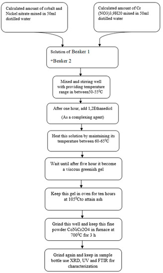
Figure 1.
Schematic flowchart of manufacturing CoCr2O4, CoNiCr2O4 nanoparticle.
3. Results and Discussion
3.1. XRD Analysis
Information on the phase and crystal structure of cobalt chromite is obtained through the X-ray diffraction technique. X-ray Cu-Kα radiation of wavelength 1.5418°A was used to produce the diffraction pattern. The range of angle 2θ of the XRD pattern ranges from 15° to 60°. The XRD pattern of cobalt chromite is shown in Figure 2. A single phase of cobalt chromite is obtained at a calcination temperature of 700 °C. XRD peaks corresponding to 700 °C were obtained at 15.5°, 30.5°, 35.5°, 40.5°, 45°, 50°, and 55.5°. A high pure cubic structure of cobalt chromite is synthesized through this process. Single 124 phase of cobalt chromite is obtained with reference code COD REV218120 2019.09 at 700 °C temperature. XRD analysis of cobalt chromite shows that the lattice parameters a and c range from 8.328 to 8.412 Å. Grainsize of 1.541874 Å for cobalt chromite is obtained from XRD peaks.
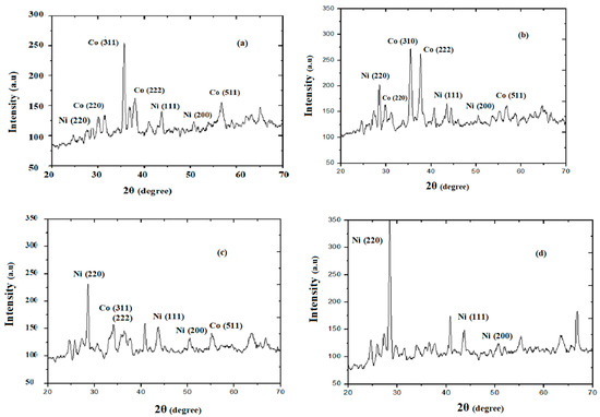
Figure 2.
XRD pattern of CoCr2O4 and Co1−xNixCr2O4: (a) x = 0.2, (b) x = 0.4, (c) x = 0.6, and (d) x = 0.8.
The X-ray diffraction pattern of Ni powders substituted with Co1−x NixCr2O4 depending on calcination temperature was attained by sol–gel process. Analysis of phase composition shows the sample with a high substitution rate at x = 0.2, 0.4, 0.6, and 0.8, with no single-phase compound for all temperatures. X-ray diffraction shows that the substitution of cobalt from nickel chromite proceeds by the steady transformation of cubic CoCr2O4 crystalline phase into cubic CoNiCr2O4 spinel state. In the pigmentary field, the main objective is to get the proper parameters of pigments used in industry [14].
The estimated size of cobalt chromite crystallites is 48.9 nm, obtained from heat treatment at 700 °C. For the nanoparticles, Co1−x NixCr2O4 crystallite size varied with heat treatment at 700 °C in the 39.5–44.5 nm range. The size of crystallite increases by less than 10 nm for all temperatures in this range.
The average linear crystallite size can be calculated using Bragg’s equation, nλ = 2d sin θ, where n is an integer, θ is the angle between the incident and scattered ray, d is the inter-spacing distance, and λ is the wavelength associated with the particle. Crystal size D of a particle can also be calculated by using Scherrer’s equation, D = Kλ/βCosθ, where K is the crystalline shape constant.
XRD results show that the powder form nanoparticles achieved from the sol–gel process is still heterogenous but more homogenous than that obtained by other techniques. However, phase composition is identical to the parent compound; yet, the morphology of synthesized pigments differed significantly.
3.2. SEM Analysis
The morphology of samples obtained by methods of synthesis is quite different than others. SEM micrographs of sol–gel-obtained samples of CoNiCr2O4 heated at 700 °C and CoCr2O4 have different morphological features, as shown in Figure 3. Very small spherical nanosized particles of CoCr2O4 were synthesized, showing a high degree of agglomeration.
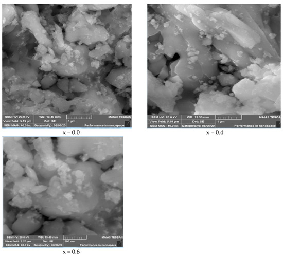
Figure 3.
SEM images of Cu1−xNixCr2O4 (x = 0.0, 0.4, and 0.6).
Different shaped and bigger sized (150–200 nm) particles of CoNiCr2O4 were obtained in synthesis. The surface morphology of doped cobalt chromite is very similar to cobalt chromite. The obtain powder largely consists of irregular, sub-micron shape particles of size 110 nm–500 nm, which resulted in a few large agglomerates with a porous microstructure.
3.3. Raman Spectroscopy
Raman spectroscopy of nickel doped in cobalt chromite, with the concentrations of nickel as 0.2, 0.4, and 0.6, was performed. Raman spectroscopy graph is plotted between intensity in (a.u) and Raman shift in cm−1 ranging from 1000 to 1600 cm−1 with a spacing of 100 cm−1, and peaks are obtained at 1440 cm−1 and 1540 cm−1, which shows the purity of the sample. No impurity peak is observed because of Raman spectroscopy. The Raman spectroscopy graph of nickel-doped cobalt chromite is shown in Figure 4.
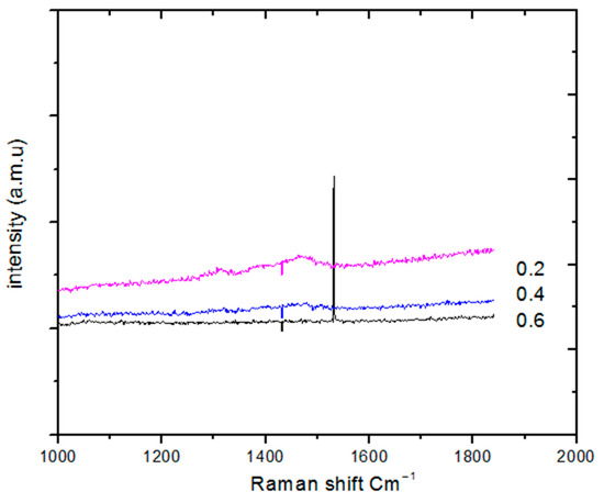
Figure 4.
Raman spectroscopy of doped cobalt chromites Co1−xNixCr2O4.
3.4. EDS Spectroscopy
Energy-dispersive spectroscopy (EDS), which is also called energy-dispersive analysis, was performed for the synthesized samples. Figure 5 shows results from EDS spectroscopy of our samples. The figure clearly shows that a single phase of copper chromite has formed. No extra impurity peak appeared in the EDS spectrum. We have obtained a single phase of copper chromite.
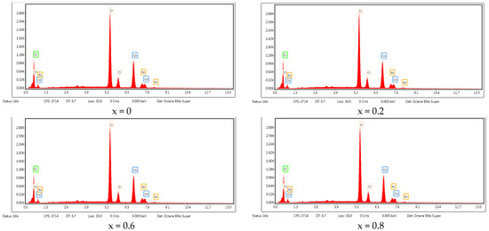
Figure 5.
EDS results of Co1−xNixCr2O4 (x = 0.2, 0.4, 0.6, 0.8).
3.5. Photoluminescence
Figure 6 shows the photoluminescence spectrum of nanoparticles. An excitation wavelength of 347 nm was used in this work at room temperature. It is important to monitor the physical properties of nanoparticles that change with variations in their dimensions on the nanometer scale, known as the quantum size effect. For example, quantum confinement increases the required energy bandgap of CoCr2O4 observed in photoluminescence. The recombination of the surface state is also evident from photoluminescence. The spectrum exhibits two emission peaks. One is located at 380 nm in the UV region, corresponding to near bandgap excitonic emission. The second peak is positioned around 530 nm, which can be attributed to the occupancy of independently ionized Co vacancies. Furthermore, the narrow size distribution of nanoparticles in luminescence, with a full width and half maximum (FWHM) of only a few nanometers, is reported by spectrum.
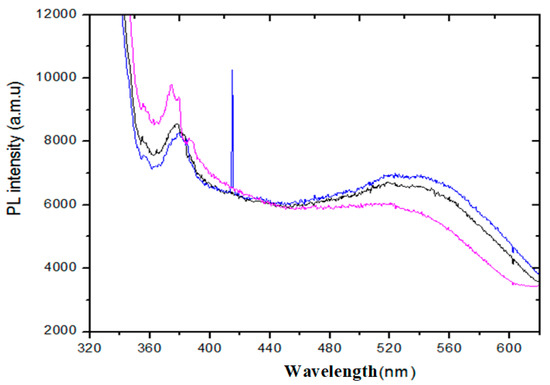
Figure 6.
Photoluminescence spectra of doped Co1−xNixCr2O4.
To further shed light on these compounds, the compounds prepared by the sol–gel synthesis route were formed with higher pronounced crystallinity. The morphological features of the samples derived by both synthesis methods are quite different. Moreover, the size and shape of chromite particles depend not only on the synthesis method used, but also on the nature of the octahedral cation . The origin of cation in the spinel structure displayed the tendency to produce pigments of numerous hues. The increase of content leads to a warm black color. The enrichment in tetrahedral ligand field gives a variety of green shades, from bluish-green to yellowish-green. Only Cu-doped pigments exhibited fewer different hues and were dark, nearly black. Nonetheless, there are no main differences in the colors between pigments produced by the sol–gel synthesis methods.
4. Conclusions
Two aqueous sol–gel and sonochemical synthesis methods were successfully employed for the preparation of the chromite spinels Co1-xNixCr2O4 (x = 0–1 with a step of 0.2). The single-phase cobalt-chromium spinel CoCr2O4 and nickel-doped chromates were synthesized irrespective of the calcination temperatures (600–700 °C). However, the phase compositions of Ni-substituted chromates were found to be dependent on the annealing temperature. At higher substitution ratios of 0.6 and 1, these samples were mixtures of spinel-type and simple oxide crystalline phases. When the concentration of the Ni dopant is increased, the Co peak is suppressed. The work is important in its applications for optical devices and enhanced photocatalytic applications.
Author Contributions
M.S. (research investigations), M.R. (project administration), K.B. (conceptualization), H.B. (data validation), A.Y. (soft-ware and data analysis), S.A. (methodology and writing original text), A.M. (art work and figures), B.H. (revisions and validation), T.A. (funding and project development), G.A. (data analysis and validation), M.M. (supervision and editing draft). All authors have read and agreed to the published version of the manuscript.
Funding
This research was funded by Princess Nourah Bint Abdulrahman University internal grant.
Data Availability Statement
The data presented in this study are available on request from the corresponding author.
Acknowledgments
The authors extend their sincere appreciation to the Deanship of Scientific Research at Princess Nourah Bint Abdulrahman University, for the support through the Fast-track Research Funding Program.
Conflicts of Interest
The authors declare no conflict of interest.
References
- Kaur, A.; Gupta, U. A review on applications of nanoparticles for the preconcentration of environmental pollutants. J. Mater. Chem. 2009, 19, 8279–8289. [Google Scholar] [CrossRef]
- Han, X.; Xu, K.; Taratula, O.; Farsad, K. Applications of nanoparticles in biomedical imaging. Nanoscale 2019, 11, 799–819. [Google Scholar] [CrossRef] [PubMed]
- Chavali, M.S.; Nikolova, M.P. Metal oxide nanoparticles and their applications in nanotechnology. SN Appl. Sci. 2019, 1, 607. [Google Scholar] [CrossRef] [Green Version]
- Castro Alarcon, N.; Herrera Arizmendi, J.L.; Luis Alberto, M.C.; Guzman Guzman, I.P.; Perez Centeno, A.; Santana Aranda, M.A. Antibacterial activity of nanoparticles of titanium dioxide, intrinsic and doped with indium and iron. Microbiol. Res. Int. 2016, 4, 55–62. [Google Scholar]
- Bindhu, M.; Umadevi, M.; Micheal, M.K.; Arasu, M.V.; Al-Dhabi, N.A. Structural, morphological and optical properties of MgO nanoparticles for antibacterial applications. Mater. Lett. 2016, 166, 19–22. [Google Scholar] [CrossRef]
- De Souza, L.K.C.; Zamian, J.R.; da Rocha Filho, G.N.; Soledade, L.E.; dos Santos, I.M.; Souza, A.G.; Scheller, T.; Angélica, R.S.; da Costa, C.E.F. Blue pigments based on CoxZn1−xAl2O4 spinels synthesized by the polymeric precursor method. Dyes Pigments 2009, 81, 187–192. [Google Scholar] [CrossRef]
- Lorenzi, G.; Baldi, G.; Di Benedetto, F.; Faso, V.; Lattanzi, P.; Romanelli, M. Spectroscopic study of a Ni-bearing gahnite pigment. J. Ceram. Soc. 2006, 26, 317–321. [Google Scholar] [CrossRef]
- Sickafus, K.E.; Wills, J.M.; Grimes, N.W. Structure of spinel. J. Am. Ceram. Soc. 1999, 82, 3279–3292. [Google Scholar] [CrossRef]
- Wang, Z.; Lazor, P.; Saxena, S.; Artioli, G. High-Pressure Raman Spectroscopic Study of Spinel (ZnCr2O4). J. Solid State Chem. 2002, 165, 165–170. [Google Scholar] [CrossRef]
- Torgashev, V.I.; Prokhorov, A.; Komandin, G.; Zhukova, E.; Anzin, V.B.; Talanov, V.M.; Rabkin, L.M.; Bush, A.A.; Dressel, M.; Gorshunov, B.P. Magnetic and dielectric response of cobalt-chromium spinel CoCr2O4 in the terahertz frequency range. Phys. Solid State 2012, 54, 350–359. [Google Scholar] [CrossRef]
- Lawes, G.; Melot, B.; Page, K.; Ederer, C.; Hayward, M.A.; Proffen, T.; Seshadri, R. Dielectric anomalies and spiral magnetic order in CoCr2O4. Phys. Rev. B 2006, 74, 024413. [Google Scholar] [CrossRef] [Green Version]
- Tsurkan, V.; Zherlitsyn, S.; Yasin, S.; Felea, V.; Skourski, Y.; Deisenhofer, J.; Krug von Nidda, H.-A.; Wosnitza, J.; Loidl, A. Unconventional magnetostructural transition in CoCr2O4 at high magnetic fields. Phys. Rev. Lett. 2013, 110, 115502. [Google Scholar] [CrossRef] [PubMed] [Green Version]
- Yamasaki, Y.; Miyasaka, S.Y.; Kaneko, Y.; He, J.-P.; Arima, T.; Tokura, Y. Magnetic Reversal of the Ferroelectric Polarization in a Multiferroic Spinel Oxide. Phys. Rev. Lett. 2006, 96, 207204. [Google Scholar] [CrossRef] [PubMed]
- Kumar, L.; Mohanty, P.; Shripathi, T.; Rath, C. Appearance of superparamagnetic phase below Curie temperature in cobalt chromite. Nanosci. Nanotechnol. Lett. 2009, 1, 199–2002. [Google Scholar] [CrossRef]
- Zakunta, D.; Vlcek, J.; Fitl, P.; Nemokovski, K.; Honecker, D.; Niznansky, D.; Disch, S. Noncollinear magnetism in nanosized cobalt chromite. Phys. Rev. B 2019, 98, 064407. [Google Scholar]
- Suchomel, M.R.; Shoemaker, D.P.; Ribaud, L.; Kemei, M.C.; Seshadri, R. Spin-induced symmetry breaking in orbitally ordered NiCr2O4 and CuCr2O4. Phys. Rev. B 2012, 86, 054406. [Google Scholar] [CrossRef] [Green Version]
- Mohanty, P.; Sheppard, C.J.; Prinsloo, A.R.E.; Roos, W.D.; Olivi, L.; Aquilanti, G. Effect of cobalt substitution on the magnetic properties of nickel chromite. J. Magn. Magn. Mater. 2018, 451, 20–28. [Google Scholar] [CrossRef]
- Mohanty, P.; Prinsloo, A.R.E.; Doyle, B.P.; Carleschi, E.; Sheppard, C.J. Structural and magnetic properties of (Co1–xNix) Cr2O4 (x = 0.5, 0.25) nanoparticles. AIP Adv. 2018, 8, 056424. [Google Scholar] [CrossRef]
- Opuchovic, O.; Kreiza, G.; Senvaitiene, J.; Kazlauskas, K.; Beganskiene, A.; Kareiva, A. Sol-Gel synthesis, characterization and application of selected sub-micro sized lanthanide (Ce, Pr, Nd, Tb) ferrites. Dyes Pigments 2015, 118, 176–182. [Google Scholar] [CrossRef]
- Maqbool, M.; Ahmad, I.; Ali, G.; Maaz, K. Energy level splitting and luminescence enhancement in AlN: Er by an external magnetic field. Opt. Mater. 2015, 46, 601–604. [Google Scholar] [CrossRef]
- Maqbool, M.; Ali, G.; Ahmad, I.; Maaz, K. Luminescence Enhancement in Amorphous AlN: W by Direct UV Excitation through Co-Doped Gadolinium. IEEE Photonics Technol. Lett. 2015, 27, 1519–1522. [Google Scholar] [CrossRef]
- Maqbool, M.; Main, K.; Kordesch, M. Titanium doped sputter-deposited AlN infrared whispering gallery mode microlaser on optical fibers. Opt. Lett. 2010, 35, 3637–3639. [Google Scholar]
- Maqbool, M.; Ahmad, I.; Richardson, H.H.; Kordesch, M.E. Direct ultraviolet excitation of an amorphous AlN: Praesiodimium phosphor by co-doped Gd3+ Cathodoluminescence. Appl. Phys. Lett. 2007, 91, 193511. [Google Scholar] [CrossRef]


Publisher’s Note: MDPI stays neutral with regard to jurisdictional claims in published maps and institutional affiliations. |
© 2021 by the authors. Licensee MDPI, Basel, Switzerland. This article is an open access article distributed under the terms and conditions of the Creative Commons Attribution (CC BY) license (https://creativecommons.org/licenses/by/4.0/).