Abstract
Various hydroxyapatite-filled and unfilled microspheres based on lactide and glycolide copolymers were prepared. The synthesized poly(lactic-co-glycolic acid) (PLGA) samples were characterized by GPC and 1H NMR spectroscopy, the morphology was characterized by SEM. It was shown that under the tin (II) 2-ethylhexanoate catalysis the glycolide is highly active in copolymerization as compared with lactide. According to the data on weight loss and the weight average molecular weight shift of PLGA over time (pH = 6.5; t = 25 °C), an increase in the rate of microsphere destruction was noted when macromolecules were enriched with glycolic acid residues, as well as when filled with hydroxyapatite. It was shown that the rate of PLGA degradation was determined by the water-accessible surface of a sample. The rate increase in PLGA hydrolytic degradation both with an increase in glycolic acid residues mole fraction in the chain and upon filling with hydroxyapatite was the result of the microspheres’ surface hydrophilization, an increase in capillary pressure upon filling of the pores as well as of the defects with water, and an increase in the number of structural defects. Approaches to the creation of composite microspheres based on PLGA degrading at a controlled rate were proposed.
1. Introduction
The successes of regenerative medicine in the last two decades are associated with the introduction of implantable composite polymer scaffolds into practice, which implies the fulfillment of a number of special requirements for the materials used. Such scaffold materials must biodegrade at a controlled rate, not cause toxic side effects and also do not form toxic compounds during degradation, have biocompatibility, a developed surface and perform the function of mechanical support at the implantation site [1]. Scaffolds are formed from dry mechanical mixtures of polymers with water-soluble salts crystals, interpolymer mixtures, polymer microspheres and nanoparticles using the phase separation methodology, foaming with gases, a “sacrificial” template, dry and wet spinning technologies, electrospinning, and other methods [2,3]. Regardless of the manufacturing method, the central issue that determines the effectiveness of polymer scaffolds used in regenerative medicine is the rate of their degradation [4].
Scaffold implants based on polymer microspheres are currently attracting increased attention due to ease of manufacture, control over morphology, the ability to discretely control their physicochemical properties, and flexibility in controlling the kinetics of release of bioactive molecules that accelerate tissue regeneration [5]. Polymeric microspheres are widely used in targeted drug delivery applications mainly due to efficient delivery and release of the encapsulated drug, as well as spatial and temporal control over release [6,7,8]. In addition to their ability to serve as excellent controlled release carriers, microspheres are rigidly shaped and can be packaged to form porous three-dimensional structures that can serve as tissue engineering scaffolds. Thus, scaffold implants based on densely packed microspheres can serve as templates for the induction of cell proliferation [9,10]. The use of microspheres in biodegradable frame implants makes it possible to create a network of pores inside the implant, promoting cell ingrowth and accelerating resorption [10,11,12]. Frame implants based on sintered microspheres have been used in various ways, including the regeneration of bone tissue [13,14,15], cartilage tissue [16,17,18,19,20,21,22], and nerve cells [23,24].
A number of natural and synthetic polymers are used for scaffolds manufacture. Among natural polymers, chitosan [25], alginates [26], gelatin [27], and collagen [28] are of the greatest importance for the manufacture of scaffolds manufacture. Although there are no problems with toxic effects when using natural polymers for scaffolds manufacture, raw materials from natural sources can vary significantly in their properties, which creates obstacles to obtaining biomedical materials with reproducible characteristics. Although the use of synthetic polymers, such as polyethersulfones, polyurethanes [29], and modified poly(ε-caprolactone) [30], makes it possible to obtain scaffolds with controlled architecture and properties, however lactide and glycolide copolymers (PLGA) remains as the gold standard for implantable synthetic polymers [31].
PLGA-based microspheres are the basis for the preparation of implants [32], scaffolds [33], and essential materials for cell engineering [34] as well as in the creation of systems with controlled drug release [35]. Emulsification, microfluidic technology, spray drying and electrospray are used to form PLGA-based microspheres [36]. It has been established that PLGA biodegradation occurs after endocytosis in the phagosomes, in which the medium is acidic [37]. During the hydrolytic degradation of PLGA, which serves as a model for biodegradation, partial crystallization was noted [38,39], as well as a pH gradient (pH inside the particles was about 1.5) [40]. The above circumstances point to acid-catalyzed hydrolytic degradation of PLGA from the surface, which was also confirmed by the experimental results obtained in the present article. Thus, PLGA-based microspheres are a promising platform for the development of new biodegradable implantable scaffolds that can release immobilized drugs. The use of hydroxyapatite as a filler in PLGA matrices makes it possible to obtain osteoconductive composites and implants with high physical and mechanical properties [41,42,43,44]. At the same time, the hydrolytic degradation patterns of composite PLGA-based microspheres that determine their application efficiency are complex and require further study.
Poly(L-lactide-co-glycolide) (PLGA), an aliphatic polyester, has been widely used in many studies due to its biocompatibility and biodegradability. A number of recent papers have reported the fabrication of scaffold implants exclusively from PLGA microspheres using thermal sintering [13,45], dichloromethane vapor treatment [46,47], or solvent/solventless sintering [48,49]. Moreover, such materials offer flexible polymer degradation kinetics, which can be controlled by changing one or more factors such as microsphere size, copolymer monomer composition, molecular weight, etc. [50,51]. Therefore, this article is devoted to the preparation of composite microspheres based on PLGA of various compositions and the dynamics study of their isothermal degradation in a slightly acidic medium.
2. Materials and Methods
2.1. Materials
L-Lactide (98%, Sigma-Aldrich, St. Louis, MO, USA), glycolide (>99%, Sigma-Aldrich, St. Louis, MO, USA), tin 2-ethylhexanoate (97%, Sigma-Aldrich, St. Louis, MO, USA), chloroform (99%, Chimmed, Moscow, Russia), ethanol (99%, Chimmed, Moscow, Russia), hexafluoroisopropanol (99%, P&M-Invest, Moscow, Russia), and hydroxyapatite with a particle size of about 1–5 μm (NPK Polistom, Moscow, Russia) were taken for synthesis without preliminary purification.
2.2. Synthesis of PLGA
Copolymerization of lactide and glycolide (Figure 1).
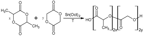
Figure 1.
Scheme of PLGA synthesis.
A 100 mL test tube equipped with a Hei-TORQUE Value 100 overhead stirrer (max. torque 100 N∙cm) (Heidolph, Schwabach, Germany) was loaded with the starting ingredients: catalyst (tin 2-ethylhexanoate), L-lactide and glycolide (Table 1).

Table 1.
Characteristics of the synthesized PLGA (catalyst 0.5 mol% of the total number of mol comonomers in the reaction mixture).
Next, after purging the test tube with argon, the synthesis was carried out at a temperature of 170 °C with continuous stirring. Since the reactivity of lactide and glycolide under these conditions differed significantly, syntheses with different comonomer ratios for a fixed time could have led to the formation of copolymers with very different molecular weight characteristics [52]. Therefore, the cessation of the stirring of the mixture due to an increase in the copolymer molecular weight and reaction mass viscosity was used as a criterion for the end of the reaction. Following completion of the synthesis, the polymer was purified by reprecipitation: a solution of 8 g of the polymer in 20 mL of chloroform was precipitated into ethanol (200 mL). The precipitated polymer was filtered off on filter paper and dried at 30 °C in a vacuum at a pressure of 40 Pa.
1H NMR spectra were taken for 10% solutions of polymers in a mixture of 20 vol% of hexafluoroisopropanol and 80 vol% CDCl3 in a WP200-SY NMR spectrometer (Bruker, Billerica, MA, USA) at an operating frequency of 300 MHz for 1H at the Center for Research on the Structure of Molecules of the A.N. Nesmeyanov Institute of Organoelement Compounds of the Russian Academy of Sciences (INEOS RAS). The spectrum of the pure solvent (a mixture of hexafluoroisopropanol and CDCl3) was subtracted from the obtained spectra using the OriginPro 9.0.0 SR2 software package (OriginLab Corporation, Northampton, MA, USA).
Gel permeation chromatography (GPC) of the copolymers was carried out in a GPC-150 chromatograph (Waters Corporation, Milford, MA, USA) at the Center for Research on the Structure of Molecules of INEOS RAS. Eluent: tetrahydrofuran (1 mL/min). Column: PL-GEL 5u MIXC, 300 × 7.5 mm (Agilent Technologies, Santa Clara, CA, USA).
2.3. Formation of Microspheres Based on PLGA
The preparation of polymer microparticles was based on the evaporation of an organic solvent from an emulsion. To do this, a solution of 100 mg of the synthesized copolymer in 10 mL of chloroform (and, when all composite particles were obtained, a dispersion of 30 mg of hydroxyapatite in a solution of 70 mg of the polymer in 10 mL of chloroform) was gradually poured into 100 mL of a 0.5% aqueous solution of polyvinyl alcohol while stirring with an overhead stirrer at a speed 2000 rpm for 3–5 min. After that, chloroform was distilled from the resulting emulsion in a rotary evaporator at a temperature of 25 °C and pressure of 10 kPa. Microparticles were collected by centrifugation for 5 min (10,000 rpm), washed with ethanol (for 10 min, so that alcoholysis of PLGA could be neglected) and dried at 25 °C and 100 Pa. The filler content in all composite particles was 30 wt% (17.6 vol%) and was similar to that described previously in the article [43]. The size of the formed microspheres averaged 1.3 mm with a maximum deviation of 0.2 mm (trinocular microscope Science MTL-201, Bresser, Rhede, Germany). In addition, the surface of the microspheres was characterized by the SEM method (TM300, Hitachi, Tokyo, Japan).
2.4. Study of Hydrolytic Degradation of PLGA
The hydrolytic decomposition of microparticles was studied gravimetrically in distilled water (pH = 6.5) at 25 °C. To do this, 0.1 g of microparticles were placed in glass vials; after adding 10 mL of distilled water, they were dispersed using a magnetic stirrer. After a certain time, the microparticles were precipitated by centrifugation for 5 min (10,000 rpm), separated from the aqueous phase, washed with alcohol, and dried at a temperature of 25 °C and pressure of 100 Pa. For the dried microparticles, the weight loss during hydrolysis and the average molecular weight (by GPC) were determined. To study the acidity of microparticle dispersions, their weighed portion (0.5 g) was dispersed in 10 mL of distilled water using a magnetic stirrer. The pH of the solutions was measured using a SevenCompact™ S220 pH/Ion meter (Mettler Toledo, Columbus, OH, USA).
3. Results and Discussion
The structure of the synthesized lactide-glycolide random copolymers was characterized by 1H NMR spectroscopy (Figure 2). Since the solubility of copolymers in traditional solvents such as chloroform (or tetrahydrofuran) decreased with an increase in the molar fraction of glycolic acid residues in the polymer chain, the 1H NMR spectra of PLGA were recorded in a medium containing 20 vol% (CF3)2CHOH and 80 vol% CDCl3. The signal with a chemical shift at 4.6 ppm corresponded to protons of methylene group in glycolic acid residues and signals with chemical shifts of 5.16 and 1.57 ppm corresponded to CH and CH3 groups of lactic acid residues (Figure 2).
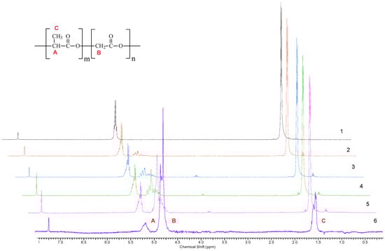
Figure 2.
1H NMR spectra of PLGA of various compositions (characteristics of copolymers 1–6 are given in Table 1).
The content of comonomer units in PLGA macromolecules was determined based on the integral intensities of the proton signals of lactic and glycolic acid residues (Table 1). As can be seen, during the copolymerization of lactide and glycolide, the polymer chain is enriched in the residues of the latter compared to its content in the monomer mixture. Thus, glycolide is more active in copolymerization than lactide, which agrees with the literature data [53]. The increased activity of glycolide in copolymerization with lactide also manifests itself in a regular increase in the molecular weight of PLGA with an increase in the glycolide mole fraction in the reaction mixture (Table 1). Thus, according to the GPC data (Figure 3), in a solution of tetrahydrofuran, with an increase of glycolide mole fraction to 30 mol%, the number average molecular weight of PLGA increases from 28,500 to 33,000. PLGA macromolecules containing 58 mol% and 77 mol% of glycolide residues were insoluble in tetrahydrofuran. As can be seen, at a glycolide mole fraction not exceeding 0.35, the composition of PLGA is close to the composition of the reaction mixture, and the yield of PLGA in all cases was more than 85% (Table 1).
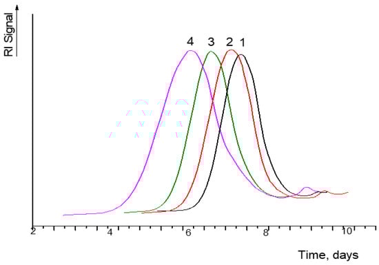
Figure 3.
Gel permeation chromatograms of copolymers 1–4 (Table 1).
For microspheres formed from PLGA of various compositions, as well as for composite microspheres based on PLGA filled with 30 wt% of hydroxyapatite, the regularities of acid hydrolysis at pH = 6.5 were studied. The SEM data indicate the formation of samples with a developed surface, and this is especially significant for PLGA filled with hydroxyapatite. Furthermore, it can be seen that the hydroxyapatite was evenly dispersed in the PLGA matrix (Figure 4).
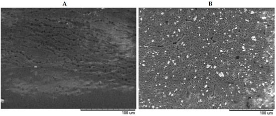
Figure 4.
SEM of microspheres surface based on unfilled PLGA (A) and PLGA filled with 30 wt% of hydroxyapatite (B).
Previously, it was found that the rate of lactide-glycolide copolymers hydrolysis increases with a decrease in the particle diameter and PLGA molecular weight [54,55]. The latter effect is explained by a decrease in the probability of hydrolysis per macromolecule with a decrease in molecular weight, and also, probably, by an increase in the tendency to crystallization with a decrease in molecular weight and an increase in the kinetic flexibility of chains [56]. This is consistent with the inverse proportionality between the degree of crystallinity and the molecular weight of PLLA samples [57]. It should be noted that the diameter of microspheres and the molecular weight for various samples of the initial copolyesters (PLGA) studied in this article were comparable and could not have a decisive effect on the rate of acid-catalyzed hydrolytic degradation.
It has been shown that the rate of hydrolysis of microspheres formed from PLGA increases significantly with an increase in the molar glycolic acid residues fraction in macromolecules (Figure 5). The latter circumstance agrees with an increase in the release rate of drugs immobilized by PLGA particles of various compositions [58].
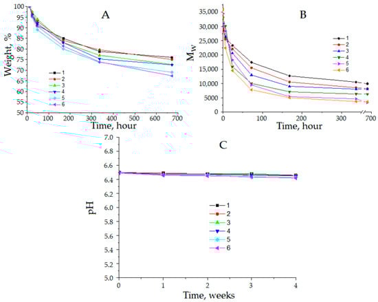
Figure 5.
The changes in the mass of microspheres based on PLGA (A), molecular weight of PLGA (B), and pH of the medium during the acid-catalyzed hydrolytic degradation of PLGA (C) of various compositions (Descriptors 1–6 refer to the PLGAs shown in Table 1).
An increase in the rate of PLGA degradation with an increase in the mole fraction of glycolide residues in the composition of macromolecules can be associated with a decrease in water contact angle, a decrease in the proportion of ordered regions, or an increase in the rate of acid hydrolysis of ester groups in the chain. Taking into account the Taft steric constant equal to −0.07, when passing from lactide residues to glycolide residues, one should expect an increase in the rate constant of ester groups hydrolysis by 1.17 times.
Therefore, the steric effect of the methyl group is insufficient to provide a significant difference in the rates of chain hydrolysis for lactic and glycolic acid residues. Apparently, an increase in the hydrophilicity of the microsphere’s surface and a decrease in the proportion of ordered regions with an increase in the proportion of glycolide units in the chain are of greater importance. According to the data presented in [59,60], the contact angle of wetting with water on the PLGA surface decreases almost linearly with an increase in the molar fraction of glycolic acid residues (Figure 6), according to Equation (1):
where: θ—contact angle of wetting PLGA surface with water, %; —molar fraction of glycolic acid residues in PLGA.
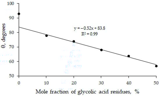
Figure 6.
The dependence of the contact angle of wetting PLGA surface with water on the composition of the copolymers (according to the data of [59,60]).
As can be seen, when the mole fraction of glycolide units is less than 10%, there are obvious deviations from linear dependence (1), and the wetting angle of PLLA with water is 93.30 [60]. Probably, deviations from Equation (1) are associated with a significant proportion of ordered regions in PLGA samples containing a small amount of glycolic acid residues. According to XRD data, for PLGA with a mole fraction of glycolic acid units of more than 10%, there are no obvious reflections that are characteristic of the crystalline phase [59]. Although, due to a large number of defects in ordered regions, the experimental determination of PLGA crystallinity degree is obviously conditional, all available literature data indicate its increase during the biological and hydrolytic degradation of lactide and glycolide copolymers [61]. In particular, this is confirmed by a pronounced slowdown of the hydrolytic acid-catalyzed degradation of PLGA, so that after 672 h more than 67% of the original microsphere mass is retained (Figure 5A). In this case, the PLGA molecular weight significantly decreases with time (Figure 5B), which may also indicate the additional formation of ordered regions due to an increase in the kinetic flexibility of macromolecules, as noted earlier in the literature [57]. Since the hydrolysis rate was low, the pH of the medium also decreased insignificantly during the degradation of PLGA (Figure 5C). An increase in the proportion of glycolic acid residues in PLGA composition leads to an increase in pressure, which contributes to the spontaneous filling of pores with water. If the application of external pressure is necessary to fill poly(L-lactide) (PLLA) pores with a contact angle of 93.30, then after the inversion of wetting with water, with an increase in the proportion of glycolic acid residues in the chain, the filling of pores occurs spontaneously, while the driving force per unit pore radius increases in accordance with the Laplace Equation (2) (Figure 7).
where: Δp—capillary pressure; r—the effective radius of cylindrical pores (defects); σ—the surface tension of water.
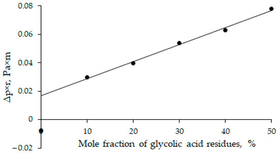
Figure 7.
The capillary pressure of filling PLGA pores as a function of the glycolic acid mole fraction in PLGA.
As can be seen, the driving force of capillary filling of pores increases by 2.6 times at a fivefold increase in the mole fraction of glycolic acid residues in the PLGA composition (Figure 7).
Upon transition to composite microspheres based on PLGA of various compositions filled with 30 wt% of hydroxyapatite, a sharp increase in the rate of acid-catalyzed hydrolytic degradation was observed (Figure 8). First, filling with hydroxyapatite increases the hydrophilicity of the surface [60], facilitating its wetting with water. Secondly, filling with hydroxyapatite contributes to the involvement of a larger PLGA surface in the reaction zone due to the penetration of water into microspheres in places of formation of main pores and defects caused by filling. The mass fraction of PLGA entering into hydrolytic degradation naturally increases (Figure 8A), and the pH of the medium decreases significantly due to the release of lactic and glycolic acids, as well as their carboxyl-containing water-soluble oligomers (Figure 8C). The molecular masses of the degradation products of composite microspheres are only slightly lower than those determined for microspheres based on pure PLGA (Figure 8C). The latter circumstance is the result of an increase in the proportion of ordered regions, both due to the degradation of PLGA in amorphous domains and, probably, as a result of achieving a higher packing density of the resulting oligomers. At the same time, the rate of acid-catalyzed PLGA hydrolysis also increases with an increase in the mole fraction of glycolic acid residues in macromolecules, as can be expected from Equation (2).
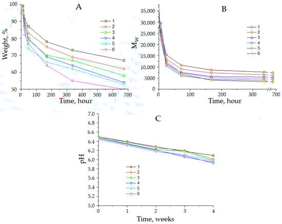
Figure 8.
The changes in the mass of composite microspheres (A), molecular weight of PLGA (B), and pH of the medium during the acid-catalyzed hydrolytic degradation of PLGA (C) of various compositions filled with 30 wt% of hydroxyapatite (Descriptors 1–6 refer to the PLGAs shown in Table 1).
As noted earlier [39,40] and confirmed by us (Figure 5C and Figure 8C), PLGA degradation is accompanied by a decrease in the pH of the medium due to an increase in the concentration of carboxyl groups, which should lead to autoacceleration of hydrolysis if it occurs in the kinetic region. In addition, the observed increase in the rate of PLGA hydrolysis with an increase in the mole fraction of glycolide residues in the chain is consistent with the data of other studies [38,58,59]. At the same time, the obtained results demonstrate a decrease in the rate of acid-catalyzed destruction of PLGA with an increase in the hydrolysis depth (Figure 5 and Figure 8), which also corresponds to the results described by other authors [39,59]. The latter is usually associated with an increase in the volume fraction of domains with a high packing density of chains caused by both the depletion of the amorphous phase during hydrolysis and by the tendency to form new ordered regions due to a decrease in the molecular weight of macromolecules and an increase in their kinetic flexibility, which promotes crystallization [38,39,57]. Therefore, the hydrolytic degradation of PLGA is limited by diffusion while the moderate changes in the pH of the medium and the reactivity of ester groups with varying the composition of macromolecules had no decisive importance.
At the same time, it was shown that a decrease in the contact angle of the PLGA surface with water contributes to a degradation rate increase in copolyesters of various compositions (Figure 5, Figure 6, Figure 7 and Figure 8). This result suggests that the rate of hydrolytic degradation was determined by the size of the PLGA surface accessible to water including the surface of the sides of open pores and defects. Surface hydrophilization leads to an increase in capillary pressure for spontaneous filling of the PLGA pores and defects and promotes water penetration into the microspheres. A significant degradation rate increase was noted upon filling PLGA with hydroxyapatite (Figure 5 and Figure 8), which creates defects, indicating the decisive contribution of the water transfer to the PLGA surface into the rate of hydrolytic degradation of microspheres. Thus, filling composites with a PLGA matrix with biocompatible hydroxyapatite is a promising approach to the control of the hydrolytic degradation rate.
4. Conclusions
It was established that the rate of hydrolytic degradation of PLGA was determined mainly by the rate of water transfer to the reaction zone and was controlled by the interfacial area, including the pores and defects. Consequently, the degradation of copolyester microspheres was limited by diffusion. The morphological effect had an impact on the degradation rate of the PLGA-based microspheres, and degradation was facilitated by an increase in the proportion of amorphous regions; however, with an increase in the depth of hydrolysis of macromolecules, the tendency to crystallization increased. The latter fact explained the slowdown in the hydrolytic degradation of microspheres with an increase in the depth of acid-catalyzed hydrolysis despite a decrease in the pH of the medium and was confirmed by the data on the shift in the samples’ weight and the weight average molecular weight of the PLGA macromolecules. The rate of hydrolytic degradation of PLGA-based microspheres can be significantly increased through filling with hydroxyapatite, which is explained by an increase in the number of defects and hydrophilization of samples. At the same time, hydrophilization of the PLGA surface leads to an increase in capillary pressure for the spontaneous filling of pores and defects with water. Therefore, filling PLGA with hydroxyapatite is not only a way to control the properties of composite microspheres but also has an impact on the rate of their hydrolytic degradation. Thus, the regularities revealed in the present article can be useful in the development of biomaterials based on hydroxyapatite-filled PLGA and the composites thereof with a controlled degradation rate.
Author Contributions
Conceptualization, V.I., V.V. and Y.M.; methodology, V.I.; validation, V.V., G.M. and O.B.; formal analysis, Y.M.; investigation, V.I., V.V., V.G., G.M. and O.B.; data curation, V.I. and V.V.; writing—original draft preparation, V.I. and Y.M.; writing—review and editing, V.V., G.M. and Y.M.; visualization, V.I., V.G. and Y.M.; supervision, V.V. All authors have read and agreed to the published version of the manuscript.
Funding
This work was supported by the Ministry of Science and Higher Education of the Russian Federation (Contract No. 075-03-2023-642).
Data Availability Statement
The characterization data are available upon request from the authors.
Conflicts of Interest
The authors declare no conflict of interest.
References
- Agrawal, C.M.; Ray, R.B. Biodegradable polymeric scaffolds for musculoskeletal tissue engineering. J. Biomed. Mater. Res. 2001, 55, 141–150. [Google Scholar] [CrossRef]
- Kontogianni, G.-I.; Bonatti, A.F.; De Maria, C.; Naseem, R.; Melo, P.; Coelho, C.; Vozzi, G.; Dalgarno, K.; Quadros, P.; Vitale-Brovarone, C.; et al. Promotion of In Vitro Osteogenic Activity by Melt Extrusion-Based PLLA/PCL/PHBV Scaffolds Enriched with Nano-Hydroxyapatite and Strontium Substituted Nano-Hydroxyapatite. Polymers 2023, 15, 1052. [Google Scholar] [CrossRef] [PubMed]
- Rosellini, E.; Cascone, M.G. Microfluidic Fabrication of Natural Polymer-Based Scaffolds for Tissue Engineering Applications: A Review. Biomimetics 2023, 8, 74. [Google Scholar] [CrossRef] [PubMed]
- Daskalakis, E.; Hassan, M.H.; Omar, A.M.; Acar, A.A.; Fallah, A.; Cooper, G.; Weightman, A.; Blunn, G.; Koc, B.; Bartolo, P. Accelerated Degradation of Poly-ε-caprolactone Composite Scaffolds for Large Bone Defects. Polymers 2023, 15, 670. [Google Scholar] [CrossRef] [PubMed]
- Shi, X.; Su, K.; Varshney, R.R.; Wang, Y.; Wang, D.-A. Sintered Microsphere Scaffolds for Controlled Release and Tissue Engineering. Pharm. Res. 2011, 28, 1224–1228. [Google Scholar] [CrossRef] [PubMed]
- White, L.J.; Kirby, G.T.S.; Cox, H.C.; Qodratnama, R.; Qutachi, O.; Rose, F.R.; Shakesheff, K.M. Accelerating Protein Release from Microparticles for Regenerative Medicine Applications. Mater. Sci. Eng. C 2013, 33, 2578–2583. [Google Scholar] [CrossRef]
- Rahman, C.V.; Ben-David, D.; Dhillon, A.; Kuhn, G.; Gould, T.W.A.; Müller, R.; Rose, F.R.; Shakesheff, K.M.; Livne, E. Controlled Release of BMP-2 from a Sintered Polymer Scaffold Enhances Bone Repair in a Mouse Calvarial Defect Model. J. Tissue Eng. Regen. Med. 2014, 8, 59–66. [Google Scholar] [CrossRef]
- Gupta, V.; Khan, Y.; Berkland, C.J.; Laurencin, C.T.; Detamore, M.S. Microsphere-Based Scaffolds in Regenerative Engineering. Annu. Rev. Biomed. Eng. 2017, 19, 135–161. [Google Scholar] [CrossRef]
- Belkas, J.S.; Shoichet, M.S.; Midha, R. Peripheral Nerve Regeneration through Guidance Tubes. Neurol. Res. 2004, 26, 151–160. [Google Scholar] [CrossRef] [PubMed]
- Valmikinathan, C.M.; Tian, J.; Wang, J.; Yu, X. Novel Nanofibrous Spiral Scaffolds for Neural Tissue Engineering. J. Neural Eng. 2008, 5, 422–432. [Google Scholar] [CrossRef]
- Habraken, W.J.E.M.; Wolke, J.G.C.; Mikos, A.G.; Jansen, J.A. Injectable PLGA Microsphere/calcium Phosphate Cements: Physical Properties and Degradation Characteristics. J. Biomater. Sci. Polym. 2006, 17, 1057–1074. [Google Scholar] [CrossRef] [PubMed]
- De Nardo, L.; Bertoldi, S.; Cigada, A.; Tanzi, M.C.; Haugen, H.J.; Farè, S. Preparation and Characterization of Shape Memory Polymer Scaffolds via Solvent Casting/particulate Leaching. J. Appl. Biomater. Funct. Mater. 2012, 10, 119–126. [Google Scholar] [CrossRef] [PubMed]
- Borden, M.; Attawia, M.; Khan, Y.; El-Amin, S.F.; Laurencin, C.T. Tissue-engineered bone formation in vivo using a novel sintered polymeric microsphere matrix. J. Bone Jt. Surg. Br. 2004, 86, 1200. [Google Scholar] [CrossRef] [PubMed]
- Ruhe, P.Q.; Hedberg-Dirk, E.L.; Padron, N.T.; Spauwen, P.H.; Jansen, J.A.; Mikos, A.G. Porous poly(DL-lactic-co-glycolic acid)/calcium phosphate cement composite for reconstruction of bone defects. Tissue Eng. 2006, 12, 789. [Google Scholar] [CrossRef] [PubMed]
- Borden, M.; Attawia, M.; Laurencin, C.T. The Sintered Microsphere Matrix for Bone Tissue Engineering: In Vitro Osteoconductivity Studies. J. Biomed. Mater. 2002, 61, 421–429. [Google Scholar] [CrossRef]
- Chun, K.W.; Yoo, H.S.; Yoon, J.J.; Park, T.G. Biodegradable PLGA microcarriers for injectable delivery of chondrocytes: Effect of surface modification on cell attachment and function. Biotechnol. Prog. 2004, 20, 1797. [Google Scholar] [CrossRef]
- Holland, T.A.; Tabata, Y.; Mikos, A.G. Dual growth factor delivery from degradable oligo(poly(ethylene glycol) fumarate) hydrogel scaffolds for cartilage tissue engineering. J. Control. Release 2005, 101, 111. [Google Scholar] [CrossRef]
- Singh, M.; Morris, C.P.; Ellis, R.J.; Detamore, M.S.; Berkland, C. Microsphere-Based Seamless Scaffolds Containing Macroscopic Gradients of Encapsulated Factors for Tissue Engineering. Tissue Eng. C Methods 2008, 14, 299–309. [Google Scholar] [CrossRef]
- Dormer, N.H.; Singh, M.; Wang, L.; Berkland, C.J.; Detamore, M.S. Osteochondral Interface Tissue Engineering Using Macroscopic Gradients of Bioactive Signals. Ann. Biomed. Eng. 2010, 38, 2167–2182. [Google Scholar] [CrossRef]
- Dormer, N.H.; Busaidy, K.; Berkland, C.J.; Detamore, M.S. Osteochondral Interface Regeneration of Rabbit Mandibular Condyle with Bioactive Signal Gradients. J. Oral. Maxill. Surg. 2011, 69, e50–e57. [Google Scholar] [CrossRef]
- Dormer, N.H.; Singh, M.; Zhao, L.; Mohan, N.; Berkland, C.J.; Detamore, M.S. Osteochondral Interface Regeneration of the Rabbit Knee with Macroscopic Gradients of Bioactive Signals. J. Biomed. Mater. Res. 2012, 100A, 162–170. [Google Scholar] [CrossRef] [PubMed]
- Dormer, N.H.; Gupta, V.; Scurto, A.M.; Berkland, C.J.; Detamore, M.S. Effect of Different Sintering Methods on Bioactivity and Release of Proteins from PLGA Microspheres. Mater. Sci. Eng. C 2013, 33, 4343–4351. [Google Scholar] [CrossRef]
- Goraltchouk, A.; Scanga, V.; Morshead, C.M.; Shoichet, M.S. Incorporation of protein-eluting microspheres into biodegradable nerve guidance channels for controlled release. J. Control. Release 2006, 110, 400. [Google Scholar] [CrossRef]
- Rosner, B.I.; Siegel, R.A.; Grosberg, A.; Tranquillo, R.T. Rational design of contact guiding, neurotrophic matrices for peripheral nerve regeneration. Ann. Biomed. Eng. 2003, 31, 1383. [Google Scholar] [CrossRef]
- Beleño Acosta, B.; Advincula, R.C.; Grande-Tovar, C.D. Chitosan-Based Scaffolds for the Treatment of Myocardial Infarction: A Systematic Review. Molecules 2023, 28, 1920. [Google Scholar] [CrossRef]
- Tomić, S.L.; Babić Radić, M.M.; Vuković, J.S.; Filipović, V.V.; Nikodinovic-Runic, J.; Vukomanović, M. Alginate-Based Hydrogels and Scaffolds for Biomedical Applications. Mar. Drugs 2023, 21, 177. [Google Scholar] [CrossRef] [PubMed]
- Hoque, M.E.; Nuge, T.; Yeow, T.K.; Nordin, N.; Prasad, R.G.S.V. Gelatin based scaffolds for Tissue Engineering: A review. J. Polym. Res. 2015, 9, 15–32. [Google Scholar]
- Parenteau-Bareil, R.; Gauvin, R.; Berthod, F. Collagen-Based Biomaterials for Tissue Engineering Applications. Materials 2010, 3, 1863–1887. [Google Scholar] [CrossRef]
- Wasyłeczko, M.; Remiszewska, E.; Sikorska, W.; Dulnik, J.; Chwojnowski, A. Scaffolds for Cartilage Tissue Engineering from a Blend of Polyethersulfone and Polyurethane Polymers. Molecules 2023, 28, 3195. [Google Scholar] [CrossRef]
- Chierchia, M.; Chirumbolo, S.; Valdenassi, L.; Franzini, M. Ozone-treated poly-ε-caprolactone scaffolds for bone regeneration. Chem. Biol. Interact. 2023, 381, 110509. [Google Scholar] [CrossRef]
- Rocha, C.V.; Gonçalves, V.; da Silva, M.C.; Bañobre-López, M.; Gallo, J. PLGA-Based Composites for Various Biomedical Applications. Int. J. Mol. Sci. 2022, 23, 2034. [Google Scholar] [CrossRef] [PubMed]
- Bassand, C.; Freitag, J.; Benabed, L.; Verin, J.; Siepmann, F.; Siepmann, J. PLGA implants for controlled drug release: Impact of the diameter. Eur. J. Pharm. Biopharm. 2022, 177, 50–60. [Google Scholar] [CrossRef] [PubMed]
- Kang, S.W.; La, W.G.; Kim, B.S. Open macroporous poly(lactic-co-glycolic Acid) microspheres as an injectable scaffold for cartilage tissue engineering. J. Biomater. Sci. Polym. Ed. 2009, 20, 399–409. [Google Scholar] [CrossRef]
- Patel, M.; Jha, A.; Patel, R. Potential application of PLGA microsphere for tissue engineering. J. Polym. Res. 2021, 28, 214. [Google Scholar] [CrossRef]
- Xu, Y.; Kim, C.S.; Saylor, D.M.; Koo, D. Polymer degradation and drug delivery in PLGA-based drug-polymer applications: A review of experiments and theories. J. Biomed. Mater. Res. B Appl. Biomater. 2017, 105, 1692–1716. [Google Scholar] [CrossRef] [PubMed]
- Su, Y.; Zhang, B.; Sun, R.; Liu, W.; Zhu, Q.; Zhang, X.; Wang, R.; Chen, C. PLGA-based biodegradable microspheres in drug delivery: Recent advances in research and application. Drug Deliv. 2021, 28, 1397–1418. [Google Scholar] [CrossRef]
- van Apeldoorn, A.A.; van Manen, H.J.; Bezemer, J.M.; de Bruijn, J.D.; van Blitterswijk, C.A.; Otto, C. Raman imaging of PLGA microsphere degradation inside macrophages. J. Am. Chem. Soc. 2004, 126, 13226–13227. [Google Scholar] [CrossRef]
- Park, T.G. Degradation of poly(lactic-co-glycolic acid) microspheres: Effect of copolymer composition. Biomaterials 1995, 16, 1123–1130. [Google Scholar] [CrossRef]
- Casalini, T.; Rossi, F.; Lazzari, S.; Perale, G.; Masi, M. Mathematical modeling of PLGA microparticles: From polymer degradation to drug release. Mol. Pharm. 2014, 11, 4036–4048. [Google Scholar] [CrossRef]
- Fu, K.; Pack, D.W.; Klibanov, A.M.; Langer, R. Visual Evidence of Acidic Environment within Degrading Poly(lactic-co-glycolic acid) (PLGA) Microspheres. Pharm. Res. 2000, 17, 100–106. [Google Scholar] [CrossRef]
- Park, J.W.; Hwang, J.U.; Back, J.H.; Jang, S.W.; Kim, H.J.; Kim, P.S.; Shin, S.; Kim, T. High strength PLGA/Hydroxyapatite composites with tunable surface structure using PLGA direct grafting method for orthopedic implants. Compos. Part B Eng. 2019, 178, 107449. [Google Scholar] [CrossRef]
- Stevanovic, M.; Selakovic, D.; Vasovic, M.; Ljujic, B.; Zivanovic, S.; Papic, M.; Zivanovic, M.; Milivojevic, N.; Mijovic, M.; Tabakovic, S.Z.; et al. Comparison of Hydroxyapatite/Poly(lactide-co-glycolide) and Hydroxyapatite/Polyethyleneimine Composite Scaffolds in Bone Regeneration of Swine Mandibular Critical Size Defects: In Vivo Study. Molecules 2022, 27, 1694. [Google Scholar] [CrossRef] [PubMed]
- Kim, S.S.; Ahn, K.M.; Park, M.S.; Lee, J.H.; Choi, C.Y.; Kim, B.S. A poly(lactide-co-glycolide)/hydroxyapatite composite scaffold with enhanced osteoconductivity. J. Biomed. Mater. Res. A 2007, 80, 206–215. [Google Scholar] [CrossRef]
- Son, J.S.; Appleford, M.; Ong, J.L.; Wenke, J.C.; Kim, J.M.; Choi, S.H.; Oh, D.S. Porous hydroxyapatite scaffold with three-dimensional localized drug delivery system using biodegradable microspheres. J. Control. Release 2011, 153, 133–140. [Google Scholar] [CrossRef] [PubMed]
- Yao, J.; Radin, S.; Leboy, P.S.; Ducheyne, P. The effect of bioactive glass content on synthesis and bioactivity of composite poly (lactic-co-glycolic acid)/bioactive glass substrate for tissue engineering. Biomaterials 2005, 26, 1935. [Google Scholar] [CrossRef]
- Jaklenec, A.; Hinckfuss, A.; Bilgen, B.; Ciombor, D.M.; Aaron, R.; Mathiowitz, E. Sequential release of bioactive IGF-I and TGF-beta(1) from PLGA microsphere-based scaffolds. Biomaterials 2008, 29, 1518. [Google Scholar] [CrossRef]
- Jaklenec, A.; Wan, E.; Murray, M.E.; Mathiowitz, E. Novel scaffolds fabricated from protein-loaded microspheres for tissue engineering. Biomaterials. 2008, 29, 185. [Google Scholar] [CrossRef]
- Brown, J.L.; Nair, L.S.; Laurencin, C.T. Solvent/non-solvent sintering: A novel route to create porous microsphere scaffolds for tissue regeneration. J. Biomed. Mater. Res. B Appl. Biomater. 2008, 86B, 396. [Google Scholar] [CrossRef]
- Nukavarapu, S.P.; Kumbar, S.G.; Brown, J.L.; Krogman, N.R.; Weikel, A.L.; Hindenlang, M.D.; Nair, L.S.; Allcock, H.R.; Laurencin, C.T. Polyphosphazene/nano-hydroxyapatite composite microsphere scaffolds for bone tissue engineering. Biomacromolecules 2008, 9, 1818. [Google Scholar] [CrossRef]
- Wu, L.; Ding, J. In vitro degradation of three-dimensional porous poly(D,L-lactide-co-glycolide) scaffolds for tissue engineering. Biomaterials 2004, 25, 5821. [Google Scholar] [CrossRef]
- Tracy, M.A.; Ward, K.L.; Firouzabadian, L.; Wang, Y.; Dong, N.; Qian, R.; Zhang, Y. Factors affecting the degradation rate of poly(lactide-co-glycolide) microspheres in vivo and in vitro. Biomaterials 1999, 20, 1057. [Google Scholar] [CrossRef] [PubMed]
- Erbetta, C.; Viegas, C.C.B.; Freitas, R.; Sousa, R.G. Synthesis Thermal and Chemical Characterization of the Poly(D,L-lactide-co-glycolide) Copolymer. Polímeros 2010, 21, 376–382. [Google Scholar] [CrossRef]
- Dechy-Cabaret, O.; Martin-Vaca, B.; Bourissou, D. Controlled Ring-Opening Polymerization of Lactide and Glycolide. Chem. Rev. 2004, 104, 6147–6176. [Google Scholar] [CrossRef] [PubMed]
- Alexis, F. Factors affecting the degradation and drug-release mechanism of poly(lactic acid) and poly[(lactic acid)-co-(glycolic acid)]. Polym. Int. 2004, 54, 36–46. [Google Scholar] [CrossRef]
- Wu, X.; Wang, N. Synthesis, characterization, biodegradation, and drug delivery application of biodegradable lactic/glycolic acid polymers. Part II: Biodegradation. J. Biomater. Sci. Polym. Ed. 2001, 12, 21–34. [Google Scholar] [CrossRef]
- Schliecker, G.; Schmidt, C.; Fuchs, S.; Wombacher, R.; Kissel, T. Hydrolytic degradation of poly(lactide-co-glycolide) films: Effect of oligomers on degradation rate and crystallinity. Int. J. Pharm. 2003, 266, 39–49. [Google Scholar] [CrossRef]
- Cam, D.; Hyon, S.H.; Ikada, Y. Degradation of high molecular weight poly(L-lactide) in alkaline medium. Biomaterials 1995, 16, 833–843. [Google Scholar] [CrossRef]
- Makadia, H.K.; Siegel, S.J. Poly Lactic-co-Glycolic Acid (PLGA) as Biodegradable Controlled Drug Delivery Carrier. Polymers 2011, 3, 1377–1397. [Google Scholar] [CrossRef]
- Zhang, Z.; Wang, X.; Zhu, R.; Wang, Y.; Li, B.; Ma, Y.; Yin, Y. Synthesis and characterization of serial random and block-copolymers based on lactide and glycolide. Polym. Sci. Ser. B 2016, 58, 720–729. [Google Scholar] [CrossRef]
- Istratov, V.; Gomzyak, V.; Vasnev, V.; Baranov, O.V.; Mezhuev, Y.; Gritskova, I. Branched Amphiphilic Polylactides as a Polymer Matrix Component for Biodegradable Implants. Polymers 2023, 15, 1315. [Google Scholar] [CrossRef]
- de Melo, L.P.; Salmoria, G.V.; Fancello, E.A.; Roesler, C.R.M. Effect of Injection Molding Melt Temperatures on PLGA Craniofacial Plate Properties during In Vitro Degradation. Int. J. Biomater. 2017, 2017, 1256537. [Google Scholar] [CrossRef] [PubMed]


Disclaimer/Publisher’s Note: The statements, opinions and data contained in all publications are solely those of the individual author(s) and contributor(s) and not of MDPI and/or the editor(s). MDPI and/or the editor(s) disclaim responsibility for any injury to people or property resulting from any ideas, methods, instructions or products referred to in the content. |
© 2023 by the authors. Licensee MDPI, Basel, Switzerland. This article is an open access article distributed under the terms and conditions of the Creative Commons Attribution (CC BY) license (https://creativecommons.org/licenses/by/4.0/).