
Nanofibrous Scaffolds in Biomedicine


Barry and Judy Silverman College of Pharmacy, Nova Southeastern University, Fort Lauderdale, FL 33328, USA
*
Author to whom correspondence should be addressed.
J. Compos. Sci. 2024, 8(7), 269; https://doi.org/10.3390/jcs8070269
Submission received: 25 May 2024
/
Revised: 4 July 2024
/
Accepted: 9 July 2024
/
Published: 12 July 2024
(This article belongs to the Special Issue Nanocomposite Materials for Drug Development and Biomedical Applications, Volume II)
Abstract
:This review explores the design, fabrication, and biomedical applications of nanofibrous scaffolds, emphasizing their impact on tissue engineering and regenerative medicine. Advanced techniques like electrospinning and 3D printing have enabled precise control over scaffold architecture, crucial for mimicking native tissue structures. Integrating bioactive materials has significantly enhanced cellular interactions, mechanical properties, and the controlled release of therapeutic agents. Applications span bone, cardiovascular, soft tissue, neural regeneration, wound healing, and advanced drug delivery. Despite these advancements, challenges such as scalability, biocompatibility, and long-term stability remain barriers to clinical translation. Future research should focus on developing smart scaffolds and utilizing AI-enhanced manufacturing for more personalized and effective regenerative therapies.
1. Introduction
In tissue engineering, the advancement and application of nanofibrous scaffolds have been spurred by several critical gaps, needs, and challenges. One of the primary needs is the enhancement of material properties and scaffold architecture to meet the diverse requirements of various tissue types. This involves improving mechanical strength, biodegradability, and designing scaffold architectures that support cellular behaviors and tissue integration [1,2,3,4].
Another significant challenge is the accurate replication of the natural extracellular matrix (ECM). Developing nanofibrous scaffolds that closely mimic the structural, mechanical, and biological properties of the ECM is crucial for effective cell growth, migration, and differentiation. This mimicry is essential for the integration of scaffolds within the target tissue, thereby promoting better regenerative outcomes [5,6,7,8].
Furthermore, biofunctionalization and surface modification are critical to enhancing the interaction between scaffolds and cellular components. Researchers are concentrating on incorporating bioactive molecules and applying surface treatments to improve cell adhesion, proliferation, and differentiation. This is particularly important for scaffolds designed for specific applications, such as bone regeneration, where the delivery of osteogenic factors and the modulation of scaffold bioactivity are vital [9,10,11,12,13].
In terms of scaffold fabrication, there is a continuous need for advanced techniques that enable the production of three-dimensional porous structures with improved spatial control. Innovations in electrospinning, for example, are facilitating the creation of more complex and precise nanofiber architectures. In vascular tissue engineering, electrospun nanofibers composed of biodegradable polymers show promise in mimicking natural extracellular matrices, enhancing cell proliferation and functionality [14]. For neural tissue engineering, recent techniques like multilayering electrospinning and ultrasound-enhanced electrospinning have optimized scaffold properties, improving injectability, compressibility, and bioactivity [15]. In bone tissue engineering, composite scaffolds made from polycaprolactone (PCL) and hyaluronic acid (HA) have shown potential in supporting cellular attachment and osteogenic differentiation [16]. Vascular graft engineering benefits from PCL/collagen blends, which offer improved biomechanical and cell-supporting features [17]. Additionally, advancements in phase separation-assisted electrospray have enabled the direct 3D patterning of nanofibrous microcapsules, offering new approaches for creating tailor-made scaffolds [18]. In wound healing, composite nano-/microfibrous scaffolds made from chitosan–beta-glycerol phosphate–gelatin have shown promise due to their non-toxic and biodegradable properties [19]. These developments are crucial for scaffolds that not only support but also actively stimulate tissue repair and regeneration across various medical applications.
Finally, addressing specific challenges such as enhancing angiogenesis, managing inflammatory responses, and ensuring vascular compatibility remains an ongoing effort in the field. Researchers are developing scaffolds with functionalities tailored to modulate the microenvironment in ways that enhance tissue healing and integration. This includes exploring conductive materials and utilizing CRISPR systems for targeted genetic modifications, which hold the potential to transform regenerative medicine applications [20,21,22,23].
The significance of this topic lies in its potential to transform regenerative medicine by overcoming the limitations of traditional tissue engineering methods. The motivation behind this research is to enhance the efficacy and functionality of nanofibrous scaffolds in clinical settings, ultimately leading to improved patient outcomes. The presented knowledge is crucial for advancing the field and is intended for an audience of biomedical researchers, clinicians, and professionals involved in tissue engineering and regenerative medicine.
This study utilized both the Web of Science and PubMed databases to identify the most relevant and recent articles in the field of tissue engineering and regenerative medicine. The search focused on keywords such as microfibrous (microfiber), nanofibrous (nanofiber), fibrous (fiber) scaffold, or structure in tissue engineering applications. These keywords were chosen to cover a broad spectrum of research related to scaffold materials, fabrication techniques, and their applications in various types of tissue regeneration. By employing these databases and keywords, the research aimed to gather a comprehensive and up-to-date collection of studies that address the current advancements and challenges in the field.
2. General Features of Nanofibrous Scaffolds
Nanofibrous scaffolds represent a critical component in the advancement of tissue engineering, offering versatile solutions through their structural and functional properties. These scaffolds can be composed of natural fibers, synthetic fibers, or a combination of both, with the focus on enhancing their functionality via physical and chemical modifications during the electrospinning process. Such modifications aim to improve the functional properties of lab-engineered tissues, making these scaffolds indispensable for scaffold-based tissue engineering [24].
To elaborate, the surface modification of polymeric nanofibers is achieved through various chemical processes, including the application of coatings and gradients and the integration of active biological cues. These processes yield scaffolds with a high surface area-to-volume ratio, without compromising their mechanical properties. This ensures that the scaffolds retain their structural integrity while being biologically enhanced, thus facilitating fine-tuned cellular responses. This dual functionality is crucial for tissue engineering applications and is scalable to industrial production [9].
Furthermore, three-dimensional nanofiber scaffolds are created using advanced electrospinning techniques such as multilayering, wet electrospinning, and ultrasound-enhanced methods, alongside numerous post-processing techniques. These methods ensure that scaffolds maintain essential characteristics like spatial distribution and porosity, which are crucial for mimicking the in vivo extracellular matrix. These properties, including injectability, compressibility, and bioactivity, are specifically optimized to enhance cellular interactions and support cell growth, migration, and differentiation, which are vital for successful tissue engineering [15].
In addition, nanofibrous composites are developed by combining various materials to match specific tissue properties. A comprehensive review has analyzed multi-component, fibrous, nanoscale composites tailored for different tissue types, highlighting how these materials meet diverse tissue engineering needs, providing insights into developing next-generation scaffold materials customized for particular regenerative purposes [25].
Polyurethane (PU) biocomposites reinforced with green biocellulose nanofibers (BC) have been created using varying ratios of hard to soft segments, incorporating hexamethylene diisocyanate (HDI) and polycaprolactone diol (PCL). Techniques such as salt leaching and thermally induced phase separation are employed to create adjustable pore sizes ranging from 125 to 355 µm. The non-cytotoxic, biodegradable nature of these composites, along with their tailored porosity and mechanical properties, makes them suitable for a wide range of tissue engineering applications where structural support and biocompatibility are crucial [3].
Poly(lactic acid) nanofibrous scaffolds, synthesized via ring-opening polymerization or polycondensation, offer a high surface area and adjustable mechanical properties that closely mimic the extracellular matrix (ECM) architecture. These characteristics make them particularly promising for various tissue engineering applications, including musculoskeletal, nervous, cardiovascular, and cutaneous tissues. The versatility and adaptability of these scaffolds underscore their potential in regenerative medicine [26].
In the field of biofabrication, a composite bio-ink comprising sodium alginate, gelatin methacrylate, and glycidyl-methacrylate silk fibroin has been utilized in microfluidic printing to create tunable hollow microfibers as shown in Figure 1. These fibers demonstrate excellent printability and biocompatibility, with rapid formation capabilities that facilitate the engineering of vessel-like tissue structures. These engineered tissues achieve high viability within just three days, highlighting the potential of this technology in developing complex vascular-like structures [27].
Additionally, poly(ε-caprolactone) constructs fabricated using pressurized melt gyration, with some constructs doped with silver for antimicrobial properties, show significant potential. Variations in gyration speed, pressure, and temperature during fabrication affect fiber diameter and surface morphology. These constructs have demonstrated notable cell attachment, growth, and proliferation, alongside significant antibacterial activity against E. coli and P. aeruginosa, highlighting their utility in applications requiring both antimicrobial efficacy and cellular compatibility [28].
Poly(ε-caprolactone) (PCL) is extensively used for electrospun nanofibers in tissue engineering, due to its biocompatibility and adjustable biodegradability. These nanofibers are characterized by a high surface area, porosity, tensile strength, and extensibility, making them well-suited to mimicking the extracellular matrix. A comprehensive review of PCL-based nanofibers has summarized their application across various domains of tissue engineering, including skin, bone, vascular, and nerve regeneration [29].
Overall, the diverse properties and modifications of nanofibrous scaffolds make them integral to the progress of tissue engineering, offering customizable solutions tailored to meet the specific needs of various tissue types and regenerative purposes. By leveraging these advancements, researchers and clinicians can develop more effective and targeted therapies, ultimately enhancing patient outcomes in regenerative medicine.
3. Nanofibrous Scaffolds in Targeted Tissue Regeneration
Targeted tissue regeneration aims to repair or replace damaged tissues by directing the body’s natural healing processes to specific areas. This approach utilizes advanced materials and techniques, such as nanofibrous scaffolds, to create environments conducive to cell growth and tissue development. By mimicking the natural extracellular matrix, these scaffolds provide structural support and biochemical signals that promote cellular activities crucial for tissue formation. The ultimate goal is to restore the function of the damaged tissue with minimal side effects, enhancing the body’s ability to heal itself and improving patient outcomes in conditions ranging from bone fractures to organ failures.
3.1. Electrospun Nanofibrous Scaffolds
Electrospun nanofibrous scaffolds, crafted from an array of synthetic and natural polymers such as polycaprolactone, polyethylene oxide, and gelatin, have demonstrated a remarkable ability to closely resemble the native extracellular matrix. The interconnected fibers and pores achieved through electrospinning not only allow for mass production but also effectively support and guide cell growth. This capability is crucial for the repair and regeneration of both hard and soft tissues, highlighting the broad applicability and potential of electrospun nanofibers in medical science [30].
3.2. Polycaprolactone (PCL) and Derivatives
Electrospun PCL fibers have been designed with tailored surface potentials by modifying voltage polarities during production. Characterized using Kelvin probe force microscopy and X-ray photoelectron spectroscopy, these fibers exhibit altered surface potentials and chemistries, impacting oxygen content and overall surface properties. These modifications enhance cell adhesion, proliferation, and filopodia formation, crucial for successful implantation and functional integration within the body [31].
In addition, PCL nanofibers have been intricately decorated on an eggshell membrane using carbodiimide chemistry for crosslinking, resulting in a bilayer structure with smooth electrospun PCL nanofibers atop the eggshell membrane. This biomimetic architecture enhances cell adhesion, proliferation, and migration, offering a novel approach for tissue engineering applications [8].
Moreover, poly-ε-caprolactone nanofibers have been functionalized with either a polymerization-initiating group or cell-binding peptide motif cyclic Arg-Gly-Asp-Ser (cRGDS) and structured into a bilayer scaffold with spatial functionality achieved through controlled radical polymerization. This innovative method creates a scaffold with dual functionality, promoting specific cell adhesion and growth in designated areas, valuable for complex tissue engineering applications [32].
Polycaprolactone/gelatin nanofibrous scaffolds combine the mechanical strength of polycaprolactone with the improved surface wettability of gelatin, enhancing cytocompatibility and supporting cellular activities. These scaffolds have been extensively reviewed for their applications in tissue engineering and drug delivery, emphasizing their therapeutic potential. This unique combination of materials enhances both physical properties and biological functionality, making it suitable for a range of biomedical applications [33].
3.3. Graphene-Based Composite Mats
Advancements in electrospun scaffolds include polymer-based composite mats incorporating various forms of graphene, such as flake graphene, graphene oxide (GO), and reduced graphene oxide (rGO). A key step in the biological assessment involved cell culture, including stem cells. Researchers observed significantly more cells adhered to scaffolds enhanced with rGO compared to other scaffolds, though cells were less spread compared to those with GO and without the substances [34].
3.4. Peptide-Based Materials
Further advancements include self-assembling peptide nanofiber hydrogels, formed from ionic complementary peptides. Factors such as amino acid sequences, pH levels, and the type of electrolytes used influence the assembly of these hydrogels. These hydrogels exhibit well-defined supramolecular architectures, with nanofibers organized to mimic the extracellular matrix at the nanoscale. This precise mimicry facilitates advanced applications in tissue engineering and regenerative medicine, by providing scaffolds that closely resemble the biological environment of cells [35].
Additionally, ultrasmall peptides, ranging from tri- to heptamers and featuring an aliphatic amino acid tail and a polar head, have demonstrated a remarkable ability to self-assemble in water to form hydrogels. These nanostructured aggregates create three-dimensional meshes capable of entrapping up to 99.9% water, are heat-resistant up to 90 °C, and exhibit a tunable, high mechanical strength. The biocompatibility of these scaffolds makes them particularly suitable for injectable therapies and as bases for tissue-engineered scaffolds, offering new avenues for medical treatments and interventions [36].
A supramolecular nanofiber hydrogel, derived from self-assembling biphenyl-tripeptides, has been developed for tissue engineering purposes. This hydrogel requires only a low concentration for robust gel formation and is biocompatible, with enhanced mechanical properties and bioactivity. It has effectively supported chondrocyte spreading, proliferation, and matrix secretion, demonstrating its potential for cartilage tissue engineering, where maintaining the structure and function of the cartilage is essential [7].
3.5. Natural and Hybrid Biomaterials
Poly-D,L-lactic acid (PDLLA) scaffolds, with and without the inclusion of Spirulina biomass (PDLLA/Sp), have been used for stem cell cultivation. These nanofibers, designed to be free of beads, exhibit mechanical and topographical properties similar to the natural extracellular matrix. The PDLLA/Sp scaffolds, in particular, demonstrate increased stem cell adhesion and viability and are non-toxic to cells, making them highly effective for biomedical applications where cell interaction with the scaffold is crucial [37].
Transitioning to another significant development, an Antheraea assama silk fibroin-based micro–nano fibrous nonwoven scaffold has been developed and characterized for its unique micro–nano architecture using scanning electron microscopy (SEM) and Fourier-transform infrared spectroscopy (FTIR). This scaffold has undergone rigorous in vitro biocompatibility testing, including hemolysis and cytotoxicity assays, to ensure its safety for biomedical applications. The results confirm that the scaffold is non-toxic and supports efficient cell adhesion and growth, making it a promising candidate for various tissue engineering applications where enhanced cellular interaction is crucial [2].
Similarly, a hybrid scaffold composed of chitosan–beta-glycerol phosphate–gelatin (chitosan-GP-gelatin) has been developed using electrospinning, resulting in a nano-/microfibrous structure. The pore size and distribution within these scaffolds vary based on the concentration of gelatin in the blend. Notably, higher concentrations of chitosan have led to improved cell attachment and proliferation, reinforcing the non-toxic and biodegradable properties of these scaffolds, which make them particularly suitable for wound-healing applications [19].
Continuing with advancements in scaffold technology, binary quaternized chitosan/chitosan nanofibers have been developed via electrospinning with poly(ethylene oxide) to enhance properties like reversible water vapor adsorption/desorption, mechanical strength, and bio-adhesion, all of which are desirable for tissue regeneration. These fibers have been confirmed safe for use with human fibroblasts and in rat models, indicating their suitability for tissue regeneration, wound healing, and controlled drug delivery [38].
Furthermore, innovative techniques have been applied to amniotic mesenchymal stem cells conditioned with various citric acid (CA)/media ratios. When a chitosan solution is extruded into a CA/media bath containing these cells, cell-laden fibers are formed through instantaneous gelation. This process allows for tunable mechanical properties and adjustable biodegradability, with chemical crosslinking confirmed by nuclear magnetic resonance (NMR) spectroscopy. These fibers promote optimal cell viability and attachment, making them highly suitable for three-dimensional tissue engineering applications where precise control over the cellular environment and material properties is required [39].
3.6. Miscellaneous Materials
Researchers have also developed metformin-loaded PLGA/collagen nanofibers (Met-PLGA/Col NFs) using electrospinning techniques to evaluate their morphology, mechanical properties, and drug release patterns. These studies have shown that these nanofibers provide a conducive environment for cell survival and are capable of modulating macrophage phenotypes. Specifically, these fibers enhance cell survival and promote the polarization of macrophages from the M1 pro-inflammatory state to the M2 anti-inflammatory state, highlighting their potential for treating inflammatory diseases and facilitating tissue repair where immune response modulation is crucial [20].
Additionally, electrospun nanofibers incorporating bioactive glasses and vascular endothelial growth factor (VEGF) have been engineered to enhance angiogenesis, aiming to improve responses in bone and skin tissue repair. These nanofibers, made from natural and synthetic polymers, have shown effectiveness in promoting angiogenesis both in vitro and in vivo. This suggests their significant potential in facilitating hard and soft tissue regeneration through improved vascularization, which is crucial for the success of many regenerative therapies [21].
3.7. Nanofiber Scaffolds and Models
Advancements also include the creation of three-dimensional aligned nanofibrous scaffolds made from various polyester materials, such as polylactic acid (PLA) and polyglycolic acid (PGA). These electrospun fibers, ranging in size from nanometers to micrometers, are engineered to mimic the aligned structures of the native extracellular matrix. This alignment is crucial, as it enhances cell behaviors and regulates stem cell fate, thereby playing a critical role in tissue regeneration processes [40].
Expanding on this, innovative techniques such as the Spinneret-based Tunable Engineered Parameters (STEP) method are used to fabricate single- and multilayer fibrinogen scaffolds. This method allows for the production of scaffolds with aligned configurations, and it is also applied to polymers like poly(lactic-co-glycolic acid) (PLGA) and polystyrene (PS) to study variations in cellular adhesion. The scaffolds produced feature hierarchical spatial properties with varying fiber diameters, spacing, and orientations, tailored to suit diverse cellular environments. This variability significantly influences cellular adhesion, proliferation, and migration and supports the elongation and myotube formation of cells, highlighting its effectiveness in tissue engineering applications [41].
Moreover, a fibrous–porous scaffold has been modeled using a poroelastic approach, to study the mechanical cues affecting stem cell differentiation. This involves detailed mechanical studies through force relaxation experiments that estimate stiffness moduli and fluid diffusion times. Insights gained from these studies assist in designing scaffolds that effectively influence stem cell fate, crucial for various regenerative applications. Such models provide a deeper understanding of the interaction between mechanical properties and biological processes, essential for developing tailored scaffolding solutions in tissue engineering [42].
Innovations in scaffold design continue with fiber-reinforced scaffolds created from various materials, ranging from traditional textile substrates to nanofibrous arrays. Scaffold-based strategies for regenerative medicine have focused on biocompatible, biodegradable polymers. Engineered into three-dimensional structures, they exhibit suitable mechanical properties and a high cellularity, effectively mimicking the natural organization of tissues. These characteristics make them excellent supports for ex vivo cell expansion, maturation, native tissue ingrowth, and the restoration of tissue functions, providing a vital tool in tissue engineering and regenerative medicine [43].
In conclusion, the development and application of electrospun nanofibrous scaffolds, peptide-based materials, natural and hybrid biomaterials, and innovative scaffold models demonstrate immense potential in targeted tissue regeneration. These scaffolds, with their enhanced physical and biological properties, offer promising solutions for various biomedical applications, from tissue repair to drug delivery.
Table 1 provides an overview of various nanofibrous scaffold structures in targeted tissue regeneration. Column 2 contains a mixture of study findings and authors’ opinions regarding the respective scaffold’s characteristics and potential research directions. The table collectively covers specifics such as stability assessments, long-term effects, biocompatibility, immunogenicity, and in vivo functionality evaluations across a range of nanofibrous scaffold designs.
4. Bone Regeneration
Bone regeneration focuses on repairing and restoring bone tissue damaged due to injury, disease, or surgical removal, leveraging the body’s innate ability to heal by using biomaterials, growth factors, and stem cells to stimulate new bone formation. Bioactive scaffolds provide a framework for new bone cells to grow, ensuring a proper structure and function, ultimately enhancing patient recovery and mobility. Significant advancements in bone tissue engineering have been achieved through the development of nanofibrous scaffolds, employing various innovative manufacturing techniques to mimic the extracellular matrix and promote bone regeneration. Electrospinning has been prominently utilized, as demonstrated in the creation of chitosan–polycaprolactone (PCL) blends [5], EMO-GO/Poly(L-lactide) scaffolds [10], and composite scaffolds with core/shell morphologies [16]. This method also facilitated the development of nanofiber meshes for drug delivery applications [44] and biopolymer scaffolds from silk fibroin solutions [45]. Co-electrospinning has been used to produce gradient nano-/microfibrous meshes [46], while polyelectrolyte complexation during electrospinning enabled the formation of chitosan–alginate nanofibers without toxic crosslinking agents [47]. Other notable techniques include 3D printing, used to manufacture highly detailed PCL fibers and hybrid scaffolds [48,49], and self-assembly, employed in peptide nanofiber scaffolds for bone regeneration [50]. Innovations such as corona discharge for depositing 3D PCL matrices [18], aminolysis followed by polydopamine coating for fiber fragments [11], and ultrasound for in situ CaCO3 synthesis in piezoelectric scaffolds [51] further expand the toolbox for creating effective bone tissue engineering scaffolds. These diverse approaches highlight the ongoing evolution and potential of nanofibrous scaffolds in regenerative medicine.
4.1. Electrospun Nanofibers in Bone Tissue Engineering
Electrospun nanofibers play a vital role in bone tissue engineering by efficiently loading growth factors. These fibers have a large surface area and high porosity, allowing them to hold a significant amount of growth factors and release them gradually. This controlled release ensures the effectiveness of the growth factors in promoting cellular activities like proliferation, differentiation, and the formation of an extracellular matrix, crucial for successful bone tissue regeneration [52]. Electrospun biomimetic nanofibrous scaffolds for bone tissue engineering are crafted using a blend of natural and synthetic polymeric materials, providing a structure that closely mimics the extracellular matrix of bone. These scaffolds offer a large surface area, high porosity, and appreciable drug loading capacity, which are essential features that facilitate cell adhesion, proliferation, and transformation. The focus on the role of nanofibrous scaffolds in bone tissue regeneration highlights the significant impact these structures have on cell adhesion and function, underscoring their potential to enhance the efficacy of bone repair and regeneration strategies [53].
4.2. Silk-Based Materials
Electrospun scaffolds produced from solutions of degummed silk fibroin, combined with nonsolvents and crosslinking agents such as genipin, represent a significant advancement in tissue engineering. These ultrathin, water-insoluble fibers undergo meticulous analysis using atomic force microscopy to confirm their properties. The addition of crosslinking agents and nonsolvents notably enhances these properties, improving the structural integrity and functional suitability of the scaffolds for biomedical applications. These scaffolds have proven particularly effective for tissue engineering, facilitating cellular attachment and growth, and thereby promoting tissue regeneration. It was found that silk fibroin powder (FB) fibers supported the three-dimensional growth and proliferation of murine fibroblasts and rat mesenchymal stem cells [45]. In particular, the combination of silk fibroin with other materials such as poly(epsilon-caprolactone) (PCL) broadens the applicability of these scaffolds.
Co-electrospun silk fibroin–poly(epsilon-caprolactone) (SF-PCL) nanofibers and PCL microfibers have been engineered to include structural and chemical gradients, incorporating ginsenoside Rg1. This creates a mesh with a varied pore size and porosity, which not only facilitates the controlled release of Rg1 but also enhances osteogenic differentiation and endothelial cell proliferation. These features make the composite mesh particularly suitable for bone regeneration, as it supports the formation of new bone tissue while also promoting vascularization, essential for the successful integration and longevity of the engineered tissues [46].
4.3. Polycaprolactone (PCL) and Derivatives
Continuing with PCL and its derivatives, their versatility and functionality further enhance scaffold performance. Shape memory polyurethanes incorporating poly(ε-caprolactone) (PCL) and polydimethylsiloxane (PDMS) segments at different ratios (9:1, 8:2, and 7:3) have been developed to create porous, fibrous scaffolds. These scaffolds exhibit varying fiber diameters, thermal behavior, and mechanical properties, yet consistently maintain excellent shape memory properties, with a more than 90% shape recovery. Such characteristics are crucial in bone tissue engineering, where these scaffolds promote osteoblast proliferation and biomineralization, making them highly suitable for applications requiring adaptability and robustness in scaffold structures [54].
The three-dimensional printing of poly-ε-caprolactone (PCL) microfibers offers a precise method for creating scaffolds that are characterized at the cellular level. By employing atomic force microscopy-based nanoindentation, researchers meticulously measure the mechanical properties, such as the near-surface Young’s modulus, and investigate the viscoelasticity of these microfibers under various strain rates. This advanced characterization deepens the understanding of interactions between cells and the scaffold, which is critical for improving the effectiveness of bone tissue engineering by tailoring the mechanical environment to support cellular activities [48].
Further advancements in scaffold design involve the fabrication of three-dimensional electrospun PCL nanofibers utilizing a corona discharge effect. This technique increases the pore size, volume, and interconnectivity of the fibers while also enhancing their crystallinity, as confirmed by thermal and rheological analyses. Such structural and material properties improve cell infiltration, proliferation, and differentiation, offering substantial potential for clinical applications in bone tissue engineering by providing an optimized scaffold that closely mimics the natural bone environment [18].
In addition to these methods, the functionalization of PCL scaffolds has shown significant improvements in osteo-inductivity and biocompatibility. Such advancements have been made with electrospun nanofibrous polycaprolactone (PCL) scaffolds, which have been functionalized with hydroxyapatite (HA) via a polydopamine (PDA) coating. This functionalization process is complemented by the systemic injection of substance P (SP), leading to the creation of scaffolds that are not only highly osteo-inductive but also biocompatible. These scaffolds facilitate the mobilization and osteogenic differentiation of endogenous stem cells directly in situ. Such capabilities allow for enhanced bone tissue formation in calvarial bone defects, offering a significant advantage by eliminating the need for ex vivo cell culture and transplantation, thus simplifying the treatment process and improving the feasibility of bone repair and regeneration [55].
In another approach, a blend of chitosan with polycaprolactone (PCL) through electrospinning has resulted in scaffolds exhibiting a suitable fiber morphology for biomedical applications, with a notably improved biocompatibility. The 10% chitosan–PCL blend, in particular, has demonstrated higher proliferation rates for MG63 osteoblast cells compared to other blends and control groups. This suggests that the specific proportion of chitosan in the blend plays a crucial role in optimizing cell interactions with the scaffold, making it particularly effective for applications requiring enhanced cell growth and integration, such as in bone tissue engineering [5].
In another development, electrospun polycaprolactone (PCL) nanofibers have been used to immobilize dexamethasone (Dex)-loaded liposomes. This setup allows for a sustained release of Dex over a 21-day period, with an initial burst release in the first 12 h. The controlled release mechanism of this scaffold promotes the osteogenic differentiation of human bone marrow stem cells (hBMSCs) without cytotoxic effects, highlighting its potential in bone tissue engineering applications [44].
An electrospun composite scaffold composed of polycaprolactone (PCL) and hyaluronic acid (HA) features a core/shell morphology. This scaffold incorporates a short self-assembling peptide with the arginine–glycine–aspartic acid (RGD) motif, enhancing its structural and bioactive properties. The core/shell fiber morphology not only resembles the extracellular matrix structure with high porosity but also improves the adhesion, proliferation, osteogenic differentiation, and calcium mineralization of pre-osteoblasts. This advanced scaffold design is pivotal in promoting enhanced cellular responses and facilitating bone tissue regeneration [16].
Lastly, integrating advanced materials such as magnetic nanoparticles and bioactive compounds into PCL scaffolds presents new frontiers in bone regeneration. Polycaprolactone (PCL) fibers have been innovatively developed into three-dimensional scaffolds using supercritical CO2, enhanced with Fe3O4 magnetic nanoparticles (MNPs) and icariin (ICA). This method significantly increases scaffold porosity and bioactivity, promoting cell proliferation, viability, infiltration, collagen deposition, and angiogenesis in vivo, thereby creating a conducive environment for bone tissue regeneration [43].
4.4. Hydroxyapatite and Nanoparticles
Gelatin (Gel), nano-hydroxyapatite (n-HA), and poly(lactide-co-glycolide) (PLGA) scaffolds are being fabricated as shown in Figure 2, using a hybrid approach that combines 3D printing with freeze-drying techniques. This method results in scaffolds that are highly porous, which is beneficial for enhancing both biodegradation and mechanical properties. These features are crucial for facilitating osteoblast adhesion and promoting their growth and differentiation. The efficacy of these scaffolds in supporting significant bone growth underscores their potential as powerful tools in regenerative medicine applications, particularly for bone regeneration, where structural and biological scaffold integration plays a pivotal role [49].
In the context of electrospun nanofibrous scaffolds, innovative composites have been developed by incorporating nanoparticles to enhance their material properties. Notable examples include scaffolds made from poly(lactide-co-glycoside) (PLGA) embedded with 23 wt.% diamond nanoparticles (DNPs), poly(l-lactide) (PLLA) with DNPs ranging from 0.4 to 12.3 wt.%, and PLLA with 5 wt.% or 15 wt.% hydroxyapatite (HAp) nanoparticles. These composites have been shown to exhibit enhanced mechanical properties, as evidenced by rupture tests and creep behavior assessments. The presence of nanoparticles tends to make the nanofibers thicker and reduce the size of void spaces. This structural characteristic significantly improves cell adhesion and growth, particularly on scaffolds loaded with hydroxyapatite, which are specifically advantageous for bone tissue engineering applications [1].
Electrospun nanofibers have been enhanced by coating them with nanohydroxyapatite via electrodeposition, merging the flexible properties of polymer-based fibers with the osteo-inductive and osteoconductive attributes of nanohydroxyapatite. This innovative combination significantly enhances the bioactivity of the scaffolds, making them highly effective supports for bone growth and regeneration. The use of electrodeposition as a technique for applying such coatings demonstrates its potential as a promising method in scaffold fabrication, emphasizing its utility in creating highly functional biomaterials for regenerative medicine [56].
Additionally, electrospun poly(lactide-co-glycolide) (PLGA) membranes have been loaded with diamond nanoparticles to create scaffolds with increased mechanical resistance. The incorporation of diamond nanoparticles results in fibers that are thicker and possess smaller pores compared to those made of pure PLGA. These structural modifications have proven beneficial for the attachment, spreading, and proliferation of osteoblast-like cells, highlighting the potential of these enhanced scaffolds in bone tissue engineering applications. The mechanical and structural properties imparted by the diamond nanoparticles contribute significantly to the functionality and effectiveness of these scaffolds [57].
Electrospun fibers created from a combination of polymers such as chitosan, alginate, polycaprolactone, and polylactic acid, and bio ceramics like hydroxyapatite, have been explored. These fibers, which incorporate bioactive molecules including synthetic drugs, growth factors, and phytocompounds, feature a nanoscale architecture with interconnecting pores that mimic the natural hierarchy of tissues. This structural mimicry supports the differentiation of stem cells towards osteoblasts, which is crucial for effective bone tissue regeneration [12].
Electrospun nanofibers containing strontium (Sr) have been designed to mimic the structure and function of the extracellular matrix. The fibers exhibit sufficient mechanical strength and favorable biological properties, promoting bone repair and regeneration. The presence of strontium is particularly effective in mediating osteolysis and enhancing osteogenesis, emphasizing the potential of incorporating bioactive elements into scaffold designs to improve their functionality in bone tissue engineering [6].
4.5. Peptide-Based Materials
Self-assembling peptide nanofiber scaffolds, derived from β-sheet peptides, have been developed for bone tissue engineering. These stable peptide nanofiber hydrogels form under specific conditions, making them suitable for three-dimensional cell culture applications and regenerative medicine. Their structure supports both in vitro cell culture growth and in vivo bone healing, highlighting the potential of peptide hydrogels to create an environment conducive to bone regeneration [58].
PuraMatrix (PM) self-assembling peptide nanofibers have been combined with dog mesenchymal stem cells (dMSCs) and platelet-rich plasma (PRP) to form a three-dimensional nanostructured scaffold. This scaffold is specifically designed to mimic the natural extracellular matrix, enhancing bone regeneration both in quality and quantity when used in vivo. The combination of PM, dMSCs, and PRP creates a synergistic effect that significantly enhances the regenerative capabilities of the scaffold, making it particularly effective for bone-healing applications [50].
4.6. Chitosan and Alginate Blends
Electrospun chitosan–alginate nanofibers incorporate in situ polyelectrolyte complexation, a method that crosslinks the fibers during the electrospinning process, eliminating the need for additional chemical crosslinking agents. These uniform fibers, with diameters around 100 nm as determined by scanning electron microscopy (SEM), demonstrate enhanced cell adhesion and proliferation compared to scaffolds made from alginate alone. The integration of chitosan improves the mechanical and biological properties of the scaffolds, offering potential for their use in tissue regeneration applications, where enhanced cellular interactions are crucial [47].
4.7. Poly(lactic acid) and Derivatives
Poly(α-lactic acid) (PLLA) nanofibers, partially degraded into fragments, have been successfully coated with adenosine using a polydopamine layer to enhance their bioactivity. This adenosine coating, facilitated by polydopamine, significantly promotes osteogenic differentiation and mineral deposition within stem cell spheroids, effectively aiding in bone regeneration. This innovative approach features the potential of combining bioactive molecules with biodegradable polymers to create scaffolds that actively support tissue engineering processes [11].
A three-dimensional macroporous scaffold has been developed using aligned electrospun poly(L-lactic acid) and polycaprolactone (w/w 9:1) nanofibrous yarns. The design of this scaffold not only enhances cell proliferation and ingrowth but also improves bone integration due to its macroporous structure. These structural properties have demonstrated effectiveness in promoting bone formation in vivo, supporting the growth and osteogenic differentiation of human embryonic stem cell-derived mesenchymal stem cells (hESC-MSCs). This scaffold represents a significant advancement in creating environments conducive to bone tissue regeneration [59].
4.8. Poly[(R)3-hydroxybutyrate] (PHB) and Poly[3-hydroxybutyrate-co-3-hydroxyvalerate] (PHBV)
Poly[(R)3-hydroxybutyrate] (PHB) and poly[3-hydroxybutyrate-co-3-hydroxyvalerate] (PHBV) scaffolds have undergone an innovative ultrasound mineralization process to incorporate calcium carbonate (CaCO3) in both vaterite and calcite forms. This process provides the scaffolds with piezoelectric properties and significantly enhances their biodegradability. The mineralization is homogeneous, leading to a transition from hydrophobic to superhydrophilic surface states and an increase in porosity. These changes notably improve osteoblast adhesion and proliferation, making these scaffolds highly biocompatible and effective for promoting bone tissue growth [51].
4.9. Calcium Phosphate and Mineralized Scaffolds
An innovative scaffold design involves the use of injectable calcium phosphate cement (CPC) that incorporates hydrogel fibers encapsulating various human stem cells, including human induced pluripotent stem cell-derived mesenchymal stem cells (hiPSC-MSCs), human embryonic stem cell-derived MSCs (hESC-MSCs), and human umbilical cord MSCs (hUCMSCs). These injectable hydrogel fibers demonstrate excellent mechanical properties, allowing for injection under low force without damaging the encapsulated cells. The scaffold has shown potential for bone regeneration, as evidenced by substantial cell viability, proliferation, and osteogenic differentiation in vitro, along with significant mineralization over time [60].
Three-dimensional carbon nanofibers derived from bacterial cellulose have been mineralized with hydroxyapatite. These carbon nanofibers (CNFs) are produced through carbonization and exhibit enhanced mineralization following nitric acid treatment, resulting in a 3D fibrous structure. This process significantly improves the biocompatibility of the fibers and their potential for bone tissue regeneration, demonstrating the benefits of combining natural materials with mineral components to support skeletal repair and growth [61].
4.10. Fiber-Guiding Scaffolds
Fiber-guiding scaffolds fabricated using solid free-form fabrication techniques are designed to create perpendicularly oriented micro-channels. These scaffolds are tailored to fit complex anatomical defects and are equipped with functionally oriented ligamentous fibers. This design allows the better control of tissue infiltration and organization, thereby guiding the regeneration of bone–ligament complexes and improving biomechanical integration at the interfaces between soft and hard tissues. Such precision in scaffold architecture is critical for ensuring the structural and functional restoration of injured or diseased tissue areas, enhancing the effectiveness of regenerative treatments [62].
4.11. Use of Adult Stem Cells
The use of adult stem cells in combination with electrospun nanofibrous scaffolds for bone tissue engineering has been comprehensively reviewed. The focus is on selecting suitable natural or synthetic materials and cell types, enhancing the understanding and application of adult stem cells in nanofibrous scaffolds and offering valuable insights into potential improvements in bone tissue engineering strategies [63].
4.12. Graphene-Based Scaffolds
Hybrid fibrous scaffolds composed of epoxidized methyl oleate–graphite oxide (EMO-GO) and poly(L-lactide) have been developed to improve scaffold integration within biological environments. These hybrid fibers exhibit enhanced hydrophobicity and form a three-dimensional porous environment, facilitating cellular integration. The resulting scaffold structure enhances mesenchymal stem cell (MSC) attachment and proliferation, demonstrating good in vivo biocompatibility. This combination of material properties and structural design is pivotal in advancing scaffold technology, particularly in stem cell-based regenerative therapies [28].
Table 2 offers an analysis of nanofibrous scaffolds designed for bone regeneration. Column 2 presents a blend of research findings and authors’ perspectives, focusing on potential limitations and areas for further investigation. The table collectively covers a range of critical aspects, including scaffold structure, chemical composition, cellular interactions, and in vivo outcomes. Specific topics addressed include scaffold stability, biocompatibility, degradation behavior, and the effectiveness of the incorporated bioactive agents. Moreover, the suggested complementary studies emphasize the importance of quality control measures, long-term assessments, and translational research efforts aimed at advancing bone tissue engineering strategies.
5. Cardiac and Vascular Regeneration
Cardiac and vascular regeneration aims to repair and restore the function of heart and blood vessels damaged by conditions such as heart attacks, congenital defects, or vascular diseases. This field employs innovative strategies, including the use of bioengineered scaffolds, stem cell therapy, and growth factors to promote the regeneration of cardiac muscle and vascular tissues. By mimicking the natural extracellular matrix, these approaches provide the structural support and biochemical cues necessary for tissue repair and regeneration. The goal is to enhance the recovery of cardiovascular function, reduce scar formation, and improve overall heart and vessel health, thereby significantly improving patient outcomes and quality of life.
5.1. Cardiac Tissue Engineering
Electrospun polyvinylidene fluoride (PVDF-TrFe) fibrous scaffolds have been developed specifically for cardiac tissue engineering, leveraging the piezoelectric effect to measure tissue contractions. These PVDF-based scaffolds are meticulously optimized to support the formation of aligned and functional cardiac tissues, while simultaneously serving as sensors for monitoring tissue contractions. Their demonstrated capability to detect contractions and support cardiomyocyte maturation makes them particularly useful for in vitro cardiotoxicity screening assays, providing a valuable tool for assessing the effects of drugs on cardiac tissue without the necessity for animal testing [65].
Building on these advancements, further advancements in scaffold technology include the development of composite scaffolds made from poly(L-lactic acid)–co-poly(ε-caprolactone) (PLACL), silk fibroin (SF), and Aloe Vera (AV). These scaffolds are characterized by their porous, beadless, uniform nanofibers with interconnected pores and feature fiber diameters of 459 ± 22 nm for PLACL, 202 ± 12 nm for PLACL/SF, and 188 ± 16 nm for PLACL/SF/AV. The addition of silk fibroin and Aloe Vera to the polymeric blend has been shown to enhance cardiac cell proliferation and the expression of cardiac proteins such as myosin and connexin 43. These properties suggest that PLACL/SF/AV scaffolds have the potential to improve cardiac tissue engineering by providing an environment that closely mimics the natural extracellular matrix and supports enhanced cellular activity [66].
In parallel, the recent progress in neural regeneration technologies has also marked significant advancements in myocardial regeneration through the development of nanofiber composites using various biodegradable polymers, primarily created through electrospinning techniques as shown in Figure 3. These advancements have led to the refinement of methods that enhance cell survival, proliferation, and migration, thereby significantly improving myocardial tissue regeneration. The resulting composites display superior biocompatibility and supportive mechanical properties, making them highly advantageous for myocardial tissue repair [67].
5.2. Vascular Tissue Engineering
Transitioning to vascular applications, the development of vascular grafts has seen notable progress with poly-epsilon-caprolactone (PCL)/collagen blend scaffolds that contain varying concentrations of collagen, ranging from 5% to 75%. Extensive testing of the biocompatibility and morphological characteristics of these scaffolds has identified an optimal blend containing 25% collagen as ideal for engineering vascular grafts. Scaffolds with this specific collagen concentration have shown the requisite properties for effective use in vascular grafts, including good cell viability and functionality. This blend optimally supports cell attachment and proliferation, which are crucial for the successful integration and function of the grafts within the cardiovascular system [17].
Moreover, polyurethanes (PUs) are increasingly used as vascular substitutes in various animal models, with research efforts focused on enhancing hemo-compatibility and reducing complications such as thrombus formation and restenosis. These materials are favored due to their properties, which are similar to normal blood vessels, including flexibility and durability under physiological conditions. A significant focus in the development of PU-based vascular grafts involves surface modifications aimed at improving their functionality. This includes techniques to enhance endothelialization and reduce blood clotting. A critical review of recent advances in this area provides comprehensive insights into the use of different cell sources, key physicochemical properties, and degradation mechanisms that affect the performance and longevity of PU-based vascular grafts [68].
Innovative research in this field also highlights the use of bacterial cellulose/oxidized bacterial cellulose nanofibrils (BC/oxBCNFs) macro-fibers enhanced by a polydopamine (PDA) coating. These fibers feature a unique heterogeneous structure, characterized by a dense surface and a porous core, which provides a high mechanical integrity and excellent biocompatibility. Surface treatment with PDA significantly improves cell adhesion, supporting healthy cell proliferation and enabling unique spiral smooth muscle cell (SMC) alignment. These properties demonstrate the vascular suitability of BC/oxBCNFs macro-fibers, suggesting their potential for use in creating blood vessel substitutes in tissue engineering applications as shown in Figure 3 [69].
Table 3 summarizes nanofibrous scaffolds designed for cardiac and vascular regeneration, highlighting their potential and key research directions. Column 2 blends findings and authors’ perspectives, focusing on challenges and future investigations. Topics include scaffold stability, material consistency, and biocompatibility, with suggested studies emphasizing optimization and in vivo validation.
6. Soft Tissue Regeneration
Soft tissue regeneration focuses on repairing and restoring tissues such as skin, muscles, tendons, and ligaments that have been damaged by injury, surgery, or disease. This field utilizes advanced biomaterials, growth factors, and stem cells to create supportive environments for tissue growth and healing. Techniques such as the application of bioengineered scaffolds and hydrogels provide the necessary structural support and biochemical signals to encourage cell proliferation and differentiation. The aim is to restore the form and function of soft tissues, promoting rapid and effective healing, while minimizing scarring, and improving the overall quality of regenerated tissue.
6.1. General Applications
In soft tissue engineering, the application of electrospun synthetic and biobased polymers is crucial, due to their ability to form scaffolds with varied fiber diameters, pore sizes, and alignments. These mechanical and physical properties, particularly porosity and alignment, significantly impact tissue engineering outcomes by influencing cell morphology, mechanosensing, and differentiation. Understanding and manipulating these characteristics allows researchers to design scaffolds that more effectively guide cellular behavior and tissue formation, thereby improving the efficacy of regenerative medicine applications [70]. The development of fiber-reinforced scaffolds focuses on structurally and mechanically mimicking natural soft tissues. Various design strategies and materials have been reviewed, emphasizing how these scaffolds can replicate the mechanical and biological environment of natural tissues. The significant influence of fiber addition on the structural characteristics, mechanical strength, and biological activities of scaffolds both in vitro and in vivo highlights the importance of fiber reinforcement in enhancing tissue engineering outcomes [71].
Building on this foundation, further developments include electrospun poly(ethylene oxide) (PEO) and carboxymethyl cellulose (CMC)/PEO blend nanofibers fabricated into scaffolds that exhibit stable, regular cylindrical fibers forming a three-dimensional porous network. This structure is particularly suitable for soft tissue engineering, as it supports cell proliferation while demonstrating non-toxicity, underscoring the potential of these materials in creating scaffolds conducive to the growth and maintenance of healthy tissues [72].
In addition to PEO-based scaffolds, biodegradable polyurethane designed specifically for soft tissue engineering has been fabricated into microfibers via electrospinning. These fibers exhibit elastomeric properties that are well-suited for soft tissue applications, featuring a uniform fiber structure with a large void volume and an interconnected porous network. The favorable mechanical properties and biodegradability of these microfibers make them highly promising for use in soft tissue engineering, where flexibility and tissue compatibility are essential for successful integration and function within the body [73].
Similarly, poly(lactic-co-glycolic acid) (PLGA) and polyisoprene (PI) have been combined and processed into microfibers using a dripping technique. This technique has revealed the immiscibility of PLGA and PI, while maintaining the chemical composition of each polymer. The resulting fibers exhibit mechanical properties that are suitable for soft tissue applications and support skeletal muscle cell proliferation. This method demonstrates the potential of blending different polymers to create scaffolds that meet the specific mechanical and biological requirements of soft tissue engineering [74].
Within the field of polymer blends, a blend of electrospun poly(ε-caprolactone) (PCL) and poly(p-dioxanone) (PPDO) has been formulated for soft tissue engineering, featuring adjustable fiber diameters and mechanical properties. By varying the PPDO content, the fibers’ hydrophilicity and in vitro degradation profile are enhanced. These modifications have resulted in the improved adhesion and proliferation of human adipose-derived stem cells on the scaffolds, indicating their suitability for soft tissue engineering, where successful tissue integration and regeneration depend on such cellular interactions [75].
6.2. Ligament and Tendon Tissue Engineering
Transitioning to specific applications, another development involves electrospun meshes composed of a blend of poly(lactic-co-glycolic acid) (PLGA), collagen I (Col I), and polyurethane (PU) in ratios of 50:50 and 85:15. Studies on their mechanical properties reveal that the tensile properties of these scaffolds vary depending on the polymer ratio and fiber orientation, with aligned fibers demonstrating superior tensile strength. Such characteristics make these scaffolds highly effective for ligament tissue regeneration, as evidenced by the proliferation of fibroblasts without the need for additional surface modifications [76].
Moreover, melt-spun polycaprolactone (PCL) fibers with specifically engineered cross-sections are being increasingly utilized in tendon and ligament tissue engineering. These fibers are designed to have a high strength and orientation, with an adjustable cross-sectional geometry that optimizes their tensile strength and degradation kinetics. These characteristics ensure that the fibers exhibit a superior mechanical performance, making them suitable for applications such as human anterior cruciate ligament replacement, where durability and functional longevity are essential. Additionally, the scalability of this fiber production method highlights its potential for widespread clinical use [4].
In another approach, a blend of alginate (Alg) and hydroxyethyl cellulose (HEC) has been crosslinked using calcium chloride (CaCl2) and glutaraldehyde to create scaffolds with potential applications in tendon tissue engineering. The varying proportions of Alg and HEC have been extensively analyzed using FTIR spectroscopy and SEM. Assessments of swelling, degradation, and tensile properties indicate that these scaffolds possess good mechanical strength and excellent biocompatibility both in vitro and in vivo, positioning them as promising candidates for supporting tendon repair and regeneration [77].
6.3. Specific Soft Tissue Applications
Reinforced biocomposites that incorporate various fibers or tubes are used to improve mechanical properties and the biocompatibility essential for both soft and hard tissue repair. These scaffolds enhance tissue repair and regeneration efficacy by mimicking the structure and functionality of the extracellular matrix. Fiber or tube reinforcement significantly enhances biocompatibility and biodegradation, though it also raises considerations regarding uniformity and cytotoxicity, which require careful attention during scaffold design and implementation [78].
Additionally, warp-knitted poly(ethylene terephthalate) (PET) scaffolds have been developed and infiltrated with hydrogels and mesenchymal stem cells (MSCs). The resulting textile-reinforced hydrogel matrix is characterized by an adjustable porosity and elasticity, features that have been meticulously analyzed using various microscopy and mechanical assays. This scaffold structure supports MSC proliferation and differentiation into adipose tissue, highlighting its adaptability and potential for a range of soft tissue engineering applications [79].
Furthermore, a three-dimensional nanofiber structure has been fabricated using a novel divergence electrospinning strategy that incorporates a double-bevel collector. This technique creates a multilayer scaffold with uniaxially aligned nanofibers, where the fiber distribution is influenced by the bevel inclination angle. These scaffolds are highly effective for engineering musculoskeletal soft tissues, as they enable the organization of fibrous cytoskeletal components, which is crucial for the functional and structural integrity of the tissues [80].
6.4. Soft Tissue Engineering with Drug Release Potential
Polycaprolactone (PCL) short fibers have been dispersed in a gelatin solution and subsequently freeze-dried to create fiber-incorporated scaffolds as shown in Figure 4. These scaffolds exhibit a hierarchical structure, with major pores ranging from 50 to 150 µm. The dispersion of short fibers within the walls of these pores effectively mimics the fibrous features of the natural extracellular matrix (ECM), which is critical for replicating the physical cues cells encounter in vivo. This structural mimicry is accompanied by modified mechanical properties that facilitate cell adhesion and proliferation, demonstrating the scaffold’s potential for various tissue engineering applications, including those involving controlled drug release [81].
Table 4 highlights advancements in nanofibrous scaffolds for soft tissue regeneration, elucidating their potential and areas for further exploration. Column 2 integrates research findings and authors’ insights, focusing on challenges and future directions. Topics covered include scaffold composition, mechanical properties, and biocompatibility, with suggested studies emphasizing cytotoxicity assessments, long-term stability evaluations, and in vivo validations.
7. Neural Regeneration
Nanofibers constructed from blends of natural and synthetic biodegradable polymers, such as poly(glycolic acid), polylactic acid, collagen, and fibrinogen, demonstrate significant potential. These materials are discussed in relation to biofunctionalization techniques that enhance cellular interactions. The focus here is on mimicking the extracellular matrix structure, with customization options available through electrospinning and advanced fabrication methods. This approach enhances cellular interaction and integration, making these nanofibers applicable to a wide range of tissue types, including neural and vascular tissues [83].
7.1. Poly(Epsilon-Caprolactone) (PCL)-Based Fibers
Poly(ε-caprolactone) (PCL) microfibers have been fabricated using a microfluidics approach, allowing precise control over fiber diameters and topographies. Variations in fiber structure significantly influence cell behavior, particularly in promoting the proliferation and glial differentiation of adult neural stem cells. This adaptation in fiber design highlights the potential of microfluidics in creating customized scaffolds tailored to meet specific cellular requirements for neural regeneration [22].
Additionally, PCL nanofibers have been enhanced by incorporating zero-valent iron (Fe) nanoparticles, which improve the fibers’ electrical conductivity and mechanical properties. Although these enhancements are generally non-toxic, the highest concentration of nanoparticles has shown some toxicity. However, nerve regeneration has been significantly promoted, with optimal results observed at a 10% Fe nanoparticle concentration. This suggests that a balanced approach in nanoparticle usage can yield substantial regenerative benefits [23].
Further advancing the utility of PCL, a layer-by-layer self-assembling peptide coating has been applied to PCL nanofibers for the delivery of CRISPR/dCas9 systems. This method has enabled the efficient loading and sustained release of plasmid DNA, improving cell adhesion and proliferation. The treatment promoted Glial Cell Line-Derived Neurotrophic Factor (GDNF) expression and neurite outgrowth in rat neurons, demonstrating the scaffold’s effectiveness in supporting neural repair and regeneration at the molecular level [84].
7.2. Composite and Encapsulated Fibers
In another innovative approach, a fibrous scaffold composed of polycaprolactone/gelatin/polypyrrole/graphene with ferulic acid encapsulation was electrospun on a rotating drum. This scaffold is conductive, biodegradable, and mechanically strong, with an enhanced alignment that supports nerve tissue repair. The scaffold has demonstrated good cell adhesion and proliferation, making it a promising option for nerve tissue regeneration applications [85].
Furthermore, Lycium barbarum polysaccharide has been encapsulated in poly(lactic-co-glycolic acid) nanofibers using coaxial electrospinning, resulting in core–shell structured nanofibers suitable for both drug delivery and tissue engineering. This encapsulation technique has enhanced the proliferation and differentiation of neuronal cells and supported neurite outgrowth, demonstrating the potential of combining natural polysaccharides with synthetic polymers to facilitate neural regeneration [86].
7.3. Irradiated Fibers
Additionally, poly-L-lactic acid (PLLA) fibers have undergone experimental treatments involving irradiation with Kr+ ions at 50 keV across a range of fluences from 1 × 10¹³ to 1 × 10¹⁵ ions/cm2. This ion beam irradiation induces significant morphological changes and the formation of new carbon structures within the fibers, improving cellular affinity. Such modifications render these fibers particularly suitable for repairing nerve, vascular, and liver tissues, as they significantly enhance cell attachment to the scaffolds [87].
Table 5 surveys advancements in nanofibrous scaffolds for neural regeneration, outlining their potential and avenues for further exploration. Column 2 integrates research findings and authors’ insights, focusing on challenges and future directions. Topics addressed include scaffold fabrication techniques, material composition, and neural cell responses, with suggested studies emphasizing cytotoxicity assessments, stability evaluations, and in vivo validations for neural tissue engineering.
8. Wound Healing and Skin Regeneration
Wound healing and skin regeneration involve the restoration of skin integrity and function after injury, burns, or surgical procedures. This process relies on a combination of cellular activities, growth factors, and extracellular matrix components to repair the damaged area. Advanced strategies in this field include the use of bioengineered skin substitutes, stem cell therapy, and growth factor delivery to accelerate healing and improve outcomes. These approaches aim to enhance cell proliferation, collagen production, and tissue remodeling, leading to faster and more effective wound closure with reduced scarring. The ultimate goal is to restore the skin’s barrier function and aesthetic appearance, improving the quality of life for patients.
8.1. General Applications
In skin tissue engineering, electrospun nanofibers are utilized to create both two-dimensional and three-dimensional scaffolds using natural, synthetic, and composite nanomaterials. These scaffolds possess a refined morphology and processing flexibility, with an architecture that closely mimics the natural extracellular matrix. This similarity significantly enhances re-epithelialization and neo-tissue formation in wound healing, making these scaffolds particularly valuable for skin repair in clinical settings [88].
8.2. Silk Fibroin-Based Scaffolds
A noteworthy innovation involves a chitosan scaffold reinforced with micron-sized non-mulberry silk fibroin fibers (SFF), prepared using a solution of N,N-dimethylacetamide (DMAc) with 10% lithium bromide (LiBr). This porous composite scaffold exhibits desirable mechanical properties and swelling characteristics, along with an improved structural stability. It has been found to significantly facilitate human mesenchymal stem cell attachment and colonization, support extracellular matrix (ECM) deposition, and enhance the expression of cartilage-specific proteins. These capabilities suggest that this material is highly promising for cartilage tissue regeneration, providing a robust platform for cell growth and differentiation [89].
Furthermore, silk fibroin microfibers and nanofiber scaffolds derived from an insect protein, the silkworm Bombyx mori, have also been extensively reviewed for their biocompatibility and use in regenerating skin, connective tissues, and small-caliber blood vessels. These materials have demonstrated promise in clinical settings in both human and veterinary medicine due to their effective outcomes in tissue regeneration. The natural properties of silk fibroin, such as biodegradability and a minimal inflammatory response, make it a highly valuable material for creating scaffolds that support complex tissue structures and functions [90].
8.3. Collagen and Composite Scaffolds
Collagen nanofibers, modified by quaternary ammonium organosilane (QOS) crosslinking, have been engineered to increase flexibility and thermal stability. These modified collagen fibers exhibit potent antibacterial activity and enhance cell proliferation at lower QOS concentrations. However, higher concentrations have shown cytotoxic effects, indicating the need for the careful optimization of the QOS content to fully harness its benefits while minimizing potential adverse effects. These properties are particularly advantageous in environments where bacterial contamination can compromise healing, making QOS-crosslinked collagen nanofibers an attractive option for advanced wound dressing and tissue engineering applications [91].
Additionally, carbon nanotube (CNT)-incorporated cellulose acetate (CA) nanofibers have been fabricated via electrospinning and further enhanced through layer-by-layer (LbL) self-assembly with chitosan (CS) and sodium alginate (ALG) as shown in Figure 5. These fibers form three-dimensional structures with multilayered surfaces characterized by roughness and an enhanced protein adsorption capability. The incorporation of CNTs has notably improved biocompatibility, promoting fibroblast attachment, spreading, and proliferation. Such properties make these nanofibers exceptionally suitable for tissue engineering applications, leveraging the unique capabilities of CNTs to enhance scaffold performance [82].
In conclusion, the advancements in nanofibrous scaffold technology, including innovations in materials such as PLA, silk fibroin, collagen, and composite nanofibers, have significantly improved their application in wound healing and skin regeneration. These developments offer promising solutions for enhancing tissue repair and regeneration, contributing to more effective and efficient clinical outcomes.
Table 6 provides an overview of recent advancements in nanofibrous scaffolds for wound healing and skin regeneration, highlighting their potential and areas for further research. Column 2 integrates research findings and authors’ insights, focusing on challenges and future directions. Topics involve scaffold fabrication methods, material properties, and biological responses, with suggested studies emphasizing scalability, material consistency, and in vivo efficacy assessments for wound healing and skin tissue engineering.
9. Advanced Drug Delivery
Building on the promising advancements in nanofibrous materials, the development of core–shell nanofibers illustrates the significant potential of electro-spun fibers in enhancing drug delivery systems. These innovations are complemented by the advancements in nanocylinder and nanoparticle systems, which provide further enhancements in drug delivery and tissue engineering. Additionally, innovative blending and infusion techniques have refined the functionality and effectiveness of nanofibrous scaffolds, making them a powerful tool in advanced drug delivery applications. These advancements collectively enhance the precision, efficiency, and therapeutic outcomes of drug delivery systems.
9.1. Electrospun Fibers in Drug Delivery
Nanofibrous materials loaded with drugs manufactured using different spinning techniques and polymers offer significant advantages for the design of micro-and nanofibrous drug delivery systems in biomedical applications. These materials feature a large specific surface area, which is beneficial for gradual and site-specific drug delivery. Advanced nanofibrous scaffolds have been shown to reduce cytotoxicity and enhance the therapeutic effects of the encapsulated drugs, marking a substantial progression in drug delivery systems [93]. The fine-tuning of electrospinning parameters has led to the development of biomaterials that support in vivo analyses of electrospun nanofibers. This potential is anticipated to facilitate progression in the applications of drug release modification and tissue regeneration [94]. A notable example is a collagen-coated electrospun poly(3-hydroxybutyric acid)–gelatin scaffold, enhanced with Coccinia grandis extract. This scaffold is designed for skin tissue engineering, combining biomimetic, physicochemical, biological, and antimicrobial properties. It effectively supports cell adhesion and proliferation, demonstrating its suitability as a multifunctional platform for skin tissue engineering and as a therapeutic wound dressing [92].
9.2. Core–Shell Nanofibers
Further advancements in scaffold technology include the development of core–shell nanofibers made from polyvinyl alcohol (PVA) and poly(L-lactic acid) (PLLA), treated with cold atmospheric plasma (CAP). This treatment increases the surface pore size and alters the hydrophilicity of the fibers, reducing the water contact angle from 110° to 50°. These modifications enhance protein and water adsorption, improving cell attachment, proliferation, and osteogenic activity. CAP treatment also facilitates a faster and more complete drug release from the fibers, highlighting its potential in enhancing scaffold functionality for medical applications [13].
9.3. Nanocylinder and Nanoparticle Systems
Innovations in nanofiber technology extend to the development of poly(L-lactic acid) (PLLA) and hydroxyapatite (HAp) nanocylinders loaded with simvastatin to enhance osteoconductivity. The electrospun nanofibrous structure of these materials is chemically treated to introduce amino groups, improving the interaction between the scaffolds and osteoblasts. This modification stimulates osteoblast function while inhibiting osteoclast activity, accelerating bone tissue regeneration. Such a targeted drug delivery within scaffold structures presents a promising approach to enhancing bone healing and regeneration [95].
Additionally, ciprofloxacin-loaded poly(DL-lactide-co-glycolide)(PLGA) nanoparticles have been successfully incorporated into electrospun scaffolds to facilitate the continuous and controlled release of antibiotics. This targeted delivery system maintains a high local concentration of the drug, optimizing its therapeutic effects. These scaffolds significantly enhance tissue regeneration by integrating controlled drug delivery within tissue engineering practices, demonstrating their efficacy in promoting healing while preventing infection [96].
9.4. Blends and Infusion Techniques
Innovative approaches in scaffold development also include polycaprolactone–gelatin blends infused with supercritical carbon dioxide, using Rhodamine B as a model agent. This method stabilizes polycaprolactone against deformation and enhances drug loading concentrations under various pressure treatments. The resulting materials exhibit improved drug loading and release profiles, making them particularly suitable for tissue engineering applications where controlled and effective drug delivery is essential [97].
Table 7 provides an overview of recent advancements in nanofibrous scaffolds for advanced drug delivery, highlighting their potential and areas for further research. Column 2 integrates research findings and authors’ insights, focusing on challenges and future directions. Topics involve scaffold fabrication methods, material properties, and biological responses, with suggested studies emphasizing scalability, material consistency, and in vivo efficacy assessments for advanced drug delivery.
10. Testing and Evaluation of Nanofibrous Scaffolds
10.1. In Vitro Tests
In vitro tests are crucial for evaluating the properties and performance of nanofibrous scaffolds in a controlled environment. These tests often focus on cell behavior, mechanical properties, morphological characteristics, and biochemical activity.
Cell Adhesion and Growth Tests: Human osteoblast-like MG-63 cells and human bone marrow mesenchymal stem cells (hBMSCs) have been tested for adhesion and growth on nanofibrous scaffolds to assess their suitability for bone tissue engineering [1]. Similar studies evaluated the cytocompatibility, cell adhesion, proliferation, and migration of human dermal fibroblast (HDF) cells [8]. Other tests showed enhanced cell adhesion and proliferation following cold atmospheric plasma (CAP) treatment [13]. Additionally, mesenchymal stem cells (MSCs) were tested for attachment and proliferation on various scaffolds [10]. Studies involving murine pre-osteoblastic MC3T3 cells assessed cell infiltration, proliferation, and differentiation [18]. Researchers also examined cell viability, proliferation, and differentiation using human fibroblasts [76], tenocytes [77], and rat neurons [84]. Moreover, studies promoted neurite outgrowth in SH-SY5Y cells and dorsal root ganglion neurons [23].
Cytotoxicity and Biocompatibility Tests: Biocompatibility has been assessed using hemolysis and cytotoxicity assays [2]. Cytocompatibility tests were conducted with HDF cells [8]. Evaluations included biocompatibility and degradation time tuning [33] and examining the antibacterial activity and cytotoxicity of various scaffolds [44]. Further tests involved in vitro biocompatibility assessments using human osteoblasts [57], and broader biocompatibility evaluations on human fibroblasts and rats [38].
Mechanical Property Tests: Mechanical properties such as rupture and creep behavior are examined [1]. High-strength, highly oriented polycaprolactone (PCL) fibers were fabricated and tested for mechanical properties and crystallinity [4]. Other scaffolds were tested for mechanical strength retention in phosphate-buffered saline (PBS) [4]. Comprehensive mechanical property evaluations, including tensile strength testing, were conducted [54,81]. Researchers also performed force relaxation experiments and developed poroelastic models to study fluid diffusion and scaffold mechanics [42], and examined creep phenomena affecting viscoelasticity [48].
Morphological and Structural Analysis: The morphological and structural characteristics of nanofibrous scaffolds were investigated using scanning electron microscopy (SEM) [3,5,8,13,37,38,46,48,56,57,81]. Fourier-transform infrared spectroscopy (FTIR) provided a chemical characterization [3,47,87]. Various techniques were used to examine microstructural and surface topographic properties, including thermal and rheological analysis [18,19].
Biochemical and Biological Activity Tests: Biological activity was assessed using MTT assays for cell survival [5,20,72]. qPCR assays were employed for macrophage phenotype modulation [20]. Researchers analyzed the osteogenic marker expression and mineral deposition [11] and evaluated the expression of pro-inflammatory and anti-inflammatory cytokines [20]. The alkaline phosphatase expression and mineral deposition were also examined [54]. Additionally, biological assays tested for osteogenic differentiation and calcium mineralization [16]. Cytocompatibility and degradation tests were performed [57], alongside assessments of biocompatibility and non-toxic properties [19].
10.2. In Vivo Tests
In vivo tests are essential for understanding the real-world application and effectiveness of nanofibrous scaffolds in biological systems.
Biocompatibility and Toxicity Tests: The in vivo biocompatibility and acute toxicity of scaffolds were evaluated [10]. Histological and X-ray analyses were used to study bone formation [59]. Further in vivo assessments of biocompatibility and efficacy were conducted through bone tissue regeneration in calvarial bone defect models [55]. Angiogenesis was evaluated in both in vitro and in vivo settings [21].
Bone and Tissue Regeneration: In situ bone tissue formation was studied using PCL-PDA-HA scaffolds and substance P (SP) treatment [55]. Transplantation and bone regeneration enhancement were also investigated [11]. Additionally, the ligamentous fiber architecture and tissue organization were evaluated [62].
10.3. Ex Vivo Tests
Ex vivo tests provide insights into scaffold performance in environments that mimic physiological conditions.
10.4. In Silico Tests
In silico tests involve computational simulations to predict scaffold behavior and optimize their design.
11. Outcomes in Using Nanofibrous Scaffolds for Biomedicine
The development of multifunctional electrospun nanofibers from polymer blends has greatly expanded the applications of these materials in regenerative therapies. These nanofibers facilitate cellular interactions and matrix remodeling, making them highly effective in various tissue engineering contexts [15,24].
Several advanced techniques have been utilized to fabricate custom 3D scaffolds with precise control over their properties. Methods such as electrospray deposition, the Surface Entropy Profusion (STEP) technique, 3D printing, electrospinning, microfluidic technologies, layer-by-layer assembly, and pressurized melt gyration have enabled the production of scaffolds with specific pore sizes, fiber orientations, and mechanical strengths. These properties are crucial for mimicking native tissue structures and addressing specific therapeutic requirements [14,16,17,18,28,41,47,48,49,60,64,65,75,80,82,84,99].
Nanofiber scaffolds are effectively employed for the localized and controlled release of growth factors and other bioactive molecules. This targeted delivery is essential for promoting tissue regeneration [52].
Surface modifications of polymeric nanofibers and the integration of biomaterials have significantly enhanced cellular responses and biological activities. These modifications improve cell adhesion, proliferation, and differentiation, which are vital for the regeneration of various tissues such as bone, neural, and ligament tissues [5,9,31,46,51,83].
The incorporation of diverse materials, including diamond, ceramic nanoparticles, natural fibers, carbon nanotubes, biocomposites, and biomimetic components, has substantially improved the mechanical properties, biocompatibility, and bioactivity of scaffolds. These enhancements are critical for effective tissue repair and regeneration [1,2,25,43,71,78].
Integrating bioactive molecules and minerals such as hydroxyapatite and simvastatin into nanofibers has boosted their osteogenic capabilities and biological responses, thereby actively stimulating tissue regeneration [12,56,95].
The development of scaffolds combining various materials and functionalizations has led to constructs that provide both the mechanical support and biochemical cues necessary for tissue regeneration. Innovations include scaffolds with piezoelectric properties, gradient structures, and composite materials [10,42,55,59].
Significant progress has been made in developing biocompatible materials and incorporating components like VEGF, bioactive glasses, and self-assembling peptides. These advancements have improved cellular interactions and supported specific cell types, thereby promoting tissue-specific regeneration [21,58,66,68,69,89,97].
Improvements in scaffolds have led to better cell attachment, proliferation, differentiation, and angiogenesis. Additionally, scaffolds are increasingly being used for therapeutic purposes such as drug delivery, biomineralization, and immunomodulation. The inclusion of antibiotics, growth factors, and other therapeutic agents within scaffold structures not only enhances tissue regeneration but also helps control infections [4,6,13,26,29,33,39,50,53,64,73,74,77,90,91,93,96,100].
12. Limitations
One significant challenge in the field of nanofibrous scaffolds is the scalability and reproducibility of their production. Achieving consistent functional properties in these scaffolds, which is essential for clinical applications, remains difficult when production is scaled up [9]. Additionally, the complexity in fabrication is noteworthy, as maintaining consistent fiber orientations and dimensions during the scaffold-manufacturing process is a challenging task [41,76].
Biocompatibility and immune response are other critical issues. While many scaffolds show promise in vitro, their in vivo responses can be variable, raising concerns about biocompatibility and potential immune reactions that require further investigation. Ensuring the long-term stability and functionality of these scaffolds post-implantation also presents problems, particularly regarding degradation rates and their integration with host tissues.
Furthermore, the scalability, clinical translation, and commercial viability of these technologies face several challenges. Achieving manufacturing consistency, managing high production costs, and meeting stringent regulatory standards are significant hurdles to making these technologies widely available for clinical and commercial use.
Another important limitation is the need for interdisciplinary integration and complexity in design. Optimizing scaffold designs to mimic the native extracellular matrix and meet all biomechanical, biochemical, and physiological requirements necessitates a greater integration of insights from materials science, biology, and engineering. This interdisciplinary approach is crucial but remains a significant challenge.
Furthermore, the long-term integration and stability of scaffolds in vivo are areas requiring more study. Ensuring that these scaffolds maintain their biocompatibility, appropriate degradation rates, and do not induce inflammatory responses over extended periods, is essential for their safety and effectiveness.
Future Directions
The development of smart and responsive scaffolds that can dynamically adapt to physiological changes, such as shifts in pH or mechanical stress, is a key focus for future research. These scaffolds could potentially offer the controlled release of drugs or growth factors based on specific healing stages or needs and adjust their mechanical properties in response to environmental stimuli.
Hybrid scaffolds, which combine natural and synthetic materials, show promise in improving biomechanical and biochemical properties tailored to specific tissue engineering needs [43]. Another innovative direction is the integration of gene editing technologies like CRISPR into scaffold designs, allowing scaffolds to actively direct tissue regeneration at the molecular level [84].
Investing in automated, robotic, and AI-enhanced manufacturing techniques could significantly improve the scalability and precision of scaffold production, making them more viable for widespread clinical use. Artificial intelligence and machine learning could transform scaffold design by predicting optimal structures and compositions, based on vast datasets of biological responses and patient outcomes.
Exploring the immunomodulatory properties of scaffolds, to guide the immune system’s response towards promoting regeneration rather than causing inflammation or rejection, could open new avenues in regenerative medicine [20]. Advances in bioprinting and personalized diagnostics are likely to lead to the development of fully personalized scaffolds, custom-designed for individual patients based on their specific medical and anatomical requirements [27].
Establishing clear regulatory guidelines for the clinical translation of nanofibrous scaffolds is crucial to ensuring their safety and efficacy in medical applications [21,26,61,75,86,88,92,93]. Encouraging a deeper collaboration and cross-disciplinary training among scientists in materials science, pharmacology, biology, and engineering is essential to creating more holistic and integrated approaches to scaffold development and application.
13. Conclusions
The field of nanofibrous scaffolds for biomedicine has seen substantial advancements in versatility, fabrication methods, and biofunctionalization, significantly enhancing tissue regeneration outcomes. Innovations such as electrospinning and 3D printing have enabled the precise control of scaffold properties, which is essential for mimicking native tissue structures. Additionally, the integration of bioactive molecules and advanced materials has improved the mechanical properties and cellular interactions of these scaffolds, which is crucial for effective tissue repair. However, challenges such as scalability, biocompatibility, and long-term stability remain significant barriers to clinical translation. Future directions focusing on smart scaffolds, hybrid materials, and AI-enhanced manufacturing hold promise for overcoming these hurdles, aiming for more personalized and effective regenerative therapies. As the field continues to evolve, interdisciplinary collaboration and advanced regulatory frameworks will be vital in harnessing the full potential of these innovative biomedical tools.
Author Contributions
The authors confirm contributions to the paper as follows: conceptualiza-tion, writing, review, and editing, H.O.; investigation, review, and editing, E.J.G. All authors have read and agreed to the published version of the manuscript.
Funding
This review article received no external funding.
Conflicts of Interest
The authors declare no conflicts of Interest. Disclosure: Authors partly used the OpenAI Large-Scale Language Model to only maximize the accuracy, clarity, and organization of the main text.
References
- Bacáková, L.; Parízek, M.; Stanková, L.; Novotná, K.; Douglas, T.E.L.; Brady, M.A.; Kromka, A.; Potocky, S.; Stránská, D.; Tanger. Polymeric Nanofibrous Scaffolds Reinforced with Diamond and Ceramic Nanoparticles for Bone Tissue Engineering. In Proceedings of the 6th NANOCON International Conference, Brno, Czech Republic, 5–7 November 2014; pp. 502–507.
- Kasoju, N.; Bhonde, R.R.; Bora, U. Fabrication of a novel micro-nano fibrous nonwoven scaffold with Antheraea assama silk fibroin for use in tissue engineering. Mater. Lett. 2009, 63, 2466–2469. [Google Scholar] [CrossRef]
- Nakhoda, H.M.; Dahman, Y. Novel Biodegradable Polyurethanes Reinforced with Green Nanofibers for Applications in Tissue Engineering. Synthesis and Characterization. Can. J. Chem. Eng. 2014, 92, 1895–1902. [Google Scholar] [CrossRef]
- Bauer, B.; Emonts, C.; Bonten, L.; Annan, R.; Merkord, F.; Vad, T.; Idrissi, A.; Gries, T.; Blaeser, A. Melt-Spun, Cross-Section Modified Polycaprolactone Fibers for Use in Tendon and Ligament Tissue Engineering. Fibers 2022, 10, 23. [Google Scholar] [CrossRef]
- Sharifi, F.; Atyabi, S.M.; Norouzian, D.; Zandi, M.; Irani, S. Osteoblast cell behavior on chitosan/poly caprolactone nanifiber for hard tissue engineering application. Minerva Biotecnol. 2018, 30, 51–57. [Google Scholar] [CrossRef]
- Zhang, Q.; Ji, Y.J.; Zheng, W.P.; Yan, M.Z.; Wang, D.Y.; Li, M.; Chen, J.B.; Yan, X.; Zhang, Q.; Yuan, X.; et al. Electrospun Nanofibers Containing Strontium for Bone Tissue Engineering. J. Nanomater. 2020, 2020, 1257646. [Google Scholar] [CrossRef]
- Sun, Y.; Li, X.; Zhao, M.D.; Chen, Y.F.; Xu, Y.; Wang, K.F.; Bian, S.Q.; Jiang, Q.; Fan, Y.J.; Zhang, X.D. Bioinspired supramolecular nanofiber hydrogel through self-assembly of biphenyl-tripeptide for tissue engineering. Bioact. Mater. 2022, 8, 396–408. [Google Scholar] [CrossRef]
- Ray, P.G.; Pal, P.; Dhara, S. Hierarchical Decoration of Eggshell Membrane with Polycaprolactone Nanofibers to fabricate a Bilayered Scaffold for Skin Tissue Engineering. MRS Adv. 2019, 4, 1215–1221. [Google Scholar] [CrossRef]
- Ashok, N.; Sankar, D.; Jayakumar, R. Surface Modified Polymeric Nanofibers in Tissue Engineering and Regenerative Medicine. In Electrospun Polymeric Nanofibers: Insight into Fabrication Techniques and Biomedical Applications; Jayakumar, R., Ed.; Advances in Polymer Science; Springer: Cham, Switzerland, 2023; Volume 291, pp. 177–189. [Google Scholar]
- Ding, Q.X.; Liao, J.F.; Yang, Y.; Qu, Y.; Peng, J.R.; Fan, M.; Luo, F.; Qian, Z.Y. Preparation and Characterization of Epoxidized Methyl Oleate-Graphite Oxide/Poly(L-lactide) Electrospun Hybrid Fibrous Scaffolds for Tissue Engineering Applications. Sci. Adv. Mater. 2014, 6, 1769–1777. [Google Scholar] [CrossRef]
- Ahmad, T.; Byun, H.; Lee, J.; Perikamana, S.K.M.; Shin, Y.M.; Kim, E.M.; Shin, H. Stem cell spheroids incorporating fibers coated with adenosine and polydopamine as a modular building blocks for bone tissue engineering. Biomaterials 2020, 230, 119652. [Google Scholar] [CrossRef]
- Purushothaman, A.E.; Abhinandan, R.; Adithya, S.P.; Sidharthan, D.S.; Balagangadharan, K.; Selvamurugan, N. Bioactive Molecule-incorporated Polymeric Electrospun Fibers for Bone Tissue Engineering. Curr. Stem Cell Res. Ther. 2023, 18, 470–486. [Google Scholar] [CrossRef]
- Wang, M.; Zhou, Y.F.; Shi, D.; Chang, R.; Zhang, J.Y.; Keidar, M.; Webster, T.J. Cold atmospheric plasma (CAP)-modified and bioactive protein-loaded core-shell nanofibers for bone tissue engineering applications. Biomater. Sci. 2019, 7, 2430–2439. [Google Scholar] [CrossRef] [PubMed]
- Ikeuchi, M.; Tane, R.; Ikuta, K. Electrospray deposition and direct patterning of polylactic acid nanofibrous microcapsules for tissue engineering. Biomed. Microdevices 2012, 14, 35–43. [Google Scholar] [CrossRef] [PubMed]
- Chen, Y.J.; Dong, X.T.; Shafiq, M.; Myles, G.; Radacsi, N.; Mo, X.M. Recent Advancements on Three-Dimensional Electrospun Nanofiber Scaffolds for Tissue Engineering. Adv. Fiber Mater. 2022, 4, 959–986. [Google Scholar] [CrossRef]
- Rachmiel, D.; Anconina, I.; Rudnick-Glick, S.; Halperin-Sternfeld, M.; Adler-Abramovich, L.; Sitt, A. Hyaluronic Acid and a Short Peptide Improve the Performance of a PCL Electrospun Fibrous Scaffold Designed for Bone Tissue Engineering Applications. Int. J. Mol. Sci. 2021, 22, 2425. [Google Scholar] [CrossRef] [PubMed]
- Bertram, U.; Steiner, D.; Poppitz, B.; Dippold, D.; Köhn, K.; Beier, J.P.; Detsch, R.; Boccaccini, A.R.; Schubert, D.W.; Horch, R.E.; et al. Vascular Tissue Engineering: Effects of Integrating Collagen into a PCL Based Nanofiber Material. Biomed Res. Int. 2017, 2017, 9616939. [Google Scholar] [CrossRef] [PubMed]
- Song, W.; Chen, L.; Seta, J.; Markel, D.C.; Yu, X.W.; Ren, W.P. Corona Discharge: A Novel Approach To Fabricate Three-Dimensional Electrospun Nanofibers for Bone Tissue Engineering. ACS Biomater. Sci. Eng. 2017, 3, 1146–1153. [Google Scholar] [CrossRef] [PubMed]
- Lotfi, M.; Bagherzadeh, R.; Naderi-Meshkin, H.; Mahdipour, E.; Mafinezhad, A.; Sadeghnia, H.R.; Esmaily, H.; Maleki, M.; Hasssanzadeh, H.; Ghayaour-Mobarhan, M.; et al. Hybrid Chitosan-β-Glycerol Phosphate-Gelatin Nano-/Micro Fibrous Scaffolds with Suitable Mechanical and Biological Properties for Tissue Engineering. Biopolymers 2016, 105, 163–175. [Google Scholar] [CrossRef] [PubMed]
- Amandi, A.F.; Shahrtash, S.A.; Kalavi, S.; Moliani, A.; Mousazadeh, H.; Sara, M.R.S.; Dadashpour, M. Fabrication and characterization of metformin-loaded PLGA/Collagen nanofibers for modulation of macrophage polarization for tissue engineering and regenerative medicine. BMC Biotechnol. 2023, 23, 55. [Google Scholar] [CrossRef]
- Nazarnezhad, S.; Baino, F.; Kim, H.W.; Webster, T.J.; Kargozar, S. Electrospun Nanofibers for Improved Angiogenesis: Promises for Tissue Engineering Applications. Nanomaterials 2020, 10, 1609. [Google Scholar] [CrossRef]
- Patel, B.B.; Sharifi, F.; Stroud, D.P.; Montazami, R.; Hashemi, N.N.; Sakaguchi, D.S. 3D Microfibrous Scaffolds Selectively Promotes Proliferation and Glial Differentiation of Adult Neural Stem Cells: A Platform to Tune Cellular Behavior in Neural Tissue Engineering. Macromol. Biosci. 2019, 19, e1800236. [Google Scholar] [CrossRef]
- Sezer, U.A.; Yavuz, K.O.; Ors, G.; Bay, S.; Aru, B.A.; Sogut, O.; Caglar, T.A.; Bozkurt, M.R.; Cagavi, E.C.; Demirel, G.; et al. Zero-valent iron nanoparticles containing nanofiber scaffolds for nerve tissue engineering. J. Tissue Eng. Regen. Med. 2020, 14, 1815–1826. [Google Scholar] [CrossRef] [PubMed]
- Hanumantharao, S.N.; Rao, S. Multi-Functional Electrospun Nanofibers from Polymer Blends for Scaffold Tissue Engineering. Fibers 2019, 7, 66. [Google Scholar] [CrossRef]
- McCullen, S.D.; Ramaswamy, S.; Clarke, L.I.; Gorga, R.E. Nanofibrous composites for tissue engineering applications. Wiley Interdiscip. Rev.-Nanomed. Nanobiotechnol. 2009, 1, 369–390. [Google Scholar] [CrossRef] [PubMed]
- Santoro, M.; Shah, S.R.; Walker, J.L.; Mikos, A.G. Poly(lactic acid) nanofibrous scaffolds for tissue engineering. Adv. Drug Deliv. Rev. 2016, 107, 206–212. [Google Scholar] [CrossRef] [PubMed]
- Wu, Z.H.; Cai, H.W.; Ao, Z.; Xu, J.H.; Heaps, S.; Guo, F. Microfluidic Printing of Tunable Hollow Microfibers for Vascular Tissue Engineering. Adv. Mater. Technol. 2021, 6, 2000683. [Google Scholar] [CrossRef] [PubMed]
- Xu, Z.W.; Mahalingam, S.; Basnett, P.; Raimi-Abraham, B.; Roy, I.; Craig, D.; Edirisinghe, M. Making Nonwoven Fibrous Poly(ε-caprolactone) Constructs for Antimicrobial and Tissue Engineering Applications by Pressurized Melt Gyration. Macromol. Mater. Eng. 2016, 301, 922–934. [Google Scholar] [CrossRef]
- Bölgen, N.; Vaseashta, A. Nanofibers for Tissue Engineering and Regenerative Medicine. In Proceedings of the 3rd International Conference on Nanotechnologies and Biomedical Engineering (ICNBME), Chisinau, Moldova, 23–26 September 2015; pp. 319–322. [Google Scholar]
- Mozafari, M.; Shabafrooz, V.; Yazdimamaghani, M.; Vashaee, D.; Tayebi, L. Nanofibrous Scaffolds for Tissue Engineering Applications: State-of-the-Art and Future Trends. In Tissue Regeneration: Where Nano-Structure Meets Biology; Liu, Q., Wang, H., Eds.; Frontiers in Nanobiomedical Research; World Scientific Publishing: Singapore, 2014; Volume 2, pp. 163–203. [Google Scholar]
- Metwally, S.; Karbowniczek, J.E.; Szewczyk, P.K.; Marzec, M.M.; Gruszczynski, A.; Bernasik, A.; Stachewicz, U. Single-Step Approach to Tailor Surface Chemistry and Potential on Electrospun PCL Fibers for Tissue Engineering Application. Adv. Mater. Interfaces 2019, 6, 1801211. [Google Scholar] [CrossRef]
- Harrison, R.H.; Steele, J.A.M.; Chapman, R.; Gormley, A.J.; Chow, L.W.; Mahat, M.M.; Podhorska, L.; Palgrave, R.G.; Payne, D.J.; Hettiaratchy, S.P.; et al. Modular and Versatile Spatial Functionalization of Tissue Engineering Scaffolds through Fiber-Initiated Controlled Radical Polymerization. Adv. Funct. Mater. 2015, 25, 5748–5757. [Google Scholar] [CrossRef]
- Farzamfar, S.; Aleahmad, M.; Kouzehkonan, G.S.; Salehi, M.; Nazeri, N. Polycaprolactone/Gelatin Nanofibrous Scaffolds for Tissue Engineering. Biointerface Res. Appl. Chem. 2021, 11, 11104–11115. [Google Scholar] [CrossRef]
- Banasiak, A.I.; Racki, A.; Malek, M.; Chlanda, A. Flake Graphene as an Efficient Agent Governing Cellular Fate and Antimicrobial Properties of Fibrous Tissue Engineering Scaffolds—A Review. Materials 2022, 15, 5306. [Google Scholar] [CrossRef]
- Koutsopoulos, S. Self-assembling peptide nanofiber hydrogels in tissue engineering and regenerative medicine: Progress, design guidelines, and applications. J. Biomed. Mater. Res. Part A 2016, 104, 1002–1016. [Google Scholar] [CrossRef] [PubMed]
- Mishra, A.; Loo, Y.H.; Deng, R.H.; Chuah, Y.J.; Hee, H.T.; Ying, J.Y.; Hauser, C.A.E. Ultrasmall natural peptides self-assemble to strong temperature-resistant helical fibers in scaffolds suitable for tissue engineering. Nano Today 2011, 6, 232–239. [Google Scholar] [CrossRef]
- Steffens, D.; Lersch, M.; Rosa, A.; Scher, C.; Crestani, T.; Morais, M.G.; Costa, J.A.V.; Pranke, P. A New Biomaterial of Nanofibers with the Microalga Spirulina as Scaffolds to Cultivate with Stem Cells for Use in Tissue Engineering. J. Biomed. Nanotechnol. 2013, 9, 710–718. [Google Scholar] [CrossRef] [PubMed]
- Andreica, B.I.; Anisiei, A.; Rosca, I.; Sandu, A.I.; Pasca, A.S.; Tartau, L.M.; Marin, L. Quaternized chitosan/chitosan nanofibrous mats: An approach toward bioactive materials for tissue engineering and regenerative medicine. Carbohydr. Polym. 2023, 302, 120431. [Google Scholar] [CrossRef] [PubMed]
- Ghosh, P.; Rameshbabu, A.P.; Das, D.; Subramanian, B.; Samanta, S.K.; Roy, S.; Pal, S.; Ghosh, S.K.; Dhara, S. Single-pot biofabrication of living fibers for tissue engineering applications. J. Mater. Res. 2018, 33, 2019–2028. [Google Scholar] [CrossRef]
- Jin, G.R.; He, R.Y.; Sha, B.Y.; Li, W.F.; Qing, H.B.; Teng, R.; Xu, F. Electrospun three-dimensional aligned nanofibrous scaffolds for tissue engineering. Mater. Sci. Eng. C-Mater. Biol. Appl. 2018, 92, 995–1005. [Google Scholar] [CrossRef]
- Nain, A.S.; Miller, E.; Sitti, M.; Campbell, P.; Amon, C.; Asme. Fabrication of Single and Multi-Layer Fibrous Biomaterial Scaffolds for Tissue Engineering. In Proceedings of the ASME International Mechanical Engineering Congress and Exposition, Boston, MA, USA, 31 October–6 November 2008; pp. 231–238. [Google Scholar]
- Yuan, D.; Somers, S.M.; Grayson, W.L.; Spector, A.A. A Poroelastic Model of a Fibrous-Porous Tissue Engineering Scaffold. Sci. Rep. 2018, 8, 5043. [Google Scholar] [CrossRef] [PubMed]
- McCullen, S.D.; Haslauer, C.M.; Loboa, E.G. Fiber-reinforced scaffolds for tissue engineering and regenerative medicine: Use of traditional textile substrates to nanofibrous arrays. J. Mater. Chem. 2010, 20, 8776–8788. [Google Scholar] [CrossRef]
- Monteiro, N.; Martins, A.; Pires, R.; Faria, S.; Fonseca, N.A.; Moreira, J.N.; Reisa, R.L.; Neves, N.M. Immobilization of bioactive factor-loaded liposomes on the surface of electrospun nanofibers targeting tissue engineering. Biomater. Sci. 2014, 2, 1195–1209. [Google Scholar] [CrossRef]
- Sazhnev, N.A.; Kil’deeva, N.R.; Drozdova, M.G.; Markvicheva, E.A. Fibrous Scaffolds for Tissue Engineering Electrospun from Fibroin-Containing Solutions. Fibre Chem. 2022, 53, 370–372. [Google Scholar] [CrossRef]
- Luo, J.J.; Zhu, J.; Wang, L.J.; Kang, J.; Wang, X.; Xiong, J. Co-electrospun nano-/microfibrous composite scaffolds with structural and chemical gradients for bone tissue engineering. Mater. Sci. Eng. C-Mater. Biol. Appl. 2021, 119, 111622. [Google Scholar] [CrossRef]
- Jeong, S.I.; Krebs, M.D.; Bonino, C.A.; Samorezov, J.E.; Khan, S.A.; Alsberg, E. Electrospun Chitosan-Alginate Nanofibers with In Situ Polyelectrolyte Complexation for Use as Tissue Engineering Scaffolds. Tissue Eng. Part A 2011, 17, 59–70. [Google Scholar] [CrossRef] [PubMed]
- Bontempi, M.; Marchiori, G.; Petretta, M.; Capozza, R.; Grigolo, B.; Giavaresi, G.; Gambardella, A. Nanomechanical Mapping of Three Dimensionally Printed Poly-ε-Caprolactone Single Microfibers at the Cell Scale for Bone Tissue Engineering Applications. Biomimetics 2023, 8, 617. [Google Scholar] [CrossRef]
- Kankala, R.K.; Xu, X.M.; Liu, C.G.; Chen, A.Z.; Wang, S.B. 3D-Printing of Microfibrous Porous Scaffolds Based on Hybrid Approaches for Bone Tissue Engineering. Polymers 2018, 10, 807. [Google Scholar] [CrossRef]
- Yoshimi, R.; Yamada, Y.; Ito, K.; Nakamura, S.; Abe, A.; Nagasaka, T.; Okabe, K.; Kohgo, T.; Baba, S.; Ueda, M. Self-Assembling Peptide Nanofiber Scaffolds, Platelet-Rich Plasma, and Mesenchymal Stem Cells for Injectable Bone Regeneration with Tissue Engineering. J. Craniofacial Surg. 2009, 20, 1523–1530. [Google Scholar] [CrossRef] [PubMed]
- Chernozem, R.V.; Surmeneva, M.A.; Shkarina, S.N.; Loza, K.; Epple, M.; Ulbricht, M.; Cecilia, A.; Krause, B.; Baumbach, T.; Abalymov, A.A.; et al. Piezoelectric 3-D Fibrous Poly(3-hydroxybutyrate)-Based Scaffolds Ultrasound-Mineralized with Calcium Carbonate for Bone Tissue Engineering: Inorganic Phase Formation, Osteoblast Cell Adhesion, and Proliferation. ACS Appl. Mater. Interfaces 2019, 11, 19522–19533. [Google Scholar] [CrossRef] [PubMed]
- Chen, G.B.; Lv, Y.G. Immobilization and Application of Electrospun Nanofiber Scaffold-based Growth Factor in Bone Tissue Engineering. Curr. Pharm. Des. 2015, 21, 1967–1978. [Google Scholar] [CrossRef]
- Anjum, S.; Rahman, F.; Pandey, P.; Arya, D.K.; Alam, M.; Rajinikanth, P.S.; Ao, Q. Electrospun Biomimetic Nanofibrous Scaffolds: A Promising Prospect for Bone Tissue Engineering and Regenerative Medicine. Int. J. Mol. Sci. 2022, 23, 9206. [Google Scholar] [CrossRef] [PubMed]
- Kai, D.; Prabhakaran, M.P.; Chan, B.Q.Y.; Liow, S.S.; Ramakrishna, S.; Xu, F.J.; Loh, X.J. Elastic poly(ε-caprolactone)-polydimethylsiloxane copolymer fibers with shape memory effect for bone tissue engineering. Biomed. Mater. 2016, 11, 015007. [Google Scholar] [CrossRef]
- Lee, J.S.; Jin, Y.; Park, H.J.; Yang, K.; Lee, M.S.; Yang, H.S.; Cho, S.W. In Situ Bone Tissue Engineering with an Endogenous Stem Cell Mobilizer and Osteoinductive Nanofibrous Polymeric Scaffolds. Biotechnol. J. 2017, 12, 1700062. [Google Scholar] [CrossRef]
- Stocco, T.D.; Rodrigues, P.J.G.; de Almeida, M.A.; Lobo, A.O. Nanohydroxyapatite Electrodeposition onto Electrospun Nanofibers: Technique Overview and Tissue Engineering Applications. Bioengineering 2021, 8, 151. [Google Scholar] [CrossRef]
- Parizek, M.; Douglas, T.E.L.; Novotna, K.; Kromka, A.; Brady, M.A.; Renzing, A.; Voss, E.; Jarosova, M.; Palatinus, L.; Tesarek, P.; et al. Nanofibrous poly(lactide-co-glycolide) membranes loaded with diamond nanoparticles as promising substrates for bone tissue engineering. Int. J. Nanomed. 2012, 7, 1931–1951. [Google Scholar] [CrossRef]
- He, B.; Yuan, X.; Wu, J.; Bai, Y.; Jiang, D.M. Self-Assembling Peptide Nanofiber Scaffolds for Bone Tissue Engineering. Sci. Adv. Mater. 2015, 7, 1221–1232. [Google Scholar] [CrossRef]
- Cai, Y.Z.; Zhang, G.R.; Wang, L.L.; Jiang, Y.Z.; Ouyang, H.W.; Zou, X.H. Novel biodegradable three-dimensional macroporous scaffold using aligned electrospun nanofibrous yarns for bone tissue engineering. J. Biomed. Mater. Res. Part A 2012, 100A, 1187–1194. [Google Scholar] [CrossRef]
- Wang, L.; Wang, P.; Weir, M.D.; Reynolds, M.A.; Zhao, L.; Xu, H.H.K. Hydrogel fibers encapsulating human stem cells in an injectable calcium phosphate scaffold for bone tissue engineering. Biomed. Mater. 2016, 11, 065008. [Google Scholar] [CrossRef]
- Wan, Y.Z.; Zuo, G.F.; Yu, F.; Huang, Y.A.; Ren, K.J.; Luo, H.L. Preparation and mineralization of three-dimensional carbon nanofibers from bacterial cellulose as potential scaffolds for bone tissue engineering. Surf. Coat. Technol. 2011, 205, 2938–2946. [Google Scholar] [CrossRef]
- Park, C.H.; Rios, H.F.; Jin, Q.M.; Sugai, J.V.; Padial-Molina, M.; Taut, A.D.; Flanagan, C.L.; Hollister, S.J.; Giannobile, W.V. Tissue engineering bone-ligament complexes using fiber-guiding scaffolds. Biomaterials 2012, 33, 137–145. [Google Scholar] [CrossRef]
- Moradi, S.L.; Golchin, A.; Hajishafieeha, Z.; Khani, M.M.; Ardeshirylajimi, A. Bone tissue engineering: Adult stem cells in combination with electrospun nanofibrous scaffolds. J. Cell. Physiol. 2018, 233, 6509–6522. [Google Scholar] [CrossRef]
- Li, K.; Zhang, Y.N.; Xu, J.W.; Wang, J.X.; Gu, X.N.; Li, P.; Fan, Y.B. Three-dimensional magnetic fibrous scaffold with icariin expanded by supercritical CO2 for bone tissue engineering under static magnetic field. Compos. Part B-Eng. 2021, 226, 109304. [Google Scholar] [CrossRef]
- Adadi, N.; Yadid, M.; Gal, I.; Asulin, M.; Feiner, R.; Edri, R.; Dvir, T. Electrospun Fibrous PVDF-TrFe Scaffolds for Cardiac Tissue Engineering, Differentiation, and Maturation. Adv. Mater. Technol. 2020, 5, 1900820. [Google Scholar] [CrossRef]
- Bhaarathy, V.; Venugopal, J.; Gandhimathi, C.; Ponpandian, N.; Mangalaraj, D.; Ramakrishna, S. Biologically improved nanofibrous scaffolds for cardiac tissue engineering. Mater. Sci. Eng. C-Mater. Biol. Appl. 2014, 44, 268–277. [Google Scholar] [CrossRef]
- Kim, P.H.; Cho, J.Y. Myocardial tissue engineering using electrospun nanofiber composites. BMB Rep. 2016, 49, 26–36. [Google Scholar] [CrossRef]
- Fathi-Karkan, S.; Banimohamad-Shotorbani, B.; Saghati, S.; Rahbarghazi, R.; Davaran, S. A critical review of fibrous polyurethane-based vascular tissue engineering scaffolds. J. Biol. Eng. 2022, 16, 6. [Google Scholar] [CrossRef]
- Wu, N.H.; Meng, S.; Li, Z.; Fang, J.; Qi, C.; Kong, T.T.; Liu, Z. Tailoring the Heterogeneous Structure of Macro-Fibers Assembled by Bacterial Cellulose Nanofibrils for Tissue Engineering Scaffolds. Small 2024, 20, e2307603. [Google Scholar] [CrossRef]
- Dolgin, J.; Hanumantharao, S.N.; Farias, S.; Simon, C.; Rao, S.T. Mechanical Properties and Morphological Alterations in Fiber-Based Scaffolds Affecting Tissue Engineering Outcomes. Fibers 2023, 11, 39. [Google Scholar] [CrossRef]
- Pei, B.Q.; Wang, W.; Fan, Y.; Wang, X.M.; Watari, F.; Li, X.M. Fiber-reinforced scaffolds in soft tissue engineering. Regen. Biomater. 2017, 4, 257–268. [Google Scholar] [CrossRef]
- Basu, P.; Repanas, A.; Chatterjee, A.; Glasmacher, B.; NarendraKumar, U.; Manjubala, I. PEO-CMC blend nanofibers fabrication by electrospinning for soft tissue engineering applications. Mater. Lett. 2017, 195, 10–13. [Google Scholar] [CrossRef]
- Rockwood, D.N.; Woodhouse, K.A.; Fromstein, J.D.; Chase, D.B.; Rabolt, J.F. Characterization of biodegradable polyurethane microfibers for tissue engineering. J. Biomater. Sci.-Polym. Ed. 2007, 18, 743–758. [Google Scholar] [CrossRef]
- Marques, D.R.; dos Santos, L.A.L.; O’Brien, M.A.; Cartmell, S.H.; Gough, J.E. In vitro evaluation of poly (lactic-co-glycolic acid)/polyisoprene fibers for soft tissue engineering. J. Biomed. Mater. Res. Part B 2017, 105, 2581–2591. [Google Scholar] [CrossRef]
- Qi, Y.; Zhai, H.Y.; Sun, Y.N.; Xu, H.X.; Wu, S.H.; Chen, S.J. Electrospun hybrid nanofibrous meshes with adjustable performance for potential use in soft tissue engineering. Text. Res. J. 2022, 92, 1537–1549. [Google Scholar] [CrossRef]
- Full, S.M.; Delman, C.; Gluck, J.M.; Abdmaulen, R.; Shemin, R.J.; Heydarkhan-Hagvall, S. Effect of fiber orientation of collagen-based electrospun meshes on human fibroblasts for ligament tissue engineering applications. J. Biomed. Mater. Res. Part B 2015, 103, 39–46. [Google Scholar] [CrossRef]
- Hojabri, M.; Tayebi, T.; Kasravi, M.; Aghdaee, A.; Ahmadi, A.; Mazloomnejad, R.; Tarasi, R.; Shaabani, A.; Bahrami, S.; Niknejad, H. Wet-spinnability and crosslinked Fiber properties of alginate/hydroxyethyl cellulose with varied proportion for potential use in tendon tissue engineering. Int. J. Biol. Macromol. 2023, 240, 124492. [Google Scholar] [CrossRef]
- Li, X.M.; Yang, Y.; Fan, Y.B.; Feng, Q.L.; Cui, F.Z.; Watari, F. Biocomposites reinforced by fibers or tubes as scaffolds for tissue engineering or regenerative medicine. J. Biomed. Mater. Res. Part A 2014, 102, 1580–1594. [Google Scholar] [CrossRef]
- Schäfer, B.; Emonts, C.; Glimpel, N.; Ruhl, T.; Obrecht, A.S.; Jockenhoevel, S.; Gries, T.; Beier, J.P.; Blaeser, A. Warp-Knitted Spacer Fabrics: A Versatile Platform to Generate Fiber-Reinforced Hydrogels for 3D Tissue Engineering. Materials 2020, 13, 3518. [Google Scholar] [CrossRef]
- Zhou, Y.G.; Tan, G.Z.; Asme. Generation of 3D Nanofiber Structure by Divergence Electrospinning for Tissue Engineering Scaffold. In Proceedings of the 13th ASME International Manufacturing Science and Engineering Conference, College Station, TX, USA, 18–22 June 2018. [Google Scholar]
- Fang, Y.; Ting, Z.; Liverani, L.; Boccaccini, A.R.; Wei, S. Novel biomimetic fiber incorporated scaffolds for tissue engineering. J. Biomed. Mater. Res. Part A 2019, 107, 2694–2705. [Google Scholar] [CrossRef]
- Luo, Y.; Qi, R.L.; Fang, Y.; Shen, M.W.; Shi, X.Y.; Donghua Univ, C.T. Carbon Nanotube-incorporated Multilayered Polymer Nanofibrous Scaffolding Materials for Tissue Engineering Applications. In Proceedings of the International Forum on Biomedical Textile Materials, Shanghai, China, 7–8 June 2011; pp. 90–95. [Google Scholar]
- Phutane, P.; Telange, D.; Agrawal, S.; Gunde, M.; Kotkar, K.; Pethe, A. Biofunctionalization and Applications of Polymeric Nanofibers in Tissue Engineering and Regenerative Medicine. Polymers 2023, 15, 1202. [Google Scholar] [CrossRef]
- Zhang, K.Y.; Chooi, W.H.; Liu, S.; Chin, J.S.; Murray, A.; Nizetic, D.; Cheng, D.; Chew, S.Y. Localized delivery of CRISPR/dCas9 via layer-by-layer self-assembling peptide coating on nanofibers for neural tissue engineering. Biomaterials 2020, 256, 120225. [Google Scholar] [CrossRef]
- Talebi, A.; Nasab, P.M.; Labbaf, S.; Roach, P. Polycaprolactone/Gelatin/Polypyrrole/Graphene Conductive Aligned Fibrous Scaffold with Ferulic Acid Encapsulation for Tissue Engineering Applications. Fiber. Polym. 2023, 24, 2995–3006. [Google Scholar] [CrossRef]
- Wang, J.; Tian, L.L.; He, L.M.; Chen, N.; Ramakrishna, S.; So, K.F.; Mo, X.M. Lycium barbarum polysaccharide encapsulated Poly lactic-co-glycolic acid Nanofibers: Cost effective herbal medicine for potential application in peripheral nerve tissue engineering. Sci. Rep. 2018, 8, 8669. [Google Scholar] [CrossRef]
- Tanaka, T.; Ujiie, R.; Yajima, H.; Mizutani, K.; Suzuki, Y.; Sakuragi, H. Ion-beam irradiation into biodegradable nanofibers for tissue engineering scaffolds. Surf. Coat. Technol. 2011, 206, 889–892. [Google Scholar] [CrossRef]
- Keirouz, A.; Chung, M.H.; Kwon, J.; Fortunato, G.; Radacsi, N. 2D and 3D electrospinning technologies for the fabrication of nanofibrous scaffolds for skin tissue engineering: A review. Wiley Interdiscip. Rev.-Nanomed. Nanobiotechnol. 2020, 12, e01626. [Google Scholar] [CrossRef]
- Singh, B.N.; Pramanik, K. Fabrication and evaluation of non-mulberry silk fibroin fiber reinforced chitosan based porous composite scaffold for cartilage tissue engineering. Tissue Cell 2018, 55, 83–90. [Google Scholar] [CrossRef]
- Freddi, G.; Armato, U. Silk fibroin microfiber and nanofiber scaffolds for tissue engineering and regeneration. In Silk Biomaterials for Tissue Engineering and Regenerative Medicine; Kundu, S.C., Ed.; Woodhead Publishing Series in Biomaterials; Woodhead Publishing: Cambridge, UK, 2014; Volume 74, pp. 157–190. [Google Scholar]
- Dhand, C.; Balakrishnan, Y.; Ong, S.T.; Dwivedi, N.; Venugopal, J.R.; Harini, S.; Leung, C.M.; Low, K.Z.W.; Loh, X.J.; Beuerman, R.W.; et al. A ntimicrobial quaternary ammonium organosilane cross-linked nanofibrous collagen scaffolds for tissue engineering. Int. J. Nanomed. 2018, 13, 4473–4492. [Google Scholar] [CrossRef]
- Ramanathan, G.; Singaravelu, S.; Raja, M.D.; Nagiah, N.; Padmapriya, P.; Ruban, K.; Kaveri, K.; Natarajan, T.S.; Sivagnanam, U.T.; Perumal, P.T. Fabrication and characterization of a collagen coated electrospun poly(3-hydroxybutyric acid)-gelatin nanofibrous scaffold as a soft bio-mimetic material for skin tissue engineering applications. RSC Adv. 2016, 6, 7914–7922. [Google Scholar] [CrossRef]
- Sebe, I.; Kállai-Szabó, B.; Zelkó, R.; Szabó, D. Polymers and Formulation Strategies of Nanofibrous Systems for Drug Delivery Application and Tissue Engineering. Curr. Med. Chem. 2015, 22, 604–617. [Google Scholar] [CrossRef]
- Parham, S.; Kharazi, A.Z.; Bakhsheshi-Rad, H.R.; Ghayour, H.; Ismail, A.F.; Nur, H.; Berto, F. Electrospun Nano-Fibers for Biomedical and Tissue Engineering Applications: A Comprehensive Review. Materials 2020, 13, 2153. [Google Scholar] [CrossRef]
- Lee, J.B.; Park, H.N.; Ko, W.K.; Bae, M.S.; Heo, D.N.; Yang, D.H.; Kwon, I.K. Poly(L-lactic acid)/Hydroxyapatite Nanocylinders as Nanofibrous Structure for Bone Tissue Engineering Scaffolds. J. Biomed. Nanotechnol. 2013, 9, 424–429. [Google Scholar] [CrossRef]
- Günday, C.; Anand, S.; Gencer, H.B.; Munafò, S.; Moroni, L.; Fusco, A.; Donnarumma, G.; Ricci, C.; Hatir, P.C.; Türeli, N.G.; et al. Ciprofloxacin-loaded polymeric nanoparticles incorporated electrospun fibers for drug delivery in tissue engineering applications. Drug Deliv. Transl. Res. 2020, 10, 706–720. [Google Scholar] [CrossRef]
- Nelson, M.T.; Munj, H.R.; Tomasko, D.L.; Lannutti, J.J. Carbon dioxide infusion of composite electrospun fibers for tissue engineering. J. Supercrit. Fluids 2012, 70, 90–99. [Google Scholar] [CrossRef]
- Arkan, E.; Azandaryani, A.H.; Moradipour, P.; Behbood, L. Biomacromolecular Based Fibers in Nanomedicine: A Combination of Drug Delivery and Tissue Engineering. Curr. Pharm. Biotechnol. 2017, 18, 909–924. [Google Scholar] [CrossRef]
- Wu, J.; Xie, L.L.; Lin, W.Z.Y.; Chen, Q.S. Biomimetic nanofibrous scaffolds for neural tissue engineering and drug development. Drug Discov. Today 2017, 22, 1375–1384. [Google Scholar] [CrossRef]
- Yousefi-Ahmadipour, A.; Asadi, F.; Pirsadeghi, A.; Nazeri, N.; Vahidi, R.; Abazari, M.F.; Afgar, A.; Mirzaei-Parsa, M.J. Current Status of Stem Cell Therapy and Nanofibrous Scaffolds in Cardiovascular Tissue Engineering. Regen. Eng. Transl. Med. 2022, 8, 248–268. [Google Scholar] [CrossRef]
Figure 1.
(a) A schematic illustration of the coaxial bioprinting of hollow microfibers using a novel composite bio-ink. (b) Two independent crosslinking processes: ionic crosslinking of alginate (black lines) and solvent crosslinking of gelatin methacrylate (GelMA) (red dashed lines) and glycidyl-methacrylate silk fibroin (SilkMA) (blue lines). (c) A microfluidic perfusion device to seed cells and mimic blood flows for the formation of engineered vascular tissues [27].

Figure 2.
A schematic illustration representing the fabrication of gelatin (Gel)/nano-hydroxyapatite (n-HA)/poly(lactide-co-glycolide) (PLGA) scaffolds. Initially, the PLGA microfibrous scaffolds were prepared by a three-dimensional (3D) printing method, and then, their humidified 3D scaffold architectures were dispersed in Gel and nano-hydroxyapatite (n-HA) for the preparation of the Gel/n-HA/PLGA scaffolds [49].
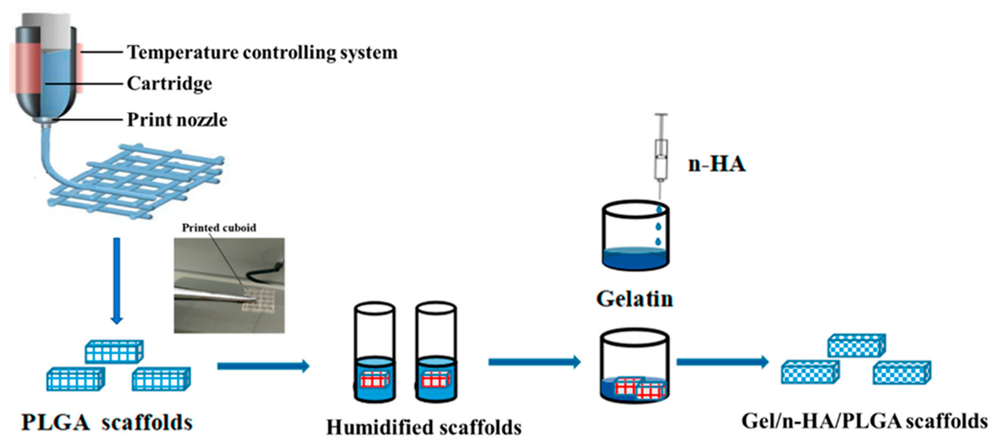
Figure 3.
Fiber surface modification for generation of multi-functional nanofibers. (a) physical adsorption, (b) blend electrospinning, (c) coaxial electrospinning, and (d) surface-grafted modification by radiation, plasma, and chemical treatment for formation of functional groups [67].
Figure 3.
Fiber surface modification for generation of multi-functional nanofibers. (a) physical adsorption, (b) blend electrospinning, (c) coaxial electrospinning, and (d) surface-grafted modification by radiation, plasma, and chemical treatment for formation of functional groups [67].

Figure 4.
Schematic diagram of scaffold fabrication. (a) Electrospinning process. (b) Ultrasonication. (c) Mixture and frozen. (d) Freeze-drying technique. (e) Crosslinking and washing [81].
Figure 4.
Schematic diagram of scaffold fabrication. (a) Electrospinning process. (b) Ultrasonication. (c) Mixture and frozen. (d) Freeze-drying technique. (e) Crosslinking and washing [81].

Figure 5.
A schematic illustration of the preparation of multilayered electrospun cellulose acetate (CA) nanofibers assembled with positively charged chitosan (CS) and negatively charged multiwalled carbon nanotubes (MWCNTs) or sodium alginate (ALG) via a layer-by-layer (LbL) technique. (a) For comparison, CS/ALG multilayers were also deposited onto the (CA) nanofibers, since sodium alginate (ALG) is a biocompatible naturally occurring polymer and has been widely used in tissue engineering applications. (b) Electrospun (CA) nanofibrous mats were first formed and used as templates for subsequent deposition of CS/MWCNTs multilayers via electrostatic self-assembly Adapted with permission from [82].
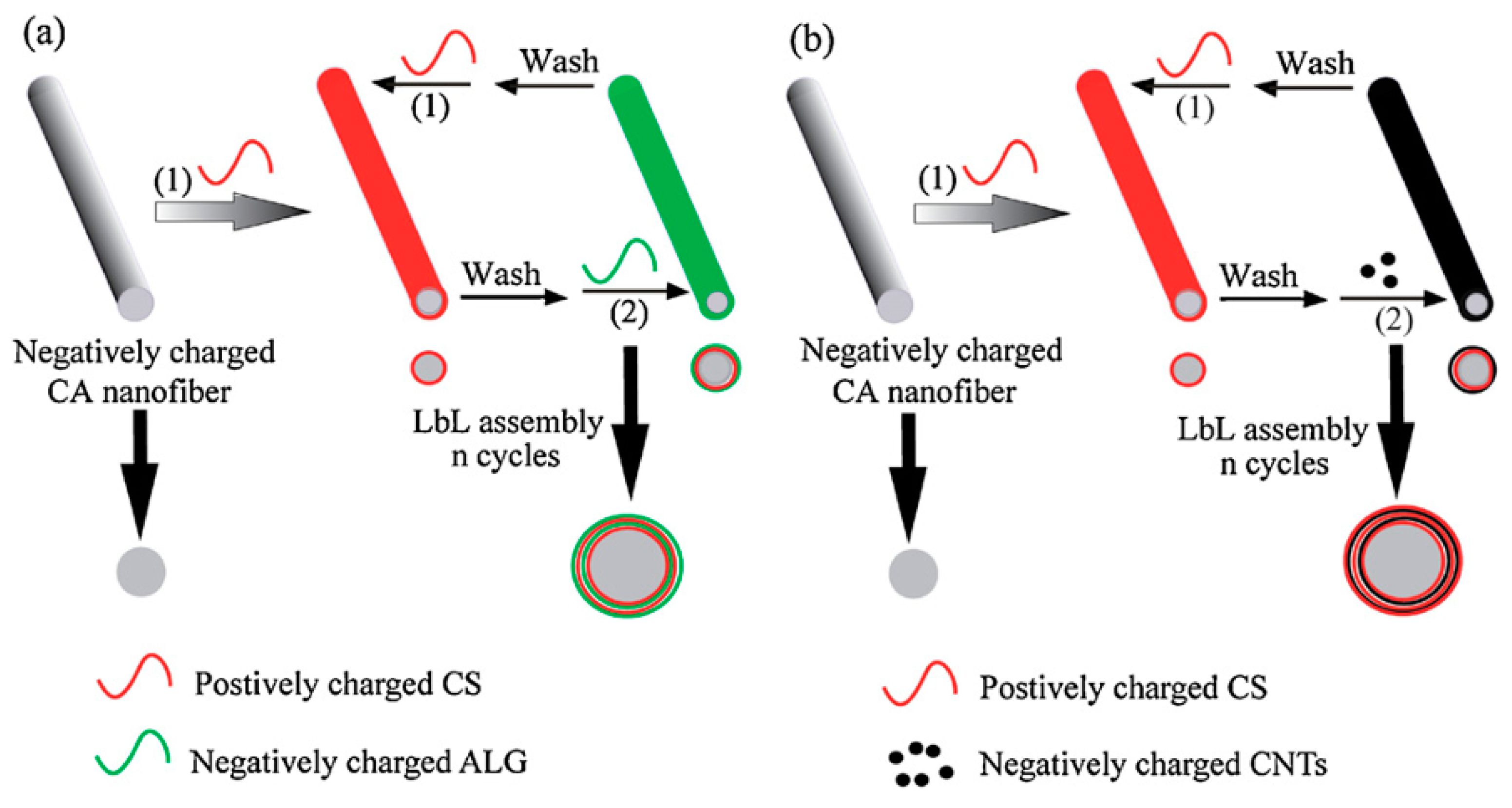

Table 1.
Nanofibrous scaffolds in targeted tissue regeneration.
| Nanofibrous Scaffold Structure and Method of Manufacturing | Scaffold Key Properties | Limitations and Critical Factors | Ref. | |
|---|---|---|---|---|
| 1 | Antheraea assama fibroin-based micro–nano fibrous nonwoven scaffold | Non-toxic, efficient in supporting cell adhesion and growth | Requires validation through in vivo studies, limited to in vitro assessments | [2] |
| 2 | Supramolecular nanofiber hydrogels through self-assembly of biphenyl-tripeptides, molecular dynamics simulations, rheology, and spectroscopy analysis | Robust hydrogels (0.7 to 13.8 kPa), good biocompatibility, enhanced L929 cell adhesion and proliferation, supports chondrocytes spreading and proliferation | Need for optimization of amino acid sequences for better performance, requires extensive in vivo studies | [7] |
| 3 | Bilayered scaffold with electrospun polycaprolactone (PCL) nanofibers decorated on eggshell membrane (ESM), carbodiimide chemistry | Facilitates extensive cell adhesion, proliferation, and migration, biomimetic architecture and protein-rich composition | Challenges in extraction process of ESM, potential for limited cell adhesion in bare natural tissue, needs further validation | [8] |
| 4 | Composite structured nano-/microfibrous scaffold made from chitosan–beta-glycerol phosphate–gelatin using electrospinning | High biocompatibility, non-toxic, biodegradable, significant cell attachment and proliferation | Potential rapid degradation and poor mechanical properties of gelatin, requires optimization for mechanical stability and handling | [19] |
| 5 | Metformin-loaded PLGA/collagen nanofibers (Met-PLGA/Col NFs) fabricated using electrospinning | Modulates macrophage phenotype from pro-inflammatory (M1) to anti-inflammatory (M2), increased cell survival, reduced pro-inflammatory cytokines | Needs further validation for long-term effects and various inflammatory conditions, requires extensive in vivo studies | [20] |
| 6 | Single-step electrospinning approach with polycaprolactone (PCL), altering voltage polarity to control surface potential | Enhanced cell adhesion and filopodia formation, tunable surface potential, well-engineered scaffolds for bone regeneration | Requires further validation for long-term in vivo effects, potential challenges in maintaining consistent surface chemistry during large-scale production | [31] |
| 7 | Modular and versatile spatial functionalization of scaffolds through fiber-initiated controlled radical polymerization with poly-e-caprolactone (PCL) and cell-binding peptide motifs | Zonal discrete bilayers with dual functionality (cell-adhesive and antifouling), supports cell adhesion and spatial organization | Complex fabrication process, needs optimization for specific tissue applications, potential scalability issues | [32] |
| 8 | Ultrasmall natural peptides self-assembled into hydrogels, forming strong temperature-resistant helical fibers | Biocompatible, heat resistant up to 90 °C, tunable high mechanical strength, resembles collagen fibers in the extracellular matrix | Requires further research for long-term stability and functionality in vivo, potential challenges in scaling up for clinical applications | [36] |
| 9 | Poly-D,L-lactic acid (PDLLA) nanofibers with or without Spirulina biomass, fabricated using electrospinning | High stem cells (SCs) adhesion and viability, atoxic for SCs, mimics natural extracellular matrix (ECM), suitable for tissue engineering | Needs further validation in long-term in vivo studies, potential challenges in consistent fabrication and scaling up for clinical use | [37] |
| 10 | Quaternized chitosan/chitosan nanofibrous mats, electrospun from ternary solutions followed by selective removal of poly(ethylene oxide) | Good mechanical properties, reversible water vapor adsorption/desorption, strong antimicrobial activity, biocompatible, suitable for wound healing and drug delivery | Requires optimization of quaternized chitosan content for different applications, further validation needed in long-term in vivo studies | [38] |
| 11 | Single-pot biofabrication of living fibers with amniotic mesenchymal stem cells, extruded in citric acid (CA)/media bath | Instantaneous gelation, tunable mechanical properties, adjustable biodegradability, supports cell attachment and proliferation | Needs further research for long-term stability and functionality in vivo, potential challenges in scaling up for clinical applications | [39] |
| 12 | Poroelastic model of a fibrous–porous tissue engineering scaffold using mechanical modeling and experimental approach | Provides insights into stiffness moduli and fluid diffusion time, helps in designing 3D scaffolds with specific mechanical properties | Requires extensive validation for different tissue types, potential challenges in accurately predicting in vivo behavior | [42] |
| 13 | Fiber-reinforced scaffolds using traditional textile substrates and nanofibrous arrays, fabrication and use of scaffold materials to act as a surrogate framework | High mechanical properties, suitable for ex vivo cell expansion and tissue maturation, mimics natural tissue organization | Needs further research to optimize fiber reinforcement strategies for specific tissue applications, potential challenges in scalability and reproducibility | [43] |
Table 2.
Nanofibrous scaffolds in bone regeneration.
| Nanofibrous Scaffold Structure and Method of Manufacturing | Scaffold Key Properties | Limitations and Critical Factors | Ref. | |
|---|---|---|---|---|
| 1 | Chitosan/polycaprolactone (PCL) nanofiber, electrospinning | Suitable fiber morphology, high biocompatibility, enhanced proliferation rate of MG63 cells on 10% chitosan–PCL scaffold | Requires optimization of chitosan–PCL blending ratios, limited to in vitro assessments | [5] |
| 2 | Epoxidized methyl oleate–graphite oxide (EMO-GO)/Poly(L-lactide) fibrous scaffolds, electrospinning | Improved hydrophobicity, favors mesenchymal stem cells (MSCs) attachment and proliferation, good biocompatibility in vivo | Requires further studies on long-term effects and efficacy, needs to confirm in vivo integration and functionality | [10] |
| 3 | Poly(lactic acid) (PLLA) nanofiber sheet partially degraded into fragmented fibers, coated with adenosine and polydopamine, assembled into spheroids with human adipose-derived stem cells (hADSCs) | High coating efficiency of adenosine, stimulates osteogenic markers expression, improved mineral deposition, enhanced bone regeneration | Suppressed adipogenic differentiation, needs further validation in more complex in vivo models and long-term studies | [11] |
| 4 | Three-dimensional (3D) electrospun nanofibers using corona discharge with polycaprolactone (PCL) | Increased pore size, pore volume, and pore interconnectivity, enhanced cell infiltration, proliferation, and differentiation | Needs further validation for clinical applications, potential challenges in maintaining consistent pore structures | [18] |
| 5 | Electrospun polycaprolactone (PCL) and hyaluronic acid (HA) composite scaffold incorporating a short self-assembling peptide | Supports cellular attachment, proliferation, and significant osteogenic differentiation and calcium mineralization | Poor processability of HA alone, requires combination with other polymers, needs further in vivo studies | [16] |
| 6 | Fibrous scaffolds electrospun from fibroin-containing solutions with chitosan, reinforced with silk fiber | Increased stiffness, water-insoluble ultrathin fibers, biocompatible, effective for tissue engineering | Requires further optimization of fibroin–chitosan ratios, potential challenges in scaling up production for clinical applications | [45] |
| 7 | Co-electrospun nano-/microfibrous composite scaffolds with silk fibroin–poly(epsilon-caprolactone) (SF-PCL) nanofibers and PCL microfibers | Structural, compositional, and chemical gradients, higher osteogenic differentiation, enhanced cell proliferation | Needs further validation in long-term in vivo studies, potential challenges in achieving consistent scaffold properties in large-scale production | [46] |
| 8 | Electrospun poly(epsilon-caprolactone)–polydimethylsiloxane (PCL-PDMS) copolymer fibers with shape memory effect | Shape memory properties, biocompatible, promotes osteoblast proliferation and biomineralization, high shape recovery and fixity ratios | Requires optimization of PCLratios for specific applications, potential challenges in maintaining shape memory properties during long-term use | [54] |
| 9 | 3D-printed poly-epsilon-caprolactone (PCL) single microfibers | Biomimetic surface mechanics, linear increase in modulus with strain rate, potential for rational scaffold design | Needs further research to correlate surface mechanics with biological response, potential challenges in achieving consistent mechanical properties in vivo | [48] |
| 10 | In situ bone tissue engineering using electrospun nanofibrous polycaprolactone (PCL) scaffolds functionalized with hydroxyapatite (HA) particles via polydopamine (PDA) coating and systemic injection of substance P (SP) | Mobilizes endogenous stem cells, enhances bone tissue regeneration, highly osteo-inductive and biocompatible scaffolds | Requires further validation for clinical applications, potential challenges in optimizing SP dosage and scaffold functionalization | [55] |
| 11 | Electrospun polycaprolactone (PCL) nanofiber meshes with dexamethasone (Dex)-loaded liposomes immobilized on the surface | Sustained release of Dex over 21 days, promotes osteogenic differentiation of human bone marrow-derived mesenchymal stem cells (hBMSCs), non-cytotoxic | Requires further optimization for in vivo applications, potential challenges in maintaining consistent release profiles in clinical settings | [44] |
| 12 | 3D-printed microfibrous porous scaffolds using composite inks of gelatin (Gel), nano-hydroxyapatite (n-HA), and poly(lactide-co-glycolide) (PLGA), combining 3D printing and freeze-drying | Exceptional biodegradation, mechanical properties, and biocompatibility, promotes osteoblast adhesion, growth, and differentiation | Needs further research to optimize scaffold properties for specific bone tissue applications, potential scalability challenges | [49] |
| 13 | Electrospun poly(lactide-co-glycolide) (PLGA) membranes loaded with diamond nanoparticles (ND) | Higher mechanical resistance, supports attachment, spreading, and proliferation of human osteoblast-like MG-63 cells, low inflammatory activity | Needs further research to optimize nanoparticle loading and distribution, potential challenges in scaling up production for clinical use | [57] |
| 14 | Self-assembling peptide nanofiber scaffolds (PuraMatrix) combined with dog mesenchymal stem cells (dMSCs) and platelet-rich plasma (PRP) | Promotes new bone regeneration, supports attachment and proliferation of dMSCs, enhances bone quality and formation | Requires further research for long-term in vivo applications, potential challenges in optimizing scaffold composition and combination with PRP | [50] |
| 15 | Electrospun chitosan–alginate nanofibers with in situ polyelectrolyte complexation | Biocompatible, promotes cell adhesion and proliferation, uniform nanofiber formation without toxic crosslinking agents | Requires further research to optimize electrospinning conditions and scaffold properties for specific tissue applications, potential scalability issues | [47] |
| 16 | Biodegradable 3D macroporous scaffold from aligned electrospun nanofibrous yarns using poly(L-lactic acid) and polycaprolactone | Supports cell proliferation and ingrowth, promotes bone formation in vitro and in vivo, provides structural support for stem cells | Needs further validation for clinical applications, potential challenges in maintaining consistent scaffold properties during large-scale production | [59] |
| 17 | Piezoelectric 3D fibrous poly(3-hydroxybutyrate)-based scaffolds ultrasound-mineralized with calcium carbonate | Biodegradable, stimulates bone tissue repair, enhances osteoblast cell adhesion and proliferation, superhydrophilic mineralized fibers | Requires optimization of mineralization process, potential challenges in achieving consistent piezoelectric properties and scaffold stability | [51] |
| 18 | Hydrogel fibers encapsulating human stem cells (hiPSCs, hESCs, hUCMSCs) in an injectable calcium phosphate scaffold | Injectable, supports stem cell viability, proliferation, and osteogenic differentiation, high mechanical properties | Needs further research on long-term in vivo effects, potential challenges in optimizing hydrogel fiber encapsulation and maintaining cell viability post-injection | [60] |
| 19 | Three-dimensional (3D) carbon nanofibers from bacterial cellulose, combined with hydroxyapatite (HAp), prepared via carbonization and surface treatment with nitric acid | Promotes mineralization, shows 3D fibrous structural features, supports bone tissue regeneration | Requires further optimization for consistent mineralization and structural properties, potential challenges in large-scale production | [61] |
| 20 | Biomimetic fiber-guiding scaffolds using solid free-form fabrication methods for bone–ligament complex regeneration | Guides ligamentous fiber orientation, enhances tissue integration, withstands biomechanical loading | Needs further research to optimize scaffold design for various clinical scenarios, potential challenges in achieving consistent fiber orientation | [62] |
| 21 | Three-dimensional magnetic fibrous scaffold with Fe3O4 nanoparticles and icariin (ICA), expanded by supercritical CO2 | Promotes cell proliferation and viability, enhances porosity and angiogenesis, supports bone tissue engineering | Needs further validation for clinical applications, potential challenges in maintaining consistent scaffold properties and magnetic field effects in vivo | [64] |
Table 3.
Nanofibrous scaffolds in cardiac and vascular regeneration.
| Nanofibrous Scaffold Structure and Method of Manufacturing | Scaffold Key Properties | Limitations and Critical Factors | Ref. | |
|---|---|---|---|---|
| 1 | Blended polycaprolactone (PCL)/collagen electrospun scaffolds with varying collagen type I concentrations | Beneficial for cell viability and functionality, ideal concentration at 25% collagen I for vascular grafts | High concentrations of collagen may lead to loss of confluent monolayer, needs optimization for different applications | [17] |
| 2 | Electrospun fibrous poly(vinylidene fluoride) (PVDF-TrFe) scaffolds | Supports cardiomyocyte differentiation and maturation, provides real-time monitoring of tissue contractions via piezoelectric effect | Requires further research to optimize scaffold properties for specific cardiac applications, potential challenges in achieving consistent piezoelectric responses | [65] |
| 3 | Electrospun biocomposite nanofibers consisting of Poly(L-lactic acid)–co-poly(epsilon-caprolactone) (PLACL), silk fibroin (SF), and Aloe Vera (AV) | Porous, beadless, uniform nanofibers, interconnected pores, supports cardiac cell proliferation and expression of cardiac proteins | Requires further optimization for specific cardiac tissue applications, potential challenges in maintaining consistent scaffold properties in large-scale production | [66] |
| 4 | Bacterial cellulose/oxidized bacterial cellulose nanofibrils (BC/oxBCNFs) macro-fibers developed via low-speed rotary coagulation spinning and solvent control | Robust mechanical integrity, high biocompatibility, excellent cell adhesion, supports endothelial and smooth muscle cell proliferation | Needs further validation for clinical applications, potential challenges in maintaining consistent scaffold properties and achieving large-scale production | [69] |
Table 4.
Nanofibrous scaffolds in soft tissue regeneration.
| Nanofibrous Scaffold Structure and Method of Manufacturing | Scaffold Key Properties | Limitations and Critical Factors | Ref. | |
|---|---|---|---|---|
| 1 | High-strength and highly oriented polycaprolactone (PCL) fibers, melt spinning technology | High tensile strength (up to 69 cN/tex), excellent strength retention after 24 weeks in phosphate-buffered saline (PBS), resistance to high loads | Potential impact of fiber cross-sectional geometry on cell growth, needs further functionalization studies | [4] |
| 2 | Electrospun polyethylene oxide (PEO) and carboxymethyl cellulose (CMC)/PEO blend nanofibers | Stable, regular, cylindrical fibers, 3D porous interconnected network, non-toxic, supports cell proliferation | Requires further research to optimize scaffold properties for specific soft tissue applications, potential scalability issues | [72] |
| 3 | Biodegradable polyurethane microfibers electrospun to create scaffolds with small fiber diameter and interconnected porous network | Elastomeric properties, supports soft tissue engineering, biodegradable via hydrolysis and enzyme-mediated chain cleavage | Needs further research to optimize degradation profile and mechanical properties, potential challenges in maintaining consistent scaffold properties in vivo | [73] |
| 4 | Poly(lactic-co-glycolic acid) (PLGA) and polyisoprene (PI) fibrous scaffolds processed through dripping technique | Suitable mechanical properties for soft tissue applications, promotes skeletal muscle cell proliferation | Requires further research to optimize scaffold properties for different soft tissue types, potential challenges in achieving consistent cell response and integration | [74] |
| 5 | Electrospun hybrid nanofibrous meshes made from poly(epsilon-caprolactone) (PCL) and poly(p-dioxanone) (PPDO) blends | Adjustable surface hydrophilicity, degradation profile, and mechanical properties, promotes cell adhesion and proliferation | Needs further validation for clinical applications, potential challenges in optimizing blend ratios and maintaining consistent scaffold properties in large-scale production | [75] |
| 6 | Electrospun scaffolds made of PLGA, collagen I, and polyurethane, with varied PLGA ratios (50:50 and 85:15) and fiber orientations | Similar tensile properties to knee ligaments, aligned fibers show higher tensile properties, supports fibroblast attachment and proliferation | Requires further optimization for specific ligament tissue applications, potential challenges in maintaining consistent scaffold properties and fiber orientation | [76] |
| 7 | Alginate (Alg) and hydroxyethyl cellulose (HEC) fibers created by wet spinning in varied proportions and crosslinked with CaCl2 and glutaraldehyde | Good degradability, mechanical strength similar to collagenous fibers, supports tenocyte proliferation and migration | Needs further validation for clinical applications, potential challenges in optimizing crosslinking conditions and maintaining consistent scaffold properties | [77] |
| 8 | Warp-knitted poly(ethylene terephthalate) (PET) scaffolds infiltrated with hydrogels and cells for fiber-reinforced matrix | Adjustable porosity and elasticity, supports MSC proliferation and differentiation, suitable for various soft tissues | Needs further research for specific tissue applications, potential challenges in maintaining consistent scaffold properties and cell response | [79] |
| 9 | Electrospun PCL short fibers incorporated in freeze-dried gelatin scaffolds | Hierarchical structure with major pores (50–150 µm), improved mechanical properties, enhanced cell adhesion and proliferation | Needs further validation for clinical applications, potential challenges in maintaining consistent scaffold properties and fiber distribution | [81] |
| 10 | Cellulose acetate (CA) nanofibers with multilayers of positively charged chitosan and negatively charged multiwalled carbon nanotubes (CS/CNTs) or CS/ALG via electrospinning and layer-by-layer (LbL) self-assembly | Three-dimensional fibrous structure, improved protein adsorption, enhanced fibroblast attachment, spreading, and proliferation | Requires optimization for homogeneous structure and composition, addressing potential cytotoxicity concerns of CNTs | [82] |
Table 5.
Nanofibrous scaffolds in neural regeneration.
| Nanofibrous Scaffold Structure and Method of Manufacturing | Scaffold Key Properties | Limitations and Critical Factors | Ref. | |
|---|---|---|---|---|
| 1 | Three-dimensional microfibrous scaffolds made from poly(epsilon-caprolactone) (PCL) using a novel microfluidics approach | Enhances proliferation and glial differentiation of adult neural stem cells, provides biological and topographic cues | Needs further validation for long-term effects and functionality in vivo, potential challenges in achieving consistent scaffold properties | [22] |
| 2 | Electrospun poly(epsilon-caprolactone) (PCL) nanofibers containing zero-valent iron (Fe) nanoparticles | Increased electrical conductivity and mechanical properties, supports nerve regeneration and neurite outgrowth | Cytotoxicity at high concentrations of Fe nanoparticles, requires optimization of Fe content for safe and effective use | [23] |
| 3 | PCL nanofibers with LbL self-assembling peptide coating for CRISPR/dCas9 delivery | Efficient loading and sustained release of pDNA complexes, enhanced cell adhesion and proliferation, promotes neurite outgrowth | Requires further validation for clinical applications, addressing potential safety concerns with CRISPR/Cas9 systems | [84] |
| 4 | Conductive aligned fibrous scaffold of PCL/gelatin/polypyrrole/graphene with ferulic acid encapsulation via electrospinning | Highly aligned fibers, improved conductivity, good mechanical properties, limited cytotoxicity, promotes nerve tissue repair | Needs further research for specific tissue applications, potential challenges in optimizing scaffold properties and ensuring consistent cell response | [85] |
| 5 | Lycium barbarum polysaccharide (LBP) encapsulated in core–shell-structured PLGA nanofibers via coaxial electrospinning | Enhanced proliferation and neuronal differentiation of PC12 cells, promotion of Schwann cells myelination | Needs further validation for clinical applications, potential challenges in maintaining consistent release profiles and scaffold properties | [86] |
| 6 | Ion beam irradiation into biodegradable nanofibers, poly-L-lactic acid (PLLA) fibers irradiated with Kr+ ions at varying fluences | Improved cell attachment, surface chemical modification, decreased average fiber diameter | Requires optimization for homogeneous irradiation effects, addressing potential cytotoxicity concerns of ion beam irradiation | [87] |
Table 6.
Nanofibrous scaffolds in wound healing and skin regeneration.
| Nanofibrous Scaffold Structure and Method of Manufacturing | Scaffold Key Properties | Limitations and Critical Factors | Ref. | |
|---|---|---|---|---|
| 1 | Phase separation-assisted electrospray and electrostatic focusing to create polylactic acid (PLA) nanofibrous microcapsules | Fiber–particle duality, promotes cell attachment, easy to handle for direct 3D patterning, high-aspect-ratio microscaffolds | Control of microcapsule diameter and surface porosity needs optimization, requires validation for diverse tissue engineering applications | [14] |
| 2 | Non-mulberry silk fibroin fiber-reinforced chitosan composite scaffold | Facilitates human mesenchymal stem cell (hMSC) attachment and colonization, enhanced sulfated glycosaminoglycan deposition, expression of collagen type II | Requires further validation for clinical applications, potential challenges in maintaining consistent scaffold properties and biological responses | [89] |
| 3 | QOS-crosslinked collagen nanofibers via electrospinning | Improved flexibility and thermal stability, potent antibacterial activity, and biocompatibility with primary human dermal fibroblasts and human fetal osteoblasts | Higher concentrations of QOS showed cytotoxic effects, optimization required for balance between antimicrobial properties and biocompatibility | [91] |
| 4 | Quaternary ammonium organosilane (QOS)-crosslinked collagen nanofibers via electrospinning | Enhanced fibroblast adhesion and proliferation, good swelling property, antibacterial properties, suitable mechanical properties, and biocompatibility | Requires further validation for in vivo applications, stability and consistency of collagen coating over time | [92] |
Table 7.
Nanofibrous scaffolds in advanced drug delivery.
| Nanofibrous Scaffold Structure and Method of Manufacturing | Scaffold Key Properties | Limitations and Critical Factors | Ref. | |
|---|---|---|---|---|
| 1 | Cold atmospheric plasma (CAP)-modified core–shell nanofibers using coaxial electrospinning, polyvinyl alcohol (PVA)/poly(L-lactic acid) (PLLA) core–shell nanofibers | Enhanced hydrophilicity, increased cell attachment and proliferation, higher osteoinductivity, improved drug release profiles | Needs further investigation into long-term effects and stability of CAP treatment, potential challenges in uniform CAP application during large-scale production | [13] |
| 2 | PLLA/HAp (hydroxyapatite) nanofibrous scaffold with simvastatin loading via electrospinning and aminolysis | Enhanced osteoblast cell proliferation, tunable aspect ratio of microcylinders, potential for bone tissue regeneration, and controlled drug release | Requires further validation for in vivo applications, optimization of drug loading and release profiles | [95] |
| 3 | Ciprofloxacin-loaded PLGA nanoparticles incorporated into electrospun fibers | Continuous slow release of antibiotics, high local concentration, biodegradability, biocompatibility, and potential for wide range of tissue engineering applications | Needs further characterization and in vivo studies for clinical translation, ensuring consistent drug release and scaffold properties | [96] |
| 4 | PCL–gelatin electrospun fibers infused with CO2 | Stabilized PCL within blend without overall deformation, increased Rhodamine B loading concentration, potential for enhanced bioactivity | Optimization required for balancing swelling and compression effects, controlled release profiles need further refinement | [97] |
| 5 | Natural polymer-based electrospun nanofibers for drug delivery and tissue engineering | Biopolymers like chitosan, alginic acid, hyaluronic acid, collagen, gelatin, and albumin are excellent candidates, improved cell interaction and biocompatibility | Difficulties in electrospinning biopolymers, need for specific conditions, solvent, copolymer addition, and crosslinking for successful spinning | [98] |
Disclaimer/Publisher’s Note: The statements, opinions and data contained in all publications are solely those of the individual author(s) and contributor(s) and not of MDPI and/or the editor(s). MDPI and/or the editor(s) disclaim responsibility for any injury to people or property resulting from any ideas, methods, instructions or products referred to in the content. |
© 2024 by the authors. Licensee MDPI, Basel, Switzerland. This article is an open access article distributed under the terms and conditions of the Creative Commons Attribution (CC BY) license (https://creativecommons.org/licenses/by/4.0/).
Share and Cite
MDPI and ACS Style
Omidian, H.; Gill, E.J. Nanofibrous Scaffolds in Biomedicine. J. Compos. Sci. 2024, 8, 269. https://doi.org/10.3390/jcs8070269
AMA Style
Omidian H, Gill EJ. Nanofibrous Scaffolds in Biomedicine. Journal of Composites Science. 2024; 8(7):269. https://doi.org/10.3390/jcs8070269
Chicago/Turabian StyleOmidian, Hossein, and Erma J. Gill. 2024. "Nanofibrous Scaffolds in Biomedicine" Journal of Composites Science 8, no. 7: 269. https://doi.org/10.3390/jcs8070269