Article

Development of PSL-Loaded PLGA Nanoparticles for the Treatment of Allergic Contact Dermatitis

1
Department of Medical Pharmacy, Graduate School of Pharmaceutical Sciences, Josai International University, 1 Gumyo Togane, Chiba 283-0002, Japan
2
Faculty of Pharmaceutical Sciences, Tokyo University of Science, 2641, Yamazaki, Noda, Chiba 278-8510, Japan
3
Faculty of Pharmaceutical Sciences, Josai International University, 1 Gumyo Togane, Chiba 283-0002, Japan
4
Faculty of Pharmacy and Pharmaceutical Sciences, Josai University, 1-1 Keyakidai Sakado, Saitama 350-0295, Japan
*
Author to whom correspondence should be addressed.
Colloids Interfaces 2024, 8(3), 39; https://doi.org/10.3390/colloids8030039
Submission received: 13 May 2024 / Revised: 6 June 2024 / Accepted: 11 June 2024 / Published: 18 June 2024

Abstract

:
Allergic contact dermatitis (ACD) can easily develop once sensitization is established by exposure to small amounts of antigen, and steroids are used for treatment. In this study, we evaluated the therapeutic efficacy of prednisolone (PSL)-loaded poly(DL-lactide-co-glycolide) (PLGA) nanoparticles (NPs) on a mouse model of contact dermatitis (CHS). Nanoparticles were prepared using a poor solvent diffusion method, and particle size distribution and mean particle size were measured using dynamic light scattering. Treatment experiments with PSL-loaded PLGA NPs were performed before and after sensitization with 1-fluoro-2,4-dinitrobenzene (DNFB), and evaluation was performed by quantifying intracutaneous IL-4 and TNF-α levels in a mouse model of CHS using ELISA. When PSL-loaded PLGA NPs were administered before sensitization, IL-4 expression was significantly decreased, and TNF-α tended to decrease in the group treated with PSL-loaded PLGA NPs compared to the non-treated group. When PSL-loaded PLGA NPs were administered after sensitization, IL-4 expression was significantly decreased in the group treated with PSL-loaded PLGA NPs compared to the non-treated group. In both cases, there were no significant differences between the PSL-loaded PLGA NP treatment group and the PSL-containing ointment group. These results suggest that, in the treatment of CHS, PSL-loaded PLGA NPs show a certain therapeutic effect when preadministration.

Graphical Abstract

1. Introduction

Contact dermatitis (CHS) is classified into irritant contact dermatitis (ICD) and allergic contact dermatitis (ACD). ACD is caused by minute amounts of haptens and has two phases of onset: a sensitization phase and an attraction phase [1,2]. The causative agents of contact dermatitis are mostly chemicals with a molecular weight of less than 1000 called haptens. Hapten penetrates the skin surface, binds to proteins, and forms hapten-protein conjugates. Hapten protein conjugates are presented to T lymphocytes by cutaneous dendritic cells, which induce sensitized lymphocytes, and sensitization is considered to be established [3,4]. The sensitization phase is asymptomatic. It has also been reported that haptens induce an early response through innate immune mechanisms [5]. After sensitization, the patient again comes into contact with the hapten, and various chemical messengers convey information to T lymphocytes, which release cytokines in the epidermis. T lymphocytes or TNF-α cause damage to epidermal cells, resulting in allergic contact dermatitis. Symptoms include itching rather than pain, erythema, and in severe cases, swelling with blistering.
Treatment generally involves the removal of allergens and the application of topical steroids. However, the prolonged use of steroids has been reported to cause skin thinning, infection, and hyperpigmentation, and the chronic use of steroids may worsen symptoms [6,7,8,9,10]. These side effects can also be caused by the arbitrary use of steroids when a patient experiences similar symptoms [11]. Furthermore, topical steroids are used in combination with moisturizers because absorption is not improved by a single agent. This increases the total amount of ointment applied, which may affect adherence by increasing the amount applied and discomfort [12]. To suppress the occurrence of steroid-induced side effects, it is important to complete treatment early. Therefore, we considered it necessary to develop a new drug delivery system for the efficient delivery of steroids into the skin.
We focused on poly(DL-lactide-co-glycolide) (PLGA) as a drug carrier to improve the absorption of steroids into the skin. PLGA is enzymatically and non-enzymatically in vivo degraded into water and carbon dioxide, which are eliminated from the body, making it highly safe [13,14,15]. In transdermal delivery, it improves the skin permeability of the drug and controls the release of the active drug from the carrier. It has also been reported that PLGA nanoparticles (NPs) pass through the stratum corneum but do not migrate to the dermis or subcutaneous tissue [16,17]. PLGA NPs have been applied in various medical fields as a base material that allows drugs to be absorbed transdermal and used for treatment [18,19,20,21]. As the L/G ratio, which is the composition ratio of PLGA used, decreases, the release rate of the encapsulated drug increases. This is because the hydrolysis of more glycolic acid accelerates the release of the contained drug [22]. The PLGA used in this study has a high L/G ratio, so the release rate is expected to be slow.
In this study, we planned to use PLGA NPs as a drug carrier for prednisolone (PSL). PLGA NPs containing PSL (PSL-loaded PLGA NPs) were prepared, and the particle size distribution and PSL content in NPs were evaluated. The efficacy of PSL-loaded PLGA NPs against CHS was investigated by observing the behavior of TNF-α and IL-4 in a CHS animal model [23,24]. The therapeutic roles of the components used in the preparation of each nanoparticle are as follows. PSL is an ester formulation with fatty acid modification for the treatment of ACD, and PSL monotherapy is the weakest category of steroids, making it the best choice for confirming therapeutic efficacy. PLGA is the ingredient used to make the nanoparticles that are the DDS carriers for carrying PSL. In vivo, PLGA is decomposed into carbon dioxide and water, so it is not toxic, and its safety is assured. ACE is a good solvent for dissolving PLGA and PSL. Since ACE is expected to be removed during dialysis and no ACE is expected to remain at the time of administration, there is no concern about irritation. Arginine (Arg) dissolves in the poor solvent, slightly tilting the nanoparticle suspension toward basicity. This is thought to cause the carboxy group ends of PLGA to be negatively charged, thereby allowing electrostatic interactions to act between the molecules and contributing to the smaller diameter of the nanoparticles. The nanoparticle preparation method used in this study can easily produce small particles at the laboratory level and is considered safe because it uses only PLGA, drugs, and amino acids as materials. PLGA NPs without drug encapsulation prepared by the same method have been proven to have no cytotoxicity [25].

2. Materials and Methods

2.1. Materials

PLGA (Mw: 10,000, monomer composition of DL-lactic acid/glycolic acid = 75/25) was purchased from Taki Chemical Co., Ltd. (Kakogawa, Japan). L-(+)-arginine (H2NC(NH)NH(CH2)3CH(NH2)COOH, purity ≥ 98.0%). L-glutamic acid (C5H9NO4, purity ≥ 99.0%) and PSL (C21H28O5, ≥97.0%) were purchased from Fujifilm Wako Pure Chemical Corp. (Osaka, Japan). Moreover, 0.5%PSL-containing ointment was purchased from Viatris Inc. (Canonsburg, PA, USA). Isoflurane for the animal was purchased from Mylan Inc. (Pittsburgh, PA, USA). ELISA MAX Deluxe Set Mouse IL-4 and ELISA MAX Deluxe Set Mouse TNF-α were purchased from Bio Legend Corp. (San Diego, CA, USA). Other chemicals were of the highest reagent grade commercially available.

2.2. Preparation of PSL-Loaded PLGA NPs Formulation

PSL-loaded PLGA NPs were prepared using a combination of nanoprecipitation and selective solvation methods [26,27,28]: 75.2 mg of PLGA and 4.8 mg of PSL were dissolved in 3 mL of acetone (ACE). The suspension was prepared by injecting 20 mL of this solution into a 0.1% (w/v) aqueous Arg solution at a flow rate of 10 mL/s in the poor solvent. The prepared suspensions (PLGA NPs) were placed in a dialysis membrane (UC36-32, molecular weight cut off: 14,000, Sekisui Material Solutions Co., Ltd., Tokyo, Japan) and washed with dialysis for 6 h to remove any PLGA that did not become NPs, PSL that did not enter NPs, and excess ACE. The MWCO of the dialysis membrane is 14,000, which is large enough to not be an obstacle for PSL to pass through.

2.3. Evaluation of Physical Properties of PSL-Loaded PLGA NPs

2.3.1. Particle Size Measurement

PSL-loaded PLGA NPs were dispersed in purified water, and particle size and polydispersity index (PDI) were measured using a zeta potential and particle size analyzer (ELSZneo, Otsuka Electronics Co., Ltd., Hirakata, Japan) under 25 °C conditions. PSL-loaded PLGA NPs were dispersed in a 0.154 M aqueous sodium chloride solution, and the ζ potentials were measured using a zeta potential and particle size analyzer under 25 °C conditions.

2.3.2. Loading Capacity and Content Efficiency

The amount of PSL contained in the prepared PSL-loaded PLGA NPs was determined via HPLC (SIL-10AF, SPD-10Avp, LD-10ADvp, CTO-10Avp, SCL-10Avp, DGU-12A, Shimadzu Corp., Kyoto, Japan). The column was Wakosil-II 5C18 AR (4.6 × 250 mm, Fujifilm Wako Pure Chemical Corp.). The prepared suspension was lyophilized, dissolved in acetonitrile (ACN), and measured via HPLC. The column temperature was 40 °C, and the injection volume was 10 μL. The mobile phase was purified water–ACN = 6:4, and the flow rate was 1.0 mL/min. The content rate and content efficiency were determined using the following equation:
Loading capacity (%) = (Amount of PSL in NPs/total NPs) × 100
Entrapment efficiency (%) = (Experimental PSL loading/Theoretical PSL loading) × 100

2.3.3. Morphological Observation

The morphology of the particles was observed via transmission electron microscopy (TEM, H-7650, Hitachi High-Technologies Co., Ltd., Tokyo, Japan). The surface of the membrane was hydrophilized via plasma irradiation for 10 s on the collodion support membrane attachment grid, and then the prepared suspension (PLGA NPs) was attached. The films were further stored in a desiccator for 24 h to completely remove the solvent. Observations were made at an accelerating voltage of 100 kV and magnification of 20,000×. To confirm the stability of these PSL-loaded PLGA NPs, the mean volume diameters and polydispersity index of these NPs were measured in the same manner as described in Section 2.3.1. The storage stability of the PSL-loaded PLGA NPs prepared in this study was confirmed by storing them at 4 and 32 °C for five days. A temperature of 32 °C was derived from the surface temperature of the human body’s skin [29]. After 1, 3, 24, 48, 96, and 120 h, the samples were taken, and mean volume diameters and polydispersity indexes were measured using the particle size analyzer. To evaluate the release properties of PSL-loaded PLGA NPs used in this study, 3 mL of PSL-loaded PLGA NPs prepared in Section 2.2 was placed in a dialysis membrane and placed in a vial with 97 mL of purified water at 32 °C for 24 h with water bath shaking [30]. After 0.5, 1, 2, 3, 6, 8, 12, and 24 h, each of the 3 mL of the outer solution was extracted and measured via HPLC, and the release rate was calculated. The same volume of purified water was added when the outer solution was extracted, and the total volume of the outer solution was always kept at 97 mL.

2.4. Animal Experiments

Animal experiments were performed using BALB/cCrslc (9-week-old, male) purchased from Japan SLC Inc. (Tokyo, Japan) in accordance with the Josai International University Animal Experiment Guidelines (Ethics Committee for Animal Experimentation of Josai International University, ethical approval code: 2300011).

2.4.1. Preparation of CHS Model Mice

CHS model mice were prepared by administering 25 μL of 0.5% 1-fluoro-2,4-dinitrobenzene (DNFB) to the abdominal skin of shaved mice for 2 consecutive days (days 1 and 2). Four days later (day 6), the back was shaved and sensitized by administering 10 μL of 0.3% DNFB and allowing it to lapse for 24 h. DNFB was dissolved in ACE and olive oil (4:1, v/v).

2.4.2. Treatment Experiments Using CHS Model Mice

Two hours before or after the sensitization dose on day 6 of the mice prepared in Section 2.4.1, PSL-loaded PLGA NPs and PSL-containing ointment were administered. Mice in the control group were treated with ACE/olive oil instead of DNFB at the time of sensitization administration. Then, 24 h after sensitization, mice were anesthetized via inhalation with isoflurane and intraperitoneally with a 3-ingredient mixture of medetomidine hydrochloride at 0.3 mL, midazolam at 0.8 mL, and butorphanol tartrate at 1.0 mL plus saline to obtain 5 mL (0.05 mL/10 g). The lesion site was then excised.

2.4.3. Evaluation of Treatment Effect by ELISA

Skin excised in Section 2.4.2 was homogenized in purified water and centrifuged at 10,000× g for 20 min at 4 °C to measure the concentration of cytokines in the skin. The cytokine concentrations in the supernatant were then measured using ELISA MAX Deluxe Set Mouse IL-4 and ELISA MAX Deluxe Set Mouse TNF-α (Bio Legend Corp., San Diego, CA, USA). TNF-α was measured as an indicator of skin inflammation and IL-4 as an indicator of prolonged allergic symptoms [31,32,33,34].

3. Results

3.1. Evaluation of Physical Properties of PSL-Loaded PLGA NPs

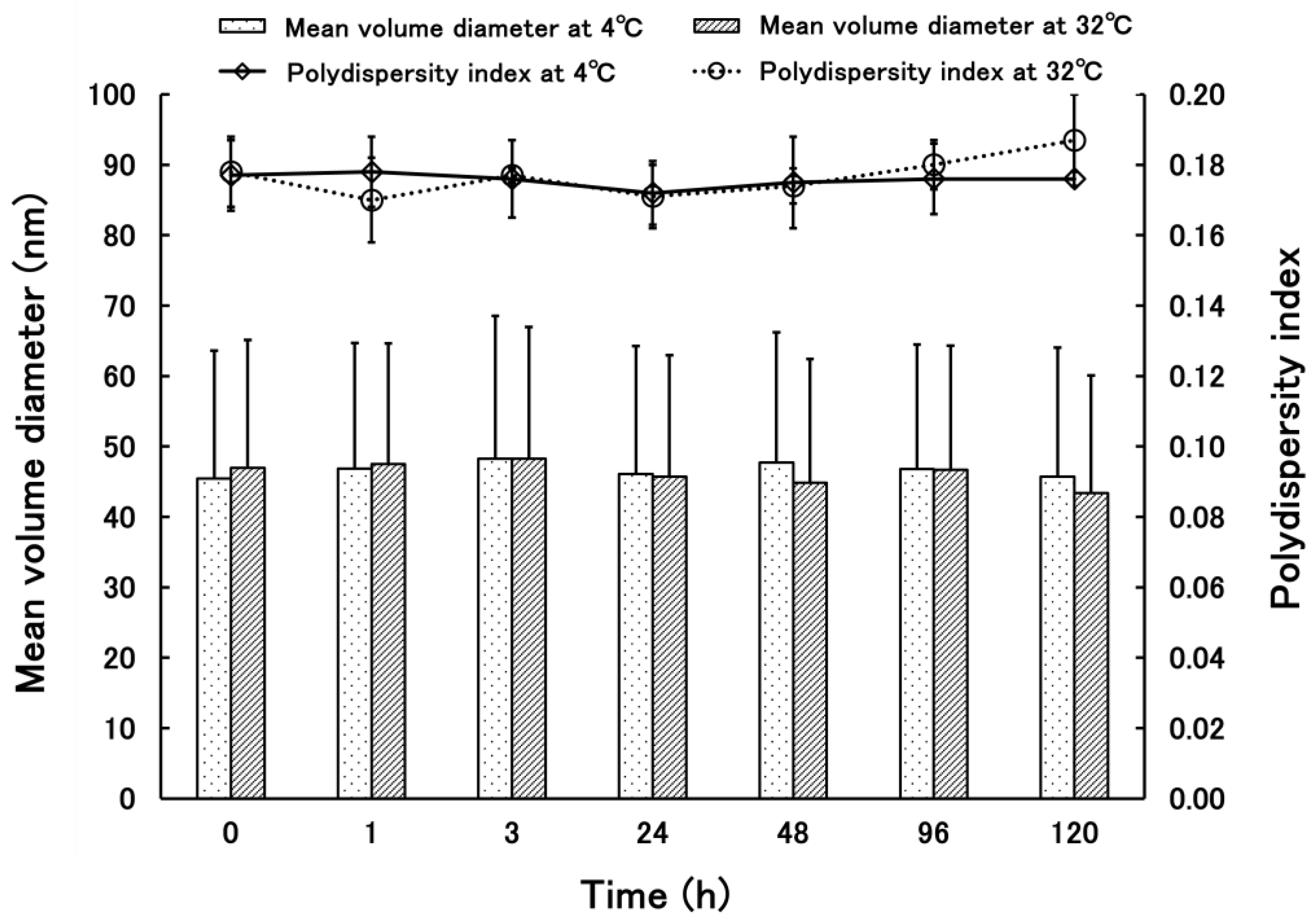
3.1.1. Properties of PSL-Loaded PLGA NPs

The PLGA NPs produced had a particle size of 30.6 ± 12.0 nm, a content of 0.9%, an entrapment efficiency of 15%, a polydispersity index of 0.170, and a ζ potential of −48.34 ± 4.14 mV. The particle size distribution and TEM images of the NPs used in the treatment experiments (dialysis 6 h, number of external solution changes: 3 times; poor solvent: 0.1% Arg solution) in light of their condition and content rate after preparation are shown in Figure 1 and Figure 2. The PLGA NPs are spherical and homogeneously dispersed, and they are considered suitable as nanoparticles for use in therapeutic experiments.

3.1.2. Properties of PSL-Loaded PLGA NPs

Figure 3 shows the results of the stability test of PSL-loaded PLGA NPs. At 4 °C, the mean volume diameter and standard deviation were stable at 45.44–48.28 nm and 16.71–18.71 nm, respectively. The polydispersity indexes were 0.170–0.187, showing no specific trends. At 32 °C, the mean volume diameter and standard deviation were stable at 43.40–48.28 nm and 17.71–20.25 nm, respectively. The polydispersity indexes were 0.172–0.178, showing no specific trends.

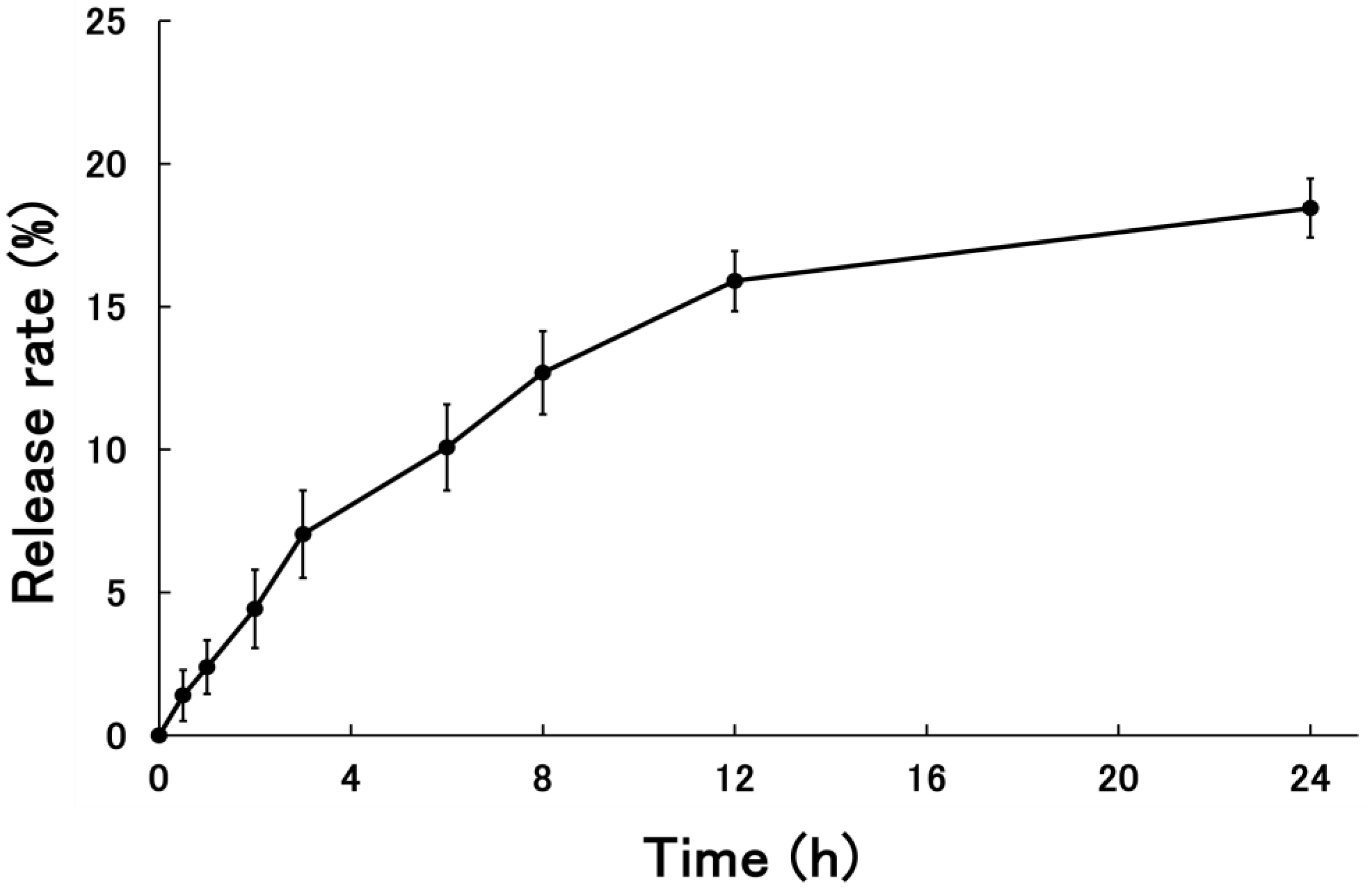
3.1.3. Release Rate of PSL-Loaded PLGA NPs

The release behavior of PSL from PSL-loaded NPs is shown in Figure 4. The cumulative PSL release rate from the NPs 24 h after the start of the release test was 18.45 ± 1.03%.

3.2. Evaluation of Therapeutic Efficacy of PSL-Loaded PLGA NPs Using CHS Model Mice

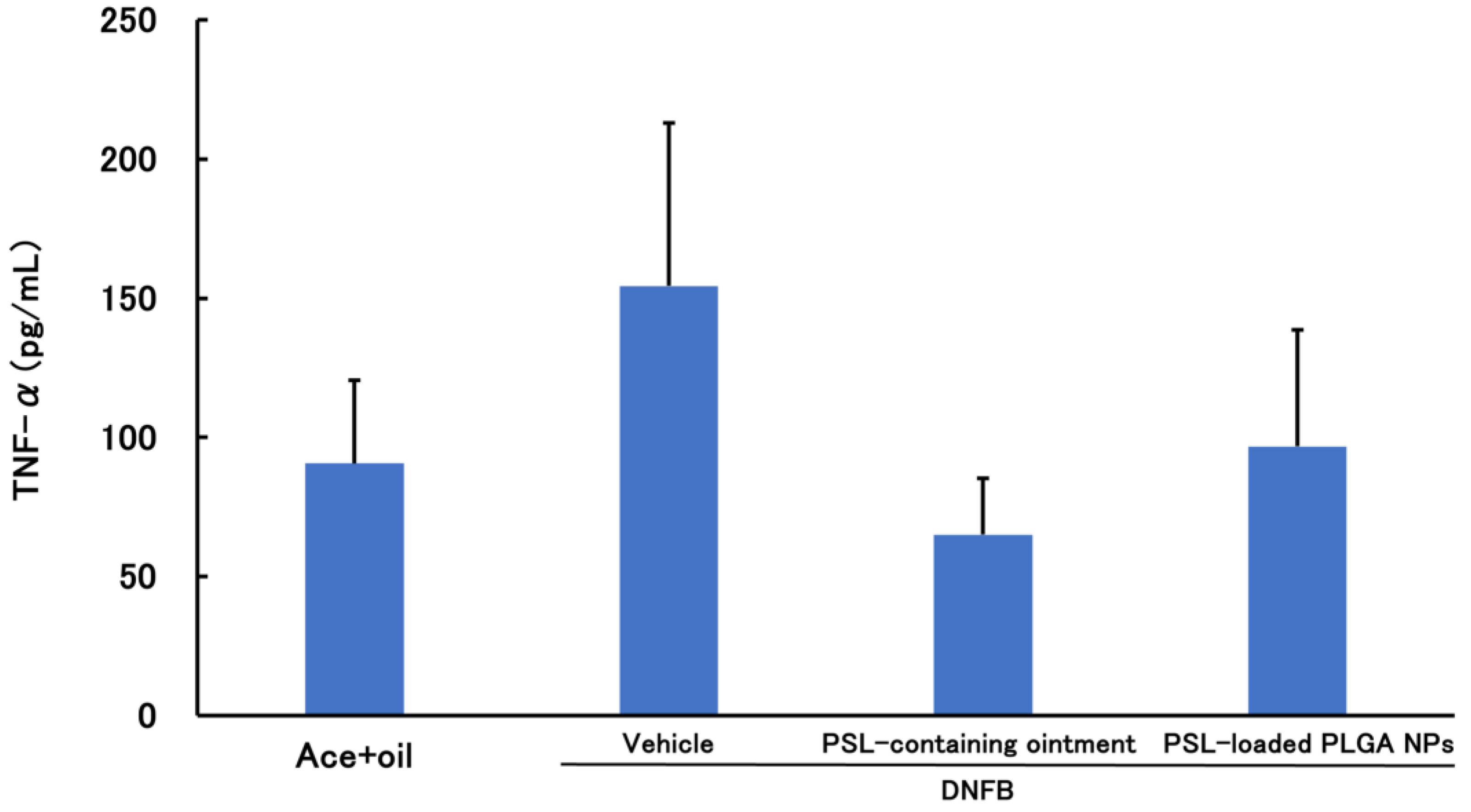
3.2.1. PSL Administered 2 h before Sensitization

Figure 5 and Figure 6 show the results of the therapeutic effect of PSL-loaded PLGA NPs administered 2 h before sensitization on ACD. In TNF-α, there was no significant difference among all groups, but there was a decreasing trend in the PSL-containing ointment group and PSL-loaded PLGA NPs group compared to the untreated group. In IL-4, there was a significant decrease in the PSL-containing ointment and PSL-loaded PLGA NPs-treated group compared to the untreated group. No significant difference in either TNF-α or IL-4 was observed between the PSL-containing ointment and PSL-loaded PLGA NP groups.

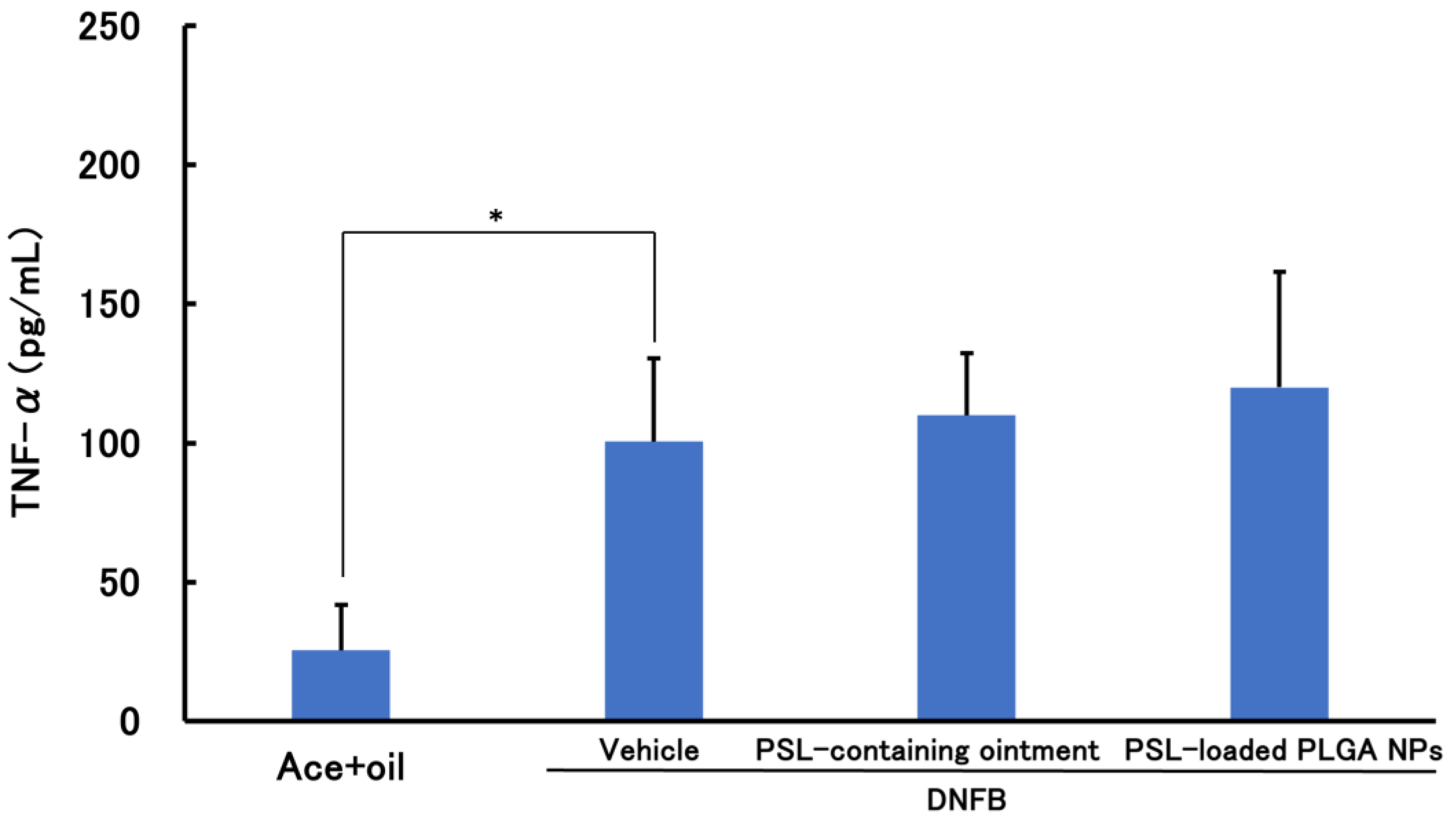
3.2.2. PSL Administered 2 h after Sensitization Administration

Figure 7 and Figure 8 show the results of the therapeutic effect of PSL-loaded PLGA NPs administered 2 h after sensitization on ACD. There was a significant decrease in the PSL-containing ointment and PSL-loaded PLGA NP group compared to the untreated group. There was no significant difference between the two groups of patients treated with PSL-containing ointment and PSL-loaded PLGA NPs.

4. Discussion

In the present nanoparticle preparation method, arginine is included in the poor solvent, and the solvent containing the nanoparticles is slightly inclined toward basicity. Therefore, the carboxy group ends of PLGA are negatively charged, and the entire surface of the nanoparticles is considered to be negatively charged [26]. Furthermore, the large surface charge may have potential applications in iontophoresis and other applications.
The stability test proved that the nanoparticles remained stable, with no agglomeration of the nanoparticles even after being left standing for 5 days. This suggests that the nanoparticles are unlikely to aggregate during administration and will remain in shape after being absorbed into the epidermis. In the release test, the release rate reached a plateau at about 20% after 24 h, which can be inferred from the fact that the PSL inside the PLGA NPs was larger in proportion than the PSL adhering to the surface of the PLGA NPs. The PSL encapsulated inside the PLGA NPs is considered to be gradually released as PLGA is hydrolyzed in the body. Although the release behavior of PSLs from the PLGA NPs was confirmed by permeation during the release test, the PSLs were simply left resting on the skin when they were actually administered to the CHS model animals. Therefore, the release rate will probably be the same or lower than the release test results when administered. These results suggest that the prepared PSL-loaded PLGA NPs are NPs with a stable structure.
When PSL-loaded PLGA NPs were administered to ACD 2 h before sensitization, both TNF-α and IL-4 decreased, whereas when PSL-loaded PLGA NPs were administered to ACD 2 h after sensitization, TNF-α did not change and only IL-4 decreased. The decrease in TNF-α suggests that inflammation is suppressed and that current inflammation is unlikely to be present. The decrease in IL-4 is the suppression of prolonged allergic symptoms, inferring that the allergic symptoms are coming to an end. Therefore, it is likely that both inflammation and prolonged allergic symptoms were suppressed in the pre-dose, whereas only prolonged allergic symptoms were suppressed in the post-dose. Post-dosing cannot suppress the effect of cytokines that have already been released from precursor cells such as macrophages, so the effect is delayed. Pre-dosing can suppress the release of cytokines before sensitization to the causative agent, and the amount of cytokines released can be suppressed in advance, making it easier to produce effects and superior results. Steroids are generally expected to inhibit cytokine induction and release from cells, rather than suppressing cytokine function. In addition, no differences were observed between the PSL-containing ointment and PSL-loaded PLGA NP groups either pre- or post-dose, even though the PSL-loaded PLGA NPs contained only 1/40th of the amount of PSL contained in the PSL-containing ointment. These results suggest that PSL-loaded PLGA NPs can show equivalent efficacy with a very small amount of steroid compared to existing PSL-containing ointments, and the gradual reduction in the total amount of steroid administered may have a significant impact on reducing side effects and shortening the duration of treatment. Based on these results, we consider that the PSL-loaded PLGA NPs exert their maximum therapeutic effect preadministration.
We believe that these PSL-loaded PLGA NPs can be used for treatment. When preadministration is used, it is administered into normal skin and the stratum corneum is the biggest barrier [35,36]. However, the particle size of the PSL-containing PLGA NPs is around 30 nm, and the size of molecules that are said to be able to pass through the gaps in the stratum corneum is 30–70 nm, so there should be no problem with stratum corneum penetration [37,38]. PSL-loaded PLGA NPs will release PSLs in the epidermis, and they are slowly released. The PLGA forming the nanoparticles is more difficult to hydrolyze with a higher proportion of lactic acid, so the nanoparticles are less fragile [15,39]. Some PSL contained in the nanoparticles are attached to the nanoparticle’s surface, while others are embedded within. From the above, PSLs attached to the surface of the nanoparticles may exhibit efficacy quickly after reaching the site of inflammation, while PSLs embedded in the nanoparticles may be released gradually and exhibit efficacy because of the hydrolysis of PLGA, thus indicating a sustained release property. To investigate this, release tests should be carried out using model mice, and skin sections should be stained for observation in the future.

5. Conclusions

These results suggest that PSL-loaded PLGA NPs can reduce symptoms in ACD via the pre-sensitization administration of antigens. Clinically, proactive therapy is recommended for atopic dermatitis, and they will likely be recommended for contact dermatitis as well soon. Further studies are needed so that it can become a treatment option in the future.

Author Contributions

Conceptualization, R.F. and I.T.; methodology, R.F. and I.T.; validation, R.F. and I.T.; formal analysis, R.F., I.T. and K.M.; investigation, R.F.; resources, K.M., A.S. and I.T.; data curation, R.F. and I.T.; writing—original draft preparation, R.F.; writing—review and editing, R.F., T.O., K.M., R.S., A.S., I.T. and K.S.; visualization, R.F., R.S. and I.T.; supervision, K.M., I.T. and K.S.; project administration, R.F. and I.T.; funding acquisition, K.M., I.T. and K.S. All authors have read and agreed to the published version of the manuscript.

Funding

This work was supported by JSPS KAKENHI, grant number JP22K06553.

Institutional Review Board Statement

The study was conducted in accordance with the Declaration of Helsinki, and approved by the Ethics Committee of Ethics Committee for Animal Experimentation of Josai International University (Ethics Code: 2300011 and date: 13 March 2023).

Data Availability Statement

The data presented in this study are available on request from the corresponding author.

Acknowledgments

We gratefully acknowledge the work of the members of our laboratory.

Conflicts of Interest

The authors declare no conflicts of interest.

References

  1. Honda, T.; Egawa, G.; Grabbe, S.; Kabashima, K. Update of immune events in the murine contact hypersensitivity model: Toward the understanding of allergic contact dermatitis. J. Investig. Dermatol. 2013, 133, 303–315. [Google Scholar] [CrossRef] [PubMed]
  2. Vocanson, M.; Hennino, A.; Rozières, A.; Poyet, G.; Nicolas, J. Effector and regulatory mechanisms in allergic contact dermatitis. Allergy Eur. J. Allergy Clin. Immunol. 2009, 64, 1699–1714. [Google Scholar] [CrossRef] [PubMed]
  3. Kaplan, D.; Igyártó, Z.; Gaspari, A. Early immune events in the induction of allergic contact dermatitis. Nat. Rev. Immunol. 2012, 12, 114–124. [Google Scholar] [CrossRef] [PubMed]
  4. Peiser, M.; Tralau, T.; Heidler, J.; Api, A.M.; Arts, J.H.E.; Basketter, D.A.; English, J.; Diepgen, T.L.; Fuhlbrigge, R.C.; Gaspari, A.A.; et al. Allergic contact dermatitis: Epidemiology, molecular mechanisms, in vitro methods and regulatory aspects. Cell. Mol. Life Sci. 2012, 69, 763–781. [Google Scholar] [CrossRef] [PubMed]
  5. Singleton, H.; Popple, A.; Gellatly, N.; Maxwell, G.; Williams, J.; Friedmann, P.S.; Kimber, I.; Dearman, R.J. Anti-hapten antibodies in response to skin sensitization. Contact Dermat. 2016, 74, 197–204. [Google Scholar] [CrossRef] [PubMed]
  6. Coondoo, A.; Phiske, M.; Verma, S.; Lahiri, K. Side-effects of topical steroids: A long overdue revisit. Indian Dermatol. Online J. 2014, 5, 416–425. [Google Scholar] [CrossRef] [PubMed]
  7. Mehta, A.; Nadkarni, N.; Patil, S.; Godse, K.; Gautam, M.; Agarwal, S. Topical corticosteroids in dermatology. Indian J. Dermatol. Venereol. Leprol. 2016, 82, 371–378. [Google Scholar] [CrossRef] [PubMed]
  8. Dey, V. Misuse of topical corticosteroids: A clinical study of adverse effects. Indian Dermatol. Online J. 2014, 5, 436–440. [Google Scholar] [CrossRef]
  9. Ference, J.; Last, A. Choosing Topical Corticosteroids. Am. Fam. Physician 2009, 79, 135–140. [Google Scholar]
  10. Hengge, U.; Ruzicka, T.; Schwartz, R.; Cork, M. Adverse effects of topical glucocorticosteroids. J. Am. Acad. Dermatol. 2006, 54, 1–15. [Google Scholar] [CrossRef]
  11. Surya, R.; Smitha, P.; Sudhir, N. Topical steroid damaged skin: A clinico-epidemiological and dermatological study. J. Pak. Assoc. Dermatol. 2021, 31, 407–414. [Google Scholar]
  12. Lodén, M. Role of Topical Emollients and Moisturizers in the Treatment of Dry Skin Barrier Disorders. Am. J. Clin. Dermatol. 2003, 4, 771–788. [Google Scholar] [CrossRef] [PubMed]
  13. Makadia, H.; Siegel, S. Poly Lactic-co-Glycolic Acid (PLGA) as biodegradable controlled drug delivery carrier. Polymers 2011, 3, 1377–1397. [Google Scholar] [CrossRef] [PubMed]
  14. Brannon-Peppas, L. Recent advances on the use of biodegradable microparticles and nanoparticles in controlled drug delivery. Int. J. Pharm. 1995, 116, 1–9. [Google Scholar] [CrossRef]
  15. Keles, H.; Naylor, A.; Clegg, F.; Sammom, C. Investigation of factors influencing the hydrolytic degradation of single PLGA microparticles. Polym. Degrad. Stab. 2015, 119, 228–241. [Google Scholar] [CrossRef]
  16. Takeuchi, I.; Hidaka, Y.; Oshizaka, T.; Takei, C.; Mori, K.; Sugibayashi, K.; Makino, K. Chitosan-coated PLGA nanoparticles for transcutaneous immunization: Skin distribution in lysozyme-sensitized mice. Colloids Surf. B Biointerfaces 2022, 220, 112916. [Google Scholar] [CrossRef] [PubMed]
  17. Takeuchi, I.; Suzuki, T.; Makino, K. Iontophoretic transdermal delivery using chitosan-coated PLGA nanoparticles for transcutaneous immunization. Colloids Surf. A Physicochem. Eng. Asp. 2021, 608, 125607. [Google Scholar] [CrossRef]
  18. Das, S.; Khuda-Bukhsh, A. PLGA-loaded nanomedicines in melanoma treatment: Future prospect for efficient drug delivery. Indian J. Med. Res. 2016, 144, 181–193. [Google Scholar] [CrossRef] [PubMed]
  19. Jain, S.; Mittal, A.; Jain, A. Enhanced Topical Delivery of Cyclosporin-A Using PLGA Nanoparticles as Carrier. Curr. Nanosci. 2011, 7, 524–530. [Google Scholar] [CrossRef]
  20. Acharya, S.; Sahoo, S. PLGA nanoparticles containing various anticancer agents and tumour delivery by EPR effect. Adv. Drug Deliv. Rev. 2011, 63, 170–183. [Google Scholar] [CrossRef]
  21. Folle, C.; Marqués, A.M.; Díaz-Garrido, N.; Espina, M.; Sánchez-López, E.; Badia, J.; Baldoma, L.; Calpena, A.C.; García, M.L. Thymol-loaded PLGA nanoparticles: An efficient approach for acne treatment. J. Nanobiotechnol. 2021, 19, 359. [Google Scholar] [CrossRef] [PubMed]
  22. Guo, Y.; Li, X.; Macgregor, R.B., Jr.; Yan, H.; Zhang, R.X. Encapsulation and release rate to cytotoxicity in human lens epithelial cells|Enhanced Reader. Heliyon 2023, 9, e18318. [Google Scholar] [CrossRef]
  23. Röse, L.; Schneider, C.; Stock, C.; Zollner, T.; Döcke, W. Extended DNFB-induced contact hypersensitivity models display characteristics of chronic inflammatory dermatoses. Exp. Dermatol. 2012, 21, 25–31. [Google Scholar] [CrossRef] [PubMed]
  24. Manresa, M. Animal Models of Contact Dermatitis: 2,4-Dinitrofluorobenzene-Induced Contact Hypersensitivity. Methods Mol. Biol. 2021, 2223, 87–100. [Google Scholar] [CrossRef] [PubMed]
  25. Kagawa, A.; Sato, A.; Makino, K.; Takeuchi, I. Therapeutic Effects of 30 nm Cyclosporin A-Loaded Nanoparticles Using PLGA-PEG-PLGA Triblock Copolymer for Transdermal Delivery in Mouse Models of Psoriasis. Appl. Sci. 2024, 14, 3791. [Google Scholar] [CrossRef]
  26. Tomoda, K.; Yabuki, N.; Terada, H.; Makino, K. Surfactant free preparation of PLGA nanoparticles: The combination of antisolvent diffusion with preferential solvation. Colloids Surf. A Physicochem. Eng. Asp. 2014, 457, 88–93. [Google Scholar] [CrossRef]
  27. Takeuchi, I.; Kobayashi, S.; Hida, Y.; Makino, K. Estradiol-loaded PLGA nanoparticles for improving low bone mineral density of cancellous bone caused by osteoporosis: Application of enhanced charged nanoparticles with iontophoresis. Colloids Surf. B Biointerfaces 2017, 155, 35–40. [Google Scholar] [CrossRef] [PubMed]
  28. Takeuchi, I.; Kagawa, A.; Makino, K. Skin permeability and transdermal delivery route of 30-nm cyclosporin A-loaded nanoparticles using PLGA-PEG-PLGA triblock copolymer. Colloids Surf. A Physicochem. Eng. Asp. 2020, 600, 124866. [Google Scholar] [CrossRef]
  29. Dipasquale, D.; Buono, M.; Kolkhorst, F. Effect of Skin Temperature on the Cholinergic Sensitivity of the Human Eccrine Sweat Gland. Jpn. J. Physiol. 2003, 53, 427–430. [Google Scholar] [CrossRef]
  30. Takeuchi, I.; Makino, K. Biocompatibility and effectiveness of paclitaxel-encapsulated micelle using phosphoester compounds as a carrier fir cancer trearment. Colloids Surf. B Biointerfaces 2019, 177, 356–361. [Google Scholar] [CrossRef]
  31. Bradley, J. TNF-mediated inflammatory disease. J. Pathol. 2008, 214, 149–160. [Google Scholar] [CrossRef] [PubMed]
  32. Zelová, H.; Hošek, J. TNF-α signalling and inflammation: Interactions between old acquaintances. Inflamm. Res. 2013, 62, 641–651. [Google Scholar] [CrossRef] [PubMed]
  33. Bradding, P.; Feather, I.H.; Howarth, P.H.; Mueller, R.; Roberts, J.A.; Britten, K.; Bews, J.P.; Hunt, T.C.; Okayama, Y.; Heusser, C.H. Interleukin 4 Is Localized to and Released by Human Mast Cells. J. Exp. Med. 1992, 176, 1381–1386. [Google Scholar] [CrossRef] [PubMed]
  34. Luzina, I.G.; Keegan, A.D.; Heller, N.M.; Rook, G.A.; Shea-Donohue, T.; Atamas, S.P. Regulation of inflammation by interleukin-4: A review of “alternatives”. J. Leukoc. Biol. 2012, 92, 753–764. [Google Scholar] [CrossRef] [PubMed]
  35. Bouwstra, J.; Loan Honeywell-Nguyen, P.; Gooris, G.; Ponec, M. Structure of the skin barrier and its modulation by vesicular formulations. Prog. Lipid Res. 2003, 42, 1–36. [Google Scholar] [CrossRef] [PubMed]
  36. Elias, P. Structure and function of the stratum corneum permeability barrier. Drug Dev. Res. 1988, 13, 97–105. [Google Scholar] [CrossRef]
  37. Labouta, H.; El-Khordagui, L.; Kraus, T.; Schneider, M. Mechanism and determinants of nanoparticle penetration through human skin. Nanoscale 2011, 3, 4989–4999. [Google Scholar] [CrossRef] [PubMed]
  38. Küchler, S.; Radowski, M.R.; Blaschke, T.; Dathe, M.; Plendl, J.; Haag, R.; Schäfer-Korting, M.; Kramer, K.D. Nanoparticles for skin penetration enhancement—A comparison of a dendritic core-multishell-nanotransporter and solid lipid nanoparticles. Eur. J. Pharm. Biopharm. 2009, 71, 243–250. [Google Scholar] [CrossRef]
  39. Schmitt, E.; Flanagan Linhardt’, R. Importance of Distinct Water Environments in the Hydrolysis of Poly(DL-lactide-co-glycolide). Macromolecules 1994, 27, 743–748. [Google Scholar] [CrossRef]
Figure 1. TEM image of PSL-loaded PLGA NPs were made at an accelerating voltage of 100 kV (magnification: 20,000×).
Colloids 08 00039 g001
Figure 2. Volume size distribution of PSL-loaded PLGA NPs by dynamic light scattering (n = 3).
Colloids 08 00039 g002
Figure 3. Time-department changes of mean volume diameter and polydispersity indexes of PSL-loaded PLGA NPs at 4 and 32 °C (n = 3, mean ± S.D.).
Colloids 08 00039 g003
Figure 4. The cumulative release rate of PSL from PSL-loaded PLGA NPs (n = 3, mean ± S.D.).
Colloids 08 00039 g004
Figure 5. TNF-α levels in a mouse model of acute allergic contact dermatitis (n = 3–4, mean ± S.D.).
Colloids 08 00039 g005
Figure 6. IL-4 levels in mouse models of acute allergic contact dermatitis (n = 4, mean ± S.D. ** p < 0.01 vs. Ace + oil; * p < 0.05 vs. DNFB, Tukey–Kramer’s test).
Colloids 08 00039 g006
Figure 7. TNF-α levels in a mouse model of acute allergic contact dermatitis (n = 4–5, mean ± S.D. * p < 0.05 vs. Ace + oil, Tukey–Kramer’s test).
Colloids 08 00039 g007
Figure 8. IL-4 levels in mouse models of acute allergic contact dermatitis (n = 4–5, mean ± S.D. ** p < 0.01 vs. Ace + oil, ** p < 0.01 vs. DNFB, Tukey–Kramer’s test).
Colloids 08 00039 g008
Disclaimer/Publisher’s Note: The statements, opinions and data contained in all publications are solely those of the individual author(s) and contributor(s) and not of MDPI and/or the editor(s). MDPI and/or the editor(s) disclaim responsibility for any injury to people or property resulting from any ideas, methods, instructions or products referred to in the content.

