Review

Could New Generations of Sensors Reshape the Management of Parkinson’s Disease?

Department of Neurology, Russian Medical Academy of Continuous Professional Education, 125009 Moscow, Russia
*
Author to whom correspondence should be addressed.
Clin. Transl. Neurosci. 2021, 5(2), 18; https://doi.org/10.3390/ctn5020018
Submission received: 3 July 2021 / Revised: 31 August 2021 / Accepted: 7 September 2021 / Published: 13 September 2021

Abstract

:
Parkinson's disease (PD) is a chronic neurologic disease that has a great impact on the patient’s quality of life. The natural course of the disease is characterized by an insidious onset of symptoms, such as rest tremor, shuffling gait, bradykinesia, followed by improvement with the initiation of dopaminergic therapy. However, this “honeymoon period” gradually comes to an end with the emergence of motor fluctuations and dyskinesia. PD patients need long-term treatments and monitoring throughout the day; however, clinical examinations in hospitals are often not sufficient for optimal management of the disease. Technology-based devices are a new comprehensive assessment method of PD patient’s symptoms that are easy to use and give unbiased measurements. This review article provides an exhaustive overview of motor complications of advanced PD and new approaches to the management of the disease using sensors.

1. A Long Day for a Patient with PD

Parkinson’s disease (PD) is a neurodegenerative disorder characterized by motor symptoms, such as bradykinesia, tremors, and rigidity. At the beginning of the disease, there is an excellent response to treatment. This phenomenon is known as the “long-duration response” of L-dopa. After a few years of use of L-dopa, patients begin to become more aware of the duration of the action, and the “short-duration response” becomes more evident. A combination of disease progression (loss of nigrostriatal dopamine terminals and their storage capacity) and fluctuating L-dopa levels lead to the motor complications of PD [1].
Motor complications include motor fluctuations and L-dopa-induced dyskinesia (Figure 1). Motor complications have a great impact on the patient’s quality of life; they can lead to disability, embarrassment, frustration, and increased caregiver burden [1,2]. In a prospective population-based longitudinal study of the incident and initially drug-naïve PD patients, the 5-year cumulative incidence of motor complications was 52.4% [3]. The CamPaIGN cohort study found a cumulative incidence of motor fluctuations and L-dopa-induced dyskinesia as 54.3% and 14.5%, respectively, at 5 years and 100% and 55.7%, respectively, at 10 years [4].
Motor fluctuations may take the form of short-duration (seconds to minutes), medium-duration and diurnal (minutes to hours), and long-duration (days) responses. Short-duration motor fluctuations include freezing and paradoxical kinesis, lasting from seconds to minutes. Medium-duration fluctuations associated with chronic L-dopa treatment include wearing-off and ON-OFF fluctuations. Diurnal fluctuations lead to different responses in the same dose of L-dopa. Usually, patients have a much better response in the morning than later in the day, but occasionally this pattern is reversed. Motor fluctuations are associated with loss of the long-duration L-dopa response. It is known that benefits from starting L-dopa, although sometimes evident from the first dose, increase over several weeks despite the same dosage. The phenomenon of late deterioration happens in the case of withdrawals or the decreasing of doses of L-dopa and leads to delayed worsening of symptoms that may occur up to two weeks later [5].
There are three worse periods of the day for patients with PD: the “end-of-the-day”, nighttime, or early morning [6]. The “early morning akinesia” is a well-known phenomenon that manifests by the worsening of symptoms in the early morning due to low levels of L-dopa as the last dose was the night before. Stocchi et al. reported that the majority of PD patients with motor fluctuations also suffered from delays in ON time (latency > 30 min) following their first morning dose of L-dopa. [7]. However, some patients can experience an improvement of symptoms in the morning. This phenomenon is also known as the sleep benefit. The prevalence of the sleep benefit ranges from 33% to 55% of PD patients. Sleep benefits can also occur after daytime naps [8]. One hypothesis on the mechanism of sleep benefit stated that dopamine storage in nigral neuronal terminals is replenished during sleep [9]. However, another study showed that patients with sleep deprivation and worse night sleep were more likely to experience sleep benefits [10,11].
Wearing-off is the early sign of predictable dissipation of efficacy of L-dopa and indicates the end of the so-called “honeymoon period” [1]. Predictable wearing-off is the regular recurrence of symptoms at the end of a dose of L-dopa. This is the most frequent type of wearing-off and is usually the earliest manifestation of motor fluctuations [12]. As the disease progresses, patients experience frequent OFF periods, which usually occur in the late afternoon or early evening. This “end-of-the-day crash” represents a predictable deterioration of motor and non-motor symptoms of the disease as evening approaches and is often due to a diminished response to L-dopa at the end of the day [12]. Less often, some PD patients demonstrate the opposite circadian pattern of motor symptoms worsening in the first half of the day. At the same time, other patients experience the deterioration of symptoms in the postprandial state.
Wearing-off typically evolves from a slow and inconspicuous recurrence of motor symptoms to a more rapid and obvious deterioration of the patient’s condition. ON–OFF constitutes a sudden onset of parkinsonism. Unpredictable wearing-off or “sudden OFFs” are less common than predictable wearing-off and usually occur in advanced stages of PD. Because of this acute worsening of parkinsonian symptoms (that can occur within a few seconds), some patients may develop sudden disabling akinesia (may happen at any time during the day) [2,12]. Some patients may experience a combination of predictable and unpredictable rapid switching from ON to OFF. The term Yo-yoing infers rapid, abrupt, and sometimes multiple transitions from one type of state to the other. This subtype of wearing-off is currently rare due to the practice of using overall lower doses of L-dopa but can be seen in some patients with advanced PD [12].
Another L-dopa treatment complication is a loss of benefit to single doses of L-dopa [13]. Some patients notice that their medication will take longer to take effect (delayed response), are less effective (partial response), or sometimes may fail to work at all (dose failure) [14]. In some patients with PD, motor symptoms may paradoxically intensify for a short period of time after taking L-dopa (beginning-of-dose worsening). Another phenomenon is end-of-dose rebound (end-of-dose deterioration) when the L-dopa effect is accompanied by a recurrence of symptoms even worse at the end of the dose than in the untreated state [13]. These subtypes of motor fluctuations are usually a consequence of erratic L-dopa absorption [12].
Freezing of gait (FOG) is characterized by a sudden inability to initiate or continue walking. FOG is an example of the clinical heterogeneity of PD patients. FOG can occur at different periods of time during the day; it can depend on the patient’s motor state condition or not and can improve with dopaminergic medication or not. It typically occurs while turning or moving among obstacles and through narrow spaces; in stressful situations. FOG is a severe gait dysfunction, and patients can feel as if their feet were glued to the floor for a couple of seconds. Gait can be limited to very short strides, or, sometimes, the patient may be completely unable to move [15]. Interestingly, recent studies have shown that freezing is a movement problem that affects more than just gait; indeed, motor blocks are present during upper limb movements and speech [16]. Freezing in the OFF state is a common feature for PD patients. It can occur during prolonged periods of time and usually improves significantly with the adjustment of dopaminergic drugs. Conversely, ON-medication FOG is rare, usually short-lived, and sometimes manifests itself as an unpredictable or paradoxical response to drug changes. Most freezers are wheelchair dependent after an average of 5 years from the onset of the symptom [15].
L-dopa-induced dyskinesia (LID) is another motor complication of PD. LID is phenomenologically recognized as chorea/choreoathetoid movements, ballism, stereotypies, or dystonia. The pathogenesis of a LID is complex and not well understood. There is evidence that it may be caused by a subtle imbalance between the activity of D1 and D2 dopamine receptors in the striatum, but existing data is conflicting [17]. LID is classified according to time of emergence in relation to L-dopa schedule and includes peak-dose, diphasic, and OFF-period dyskinesias. Peak-dose dyskinesia is the most common form of dyskinesia. It emerges at the time of the maximum symptomatic improvement that is correlated with the highest plasma L-dopa levels and, presumably, high brain dopamine concentrations [18,19,20]. Usually, it is a choreiform movement that involves the neck and limbs. In some patients, ballistic movements and myoclonus can occur. When dyskinesia becomes more evident, it can involve even facial and diaphragmatic regions.
Diphasic dyskinesia occurs more rarely; it is seen in a range from 15% to 20% of patients. Involuntary movements occur when a serum level of L-dopa is going up or down, coinciding with two peaks of abnormal movements, one present at the onset of drug effect and another present at the end of drug effect [14] (Figure 1). Diphasic dyskinesia is presented by stereotypical alternating, jerking, dystonic, or ballistic kicking movements. Lower extremities are usually more affected [20]. Gait is also changed with high stepping and is known as “funny” gait [12].
The OFF-period dyskinesia (OFF-period dystonia) is static posturing causing twisting, spasms, or cramping, usually in the feet, although, in patients with advanced PD, it may also occur in the legs, trunk, or arms. It is often worse on the side of the body most affected by PD. OFF-period dystonia often emerges in the morning. However, it can also occur spontaneously during any OFF period of the day and may be provoked by attempting to walk or by anxiety and other non-motor features. OFF-period dystonia may be painful, resembling a muscle cramp, and can be very distressing for patients [18].
Nocturnal symptoms of PD are very common, especially in those patients who are already experiencing the wearing-off symptoms. [21]. Nocturnal hypokinesia appears in the middle stage of the disease. It affects up to 70% of patients with PD and contributes to poor sleep quality [22]. Nocturnal hypokinesia (difficulty turning) is a decrease in the ability to perform sufficient axial rotation and/or trunk flexion to turn in or get out of bed because of axial and limb muscle incoordination. It may occur in all sleep stages but intensifies in the second half of the night. Clinical observations have identified nocturnal hypokinesia as a hypodopaminergic state [22]. The emergence of OFF symptoms (rest tremor, rigidity, and bradykinesia) occurs during the nighttime in 48.2% of patients [6].
Restless legs syndrome (RLS) is a sleep-related movement disorder characterized by the urge to move one’s legs and abnormal leg sensations while resting during the night that disturbs sleep. Dopaminergic dysfunction has been suggested to play a role in RLS based on the clinical responses of patients with RLS to dopaminergic treatment [23]. RLS in PD patients usually develops after motor disease manifestation. A longitudinal study that included 109 drug-naïve PD patients showed RLS prevalence increased from 4.6% at baseline evaluation to 16.3% after 4 years, suggesting disease progression along with increased dopaminergic medication had a role [24]. Leg motor restlessness (LMR) is a condition when an urge to move a leg exists but does not fulfill RLS criteria. LMR could be considered focal akathisia with a distinct diurnal fluctuation characterized by worsening of symptoms in the evening or night. [25]. The LMR prevalence in PD range from 11.1% to 32.3% of patients. [26,27].
The definition of non-motor fluctuations (NMF) initially was represented as dynamic changes of non-motor symptoms that accompany a motor OFF state. According to Brun et al., among 303 PD patients with a mean disease duration of 10 ± 7 years, 19% have NMF, and 86% of patients have motor fluctuations [28]. A study conducted by Seki et al. in 464 PD patients found that the frequency of motor fluctuations was 69% and for NMF was 40% [29]. The pathogenic mechanisms of NMF are probably based on the dysfunction of dopaminergic and other neurotransmitter systems [30,31,32].
There are three main categories of NMF: neuropsychiatric, autonomic, and sensory. Neuropsychiatric fluctuations are reported to be the most frequent and disabling NMF. From 32% to 100% of fluctuating PD patients experience neuropsychiatric NMF [29]. Neuropsychiatric NMFs are divided into three groups: mood, cognitive, and psychiatric fluctuations. Mood swings usually correlate with motor fluctuations. The frequency of panic attacks in patients with PD achieves 24% [33]. Panic attacks are suggested to be the most debilitating among non-motor OFF symptoms. It usually occurs when wearing-off has been present for a few years. OFF-period anxiety is known to be the most common type of mood fluctuation, which is observed in 75% of patients and often correlates with NMF’s disability [34,35].
Depression in OFF periods can appear suddenly as a dose of medication wears off and be interrupted just as quickly as the next dose takes effect. In the majority of patients with mood swings, prominent depressive symptoms are evident in OFF states only. This means that the depressive symptoms refer to the underlying biochemical, molecular, and structural causes of fluctuations and are not psychological reactions to immobility [36]. Apathy can occur or become more evident and severe in the OFF motor state. Apathy correlates with motor symptoms, depression, and cognitive impairment in untreated PD patients [37,38]. Some patients can develop apathy after reducing the L-dopa dose [39].
On the other hand, patients can experience mood elevation during ON periods that is associated with alertness and euphoria [34,40]. Some patients describe a feeling of euphoria that just precedes the beginning of ON [34]. Importantly, an extreme form of mood elevation during the ON period may lead to psychomotor agitation and hyperactivity, increased excitability, and even hypomania or mania.
Most studies confirm that large numbers of patients experience cognitive impairment in selective domains during the OFF state. Delayed recall memory impairment, particularly for names, and perseveration or festination of speech are frequently seen in these patients [36]. Fluctuations in cognition are considered to be complex phenomena. For example, OFF-state bradyphrenia has a tendency to improve in the ON state [41,42], but L-dopa may also have negative effects on executive functions [43]. There are greater fluctuations in cognitive performance in patients with motor fluctuations than in patients without them [44]. This fact confirms that fluctuations in cognition are mainly regulated by dopaminergic mechanisms and correlate with the degree of dopaminergic denervation [45].
Hallucinations mainly appear in ON periods [34], but some patients report them during the OFF state [46]. Visual hallucinations are considered to occur in the evening and nighttime, but they may also fluctuate throughout the day [47].
The most common sensory fluctuations are akathisia and pain. Akathisia is a subjective sensation of inner restlessness and a feeling of an emergency to move. It may be asymmetric and usually coincides with the affected side. It is considered to be the most frequent sensory fluctuation reported by 54% of patients with PD [34]. Akathisia is often observed in the OFF period but can also be seen during peak dose. Sensations during OFF akathisia may be so severe that bradykinetic patients may ask for a passive movement of their extremities to keep tolerable discomfort levels [47]. Pain has been reported in 23–46% of PD patients [48]. It can sometimes happen in the ON period but is generally seen during the OFF period. The pain syndrome is not always combined with dystonia. It may take the forms of a constant aching often attributed to bursitis or arthritis or may resemble a radicular or neuropathic pain. Patients complain about these painful sensations most often during the OFF periods [35]. Sometimes patients describe severe symptoms, such as burning or stabbing sensations. Pain with dystonia is usually localized in the feet and toes. Other localizations of dystonic pain include the abdomen, neck, back, and head [47].
Fluctuations of autonomic symptoms occur in about half of patients with NMF [28]. Profuse sweating and flushing during the ON state are usually associated with severe chorea [49]. The most prominent drenching sweats, however, occur as part of the spectrum of the OFF period [50]. Nausea is known to be a common problem at the beginning of dopaminergic therapy, but patients with advanced PD may have nausea after each high dose of L-dopa in association with the peak plasma concentration. This clinical sign appears only with the first dose of the day, but it can also occur with every dose or worsen with each successive dose throughout the day [36].
PD patients usually experience significantly lower blood pressure in the ON than in the OFF state [51]. The main cause of unexplained dizziness during the “ON’’ state is considered to be orthostatic hypotension. Orthostatic hypotension may occur due to secondary involvement of the sympathetic nervous system, but L-dopa and dopamine agonists can worsen it [52]. Motor fluctuations are considered to be an independent risk factor for fluctuations of blood pressure [53]. Several studies have also revealed that 24-h ambulatory blood pressure monitoring may determine a large variety of abnormal circadian BP patterns, including awakening hypotension, increased blood pressure variability, and circadian loss of nocturnal blood pressure dipping [54,55].
Bladder urgency is clearly associated with the OFF period [34]. Nocturia is another common urinary complaint of PD patients. A daily profile of urine formation and excretion is altered by PD. In healthy people, the highest level of urinary excretion is noticed in the afternoon and the lowest one at midnight. However, patients with PD are able to eliminate only 47% of daily volume in the daytime and 57% at night [56].

2. Pathogenesis and Risk Factors of Motor Complications

Motor fluctuations are complications of long-term L-dopa therapy. Pathogenesis of this phenomenon is based on the disease progression and the response to L-dopa treatment. Nowadays, there are two different described responses to L-dopa [36]:
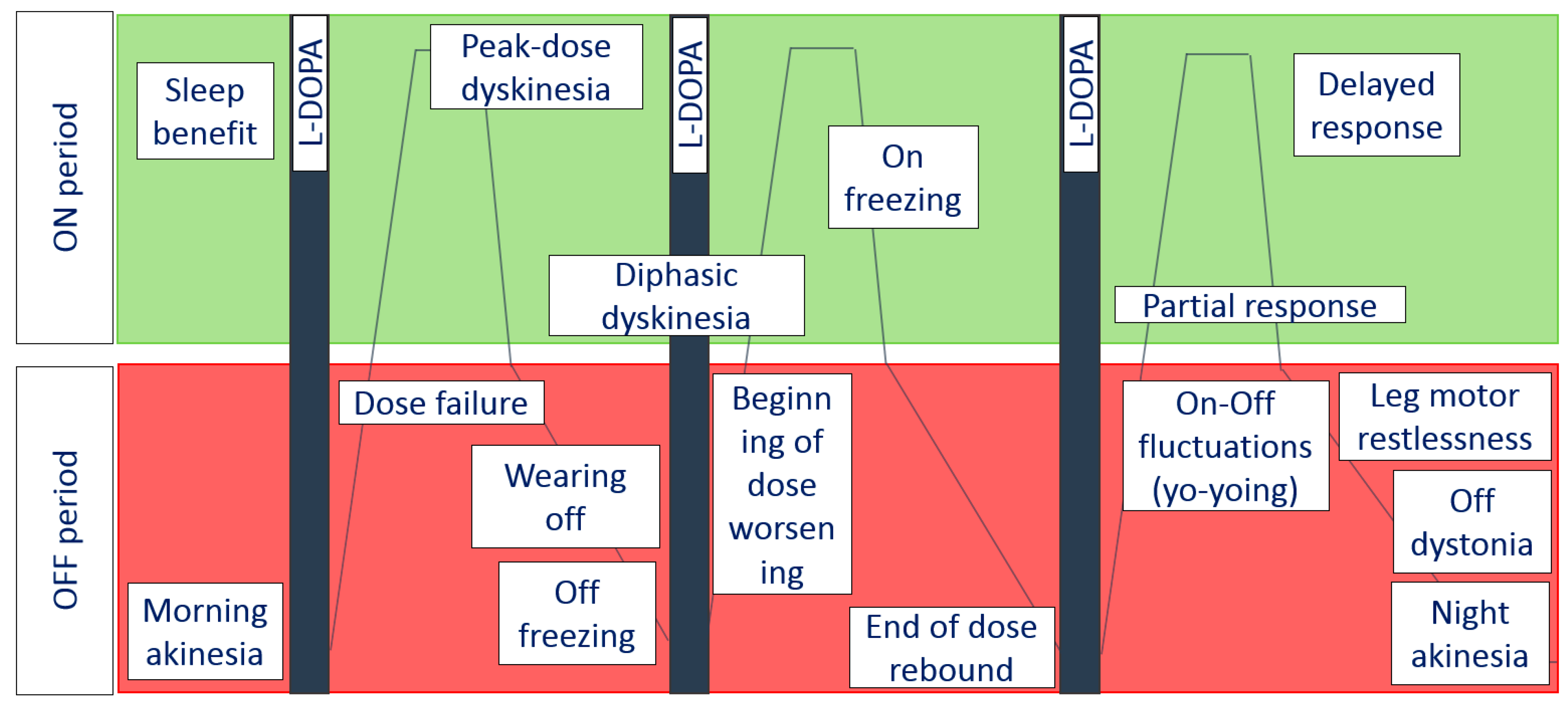
  • Short-duration response is characterized by motor improvement and coincides with the elevation of plasma L-dopa after drug consumption. It lasts from minutes to hours. Peak motor response also happens due to short-duration response.
  • Long-duration response keeps the positive effect of L-dopa beyond the normal half-life of the individual dose: this kind of response usually dominates in early PD. During this period, patients are able to control motor symptoms using two or three daily doses of L-dopa [18].
L-dopa has a rather short plasma half-life (about 1.5 h) [18]. Being a neutral amino acid, it competes for absorption and transferring through the blood-brain barrier. Changes in brain concentration of L-dopa do not always correlate with those in the blood. Perturbations in dopamine levels in the brain do not always correlate with L-dopa concentrations in the blood. In normal conditions, the dopaminergic system tends to assure a stable flow of dopamine into the striatum. Oral doses of L-dopa provoke the appearance of short peaks of L-dopa in the brain and may change dopamine synthesis in the central nervous system. This process may be compensated by the dopamine storage in nigrostriatal dopamine neurons. with disease progression, the consequent loss of nigrostriatal neurons leads to a reduction of buffering capacity and the pulsatile stimulation of dopamine receptors [17]. There is usually a linear improvement of symptoms with increasing dose, but chronic treatment alters motor response in an “all or none” model, and the patient becomes dependent on plasma L-dopa, brain L-dopa, and dopamine levels. The most frequent risk factors that may provoke motor fluctuations are the following: disease progression, disease severity, higher individual doses of L-dopa, peripheral pharmacokinetic factors affecting absorption of L-dopa, and possibly genetic risk factors [57].
These factors lead to alterations in dopamine concentrations and normal constant stimulation of postsynaptic dopamine receptors. The L-dopa effect begins to fluctuate and results in hyperkinetic movements (dyskinesia) in response to L-dopa dosing. Disease progression is known to be the main factor in motor complication development. Epidemiological studies have revealed that about 50% of patients demonstrate some degree of motor complications within 2–5 years, and between 80 to 100% of PD patients will suffer from motor complications after 10 years of L-dopa therapy [58,59]. The clinical subtype of PD may also influence the appearance of motor fluctuations. Patients with postural instability gait difficulty motor subtype are less prone to develop motor complications; however, they are considered to have lower effectiveness of anti-parkinsonian treatment than patients with the tremor-dominant subtype [60].
Among factors that lead to pitfalls in L-dopa treatment are the influence of meals and Helicobacter pylori. There is some evidence of a link between the elimination of H. pylori and significant improvement in clinical response to L-dopa and decrease of motor complications. Patients with early disease stages and with a family history of PD are more likely to develop protein interaction with L-dopa [61]. Some issues may influence L-dopa metabolism, such as slow gastric emptying, which results in raised pre-systemic decarboxylation and reduced intestinal absorption [62].
A young age of onset was determined as a risk factor for motor complications. Kostic et al. found that median intervals of the development of LID and motor fluctuations are shorter in young-onset patients than older-onset patients [63]. The female gender may also influence wearing-off and LID. Studies have revealed that the wearing-off phenomenon has a higher prevalence among women than men [64]. The reason for a higher frequency of motor complications among women is not known, but there are some hypotheses about relatively lower body weights and the effects of estrogen [65,66].
Genetic risk factors may also contribute to the development of motor complications. The autosomal recessive parkinsonism genes PARK2, PARK6, and PARK7, are associated with young-onset PD and the early development of dyskinesia [67]. In sporadic PD, the polymorphisms in dopaminergic D2 receptors were reported as factors that reduced the risk of developing LID [68]. Polymorphisms in the dopamine transporter have also been implicated.
Taking into consideration that the L-dopa effect gradually becomes shorter, patients have to decrease intervals between doses or increase their individual dose. Loss of smooth duration of the L-dopa effect causes the appearance of different motor fluctuations. Diurnal rhythms of PD symptoms have been investigated in numerous studies. The results of these studies may be suggestive of possible circadian influences on the expression of clinical features of PD. Patients with PD have lower peak activity levels and lower amplitude of the rest-activity cycle in comparison with healthy older adults [69]. Motor symptoms are known to be more prominent in the afternoon and evening than in the first half of the day. This daily motor condition does not always depend on the time of drug administration and may be caused by circadian regulation of dopaminergic systems. Motor symptoms severity can increase throughout the day even if L-dopa metabolism and pharmacokinetics are not altered. One of the studies by Bonucelli et al., focused attention on tremor, bradykinesia, and gait disturbances in three groups of PD patients: newly diagnosed PD, patients with, and patients without motor fluctuations. All these patients had standard L-dopa doses at 8:00, 12:00, and 16:00. The results showed that patients in the early stages do not have prominent fluctuations throughout the day, but those with advanced PD experience serious worsening in the second half of the day [70].

3. Strategies of the Prevention and Management of Motor Complications

Since chronic L-dopa treatment for PD patients is associated with the development of motor complications, the main strategy is to delay L-dopa use until absolutely necessary to preserve the patient’s function [57]. The use of L-dopa leads to pulsatile dopaminergic stimulation that may disrupt the physiological functions of dopaminergic neurons. Continuous dopaminergic stimulation is a therapeutic concept for the management of PD that proposes that continuous, as opposed to discontinuous or pulsatile, stimulation of striatal dopamine receptors will delay or prevent the onset of motor complications [58].
There is no doubt that initiating treatment with a dopamine agonist will delay the need for L-dopa by 1–3 years and that the incidence of motor complications during that time will be very low [71]. When patients have already developed motor complications, the main strategy is an adjustment of the L-dopa schedule. Other approaches include the addition of an adjunctive therapy (dopamine agonist, COMT inhibitor, MAO-B inhibitor, amantadine) or the use of device-assisted and surgical therapies [72]. However, the adjusting of dose and treatment regimen of L-dopa or adding new drugs can become a clinical dilemma. Higher doses of L-dopa can lead to new complications, such as peak-dose dyskinesia, orthostatic hypotension, or hallucinations. The decreasing of dose or modification of regimen of L-dopa helps to control LID. However, it can increase wearing-off. Therefore, the therapy of PD should be individualized and tailored to the specific needs of each patient [14].
The diversity of the clinical manifestation of PD requires multi-modal assessments of a patient’s condition. It can be done using different scales for the evaluation of motor, cognitive, affective and autonomic symptoms, motor fluctuations and dyskinesia, Hauser’s diary, and other instruments. However, the use of scales and questionnaires is time-consuming and is associated with a lack of objectivity. Motor diaries have been extensively utilized to gather treatment outcomes in clinical trials as well as in clinical practice. However, poor adherence has often been reported [73].
Technology development leads scientists to the creation of devices that would make it possible to assess the variety of changing parameters of patients during the day. Sensors provide objective data of tremor, bradykinesia, rigidity, ON/OFF/dyskinetic condition, physical activity, sleep, and wakefulness [74,75,76]. Technology-based devices offer the opportunity to improve the objectivity and relevance of the assessment and treatment of individuals with PD by quantifying symptom presentation in real-life conditions. Clinical visits provide only a brief snapshot of a patient’s state and cannot adequately display motor complications and non-motor symptoms of PD. Moreover, performance during the clinical visit does not always reflect how patients perform at home. Such devices can give clinicians the full presentation of patient’s state, assess the efficacy of medications, and patient’s adherence to them. Sensors can be even more sensitive in the detection of a patient’s transition from the “honeymoon period” to the first signs of wearing-off. Accurate evaluation of a patient’s condition is an essential requirement for effective treatment. Finally, technology-based devices represent a new care model that uses a closed-loop principle as they allow not only to monitor the patient but also adjust the therapy.

4. The World of Sensors

Mobile health (mHealth) has been defined as medical and public health practice supported by mobile devices, such as smartphones and patient monitoring devices. There is a great number of technology-based devices used in PD patients, and they can be divided by type of assessed clinical parameter (tremor, bradykinesia, gait), sensor type (accelerometer, gyroscope, magnetometer, etc.), and other characteristics (Table 1). The evaluation of clinical symptoms can be done during passive monitoring when patients do their daily activities or using special tests, such as finger tapping, the Timed Up and Go test, or sustained phonation. Other systems perform the automated assessments of motor tasks of the Unified Parkinson’s Disease Rating Scale [74]. Devices that assess motor symptoms during the activities of daily living in home-like conditions seem more suitable for clinical purposes and correspond to the tasks of monitoring a patient’s condition. The introduction of active motor tests in the process of passive monitoring of motor functions gives opportunities for the enhanced evaluation of a patient’s condition between visits to a doctor.
Accelerometers and gyroscopes are the most common wearable sensors for the assessment of motor symptoms in PD patients. Accelerometers operate by measuring acceleration along each axis of the device and can, therefore, detect static postures by measuring the acceleration due to gravity and detect motion by measuring the corresponding dynamic acceleration. Gyroscopes measure the Coriolis acceleration from rotational angular velocity. They can therefore measure transitions between postures and are often used to compliment accelerometers in mobility monitoring systems. These wearable devices can record not only the orientation, amplitude, and frequency of movements but also the speed of the part of the body where they are attached. These data allow clinicians to assess the presence and severity of the cardinal features and complications of PD (tremor, bradykinesia, and dyskinesia) [77]. Accelerometers and gyroscopes can be paired in an inertial measurement unit (IMU). This combination of sensors can be fused to provide a stable estimate of sensor orientation, both linear and angular motion information [78]. Each sensor has several specifications, including size, number of axes (one, two, or three axes), amplitude range, sampling frequency, bandwidth, sensitivity, and accuracy. It is important to consider these specifications when evaluating PD patients to ensure consistency with the device’s intended use.
Technology-based devices can consist of multiple sensors distributed on the body, but some authors prefer to use a single sensor unit worn at the waist, sacrum, or chest. There is little consensus as to the optimal placement and number of sensors required to obtain sufficient results [79]. Because wearable sensors produce large quantities of data that are not amenable to human interpretation, machine-learning algorithms are used (Table 1). Each of the algorithms has computational as well as functional strengths and weaknesses. To date, no system has been identified as the universally accepted and optimized algorithm for the analysis of human movement [78].
Technology-based devices can be classified into three groups: (1) implemented devices for automatic assessment of PD symptoms; (2) mHealth applications on smartphone/tablet; and (3) a telemedicine service consisting of automated systems for the assessment and/or monitoring of specific symptoms and mobile or web-based applications that allow both the patients and the clinicians to access the system through user interfaces. These systems would practically provide a modern telemedicine service using cloud platforms and server applications in which special algorithms are implemented to analyze the acquired data. Such systems allow a large amount of data to be transferred and managed, providing both the clinicians and the patients with useful information about disease progression and health conditions [68].
The format of output information about motor symptoms and motor complications that the devices present to the clinician vary. Systems can provide information about the amplitude of tremor, the period of time when tremors are present, bradykinesia, or freezing of gait were detected. At the same time, other devices use a motor-fluctuation detection algorithm that determines ON, OFF, and dyskinetic states. Klapper et al. used five accelerometers to detect bradykinesia, hypokinesia, and dyskinetic movements. The authors used classification trees and neural networks to detect bradykinetic/hypokinetic states vs. not bradykinetic/hypokinetic states compared to dichotomized scores from the neurologist who observed the participants for the duration of the recording period. Classification trees detected bradykinesia/hypokinesia with accuracies of 74.8–85.3% and dyskinesia with accuracies of 80.6–91.6%. Using neural networks improved the accuracy of the algorithm to 88.0–92.1% for bradykinesia/hypokinesia detection and to 91.1–94.1% for dyskinesia detection [80].
In an attempt to use fewer sensors, Rodríguez-Molinero et al. used a single tri-axial accelerometer worn on a belt and an ON/OFF detection algorithm based on the analysis of patients’ movements while walking. The motor fluctuation detector showed a mean sensitivity of 0.96 (median 1; interquartile range, IQR, 0.93–1) and a specificity of 0.94 (median 0.96; IQR, 0.90–1). However, the algorithm was unable to detect status changes when the patient was at rest [81].
Pfister et al. used deep learning to classify motion data from a single wrist-worn IMU sensor recording in 30 PD patients. Convolutional neural networks modeled the PD motor states as a three-class categorical concept (OFF/ON/DYS). Using a 1-min window size as an input for a convolutional neural network trained on data from a subset of patients, they achieved a three-class balanced accuracy of 0.654 on data from previously unseen subjects. This corresponds to detecting the OFF, ON, or DYSKINETIC motor state at a sensitivity/specificity of 0.64/0.89, 0.67/0.67, and 0.64/0.89, respectively. On average, the model outputs were highly correlated with the annotation on a per subject scale (r = 0.83/0.84; p < 0.0001) [82].
There are some commercially available devices that provide continuous objective measurement of patients with PD. The Personal KinetiGraph (PKG) Movement Recording System is a technology that provides daily and summary scores for bradykinesia, dyskinesia, fluctuations, data on tremor, immobility, movement during daytime, somnolence, and sleep. The PKG System consists of an interactive data logger (PKG Watch) that resembles a wristwatch it measures movement accelerations of the wrist and analyzes the spectral power of the low frequencies of accelerometer data providing continuous variables—namely the median bradykinesia score (BKS) and dyskinesia score (DKS). Griffiths et al. found that BKS and DKS closely correlate with UPDRS motor score and mAIMS, respectively [83]. The PKG System also contains a reminder to the subject when PD medications are due, and a means for recording when PD medications are taken. At the end of the patient wear period, the PKG data logger is returned to the clinic and data are downloaded and analyzed using an algorithm to translate raw movement data into a printable output of the patient’s movement over the worn period [76]. Price et al. evaluated the clinical utility of the PKG in routine clinical care and found the PKG identified issues that had not been reported previously in 63% of patients [84]. Spengler et al. reported PKG use to support deep brain stimulation programming was feasible and may decrease time to deep brain stimulation (DBS) optimization, contributing to a more effective DBS therapy and possibly fewer programming visits [85].
Physilog uses body-attached gyroscopes to assess spatio-temporal parameters of gait, sway, physical activity, tremor, and bradykinesia. Depending on the expected outcomes, one to seven inertial sensors, including accelerometers and gyroscopes, can be used. Gait measurements using this device were performed on 10 PD patients with DBS. Some of the gait parameters had a high correlation with UPDRS subscores (r = −0.90). This algorithm was able to detect gait cycles and related gait events with very high sensitivity (>96%) and with a positive prediction value >98%. These results have been demonstrated to be accurate enough to show significant differences between Stimulation ON and Stimulation OFF states in PD patients [86].
Kinesia-360 consists of two sensors, one mounted on a wrist and another one placed on the patients’ ankle, a mobile phone application and an external server for data protection. The kinesia system provides information about tremors, dyskinesia, and mobility based on temporal and frequency features [87]. Another product, Kinesia ONE, integrates accelerometers and gyroscopes in a compact patient-worn unit and provides results in the form of indexes from bradykinesia, dyskinesia, and tremors. The sensor component of the device is installed in a ring, which fits on a finger [88].
The Perform system is an intelligent closed-loop system that integrates four tri-axial accelerometers for extremities and one accelerometer/gyroscope on the waist that evaluate tremor, bradykinesia, freezing of gait, and dyskinesia. Data acquired are pre-processed by algorithms and allow health professionals to remotely monitor the overall status of the patients, adjust medication schedules, and personalize treatment [89].
The assessment of LID using sensors is complicated because voluntary movements occur in the same frequency band (1–4 Hz band), so during the performance of daily activities, the objective evaluation of dyskinesias is confounded. Hoff et al. used four pairs of accelerometers to investigate movement characteristics of dyskinesias. Although objective measures of dyskinesias were reliable and responsive, they failed to distinguish LID from voluntary movements [90].
Keijsers et al. used the same data but with neural networks for better differentiation between voluntary movements and dyskinesias. This technology improves LID detection and the assessment of its severity in various activities [91]. In the next study, Keijsers et al. tested an algorithm for the detection and assessment of the severity of LID in PD patients performing different activities of daily living tasks during 2.5 h. The neural network correctly classified dyskinesia or the absence of dyskinesia in 15-min intervals in 93.7, 99.7, and 97.0% for the arm, trunk, and leg, respectively [92].
Rodríguez-Molinero et al. developed a device that can detect bradykinesia, ON/OFF motor conditions, and freezing of gait [81,93]. In the next study, they designed and validated an algorithm that can register the occurrence of dyskinesia during a patient’s activities of daily living [94]. Dyskinesia was detected in a dichotomous way—namely, only its occurrence or not is detected at every moment, without information on its severity. Then, the possibility of an algorithm to assess the severity of dyskinesia using a continuous numerical value was investigated. The correlation coefficient between the sensor output and the Unified Dyskinesia Rating Scale score was 0.70 (CI 95%: 0.33–0.88; p = 0.01). Since the sensor was located on the waist, the correlation between the sensor output and the results of the trunk and legs scale sub-items was calculated: 0.91 (CI 95% 0.76–0.97: p < 0.001) [95].
It should be noted that studies using wearable techniques did not differentiate between dyskinesia subtypes, such as peak-dose dyskinesia, OFF-dystonia, and diphasic dyskinesias. More studies are needed with new analytical algorithms that would be able to separate the more dystonic type of dyskinesias (as in diphasic or OFF dyskinesias) from the more choreatic peak-dose dyskinesias. This would certainly be a great help to clinicians because making this distinction based on history taking can be very difficult.

5. mHealth Applications for PD Monitoring

In agreement with the large use of smartphones in the population, mobile applications for monitoring and assessing motor symptoms in PD patients were designed. The mobile apps can be used directly on the patient’s own smartphone or smartwatch without additional devices. On the contrary, there are many studies where apps are integrated into systems consisting of additional sensors. Some allow additional functions, such as external (non-invasive) sensory stimulation for the treatment of FOG [96]. The apps can offer corrective feedback as well, encouraging the patients to improve their physical activity or medical adherence [75].
Many studies validated apps using a smartphone’s accelerometer and gyroscope signals for the characterization of tremors. High sensitivity and specificity in detection and measurement of hand tremor was shown [97,98,99]. The control your Parkinson disease app (CYPD) uses a gyroscope in a smartwatch and a special algorithm for parkinsonian tremor detection. CYPD collects data on tremors of the limbs, manages a medicine schedule, and contains tests and questionnaires for self-assessment of motor and non-motor PD symptoms. As a result, it generates reports on the patient’s condition. CYPD makes the combined graph with the percentage of tremor, the tremor duration during the day, and the schedule of medications taken. The information can be transferred to the doctor as a report [100]. The KinesiaU motor assessment system is a validated consumer app for PD patients to measure their symptoms using an Android smartphone and smartwatch. The system tracks tremors, slowness, and dyskinesia, as well as therapies and activities in the daily reports. Furthermore, it can provide interactive communication with healthcare professionals every month for better monitoring and treatment [101].
Other apps use special tests to evaluate a patient’s motor performance. Zhan et al. developed an Android smartphone app HopkinsPD that assesses five activities (voice, finger tapping, gait, balance, and reaction time) and a mobile Parkinson disease score (mPDS) that objectively weighs features derived from each activity. The mPDS was derived from 6148 smartphone activity assessments from 129 individuals. The measure correlated well with the Movement Disorder Society Unified Parkinson Disease’s Rating Scale total (r = 0.81; p < 0.001) and part III only (r = 0.88; p < 0.001), the Timed Up and Go assessment (r = 0.72; p = 0.002), and the Hoehn and Yahr stage (r = 0.91; p < 0.001). The mPDS responds to dopaminergic medication administration; it improved by a mean of 16.3 ± 5.6 points [97].
Punin et al. developed a system based on an Android mobile app and a tri-axial accelerometer device for gait data acquisition. Gathered data is processed to detect FOG episodes in real-time. Detection activates an external vibratory stimulation of the legs to reduce FOG time. The system obtained a specificity of 86.66% and a sensitivity of 60.61% in FOG detection. It showed an improvement in the time reduction of the FOG episodes of each patient [96].
The Fox Wearable Companion App (FWC App) is a mobile and wearable application for PD patients. It uses a smartwatch and smartphone that gather and transmit data to the cloud where algorithms generate metrics, such as activity level during waking and sleeping hours, gait, and tremor detection. The feasibility and acceptability of using the FWC App were determined in an observational, two-cohort Parkinson at home study with 953 PD patients. Patients used the FWC App for a minimum of 6 weeks (North America, NAM) or 13 weeks (The Netherlands, NL). Additionally, medication intake and symptoms were collected via self-reporting in the app. The enrollment rate was 88% in the NL (n = 304) and 51% (n = 649) in NAM. Overall, 84% (n = 805) of participants contributed sensor data. Participants were compliant for 68% (16.3 h/participant/day) of the study period in NL and for 62% (14.8 h/participant/day) in NAM [102].
PD_Manager is a mHealth platform that covers most of the aspects regarding the management of PD with a holistic approach. Patients are monitored using wrist and insole sensors paired with a smartphone. A mobile app also provides various non-motor self-evaluation tests for assessing cognition, mood, and nutrition. The smart pillbox can be filled in advance by caregivers according to the prescribed medication plan and then handed over to the patients for intake. The schedule is automatically downloaded from the cloud service, and the patient is alerted accordingly. The core of the system is the mHealth platform, which is a cloud IT system that provides all the necessary functionality for users and services communication, along with computing power for data processing and storage. Clinicians can use a separate mobile app that provides fast patient assessment, the current and past medication plans, and register a new one if required. The system showed good accuracy for the assessment of gait problems, tremors, bradykinesia, dyskinesia, and for the detection of ON and OFF states. A total of 78% of patients reported that they would use this type of mHealth platform, with the main reason being their feeling of assurance due to close and more personalized treatment [103].

6. What Does an Ideal Sensor Look Like?

Nowadays, the variety of solutions of PD monitoring is rapidly growing. Considering this, there is a need for consensus on the type of sensors and the main goal of their use. The Movement Disorders Society Task Force on Technology recently suggested principles of mHealth technologies: (1) identification of patient-centered and clinically relevant digital outcomes; (2) selection criteria for device combinations that offer an acceptable benefit-to-burden ratio to patients and that deliver reliable, clinically relevant insights; (3) development of an accessible, scalable, and secure platform for data integration and data analytics; and (4) agreement on a pathway for approval by regulators, adoption into e-health systems, and implementation by health care organizations [104].
Thus, technology-based devices should be patient-centered and ideally work as a closed-loop system. They assess the motor performance, non-motor symptoms and detect radical changes of condition. Such systems are integrated with medical records that provide continuous monitoring of the patient. Moreover, it can potentially be integrated into treatment delivery systems that remind about the next drug dose and check that the patient took it. In the future, mHealth technology, by using smart algorithms, can analyze the state of the patient and determine the course of disease, give recommendations about adjusting of therapy, or alert patients and healthcare providers the need for a clinic visit. Possible application areas for the mHealth system include remote DBS programming and titration of L-dopa-carbidopa intestinal gel, particularly for people who live in rural areas. The next step is the development of a closed-loop DBS system when signals are transmitted from wearable sensors and central sources (such as local field potential) toward the implanted programmable generator. The generator analyzes the data and adaptively responds by modulation of stimulation settings [105,106,107].
Technology-based devices open an era of new remote medicine for doctors and patients. This approach has some limitations that need to be discussed. Side aspects of the use of sensors are associated with the introduction into the patient’s personal life. Therefore, information collected should be depersonalized and reliably protected on storage cloud servers. Some types of received information cannot be used. For example, video recording for long-term monitoring during free daily activities is not possible. On the other hand, technology-based devices can be addictive for some patients, especially with behavior and cognitive disorders.
Although a smart mHealth system can replace some of the work of health providers, it cannot be fully relied on because they are not error-free. Nevertheless, nowadays, clinicians cannot fully rely on a mHealth system because they are not error-free. Developed sensors and algorithms are not suitable for the detection of disorders such as OFF-period dystonia, akathisia, or most of the non-motor symptoms. Technical problems can occur, as well as problems with the prolonged wearing of sensors [108]. If retired people, for example, can use systems with several sensors at their home or outdoors, younger patients could refuse the wearing of noticeable and massive devices. Hence, there is a need for systems that can reduce patient burden by using a minimal number of sensors while continuously capturing clinically meaningful measures of motor symptom severity under free-living conditions.
The mHealth systems cannot be used for every patient. There are expected limitations in the use of such technologies in patients with dementia and psychotic disorders. Additionally, such systems require training and skills to work them as well as extra time for analyzing information. Taking into consideration the heavy workload of doctors, it can be another limitation of use. Therefore, the development of a user-friendly interface for doctors that provide quick and easy access to the mHealth system is required.

7. Conclusions

Since there is no proven etiological treatment for PD, the only opportunity to improve a patient’s quality of life is the use of dopamine replacement therapy. Therefore, the main goal for clinicians should be the optimizing of individual symptomatic therapy that includes dopamine drugs, medication for the treatment of affective and autonomic disturbances, sleep and wakefulness disorders, cognitive and psychiatric impairments, as well as methods of physical rehabilitation. Technology-based devices can change approaches to the management of PD. Although they raised some limitations, the use of such systems provides new tools in monitoring and treatment, as well as promoting the active engagement of the patients and their caregivers in the healthcare path. This aims to improve both patient’s quality of life and clinician’s quality of care toward an optimal personalized therapy.

Author Contributions

Conceptualization, O.S.L.; methodology, O.S.L. and O.V.I.; writing—original draft preparation, O.S.L., O.V.I., I.I.C. and A.V.K.; writing—review and editing, O.S.L., O.V.I., I.I.C.; supervision, O.S.L.; project administration, O.V.I.; funding acquisition, O.S.L. All authors have read and agreed to the published version of the manuscript.

Funding

This research received no external funding.

Conflicts of Interest

The authors declare no conflict of interest.

References

  1. Müller, T.; Russ, H. Levodopa, motor fluctuations and dyskinesia in Parkinson’s disease. Expert Opin. Pharmacother. 2006, 7, 1715–1730. [Google Scholar] [CrossRef] [PubMed]
  2. Rodríguez-Violante, M.; Ospina-García, N.; Dávila-Avila, N.M.; Cruz-Fino, D.; Cruz-Landero, A.; Cervantes-Arriaga, A. Motor and non-motor wearing-off and its impact in the quality of life of patients with Parkinson’s disease. Arquivos Neuro-Psiquiatr. 2018, 76, 517–521. [Google Scholar] [CrossRef] [Green Version]
  3. Bjornestad, A.; Forsaa, E.B.; Pedersen, K.F.; Tysnes, O.B.; Larsen, J.P.; Alves, G. Risk and course of motor complications in a population-based incident Parkinson’s disease cohort. Parkinsonism Relat. Disord. 2016, 22, 48–53. [Google Scholar] [CrossRef]
  4. Kim, H.J.; Mason, S.; Foltynie, T.; Winder-Rhodes, S.; Barker, R.A.; Williams-Gray, C.H. Motor complications in Parkinson’s disease: 13-year follow-up of the CamPaIGN cohort. Mov. Disord. 2020, 35, 185–190. [Google Scholar] [CrossRef] [Green Version]
  5. Quinn, N.P. Classification of fluctuations in patients with Parkinson’s disease. Neurology 1998, 51 (Suppl. 2), S25–S29. [Google Scholar] [CrossRef]
  6. Bhidayasiri, R.; Sringean, J.; Trenkwalder, C. Mastering nocturnal jigsaws in Parkinson’s disease: A dusk-to-dawn review of night-time symptoms. J. Neural Transm. 2020, 127, 763–777. [Google Scholar] [CrossRef]
  7. Stocchi, F.; Coletti, C.; Bonassi, S.; Radicati, F.G.; Vacca, L. Early-morning OFF and levodopa dose failures in patients with Parkinson’s disease attending a routine clinical appointment using Time-to-ON Questionnaire. Eur. J. Neurol. 2019, 26, 821–826. [Google Scholar] [CrossRef]
  8. van Gilst, M.M.; Louter, M.; Baumann, C.R.; Bloem, B.R.; Overeem, S. Sleep benefit in Parkinson’s disease: Time to revive an enigma? J. Parkinsons Dis. 2012, 2, 167–170. [Google Scholar] [CrossRef]
  9. van Gilst, M.M.; Bloem, B.R.; Overeem, S. Sleep benefit” in Parkinson’s disease: A systematic review. Parkinsonism Relat. Disord. 2013, 19, 654–659. [Google Scholar] [CrossRef] [PubMed]
  10. Högl, B.E.; Gómez-Arévalo, G.; García, S.; Scipioni, O.; Rubio, M.; Blanco, M.; Gershanik, O.S. A clinical, pharmacologic, and polysomnographic study of sleep benefit in Parkinson’s disease. Neurology 1998, 50, 1332–1339. [Google Scholar] [CrossRef]
  11. Zhong, R.; Chen, Q.; Zhang, X.; Zhang, X.; Lin, W. Correction: The related factors of sleep benefit in Parkinson’s disease: A systematic review and meta-analysis. PLoS ONE 2019, 14, e0216524. [Google Scholar] [CrossRef] [PubMed] [Green Version]
  12. Fox, S.H.; Lang, A.E. Levodopa-related motor complications--phenomenology. Mov Disord. 2008, 23, S509–S514. [Google Scholar] [CrossRef] [PubMed]
  13. Kuoppamäki, M.; Al-Barghouthy, G.; Jackson, M.L.; Zeng, B.Y.; Quinn, N.; Jenner, P. Beginning-of-dose and rebound worsening in MPTP-treated common marmosets treated with levodopa. Mov. Disord. 2002, 17, 1312–1317. [Google Scholar] [CrossRef]
  14. Jankovic, J. Motor fluctuations and dyskinesias in Parkinson’s disease: Clinical manifestations. Mov. Disord. 2005, 20 (Suppl. 11), S11–S16. [Google Scholar] [CrossRef]
  15. Cucca, A.; Biagioni, M.C.; Fleisher, J.E.; Agarwal, S.; Son, A.; Kumar, P.; Brys, M.; Di Rocco, A. Freezing of gait in Parkinson’s disease: From pathophysiology to emerging therapies. Neurodegener. Dis. Manag. 2016, 6, 431–446. [Google Scholar] [CrossRef]
  16. Ginis, P.; Nackaerts, E.; Nieuwboer, A.; Heremans, E. Cueing for people with Parkinson’s disease with freezing of gait: A narrative review of the state-of-the-art and novel perspectives. Ann. Phys. Rehabil. Med. 2018, 61, 407–413. [Google Scholar] [CrossRef]
  17. Donaldson, I.; Marsden, C.D.; Schneider, S.A.; Bhatia, K. Parkinson’s disease. In Marsden’s Book of Movement Disorders; Oxford University Press: Oxford, UK, 2012; pp. 159–370. [Google Scholar]
  18. Burn, D. Parkinson’s Disease: Advanced Disease, Motor Complications, and Management. In Oxford Textbook of Movement Disorders; Oxford University Press: Oxford, UK, 2013; pp. 83–111. [Google Scholar]
  19. Nutt, J.G. Levodopa-induced dyskinesia: Review, observations, and speculations. Neurology 1990, 40, 340–345. [Google Scholar] [CrossRef]
  20. Luquin, M.R.; Scipioni, O.; Vaamonde, J.; Gershanik, O.; Obeso, J.A. Levodopa-induced dyskinesias in Parkinson’s disease: Clinical and pharmacological classification. Mov. Disord. 1992, 7, 117–124. [Google Scholar] [CrossRef]
  21. Bhidayasiri, R.; Mekawichai, P.; Jitkritsadakul, O.; Panyakaew, P.; Kaewwilai, L.; Boonrod, N.; Petchrutchatachart, S.; Jagota, P.; Boonpeng, K.; Singmaneesakulchai, S.; et al. Nocturnal journey of body and mind in Parkinson’s disease: The manifestations, risk factors and their relationship to daytime symptoms. Evidence from the NIGHT-PD study. J. Neural Transm. 2014, 121 (Suppl. 1), S59–S68. [Google Scholar] [CrossRef]
  22. Bhidayasiri, R.; Trenkwalder, C. Getting a good night sleep? The importance of recognizing and treating nocturnal hypokinesia in Parkinson’s disease. Parkinsonism Relat. Disord. 2018, 50, 10–18. [Google Scholar] [CrossRef]
  23. Allen, R.P.; Picchietti, D.L.; Garcia-Borreguero, D.; Ondo, W.G.; Walters, A.S.; Winkelman, J.W.; Zucconi, M.; Ferri, R.; Trenkwalder, C.; Lee, H.B. Restless legs syndrome/Willis-Ekbom disease diagnostic criteria: Updated International Restless Legs Syndrome Study Group (IRLSSG) consensus criteria--history, rationale, description, and significance. Sleep Med. 2014, 15, 860–873. [Google Scholar] [CrossRef]
  24. Moccia, M.; Erro, R.; Picillo, M.; Santangelo, G.; Spina, E.; Allocca, R.; Longo, K.; Amboni, M.; Palladino, R.; Assante, R.; et al. A Four-Year Longitudinal Study on Restless Legs Syndrome in Parkinson Disease. Sleep 2016, 39, 405–412. [Google Scholar] [CrossRef] [Green Version]
  25. Gjerstad, M.D.; Tysnes, O.B.; Larsen, J.P. Increased risk of leg motor restlessness but not RLS in early Parkinson disease. Neurology 2011, 77, 1941–1946. [Google Scholar] [CrossRef]
  26. Matsubara, T.; Suzuki, K.; Fujita, H.; Watanabe, Y.; Sakuramoto, H.; Matsubara, M.; Hirata, K. Restless legs syndrome, leg motor restlessness and their variants in patients with Parkinson’s disease and related disorders. J. Neurol. Sci. 2018, 393, 51–57. [Google Scholar] [CrossRef]
  27. Suzuki, K.; Miyamoto, M.; Miyamoto, T.; Hirata, K. Restless Legs Syndrome and Leg Motor Restlessness in Parkinson’s Disease. Parkinsons Dis. 2015, 2015, 490938. [Google Scholar] [CrossRef]
  28. Brun, L.; Lefaucheur, R.; Fetter, D.; Derrey, S.; Borden, A.; Wallon, D.; Bourre, B.; Maltête, D. Non-motor fluctuations in Parkinson’s disease: Prevalence, characteristics and management in a large cohort of parkinsonian outpatients. Clin. Neurol. Neurosurg. 2014, 127, 93–96. [Google Scholar] [CrossRef]
  29. Seki, M.; Takahashi, K.; Uematsu, D.; Mihara, B.; Morita, Y.; Isozumi, K.; Ohta, K.; Muramatsu, K.; Shirai, T.; Nogawa, S.; et al. Clinical features and varieties of non-motor fluctuations in Parkinson’s disease: A Japanese multicenter study. Parkinsonism Relat. Disord. 2013, 19, 104–108. [Google Scholar] [CrossRef] [PubMed]
  30. Riley, D.E.; Lang, A.E. The spectrum of levodopa-related fluctuations in Parkinson’s disease. Neurology 1993, 43, 1459–1464. [Google Scholar] [CrossRef]
  31. Chaudhuri, K.R.; Schapira, A.H. Non-motor symptoms of Parkinson’s disease: Dopaminergic pathophysiology and treatment. Lancet Neurol. 2009, 8, 464–474. [Google Scholar] [CrossRef]
  32. Levin, O.S.; Ivanov, A.K.; Shindriaeva, N.N. Actual problems of inpatient psychiatric care in Russia. Zhurnal Nevrologii i Psikhiatrii imeni SS Korsakova 2011, 111, 38–42. [Google Scholar]
  33. Vázquez, A.; Jiménez-Jiménez, F.J.; García-Ruiz, P.; García-Urra, D. “Panic attacks” in Parkinson’s disease. A long-term complication of levodopa therapy. Acta Neurol. Scand. 1993, 87, 14–18. [Google Scholar] [CrossRef]
  34. Witjas, T.; Kaphan, E.; Azulay, J.P.; Blin, O.; Ceccaldi, M.; Pouget, J.; Poncet, M.; Chérif, A.A. Nonmotor fluctuations in Parkinson’s disease: Frequent and disabling. Neurology 2002, 59, 408–413. [Google Scholar] [CrossRef]
  35. Quinn, N.P.; Lang, A.E.; Koller, W.C.; Marsden, C.D. Painful Parkinson’s disease. Lancet 1986, 327, 1366–1369. [Google Scholar] [CrossRef]
  36. Factor, S.A.; Weiner, W.J. Parkinson’s Disease: Diagnosis & Clinical Management, 2nd ed.; Demos Medical Publishing: New York, NY, USA, 2008. [Google Scholar]
  37. Storch, A.; Schneider, C.B.; Wolz, M.; Stürwald, Y.; Nebe, A.; Odin, P.; Mahler, A.; Fuchs, G.; Jost, W.H.; Chaudhuri, K.R.; et al. Nonmotor fluctuations in Parkinson disease: Severity and correlation with motor complications. Neurology 2013, 80, 800–809. [Google Scholar] [CrossRef]
  38. Storch, A.; Schneider, C.B.; Klingelhöfer, L.; Odin, P.; Fuchs, G.; Jost, W.H.; Martinez-Martin, P.; Koch, R.; Reichmann, H.; Chaudhuri, K.R.; et al. Quantitative assessment of non-motor fluctuations in Parkinson’s disease using the Non-Motor Symptoms Scale (NMSS). J. Neural. Transm. 2015, 122, 1673–1684. [Google Scholar] [CrossRef]
  39. Funkiewiez, A.; Ardouin, C.; Caputo, E.; Krack, P.; Fraix, V.; Klinger, H.; Chabardes, S.; Foote, K.; Benabid, A.L.; Pollak, P. Long-term effects of bilateral subthalamic nucleus stimulation on cognitive function, mood, and behaviour in Parkinson’s disease. J. Neurol. Neurosurg. Psychiatry 2004, 75, 834–839. [Google Scholar] [CrossRef] [Green Version]
  40. Nissenbaum, H.; Quinn, N.P.; Brown, R.G.; Toone, B.; Gotham, A.M.; Marsden, C.D. Mood swings associated with the ‘on-off’ phenomenon in Parkinson’s disease. Psychol. Med. 1987, 17, 899–904. [Google Scholar] [CrossRef]
  41. Menza, M.A.; Sage, J.; Marshall, E.; Cody, R.; Duvoisin, R. Mood changes and “on-off” phenomena in Parkinson’s disease. Mov. Disord. 1990, 5, 148–151. [Google Scholar] [CrossRef]
  42. Stacy, M.A.; Murck, H.; Kroenke, K. Responsiveness of motor and nonmotor symptoms of Parkinson disease to dopaminergic therapy. Prog. Neuropsychopharmacol. Biol. Psychiatry 2010, 34, 57–61. [Google Scholar] [CrossRef]
  43. Cools, R. Dopaminergic modulation of cognitive function-implications for L-DOPA treatment in Parkinson’s disease. Neurosci. Biobehav. Rev. 2006, 30, 1–23. [Google Scholar] [CrossRef]
  44. Kulisevsky, J.; Avila, A.; Barbanoj, M.; Antonijoan, R.; Berthier, M.L.; Gironell, A. Acute effects of levodopa on neuropsychological performance in stable and fluctuating Parkinson’s disease patients at different levodopa plasma levels. Brain 1996, 119, 2121–2132. [Google Scholar] [CrossRef] [Green Version]
  45. Nieoullon, A. Dopamine and the regulation of cognition and attention. Prog. Neurobiol. 2002, 67, 53–83. [Google Scholar] [CrossRef]
  46. Raudino, F. Non motor off in Parkinson’s disease. Acta Neurol. Scand. 2001, 104, 312–315. [Google Scholar] [CrossRef] [PubMed]
  47. Sage, J.I. Fluctuations of Nonmotor symptoms. In Parkinson’s Disease, Diagnosis and Clinical Management; Demos Medical Publishing: New York, NY, USA, 2002; Volume 41, pp. 455–463. [Google Scholar]
  48. Goetz, C.G.; Tanner, C.M.; Levy, M.; Wilson, R.S.; Garron, D.C. Pain in Parkinson’s disease. Mov. Disord. 1986, 1, 45–49. [Google Scholar] [CrossRef]
  49. Barbeau, A. The clinical physiology of side effects in long-term L-DOPA therapy. Adv. Neurol. 1974, 5, 347–365. [Google Scholar] [PubMed]
  50. Sage, J.I.; Mark, M.H. Drenching sweats as an off phenomenon in Parkinson’s disease: Treatment and relation to plasma levodopa profile. Ann. Neurol. 1995, 37, 120–122. [Google Scholar] [CrossRef] [PubMed]
  51. Baratti, M.; Calzetti, S. Fluctuation of arterial blood pressure during end-of-dose akinesia in Parkinson’s disease. J. Neurol. Neurosurg. Psychiatry 1984, 47, 1241–1243. [Google Scholar] [CrossRef] [Green Version]
  52. Fahn, S.; Jankovic, J.; Hallett, M. Nonmotor problems in Parkinson disease. In Principles and Practice of Movement Disorders; Elsevier: Amsterdam, The Netherlands, 2011; pp. 183–196. [Google Scholar]
  53. Fox, S.H.; Lang, A.E. Motor and non-motor fluctuations. Handbook Clin. Neurol. 2007, 84, 157–184. [Google Scholar] [CrossRef]
  54. Milazzo, V.; Di Stefano, C.; Vallelonga, F.; Sobrero, G.; Zibetti, M.; Romagnolo, A.; Merola, A.; Milan, A.; Espay, A.J.; Lopiano, L.; et al. Reverse blood pressure dipping as marker of dysautonomia in Parkinson disease. Parkinsonism Relat. Disord. 2018, 56, 82–87. [Google Scholar] [CrossRef]
  55. Vallelonga, F.; Di Stefano, C.; Merola, A.; Milan, A.; Espay, A.J.; Lopiano, L.; Veglio, F.; Maule, S. Blood pressure circadian rhythm alterations in alpha-synucleinopathies. J. Neurol. 2019, 266, 1141–1152. [Google Scholar] [CrossRef]
  56. Hineno, T.; Mizobuchi, M.; Nishimatsu, O.; Horiguchi, J.; Kakimoto, Y. Day-night variation of urine volume in Parkinson’s disease. Jpn. J. Psychiatry Neurol. 1994, 48, 583–587. [Google Scholar] [CrossRef]
  57. Freitas, M.E.; Hess, C.W.; Fox, S.H. Motor Complications of Dopaminergic Medications in Parkinson’s Disease. Semin. Neurol. 2017, 37, 147–157. [Google Scholar] [CrossRef]
  58. Pahwa, R.; Lyons, K.E. Levodopa-related wearing-off in Parkinson’s disease: Identification and management. Curr. Med. Res. Opin. 2009, 25, 841–849. [Google Scholar] [CrossRef]
  59. Yoritaka, A.; Shimo, Y.; Takanashi, M.; Fukae, J.; Hatano, T.; Nakahara, T.; Miyamato, N.; Urabe, T.; Mori, H.; Hattori, N. Motor and non-motor symptoms of 1453 patients with Parkinson’s disease: Prevalence and risks. Parkinsonism Relat. Disord. 2013, 19, 725–731. [Google Scholar] [CrossRef] [PubMed] [Green Version]
  60. Thenganatt, M.A.; Jankovic, J. Parkinson disease subtypes. JAMA Neurol. 2014, 71, 499–504. [Google Scholar] [CrossRef] [PubMed]
  61. Virmani, T.; Tazan, S.; Mazzoni, P.; Ford, B.; Greene, P.E. Motor fluctuations due to interaction between dietary protein and levodopa in Parkinson’s disease. J. Clin. Mov. Disord. 2016, 3, 8. [Google Scholar] [CrossRef] [Green Version]
  62. Contin, M.; Martinelli, P. Pharmacokinetics of levodopa. J. Neurol. 2010, 257 (Suppl. 2), S253–S261. [Google Scholar] [CrossRef]
  63. Kostic, V.; Przedborski, S.; Flaster, E.; Sternic, N. Early development of levodopa-induced dyskinesias and response fluctuations in young-onset Parkinson’s disease. Neurology 1991, 41, 202–205. [Google Scholar] [CrossRef]
  64. Warren Olanow, C.; Kieburtz, K.; Rascol, O.; Poewe, W.; Schapira, A.H.; Emre, M.; Nissinen, H.; Leinonen, M.; Stocchi, F.; Stalevo Reduction in Dyskinesia Evaluation in Parkinson’s Disease (STRIDE-PD) Investigators. Factors predictive of the development of Levodopa-induced dyskinesia and wearing-off in Parkinson’s disease. Mov. Disord. 2013, 28, 1064–1071. [Google Scholar] [CrossRef]
  65. Colombo, D.; Abbruzzese, G.; Antonini, A.; Barone, P.; Bellia, G.; Franconi, F.; Simoni, L.; Attar, M.; Zagni, E.; Haggiag, S.; et al. The “gender factor” in wearing-off among patients with Parkinson’s disease: A post hoc analysis of DEEP study. Sci. World J. 2015, 2015, 787451. [Google Scholar] [CrossRef] [Green Version]
  66. Kumagai, T.; Nagayama, H.; Ota, T.; Nishiyama, Y.; Mishina, M.; Ueda, M. Sex differences in the pharmacokinetics of levodopa in elderly patients with Parkinson disease. Clin. Neuropharmacol. 2014, 37, 173–176. [Google Scholar] [CrossRef] [Green Version]
  67. Dekker, M.C.; Bonifati, V.; van Duijn, C.M. Parkinson’s disease: Piecing together a genetic jigsaw. Brain 2003, 126, 1722–1733. [Google Scholar] [CrossRef] [Green Version]
  68. Oliveri, R.L.; Annesi, G.; Zappia, M.; Civitelli, D.; Montesanti, R.; Branca, D.; Nicoletti, G.; Spadafora, P.; Pasqua, A.A.; Cittadella, R.; et al. Dopamine D2 receptor gene polymorphism and the risk of levodopa-induced dyskinesias in PD. Neurology 1999, 53, 1425–1430. [Google Scholar] [CrossRef] [PubMed]
  69. Whitehead, D.L.; Davies, A.D.; Playfer, J.R.; Turnbull, C.J. Circadian rest-activity rhythm is altered in Parkinson’s disease patients with hallucinations. Mov. Disord. 2008, 23, 1137–1145. [Google Scholar] [CrossRef] [PubMed]
  70. Bonuccelli, U.; Del Dotto, P.; Lucetti, C.; Petrozzi, L.; Bernardini, S.; Gambaccini, G.; Rossi, G.; Piccini, P. Diurnal motor variations to repeated doses of levodopa in Parkinson’s disease. Clin. Neuropharmacol. 2000, 23, 28–33. [Google Scholar] [CrossRef]
  71. Nutt, J.G. Motor fluctuations and dyskinesia in Parkinson’s disease. Parkinsonism Relat. Disord. 2001, 8, 101–108. [Google Scholar] [CrossRef]
  72. Fox, S.H.; Katzenschlager, R.; Lim, S.Y.; Barton, B.; de Bie, R.; Seppi, K.; Coelho, M.; Sampaio, C.; Movement Disorder Society Evidence-Based Medicine Committee. International Parkinson and movement disorder society evidence-based medicine review: Update on treatments for the motor symptoms of Parkinson’s disease. Mov. Disord. 2018, 33, 1248–1266. [Google Scholar] [CrossRef]
  73. Erb, M.K.; Karlin, D.R.; Ho, B.K.; Thomas, K.C.; Parisi, F.; Vergara-Diaz, G.P.; Daneault, J.F.; Wacnik, P.W.; Zhang, H.; Kangarloo, T.; et al. mHealth and wearable technology should replace motor diaries to track motor fluctuations in Parkinson’s disease. NPJ Digit. Med. 2020, 3, 6. [Google Scholar] [CrossRef]
  74. Albani, G.; Ferraris, C.; Nerino, R.; Chimienti, A.; Pettiti, G.; Parisi, F.; Ferrari, G.; Cau, N.; Cimolin, V.; Azzaro, C.; et al. An Integrated Multi-Sensor Approach for the Remote Monitoring of Parkinson’s Disease. Sensors 2019, 19, 4764. [Google Scholar] [CrossRef] [Green Version]
  75. Rovini, E.; Maremmani, C.; Cavallo, F. Automated Systems Based on Wearable Sensors for the Management of Parkinson’s Disease at Home: A Systematic Review. Telemed. J. E Health 2019, 25, 167–183. [Google Scholar] [CrossRef]
  76. Santiago, A.; Langston, J.W.; Gandhy, R.; Dhall, R.; Brillman, S.; Rees, L.; Barlow, C. Qualitative Evaluation of the Personal KinetiGraphTM Movement Recording System in a Parkinson’s Clinic. J. Parkinsons Dis. 2019, 9, 207–219. [Google Scholar] [CrossRef] [PubMed] [Green Version]
  77. Ní Scanaill, C.; Carew, S.; Barralon, P.; Noury, N.; Lyons, D.; Lyons, G.M. A review of approaches to mobility telemonitoring of the elderly in their living environment. Ann. Biomed. Eng. 2006, 34, 547–563. [Google Scholar] [CrossRef]
  78. Thorp, J.E.; Adamczyk, P.G.; Ploeg, H.L.; Pickett, K.A. Monitoring Motor Symptoms during Activities of Daily Living in Individuals with Parkinson’s Disease. Front. Neurol. 2018, 9, 1036. [Google Scholar] [CrossRef] [Green Version]
  79. Yang, C.C.; Hsu, Y.L. A review of accelerometry-based wearable motion detectors for physical activity monitoring. Sensors. 2010, 10, 7772–7788. [Google Scholar] [CrossRef]
  80. Klapper, D.A.; Weaver, J.; Fernandez, H.; Ohno-Machado, L. Classification of movement states in Parkinson’s disease using a wearable ambulatory monitor. AMIA Annu. Symp. Proc. 2003, 2003, 896. [Google Scholar]
  81. Rodríguez-Molinero, A.; Samà, A.; Pérez-Martínez, D.A.; Pérez López, C.; Romagosa, J.; Bayés, À.; Sanz, P.; Calopa, M.; Gálvez-Barrón, C.; de Mingo, E.; et al. Validation of a portable device for mapping motor and gait disturbances in Parkinson’s disease. JMIR Mhealth Uhealth 2015, 3, e9. [Google Scholar] [CrossRef]
  82. Pfister, F.M.J.; Um, T.T.; Pichler, D.C.; Abedinpour, K.; Lang, M.; Endo, S.; Ceballos-Baumann, A.O.; Hirche, S.; Bischl, B.; Kulić, D.; et al. High-Resolution Motor State Detection in Parkinson’s Disease Using Convolutional Neural Networks. Sci. Rep. 2020, 10, 5860. [Google Scholar] [CrossRef] [Green Version]
  83. Griffiths, R.I.; Kotschet, K.; Arfon, S.; Xu, Z.M.; Johnson, W.; Drago, J.; Evans, A.; Kempster, P.; Raghav, S.; Horne, M.K. Automated assessment of bradykinesia and dyskinesia in Parkinson’s disease. J. Parkinsons Dis. 2012, 2, 47–55. [Google Scholar] [CrossRef] [Green Version]
  84. Price, J.; Martin, H.; Ebenezer, L. A service evaluation by Parkinson’s disease nurse specialists, of Parkinson’s KinetiGraph (PKG) movement recording system use in routine clinical care of patients with Parkinson’s disease. In Proceedings of the 4th World Parkinson Congress, Portland, OR, USA, 20–23 September 2016; pp. 1–284. Available online: https://content.iospress.com/articles/journal-of-parkinsons-disease/jpd169900 (accessed on 3 July 2021).
  85. Spengler, D.; Velez-Aldahondo, V.A.; Singer, C.; Luca, C. Initial deep brain stimulation programming optimization using the Personal KinetiGraph (PKG) Movement Recording System. In Proceedings of the 68th AAN Annual Meeting Abstract, Vancouver, BC, Canada, 15–21 April 2016; Available online: http://www.abstractsonline.com/pp8/#!/4046/presentation/8131 (accessed on 3 July 2021).
  86. Salarian, A.; Russmann, H.; Vingerhoets, F.J.; Dehollain, C.; Blanc, Y.; Burkhard, P.R.; Aminian, K. Gait assessment in Parkinson’s disease: Toward an ambulatory system for long-term monitoring. IEEE Trans. Biomed. Eng. 2004, 51, 1434–1443. [Google Scholar] [CrossRef]
  87. Mera, T.O.; Burack, M.A.; Giuffrida, J.P. Objective motion sensor assessment highly correlated with scores of global levodopa-induced dyskinesia in Parkinson’s disease. J. Parkinsons Dis. 2013, 3, 399–407. [Google Scholar] [CrossRef] [PubMed]
  88. Rodríguez-Martín, D.; Pérez-López, C.; Samà, A.; Català, A.; Moreno Arostegui, J.M.; Cabestany, J.; Mestre, B.; Alcaine, S.; Prats, A.; Cruz Crespo, M.; et al. A Waist-Worn Inertial Measurement Unit for Long-Term Monitoring of Parkinson’s Disease Patients. Sensors 2017, 17, 827. [Google Scholar] [CrossRef] [Green Version]
  89. Tzallas, A.T.; Tsipouras, M.G.; Rigas, G.; Tsalikakis, D.G.; Karvounis, E.C.; Chondrogiorgi, M.; Psomadellis, F.; Cancela, J.; Pastorino, M.; Waldmeyer, M.T.; et al. PERFORM: A system for monitoring, assessment and management of patients with Parkinson’s disease. Sensors 2014, 14, 21329–21357. [Google Scholar] [CrossRef] [Green Version]
  90. Hoff, J.I.; van den Plas, A.A.; Wagemans, E.A.; van Hilten, J.J. Accelerometric assessment of levodopa-induced dyskinesias in Parkinson’s disease. Mov. Disord. 2001, 16, 58–61. [Google Scholar] [CrossRef]
  91. Keijsers, N.L.; Horstink, M.W.; van Hilten, J.J.; Hoff, J.I.; Gielen, C.C. Detection and assessment of the severity of levodopa-induced dyskinesia in patients with Parkinson’s disease by neural networks. Mov. Disord. 2000, 15, 1104–1111. [Google Scholar] [CrossRef]
  92. Keijsers, N.L.; Horstink, M.W.; Gielen, S.C. Automatic assessment of levodopa-induced dyskinesias in daily life by neural networks. Mov. Disord. 2003, 18, 70–80. [Google Scholar] [CrossRef] [PubMed]
  93. Rodríguez-Martín, D.; Samà, A.; Pérez-López, C.; Català, A.; Moreno Arostegui, J.M.; Cabestany, J.; Bayés, À.; Alcaine, S.; Mestre, B.; Prats, A.; et al. Home detection of freezing of gait using support vector machines through a single waist-worn triaxial accelerometer. PLoS ONE 2017, 12, e0171764. [Google Scholar] [CrossRef]
  94. Pérez-López, C.; Samà, A.; Rodríguez-Martín, D.; Moreno-Aróstegui, J.M.; Cabestany, J.; Bayes, A.; Mestre, B.; Alcaine, S.; Quispe, P.; Laighin, G.Ó.; et al. Dopaminergic-induced dyskinesia assessment based on a single belt-worn accelerometer. Artif. Intell. Med. 2016, 67, 47–56. [Google Scholar] [CrossRef]
  95. Rodríguez-Molinero, A.; Pérez-López, C.; Samà, A.; Rodríguez-Martín, D.; Alcaine, S.; Mestre, B.; Quispe, P.; Giuliani, B.; Vainstein, G.; Browne, P.; et al. Estimating dyskinesia severity in Parkinson’s disease by using a waist-worn sensor: Concurrent validity study. Sci. Rep. 2019, 9, 13434. [Google Scholar] [CrossRef] [Green Version]
  96. Punin, C.; Barzallo, B.; Clotet, R.; Bermeo, A.; Bravo, M.; Bermeo, J.P.; Llumiguano, C. A Non-Invasive Medical Device for Parkinson’s Patients with Episodes of Freezing of Gait. Sensors 2019, 19, 737. [Google Scholar] [CrossRef] [Green Version]
  97. Zhan, A.; Mohan, S.; Tarolli, C.; Schneider, R.B.; Adams, J.L.; Sharma, S.; Elson, M.J.; Spear, K.L.; Glidden, A.M.; Little, M.A.; et al. Using Smartphones and Machine Learning to Quantify Parkinson Disease Severity: The Mobile Parkinson Disease Score. JAMA Neurol. 2018, 75, 876–880. [Google Scholar] [CrossRef]
  98. Fraiwan, L.; Khnouf, R.; Mashagbeh, A.R. Parkinson’s disease hand tremor detection system for mobile application. J. Med. Eng. Technol. 2016, 40, 127–134. [Google Scholar] [CrossRef] [PubMed]
  99. Daneault, J.F.; Carignan, B.; Codère, C.É.; Sadikot, A.F.; Duval, C. Using a smart phone as a standalone platform for detection and monitoring of pathological tremors. Front. Hum. Neurosci. 2013, 6, 357. [Google Scholar] [CrossRef] [PubMed] [Green Version]
  100. Available online: https://apps.apple.com/us/app/cypd/id1472983543 (accessed on 3 July 2021).
  101. Available online: https://kinesiau.com/ (accessed on 3 July 2021).
  102. Silva de Lima, A.L.; Hahn, T.; Evers, L.J.W.; de Vries, N.M.; Cohen, E.; Afek, M.; Bataille, L.; Daeschler, M.; Claes, K.; Boroojerdi, B.; et al. Feasibility of large-scale deployment of multiple wearable sensors in Parkinson’s disease. PLoS ONE 2017, 12, e0189161. [Google Scholar] [CrossRef] [PubMed] [Green Version]
  103. Tsiouris, K.M.; Gatsios, D.; Rigas, G.; Miljkovic, D.; Koroušić Seljak, B.; Bohanec, M.; Arredondo, M.T.; Antonini, A.; Konitsiotis, S.; Koutsouris, D.D.; et al. PD_Manager: An mHealth platform for Parkinson’s disease patient management. Healthc. Technol. Lett. 2017, 4, 102–108. [Google Scholar] [CrossRef] [PubMed]
  104. Espay, A.J.; Hausdorff, J.M.; Sánchez-Ferro, Á.; Klucken, J.; Merola, A.; Bonato, P.; Paul, S.S.; Horak, F.B.; Vizcarra, J.A.; Mestre, T.A.; et al. Movement Disorder Society Task Force on Technology. A roadmap for implementation of patient-centered digital outcome measures in Parkinson’s disease obtained using mobile health technologies. Mov. Disord. 2019, 34, 657–663. [Google Scholar] [CrossRef]
  105. Shah, S.A.; Tinkhauser, G.; Chen, C.C.; Little, S.; Brown, P. Parkinsonian Tremor Detection from Subthalamic Nucleus Local Field Potentials for Closed-Loop Deep Brain Stimulation. In Proceedings of the 40th Annual International Conference of the IEEE Engineering in Medicine and Biology Society, Honolulu, HI, USA, 17–21 July 2018; pp. 2320–2324. [Google Scholar] [CrossRef]
  106. O’Day, J.J.; Kehnemouyi, Y.M.; Petrucci, M.N.; Anderson, R.W.; Herron, J.A.; Bronte-Stewart, H.M. Demonstration of Kinematic-Based Closed-loop Deep Brain Stimulation for Mitigating Freezing of Gait in People with Parkinson’s Disease. In Proceedings of the 42nd Annual International Conference of the IEEE Engineering in Medicine and Biology Society, online, 20–24 July 2020; pp. 3612–3616. [Google Scholar] [CrossRef]
  107. LeMoyne, R.; Mastroianni, T.; Whiting, D.; Tomycz, N. Parametric evaluation of deep brain stimulation parameter configurations for Parkinson’s disease using a conformal wearable and wireless inertial sensor system and machine learning. In Proceedings of the 42nd Annual International Conference of the IEEE Engineering in Medicine and Biology Society, online, 20–24 July 2020; pp. 3606–3611. [Google Scholar] [CrossRef]
  108. Fisher, J.M.; Hammerla, N.Y.; Rochester, L.; Andras, P.; Walker, R.W. Body-Worn Sensors in Parkinson’s Disease: Evaluating Their Acceptability to Patients. Telemed. J. E Health 2016, 22, 63–69. [Google Scholar] [CrossRef] [Green Version]
Figure 1. Motor complications of PD.
Ctn 05 00018 g001
Table 1. Parameters of technology-based devices.
ParametersVariants
Clinical symptom
Tremor
Bradykinesia
Dyskinesia
ON/OFF/dyskinetic state
Balance, posture, gait
Freezing of gait
Speech
Physical activity
Sleep
Daytime sleepiness
Sensor type
Accelerometer
Gyroscope
Magnetometer
Camera (video recording)
Optical sensors
Electrode sensors
Type of the way to use device
Wearable
Non-wearable
Hybrid devices
Location of wearable sensor
Upper arm, forearm, wrist, finger
Thigh, shin, heel
Sternum
Waist
Lower back
Configuration of wearable sensor
Band
Watch-shaped
Sensor of smartphone/smartwatch
Ring-shaped
Glove-shaped
Sole
Belt/put on a belt
Integrated into clothing
Way of monitoring
Active (specific motor tasks)
Passive
Combined
Operating system
Computerized devices
Mobile applications
Telemedicine service
Data analysis
Decision trees
Neural networks
Support vector machines
Linear discriminant analysis
Bayesian networks
Hidden Markov models
Functions
Motor diary
Assessment of specific motor tasks
Non-motor assessment
Deliver information to doctor
Schedule of medications
Adjusting of therapy
Medication delivery systems
Monitor the clinical evolution of the disease
Information for patients regarding their success in controlling PD symptoms
Promotion of patient for physical activity, rehabilitation tools
Publisher’s Note: MDPI stays neutral with regard to jurisdictional claims in published maps and institutional affiliations.

