Article

Effects of Different Lipopolysaccharide Doses on Short- and Long-Term Spatial Memory and Hippocampus Morphology in an Experimental Alzheimer’s Disease Model

by
Khulud Abdullah Bahaidrah
1,*,
Noor Ahmed Alzahrani
1,
Rahaf Saeed Aldhahri
1,2,
Rasha Abdulrashed Mansouri
1 and
Badrah Saeed Alghamdi
3,4
1
Department of Biochemistry, Faculty of Sciences, King Abdulaziz University, Jeddah 21589, Saudi Arabia
2
Department of Biochemistry, Faculty of Sciences, University of Jeddah, Jeddah 21959, Saudi Arabia
3
Neuroscience Unit, Department of Physiology, Faculty of Medicine, King Abdulaziz University, Jeddah 21589, Saudi Arabia
4
Pre-Clinical Research Unit, King Fahd Medical Research Center, King Abdulaziz University, Jeddah 21589, Saudi Arabia
*
Author to whom correspondence should be addressed.
Clin. Transl. Neurosci. 2022, 6(3), 20; https://doi.org/10.3390/ctn6030020
Submission received: 16 June 2022 / Revised: 15 August 2022 / Accepted: 23 August 2022 / Published: 27 August 2022

Abstract

:
Background: Alzheimer’s disease (AD) is a progressive neurodegenerative disease and the most common cause of dementia. Various animal models are widely used to investigate its underlying mechanisms, including lipopolysaccharide (LPS)-induced neuroinflammation models. Aim: In this study, we aimed to investigate the effect of different doses (0.25, 0.5, and 0.75 mg/kg) of LPS on short- and long-term spatial memory and hippocampal morphology in an experimental AD mouse model. Materials and methods: Twenty-four adult male Swiss mice (SWR/J) weighing 18–25 g were divided into four groups: control, 0.25 mg/kg LPS, 0.50 mg/kg LPS, and 0.75 mg/kg LPS. All groups were treated with LPS or vehicle for 7 days. Behavioral tests were started (Morris water maze for 6 days and Y maze for 1 day) on the last 2 days of injections. After the behavioral procedures, tissues were collected for further histological investigations. Result: All LPS doses induced significant short- and long-term spatial memory impairment in both the Y maze and Morris water maze compared with the control group. Furthermore, histological examination of the hippocampus indicated degenerating neurons in both the 0.50 mg/kg and 0.75 mg/kg LPS groups, while the 0.25 mg/kg LPS group showed less degeneration. Conclusion: our results showed that 0.75 mg/kg LPS had a greater impact on early-stage spatial learning memory and short-term memory than other doses. Our behavioral and histological findings suggest 0.75 mg/kg LPS as a promising dose for LPS-induced AD models.

1. Introduction

Alzheimer’s disease (AD) is a neurodegenerative disorder with a complex pathology that affects brain integrity [1]. Globally, the number of AD patients is expected to double approximately every 20 years, which would lead to an increase in the number of AD patients to 74.7 million by 2030 and 131.5 million by 2050. The main hallmarks of the disease are the presence of beta-amyloid plaques and neurofibrillary tangles [2]. In addition, in recent decades, neuroinflammation has received attention as a critical feature of AD. Several studies have demonstrated that the brains of patients with AD presented an inflammatory response in addition to beta-amyloid plaques and neurofibrillary tangle formation [3,4,5,6]. Furthermore, neuroinflammation plays an important role in initiation and progression of cognitive impairment [7]. The symptoms of AD generally begin with a slow cognitive decline starting with forgetting and difficulties with daily habits. The patient’s cognitive ability then deteriorates as the disease progresses, leading to gradual severe memory loss, speech impairment, loss of coordination and motor skills, and visual and spatial deficits [2,4,8].
Spatial memory is one of the most widely studied cognitive mechanisms of memory [9]. Several studies have shown that the hippocampus is responsible for spatial memory consolidation [10,11,12], which is defined as an animal’s capability to track its previous location by memorizing where it has been [13]. Spatial memory is typically classified as a subtype of episodic memory because it is stored within the spatiotemporal frame [13]. Spatial memory can be divided into two types, short-term and long-term [13]. Short-term memory is limited in capacity and lasts only for limited periods of time [13,14,15,16]. However, long-term memory can store larger amounts of information for possibly indefinite periods of time [13].
Lipopolysaccharide (LPS) is one of the most commonly used endotoxins and has been shown to induce systematic inflammation and amyloidosis in rodent models. It was applied in several in vivo and in vitro studies to investigate neuroinflammation-associated diseases such as AD [17]. Studies have demonstrated that LPS administration leads to systemic inflammation, which enhances the synthesis and release of cytokines [18,19,20]. Consequently, these cytokines can trigger a sequence of pathogenic processes in the peripheral and central nervous system that eventually lead to memory deterioration and cognitive dysfunction [21,22]. Furthermore, LPS administration has been shown to cause microglial overreaction in addition to enhanced proinflammatory cytokine production [20,21,22]. This causes a series of events ending with synaptic plasticity impairments and neuronal cell death [22]. Certainly, the outcome of LPS administration can be influenced by the dose, route, and duration of administration and the age and species of animals [23]. In fact, previous animal studies have generally focused on using a single dose of LPS to induce cognitive impairment [24,25,26]. Thus, assessment of chronic administration of different LPS doses is important to determine the optimal dose of LPS needed to induce animal models, the mechanisms underlying its possible side effects, and consequences on cognitive health. Therefore, this study was performed to investigate the effect of frequent administration of different doses (0.25, 0.5, and 0.75 mg/kg) of LPS on short- and long-term spatial memory and hippocampal changes in an experimental AD mouse model.

2. Materials and Methods

2.1. Animals

Twenty-four adult male Swiss mice (SWR/J) aged 8 weeks and weighing 18–25 g were obtained from the animal facility of King Fahd Medical Research Center (KFMRC), King Abdulaziz University, Jeddah, Saudi Arabia. Mice were housed at appropriate temperature (23 ± 2 °C) and humidity conditions (65%) with a standard 12 h light/dark cycle and ad libitum access to water and standard food. All experimental mice were treated according to the guidelines of the animal unit committee of KFMRC. All experiments were performed according to the guidelines of the biomedical ethics research committee (Reference No. 603-20) at King Abdulaziz University and followed the rules and regulations of the Animal Care and Use Committee at KFMR, which comply with the guidelines of the “System of Ethics of Research on Living Creatures” prepared by the King Abdulaziz City for Science and Technology and were approved by the Royal Decree No. M/59 dated 24 August 2010.

2.2. Treatment Preparation

A stock solution of the treatment was prepared by dissolving 5 mg of LPS (E. coli O111:B4, Invivogen, Toulouse, France) in 1 mL of endotoxin-free water and then divided into 100 µL aliquots and stored at −20 °C. All LPS doses (0.25 mg/kg, 0.50 mg/kg, and 0.75 mg/kg) were prepared daily in the morning from the stock solution and diluted using saline according to Ramírez et al. (2019) to the required concentration (0.1 mL/ 10 g body weight, intraperitoneal, i.p.) [27].

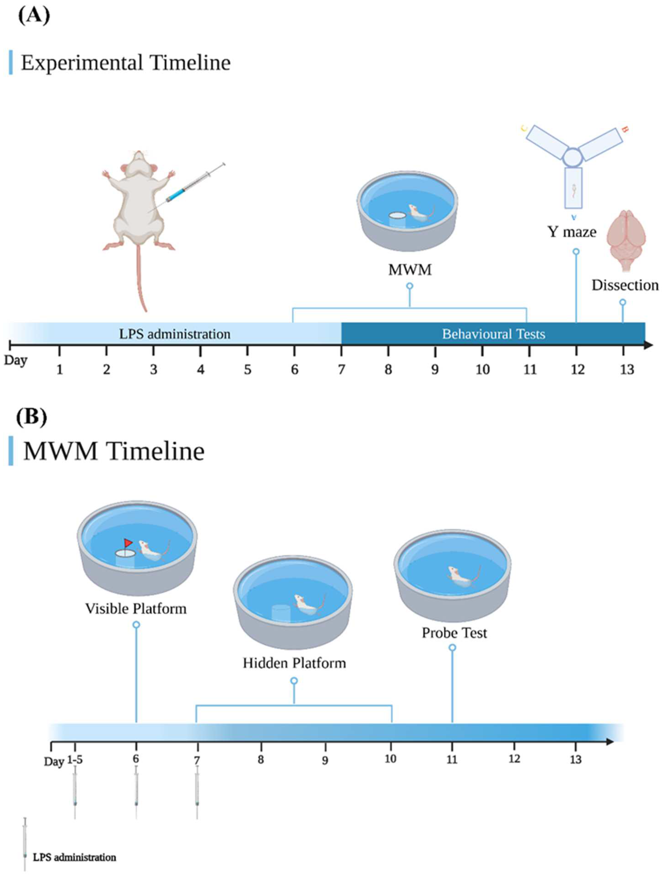
2.3. Experimental Design

Mice were randomly divided into four groups (n = 6): (I) i.p. saline group (control), (II) i.p. 0.25 mg/kg LPS group, (III) i.p. 0.50 mg/kg LP group, and (IV) i.p. 0.75 mg/kg LPS group. The i.p. injection was given in the lower left quadrant of the abdomen of the mice. All treatments were administered for 7 consecutive days. Behavioral tests were started on day 6 of LPS injections, which were administered for 7 days. At the end of day 13, mice were sacrificed, and the tissues were collected for further investigation (Figure 1A).

2.4. Weight and Temperature

Daily measurements of temperature and the percentage of weight gain were recorded to assess any sign of toxicity induced by LPS administration. At the end of day 7, the percentage of weight gain was calculated using the following equation: [weight at the end of the experiment − initial weight/initial weight] × 100. Temperature was measured using a (Hello, DT-8826) digital infrared thermometer (SCC Inc., Gampaha, Sri Lanka).

2.5. Behavioral Tests

2.5.1. Morris Water Maze (MWM) Test

The MWM is used to assess long-term spatial memory in rodents [28]. The test involves three stages: visible platform (day 1), hidden platform (day 2–5), and the probe test (day 6) (Figure 1B). In brief, a circular pool (height: 35 cm, diameter: 120 cm) was filled with water and maintained at 26 ± 2 °C, and fixed distance cues were present. Each day, semi-random start position sets with six positions (N, NW, S, SE, W, SW) were used. During the first stage, the platform (height: 14 cm, diameter: 4–5 cm) was exposed at 1 cm above the surface of the water in a specific position (Q2) with a flag placed on it to increase its visibility [28]. In the second stage, the platform was submerged in water colored by a non-toxic dye. During the two stages, mice were trained in 60 s trials with five trials per day [28]. If the mouse did not find the platform, it was guided to the platform and allowed to remain there for 15 s [28]. The total distance moved (TDM) and time to reach the platform (escape latency) were recorded daily throughout the 5 days of the experiment. In the probe test, the platform was removed, and the mice were allowed to find the platform location in one 30 s trial with SW fixed as the starting point position [28]. In the probe test, the time spent in the target quadrant (Q2) was recorded. ANY-Maze video tracking software (Stoelting Co., Wood Dale, IL, USA) was used to track the animal path during the test.

2.5.2. Y Maze

The Y maze is a test used to assess short-term spatial working memory in rodents [29]. This test was performed in the Y maze apparatus, which consists of three identical arms that are 10 cm wide, 40 cm high, and separated equally at 120°. In brief, mice were placed in one arm of the Y maze facing the wall; then, entries into the different arms were recorded for 3 min [29]. Alternation was recorded when the mouse visited the three arms sequentially without repetition [29]. Spontaneous alternation was measured as the percentage of alternation [% alternation = numbers of alternations/(total arm visits − 2)], where a smaller percentage of spontaneous alternation activity was associated with memory impairment.

2.5.3. Hematoxylin–Eosin (H&E) Staining

Mice were anesthetized via isoflurane inhalation, and brains were removed and placed in 4% buffered formalin fixative for 24 h. Paraffin-embedded blocks were sagittally sectioned at 2–5 µm thickness. The sections were stained with an H&E staining kit according to the manufacturer’s protocol (ab245880, Abcam, Cambridge, MA, USA) to evaluate the histological morphology. Cell loss in the hippocampal area was assessed under a microscope, in which there were 3 slices analyzed per group.

2.6. Statistical Analyses

All data are expressed as the mean ± standard error of the mean and were statistically analyzed using GraphPad Prism 8.3.8 (GraphPad Software Inc., San Diego, CA, USA). One-way analysis of variance (ANOVA) followed by a post hoc Dunnett’s test was used to compare differences among groups for weight gain (%), the probe test, and the Y maze. For temperature, TDM, and escape latency, two-way ANOVA followed by a post hoc Dunnett’s test was used. Differences between groups were considered statistically significant if the p-value was <0.05.

3. Results

3.1. Effect of LPS on Weight and Temperature

During the 7 days of LPS administration, weight gain and temperature were recorded to identify any sign of toxicity. Figure 2A shows that no significant differences were found between the control group and LPS groups for weight gain (0.25 mg/kg LPS (p = 0.3705); 0.50 mg/kg LPS (p = 0.2795) and 0.75 mg/kg LPS (p = 0.6247). The results also indicated no significant differences between the control group and LPS groups in the body temperature for days × groups [F(18, 120) = 0.5451, p = 0.9303], as shown in Figure 2B.

3.2. Behavioral Tests

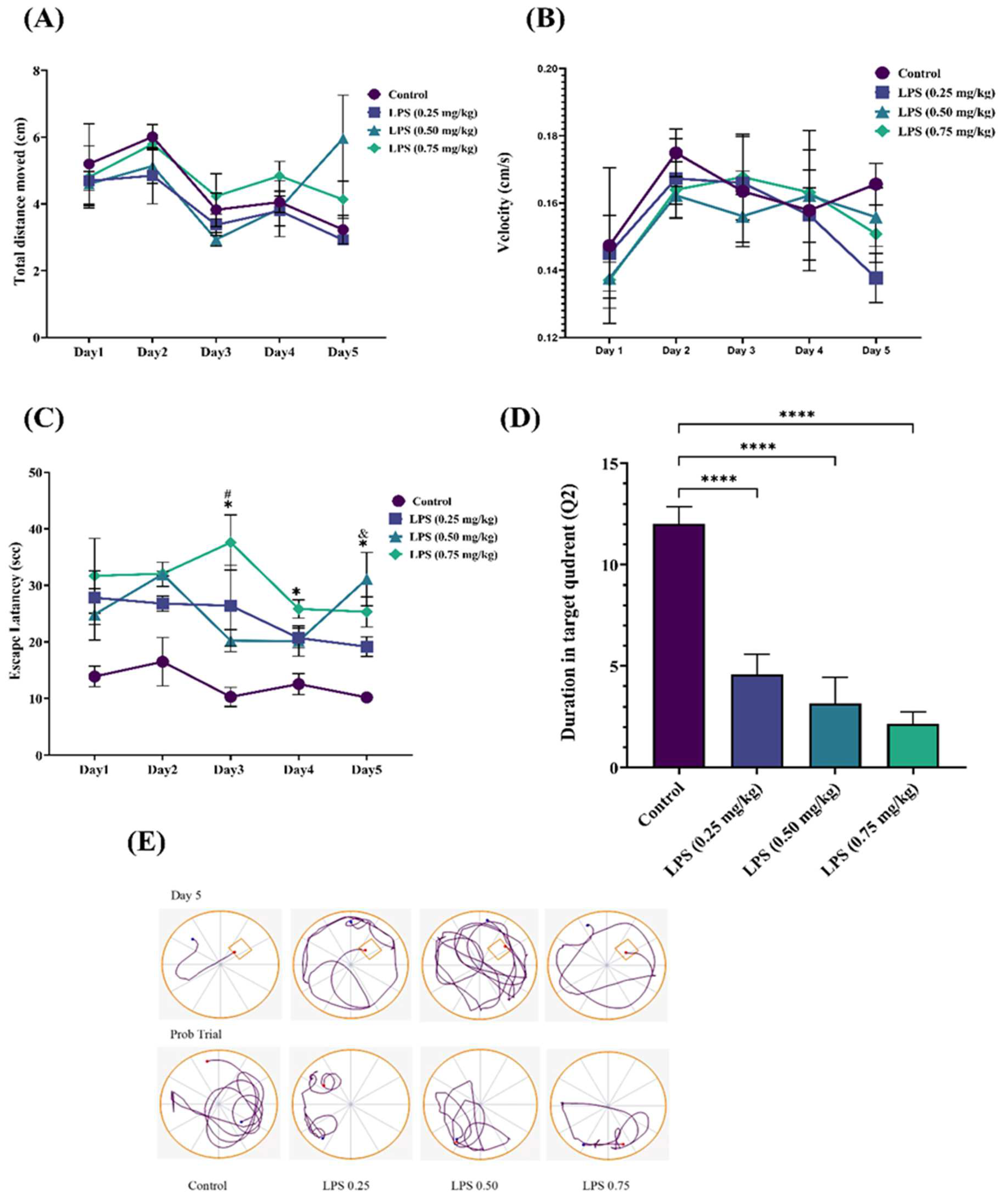
3.2.1. Effect of LPS on Long-Term Spatial Memory

The MWM test results showed that 7 days of LPS injection induced spatial memory impairment compared with the control group. The TDM and velocity results showed no significant difference between the control and LPS groups during the 5 days of the MWM test for days × groups ([F(12, 32) = 1.326, p = 0.2522] for TDM as shown in Figure 3A, and [F(12, 32)= 0.3672, p = 0.9659] for velocity as shown in Figure 3B. Regarding the escape latency (Figure 3C), data analyzed using two-way ANOVA indicated a significant effect of days × groups [F(12, 32) = 2.125, p = 0.0442]. Significant effects were found for days [F(2.711, 21.68) = 3.72, p = 0.0300] and groups [F(3, 8) = 9.686, p = 0.0049]. Further analysis with Dunnett’s post hoc test revealed that in the first 2 days of the test, there were no significant differences between the control and LPS groups. However, the 0.25 mg/kg LPS group showed a significant increase in the escape latency time only on the fifth day (p = 0.0460), while the 0.50 mg/kg LPS group exhibited a significant increase in the escape latency only on day 3 (p = 0.0417). Additionally, the LPS 0.75 mg/kg group showed significantly increased escape latency times on days 3, 4, and 5 (p = 0.0425, p = 0.0142, and p = 0.0453, respectively).
In the probe test, data represent the time (s) spent in the target quadrant (Q2) as shown in Figure 3D. The results revealed that the LPS groups spent significantly less time in the target quadrant (0.25 mg/kg LPS, p < 0.0001; 0.50 mg/kg LPS, p < 0.0001, and 0.75 mg/kg LPS, p < 0.0001) than the control group. Figure 3E shows representative data of the tracking plot for days 5 and 6.

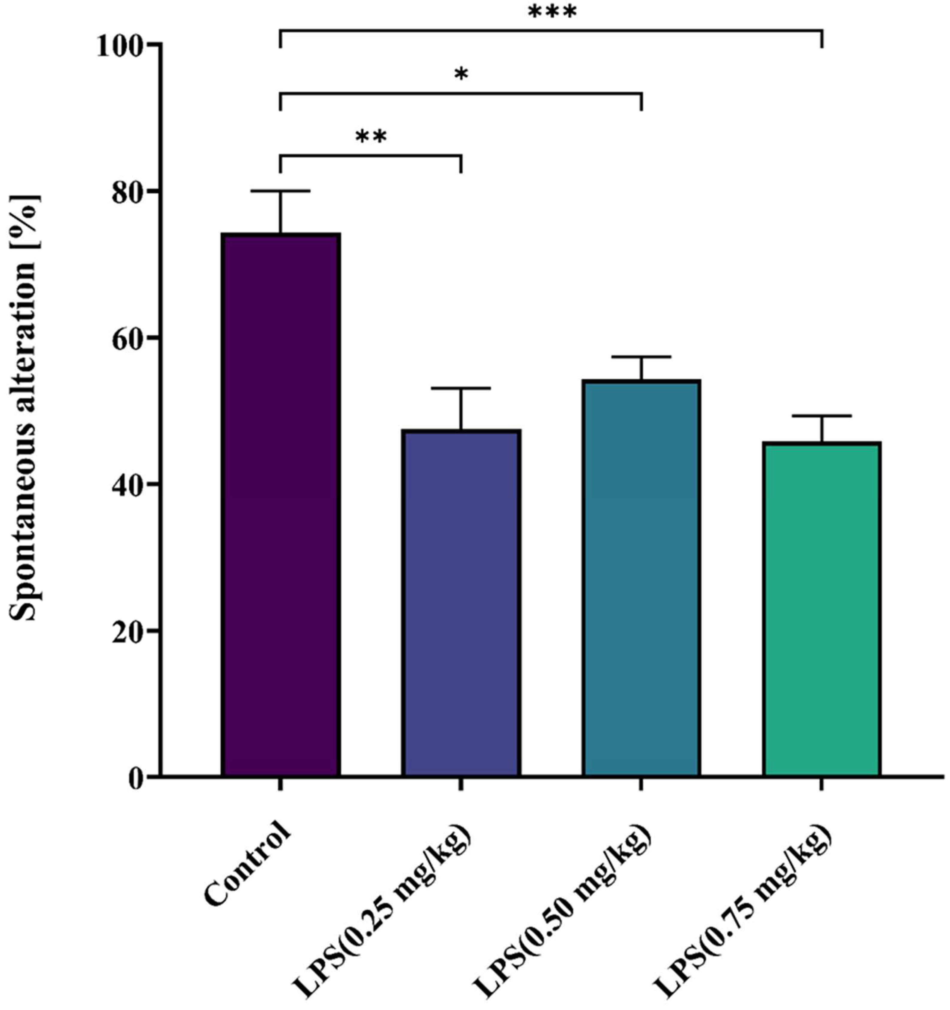
3.2.2. Effect of LPS on Short-Term Spatial Memory

The results of the Y maze test showed that LPS administration for 7 days induced working memory impairment in mice (Figure 4). A significant decrease in the spontaneous alternation percentage was observed in all LPS groups compared with the control group, and the 0.75 mg/kg LPS group (p > 0.0008) showed a more significant difference than the 0.5 mg/kg (p > 0.0154) and 0.25 mg/kg LPS groups (p > 0.0014). There were no differences among the LPS groups.

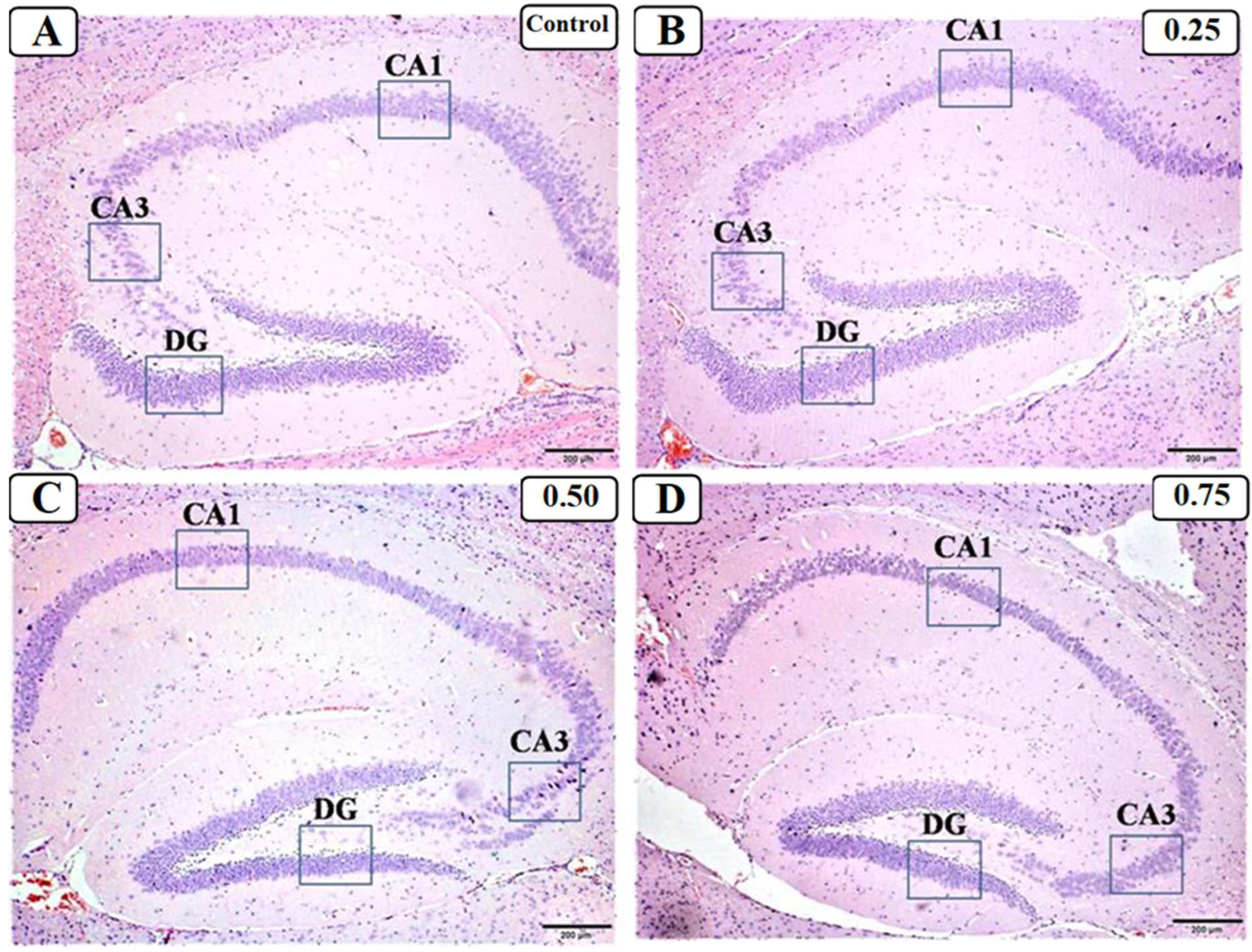
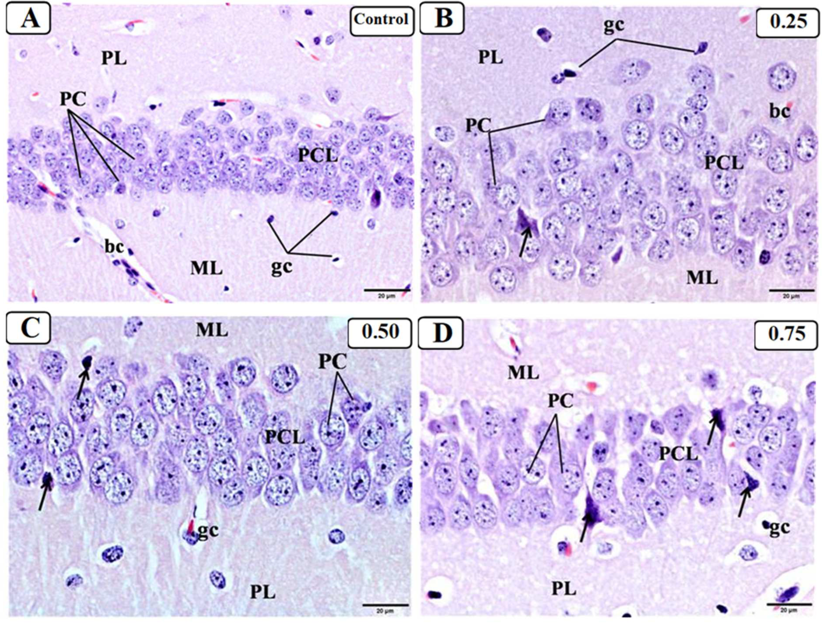
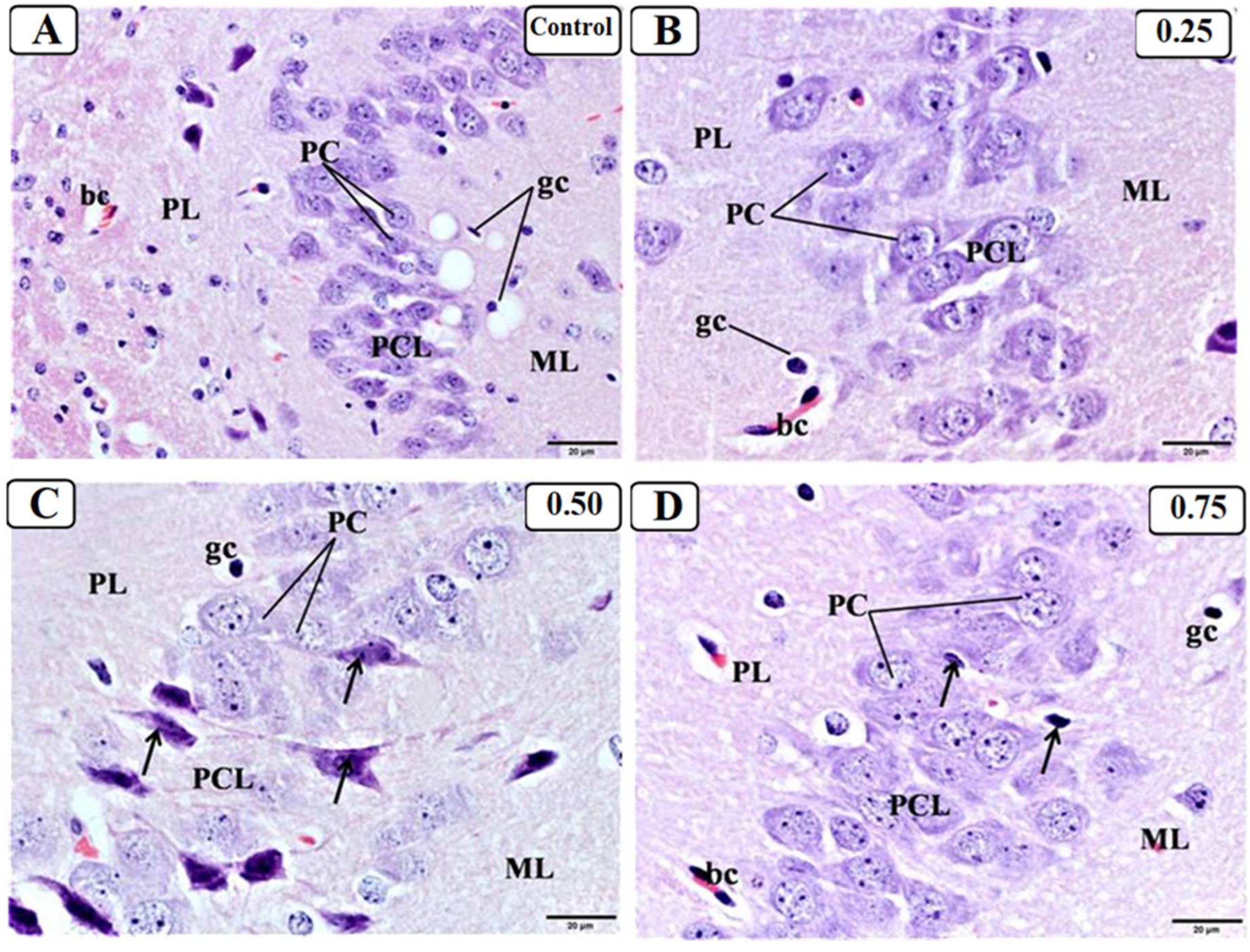
3.3. Effect of LPS on the Hippocampus

Examination of H&E-stained sagittal brain sections from the control group (Figure 5A) showed a normal histological structure of the hippocampal formation, which was visible as a curved structure in the medial portion of the temporal lobe. Furthermore, it consisted of two interconnected grey matter structures: the hippocampus proper (cornu ammonis, CA) and the dentate gyrus (DG). The CA exhibited a distinct C shape that was further subdivided into four regions: CA1, CA2, CA3, and CA4, whereas the DG appeared as a V-shaped structure that enfolded around CA4. Furthermore, examination of H&E-stained sagittal brain sections from different LPS treated groups (Figure 5B–D) showed the structure of the hippocampus and DG.
Higher magnification of the CA1 region in the control group (Figure 6A) showed that it was divided into three layers: a polymorphic layer (PL) containing glial cells, a pyramidal cell layer (PCL) containing the primary cell layer, and a molecular layer (ML) containing the apical dendrites and glial cells of pyramidal neurons. The PCL contained tiny, spherical, densely packed pyramidal cell (PC) bodies. The CA1 of LPS 0.25 treated group (Figure 6B) showed a picture more or less similar to that of the control group and the presence of multi-layers of pyramidal cells (PCs) with pale vesicular nuclei and prominent nucleolus. However, the presence of some PCs with dark basophilic cytoplasm (arrow) could be observed. The CA1 of LPS 0.50 treated group (Figure 6C) showed more degenerated PCs in the PCL, which appeared to be condensed with pyknotic nuclei. The CA1 of LPS 0.75 treated group (Figure 6D) showed many degenerated PCs in the PCL, which appeared to be condensed with dark stained cytoplasm and ill-defined nuclei.
Higher magnification of CA3 region in control group (Figure 7A) showed that it was divided into three layers: PL containing glial cells, PCL containing the primary cell layer, and a ML containing the apical dendrites and glial cells of pyramidal neurons. The PCL in CA3 had large, triangular, loosely packed PC bodies with rounded vesicular nuclei, prominent nucleoli, and a border of cytoplasm. The CA3 of the LPS 0.25 treated group (Figure 7B) showed a picture similar to that of the control group with the presence of PCs with pale vesicular nuclei and prominent nucleolus with no sign of neural degeneration. The CA3 in the LPS 0.50 treated group (Figure 7C) showed some degenerated PCs in the PCL, which appeared to be condensed with darkly stained cytoplasm and ill-defined nuclei. The CA3 of the LPS 0.75 treated group (Figure 7D) showed many degenerated PCs in the PCL, which appeared condensed with pyknotic nuclei.
The control group DG (Figure 8A) consists of three layers: the ML, the main layer consisting of the granule cell layer (GCL), and the PL, which is the hilus of the DG. The GCL consisted of tiny, spherical, tightly packed granule cells. The DG of LPS 0.25 treated group (Figure 8B) showed a picture similar to that of the control group except for the presence of some degenerated granule cells (arrow). The DG of both LPS 0.5 and LPS 0.75 treated group (Figure 8C,D) showed many degenerated granule cells in the GCL, which appeared to be condensed with pyknotic nuclei.

4. Discussion

4.1. This Work

Various animal models have been established to study AD pathology. LPS induction is one of the most commonly used animal models of AD [17,23,30]. Increasing evidence suggests a role of LPS in systemic inflammation, amyloid genesis, cognitive deficits, learning and memory impairment, and behavioral alterations [30,31]. Although previous studies have focused on examining the effect of LPS using a single dose or chronic administration of 0.25 mg/kg LPS to induce cognitive impairment [24,25,26,29,32], it is advantageous to explore chronic administration of different LPS doses and highlight their effects on memory deficits and brain pathology. Therefore, this study was performed to investigate the effect of frequent administration of 0.25, 0.5, and 0.75 mg/kg LPS on short- and long-term spatial memory and brain pathology in an experimental AD mouse model.
Various studies have reported that a single dose of LPS can lead to sickness behavior in rodents, which is characterized by hypothermia, body weight loss, and decreased food consumption [33,34,35]. In our study, weight gain (%) and temperature were assessed to observe the general health of mice. Our data revealed that the experimental animals did not exhibit any significant decrease in weight or temperature compared with the control group, which could indicate good health status and the absence of signs of toxicity.
The MWM test is a common research tool and is performed to investigate spatial memory performance, specifically in AD research [36]. Regarding our MWM results, mice showed normal locomotor activity in the TDM and velocity after LPS administration. However, previous studies reported that LPS may produce non-specific behavioral effects including decreased locomotor activity and exploration capability [1,37]. Furthermore, it is unclear whether the probe trial reflects reference memory or a combination of reference and working memory. Evidence suggests that a probe trial given 24 h after the last session of the escape latency evaluation reflects reference memory [38,39]. Here, both escape latency and time spent in the target quadrant were calculated to reflect long-term spatial learning memory. During the escape latency evaluation, the 0.50 mg/kg and 0.25 mg/kg LPS groups required a longer time to find the platform only on day 3 and 5, respectively, compared with the control group. However, the 0.75 mg/kg LPS group showed a significant increase in escape latency during the last 3 days of the hidden platform stage. These results revealed that the administration of 0.75 mg/kg LPS deteriorates spatial learning memory in the early stage compared with other doses. Additionally, in the probe test, all groups of mice exposed to LPS remained within the target quadrant for a shorter time. In line with our results, previous studies indicated that different doses of LPS remarkably decreased spatial memory performance in the MWM. For example, according to Kamdi et al. (2021), male Swiss albino mice treated with 25 mg/kg LPS showed significant memory impairment in the MWM behavioral test [40]. Another study used male C57BL/6J mice administered various LPS doses via different routes of administration (0.5 mg/kg and 0.75 mg/kg i.p. for 7 days and a single 12 μg intracerebroventricular dose) and demonstrated considerable memory deficits during the MWM test [36]. Furthermore, wild-type C57BL/6N mice injected with 0.25 mg/kg LPS (i.p.) exhibited remarkable spatial memory impairment in the MWM test [41].
The Y maze test is used to assess an animal’s ability to navigate a new area; thus, it is often used to assess short-term spatial working memory and learning [42]. To our knowledge, no previous studies have examined treatment with 0.50 mg/kg and 0.75 mg/kg doses of LPS in the Y maze test. Although all LPS groups (0.25 mg/kg, 0.50 mg/kg, and 0.75 mg/kg) showed a decreased percentage of spontaneous alternation, the 0.75 mg/kg dose of LPS produced a more significant change compared with the control group than the other LPS doses. In agreement with our results, several studies have shown that LPS affects spatial working memory. For example, Muhammad et al. (2019) showed that C57BL/6N mice injected with LPS (0.25 mg/kg i.p.) displayed neuroinflammation, which led to cognitive decline and working memory impairments [29]. Another study using adult male Sprague–Dawley rats treated with 0.25 mg/kg LPS showed a significant decrease in the percentage of spontaneous alternation [24]. Moreover, several studies reported that chronic administration of 0.25 mg/kg LPS impaired spatial working memory during the Y maze test [32,43,44].
Histological examination is an important tool to understand the pathology of specific organs and visualize cell lesions [45]. It was known that the hippocampus plays a key role in learning and memory, is particularly susceptible to inflammatory injury due to its high intensity of receptors for inflammatory mediators [46]. Chronic peripheral inflammation is caused by long-term peripheral diseases and metabolic abnormalities, bacterial infection, and natural aging. These conditions are connected to behavioral problems such as cognitive impairment, learning and memory impairments, depression, and anxiety, which are all linked to impaired hippocampus integrity [46,47].
Examination of the histological effect of LPS on hippocampal tissue demonstrated the presence of neurodegeneration in all LPS groups. However, treatment with both 0.50 mg/kg and 0.75 mg/kg LPS resulted in darker neuronal cells compared with the control group, while 0.25 mg/kg LPS produced results similar to those of the control group in some regions. Additionally, the 0.25 mg/kg dose of LPS resulted in degeneration of neurons only in the DG region, while the 0.50 mg /kg and 0.75 mg/kg doses produced degeneration of neurons in all hippocampal regions, including the CA1, CA3, and DG regions. In accordance with our results, several reports have found that i.p. injection of LPS results in cell shrinkage, darker-looking cells, and increased apoptotic neurons [48,49,50]. Furthermore, hippocampal damage often triggers severe impairments in spatial memory [10], which links our behavioral results with the histological observations.
Neuroinflammation and microglial activation are both involved in the etiology of several neurodegenerative disorders, including AD [51]. In addition, neuroinflammation triggers excessive release of proinflammatory cytokines, chemokines, and oxygen radicals and contributes to neuronal dysfunction and degeneration, leading to behavioral and memory impairment [52,53,54]. Previous investigations have confirmed that LPS administration can significantly activate microglia and astrocytes by increasing expression of the hippocampal proteins Iba-1 and GFAP [24,36,55]. Furthermore, peripheral LPS injection has been reported to increase the expression of inflammatory mediators, including nitric oxide synthase and cyclooxygenase-2, in the hippocampus [24,36,56]. Peripheral LPS administration also increases the release of pro-inflammatory mediators such as interleukin-1β, interleukin-6, and tumor necrosis factor-α in the hippocampus [36,57,58]. Moreover, the interference with brain-derived neurotrophic factor (BDNF) gene expression and function may be one of the underlying mechanisms of LPS-induced cognitive impairments [59]. In particular, BDNF has been reported to be involved in the control of GABAergic neuron growth and survival, as well as the regulation of synaptic plasticity in short- and long-term memory [60,61]. During the inflammatory response to LPS, BDNF induction in the hippocampus is impaired by the production of nuclear factor-kappa B (NF-κB)-dependent pro-inflammatory cytokines [62]. This reduces BDNF signaling in the synaptic cleft, which further reduces BDNF-dependent survival-related signaling and inhibits apoptotic pathways [62]. In summary, decreased BDNF availability may play a pivotal role in the development of cognitive impairment [59]. Therefore, our observed behavioral and histological alterations could be explained by prolonged neuroinflammation caused by LPS administration.

4.2. Contributions and Limitations

In summary, this study examined the effect of different LPS doses on short- and long-term spatial memory along with hippocampal histological changes in mice. Interestingly, the 0.75 mg/kg dose of LPS produced stronger deterioration of both short-term and long-term spatial memory in mice exhibiting normal temperature and weight gain results, which indicated good general health. This behavioral result suggests 0.75 mg/kg LPS as a promising dose for future study of an LPS-induced AD model. In addition, histological examination of mice treated with 0.50 mg/kg and 0.75 mg/kg LPS showed a higher degree of degeneration than that observed in mice treated with 0.25 mg/kg LPS. In this study, our histological data were limited to representative examination and quantitative data of the narcotic neurons in a different section of the hippocampus can be measured using ImageJ program. To the best of our knowledge, this study is one of the first studies to compare the effect of frequent administration of various LPS doses (0.25 mg/kg, 0.50 mg/kg, and 0.75 mg/kg) on different types of spatial memory impairment in conjunction with the histological results.

4.3. Future Work

For future studies, we recommend further investigation with a higher sample size and female gender. More studies are needed to elucidate the role of microglial activation along with quantitative histological investigation. In addition, we recommend assessment of the underlying neuroinflammation mechanism of spatial memory impairment that is induced by chronic administration of 0.75 mg/kg LPS.

Author Contributions

N.A.A., K.A.B., R.A.M. and B.S.A. designed the research study. K.A.B. performed the research. N.A.A. helped carried out the experiments. K.A.B. analyzed the data and wrote the manuscript. R.A.M. and B.S.A. supervised the project. R.S.A., B.S.A. and R.A.M. provided critical feedback and helped shape the research. R.S.A., B.S.A. and R.A.M. contributed to editorial changes in the final manuscript. All authors have read and agreed to the published version of the manuscript.

Funding

This research received no external funding.

Institutional Review Board Statement

The animal study protocol was approved by the Unit of Biomedical Ethics, Faculty of Medicine, King Abdul-Aziz University, Jeddah, SA (Reference No. 603-20).

Informed Consent Statement

Not applicable.

Data Availability Statement

Not applicable.

Acknowledgments

We would like to show our appreciation to Khlood Alyami and Ghada Alshahrani for their support and assistance in manuscript revision and figure design.

Conflicts of Interest

The authors declare no conflict of interest.

Abbreviations

Lipopolysaccharide (LPS); Alzheimer’s disease (AD); King Fahd Medical Research Center (KFMRC); Morris water maze (MWM); total distance moved (TDM); analysis of variance (ANOVA); hematoxylin–eosin (H&E); cornu ammonis (CA); dentate gyrus (DG); polymorphic layer (PL); pyramidal cell layer (PCL); molecular layer (ML); granule cell layer (GCL).

References

  1. Knopman, D.S.; Amieva, H.; Petersen, R.C.; Chételat, G.; Holtzman, D.M.; Hyman, B.T.; Nixon, R.A.; Jones, D.T. Alzheimer disease. Nat. Rev. Dis. Primers 2021, 7, 1–21. [Google Scholar] [CrossRef] [PubMed]
  2. Du, X.; Wang, X.; Geng, M. Alzheimer’s disease hypothesis and related therapies. Transl. Neurodegener. 2018, 7, 1–7. [Google Scholar] [CrossRef] [PubMed]
  3. Zakaria, R.; Wan Yaacob, W.; Othman, Z.; Long, I.; Ahmad, A.; Al-Rahbi, B. Lipopolysaccharide-induced memory impairment in rats: A model of Alzheimer’s disease. Physiol. Res. 2017, 66, 553–565. [Google Scholar] [CrossRef]
  4. Calabrò, M.; Rinaldi, C.; Santoro, G.; Crisafulli, C. The biological pathways of Alzheimer disease: A review. AIMS Neurosci. 2021, 8, 86. [Google Scholar] [CrossRef]
  5. Kinney, J.W.; Bemiller, S.M.; Murtishaw, A.S.; Leisgang, A.M.; Salazar, A.M.; Lamb, B.T. Inflammation as a central mechanism in Alzheimer’s disease. Alzheimer’s Dement. Transl. Res. Clin. Interv. 2018, 4, 575–590. [Google Scholar] [CrossRef]
  6. Yiannopoulou, K.G.; Papageorgiou, S.G. Current and future treatments in Alzheimer disease: An update. J. Cent. Nerv. Syst. Dis. 2020, 12, 1179573520907397. [Google Scholar] [CrossRef]
  7. Zhu, X.; Liu, J.; Chen, S.; Xue, J.; Huang, S.; Wang, Y.; Chen, O. Isoliquiritigenin attenuates lipopolysaccharide-induced cognitive impairment through antioxidant and anti-inflammatory activity. BMC Neurosci. 2019, 20, 1–13. [Google Scholar] [CrossRef]
  8. Glachet, O.; El Haj, M. Effects of olfactory stimulation on past and future thinking in Alzheimer’s disease. Chem. Senses 2020, 45, 313–320. [Google Scholar] [CrossRef]
  9. Olton, D.S. Spatial memory. Sci. Am. 1977, 236, 82–99. [Google Scholar] [CrossRef]
  10. Vikbladh, O.M.; Meager, M.R.; King, J.; Blackmon, K.; Devinsky, O.; Shohamy, D.; Burgess, N.; Daw, N.D. Hippocampal contributions to model-based planning and spatial memory. Neuron 2019, 102, 683–693.e4. [Google Scholar] [CrossRef]
  11. Miller, J.; Watrous, A.J.; Tsitsiklis, M.; Lee, S.A.; Sheth, S.A.; Schevon, C.A.; Smith, E.H.; Sperling, M.R.; Sharan, A.; Asadi-Pooya, A.A. Lateralized hippocampal oscillations underlie distinct aspects of human spatial memory and navigation. Nat. Commun. 2018, 9, 1–12. [Google Scholar] [CrossRef] [PubMed]
  12. Stevenson, R.F.; Zheng, J.; Mnatsakanyan, L.; Vadera, S.; Knight, R.T.; Lin, J.J.; Yassa, M.A. Hippocampal CA1 gamma power predicts the precision of spatial memory judgments. Proc. Natl. Acad. Sci. USA 2018, 115, 10148–10153. [Google Scholar] [CrossRef] [PubMed]
  13. Sharma, S.; Rakoczy, S.; Brown-Borg, H. Assessment of spatial memory in mice. Life Sci. 2010, 87, 521–536. [Google Scholar] [CrossRef] [PubMed]
  14. Cowan, N. What are the differences between long-term, short-term, and working memory? Prog. Brain Res. 2008, 169, 323–338. [Google Scholar] [PubMed]
  15. Freedman, M.L.; Martin, R.C. Dissociable components of short-term memory and their relation to long-term learning. Cogn. Neuropsychol. 2001, 18, 193–226. [Google Scholar] [CrossRef] [PubMed]
  16. Cowan, N. The many faces of working memory and short-term storage. Psychon. Bull. Rev. 2017, 24, 1158–1170. [Google Scholar] [CrossRef]
  17. Vasconcelos, A.R.; Yshii, L.M.; Viel, T.A.; Buck, H.S.; Mattson, M.P.; Scavone, C.; Kawamoto, E.M. Intermittent fasting attenuates lipopolysaccharide-induced neuroinflammation and memory impairment. J. Neuroinflamm. 2014, 11, 1–14. [Google Scholar] [CrossRef]
  18. Lively, S.; Schlichter, L.C. Microglia responses to pro-inflammatory stimuli (LPS, IFNγ+ TNFα) and reprogramming by resolving cytokines (IL-4, IL-10). Front. Cell. Neurosci. 2018, 12, 215. [Google Scholar] [CrossRef]
  19. Liu, X.; Yin, S.; Chen, Y.; Wu, Y.; Zheng, W.; Dong, H.; Bai, Y.; Qin, Y.; Li, J.; Feng, S. LPS-induced proinflammatory cytokine expression in human airway epithelial cells and macrophages via NF-κB, STAT3 or AP-1 activation. Mol. Med. Rep. 2018, 17, 5484–5491. [Google Scholar] [CrossRef]
  20. Zhao, G.; Zhang, T.; Ma, X.; Jiang, K.; Wu, H.; Qiu, C.; Guo, M.; Deng, G. Oridonin attenuates the release of pro-inflammatory cytokines in lipopolysaccharide-induced RAW264. 7 cells and acute lung injury. Oncotarget 2017, 8, 68153. [Google Scholar] [CrossRef] [Green Version]
  21. Cavaillon, J.-M. Exotoxins and endotoxins: Inducers of inflammatory cytokines. Toxicon 2018, 149, 45–53. [Google Scholar] [CrossRef] [PubMed]
  22. Azmand, M.J.; Rajaei, Z. Effects of crocin on spatial or aversive learning and memory impairments induced by lipopolysaccharide in rats. Avicenna J. Phytomed. 2021, 11, 79. [Google Scholar] [PubMed]
  23. Batista, C.R.A.; Gomes, G.F.; Candelario-Jalil, E.; Fiebich, B.L.; De Oliveira, A.C.P. Lipopolysaccharide-induced neuroinflammation as a bridge to understand neurodegeneration. Int. J. Mol. Sci. 2019, 20, 2293. [Google Scholar] [CrossRef] [PubMed]
  24. Andy, S.N.; Pandy, V.; Alias, Z.; Kadir, H.A. Deoxyelephantopin ameliorates lipopolysaccharides (LPS)-induced memory impairments in rats: Evidence for its anti-neuroinflammatory properties. Life Sci. 2018, 206, 45–60. [Google Scholar] [CrossRef] [PubMed]
  25. Mastinu, A.; Bonini, S.A.; Rungratanawanich, W.; Aria, F.; Marziano, M.; Maccarinelli, G.; Abate, G.; Premoli, M.; Memo, M.; Uberti, D. Gamma-oryzanol prevents LPS-induced brain inflammation and cognitive impairment in adult mice. Nutrients 2019, 11, 728. [Google Scholar] [CrossRef]
  26. Thingore, C.; Kshirsagar, V.; Juvekar, A. Amelioration of oxidative stress and neuroinflammation in lipopolysaccharide-induced memory impairment using Rosmarinic acid in mice. Metab. Brain Dis. 2021, 36, 299–313. [Google Scholar] [CrossRef]
  27. Ramírez, K.; Quesada-Yamasaki, D.; Fornaguera-Trías, J. A Protocol to Perform Systemic Lipopolysacharide (LPS) Challenge in Rats. Odovtos-Int. J. Dent. Sci. 2019, 21, 53–66. [Google Scholar] [CrossRef]
  28. Bromley-Brits, K.; Deng, Y.; Song, W. Morris water maze test for learning and memory deficits in Alzheimer’s disease model mice. JoVE 2011, 20, e2920. [Google Scholar] [CrossRef]
  29. Muhammad, T.; Ikram, M.; Ullah, R.; Rehman, S.U.; Kim, M.O. Hesperetin, a citrus flavonoid, attenuates LPS-induced neuroinflammation, apoptosis and memory impairments by modulating TLR4/NF-κB signaling. Nutrients 2019, 11, 648. [Google Scholar] [CrossRef]
  30. Nazem, A.; Sankowski, R.; Bacher, M.; Al-Abed, Y. Rodent models of neuroinflammation for Alzheimer’s disease. J. Neuroinflamm. 2015, 12, 1–15. [Google Scholar] [CrossRef] [Green Version]
  31. Hoogland, I.C.; Houbolt, C.; van Westerloo, D.J.; van Gool, W.A.; van de Beek, D. Systemic inflammation and microglial activation: Systematic review of animal experiments. J. Neuroinflamm. 2015, 12, 1–13. [Google Scholar] [CrossRef] [PubMed]
  32. Wang, J.; Li, L.; Wang, Z.; Cui, Y.; Tan, X.; Yuan, T.; Liu, Q.; Liu, Z.; Liu, X. Supplementation of lycopene attenuates lipopolysaccharide-induced amyloidogenesis and cognitive impairments via mediating neuroinflammation and oxidative stress. J. Nutr. Biochem. 2018, 56, 16–25. [Google Scholar] [CrossRef] [PubMed]
  33. Erickson, M.A.; Hartvigson, P.E.; Morofuji, Y.; Owen, J.B.; Butterfield, D.A.; Banks, W.A. Lipopolysaccharide impairs amyloid beta efflux from brain: Altered vascular sequestration, cerebrospinal fluid reabsorption, peripheral clearance and transporter function at the blood–brain barrier. J. Neuroinflammation 2012, 9, 1–15. [Google Scholar] [CrossRef]
  34. Wang, Z.; Zhang, Q.; Yuan, L.; Wang, S.; Liu, L.; Yang, X.; Li, G.; Liu, D. The effects of curcumin on depressive-like behavior in mice after lipopolysaccharide administration. Behav. Brain Res. 2014, 274, 282–290. [Google Scholar] [CrossRef]
  35. O’Connor, J.C.; Lawson, M.A.; Andre, C.; Moreau, M.; Lestage, J.; Castanon, N.; Kelley, K.W.; Dantzer, R. Lipopolysaccharide-induced depressive-like behavior is mediated by indoleamine 2, 3-dioxygenase activation in mice. Mol. Psychiatry 2009, 14, 511–522. [Google Scholar] [CrossRef] [PubMed]
  36. Zhao, J.; Bi, W.; Xiao, S.; Lan, X.; Cheng, X.; Zhang, J.; Lu, D.; Wei, W.; Wang, Y.; Li, H. Neuroinflammation induced by lipopolysaccharide causes cognitive impairment in mice. Sci. Rep. 2019, 9, 1–12. [Google Scholar]
  37. Jahangiri, Z.; Gholamnezhad, Z.; Hosseini, M. The effects of exercise on hippocampal inflammatory cytokine levels, brain oxidative stress markers and memory impairments induced by lipopolysaccharide in rats. Metab. Brain Dis. 2019, 34, 1157–1169. [Google Scholar] [CrossRef]
  38. Vorhees, C.V.; Williams, M.T. Assessing spatial learning and memory in rodents. ILAR J. 2014, 55, 310–332. [Google Scholar] [CrossRef]
  39. Paul, C.-M.; Magda, G.; Abel, S. Spatial memory: Theoretical basis and comparative review on experimental methods in rodents. Behav. Brain Res. 2009, 203, 151–164. [Google Scholar] [CrossRef]
  40. Kamdi, S.P.; Raval, A.; Nakhate, K.T. Phloridzin attenuates lipopolysaccharide-induced cognitive impairment via antioxidant, anti-inflammatory and neuromodulatory activities. Cytokine 2021, 139, 155408. [Google Scholar] [CrossRef]
  41. Khan, A.; Ali, T.; Rehman, S.U.; Khan, M.S.; Alam, S.I.; Ikram, M.; Muhammad, T.; Saeed, K.; Badshah, H.; Kim, M.O. Neuroprotective effect of quercetin against the detrimental effects of LPS in the adult mouse brain. Front. Pharmacol. 2018, 9, 1383. [Google Scholar] [CrossRef] [PubMed] [Green Version]
  42. Kraeuter, A.-K.; Guest, P.C.; Sarnyai, Z. The Y-maze for assessment of spatial working and reference memory in mice. In Pre-Clinical Models; Springer: Berlin/Heidelberg, Germany, 2019; pp. 105–111. [Google Scholar]
  43. Chowdhury, A.A.; Gawali, N.B.; Shinde, P.; Munshi, R.; Juvekar, A.R. Imperatorin ameliorates lipopolysaccharide induced memory deficit by mitigating proinflammatory cytokines, oxidative stress and modulating brain-derived neurotropic factor. Cytokine 2018, 110, 78–86. [Google Scholar] [CrossRef] [PubMed]
  44. Emokpae, O.; Ben-Azu, B.; Ajayi, A.M.; Umukoro, S. D-Ribose-L-cysteine attenuates lipopolysaccharide-induced memory deficits through inhibition of oxidative stress, release of proinflammatory cytokines, and nuclear factor-kappa B expression in mice. Naunyn-Schmiedeberg’s Arch. Pharmacol. 2020, 393, 909–925. [Google Scholar] [CrossRef] [PubMed]
  45. Li, Y.; Li, N.; Yu, X.; Huang, K.; Zheng, T.; Cheng, X.; Zeng, S.; Liu, X. Hematoxylin and eosin staining of intact tissues via delipidation and ultrasound. Sci. Rep. 2018, 8, 1–8. [Google Scholar] [CrossRef]
  46. Green, H.F.; Nolan, Y.M. Inflammation and the developing brain: Consequences for hippocampal neurogenesis and behavior. Neurosci. Biobehav. Rev. 2014, 40, 20–34. [Google Scholar] [CrossRef] [PubMed]
  47. Barrientos, R.; Kitt, M.; Watkins, L.; Maier, S. Neuroinflammation in the normal aging hippocampus. Neuroscience 2015, 309, 84–99. [Google Scholar] [CrossRef]
  48. Liu, Q.; Chen, Y.; Shen, C.; Xiao, Y.; Wang, Y.; Liu, Z.; Liu, X. Chicoric acid supplementation prevents systemic inflammation-induced memory impairment and amyloidogenesis via inhibition of NF-κB. FASEB J. 2017, 31, 1494–1507. [Google Scholar] [CrossRef]
  49. Eduviere, A.T.; Umukoro, S.; Adeoluwa, O.A.; Omogbiya, I.A.; Aluko, O.M. Possible mechanisms involved in attenuation of lipopolysaccharide-induced memory deficits by methyl jasmonate in mice. Neurochem. Res. 2016, 41, 3239–3249. [Google Scholar] [CrossRef]
  50. He, P.; Yan, S.; Zheng, J.; Gao, Y.; Zhang, S.; Liu, Z.; Liu, X.; Xiao, C. Eriodictyol attenuates LPS-induced neuroinflammation, amyloidogenesis, and cognitive impairments via the inhibition of NF-κB in male C57BL/6J mice and BV2 microglial cells. J. Agric. Food Chem. 2018, 66, 10205–10214. [Google Scholar] [CrossRef]
  51. Qin, L.; Wu, X.; Block, M.L.; Liu, Y.; Breese, G.R.; Hong, J.S.; Knapp, D.J.; Crews, F.T. Systemic LPS causes chronic neuroinflammation and progressive neurodegeneration. Glia 2007, 55, 453–462. [Google Scholar] [CrossRef]
  52. Tyagi, E.; Agrawal, R.; Nath, C.; Shukla, R. Influence of LPS-induced neuroinflammation on acetylcholinesterase activity in rat brain. J. Neuroimmunol. 2008, 205, 51–56. [Google Scholar] [CrossRef] [PubMed]
  53. Park, T.; Chen, H.; Kevala, K.; Lee, J.-W.; Kim, H.-Y. N-Docosahexaenoylethanolamine ameliorates LPS-induced neuroinflammation via cAMP/PKA-dependent signaling. J. Neuroinflamm. 2016, 13, 1–15. [Google Scholar] [CrossRef] [PubMed]
  54. Shabab, T.; Khanabdali, R.; Moghadamtousi, S.Z.; Kadir, H.A.; Mohan, G. Neuroinflammation pathways: A general review. Int. J. Neurosci. 2017, 127, 624–633. [Google Scholar] [CrossRef]
  55. Khan, M.S.; Muhammad, T.; Ikram, M.; Kim, M.O. Dietary supplementation of the antioxidant curcumin halts systemic LPS-induced neuroinflammation-associated neurodegeneration and memory/synaptic impairment via the JNK/NF-κB/Akt signaling pathway in adult rats. Oxidative Med. Cell. Longev. 2019, 2019. [Google Scholar] [CrossRef] [PubMed]
  56. Yeo, I.J.; Yun, J.; Son, D.J.; Han, S.-B.; Hong, J.T. Antifungal drug miconazole ameliorated memory deficits in a mouse model of LPS-induced memory loss through targeting iNOS. Cell Death Dis. 2020, 11, 1–14. [Google Scholar] [CrossRef] [PubMed]
  57. Zhao, W.; Xu, Z.; Cao, J.; Fu, Q.; Wu, Y.; Zhang, X.; Long, Y.; Zhang, X.; Yang, Y.; Li, Y. Elamipretide (SS-31) improves mitochondrial dysfunction, synaptic and memory impairment induced by lipopolysaccharide in mice. J. Neuroinflamm. 2019, 16, 1–19. [Google Scholar] [CrossRef]
  58. Zhang, J.; Zheng, Y.; Luo, Y.; Du, Y.; Zhang, X.; Fu, J. Curcumin inhibits LPS-induced neuroinflammation by promoting microglial M2 polarization via TREM2/TLR4/NF-κB pathways in BV2 cells. Mol. Immunol. 2019, 116, 29–37. [Google Scholar] [CrossRef]
  59. Wei, P.; Liu, Q.; Li, D.; Zheng, Q.; Zhou, J.; Li, J. Acute nicotine treatment attenuates lipopolysaccharide-induced cognitive dysfunction by increasing BDNF expression and inhibiting neuroinflammation in the rat hippocampus. Neurosci. Lett. 2015, 604, 161–166. [Google Scholar] [CrossRef]
  60. Saral, S.; Topçu, A.; Alkanat, M.; Mercantepe, T.; Akyıldız, K.; Yıldız, L.; Tümkaya, L.; Yazıcı, Z.A.; Yılmaz, A. Apelin-13 activates the hippocampal BDNF/TrkB signaling pathway and suppresses neuroinflammation in male rats with cisplatin-induced cognitive dysfunction. Behav. Brain Res. 2021, 408, 113290. [Google Scholar] [CrossRef]
  61. Stajic, D.; Selakovic, D.; Jovicic, N.; Joksimovic, J.; Arsenijevic, N.; Lukic, M.L.; Rosic, G. The role of galectin-3 in modulation of anxiety state level in mice. Brain Behav. Immun. 2019, 78, 177–187. [Google Scholar] [CrossRef]
  62. Lima Giacobbo, B.; Doorduin, J.; Klein, H.C.; Dierckx, R.A.; Bromberg, E.; de Vries, E.F. Brain-derived neurotrophic factor in brain disorders: Focus on neuroinflammation. Mol. Neurobiol. 2019, 56, 3295–3312. [Google Scholar] [CrossRef] [Green Version]
Figure 1. Experimental timeline. (A) Experimental timeline and behavioral test summary. The study was conducted for 13 days. LPS was administered on the first 7 days, and behavioral tests were started on the sixth day of LPS administration. At the end of day 13, mice were sacrificed for further histological investigation. (B) Timeline of the MWM test. The MWM test was conducted for 6 days starting on the last 2 days (day 6 and 7) of LPS administration. Three stages were preformed: visible platform, hidden platform, and the probe test. MWM: Morris water maze; LPS: lipopolysaccharide. Created with BioRender.com (accessed on 3 June 2022).
Ctn 06 00020 g001
Figure 2. Weight and temperature. Weight gain and temperature were recorded daily during LPS administration. The body weight and temperature of mice showed no significant differences between the LPS groups (0.25 mg/kg, 0.50 mg/kg, and 0.75 mg/kg) and the control group. The data are presented as the mean ± SEM. (A) One-way ANOVA was used. (B) Two-way ANOVA was used. ns = non-significant. LPS: lipopolysaccharide; SEM: standard error of the mean; ANOVA: analysis of variance.
Ctn 06 00020 g002
Figure 3. Morris water maze. (A) Total distance moved; no significant differences were observed in the total distance moved among all groups. (B) Velocity; no significant differences were observed in swimming velocity among all groups. (C) Escape latency; no significant difference was observed in the escape latency time among all groups in the first two days. The 0.75 mg/kg LPS group showed a significant increase in escape latency time in the 3 remaining days, while the 0.50 mg/kg and 0.25 mg/kg LPS groups exhibited significantly increased escape latency time only on days 3 and 5, respectively. (D) Probe test; the LPS groups spent significantly less time in the target quadrant than the control group. (E) Representative tracking plot of days 5 and 6. Data are presented as the mean ± SEM. (AC) Two-way ANOVA was used followed by a post hoc Dunnett’s test. (D) One-way ANOVA was used followed by a post hoc Dunnett’s test. LPS: lipopolysaccharide; sec: seconds; SEM: standard error of the mean; ANOVA: analysis of variance. 0.25 mg/kg LPS group, # p < 0.05, & p < 0.05, 0.50 mg/kg LPS group, and * p < 0.05, 0.75 mg/kg LPS group compared with the control group, **** p < 0.0001.
Ctn 06 00020 g003
Figure 4. Y maze. Significant differences in spontaneous alternation were observed in all LPS groups (0.25 mg/kg, 0.50 mg/kg, and 0.75 mg/kg) compared with the control group. Data are presented as the mean ± SEM. A One-way ANOVA followed by a post hoc Dunnett’s test was used. * p < 0.05, ** p < 0.01, *** p <0.001. LPS: lipopolysaccharide; SEM: standard error of the mean.
Ctn 06 00020 g004
Figure 5. Representative photomicrographs of a sagittal section of the hippocampus in (A) control, (B) 0.25 LPS, (C) 0.50 LPS, and (D) 0.75 LPS groups. It shows different parts of the hippocampal structure, including the cornu ammonis (CA) and dentate gyrus (DG). The CA is subdivided into the CA1 and CA3 regions (hematoxylin and eosin staining (H&E) ×40).
Ctn 06 00020 g005
Figure 6. Representative photomicrographs of the CA1 region. (A) Control group contains three well-defined layers: the polymorphic layer (PL), pyramidal cell layer (PCL), and molecular layer (ML). The PCL is composed of densely packed pyramidal cells (PC) with vesicular nuclei, conspicuous nucleoli, and sparse cytoplasm. The PL and ML contain glial cells (gc) and blood capillaries (bc). (B) Nearly normal structure in LPS 0.25 treated group. Notice the presence of some PCs with dark basophilic cytoplasm without clear nucleus (arrow). The PL and ML displayed normal gc and bc. (C) Some degenerated PCs (arrow) in LPS 0.5 treated group in the PCL, which appeared condensed with pyknotic nuclei. (D) Many degenerated PCs (arrow) in LPS 0.75 treated group in the PCL, which appeared condensed with dark stained cytoplasm and ill-defined nuclei (H&E × 400).
Ctn 06 00020 g006
Figure 7. Representative photomicrographs of the CA3 region. (A) The control group reveals three regularly arranged layers; the polymorphic layer (PL), pyramidal cell layer (PCL), and molecular layer (ML). Pyramidal cells (PCs) are large with vesicular nuclei, prominent nucleoli, and scant cytoplasm. The PL and ML contain glial cells (gc) and blood capillaries (bc). (B) Regularly arranged three layers; PL, PCL and ML in LPS 0.25 treated group with normal appearance of PC. (C) Many degenerated PCs (arrow) in the PCL of LPS 0.5 treated group, which appeared condensed with pyknotic nuclei. (D) Some degenerated PCs (arrow) in the PCL of LPS 0.75 treated group, which appeared condensed with darkly stained cytoplasm and ill-defined nuclei (H&E × 400).
Ctn 06 00020 g007
Figure 8. Representative photomicrographs of the dentate gyrus (DG) region. (A) The control group displaying three layers: the molecular layer (ML), granule cell layer (GCL), and polymorphic layer (PL). The GCL exhibits aggregation of rounded to oval granule cells (GC). (B) Nearly normal appearance in LPS 0.25 treated group, except the presence of some degenerated GC (arrow). (C) Many degenerated GC in the GCL in LPS 0.5 treated group, which appeared condensed with pyknotic nuclei (arrow). (D) Many degenerated GC (arrow) in the GCL in LPS 0.75 treated group, which appeared condensed with pyknotic nuclei. (H&E × 400).
Ctn 06 00020 g008
Publisher’s Note: MDPI stays neutral with regard to jurisdictional claims in published maps and institutional affiliations.

