Review

Pediatric Narcolepsy Type 1: A State-of-the-Art Review

by
Valentina Baldini
1,2,
Francesco Biscarini
1,
Giorgia Varallo
2,
Fabio Pizza
1,3 and
Giuseppe Plazzi
2,3,*
1
Department of Biomedical and Neuromotor Sciences, University of Bologna, 40123 Bologna, Italy
2
Department of Biomedical, Metabolic and Neural Sciences, University of Modena and Reggio Emilia, 41125 Modena, Italy
3
Istituto di Ricovero a Carattere Scientifico Istituto Delle Scienze Neurologiche di Bologna (ISNB), 40139 Bologna, Italy
*
Author to whom correspondence should be addressed.
Clin. Transl. Neurosci. 2024, 8(3), 25; https://doi.org/10.3390/ctn8030025
Submission received: 16 May 2024 / Revised: 16 June 2024 / Accepted: 25 June 2024 / Published: 30 June 2024
(This article belongs to the Section Neuroscience/translational neurology)

Abstract

:
Narcolepsy is a chronic central disorder of hypersomnolence most frequently arising during childhood/adolescence. This review article examined the literature concerning the etiology, prevalence, clinical course, and treatment of children with type 1 narcolepsy (NT1). Core symptoms of pediatric NT1 include excessive daytime sleepiness (EDS) and cataplexy, together with disrupted night sleep, sleep paralysis, and hypnagogic and hypnopompic hallucinations that can also occur. This disease frequently presents several comorbidities, such as obesity and precocious puberty, conditions ranging from psychological distress to psychiatric disorders, and cognitive aspects that further worsen the clinical picture. NT1 impairs the quality of life of children, thus calling for an early diagnosis and adequate treatment. To date, pharmacological treatments have been registered for childhood NT1 and can improve symptoms. Non-pharmacological approaches are also essential to improve patients’ well-being, ranging from behavioral treatments (e.g., planned napping) to psychosocial interventions (e.g., school programs). Multidisciplinary treatment management and early diagnosis are key factors in order to allow for adequate quality of life and development in children with NT1.

1. Introduction

Narcolepsy type 1 (NT1) is a rare chronic central disorder of hypersomnolence (CDH) characterized by excessive daytime sleepiness (EDS), low levels of cerebrospinal (CSF) hypocretin and cataplexy (i.e., loss of muscle control triggered by emotions during wakefulness), a pathognomonic disease symptom. Conversely, narcolepsy type 2 (NT2) shares EDS but presents with normal levels of hypocretin and the absence of cataplexy [1] (Table 1).
An autoimmune basis triggered by environmental factors is the main pathogenic hypothesis of NT1, based on its strong association with human leukocyte haplotype antigen (HLA DQB1*06:02) and other genetic predisposing factors [2], association with disease peaks corresponding to H1N1 flu [3], and association with the elevated Anti-streptolysin O (ASO) titers [4]. There are several differences in clinical presentation between adult and pediatric patients with NT1, ranging from cataplexy features to EDS impact on behavior. Moreover, disease symptoms can change across a lifetime. Some children can develop cataplexy after EDS and may be diagnosed as having NT2 initially if onset CSF hypocretin is not measured [5]. Considering the differences between children and adults may be essential during the diagnosis process and for the treatment of pediatric patients [6].

2. Epidemiology

The reported prevalence of narcolepsy in studies is from different countries and with different ethnicities. The global mean prevalence is approximately 30 per 100,000, with the lowest prevalence reported in Israel (0.23 per 100,000), while the highest is in Japan (160 per 100,000) [7].
The difference in the global prevalence of narcolepsy is generally caused by the distribution of HLA-subtype DQB1* [8]. However, studies specifically addressing the prevalence of narcolepsy in children are lacking.
The incidence of narcolepsy with cataplexy in Europe was estimated to be 0.74 per 100,000 person-years [9]. The limited availability of epidemiologic studies on the pediatric population and the retrospective nature of the majority of these contribute to the knowledge gap that promotes diagnostic delay.
Although some studies suggest a higher prevalence in males, narcolepsy, however, affects males and females equally [10]. The peak age of onset has been reported to be 15 years, with most cases developing during adolescence [10,11]. More specifically, there are two incidence peaks: a primary peak at 15 years and a secondary peak at 35 years [12].

H1N1 Influenza and Pandemrix

Various studies indicated an increased incidence of narcolepsy in children population in Europe and Asia after the H1N1 influenza pandemic in 2010 [13,14,15]. Indeed, analyses carried out in Europe suggested a possible relation with the H1N1 vaccine as the European ASO3 adjuvant A H1N1 vaccine (Pandemrix®) [15]. More specifically, a meta-analysis confirmed that the higher risk of developing narcolepsy is related only to Pandemrix®, with an estimated risk of 1 per 18.400 vaccine doses in children and adolescents [16]. The association between Pandemrix® and narcolepsy is only for NT1 [13]. Also, in China and Taiwan, which had a low rate of vaccination, there was an increased frequency of pediatric narcolepsy, but this was not related to the H1N1 vaccine [14].
This association has underscored the importance of rigorous post-marketing surveillance and further research in order to ensure the safety of vaccines. However, one of the main reasons for the uncertainty of this association was that patients who were presented with EDS in 2010 could only be diagnosed a few years later due to its progressive development. The delay in diagnosis during that period, indeed, could have caused a bias [17,18,19].

3. Clinical Picture and Severity Assessment

3.1. Core Features

NT1 typically manifests with five core clinical symptoms: EDS, cataplexy, disturbed nighttime sleep (DNS), sleep-related hallucinations, and sleep paralysis. In children, these manifestations can be peculiar and more challenging to recognize [20].

3.2. Excessive Daytime Sleepiness

EDS stands as the central manifestation of narcolepsy, typically emerging first at the onset of the disease. EDS in NT1 exhibits distinctive traits that set it apart from other CDHs, as it is often irresistible, with sudden sleep attacks even during activities. These sleep episodes, often short, are typically refreshing and may include oneiric content, suggesting an early transition to REM sleep. EDS can also manifest with automatic behaviors during wakefulness, involving the unintentional continuation of activities without the subsequent memory of the events. In children with NT1, however, prolonged napping, followed by sleep inertia, are also common instead of brief sleep episodes, and EDS can be concealed behind hyperactivity, irritability, aggressive behaviors, distractibility, and restlessness. These behavioral aspects, possibly representing the patients’ response to EDS or an attempt to resist it, can be mistaken as attention-deficit/hyperactivity disorder (ADHD), contributing to the delay of the correct diagnosis [20], without disregarding the possibility of real comorbidity [20].

3.3. Cataplexy

Cataplexy, pathognomonic of NT1, typically manifests in adults as sudden episodes of transient muscle control loss with preserved consciousness. These attacks, triggered by emotional stimuli (typically positive ones such as laughter or jokes), can be generalized with falls, sparing only respiratory muscles or partially affecting facial, neck, or limb muscles. The duration varies from a few seconds to 1–2 min, but more prolonged attacks are seldom reported. During generalized spells, deep tendon reflexes are abolished. Pediatric cataplexy can appear instead with a more complex semiology characterized by spontaneous (i.e., even in the absence of emotional triggers) hypotonia involving the facial muscles manifesting with ptosis, mouth opening, and tongue protrusion (defined as cataplectic facies), in addition to effects on the whole body, intermixed with hyperactive movements, even in the absence of emotional triggers, potentially mimicking neuromuscular diseases and hyperkinetic movement disorders [21,22]. As the child grows, this phenotype of cataplexy evolves into a classical picture; however, cataplectic facies may persist during adulthood [23]. Notably, cataplexy is absent in about 10% of NT1 cases with hypocretin deficiency (i.e., <110 pg/mL) [24], though it may manifest later in the course of the disease [22] or be characterized by mild, unnoticed phenomena [24]. Furthermore, individuals with narcolepsy displaying intermediate levels between 110 and 200 pg/mL may present cataplexy in approximately 70% of cases, often accompanied by other typical features of NT1.

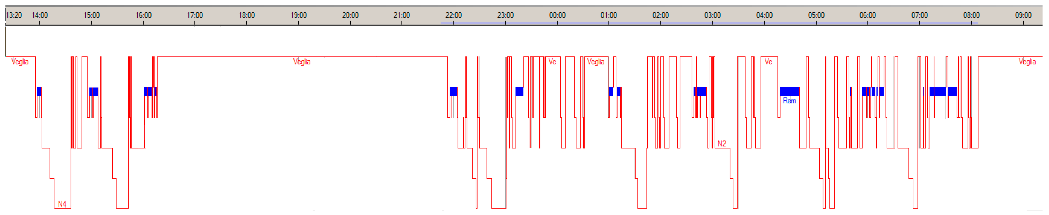
3.4. Disturbed Nighttime Sleep and Nocturnal Sleep Symptoms

DNS is a frequently overlooked symptom of NT1, despite being the third most disabling and affecting 53 to 78% of cases of pediatric NT1 [23]. DNS is characterized by frequent, often brief, nocturnal awakenings and poor perceived sleep quality. It can be an independent symptom or a consequence of other sleep-related features and comorbidities, including obstructive sleep apneas, periodic and non-periodic limb movement during sleep, sleep-related hallucinations, paralysis, and REM sleep behavior disorder (RBD) [25]. NT1 is the first cause of RBD (this occurs in up to 60% of patients) in the young population [26]. NT1-associated RBD differs from RBD in neurodegenerative diseases that are seen in adults and the elderly; it typically manifests with mild, simple motor behaviors distributed across all REM phases throughout the night and occasionally during daytime naps. More complex and energetic episodes are rarer, usually occurring in the second half of the night and not daily [26,27]. However, in children, RBD may be the first symptom of NT1 [28]. RBD in pediatric NT1 can also be severe and pervasive, also occurring during daytime SOREMPs and resembling automatic behaviors, possibly representing a form of “status dissociatus” (i.e., the co-occurrence of features of different behavioral states, which prevents the unambiguous recognition of the state itself) [27].

3.5. Sleep Paralysis

Both sleep paralysis, characterized by a temporary inability to move the body, speak, and breathe normally, and hypnagogic or hypnopompic hallucinations—auditory, tactile, visual, or multimodal—may represent abnormal expressions of dissociated REM sleep elements (i.e., muscle atonia and dream-like mental content, respectively) intruding into the transition between wakefulness and sleep. These symptoms, often co-occurring, can appear in the general population but are more intense and frequent in NT1, involving up to two-thirds of patients.
Its distinctive pediatric phenotype and the limited ability of children to effectively express symptoms contribute to the complexity of identifying NT1 during childhood. Cataplexy spells may be mistaken for ordinary falls, clumsiness, seizures, or neuromuscular disorders; partial attacks accompanied by hyperkinetic compensatory movement could suggest a choreic-dystonic disease [22]. EDS can be interpreted as laziness, depression, or ADHD, while hypnagogic hallucinations might be misinterpreted as nightmares, “night terrors”, or, in some cases, indicative of psychosis in children and adolescents [20].

3.6. Severity Assessment

The severity of symptoms in children with NT1 can be quantified in clinical practice and research by employing various questionnaires. The Narcolepsy Severity Scale (NSS) and the Ullanlinna Narcolepsy Scale (UNS) are the only disease-specific ones [29]. The UNS assesses daytime sleepiness and cataplexy through 11 items. It was developed in adults and not validated in the pediatric population, although it was employed as an outcome measure in pharmacological trials in children [30]. The NSS, instead, is a 15-item measure of the five core symptoms of NT1 that stratify it into severity classes [31], and it has been recently validated in the pediatric population as a modified 14-item version (NSS-P) [32]. More tools are available for only measuring EDS in children. The Epworth Sleepiness Scale (ESS) is the most widely used to assess trait EDS, with 8 items (with pathological scores > 11/24) [33]. An adapted version for children and adolescents (ESS-CHAD) has been validated [34]. A comprehensive review of the questionnaires to measure different aspects of pediatric narcolepsy was published by Ouyang et al. [29].

4. Pathogenic Mechanisms

4.1. The Hypocretin System

The acquired deficiency of hypocretin is widely accepted as the primary cause of NT1 in children as in adult cases [35]. Hypocretin is a neuropeptide produced by a small group of about 70,000 cells in the posterolateral hypothalamus that, upon interacting with other neurotransmitter pathways, promotes wakefulness, regulates energy intake, and stabilizes sleep [36]. The loss of hypocretin, due to damage to the producing neurons, is measurable in vivo in cerebrospinal fluid (CSF) [37], and it was demonstrated to induce the narcoleptic phenotype in animal models [38].
The disappearance of hypocretinergic cells that are widely projected to wake-promoting systems (including the locus coeruleus, dorsal raphe, tuberomammillary nucleus, basal forebrain, and pons) is probably the core mechanism of EDS and sleep–wake instability [39]. The hypothesized neural circuits underlying cataplexy are considered similar to an abnormal activation of muscle atonia typical of REM sleep during wakefulness, which is induced by the pontine sublaterodorsal nucleus. This nucleus is physiologically inhibited during wakefulness and non-REM sleep by the ventrolateral periaqueductal gray area and lateral pontine tegmentum, which receive excitatory signals from the hypothalamic hypocretinergic neurons and inhibitory projections from the central nucleus of the amygdala. Sudden emotions activate neurons in the medial prefrontal cortex, which stimulates hypothalamic hypocretin neurons and the central nucleus of the amygdala. In the absence of hypocretin, the unbalanced activation of the central nucleus of the amygdala would provoke an abnormal disinhibition of the sublaterodorsal nucleus and, thus, transient muscle atonia during wakefulness [40].
The causes and mechanisms of hypocretin loss have yet to be elucidated and involve multiple components that point to an autoimmune-mediated process.

4.2. Neuropathology

A reduction of about 90% of hypocretin-expressing cells in the lateral hypothalamus was reported in the human brains of a few individuals with NT1 [41,42,43,44]. No inflammatory infiltrates or neurodegenerative changes have ever been detected, although some studies have described localized gliosis [41,42].
Neuronal loss seems to be highly selective in hypocretinergic neurons. Melanin-concentrating hormone (MCH) neurons, closely intermixed with hypocretin neurons, are not reduced in NT1 [41]. Alongside the loss of hypocretin, an increase in the number of histaminergic cells, mainly in the tuberomammillary nucleus, has been described in humans and animals with narcolepsy [45,46]. Histamine is involved in wake promotion; thus, an increase in histaminergic neurons could be a compensatory mechanism for hypocretin loss. Some reports of lower levels of CSF histamine [47,48] and the effectiveness of a histaminergic agonist pitolisant in improving symptoms of NT1 could also suggest a dysfunction of the histaminergic transmission secondary to hypocretin deficiency [40].
An alternative hypothesis has been recently proposed by Seifinejad et al [49]. The study highlighted the persistence of the neuropeptide QRFP, which co-localizes on hypocretinergic neurons, in post-mortem hypothalamic tissues of patients with narcolepsy. Conversely, QRFP (Pyroglutamylated RFamide peptide) is absent in the brains of murine models with ablated hypocretin neurons. A hypermethylation of the promoter of the hypocretin gene was also described. The authors therefore suggested that the loss of hypocretin in NT1 could rely on an epigenetic silencing of the hypocretin gene rather than neuron destruction [50].

4.3. The Immune-Mediated Mechanism

Evidence suggests a genetic susceptibility to the disease, although familial cases are rare, and only 25–31% of monozygotic twins are concordant for narcolepsy [50]. Human leukocyte antigen (HLA) allele DQB1*06:02 of the major histocompatibility complex (MHC) class II, which is involved in presenting antigen to the T-cell receptor (TCR) of CD4+ cells, is recognized as the strongest genetic risk factor for NT1 across all ethnic groups. The HLA-DQB1*06:02 allele is present in 98% of NT1 vs. 5–38% in the general population, and its presence in homozygosis provides a 2-fold higher risk than in heterozygosis. Other HLA class II alleles and HLA class I alleles have shown weaker associations with NT1, while some other HLA class II variants, encoding for molecules of the DQ1 group, appear to have a protective role in individuals with DQB1*0602 heterozygosis [51,52]. Beyond HLA typing, genome-wide association studies identified polymorphisms of the TCRα region and of 12 other loci involved in the immune response (TRB, CTSH, IFNAR1, ZNF365, TNFSF4, CD207, NAB1, IKZF4–ERBB3, CTSC, DENND1B, SIRPG, and PRF1) associated with NT1 [53,54].
In this context of genetic predisposition, environmental triggering factors are likely to play a role.
The seasonal increase in the incidence of NT1 during spring, especially in children, suggests a link with upper airway infections. Elevated anti-streptolysin-O antibodies and anti-DNAase B antibodies in patients with narcolepsy close to the onset of the disease in comparison with controls suggest a streptococcal infection as a potential trigger [55]. In 2009–2010, a rise in NT1 cases was reported as being associated with the H1N1 influenza pandemic and with vaccination with the AS03-adjuvanted vaccine Pandemrix in HLA-DQB1 carriers. During the first year after vaccination, the relative risk of narcolepsy was increased 5 to 14-fold in children and adolescents [16].
Based on genetic susceptibility and temporal association with vaccination and infections, there is convincing proof of immune-mediated damage to hypocretinergic neurons in NT1. However, direct proof of an autoimmune process is lacking. Liblau et al. published a comprehensive review of the current evidence regarding immune-mediated mechanisms in NT1 [51]. Studies in animal models and on human blood samples strongly point to the existence of hypocretin-reactive CD4+ and CD8+ T cells in NT1 [51], possibly triggered by environmental factors through a process of cross-reactivity, superantigen activation, or bystander activation [56]. However, self-reactive T cells have never been detected in the CSF or in neuropathology studies, possibly due to a time-dependent variation in the autoimmune response. Moreover, it is not possible to rule out the notion that T cell reactivity is a consequence of the disease pathology instead of a primary mechanism [52].
Conversely, no autoantibodies having a diagnostic value or pathogenic significance have been discovered in NT1 so far. A few studies detected different autoantibodies, nonspecific to hypocretinergic neurons, in the serum of a minority of patients with narcolepsy but also in some controls. The only replicated results regard the detection of anti-Tribbles 2 (TRIB2) and anti-muscarinic receptor antibodies. None of these antibodies have yet been proven to induce a narcolepsy phenotype in animal models [51].
Overall, widely accepted evidence on the mechanisms behind hypocretin loss in NT1 is still lacking.

4.4. Secondary Narcolepsy in Children

Rare diseases need to be taken into consideration as possible causes of secondary narcolepsy.
Narcolepsy with cataplexy can appear in pediatric patients with intra-axial and extra-axial tumors involving the basal skull and the hypothalamus, often craniopharyngiomas [57,58], or after brain tumor resections [59]. Head trauma with axonal injury was also described as a cause of secondary narcolepsy [60]. EDS is frequently reported in children with Prader–Willi syndrome, a neurodevelopmental genetic disorder with hypothalamic dysfunction. MSLTs can be compatible with narcolepsy in up to 35% of patients with Prader–Willi syndrome, and some also present cataplexy (Figure 1). In these patients, HLA-DQB1*0602 is typically absent; the hypocretin level is lower than in healthy controls and higher than in NT1 [61].
Niemann–Pick type C is a multisystemic autosomal recessive lysosomal lipid storage disease that can manifest during childhood with the typical “gelastic cataplexy” prevailing on the EDS and variably reduced hypocretin [62]. Its identification is crucial as long as disease-modifying treatments (e.g., miglustat) are available [63].
Myotonic dystrophy type 1 (DM1), or Steinert’s disease, is a triplet repeat expansion disorder that encompasses EDS in about 50% of pediatric cases. EDS is associated with prolonged, unrefreshing episodes of daytime and nighttime sleep, and it can be objectified by a pathological MSLT with possible multiple SOREMPs. In DM1, EDS is not explained by the highly prevalent sleep-disordered breathing but by nocturnal sleep fragmentation [64]. Deficient and intermediate hypocretin levels were reported in DM1 but without cataplexy [65]. Other genetic syndromes where cataplexy-like episodes have been described are the autosomal dominant Moebius syndrome, the X-linked recessive Norrie syndrome, and the X-linked dominant Coffin–Lowry syndrome [66,67,68].
Among SNC inflammatory diseases, multiple sclerosis, neuromyelitis optica spectrum disorders, and, especially in children, acute disseminated encephalomyelitis can provoke hypersomnia with features of narcolepsy, sometimes with cataplexy, and typically with demyelinating lesions involving the hypothalamus [69,70,71] Narcolepsy and reduced hypocretin were reported to normalize after immunomodulant treatment [72]. Autoimmune and paraneoplastic encephalitis are other rare causes of symptomatic narcolepsy with cataplexy [73]. In children and adolescents, cases have been reported in association with neuroblastoma [74], thymic seminoma [74], and rapid-onset obesity with hypothalamic dysfunction, hypoventilation, and autonomic dysregulation [75].

5. Comorbidities

5.1. Endocrinological Aspects

The endocrinological aspects of obesity, precocious puberty, and type 2 diabetes are well-known features of childhood narcolepsy [76]. In the literature, obesity rates have ranged from 25% to 75% of children. Weight gain develops soon after the onset of symptoms [77]. Furthermore, several studies have shown that the early onset of NT1 is associated with a higher grade of obesity and a greater severity of disease [5,24]. Poli and colleagues showed that younger age onset of NT1 was a predictor for obesity but also for precocious puberty (PP), suggesting that a hypothalamic dysfunction underlies this mechanism [78]. The association between NT1 and PP was assessed systematically for the first time in 2013 [79], although it had been anecdotally mentioned in previous reports [80,81]. The opposite association has also been documented in the precocious puberty population [82].
The mechanism that underlies this association has been hypothesized to be related to the role of the hypocretin system, which is not only involved in sleep/wake regulation but also in modulating feeding behaviors [83]. Recently, a case report described a possible association between NT1 and growth hormone (GH) deficiency [84].
Metabolic syndrome (MS), which is a constellation of disturbances associated with obesity, is frequently diagnosed in individuals with NT1 [85]. Metabolic alterations still persist after adjustment of the body mass index (BMI), suggesting a possible role of hypocretin deficiency in these disorders [86]. However, the presence of MS in children is low compared to adults with NT1; the higher prevalence of MS in adults could possibly be explained by the increase in MS prevalence according to age, as observed in the general population and explained by organismal aging [87]. Studies on the general population have shown that insulin sensitivity was negatively associated with triglycerides and positively associated with HDL-C in children with MS, suggesting that insulin resistance mediates lipid metabolism independently of body fatness and even after adjusting the BMI [88]. Furthermore, insulin sensitivity was reported to be negatively associated with systolic and diastolic blood pressure in children [89].

5.2. Psychiatric Disorders

Children with NT1 can develop various psychiatric disorders. The neuropsychiatric comorbidity that has been demonstrated in children with NT1 includes attention-deficit/hyperactivity disorder (ADHD) (29%), mood disorders (20%), anxiety disorders (10%), oppositional defiant disorder (7%), and pervasive developmental disorders not otherwise specified (3%) [90]. Despite a few studies, the prevalence of autism among patients with narcolepsy is unknown [91].
Emotional problems with depression and social difficulties were reported in 44% and 66% of children with narcolepsy [92]. In adult patients, the frequency of depressive symptoms ranged from 15% to 56.9% and had a large impact on the quality of life of these patients [93]. However, the incidence of depressive disorders related to narcoleptic patients remains unsettled [94]. Vourdas et al. found that 16% of adult patients with narcolepsy and 18% of the control group had one or more episodes that met the DSM-IV criteria for major depression [95]. Likewise, Fortuyn and colleagues reported that 7% of narcoleptic patients had a current major depressive episode, with 3% of the control group reporting the same [96].
A high level of anxiety, both general and specific, and, in particular, social phobia and panic attacks were reported in children with narcolepsy [97], a finding that parallels the association disclosed in adults [98].
Hallucinations are a common feature of both narcolepsy and schizophrenia. However, hallucinations have strong differences in narcolepsy, being mostly sleep-related, vs. those of schizophrenia. The co-occurrence of narcolepsy and schizophrenia was reported in the literature, with affected individuals reporting a poorer response to antipsychotic treatment [99,100]. The presence of a block of thought, insertion, and delusions represent a red flag that suggests comorbidity with schizophrenia. According to a retrospective study, the frequency of schizophrenia was 1.8%, typically developing within 3 years after the onset of narcolepsy [101]. A study investigating a large cohort reported that 9.8% of narcoleptic children developed schizophrenia after the onset of narcolepsy but persisted even after stopping psychostimulants [102]. It is unknown whether hypocretin deficiency and NT1 symptoms affecting very young patients may lead to neuropsychiatric disorders such as schizophrenia or be intrinsically part of an underlying neurodevelopment disorder [103].
Attention-deficit/hyperactivity disorder (ADHD) and oppositional defiant disorder (ODD) are common comorbidities in children with NT1 [104]. Due to the deficits of alertness and sleep disturbances that have been hypothesized to contribute to ADHD, comorbid ADHD symptoms are likely to be found in young people with narcolepsy [105]. Some retrospective reports in adults with narcolepsy have identified frequent ADHD symptoms in childhood [106,107]. Given these findings, there may be common underlying pathophysiological mechanisms linking narcolepsy and ADHD, such that treatment for one condition may improve and/or affect the other, especially since treatments for narcolepsy, such as modafinil and methylphenidate, are also used in children with ADHD [108].
Studies investigating the presence of eating disorders (EDs) in adult patients with NT1 produced contradictory results. Dahmen and colleagues did not find an increased prevalence of EDs in patients with NT1 compared with the general population [109]. Conversely, a recent cross-sectional study with a large sample of NT1 individuals found that patients with NT1 reported higher scores on the Eating Disorder Examination Questionnaire (EDE-Q) and the Italian Night Eating Questionnaire (I-NEQ) [110]. To identify the possible presence of EDs in patients with NT1, a multidisciplinary team with a psychiatry specialist, dietician, and nutritionist is necessary. Future research is needed to assess the presence of EDs in the pediatric population.

5.3. Cognitive Aspects

Children with NT1 showed poorer cognitive performances in attention, vigilance, executive functions, and decision making than the controls [111]. An impairment in working memory and verbal comprehension was also found; however, a specific cognitive profile was not identified [112]. Moreover, the co-occurrence of psychiatric disorders seems to be a risk for poorer cognitive functions [113]. A functional brain image study in young patients with NT1 revealed an alteration in brain metabolic activities; compared to controls, patients had significant hypometabolism in different frontal lobe areas, correlating with poor neurocognitive testing results and major sleepiness [114]. Mazzetti and colleagues highlighted the role of napping for the enhancement of memory [115].

6. Diagnosis

6.1. Diagnostic Criteria

A diagnosis of NT1 is based on the criteria of the International Classification of Sleep Disorders (ICSD), which does not differentiate between adult and pediatric patients.
According to the third edition of the ICSD (ICSD-3), in 2014, NT1 was defined by a complaint of EDS for at least three months and either hypocretin < 110 pg/mL or a combination of the presence of clear cataplexy and multiple sleep latency test (MSLT) scores with a mean sleep latency ≤ 8 min and ≥2 naps, with an occurrence of REM sleep ≤ 15 min from the onset of sleep (defined as a sleep-onset REM period, SOREMP); an SOREMP on a preceding nocturnal polysomnogram may replace one of the SOREMPs on the MSLT [1].
In 2023, a text revision of the ICSD-3 (ICSD-3-TR) codified the recording of an SOREMP on a nocturnal PSG combined with the presence of cataplexy as sufficient for a diagnosis of NT1, regardless of the MSLT result and hypocretin deficiency [1]. This modification is derived from the evidence that the nocturnal SOREMP provides high specificity (with low-to-moderate sensitivity) for hypocretin deficiency [116] (Table 1).

6.2. Diagnostic Tools in Pediatric NT1

Conventional and alternative diagnostic tools have been investigated in the pediatric population.
Nocturnal SOREMPs, which are highly specific (97.3%) and modestly sensitive (54.8%) for narcolepsy with cataplexy, were replicated in children before the introduction of hypocretin dosage as a diagnostic criterion [117].
Concerns about the applicability of the standardized MSLT in children exist. However, the test has been validated for pediatric narcolepsy by the Sleep Centers of Bologna and Montpellier, and alternative MSLT cutoffs for NT1 for patients < 18 years old have been proposed. Specifically, either at least two SOREMPs or a mean sleep latency ≤ 8.2 min have demonstrated diagnostic accuracy for NT1 comparable to standard MSLT criteria [118].
Besides nocturnal SOREMP, other PSG features have been studied as possible diagnostic markers.
In particular, sleep fragmentation with heightened sleep stage transitions characterizes NT1 in comparison to other CDHs [119]. This feature was applied to a pediatric cohort of 316 individuals suspected of a CDH. An increased index of transitions from any sleep stage to non-REM sleep stage 1 or wake resulted in good accuracy for identifying NT1, significantly enhanced by the combination with the night SOREMP [120].
Recently, continuous ambulatory daytime PSG has shown promising diagnostic utility. The detection of at least one daytime spontaneous SOREMP resulted in evaluations as accurate as MSLT criteria for pediatric NT1 [121]. REM sleep without atonia (RSWA) is the loss of physiological muscle atonia during REM sleep and is a neurophysiological marker for RBD. RSWA has also been investigated in narcolepsy, employing various scoring methods, both automatic and visual. Applied to a pediatric context, visual RSWA showed high sensitivity and moderate specificity for narcolepsy [122]. Silvani et al. integrated the automatic REM atonia index with REM sleep latency in night PSG with a non-linear function, demonstrating that this combination significantly improved specificity (91.7%) without compromising sensitivity (85.4%) for cases of pediatric NT1 [123].
Actigraphy, a quantitative assessment of motor activity and an indirect measurement of sleep time based on a wearable device on the wrist, can be adopted for seven to ten days before PSG-MSLT execution to objectively exclude sleep deprivation or circadian rhythm disorders possibly impairing MSLT results [123]. Additionally, a study described the good discriminatory capability of an actigraphy-recorded altered rest–activity rhythm with enhanced nocturnal motor activity and diminished activity in the first afternoon of children with NT1 [124].
The measurement of hypocretin deficiency is a fundamental exam for confirming NT1, also in children [125]. Hypocretin levels have been demonstrated to remain stable from early infancy up to old age [126]. Reports of decreased hypocretin levels at the onset of NT1 exist [127]. It must be acknowledged that lumbar puncture in children can be problematic and may need the support of sedation [128].
The Pediatric Narcolepsy Screening Questionnaire (PNSQ) is a recently proposed 11-item questionnaire administered to the parents of children with a suspected CDH to investigate the presence of symptoms suggestive of narcolepsy (including those typical of NT1), and it showed good discriminatory power against other sleep disorders and healthy controls [129].
A significant challenge persists in the form of under-diagnosis and delayed diagnosis of narcolepsy in adult and pediatric cases, with a median duration from the initiation of symptoms to recognition surpassing 10 years [130]. This prolonged diagnostic delay was underscored by a recent investigation conducted by the European Narcolepsy Network, revealing a mean diagnostic delay of almost 10 years across various European countries, with no significant improvement since the 1990s. Notably, this delay was found to be more pronounced in children and adolescents, possibly due to a misinterpretation of symptoms in pediatric cases, and in females, postulated to be linked to gender-related social disparities. Factors such as the infrequent occurrence and lower frequency of cataplexy, as well as less severe symptoms, were identified as positive predictors of this extended diagnostic delay [131].

6.3. Differential Diagnosis

The obvious prevailing differential diagnoses for NT1 encompass NT2 (formerly known as narcolepsy without cataplexy) and idiopathic hypersomnia (IH), both of which are less common. NT2 is diagnosed when an MSLT shows reduced mean sleep latency; at least two SOREMPs (including the one on the preceding night PSG) similar to NT1, but without cataplexy; and hypocretin > 110 pg/mL, if measured. A diagnosis of IH requires either a reduced mean sleep latency below 8 min, with no more than one SOREMP, or a prolonged total sleep time above 11 h throughout 24 h [1]. Remitting periods of variable durations of hypersomnolence, associated with contextual disinhibition, derealization, apathy, and cognitive dysfunction in children and adolescents, suggest Kleine–Levin syndrome [132]. EDS may also manifest in the context of hypersomnia associated with medical and psychiatric conditions, medication or substance abuse, behaviorally induced insufficient sleep syndrome (ISS), circadian rhythm disorders, and other sleep disorders characterized by a compromised quality of nocturnal sleep [1]. Among the latter, obstructive sleep apnea syndrome (OSAS) and periodic limb movement disorder (PLMD) are the most frequently encountered and are often comorbid in NT1 [133,134]. To help rule out other conditions presenting with EDS, careful history taking is needed that would be especially focused on sleep–wake features, a night PSG, and a standardized attended MSLT, preceded by suspension of REM-suppressing, stimulant, and sedative drugs and possibly by prolonged actigraphic monitoring.

6.4. Neuroimaging

Brain magnetic resonance imaging (MRI) is required to rule out lesions responsible for secondary narcolepsy.
While routine brain MRI in NT1 does not disclose any relevant alterations to clinical practice, multiple neuroimaging studies, mainly based on adult cohorts, have been published.
A systematic review of these studies was published by Wada et al. [135], where the replicated results in NT1 were the following. Various morphological MRI studies documented reductions in the superior frontal gyri, superior and inferior temporal gyri, middle occipital gyri, hypothalamus, amygdala, insula, hippocampus, cingulate cortex, thalamus, and nucleus accumbens. Three studies with diffusion tensor imaging showed decreased fractional anisotropy (i.e., a measure reflecting reduced myelin fiber integrity) in the white matter of the frontal-orbital and cingulate area. Reduced brain metabolism in the middle and left superior frontal gyri and in the cingulate cortex were replicated in PET and SPECT studies. Two studies of functional MRI (fMRI) detected increased activity in the inferior frontal gyri, insula, amygdala, and nucleus accumbens. Two studies employing proton magnetic resonance spectroscopy (1H MRS) showed lowered N-acetyl-aspartate/creatine-phosphocreatine levels in the hypothalamus [136].
A few research studies have recently evaluated advanced neuroimaging in children and adolescents with NT1. Compared with healthy controls, analysis of morphological MRI in drug-naïve children and adolescents showed reduced gray matter in the cerebellum and medial prefrontal cortex and reduced cortical thickness in the frontal lobe, in line with previous evidence. In contrast with results in adults that showed reduced volume in the hippocampus, the study also documented increased volume and shape expansion in the right hippocampus, hypothesized to reflect a compensatory mechanism to maintain cognitive performances [137]. Neuroimaging data on children with NT1 consistently align with the cognitive deficits identified in other studies of this population. Imaging studies reveal structural and functional brain abnormalities, particularly in areas associated with memory, attention, and executive function, which corroborate the cognitive impairments observed clinically. These findings provide a robust neurobiological basis for the cognitive challenges experienced by children with NT1, highlighting the importance of addressing these deficits in both diagnostic and therapeutic strategies. This convergence of neuroimaging evidence with clinical observations underscores the critical need for comprehensive cognitive assessments and targeted interventions to support the cognitive development of affected children.
Meletti et al., provoking cataplexy during fMRI in pediatric patients with NT1, observed activation of the anterior insular cortex, the nucleus accumbens, and the amygdala, as well as in the locus coeruleus and the anteromedial pons during emotion-induced cataplexy but not during laughter alone, confirming the hypothesis of the involvement of the cortico-limbic network in cataplexy [138]. The same group assessed resting-state fMRI in drug-naïve pediatric patients with NT1 compared to healthy controls and found decreased functional connectivity between the lateral hypothalamus and the left superior parietal lobule, the hippocampus and the parahippocampal gyrus, the amygdala and the post-central gyrus, and several occipital regions, finding increased connectivity between the amygdala and the inferior frontal gyrus, claustrum, insula, and putamen as well. These results could suggest dysfunctional interactions between regions subserving the maintenance of arousal, memory, and emotional processing [139]. Quantitative brain MRI in adolescents with NT1 showed reduced R2 relaxation rates in the rostral reticular formation near the superior cerebellar peduncle, which suggested to the authors a lower content of neuromelanin in the proximity of the locus coeruleus, an area functionally connected with orexin signaling [140].

7. Treatment

In 2021, guidelines for the treatment of narcolepsy in adults and children were published by both the European Academy of Neurology [141] and the American Academy of Sleep Medicine [142]. Available treatments for NT1 in children are symptomatic and include non-pharmacological and pharmacological approaches. In some situations where medication is considered unsuitable, such as in pregnancy or potentially in early childhood, non-pharmacological management approaches are mandatory [141]. No effective curative therapy exists for NT1.

7.1. Non-Pharmacological Approaches

Behavioral therapy is recommended for both patients treated with medication and untreated patients.
Children with NT1 should have adequate sleep hygiene and a regular sleep/wake schedule, avoiding staying late and limiting screen time before bed.
Planned daytime naps, possibly brief (about 15–20 min each time), are a necessary component of behavioral therapy of NT1, as short naps in narcolepsy are typically refreshing [141]. A comprehensive understanding of narcolepsy, spanning disease mechanisms and treatments, is crucial for patients and their caregivers. A healthy lifestyle and physical activity can promote symptom control and patient empowerment [143]. Joining patient organizations facilitates information exchange and support.

7.2. Pharmacological Symptomatic Approaches

Pharmacological treatment is crucial for managing EDS, cataplexy, and DNS. Available medications for narcolepsy can be schematically classified as (i) wake-promoting agents that include CNS stimulants (modafinil, armodafinil, solriamfetol, methylphenidate, and amphetamine derivates) and pitolisant (which is also anti-cataplectic), (ii) oxibates, and (iii) antidepressants [141].
Under the age of six, there is no pharmacological treatment approved by the main regulatory agency of the drug. The use of sodium oxybate for NT1 in patients seven years old or older is approved by the Food and Drug Administration (FDA) and the European Medicines Agency (EMA). In 2023, the EMA also granted approval for the marketing authorization of pitolisant for use in patients older than six years old with narcolepsy with and without cataplexy. For the treatment of EDS in adults with narcolepsy, modafinil and solriamfetol are approved by the EMA and FDA, and armodafinil and amphetamine derivates are only approved by the FDA. Antidepressants are considered off-label for the treatment of cataplexy in all ages.
As there are no comparative studies of treatment efficacy, the choice between first- and second-line approaches depends on individual factors, including the patient’s needs, comorbidities, and severity of different symptoms. Evidence-based differentiations are made considering clinical trials and regulatory approval. The treatment of NT1 is guided by symptomatology (Figure 2), and an overview of medication suggested for pediatric NT1 is summarized in Table 2. It is recommended to start with monotherapy, at the lowest effective dose, possibly chosen to act on all the invalidating symptoms of patients. In cases of incomplete benefit at the maximum tolerated dose, the add-on of one or more medications is possible, avoiding a combination of dopaminergic stimulants [141,142].
Modafinil is a central dopamine re-uptake inhibitor that promotes wakefulness through hypothalamic pathways [145]. Prescribed at an initial dose of 100 mg and gradually titrated up to 400 mg/day in two doses (typically at morning awakening and at lunchtime), modafinil improves subjective and objective EDS. Retrospective data showed modafinil as effective and safe in children with narcolepsy [146]. Armodafinil is the R-enantiomer of modafinil with a longer half-life. It is given in the morning as a single dose of 100–250 mg/day.
More recently introduced, solriamfetol is a selective inhibitor of the reuptake of noradrenaline and dopamine that acts on dopamine and noradrenaline transporters. It was proven to be safe and effective in improving subjective and objective EDS in adults with narcolepsy, at doses of 75-to-150 mg/day via one or two doses in the morning [147]. The side effects of modafinil and solriamfetol are similar and include headache, nausea, decreased appetite, nasopharyngitis, dry mouth, insomnia, irritability, and anxiety. Both medications show a low addiction potential [146,147,148]. Caution should be used in cases of cardiovascular morbidity, although the issue is more relevant in the adult population than in pediatric patients [149].
Methylphenidate is an inhibitor of the reuptake of noradrenaline and dopamine, which stimulate cortical and subcortical brain structures. Doses for EDS in narcolepsy vary from 5 to 60 mg/day in 2–3 portions [146].
Amphetamine derivates (amphetamine–dextroamphetamine and amphetamine sulfate) are alternative options for the treatment of EDS in narcolepsy, also enhancing dopaminergic and noradrenergic transmission. Tachycardia, hypertension, sweating, palpitations, irritability, mood swings, weight loss, anorexia, and insomnia are among the common side effects of methylphenidate and amphetamines. Mild risks of abuse and dependence exist, and cardiovascular risk should be considered [141,146,150].
Pitolisant is a selective competitive antagonist/inverse agonist of the pre-synaptic H3-receptor that is considered to promote wakefulness, enhancing CNS histamine release. It is titrated over several weeks from 4.5 mg to a maximum of 18 mg/day (if the child’s weight is up to 40 Kg) or 36 given once daily at morning awakening. In pediatric patients with NT1, as in adults, pitolisant was proven effective in reducing EDS and the frequency of cataplexy, both in a randomized controlled trial and in a real-world study [151,152]. Side effects are usually mild, including insomnia, headache, and gastrointestinal discomfort [30].
Sodium oxybate is a gamma-hydroxybutyric acid B-subtype (GABAb) receptor agonist that strongly promotes slow-wave sleep, suppresses REM sleep, and improves sleep continuity. It is administered in two doses, one at nocturnal bedtime and the second 2.5–4 h later when the sedative effect fades. In children, the initial dose is usually 2 g/day (divided in two) and can be gradually titrated up to 8 g/day (in adults, the maximum dose is 9 g). In pediatric patients with NT1, as in adults, sodium oxybate was demonstrated to reduce EDS and cataplexy. Moreover, it is the only available treatment effective in improving DNS in NT1 [153,154,155,156,157].
Nausea, vomiting, dizziness, disorders of arousal, enuresis, and confusion are common symptoms in the hours after drug assumption, and thus, its administration in pediatric patients needs to be controlled by a trained caregiver. Pulmonary comorbidity and OSA should be ruled out or treated before starting sodium oxybate, as it can depress respiratory drive. Weight loss (which can also be beneficial for the obesity associated with NT1), hypertension, hypernatremia, mood disturbances, and other psychiatric problems are the chronic side effects to be monitored [155,157]. To reduce the cardiovascular morbidity related to the increased intake of sodium with sodium oxybate, an alternative lower-sodium oxybate (with calcium, magnesium, potassium, and sodium) was developed and was demonstrated equivalent to sodium oxybate to reduce EDS and cataplexy in adults [158]. Therefore, it was recently approved by the FDA as a treatment for NT1 in patients older than seven years old [159]. As the forced awakening in the twice-nightly dosing regimen of sodium oxybate can create distress to the patient, an extended-release sodium oxybate, administered only once nightly at bedtime, was developed and showed efficacy in improving EDS and cataplexy in patients 16 years old or older with NT1 [160].
Antidepressant medications in NT1 are employed as anti-cataplectic therapy without a significant stimulant effect. Venlafaxine, a selective serotonin–norepinephrine reuptake inhibitor, is the most widely prescribed, at doses ranging from 37.5 mg to 225 mg/day (typically, a benefit already appears at low doses). Selective serotonin reuptake inhibitors and clomipramine, a tricyclic antidepressant, can also be administered as anti-cataplectic therapy. Dizziness, dry mouth, headache, irritability, and weight changes are the most common side effects [146]. The rapid suspension of antidepressants can induce a rebound of prolonged or sub-continuous invalidating cataplexy, known as status cataplecticus [161].
Recently, a phase 2 randomized controlled trial (RCT) of TAK-994, a novel oral orexin receptor 2 selective agonist, was conducted in adult patients with NT1. This new medication provided greater improvements in measurements of sleepiness and cataplexy than the placebo. However, the RCT was terminated early due to hepatotoxic effects on 5 out of the 73 included subjects [162]. It is likely that, in the near future, orexinergic agonists will be further developed and proposed in the pediatric population too.

7.3. Immunomodulatory Treatment

A trial of immunomodulatory treatments was based on the hypothesis of the autoimmune destruction of hypocretin neurons. Immunotherapy administration was described in several case reports and case series of patients, mainly pediatric, close to the onset of the disease. Most of the studies reported the use of intravenous immunoglobulin (IVIg) or steroids with various schemes, and some were accompanied by a transient improvement in subjective symptoms, such as ESS or cataplexy, without any objective effect. Lecendreux et al. [163] conducted a non-randomized, open-label, controlled, longitudinal, observational study of IVIg use (dose of 1 g/kg as three separate infusions at monthly intervals) added to standard care in 24 pediatric patients compared with 32 pediatric patients who continued standard care. IVIg administration was not associated with a reduction in symptoms. In a subgroup of patients with high UNS scores, those treated with IVIg achieved symptom remission more rapidly than those on standard care [164]. No RCT has been conducted with immunotherapy on NT1. Therefore, no disease-modifying therapy for narcolepsy is recommended [141].

8. Management and Treatment of Comorbidities

8.1. Endocrinological Comorbidities

To manage precocious puberty, the agonist analogs of gonadotropin-releasing hormones (GnRH) can be used as the first line of treatment. Once started, the effects of therapy should be monitored by a follow-up every 6 months. An increase and subsequent decrease in fat mass at the start of the treatment have been reported, but GnRH analogs do not appear to have a negative impact on the BMI [165]. For obesity, physical activities and a well-balanced intake of nutrients are strongly recommended. Moreover, stimulants and sodium oxybate might help to reduce weight [165,166,167].

8.2. Psychiatric Comorbidities

Depression and anxiety can be managed with psychological support and antidepressant medications, e.g., FDA-approved antidepressants for children and adolescents, such as fluoxetine [168]. It is essential to monitor the suicidal risk after the prescription of an antidepressant drug, which can be higher in the first period of treatment [169]. Specific recommendations for children with narcolepsy are not available. For the co-occurrence of psychosis in narcolepsy, aripiprazole is often prescribed first-line due to its less sedative side effects. Antipsychotic drugs such as risperidone or olanzapine can also be prescribed; however, they could worsen EDS symptoms.

8.3. Behavioral and Psychological Difficulties

Psychosocial functioning can be severely compromised in children with NT1. Cognitive and behavioral treatment has been poorly investigated, and no systematic studies are based on this approach. However, cognitive therapy is recommended for children [170]

9. Conclusions and Future Directions

Diagnosing narcolepsy in children can be difficult due to insufficient knowledge and under-recognition, especially because its presentation may differ from adult patients. EDS in children may manifest as irritability and poor attention, which may be wrongly interpreted as misbehavior. The treatment of pediatric narcolepsy should involve medication, behavior modification, and mental support. Indeed, psychosocial interventions such as behavior modification have been shown to benefit patients and their families; prospective long-term follow-up is necessary to evaluate the prognosis of outcomes of children with narcolepsy. Longitudinal studies are essential to comprehend the long-term progression and psychosocial impact of the disease on children and adolescents. Additionally, there is a need for more comprehensive studies on the efficacy and safety of emerging treatment options, including immunotherapy and novel pharmacological agents. Investigating the role of environmental factors and their interplay with genetic susceptibility could also provide insights into prevention strategies. Furthermore, enhancing diagnostic criteria and developing more accessible, non-invasive diagnostic tools will be crucial in facilitating early diagnosis and intervention, ultimately improving the quality of life for pediatric patients.

Author Contributions

Conceptualization: G.P. and F.P.; Writing-original draft preparation: V.B., F.B. and G.V.; Supervision: G.P. and F.P. All authors have read and agreed to the published version of the manuscript.

Funding

This research received no external funding.

Data Availability Statement

Not applicable.

Conflicts of Interest

G.P. has received honoraria for advisory board and consulting fees from Bio- projet, Jazz, Takeda, and Idorsia. F.P. as received honoraria for presentations from Jazz Phar- maceuticals, for participation in the advisory board by Tadeka, and meeting attendance support from Bioprojet.

References

  1. American Academy of Sleep Medicine. International Classification of Sleep Disorders, 3rd ed. Text Revision (ICSD-3-TR). 2023. Available online: https://aasm.org/clinical-resources/international-classification-sleep-disorders/ (accessed on 15 August 2023).
  2. Partinen, M.; Kornum, B.R.; Plazzi, G.; Jennum, P.; Julkunen, I.; Vaarala, O. Narcolepsy as an autoimmune disease: The role of H1N1 infection and vaccination. Lancet Neurol. 2014, 13, 600–613. [Google Scholar] [CrossRef] [PubMed]
  3. Wang, X.; Xiao, F.; Wang, Y.; Deng, X.; Chen, Z.; Dong, X.; Wang, W.; Li, C.; Xu, Z.; Wu, H.; et al. Changed epidemiology of narcolepsy before, during and after the 2009 H1N1 pandemic: A nationwide narcolepsy surveillance network study in mainland China, 1990–2017. Sleep 2023, 9, 46. [Google Scholar] [CrossRef] [PubMed]
  4. Zhang, Z.; Gool, J.K.; Fronczek, R.; Dauvilliers, Y.; Bassetti, C.L.A.; Mayer, G.; Plazzi, G.; Pizza, F.; Santamaria, J.; Partinen, M.; et al. New 2013 incidence peak in childhood narcolepsy: More than vaccination? Sleep 2021, 12, 44. [Google Scholar] [CrossRef] [PubMed]
  5. Nevsimalova, S. Narcolepsy in childhood. Sleep Med. Rev. 2009, 13, 169–180. [Google Scholar] [CrossRef] [PubMed]
  6. Vignatelli, L.; Antelmi, E.; Ceretelli, I.; Bellini, M.; Carta, C.; Cortelli, P.; Ferini-Strambi, L.; Ferri, R.; Guerrini, R.; Ingravallo, F.; et al. Red Flags for early referral of people with symptoms suggestive of narcolepsy: A report from a national multidisciplinary panel. Neurol. Sci. 2019, 40, 447–456. [Google Scholar] [CrossRef]
  7. Honda, Y. Census of narcolepsy, cataplexy and sleep life among teenagers in Fujisawa City. Sleep Res. 1979, 8, 191. [Google Scholar]
  8. Wilner, A.; Steinman, L.; Lavie, P.; Peled, R.; Friedmann, A.; Brautbar, C. Narcolepsy-cataplexy in Israeli Jews is associated exclusively with the HLA DR2 haplotype. A study at the serological and genomic level. Hum. Immunol. 1988, 21, 15–22. [Google Scholar] [CrossRef] [PubMed]
  9. Koepsell, T.D.; Longstreth, W.T.; Ton, T.G. Medical exposures in youth and the frequency of narcolepsy with cataplexy: A population-based case-control studies in genetically predisposed people. J. Sleep Res. 2010, 19, 80–86. [Google Scholar] [CrossRef]
  10. Silber, M.H.; Krahn, L.E.; Olson, E.J.; Pankratz, V.S. The epidemiology of narcolepsy in Olmsted County, Minnesota: A population-based study. Sleep 2002, 25, 197–202. [Google Scholar] [CrossRef]
  11. Ohayon, M.M.; Priest, R.G.; Zulley, G.; Smirne, S.; Paiva, T. Prevalence of narcolepsy symptomatology and diagnosis in the European general population. Neurology 2002, 58, 1826–1856. [Google Scholar] [CrossRef]
  12. Dauvilliers, Y.; Montplaisir, J.; Molinari, N.; Carlander, B.; Ondze, B.; Besset, A.; Billiard, M. Age at onset of narcolepsy in two large populations of patients in France and Quebec. Neurology 2001, 57, 2029–2033. [Google Scholar] [CrossRef] [PubMed]
  13. Wu, H.; Zhuang, J.; Stone, W.S.; Zhang, L.; Zhao, Z.; Wang, Z.; Yang, Y.; Li, X.; Zhao, X.; Zhao, Z. Symptoms and occurences of narcolepsy: A retrospective study of 162 patients during a 10-year period in Eastern China. Sleep Med. 2014, 15, 607–613. [Google Scholar] [CrossRef] [PubMed]
  14. Han, F.; Lin, L.; Warby, S.C.; Faraco, J.; Li, J.; Dong, S.J.; An, P.; Zhao, L.; Wang, L.; Li, Q.Y.; et al. Narcolepsy onset is seasonal and increased following the 2009 H1N1 pandemic in China. Ann. Neurol. 2011, 70, 410–417. [Google Scholar] [CrossRef] [PubMed]
  15. Sarkanen, T.; Alakuijala, A.P.E.; Julkunen, I.; Partinen, M. Narcolepsy Associated with Pandemrix Vaccine. Curr. Neurol. Neurosci. Rep. 2018, 18, 43. [Google Scholar] [CrossRef] [PubMed]
  16. Sarkanen, T.O.; Alakuijala, A.P.E.; Dauvilliers, Y.; Partinen, M. Incidence of narcolepsy after H1N1 influenza and vaccinations: Systematic review and meta-analysis. Sleep Med. Rev. 2018, 38, 177–186. [Google Scholar] [CrossRef] [PubMed]
  17. Gauffin, H.; Bostròm, I.; Bertnsson, S.G.; Kristoffersson, A.; Fredrikson, M.; Landtblom, A.M. Characterization of the increase in Narcolepsy following the 2009 H1N1 Pandemic in Sweden. J. Clin. Med. 2024, 23, 652. [Google Scholar] [CrossRef] [PubMed]
  18. Hovi, M.; Heiskala, H.; Aronen, E.T.; Saarenpàà-Heikkilà, O.; Olsen, P.; Nokelainen, P.; Kirjavainen, T. Finnish children who experienced narcolepsy after receiving the Pandemrix vaccine during the 2009-2010 H1N1 pandemic demonstrated high level of psychosocial problems. Acta Paediatr. 2022, 111, 850–858. [Google Scholar] [CrossRef] [PubMed]
  19. Wijans, L.; Lecomte, C.; De Vries, C.; Weibel, D.; Sammon, C.; Hviid, A.; Svanstrom, H.; Nielsen, D.; Haijbel, H.; Dahlstrom, L.A.; et al. The incidence of narcolepsy in Europe: Before, during, and after the influenza A (H1N1) pdm09 pandemic and vaccination campaigns. Vaccine 2013, 31, 1246–1254. [Google Scholar] [CrossRef] [PubMed]
  20. Plazzi, G.; Clawges, H.M.; Owens, J.A.; Virginia, W. Pediatric Neurology Clinical Characteristics and Burden of Illness in Pediatric Patients with Narcolepsy. Pediatr. Neurol. 2018, 85, 21–32. [Google Scholar] [CrossRef]
  21. Serra, L.; Montagna, P.; Mignot, E.; Lugaresi, E.; Plazzi, G. Cataplexy features in childhood narcolepsy. J. Mov. Disord. 2008, 23, 858–865. [Google Scholar] [CrossRef]
  22. Plazzi, G.; Pizza, F.; Palaia, V.; Franceschini, C.; Poli, F.; Moghadam, K.K.; Cortelli, P.; Nobili, L.; Bruni, O.; Dauvilliers, Y.; et al. Complex movement disorders at disease onset in childhood narcolepsy with cataplexy. Brain 2011, 134, 3477–3489. [Google Scholar] [CrossRef] [PubMed]
  23. Pizza, F.; Franceschini, C.; Peltola, H.; Vandi, S.; Finotti, E.; Ingravallo, F.; Nobili, L.; Bruni, O.; Lin, L.; Edwards, M.J.; et al. Clinical and polysomnographic course of childhood narcolepsy with cataplexy. Brain 2013, 136, 3787–3795. [Google Scholar] [CrossRef] [PubMed]
  24. Postiglione, E.; Barateau, L.; Pizza, F.; Lopez, R.; Antelmi, E.; Rassu, A.L.; Vandi, S.; Chenini, S.; Mignot, E.; Dauvilliers, Y.; et al. Narcolepsy with intermediate cerebrospinal level of hypocretin-1. Sleep 2022, 45, zsab285. [Google Scholar] [CrossRef]
  25. Maski, K.; Mignot, E.; Plazzi, G.; Dauvilliers, Y. Disrupted nighttime sleep and sleep instability in narcolepsy. J. Clin. Sleep Med. 2022, 18, 289–304. [Google Scholar] [CrossRef] [PubMed]
  26. Cipolli, C.; Franceschini, C.; Mattarozzi, K.; Mazzetti, M.; Plazzi, G. Overnight distribution and motor characteristics of REM sleep behaviour disorder episodes in patients with narcolepsy-cataplexy. Sleep Med. 2011, 12, 635–640. [Google Scholar] [CrossRef] [PubMed]
  27. Antelmi, E.; Pizza, F.; Vandi, S.; Neccia, G.; Ferri, R.; Bruni, O.; Filardi, M.; Cantalupo, G.; Liguori, R.; Plazzi, G. The spectrum of REM sleep-related episodes in children with type 1 narcolepsy. Brain 2017, 140, 1669–1679. [Google Scholar] [CrossRef] [PubMed]
  28. Nevsimalova, S.; Prihodova, I.; Kemlink, D.; Lin, L.; Mignot, E. REM behavior disorder (RBD) can be one of the first symptoms of childhood narcolepsy. Sleep Med. 2007, 8, 784–786. [Google Scholar] [CrossRef] [PubMed]
  29. Ouyang, O.; Gao, X.; Zhang, J. Symptom measures in pediatric narcolepsy patients: A review. Ital. J. Pediatr. 2021, 47, 124. [Google Scholar] [CrossRef] [PubMed]
  30. Dauvilliers, Y.; Lecendreux, M.; Lammers, G.J.; Franco, P.; Poluektov, M.; Caussè, C.; Lecomte, I.; Lecomte, J.M.; Lehert, P.; Schwartz, J.C.; et al. Safety and efficacy of pitolisant in children aged 6 years or older with narcolepsy with or without cataplexy: A double-blind, randomised, placebo-controlled trial. Lancet Neurol. 2023, 22, 303–311. [Google Scholar] [CrossRef]
  31. Dauvilliers, Y.; Beziat, S.; Pesenti, C.; Lopez, R.; Barateau, L.; Carlander, B.; Luca, G.; Tafti, M.; Morin, C.M.; Billiard, M.; et al. Measurement of narcolepsy symptoms: The Narcolepsy Severity Scale. Neurology 2017, 88, 1358–1365. [Google Scholar] [CrossRef]
  32. Barateau, L.; Lecendreux, M.; Chenini, S.; Rassu, A.L.; Lopez, R.; Pesenti, C.; Jaussent, I.; Bèziat, S.; Dauvilliers, Y. Measurement of Narcolepsy Symptoms in School-Aged Children and Adolescents: The Pediatric Narcolepsy Severity Scale. Neurology 2021, 91, e476–e488. [Google Scholar] [CrossRef] [PubMed]
  33. Johns, M.W. A new method for measuring daytime sleepiness: The Epworth sleepiness scale. Sleep 1991, 14, 540–545. [Google Scholar] [CrossRef] [PubMed]
  34. Wang, Y.G.; Benmedjahed, K.; Lambert, J.; Evans, C.J.; Hwang, S.; Black, J.; Johns, M.W. Assessing narcolepsy with cataplexy in children and adolescents: Development of a cataplexy diary and the ESS-CHAD. Nat. Sci. Sleep 2017, 9, 201–211. [Google Scholar] [CrossRef] [PubMed]
  35. Thannickal, T.C.; Moore, R.Y.; Nienhuis, R.; Ramanathan, L.; Gulyani, S.; Aldrich, M.; Cornford, M.; Siegel, J.M. Reduced Number of Hypocretin Neurons in Human Narcolepsy. Neuron 2000, 27, 469–474. [Google Scholar] [CrossRef] [PubMed]
  36. De Lecea, L.; Kilduff, T.S.; Peyron, C.; Gao, X.; Foye, P.E.; Danielson, P.E.; Fukuhara, C.; Battenberg, E.L.; Gautvik, V.T.; Bartlett, F.S.; et al. The hypocretins: Hypothalamus-specific peptides with neuroexcitatory activity. Proc. Natl. Acad. Sci. USA 1998, 95, 322–327. [Google Scholar] [CrossRef] [PubMed]
  37. Dauvilliers, Y.; Baumann, C.R.; Carlander, B.; Bischof, M.; Blatter, T.; Lecendreux, M.; Maly, F.; Besset, A.; Touchon, J.; Billiard, M.; et al. CSF hypocretin-1 levels in narcolepsy, Kleine-Levin syndrome, and other hypersomnias and neurological conditions. J. Neurol. Neurosurg. Psychiatry 2003, 74, 1667–1673. [Google Scholar] [CrossRef]
  38. Tisdale, R.K.; Yamanaka, A.; Kilduff, T.S. Animal models of narcolepsy and the hypocretin/orexin system: Past, present, and future. Sleep 2021, 44, zsaa278. [Google Scholar] [CrossRef] [PubMed]
  39. Burgess, C.R.; Scammell, T.E. Narcolepsy: Neural mechanisms of sleepiness and cataplexy. J. Neurosci. 2012, 32, 12305–12311. [Google Scholar] [CrossRef] [PubMed]
  40. Bassetti, C.L.A.; Adamantidis, A.; Burdakov, D.; Han, F.; Gay, S.; Kallweit, U.; Khatami, R.; Koning, F.; Kornum, B.R.; Lammers, G.J.; et al. Narcolepsy—Clinical spectrum, aetiopathophysiology, diagnosis and treatment. Nat. Rev. Neurol. 2019, 15, 519–539. [Google Scholar] [CrossRef]
  41. Thannickal, T.C.; Nienhuis, R.; Siegel, J.M. Localized loss of hypocretin (orexin) cells in narcolepsy without cataplexy. Sleep 2009, 32, 993–998. [Google Scholar] [CrossRef]
  42. Thannickal, T.C.; Siegel, J.M.; Nienhuis, R.; Moore, R.Y. Pattern of Hypocretin (Orexin) Soma and Axon Loss, and Gliosis, in Human Narcolepsy. Brain Pathol. 2003, 13, 340–351. [Google Scholar] [CrossRef]
  43. Honda, M.; Arai, T.; Fukazawa, M.; Honda, Y.; Tsuchiya, K.; Salehi, A.; Akiyama, H.; Mignot, E. Absence of ubiquitinated inclusions in hypocretin neurons of patients with narcolepsy. Neurology 2009, 73, 511–517. [Google Scholar] [CrossRef] [PubMed]
  44. Peyron, C.; Faraco, J.; Rogers, W.; Ripley, B.; Overeem, S.; Charnay, Y.; Nevsimalova, S.; Aldrich, M.; Reynolds, D.; Albin, R.; et al. A mutation in a case of early onset narcolepsy and a generalized absence of hypocretin peptides in human narcoleptic brains. Nat. Med. 2000, 6, 991–997. [Google Scholar] [CrossRef] [PubMed]
  45. John, J. Greatly increased numbers of histamine cells in human narcolepsy with cataplexy. Ann. Neurol. 2013, 74, 786–793. [Google Scholar] [CrossRef] [PubMed]
  46. Valko, P.O.; Gavrilov, Y.V.; Yamamoto, M.; Reddy, H.; Haybaeck, J.; Mignot, E.; Baumann, C.R.; Scammel, T.E. Increase of histaminergic tuberomammillary neurons in narcolepsy. Ann. Neurol. 2013, 74, 794–804. [Google Scholar] [CrossRef] [PubMed]
  47. Nishino, S.; Sakurai, E.; Nevsimalova, S.; Yoshida, Y.; Watanabe, T.; Yanai, K.; Mignot, E. Decreased CSF histamine in narcolepsy with and without low CSF hypocretin-1 in comparison to healthy controls. Sleep 2009, 32, 175–180. [Google Scholar] [CrossRef] [PubMed]
  48. Bassetti, C.L.; Baumann, C.R.; Dauvilliers, Y.; Croyal, M.; Robert, P.; Schwartz, J.C. Cerebrospinal fluid histamine levels are decreased in patients with narcolepsy and excessive daytime sleepiness of other origin. J. Sleep Res. 2010, 19, 620–623. [Google Scholar] [CrossRef] [PubMed]
  49. Seifinejad, A.; Ramosaj, M.; Shan, L.; Li, S.; Possovre, M.L.; Pfister, C.; Fronzek, R.; Garrett-Sinha, L.A.; Frieser, D.; Honda, M.; et al. Epigenetic silencing of selected hypotalamic neuropeptides in narcolepsy with cataplexy. Proc. Natl. Acad. Sci. USA 2023, 9, 120. [Google Scholar]
  50. Mignot, E. Genetic and familial aspects of narcolepsy. Neurology 1998, 50, S16–S22. [Google Scholar] [CrossRef]
  51. Liblau, R.S.; Latorre, D.; Kornum, B.R.; Dauvilliers, Y.; Mignot, E.J. The immunopathogenesis of narcolepsy type 1. Nat. Rev. Immunol. 2023, 24, 33–48. [Google Scholar] [CrossRef]
  52. Mignot, E.; Lin, L.; Rogers, W.; Honda, Y.; Qiu, X.; Lin, X.; Okun, M.; Hohjoh, H.; Miki, T.; Leffell, M.; et al. Complex HLA-DR and -DQ interactions confer risk of narcolepsy-cataplexy in three ethnic groups. Am. J. Hum. Genet. 2001, 68, 686–699. [Google Scholar] [CrossRef] [PubMed]
  53. Ollila, H.M.; Sharon, E.; Lin, L.; Sinnott-Armstrong, N.; Ambati, A.; Yogeshwar, S.M.; Hillary, R.P.; Jolanki, O.; Faraco, J.; Einen, M.; et al. Narcolepsy risk loci outline role of T cell autoimmunity and infectious triggers in narcolepsy. Nat. Commun. 2023, 14, 2709. [Google Scholar] [CrossRef]
  54. Hallmayer, J.; Faraco, J.; Lin, L.; Hesselson, S.; Winkelmann, J.; Kawashima, M.; Mayer, G.; Plazzi, G.; Nevsimalova, S.; Bourgin, P.; et al. Narcolepsy is strongly associated with the T-cell receptor alpha locus. Nat. Genet. 2009, 41, 708–771. [Google Scholar] [CrossRef]
  55. Aran, A.; Lin, L.; Nevsimalova, S.; Plazzi, G.; Hong, S.C.; Weiner, K.; Zeitzer, J.; Mignot, E. Elevated anti-streptococcal antibodies in patients with recent narcolepsy onset. Sleep 2009, 32, 979–983. [Google Scholar] [CrossRef]
  56. Fontana, A.; Gast, H.; Reith, W.; Recher, M.; Birchler, T.; Bassetti, C.L. Narcolepsy: Autoimmunity, effector T cell activation due to infection, or T cell independent, major histocompatibility complex class II induced neuronal loss? Brain 2010, 133, 1300–1311. [Google Scholar] [CrossRef]
  57. Cordani, R.; Veneruso, M.; Napoli, F.; Di Iorgi, N.; Milanaccio, C.; Consales, A.; Disma, N.; De Grandis, E.; Maghnie, M.; Nobilil, L. Sleep Disturbances in Pediatric Craniopharyngioma: A Systematic Review. Front. Neurol. 2022, 13, 876011. [Google Scholar] [CrossRef]
  58. Madan, R.; Pitts, J.; Patterson, M.C.; Lloyd, R.G.; Keating, G.; Kotagal, S. Secondary Narcolepsy in Children. J. Child Neurol. 2021, 36, 123–127. [Google Scholar] [CrossRef] [PubMed]
  59. Arii, J.; Kanbayashi, T.; Tanabe, Y.; Ono, J.; Nishino, S.; Kohno, Y. A hypersomnolent girl with decreased CSF hypocretin level after removal of a hypothalamic tumor. Neurology 2001, 56, 1775–1776. [Google Scholar] [CrossRef]
  60. Yassin, W.; Sugihara, G.; Oishi, N.; Kubota, M.; Ubukata, S.; Murai, T.; Ueda, K. Hypothalamic-amygdalar-brainstem volume reduction in a patient with narcolepsy secondary to diffuse axonal injury. J. Clin. Sleep Med. 2015, 11, 581–582. [Google Scholar] [CrossRef]
  61. Cataldi, M.; Arnaldi, D.; Tucci, V.; De Carli, F.; Patti, G.; Napoli, F.; Pace, M.; Maghnie, M.; Nobilil, L. Sleep disorders in Prader-Willi syndrome, evidence from animal models and humans. Sleep Med. Rev. 2021, 57, 101432. [Google Scholar] [CrossRef]
  62. Nevsimalova, S.; Malinova, V. Cataplexy and sleep disorders in Niemann-Pick type C disease. Curr. Neurol. Neurosci. 2015, 15, 522. [Google Scholar] [CrossRef] [PubMed]
  63. Imanishi, A.; Kawazoe, T.; Hamada, Y.; Kumagai, T.; Tsutsui, K.; Sakai, N.; Eto, K.; Noguchi, A.; Shimizu, T.; Takahashi, T.; et al. Early detection of Niemann-pick disease type C with cataplexy and orexin levels: Continuous observation with and without Miglustat. Orphanet J. Rare Dis. 2020, 15, 269. [Google Scholar] [CrossRef] [PubMed]
  64. Dauvilliers, Y.A.; Laberge, L. Myotonic dystrophy type 1, daytime sleepiness and REM sleep dysregulation. Sleep Med. Rev. 2012, 16, 539–545. [Google Scholar] [CrossRef]
  65. Omori, Y.; Kanbayashi, T.; Imanishi, A.; Tsutsui, K.; Sagawa, Y.; Kikuchi, Y.S.; Takeshima, M.; Yoshizawa, K.; Uemura, S.; Shimizu, T. Orexin/hypocretin levels in the cerebrospinal fluid and characteristics of patients with myotonic dystrophy type 1 with excessive daytime sleepiness. Neuropsychiatr. Dis. Treat. 2018, 14, 451–457. [Google Scholar] [CrossRef] [PubMed]
  66. Lima, F.C.B.; do Nascimento Junior, F.C.B.; Teixeira, S.S.; Coelho, F.M.; Oliveira, G. Thinking outside the box: Cataplexy without narcolepsy. Sleep Med. 2019, 61, 118–121. [Google Scholar] [CrossRef]
  67. Hahn, J.S.; Hanauer, A. Stimulus-induced drop episodes in Coffin-Lowry syndrome. Eur. J. Med. Genet. 2012, 55, 335–337. [Google Scholar] [CrossRef]
  68. Tyagi, A.; Harrington, H. Cataplexy in association with Moebius syndrome. J. Neurol. 2003, 250, 110–111. [Google Scholar] [CrossRef] [PubMed]
  69. Kubota, H.; Kanbayashi, T.; Tanabe, Y.; Takanashi, J.I.; Kohno, Y. A case of acute disseminated encephalomyelitis presenting hypersomnia with decreased hypocretin level in cerebrospinal fluid. J. Child Neurol. 2002, 17, 537–539. [Google Scholar] [CrossRef] [PubMed]
  70. Beigneux, Y.; Arnulf, I.; Guillaume-Jugnot, P.; Leu-Semenescu, S.; Maillart, E.; Lubetzki, C.; Benveniste, O.; Papeix, C. Secondary hypersomnia as an initial manifestation of neuromyelitis optica spectrum disorders. Mult. Scler. Relat. Disord. 2020, 38, 101869. [Google Scholar] [CrossRef]
  71. Kanbayashi, T. Symptomatic narcolepsy in patients with neuromyelitis optica and multiple sclerosis: New neurochemical and immunological implications. Arch. Neurol. 2009, 66, 1563–1566. [Google Scholar] [CrossRef]
  72. Nishino, S.; Kanbayashi, T. Symptomatic narcolepsy, cataplexy and hypersomnia, and their implications in the hypothalamic hypocretin/orexin system. Sleep Med. Rev. 2005, 9, 269–310. [Google Scholar] [CrossRef]
  73. Sinsioco, C.; Silver, K.; Forrest, K.M.; Gray, J.; Nechay, A.; Sheldon, S.; Chelmicka Schorr, E. Narcolepsy with cataplexy as presenting symptom of occult neuroblastoma. Pediatr. Neurol. 2013, 49, 64–67. [Google Scholar] [CrossRef]
  74. Rossi, S.; Asioli, G.M.; Rizzo, G.; Sallemi, G.; Moresco, M.; Franceschini, C.; Pizza, F.; Plazzi, G. Onset of narcolepsy type 1 in a paraneoplastic encephalitis associated with a thymic seminoma. J. Clin. Sleep Med. 2021, 17, 2557–2560. [Google Scholar] [CrossRef]
  75. Dhondt, K.; Verloo, P.; Verhelst, H.; Van Coster, R.; Overeem, S. Hypocretin-1 deficiency in a girl with ROHHAD syndrome. Pediatrics 2013, 132, e788–e792. [Google Scholar] [CrossRef]
  76. Singh, A.K.; Mahlios, J.; Mignot, E. Genetic association, seasonal infections and autoimmune basis of narcolepsy. J. Autoimmun. 2013, 43, 26–31. [Google Scholar] [CrossRef]
  77. Young, D.; Zorick, F.; Wittig, R.; Roehrs, T.; Roth, T. Narcolepsy in a pediatric population. Am. J. Dis. Child. 1998, 142, 210–213. [Google Scholar] [CrossRef]
  78. Poli, F.; Pizza, F.; Mignot, E.; Ferri, R.; Pagotto, U.; Taheri, S.; Finotti, E.; Bernardi, F.; Pirazzoli, P.; Cicognani, A.; et al. High prevalence of precocious puberty and obesity in childhood narcolepsy with cataplexy. Sleep 2013, 36, 175–181. [Google Scholar] [CrossRef]
  79. Ponziani, V.; Gennari, M.; Pizza, F.; Balsamo, A.; Bernardi, F.; Plazzi, G. Growing up with type 1 narcolepsy: Its anthropometric and endocrine features. J. Clin. Sleep Med. 2016, 12, 1649–1657. [Google Scholar] [CrossRef]
  80. Plazzi, G.; Parmeggiani, A.; Mignot, E.; Lin, L.; Scano, M.C.; Posar, A.; Bernardi, F.; Lodi, R.; Tonon, C.; Barbiroli, B.; et al. Narcolepsy-cataplexy associated with precocious puberty. Neurology 2006, 66, 1577–1579. [Google Scholar] [CrossRef]
  81. Perriol, M.P.; Cartigny, M.; Lamblin, M.D.; Poirot, I.; Weill, J.; Derambure, P.; Monaca, C. Childhood-onset narcolepsy, obesity and puberty in four consecutive children: A close temporal link. J. Pediatr. Endocrinol. Metab. 2010, 23, 257–265. [Google Scholar] [CrossRef]
  82. Wannes, S.; Elmaleh-Bergès, M.; Simon, D.; Zénaty, D.; Martinerie, L.; Storey, C.; Gelwane, G.; Paulsen, A.; Ecosse, E.; De Roux, N.; et al. High prevalence of syndromic disorders in patients with non-isolated central precocious puberty. Eur. J. Endocrinol. 2018, 179, 373–380. [Google Scholar] [CrossRef]
  83. Dominguez, L.J.; Barbagallo, M. The biology of the metabolic syndrome and aging. Curr. Opin. Clin. Nutr. Metab. Care 2016, 19, 5–11. [Google Scholar] [CrossRef]
  84. Tsuneki, H.; Murata, S.; Anzawa, Y.; Soeda, Y.; Tokai, E.; Wada, T.; Kimura, I.; Yanagisawa, M.; Sakurai, T.; Sasaoka, T. Age-related insulin resistance in hypothalamus and peripheral tissues of orexin knockout mice. Diabetologia 2008, 51, 657–667. [Google Scholar] [CrossRef] [PubMed]
  85. Tsuneki, H.; Wada, T.; Sasaoka, T. Role of orexin in the regulation of glucose homeostasis. Acta Physiol. 2010, 198, 335–348. [Google Scholar] [CrossRef]
  86. Cruz, M.L.; Huang, T.; Johnson, M.S.; Gower, B.A.; Goran, M.I. Insulin sensitivity and blood pressure in black and white children. Hypertension 2002, 40, 18–22. [Google Scholar] [CrossRef]
  87. Mohammadi, S.; Moosaie, F.; Saghazadeh, A.; Mahmoudi, M.; Rezaei, N. Metabolic profile in patients with narcolepsy: A systematic review and meta-analysis. Sleep Med. 2021, 81, 268–284. [Google Scholar] [CrossRef] [PubMed]
  88. Szakàcs, A.; Dahlgren, J.; Eklund, J.; Aronson, S.; Hallbook, T.; Darin, N. Endocrine and metabolic aspects of narcolepsy type 1 children. Eur. J. Paediatr. Neurol. 2021, 33, 68–74. [Google Scholar] [CrossRef]
  89. Vandi, S.; Redolfi, S.; Pizza, F.; Moresco, M.; Antelmi, E.; Ferri, R.; Mignot, E.; Plazzi, G.; Silvani, A. Cardiovascular autonomic dysfunction, altered sleep architecture, and muscle overactivity during nocturnal sleep in pediatric patients with narcolepsy type 1. Sleep 2019, 42, zsz169. [Google Scholar] [CrossRef] [PubMed]
  90. Szakàcs, A.; Hallbòòk, T.; Tideman, P.; Darin, N.; Wentz, E. Psychiatric comorbidity and cognitive profile in children with narcolepsy with and without association to the H1N1 influenza vaccination. Sleep 2015, 38, 615–621. [Google Scholar] [CrossRef]
  91. Prihodova, I.; Dudova, I.; Mohaplova, M.; Hrdlicka, M.; Nevsimalova, S. Childhood narcolepsy and autism spectrum disorders: Four case reports. Sleep Med. 2018, 51, 167–170. [Google Scholar] [CrossRef]
  92. Nevsimalova, S.; Jara, C.; Prihodova, I.; Kemlink, D.; Sonka, K.; Skibova, J. Clinical features of childhood narcolepsy. Can cataplexy be foretold? Eur. J. Paediatr. Neurol. 2011, 15, 320–325. [Google Scholar] [CrossRef]
  93. Vignatelli, L.; Plazzi, G.; Peschechera, F.; Delaj, L.; D’Alessandro, R. A 5-year prospective cohort study on health-related quality of life in patients with narcolepsy. Sleep Med. 2011, 12, 19–23. [Google Scholar] [CrossRef]
  94. Jin, Y. Causal relationship between narcolepsy and depression: A two-sample Mendelian randomization study. J. Psychosom. Res. 2023, 175, 111517. [Google Scholar] [CrossRef]
  95. Vourdas, A.; Shneerson, J.M.; Gregory, C.A.; Smith, C.E.; King, M.A.; Morrish, E.; McKenna, P.J. Narcolepsy and psychopathology: Is there an association? Sleep Med. 2002, 3, 353–360. [Google Scholar] [CrossRef]
  96. Fortuyn, H.A.; Lappenschaar, M.A.; Furer, W.J.; Hodiamont, P.P.; Rijnders, C.A.; Renier, W.O.; Buitelaar, J.K.; Overeem, S. Anxiety and mood disorders in narcolepsy: A case-control study. Gen. Hosp. Psychiatry 2010, 32, 49–56. [Google Scholar] [CrossRef]
  97. Dorris, L.; Zuberi, S.M.; Scott, N.; Moffat, C.; McArthur, I. Psychosocial and intellectual functioning in childhood narcolepsy. Dev. Neurorehabil. 2008, 11, 187–194. [Google Scholar] [CrossRef]
  98. Fortuyn, H.A.; Mulders, P.C.; Renier, W.O.; Buitelaar, J.K.; Overeem, S. Narcolepsy and psychiatry: An evolving association of increasing interest. Sleep Med. 2011, 12, 714–719. [Google Scholar] [CrossRef]
  99. Plazzi, G.; Fabbri, C.; Pizza, F.; Serretti, A. A schizophrenia-like symptoms in narcolepsy type 1: Shared and distinctive clinical characteristics. Neuropsychobiology 2015, 71, 218–224. [Google Scholar] [CrossRef]
  100. Dahmen, N.; Kasten, M.; Mittag, K.; Muller, M.J. Narcoleptic and schizophrenic hallucinations. Implications for differential diagnosis and pathophysiology. Eur. J. Health Econ. 2002, 3, 94–98. [Google Scholar] [CrossRef]
  101. Huang, Y.S.; Guilleminault, C.; Chen, C.H.; Lai, P.C.; Hwang, F.M. Narcolepsy-cataplexy and schizophrenia in adolescents. Sleep Med. 2014, 15, 15–22. [Google Scholar] [CrossRef]
  102. Pizza, F.; Magnani, M.; Indrio, C.; Plazzi, G. The hypocretin system and psychiatric disorders. Curr. Psychiatry Rep. 2014, 16, 433. [Google Scholar] [CrossRef]
  103. Kim, J.; Lee, G.H.; Sung, S.M.; Jung, D.S.; Pak, K. Prevalence of attention deficit hyperactivity disorder symptoms in narcolepsy: A systematic review. Sleep Med. 2020, 65, 84–88. [Google Scholar] [CrossRef]
  104. Cortese, S.; Konofal, E.; Lecendreux, M. Alertness and feeding behaviors in ADHD: Does the hypocretin/orexin system play a role? Med. Hypothes. 2008, 71, 770–775. [Google Scholar] [CrossRef]
  105. Lecendreux, M.; Konofal, E.; Bouvard, M.; Falissard, B.; Mouren-Simeoni, M.C. Sleep and alertness in children with ADHD. J. Child Psychol. Psychiatry 2000, 41, 803–812. [Google Scholar] [CrossRef]
  106. Modestino, E.J.; Winchester, J. A retrospective survey of childhood ADHD symptomatology among adult narcoleptics. J. Atten. Disord. 2013, 17, 574–582. [Google Scholar] [CrossRef]
  107. Ohayon, M.M. Narcolepsy is complicated by high medical and psychiatric comorbidities: A comparison with the general population. Sleep Med. 2013, 14, 488–492. [Google Scholar] [CrossRef]
  108. De la Herran-Arita, A.K.; Garcia-Garcia, F. Current and emerging options for the drug treatment of narcolepsy. Drugs 2013, 73, 1771–1781. [Google Scholar] [CrossRef]
  109. Dahmen, N.; Becht, J.; Engel, A.; Thommes, M.; Tonn, P. Prevalence of eating disorders and eating attacks in narcolepsy. Neuropsychi Dis. Treat. 2008, 4, 257–261. [Google Scholar]
  110. Baldini, V.; Venezia, N.; Iriti, A.; Quattrocchi, S.; Zenesini, C.; Biscarini, F.; Atti, A.R.; Menchetti, M.; Franceschini, C.; Varallo, G.; et al. Eating disorders in narcolepsy type 1: Evidence from a cross-sectional Italian study. J. Sleep Res. 2024, 14150. [Google Scholar] [CrossRef]
  111. Naumann, A.; Bellebaum, C.; Daum, I. Cognitive deficits in narcolepsy. J. Sleep Rev. 2006, 15, 329–333. [Google Scholar] [CrossRef]
  112. Hood, B.; Bruck, D. A comparison of sleep deprivation and narcolepsy in terms of cognitive performance and subjective sleepiness. Sleep Med. 2002, 3, 3259–3266. [Google Scholar] [CrossRef]
  113. Huang, Y.S.; Liu, F.Y.; Lin, C.Y.; Hsiao, I.T.; Guilleminault, C. Brain imaging and cognition in young narcoleptic patients. Sleep Med. 2016, 24, 137–144. [Google Scholar] [CrossRef]
  114. Bayard, S.; Langenier, M.C.; De Cock, V.C.; Scholz, S.; Dauvilliers, Y. Executive Control of Attention in Narcolepsy. PLoS ONE 2012, 7, e33525. [Google Scholar] [CrossRef]
  115. Mazzetti, M.; Bellucci, C.; Cipolli, C.; Pizza, F.; Russo, P.M.; Tuozzi, G.; Vandi, S.; Plazzi, G. Age-related differences in sleep-dependent consolidation of motor skills in patients with narcolepsy type 1. Sleep Med. 2016, 24, 80–86. [Google Scholar] [CrossRef]
  116. Andlauer, O.; Moore, H.; Jouhier, L.; Drake, C.; Peppard, P.E.; Han, F.; Hong, S.C.; Poli, F.; Plazzi, G.; O’Hara, R.; et al. Nocturnal rapid eyemovement sleep latency for identifying patients with narcolepsy/hypocretin deficiency. JAMA Neurol. 2013, 70, 891–902. [Google Scholar] [CrossRef]
  117. Reiter, J.; Katz, T.; Scammell, E.; Maski, K. Usefulness of a nocturnal SOREMP for diagnosing narcolepsy with cataplexy in a pediatric population. Sleep 2015, 38, 859–865. [Google Scholar] [CrossRef]
  118. Pizza, F.; Barateau, L.; Jaussent, I.; Vandi, S.; Antelmi, E.; Mignot, E.; Dauvilliers, Y.; Plazzi, G. Validation of Multiple Sleep Latency Test for the diagnosis of pediatric narcolepsy type 1. Neurology 2019, 93, e1034–e1044. [Google Scholar] [CrossRef]
  119. Wise, M.S. Childhood narcolepsy. Neurology 1998, 50, S37–S42. [Google Scholar] [CrossRef]
  120. Maski, K.; Colclasure, A.; Little, E.; Steinhart, E.; Scammel, T.E.; Navidi, W.; Behn, C.D. Stability of nocturnal wake and sleep stages defines central nervous system disorders of hypersomnolence. Sleep 2021, 44, zsab021. [Google Scholar] [CrossRef]
  121. Pizza, F.; Vignatelli, L.; Vandi, S.; Zenesini, C.; Biscarini, F.; Franceschini, C.; Antelmi, E.; Ingravallo, F.; Mignot, E.; Bruni, O.; et al. Role of Daytime Continuous Polysomnography in the Diagnosis of Pediatric Narcolepsy Type 1. Neurology 2024, 102, 207815. [Google Scholar] [CrossRef]
  122. Bin-Hasan, S.; Videnovic, A.; Maski, K. Nocturnal REM Sleep Without Atonia Is a Diagnostic Biomarker of Pediatric Narcolepsy. J. Clin. Sleep Med. Feb. 2018, 14, 245–252. [Google Scholar] [CrossRef]
  123. Silvani, A.; Vandi, S.; Pizza, F.; Antelmi, E.; Ferri, R.; Plazzi, G. Combining Information on Nocturnal Rapid Eye Movement Sleep Latency and Atonia to Facilitate Diagnosis of Pediatric Narcolepsy Type 1. Sleep 2021, 44, 203. [Google Scholar] [CrossRef]
  124. Sadeh, A. The role and validity of actigraphy in sleep medicine: An update. Sleep Med. Rev. 2011, 15, 259–267. [Google Scholar] [CrossRef]
  125. Filardi, M.; Pizza, F.; Bruni, O.; Natale, V.; Plazzi, G. Circadian rest-Activity rhythm in pediatric type 1 narcolepsy. Sleep 2016, 39, 1241–1247. [Google Scholar] [CrossRef]
  126. Mignot, E.; Lammers, G.J.; Ripley, B.; Okun, M.; Nevsimalova, S.; Overeem, S.; Vankova, J.; Black, J.; Harsh, J.; Bassetti, C.; et al. The Role of Cerebrospinal Fluid Hypocretin Measurement in the Diagnosis of Narcolepsy and Other Hypersomnias. Arch. Neurol. 2002, 59, 1553–1562. [Google Scholar] [CrossRef]
  127. Testoni, C.; Sallemi, G.; Pizza, F.; Capelli, P.; Moresco, M.; Antelmi, E.; Plazzi, G.; Zanello, M. Use and safety of nitrous oxide during lumbar puncture for the diagnosis of childhood narcolepsy. Sleep Med. 2019, 59, 120–122. [Google Scholar] [CrossRef]
  128. Kanbayashi, T.; Yano, T.; Ishiguro, H.; Kawanishi, K.; Chiba, S.; Aizawa, R.; Sawaishi, Y.; Hirota, K.; Nishino, S.; Shimizu, T. Hypocretin-1 (orexin-A) levels in human lumbar CSF in different age groups: Infants to elderly persons. Sleep 2002, 25, 337–339. [Google Scholar] [CrossRef]
  129. Morris, S.; Plazzi, G.; De La Loge, C.; Marrel, A.; Profant, J.; Steininger, T.L.; Lin, J.; Owens, J.A. Validation of the Pediatric Narcolepsy Screening Questionnaire (PNSQ): A cross-sectional, observational study. Sleep Med. 2022, 95, 127–138. [Google Scholar] [CrossRef]
  130. Luca, G.; Haba-Rubio, J.; Dauvillier, Y.; Lammers, G.J.; Overeem, S.; Donjacour, C.E.; Mayer, G.; Javidi, S.; Iranzo, A.; Santamaria, J.; et al. Clinical, polysomnographic and genome-wide association analyses of narcolepsy with cataplexy: A European Narcolepsy Network Study. J. Sleep Res. 2013, 22, 482–495. [Google Scholar] [CrossRef]
  131. Zhang, Z.; Dauvilliers, Y.; Plazzi, G.; Mayer, G.; Lammers, G.J.; Santamaria, J.; Partinen, M.; Overeem, S.; Del Rio Villegas, G.; Sonka, K.; et al. Idling for Decades: A European Study on Risk Factors Associated with the Delay Before a Narcolepsy Diagnosis. Nat. Sci. Sleep 2022, 14, 1031–1047. [Google Scholar] [CrossRef]
  132. Kubota, H.; Kanbayashi, T.; Tanabe, Y.; Ito, M.; Takanashi, J.; Kohno, Y.; Shimizu, T. Decreased cerebrospinal fluid hypocretin-1 levels near the onset of narcolepsy in 2 prepubertal children. Sleep 2003, 26, 555–557. [Google Scholar] [CrossRef]
  133. Arnulf, I.; Rico, T.J.; Mignot, E. Diagnosis, disease course, and management of patients with Kleine-Levin syndrome. Lancet Neurol. 2012, 11, 918–928. [Google Scholar] [CrossRef]
  134. Nevsimalova, S.; Pisko, J.; Buskova, J.; Kemlink, D.; Prihodova, I.; Sonka, K.; Skibova, J. Narcolepsy: Clinical differences and association with other sleep disorders in different age groups. J. Neurol. 2013, 260, 767–775. [Google Scholar] [CrossRef]
  135. Wada, M.; Mimura, M.; Noda, Y.; Takasu, S.; Plitman, E.; Honda, M.; Natsubori, A.; Ogyu, K.; Tarumi, R.; Graff-Guerrero, A.; et al. Neuroimaging correlates of narcolepsy with cataplexy: A systematic review. Neurosci. Res. 2019, 142, 16–29. [Google Scholar] [CrossRef]
  136. Filardi, M.; Demir, N.; Pizza, F.; Vandi, S.; Antelmi, E.; Noce, S.; Bruni, O.; Plazzi, G. Prevalence and neurophysiological correlates of sleep disordered breathing in pediatric type 1 narcolepsy. Sleep Med. 2020, 65, 8–12. [Google Scholar] [CrossRef]
  137. Tondelli, M.; Pizza, F.A.; Vaudano, A.E.; Plazzi, G.; Meletti, S. Cortical and Subcortical Brain Changes in Children and Adolescents with Narcolepsy Type 1. Sleep 2018, 41, 192. [Google Scholar] [CrossRef]
  138. Meletti, S.; Vaudano, A.E.; Pizza, F.; Ruggieri, A.; Vandi, S.; Teggi, A.; Franceschini, C.; Benuzzi, F.; Nichelli, P.F.; Plazzi, G. The Brain Correlates of Laugh and Cataplexy in Childhood Narcolepsy. J. Neurosci. 2015, 35, 11583–11594. [Google Scholar] [CrossRef]
  139. Ballotta, D.; Talami, F.; Pizza, F.; Vaudano, A.E.; Benuzzi, F.; Meletti, S. Hypothalamus and amygdala functional connectivity at rest in narcolepsy type 1. Neuroimage Clin. 2021, 31, 102748. [Google Scholar] [CrossRef]
  140. Drissi, N.M.; Warntjes, M.; Wessèn, A.; Szakacs, A.; Darin, N.; Hallbock, T.; Landtblom, A.M.; Gauffin, H.; Engstrom, M. Structural anomaly in the reticular formation in narcolepsy type 1, suggesting lower levels of neuromelanin. Neuroimage Clin. 2019, 23, 101875. [Google Scholar] [CrossRef]
  141. Bassetti, C.L.A.; Kallweit, U.; Vignatelli, L.; Plazzi, G.; Lecendreux, M.; Baldin, E.; Dolenc-Groselj, L.; Jennum, P.; Khatami, R.; Manconi, M.; et al. European guideline and expert statements on the management of narcolepsy in adults and children. Eur. J. Neurol. 2021, 28, 2815–2830. [Google Scholar] [CrossRef]
  142. Maski, K.; Trotti, L.M.; Kotagal, S.; Auger, R.R.; Rowley, J.A.; Hashmi, S.D.; Watson, N.F. Treatment of central disorders of hypersomnolence: An American Academy of Sleep Medicine clinical practice guideline. J. Clin. Sleep Med. 2021, 17, 1881–1893. [Google Scholar] [CrossRef] [PubMed]
  143. Filardi, M.; Pizza, F.; Antelmi, E.; Pillastrini, P.; Natale, V.; Plazzi, G. Physical Activity and Sleep/Wake Behavior, Anthropometric, and Metabolic Profile in Pediatric Narcolepsy Type 1. Front. Neurol. 2018, 24, 707. [Google Scholar] [CrossRef] [PubMed]
  144. Plazzi, G.; Pizza, F.; Lecendreux, M.; Gringras, P.; Barateau, L.; Bruni, O.; Franco, P.; Iranzo, A.; Jennum, P.; Khatami, R.; et al. Pharmacological management of narcolepsy in children and adolescents. J. Sleep Res. 2023, 4, e14055. [Google Scholar] [CrossRef] [PubMed]
  145. Scammell, T.E.; Estabrooke, I.V.; McCarthy, M.T.; Chemelli, R.M.; Yanagisawa, M.; Miller, M.S.; Saper, C.B. Hypothalamic arousal regions are activated during modafinil-induced wakefulness. J. Neurosci. 2000, 20, 8620–8628. [Google Scholar] [CrossRef] [PubMed]
  146. Aran, A.; Einen, M.; Lin, L.; Plazzi, G.; Nishino, S.; Mignot, E. Clinical and therapeutic aspects of childhood narcolepsy-cataplexy: A retrospective study of 51 children. Sleep 2010, 33, 1457–1464. [Google Scholar] [CrossRef] [PubMed]
  147. Thorpy, M.J.; Shapiro, C.; Mayer, G.; Corser, B.C.; Emsellem, H.; Plazzi, G.; Chen, D.; Carter, L.P.; Wang, H.; Lu, Y.; et al. A randomized study of solriamfetol for excessive sleepiness in narcolepsy. Ann. Neurol. 2019, 85, 359–370. [Google Scholar] [CrossRef] [PubMed]
  148. Thakrar, C.; Patel, K.; D’ancona, G.; Kent, B.D.; Nesbitt, A.; Selsick, H.; Steier, J.; Rosenzweig, I.; Williams, A.J.; Leschziner, G.D.; et al. Effectiveness and side-effect profile of stimulant therapy as monotherapy and in combination in the central hypersomnias in clinical practice. J. Sleep Res. 2018, 27, 12627. [Google Scholar] [CrossRef] [PubMed]
  149. Jennum, P.; Ibsen, R.; Knudsen, S.; Kjellberg, J. Comorbidity and mortality of narcolepsy: A controlled retro- and prospective national study. Sleep 2013, 36, 835–840. [Google Scholar] [CrossRef] [PubMed]
  150. Morgenthaler, T.I.; Kapur, V.K.; Brown, T.; Swick, T.J.; Alessi, C.; Aurora, R.N.; Boehlecke, B.; Chesson, A.L.; Friedman, L.; Maganti, R.; et al. Practice parameters for the treatment of narcolepsy and other hypersomnias of central origin. Sleep 2007, 30, 1705–1711. [Google Scholar] [CrossRef]
  151. Dauvilliers, Y.; Bassetti, C.; Lammers, G.J.; Arnulf, I.; Mayer, G.; Rodenbeck, A.; Lahert, P.; Ding, C.L.; Lecomte, J.M.; Schwartz, J.C. Pitolisant versus placebo or modafinil in patients with narcolepsy: A double-blind, randomised trial. Lancet Neurol. 2013, 12, 1068–1075. [Google Scholar] [CrossRef]
  152. Triller, A.; Pizza, F.; Lecendreux, M.; Lieberich, L.; Rezaei, R.; Pech de Laclause, A.; Vandi, S.; Plazzi, G.; Kallweit, U. Real-world treatment of pediatric narcolepsy with pitolisant: A retrospective, multicenter study. Sleep Med. 2023, 103, 62–68. [Google Scholar] [CrossRef] [PubMed]
  153. Filardi, M.; Pizza, F.; Antelmi, E.; Ferri, R.; Natale, V.; Plazzi, G. In-field assessment of sodium oxybate effect in pediatric type 1 narcolepsy: An actigraphic study. Sleep 2018, 41, zsy050. [Google Scholar] [CrossRef] [PubMed]
  154. Lecendreux, M.; Poli, F.; Oudiette, D.; Benazzouz, F.; Donjacour, C.E.H.M.; Franceschini, C.; Finotti, E.; Pizza, F.; Bruni, O.; Plazzi, G. Tolerance and efficacy of sodium oxybate in childhood narcolepsy with cataplexy: A retrospective study. Sleep 2012, 35, 709–711. [Google Scholar] [CrossRef] [PubMed]
  155. Lecendreux, M.; Plazzi, G.; Dauvilliers, Y.; Rosen, C.L.; Ruoff, C.; Black, J.; Parvataneni, R.; Guinta, D.; Wang, Y.G.; Mignot, E. Long-term safety and maintenance of efficacy of sodium oxybate in the treatment of narcolepsy with cataplexy in pediatric patients. J. Clin. Sleep Med. 2022, 18, 2217–2227. [Google Scholar] [CrossRef]
  156. Mayer, G.; Plazzi, G.; Iranzo, A.; Ortega-Albàs, J.; Quinnel, T.; Pesch, H.; Serralheiro, P.; Schlit, A.F.; Wuiame, D.; Bentz, J.W.G. Long-term compliance, safety, and tolerability of sodium oxybate treatment in patients with narcolepsy type 1: A postauthorization, noninterventional surveillance study. Sleep 2018, 41, zsy128. [Google Scholar] [CrossRef] [PubMed]
  157. Plazzi, G.; Ruoff, C.; Lecendreux, M.; Dauvilliers, Y.; Rosen, C.L.; Black, J.; Parvataneni, R.; Guinta, D.; Wang, Y.G.; Mignot, E. Treatment of paediatric narcolepsy with sodium oxybate: A double-blind, placebo-controlled, randomised-withdrawal multicentre study and open-label investigation. Lancet Child Adolesc. Health 2018, 2, 483–494. [Google Scholar] [CrossRef] [PubMed]
  158. Bogan, R.K.; Thorpy, M.J.; Dauvilliers, Y.; Partinen, M.; Del Rio Villegas, R.; Foldvary- Schaefer, N.; Skowronski, R.; Tang, L.; Skobieranda, F.; Sonka, K. Efficacy and safety of calcium, magnesium, potassium, and sodium oxybates (lower-sodium oxybate [LXB]; JZP-258) in a placebo-controlled, double-blind, randomized withdrawal study in adults with narcolepsy with cataplexy. Sleep 2021, 44, zsaa206. [Google Scholar]
  159. Dauvilliers, Y.; Bogan, R.K.; Šonka, K.; Partinen, M.; Foldvary-Schaefer, N.; Thorpy, M.J. Calcium, Magnesium, Potassium, and Sodium Oxybates Oral Solution: A Lower-Sodium Alternative for Cataplexy or Excessive Daytime Sleepiness Associated with Narcolepsy. Nat. Sci. Sleep 2022, 14, 531–546. [Google Scholar] [CrossRef]
  160. Kushida, C.A.; Shapiro, C.; Roth, T.; Thorphy, M.J.; Corser, B.C.; Ajayi, A.O.; Rosenberg, R.; Roy, A.; Seiden, D.; Dubow, J.; et al. Once-nightly sodium oxybate (FT218) demonstrated improvement of symptoms in a phase 3 randomized clinical trial in patients with narcolepsy. Sleep 2022, 45, 200. [Google Scholar] [CrossRef]
  161. Wang, J.; Greenberg, H. Status cataplecticus precipitated by abrupt withdrawal of venlafaxine. J. Clin. Sleep Med. 2013, 9, 715–716. [Google Scholar] [CrossRef]
  162. Dauvilliers, Y.; Mignot, E.; Del Rio Villegas, R.; Du, Y.; Hanson, E.; Inoue, Y.; Kadali, H.; Koundourakis, E.; Meyer, S.; Rogers, R.; et al. Oral Orexin Receptor 2 Agonist in Narcolepsy Type 1. N. Engl. J. Med. 2023, 389, 309–321. [Google Scholar] [CrossRef] [PubMed]
  163. Lecendreux, M.; Berthier, J.; Corny, J.; Bourdon, O.; Dossier, C.; Delclaux, C. Intravenous immunoglobulin therapy in pediatric narcolepsy: A nonrandomized, open-label, controlled, longitudinal observational study. J. Clin. Sleep Med. 2017, 13, 441–453. [Google Scholar] [CrossRef] [PubMed]
  164. Giannoccaro, M.P.; Sallemi, G.; Liguori, R.; Plazzi, G.; Pizza, F. Immunotherapy in Narcolepsy. Curr. Treat. Options Neurol. 2020, 22, 1. [Google Scholar] [CrossRef] [PubMed]
  165. Guaraldi, F.; Beccuti, G.; Gori, D.; Ghizzoni, L. Managment of endocrine disease: Long-term outcomes of the treatment of central precocious puberty. Eur. J. Endocrinol. 2016, 174, R79e87. [Google Scholar] [CrossRef] [PubMed]
  166. Ponziani, V.; Pizza, F.; Zenesini, C.; Vignatelli, L.; Pession, A.; Plazzi, G. BMI changes in pediatric type 1 narcolepsy under sodium oxybate treatment. Sleep 2021, 44, zsaa295. [Google Scholar] [CrossRef] [PubMed]
  167. Dauvilliers, Y.; Lammers, G.J.; Lecendreux, M.; Maski, K.; Kansagra, S.; Black, J.; Parvataneni, R.; Chen, A.; Wang, Y.G.; Plazzi, G. Effect of sodium oxybate on body mass index in pediatric patients with narcolepsy. J. Clin. Sleep Med. 2024, 20, 445–454. [Google Scholar] [CrossRef] [PubMed]
  168. Henry, A.; Kisicki, M.D.; Varley, C. Efficacy and safety of antidepressant drug treatment in children and adolescents. Mol. Psychiatry 2012, 17, 1186–1193. [Google Scholar] [CrossRef] [PubMed]
  169. Isacsson, G.; Rich, C.L. Antidepressant drugs and the risk of suicide in children and adolescents. Paediatr. Drugs 2014, 16, 115–122. [Google Scholar] [CrossRef]
  170. Marin-Agudelo, H. Multicomponent Cognitive Behavioral treatment efficacy for narcolepsy (MCBT-N). Sleep Med. 2011, 12, S55. [Google Scholar] [CrossRef]
Figure 1. Hypnograms of a child with Narcolepsy type 1, assessed using the Multiple Sleep Latency Test showing 5/5 sleep-onset REM periods (SOREMPs) with long REM sleep duration, reduced REM sleep latency, and direct transitions from non-REM sleep stage 1 to REM sleep, as well as continuous daytime and nighttime polysomnography showing spontaneous daytime SOREMPs, nighttime SOREMPs, and disrupted nocturnal sleep (blue vertical lines = lights off and lights on of each MSLT nap; blue horizontal line = REM sleep).
Ctn 08 00025 g001
Figure 2. Suggested approach for the pharmacological management of children with narcolepsy type 1, adapted from Plazzi et al., 2023 [144]. Legend: EDS, excessive daytime sleepiness; DNS, disturbed nighttime sleep; PIT, Pitolisant; SXB, Sodium Oxybate; MPH, Methylphenidate; MOD, Modafinil; SOL, Solriamfetol; AMD, Amphetamine-derivates; VEN, Venlafaxine; CLO, Clomipramine (low dose); AD, Antidepressant. 1, based on grade A (randomized controlled trials) and EMA approval; 2, based on expert opinion and clinical experience; 3, preliminary, needs further results from clinical trials and clinical experience; # exclude sleep apnea before starting; * only authorized in some European Countries, with limitations.
Ctn 08 00025 g002
Table 1. Diagnostic criteria of narcolepsy type 1 and type 2 in the International Classification of Sleep Disorders, 3rd edition—2023 Text Revision.
ICSD-3, 2014 (American Academy of Sleep Medicine, 2014) [1]
Narcolepsy Type 1Narcolepsy Type 2
A-B must be met
A. The patient has daily periods of an irrepressible need to sleep or daytime lapses into drowsiness or sleep.
B. The presence of one or both of the following elements:
1. Cataplexy;
a. Mean sleep latency of less than or equal to 8 min and two or more SOREMPs on an MSLT performed in accordance with current recommended protocols;
b. An SOREMP (within 15 min of sleep onset) on nocturnal PSG.
2. CSF Orexin-A/Hypocretin-1 level, measured by RIA, is less than or equal to 110 pg/mL (using a Stanford reference sample) or less than one-third of mean values obtained in normal subjects with the same standardized immunoreactivity assay.
A-E must be met
A. The patient has daily periods of an irrepressible need to sleep or daytime lapses into sleep occurring for at least three months.
B. A mean sleep latency of ≤8 min and two or more sleep onset REM periods (SOREMPs) are found on an MSLT performed according to standard techniques. An SOREMP (within 15 min of sleep onset) on the preceding nocturnal polysomnogram may replace one of the SOREMPs on the MSLT.
C. Cataplexy is absent.
D. Either CSF hypocretin-1 concentration has not been measured or CSF hypocretin-1 concentration measured by immunoreactivity is either >110 pg/mL or >1/3 of mean values obtained in normal subjects with the same standardized assay.
E. The hypersomnolence and/or MSLT findings are not better explained by other causes such as insufficient sleep, obstructive sleep apnea, delayed sleep phase disorder, or the effect of medication or substances or their withdrawal.
Table 2. Summary of recommendations for the pharmacological treatment of pediatric narcolepsy in the European Guidelines [141] in the American Academy of Sleep Medicine (AASM) Guidelines [142], and the state of approval by the European Medicine Agency (EMA) and the Food and Drug Administration (FDA).
DrugEuropean Guidelines (2021 and 2023 Letter Update) for ChildrenAASM Guidelines (2021) for ChildrenEMA ApprovalFDA Approval
Sodium OxybateEDS++
Cataplexy++
DNS+
SP/HH+
EDS+
Cataplexy+
For EDS and cataplexy, in NT1
≥7 years old
For EDS and cataplexy, in NT1
≥7 years old
Lower-sodium oxybate// For EDS and cataplexy, in NT1
≥7 years old
PitolisantEDS++
cataplexy++
For narcolepsy with and without cataplexy,
≥6 years old
For EDS in adults with narcolepsy
MethylphenidateEDS+ Authorized for EDS in narcolepsy in some European countriesFor EDS in adults with narcolepsy
ModafinilEDS+
Cataplexy--
For EDS in adults with narcolepsyFor EDS in adults with narcolepsy
Armodafinil// For EDS in adults with narcolepsy
Solriamfetol//For EDS in adults with narcolepsyFor EDS in adults with narcolepsy
Amphetamine derivatesEDS+/ For EDS in adults with narcolepsy
AntidepressantsCataplexy+
SP/HH+
/
Disclaimer/Publisher’s Note: The statements, opinions and data contained in all publications are solely those of the individual author(s) and contributor(s) and not of MDPI and/or the editor(s). MDPI and/or the editor(s) disclaim responsibility for any injury to people or property resulting from any ideas, methods, instructions or products referred to in the content.

