Article

Comparison of the Use of Traditional Solvents and Nanosecond 213 nm Nd:YAG Laser in Thinning Naturally Aged Varnish on a Contemporary Oil Easel Painting

by
Marina Martínez-Weinbaum
1,
Míriam Lozano-Carbó
2,
Laura Maestro-Guijarro
1,
Paula María Carmona-Quiroga
1,*,
Mohamed Oujja
1 and
Marta Castillejo
1
1
Instituto de Química Física Rocasolano (IQFR), Spanish National Research Council (CSIC), 28006 Madrid, Spain
2
Facultad de Bellas Artes, Conservación-Restauración de Bienes Culturales, Universitat de Barcelona, 08028 Barcelona, Spain
*
Author to whom correspondence should be addressed.
Heritage 2023, 6(2), 957-967; https://doi.org/10.3390/heritage6020053
Submission received: 22 December 2022 / Revised: 15 January 2023 / Accepted: 19 January 2023 / Published: 22 January 2023
(This article belongs to the Special Issue Cleaning Strategies for Cultural Heritage)

Abstract

:
The removal of aged varnish on artistic paintings is a delicate intervention and the use of UV laser for this purpose is of special relevance. In particular, the use of nanosecond-pulsed lasers operating at 213 nm has been noted to produce good results in mock-up samples, but it has not been tested in real artworks. In this paper, we report on the application of this procedure for the first time on a contemporary oil easel painting with naturally aged varnish. The obtained results were compared with those achieved using traditional solvents, specifically a mixture of ligroin:acetone. Additionally, hot water was used to remove surface dirt. The performance of the different cleaning procedures was assessed with a range of techniques, including low-power microscopy, UV lamp illumination, laser-induced fluorescence (LIF), and laser-induced breakdown spectroscopy (LIBS). Of the tested treatments, the best performance is obtained by nanosecond laser irradiation at 213 nm using an adequate laser fluence (typically 0.14 J/cm2) that allows controlled and efficient removal of the outermost aged varnish layer without affecting the underlying non-aged varnish and paint layers.

1. Introduction

The removal of aged varnishes, soot, dust, repaints, burnt material, consolidation treatments, etc., in pictorial strata of easel, panel, and mural paintings aims to recover the original tonalities that enhance the readability of the artwork. These interventions are the most complex and delicate [1] performed in the conservation–restoration of cultural property due to their irreversible nature. Routine cleaning procedures include the use of organic solvents, such as acetone, ethanol, isopropanol, or hexane, for varnish removal (e.g., with cotton swabs or in gel systems for a more controlled application), aqueous solutions with surfactants for dirt extraction, scalpel for scraping of darkened surfaces, etc. [2,3].
In light of the sometimes ineffective results of the traditional procedures or the unwanted associated side effects (e.g., swelling, leaching, generation of residues, induced reactivity, poor selectivity, etc.) [2], the use of laser treatments for painted surfaces has emerged as a convenient cleaning tool. Their more precise control and higher material selectivity [4] can improve, complement [5,6,7], or replace the long-established procedures. Different laser systems, namely the excimers Er:YAG and Nd:YAG, have been tested in particular cases with optimized process parameters to elucidate their efficiency and side effects.
Accordingly, excimer laser irradiation emission in the UV region (e.g., KrF laser at 248 nm), which is highly absorbed by most varnishes and coatings, has been used to thin down artificially and naturally aged varnish [8,9] and/or to remove overpaints [10]. Er:YAG laser has proven to be effective in removing artificially aged and/or fresh varnishes from mock-ups of easel oil [11], tempera [6,12], and mural paintings [13,14] without damaging the underlying paint layers. Naturally aged varnishes from tempera [5] to oil easel and panel paintings [6,7,15,16] and tempera overpaint [6] have been also successfully treated with this type of laser. However, Er:YAG laser works at 2.94 μm, a wavelength that can only be absorbed by OH and NH functional groups. Consequently, when these groups are not present in the unwanted materials of the overlayers, wetting agents [6,12] need to be added for photothermal disaggregation to ensure efficient removal.
Regarding Nd:YAG lasers, the fundamental wavelength of 1064 nm (long Q-switched, 60–120 ns) has proven to be effective in removing overpaints [17], while the second harmonic at 532 nm (pulses of 4–6 ns) has shown optimum performance on burnt paintings [18]. The fourth and fifth harmonics at 266 nm and 213 nm are the most suitable for varnish removal [11,19,20,21] since shorter UV wavelengths are more strongly absorbed by most of the aged varnish coatings and of their degradation by-products [9].
In particular, the fifth harmonic of Nd:YAG laser at 213 nm (Q-switched, pulses of 5–15 ns) is noted for producing good results in thinning down, via non-thermal laser ablation, layers of a wide variety of aged varnishes (shellac, mastic, dammar, sandarac, or copal). Because this wavelength is efficiently absorbed by them [19,20,21,22], their original physicochemical properties do not change and the underlying paint layers are not altered [21]. However, to our knowledge, this Nd:YAG laser wavelength has not yet been applied in real-world case studies, unlike excimer or Er:YAG lasers [5,6,7,10,15,16]. Studies of laser cleaning of real paintings are of special relevance, as response to laser irradiation, and therefore cleaning efficiency, differs from mock-ups to old artistic paintings [6,16]. Nonetheless, as laser treatments are not routinely used in painting restoration due to the high sensitivity of painting materials to laser light and to the similar optical properties of varnishes and painted substrates [9], systematic validation studies are of the upmost importance.
The study presented here aims to determine the capabilities of nanosecond-pulsed laser irradiation at 213 nm in thinning down naturally aged varnish layers on a real artwork, a contemporary easel oil painting, and to establish a comparison with the use of traditional solvents using an array of techniques, including low-power microscopy, UV lamp illumination, laser-induced fluorescence (LIF), and laser-induced breakdown spectroscopy (LIBS).

2. Materials and Methods

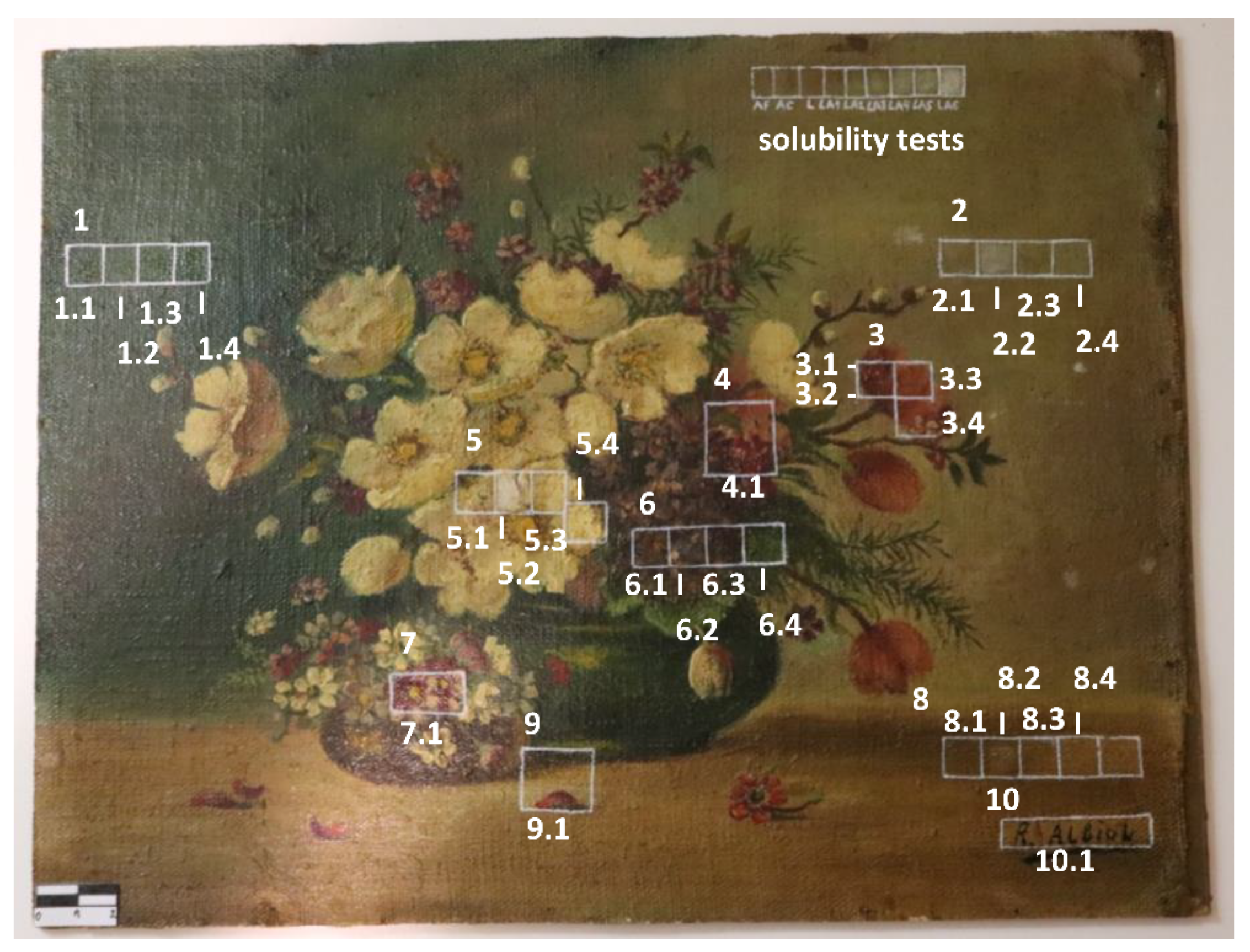
2.1. Description of the Artwork and the State of the Varnish Coating

A contemporary oil easel painting was temporarily loaned from the Restoration-Conservation Laboratory of the Faculty of Fine Arts of the University of Barcelona. The oil painting, of 24 × 31.5 cm2, was applied on a canvas directly adhered to a cardboard and represents a still life with a vase full of several types of flowers of different shades. The author is an anonymous artist who painted it in 1940–1950s (Figure 1).
To examine the distribution of the varnish on the surface of the painting, UV illumination with two wood lamps (220 volts, 50 Hz, 3 Amps, 4 × 36 Watts, CLE design limited, 69 Haydons Road, London SW19 1HQ, serial no P.C 25 27) was arranged at an angle of 45° in front of the artwork. For the identification of the varnish coating, a sample (weight 0.4 mg) was removed with a scalpel and analyzed on a Nicolet 7600 infrared spectrophotometer, using 200 mg of KBr, in the 4000–400 cm−1 range and a spectral resolution of 4 cm−1, by summing up 64 scans.

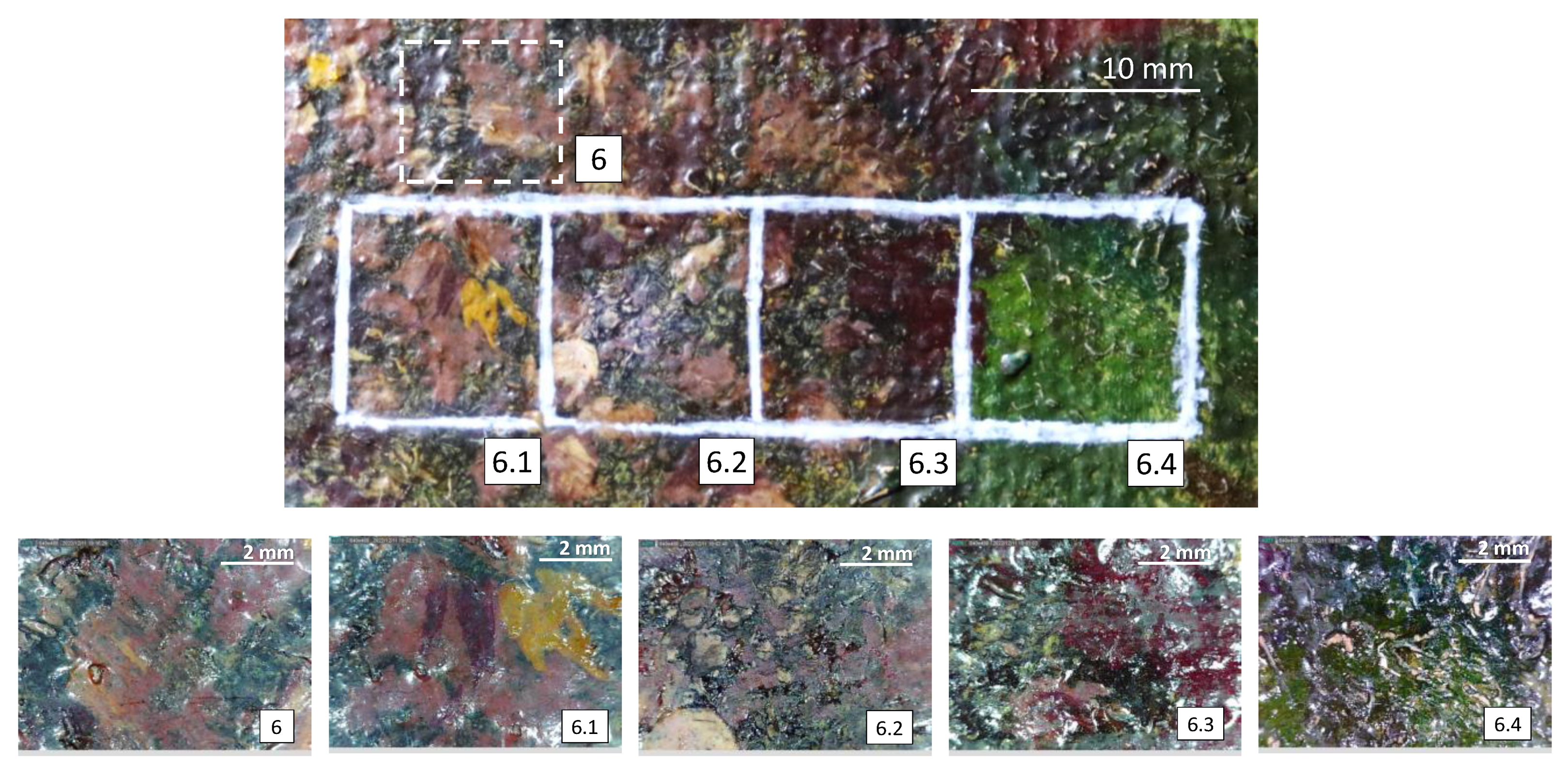
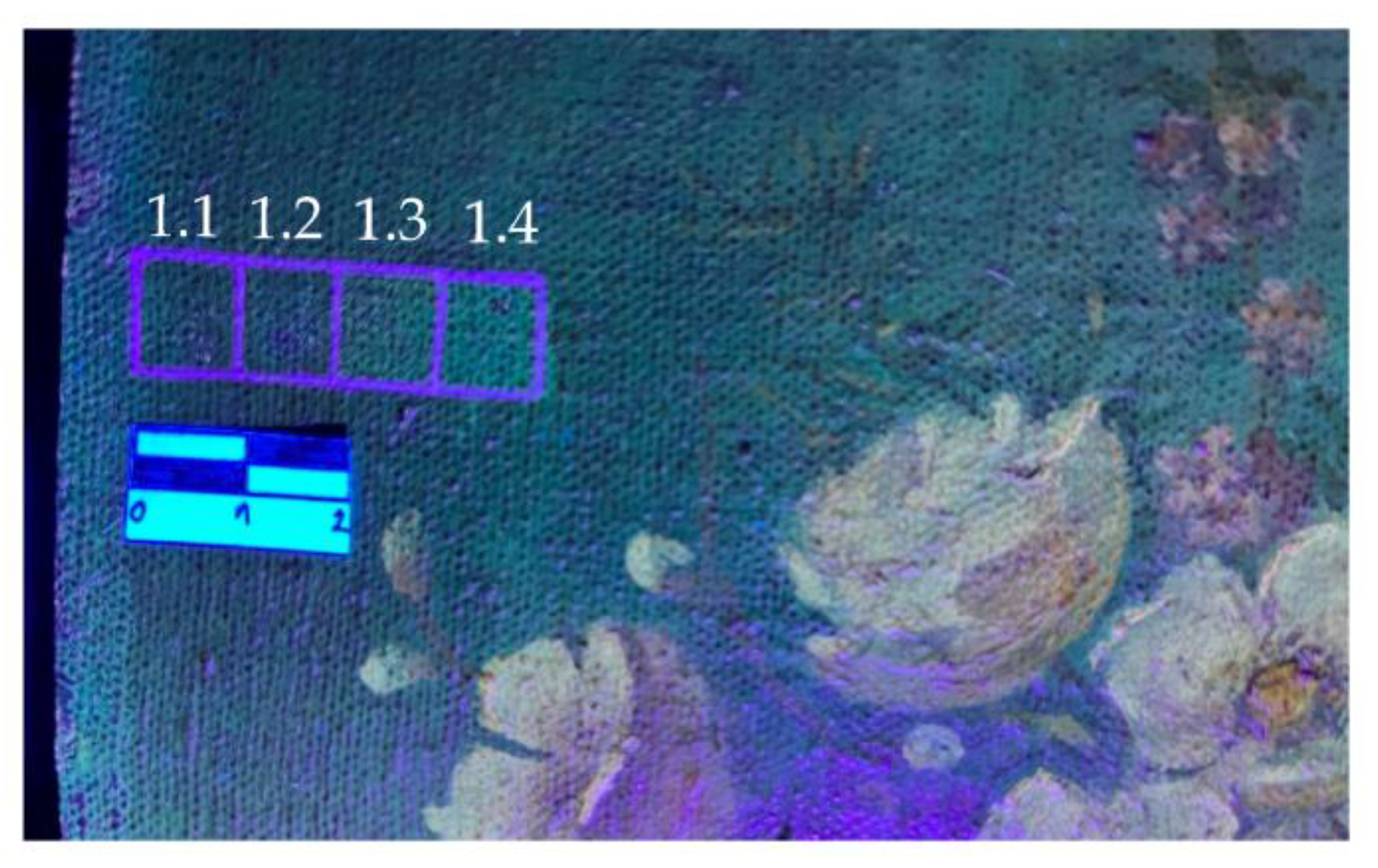
2.2. Varnish Removal Tests

With the aim of only removing the degraded part of the varnish layer without affecting the sound part and the underlying paint layers, different cleaning procedures, either conventional solvents [23] or nanosecond Nd:YAG laser irradiation at 213 nm, were tested in 1 × 1 cm2 areas of the painting.
Hot deionized water, applied with a swab (in areas numbered n.1, where “n” ranges from 1 to 10, Figure 1), was used to avoid creating osmotic tension on the surface when cleaning the surface dirt. An adapted Cremonesi solubility test [23] was used to rationally select solvent from among 24 solvent mixtures. The most suitable one for thinning varnish is composed of (nonpolar) ligroin and (polar) acetone (50:50) and was also applied (in n.2 areas, Figure 1) with a swab.
Laser cleaning was performed next to the areas that were previously treated with solvents (n.3 and n.4, Figure 1) with a Q-switched Nd:YAG laser (Lotis TII, LS-2147) operating at the 5th harmonic, at 213 nm, with pulse duration of 17 ns and at a repetition rate of 10 Hz. Varnish removal was based on a laser scanning mode in order to achieve homogeneous cleaning areas. The laser pulses were focused on the varnish surface using a plano-convex cylindrical lens with a focal length of 15 cm. The scanning speed, and consequently the spatial overlap of successive pulses, was chosen to ensure the delivery of approximately 40 pulses to each point of the sample with an overlap of 50% with respect to the maximum pulse intensity (Gaussian beam). Two different conditions were applied in adjacent areas: 0.10 J/cm² with a spot size of 5.5 × 0.8 mm² and speed of 0.4 mm/s (n.3, Figure 1) and 0.14 J/cm² with a spot size of 5.5 × 0.5 mm2 and speed of 0.25 m/s (n.4, Figure 1).

2.3. Experimental Methods to Assess Cleaning Tests

To assess the effectiveness of different aged varnish removal methods, whether the non-aged part was preserved or removed in excess, a set of physicochemical analyses was performed.
A low-power magnification digital microscope (DINO-LITE basic, magnification 10×) served to observe the areas in which the varnish was removed by different procedures, and the adjacent untreated ones, using a grazing light. Complementary UV lamp illumination was used as described in Section 2.1 after the cleaning procedures.
LIF measurements were performed in the uncleaned and cleaned areas using laser excitation at 266 nm (4th harmonic of a Quantel Brilliant Q-switched Nd:YAG laser, 6 ns pulses, 2 Hz repetition rate, shot to shot laser energy fluctuation less than 10%) and a 0.30 m spectrograph (TMc300 Bentham) with a 300 grooves/mm grating coupled to an intensified charged coupled detector (ICCD, 2151 Andor Technologies). The temporal gate was operated at zero-time delay with respect to the arrival of the laser pulse to the surface of the sample and with a width of 3 μs. The sample was illuminated at an incidence angle of 45° using laser pulses of around 1 mJ with a laser spot diameter of 200 μm (fluence of 0.06 J/cm2). LIF spectra were recorded in the spectral range of 300–700 nm and are the result of 10 accumulations. Cut-off filters at 300 and 420 nm were used to reduce the scattered laser light from the surface of the sample and to avoid the second-order diffraction of the laser.
In-depth LIBS spectra were recorded in the uncleaned and cleaned areas using the same laser excitation wavelength as for LIF measurements at 266 nm and the same combination of spectrograph and ICCD, although the grating had 1200 grooves/mm. In this case, the laser beam was focused on the samples with a 10 cm focal length lens reaching a fluence of 9.5 J/cm2 (spot diameter of 200 μm). Successive spectra in the range of 365–435 nm were acquired with a resolution of 0.2 nm, with gate delay and width of 100 ns and 3 μs, respectively. A cut-off filter at 300 nm was placed in front of the entrance window of the spectrograph to reduce the scattered laser light from the surface of the sample and to avoid the second-order diffraction of the emissions of lower wavelengths.

3. Results and Discussion

3.1. Varnish Condition and Identification

The greenish inhomogeneous fluorescence emission observed upon UV lamp illumination of the varnished surface indicates that the varnish has been naturally aged and the oxidized layer is irregularly and unevenly distributed over the entire surface of the painting. This fluorescent emission is absent from the areas covered by the frame (Figure 2).
Figure 3 displays the FTIR spectrum of the varnish, where the main vibration bands are shown; Table 1 indicates the band assignments. The sequence of bands, corresponding to stretching vibrations of C-H (2955–2853 cm−1), C=O (1735–1900 cm−1), and C-O (1257–1163 cm−1) and bending vibrations of C-H (1456–1384 cm−1), indicates that the varnish is a shellac resin [24,25]. Additional bands of barite (BaSO4) are also observed in the range of 1116–610 cm−1, pointing to the presence of this compound, which is traditionally used as extender pigment or preparatory layer (in the form of lithopone, ZnS.BaSO4) [26].

3.2. Varnish Removal Tests

3.2.1. Low-Power Microscopy

After close inspection of the tested areas by low-power microscopy (Figure 4), the application of hot water only slightly cleaned part of the surface dirt. In turn, the areas of the oil painting intervened with the solvent mixture presented a matte finish with traces of varnish resulting from its heterogeneous removal (area 6.2, Figure 4). Due to the poor selectivity of the swab cleaning and the heterogeneous thickness of the varnish that determines differential amount of solvent penetration in the paint layer [2], the coating in certain areas was excessively removed, compromising the condition of the paint layer (area 5.2, Figure 1). This agrees with previous observations that, despite the suitability of swab cleaning procedures, traditional solvents sometimes yield uneven results [17,27].
UV (213 nm) laser irradiation resulted in gradual thinning down of the varnish layer, since at both fluences used (0.10 and 0.14 J/cm²), particularly at the higher fluence (area 6.4, Figure 4), the surface regained gloss. However, in areas with thicker varnish layer (e.g., area 2.4, Figure 1), the transformation from opaque to bright colors was less evident.

3.2.2. UV Lamp Illumination

Cleaning procedures were also monitored by UV fluorescence imaging (Figure 2 and Figure 5), which confirmed the result of the above visual inspection. The same characteristic dark greenish fluorescence from the oxidized varnish was observed over the entire surface before and after application of hot water (Figure 2 and Figure 5). In the areas treated with the binary solvent mixture or irradiated with laser, the cloudy fluorescence disappears, as aged varnish is removed and the underlying non-aged varnish comes to the surface. With regards to laser ablation at 213 nm, a greater amount of varnish is removed with the highest fluence used (0.14 J/cm²). The solvent mixture action is less controllable and, in some areas, it removes more varnish than the laser procedure (in most of the tested areas, Figure 2) or vice versa (Figure 5).

3.2.3. Laser-Induced Fluorescence (LIF)

LIF spectra were recorded on untreated (reference) and cleaned areas throughout the painting. Figure 6 shows representative results from area 1 (Figure 5). As seen in the figure, the spectrum corresponding to the uncleaned area consists of a broad band in the spectral range of 300–700 nm centered at 469 nm (λmax) and with a shoulder at around 373 nm (λ1), both attributed to hydroxy acids [21]. Table 2 displays the fluorescence intensity and wavelength of those spectral features from the reference and cleaned areas. After cleaning with hot water (Figure 6a), the intensity of the maximum emission (Imax) and I1 does not significantly change (note that spectra were collected not in the same areas but instead in adjacent areas). Solvent mixture and laser treatments increase the intensity of the main band and of the shoulder (Figure 6a, Table 2). In contrast, it is traditionally reported [9,10,22] that the less photo-oxidized the varnishes are, the less fluorescence they emit. Since LIF measurements in this study were conducted with laser excitation at 266 nm, which is not efficiently absorbed by varnishes [22], these observations, particularly the increase in Imax, in addition to the shift of this signal towards higher wavelengths (Figure 6a, Table 2), could reflect a relevant contribution from the underlying preparation layer to the total LIF emission [28]. Fluorescence of less aged photo-oxidized varnish [9,10] also contributes to the total emission, as seen by the change in relative intensity of Imax and I1 (Figure 6b, Table 2). As it turns out, the more varnish is removed via laser ablation, the more noticeable are these effects.

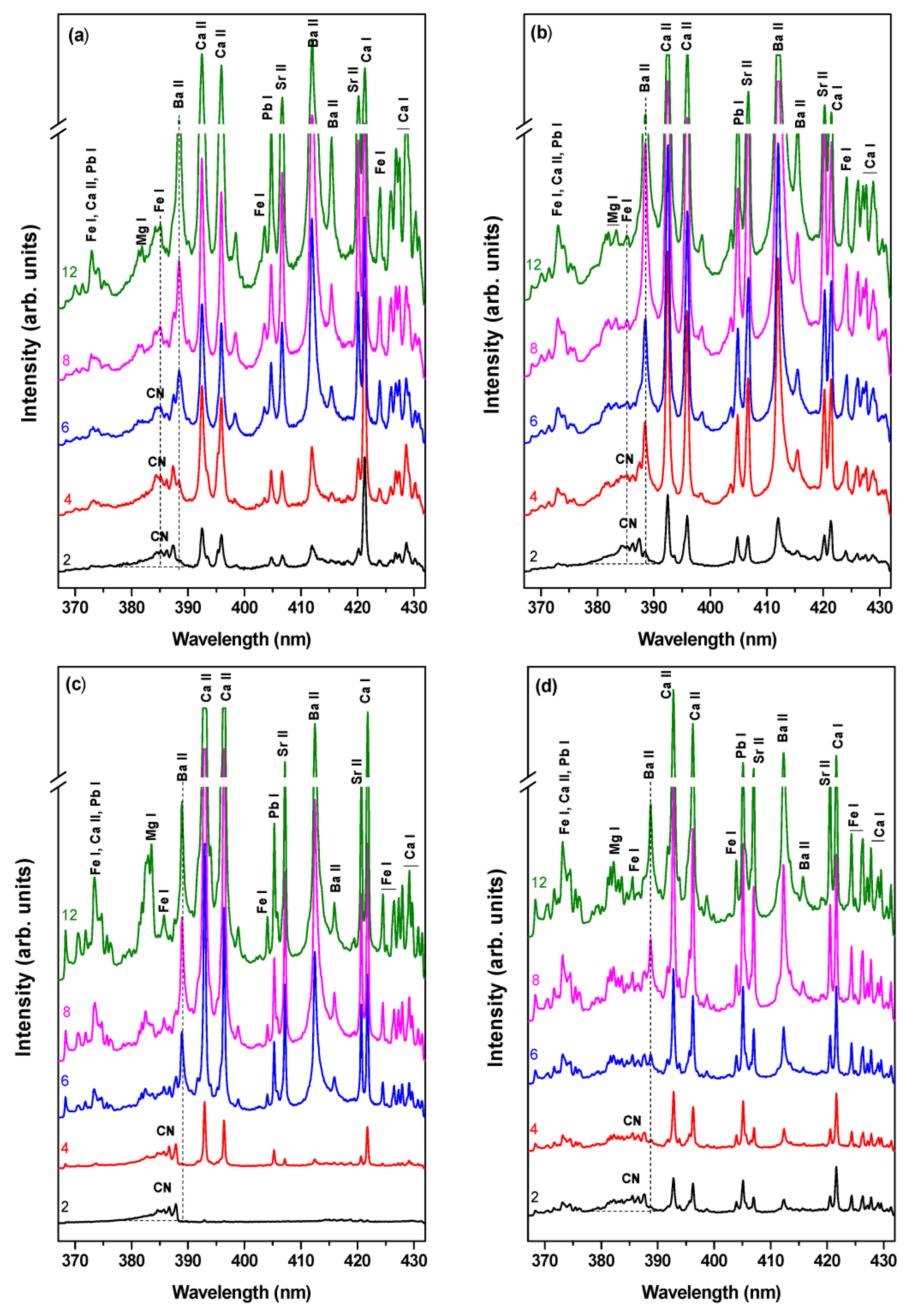
3.2.4. Laser-Induced Breakdown Spectroscopy (LIBS)

The efficiency of the cleaning procedures based on solvents and laser cleaning was assessed by LIBS. Figure 7 shows successive selected LIB spectra from a stratigraphic study (from 2 to 12 pulses) on the treated surfaces in the region of 365–435 nm (area 1, Figure 1 and Figure 5).
In all cleaned areas, the first two pulses revealed the presence of CN bands between 380–390 nm originated from the varnish [8,29]. However, in the area treated with laser fluence at 0.14 J/cm2 these emissions are less evident, as they overlap with the lines of Fe from a pigment on the paint layer, indicating that the varnish layer is thinner in this area (Figure 7d). CN emission bands disappear after four laser pulses in the solvent and laser-cleaned areas and after six pulses in the water-cleaned area, while Mg and Fe lines, coming from the painting layers, increase in intensity.
Ca lines at 393.37, 396.85, and 422.67 nm (NIST database [30]) were also visible right from the surface (two pulses) in all cleaned areas, similar to reports on shellac varnished temperas [8]. However, those lines arising from the area cleaned with laser at 0.10 J/cm2 presented at first (from two to four pulses) a low intensity (Figure 7c). This, together with the later onset and lower emission intensity of Pb, Sr lines (attributed to pigments) and of Ba lines (attributed to lithopone) (note that Zn lines are visible in a different spectral region not shown in Figure 7c) confirms, once again, the heterogeneous (and in this case thicker) distribution of the varnish.

4. Conclusions

In this work, a traditional procedure for the removal of naturally aged varnish on a contemporary oil easel painting, based on the application of a mixture of ligroin:acetone with laser cleaning using nanosecond pulses of 213 nm delivered by the fifth harmonic of a Nd:YAG Q-switched laser have been compared. Furthermore, the cleaning effect of hot water was assessed.
Primarily, water application does not produce a cleaning effect as confirmed by the microscope and UV fluorescence images, and LIF. In fact, in-depth LIBS analyses in the water-treated area reveal that the CN bands from the varnish disappear with higher number of pulses than those necessary in the areas subjected to the other cleaning procedures (ligroin:acetone and laser fluence of 0.14 J/cm2), indicating the presence of a thicker varnish layer.
Traditional binary solvents perform unevenly. The mixture of ligroin:acetone irregularly (scarcely or excessively) thins down the varnish layer (reaching in some areas the pictorial layer), as shown by the aforementioned experimental techniques. Successful removal of the naturally aged varnish on the contemporary oil easel painting has been achieved by irradiation at a fluence of 0.14 J/cm2 with 17 ns, UV laser pulses of 213 nm, a wavelength that is efficiently absorbed by the varnish. This treatment returns the painting surface to its original shine by removing the outermost deteriorated varnish layer in a controlled way, leaving the underlying material unaffected (both deeper varnish and paint layers). This results in low UV fluorescence emissions from the surface that, in contrast, produce high LIF signals upon laser excitation at 266 nm. With the lower fluence of 0.10 J/cm2, the same effects are obtained, although to a lesser extent since varnish removal was less effective.
The results of this study open the possibility of extending the use of nanosecond 213 nm laser irradiation for the selective removal of oxidized varnishes from artistic paintings without side effects. The methodologic approach used reveals the necessity of applying these different analytical and fully complementary techniques to accurately assess the performance of the selected cleaning procedures.

Author Contributions

Conceptualization, M.L.-C., M.O. and M.C.; investigation, all authors; formal analysis, M.M.-W., L.M.-G., P.M.C.-Q. and M.O.; writing—original draft preparation, P.M.C.-Q.; writing—review and editing, all authors; funding acquisition, M.O. and M.C. All authors have read and agreed to the published version of the manuscript.

Funding

This research was funded by the Spanish State Research Agency (AEI) through the project SCOCHLAS (Study and Conservation of Cultural Heritage with Lasers, PID2019-104124RB-I00/AEI/1013039/501100011033), by the Community of Madrid through the project Top Heritage-CM (Tecnologías en Ciencias del Patrimonio, S2018/NMT_4372), and by the H2020 project IPERION HS (Integrated Platform for the European Research Infrastructure ON Heritage Science, GA 871034). L. Maestro-Guijarro’s participation was financed by a Youth Guarantee contract (CAM20_IQFR_AI_06) from the Community of Madrid.

Data Availability Statement

The data that support the findings will be available in the CSIC digital repository within 6 months from the date of publication.

Acknowledgments

The professional support provided by the CSIC ‘Open Heritage: Research and Society (PTI-PAIS)’ Interdisciplinary Thematic Platform is gratefully acknowledged.

Conflicts of Interest

The authors declare no conflict of interest.

References

  1. Zanini, A.; Trafeli, V.; Bartoli, L. The Laser as a Tool for the Cleaning of Cultural Heritage. IOP Conf. Ser. Mater. Sci. Eng. 2018, 364, 012078. [Google Scholar] [CrossRef]
  2. Baij, L.; Hermans, J.; Ormsby, B.; Noble, P.; Iedema, P.; Keune, K. A Review of Solvent Action on Oil Paint. Herit. Sci. 2020, 8, 43. [Google Scholar] [CrossRef]
  3. Pereira-Pardo, L.; Melita, L.N.; Korenberg, C. Tackling Conservation Challenges Using Erbium Lasers: Case Studies at the British Museum. J. Inst. Conserv. 2020, 43, 25–43. [Google Scholar] [CrossRef]
  4. Pouli, P.; Oujja, M.; Castillejo, M. Practical Issues in Laser Cleaning of Stone and Painted Artefacts: Optimisation Procedures and Side Effects. Appl. Phys. A Mater. Sci. Process. 2012, 106, 447–464. [Google Scholar] [CrossRef]
  5. Andreotti, A.; Brown, W.P.; Camaiti, M.; Colombini, M.P.; DeCruz, A. Diagnosis of Materials and Effectiveness of Er:YAG Laser Cleaning as Complementary Treatment in a Panel Painting Attributed to Lluís Borrassà (Fifteenth Century). Appl. Phys. A 2016, 122, 572. [Google Scholar] [CrossRef]
  6. Bracco, P.; Lanterna, G.; Matteini, M.; Nakahara, K.; Sartiani, O.; de Cruz, A.; Wolbarsht, M.L.; Adamkiewicz, E.; Colombini, M.P. Er:YAG Laser: An Innovative Tool for Controlled Cleaning of Old Paintings: Testing and Evaluation. J. Cult. Herit. 2003, 4, 202–208. [Google Scholar] [CrossRef]
  7. Brunetto, A.; Bono, G.; Frezzato, F. Er:YAG Laser Cleaning of ‘San Marziale in Gloria’ by Jacopo Tintoretto in the Church of San Marziale, Venice. J. Inst. Conserv. 2020, 43, 44–58. [Google Scholar] [CrossRef]
  8. Castillejo, M.; Martín, M.; Oujja, M.; Silva, D.; Torres, R.; Manousaki, A.; Zafiropulos, V.; van den Brink, O.F.; Heeren, R.M.A.; Teule, R.; et al. Analytical Study of the Chemical and Physical Changes Induced by KrF Laser Cleaning of Tempera Paints. Anal. Chem. 2002, 74, 4662–4671. [Google Scholar] [CrossRef]
  9. Kokkinaki, O.; Dimitroulaki, E.; Melessanaki, K.; Anglos, D.; Pouli, P. Laser-Induced Fluorescence as a Non-Invasive Tool to Monitor Laser-Assisted Thinning of Aged Varnish Layers on Paintings: Fundamental Issues and Critical Thresholds. Eur. Phys. J. Plus 2021, 136, 938. [Google Scholar] [CrossRef]
  10. Moretti, P.; Iwanicka, M.; Melessanaki, K.; Dimitroulaki, E.; Kokkinaki, O.; Daugherty, M.; Sylwestrzak, M.; Pouli, P.; Targowski, P.; van den Berg, K.J.; et al. Laser Cleaning of Paintings: In Situ Optimization of Operative Parameters through Non-Invasive Assessment by Optical Coherence Tomography (OCT), Reflection FT-IR Spectroscopy and Laser Induced Fluorescence Spectroscopy (LIF). Herit. Sci. 2019, 7, 44. [Google Scholar] [CrossRef]
  11. Lopez, M.; Bai, X.; Koch-Dandolo, C.; Serfaty, S.; Wilkie-Chancellier, N.; Detalle, V. Nd:YAG vs Er:YAG: A Comparative Study of Laser Varnish Removal on Easel Paintings. In Proceedings of the Optics for Arts, Architecture, and Archaeology VII, Munich, Germany, 8 August 2019; Targowski, P., Groves, R., Liang, H., Eds.; SPIE: Bellingham, WA, USA, 2019; p. 3. [Google Scholar]
  12. Wang, C.; Cao, Y.; Tie, F.; Camaiti, M. Er:YAG Laser Cleaning of Painted Surfaces: Functional Considerations to Improve Efficacy and Reduce Side Effects. Coatings 2021, 11, 1315. [Google Scholar] [CrossRef]
  13. Striova, J.; Fontana, R.; Barbetti, I.; Pezzati, L.; Fedele, A.; Riminesi, C. Multisensorial Assessment of Laser Effects on Shellac Applied on Wall Paintings. Sensors 2021, 21, 3354. [Google Scholar] [CrossRef] [PubMed]
  14. Striova, J.; Salvadori, B.; Fontana, R.; Sansonetti, A.; Barucci, M.; Pampaloni, E.; Marconi, E.; Pezzati, L.; Colombini, M.P. Optical and Spectroscopic Tools for Evaluating Er: YAG Laser Removal of Shellac Varnish. Stud. Conserv. 2015, 60, S91–S96. [Google Scholar] [CrossRef] [Green Version]
  15. Chillè, C.; Papadakis, V.M.; Theodorakopoulos, C. An Analytical Evaluation of Er:YAG Laser Cleaning Tests on a Nineteenth Century Varnished Painting. Microchem. J. 2020, 158, 105086. [Google Scholar] [CrossRef]
  16. Hellen, R. Preliminary Study into the Reduction and Removal of Naturally Aged Varnishes from Painted Surfaces Using an Er:YAG Laser in a Two-Step Cleaning Process with Solvents. J. Inst. Conserv. 2020, 43, 79–93. [Google Scholar] [CrossRef]
  17. Siano, S.; Osticioli, I.; Pavia, A.; Ciofini, D. Overpaint Removal from Easel Paintings Using an LQS Nd:YAG Laser: The First Validation Study. Stud. Conserv. 2015, 60, S49–S57. [Google Scholar] [CrossRef]
  18. Antonopoulou-Athera, N.; Chatzitheodoridis, E.; Doulgerides, M.; Evangelatos, C.; Serafetinides, A.A.; Terlixi, A. Laser Cleaning Treatment of Burnt Paintings. In Proceedings of the 18th International School on Quantum Electronics: Laser Physics and Applications, Sozopol, Bulgaria, 8 January 2015; Dreischuh, T., Gateva, S., Serafetinides, A., Eds.; Proccedings of SPIE Vol. 9447. SPIE: Bellingham, WA, USA, 2015; p. 944712. [Google Scholar]
  19. Ciofini, D.; Oujja, M.; Cañamares, M.V.; Siano, S.; Castillejo, M. Detecting Molecular Changes in UV Laser-Ablated Oil/Diterpenoid Resin Coatings Using Micro-Raman Spectroscopy and Laser Induced Fluorescence. Microchem. J. 2018, 141, 12–24. [Google Scholar] [CrossRef] [Green Version]
  20. Oujja, M.; Psilodimitrakopoulos, S.; Carrasco, E.; Sanz, M.; Philippidis, A.; Selimis, A.; Pouli, P.; Filippidis, G.; Castillejo, M. Nonlinear Imaging Microscopy for Assessing Structural and Photochemical Modifications upon Laser Removal of Dammar Varnish on Photosensitive Substrates. Phys. Chem. Chem. Phys. 2017, 19, 22836–22843. [Google Scholar] [CrossRef] [Green Version]
  21. Oujja, M.; García, A.; Romero, C.; Vázquez de Aldana, J.R.; Moreno, P.; Castillejo, M. UV Laser Removal of Varnish on Tempera Paints with Nanosecond and Femtosecond Pulses. Phys. Chem. Chem. Phys. 2011, 13, 4625. [Google Scholar] [CrossRef] [Green Version]
  22. Ciofini, D.; Oujja, M.; Cañamares, M.V.; Siano, S.; Castillejo, M. Spectroscopic Assessment of the UV Laser Removal of Varnishes from Painted Surfaces. Microchem. J. 2016, 124, 792–803. [Google Scholar] [CrossRef]
  23. Cremonesi, P. L’uso dei Solventi Organici Nella Pulitura di Opere Policrome, 2nd ed.; Il Prato: Padova, Italia, 2004; ISBN 978-88-87243-85-7. [Google Scholar]
  24. Derrick, M.R.; Stulik, D.C.; Landry, J.M. (Eds.) Scientific tools for conservation. In Infrared Spectroscopy in Conservation Science; Getty Conservation Institute: Los Angeles, CA, USA, 1999; ISBN 978-0-89236-469-5. [Google Scholar]
  25. Issa, Y.M.; Abdel-Maksoud, G.; Magdy, M. Analytical Study of Saint Gregory Nazianzen Icon, Old Cairo, Egypt. J. Mol. Struct. 2015, 1100, 70–79. [Google Scholar] [CrossRef]
  26. Zuena, M.; Buemi, L.P.; Stringari, L.; Legnaioli, S.; Lorenzetti, G.; Palleschi, V.; Nodari, L.; Tomasin, P. An Integrated Diagnostic Approach to Max Ernst’s Painting Materials in His Attirement of the Bride. J. Cult. Herit. 2020, 43, 329–337. [Google Scholar] [CrossRef]
  27. Teppo, E. Introduction: Er:YAG Lasers in the Conservation of Artworks. J. Inst. Conserv. 2020, 43, 2–11. [Google Scholar] [CrossRef]
  28. Gómez-Morón, A.; Ortiz, R.; Colao, F.; Fantoni, R.; Ortiz, P. Laser-Induced Fluorescence Mapping of Pigments in a Secco Painted Murals. Ge Conserv. 2020, 17, 233–250. [Google Scholar] [CrossRef]
  29. Anglos, D. Laser-Induced Breakdown Spectroscopy in Art and Archaeology. Appl. Spectrosc. 2001, 55, 186A–205A. [Google Scholar] [CrossRef]
  30. NIST Database. Available online: https://physics.nist.gov/PhysRefData/ASD/lines_form.html (accessed on 19 October 2022).
Figure 1. Selected cleaned areas. n.1: with hot deionized water; n.2: with ligroin:acetone mixture (50:50); n.3: irradiation with 213 nm, 0.10 J/cm2; n.4: irradiation with 213 nm, 0.14 J/cm2.
Heritage 06 00053 g001
Figure 2. UV light image of the artwork showing greenish fluorescence and its absence in areas covered by the frame.
Heritage 06 00053 g002
Figure 3. FTIR spectrum of the varnish.
Heritage 06 00053 g003
Figure 4. Digital microscopy images of the surface of the oil painting before (area 6, Figure 1) and after the cleaning tests with hot water (6.1), solvent mixture (6.2), laser at 213 nm, 0.10 J/cm2 (6.3), and laser at 213 nm, 0.14 J/cm2 (6.4).
Heritage 06 00053 g004
Figure 5. Fluorescence observed upon UV lamp illumination of treated and untreated area 1 of the painting cleaned with water (1.1), with solvent mixture (1.2), with 213 nm laser at 0.10 J/cm2 (1.3), and with 213 nm laser at 0.14 J/cm2 (1.4).
Heritage 06 00053 g005
Figure 6. LIF spectra of the oil painting before and after cleaning tests without (a) and with (b) normalization of the fluorescence intensity.
Heritage 06 00053 g006
Figure 7. LIB spectra after 2 to 12 laser pulses of the area 1 cleaned with: (a) deionized water, (b) solvent mixture, (c) 213 nm laser, 0.10 J/cm2, and (d) 213 nm laser, 0.14 J/cm2.
Heritage 06 00053 g007
Table 1. Band assignments for the varnish mid-infrared spectrum.
Band (cm−1)Functional GroupBand (cm−1)Functional Group
3435ν(O-H)1384δ(C-H)
2955ν(CH3)1314Not assigned
2925ν(CH3)1257ν(C-O)
2853ν(CH2)1163ν(C-O)
1735ν(C=O)1116ν(SO4), δ(C-H)
1708ν(C=O)1078ν(SO4), δ(C-H)
1638δ(O-H), ν(C=C)983ν(SO4)
1547Not assigned878Not assigned
1456δ(C-H)635δ(SO4)
1417δ(C-H)610δ(SO4)
Table 2. Fluorescence spectral features of the painting surface before (ref) and after the varnish removal tests. The intensities are presented in arbitrary units.
Area 1λ1 (nm)I1λmax (nm)ImaxΔλmax (nm)Imax/I1
ref37322,17546930,990-1.4
H2O37420,86847433,63151.6
Ligroin:acetone37637,38947478,85852.1
213 nm, 0.10 J/cm237242,28947575,94061.8
213 nm, 0.14 J/cm237149,480477104,49482.1
Disclaimer/Publisher’s Note: The statements, opinions and data contained in all publications are solely those of the individual author(s) and contributor(s) and not of MDPI and/or the editor(s). MDPI and/or the editor(s) disclaim responsibility for any injury to people or property resulting from any ideas, methods, instructions or products referred to in the content.

