
A Royal Mystery: A Multianalytical Approach for Dyestuff Identification in Seventeenth Century Waistcoats


Abstract
1. Introduction
2. Materials and Methods
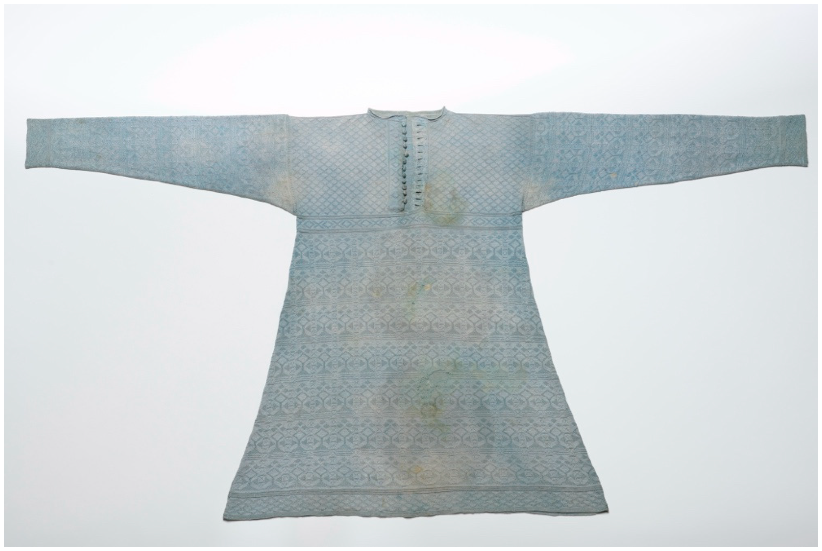
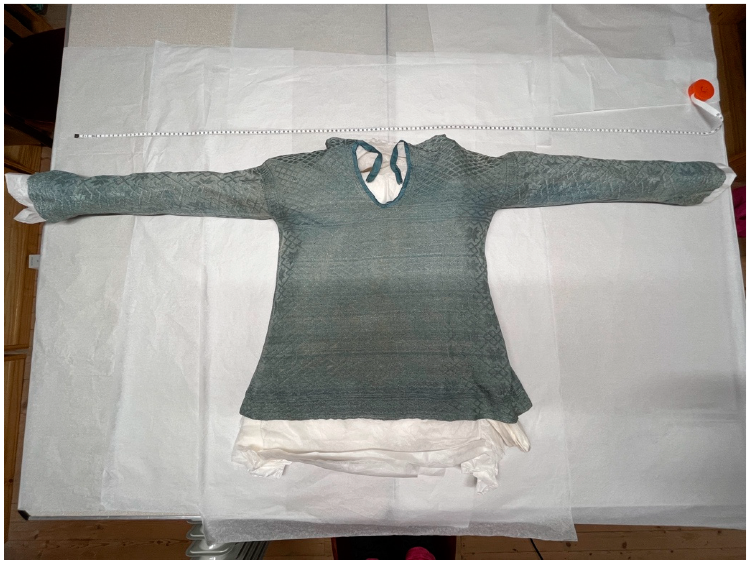
2.1. Materials
2.2. Sampling
2.3. Confocal Micro-Raman Spectroscopy
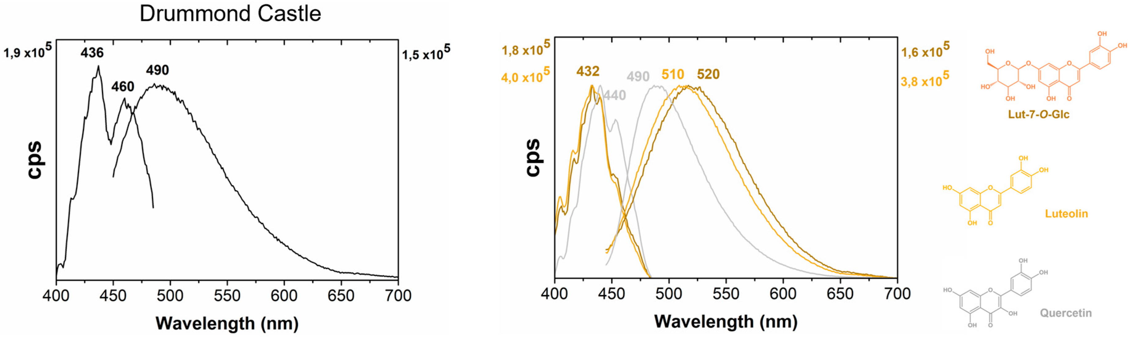
2.4. Molecular Fluorescence in the Visible
2.5. High-Performance Liquid Chromatography (HPLC) Coupled with Mass Spectrometry (MS)
3. Results and Discussion
3.1. HPLC-MS Analysis
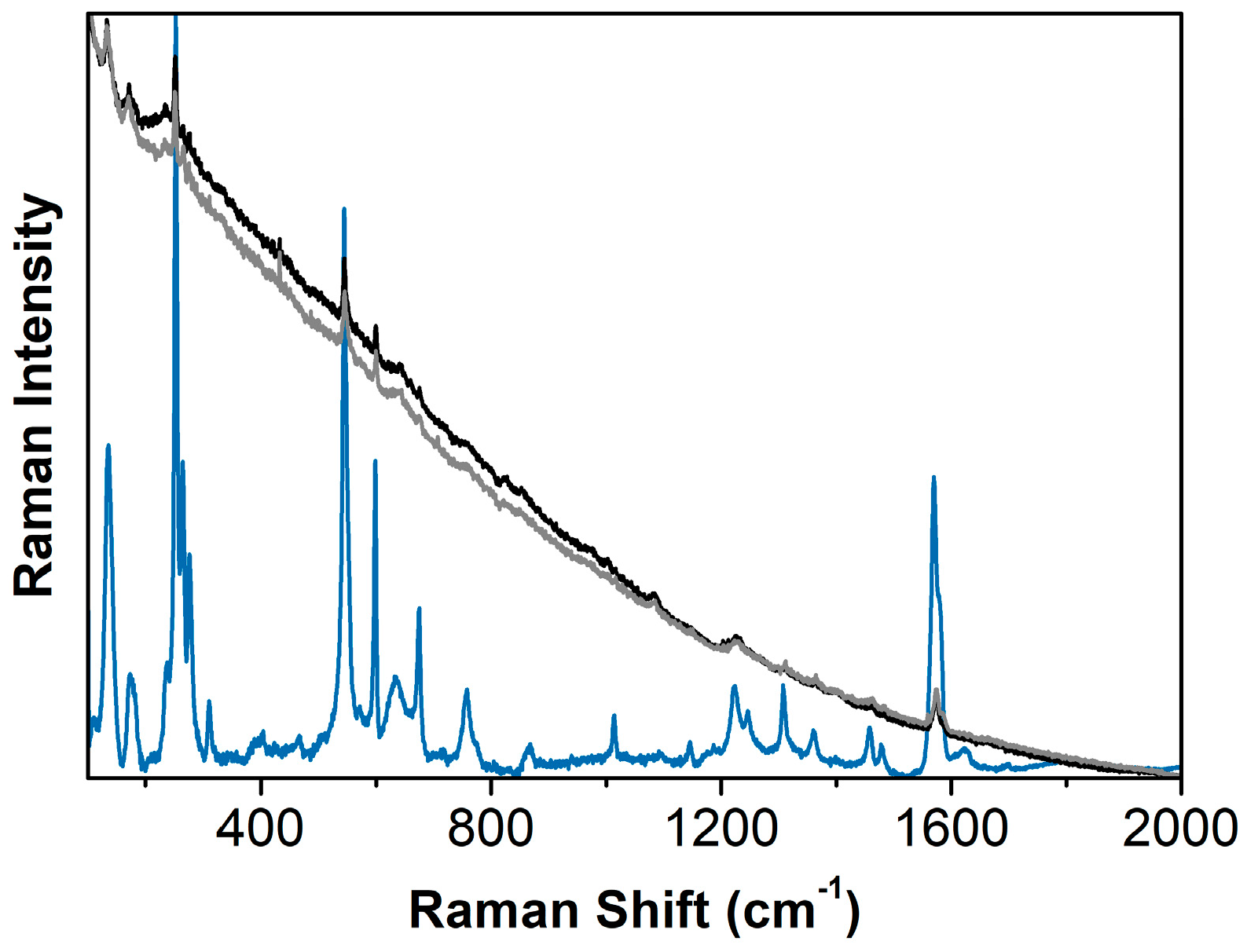
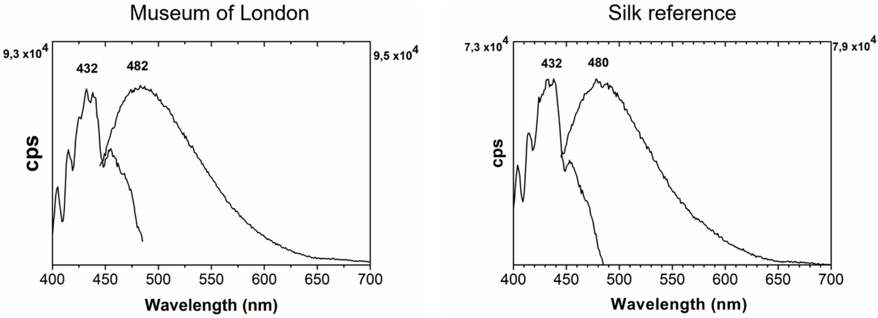
3.2. In-Situ Analysis: Raman and Molecular Fluorescence
4. Conclusions
Supplementary Materials
Author Contributions
Funding
Data Availability Statement
Acknowledgments
Conflicts of Interest
References
- Adcock, S. (Ed.) Wonderful London: The World’s Greatest City Described by Its Best Writers and Pictured by Its Finest Photographers; Educational Book Company: London, UK, 1926; Volume 1, p. 722. [Google Scholar]
- Sanderson, S. Compleat History of the Life and Raigne of King Charles from His Cradle to the Grave, Humphrey Moseley; Richard Tomlins & George Sawbridge: London, UK, 1658. [Google Scholar]
- The National Archives, TNA 3/910/1 (1632–1633) The particulars of the account of George Kirke esq, Gentleman of the Robes.
- The Royal Collection, inventory number RCIN 404124.
- Strong, R. Charles I’s clothes for the years 1633 to 1635. Costume 1980, 14, 73–89. [Google Scholar] [CrossRef]
- Divall, G. Charles I’s Waistcoat Keeps Its Secrets; London Press Service: London, UK, 1989; pp. 1–4, Unpublished Draft Press Release. [Google Scholar]
- Metropolitan Police Forensic Science Laboratory (London, UK). 1988; Unpublished Report.
- Ringgaard, M. Silk knitted waistcoats: A seventeenth century fashion item. In Fashionable Encounters: Perspectives and Trends in Fashionable Dress in the Early Modern Nordic World; Englehardt Matthiassen, T., Nosch, M.-L., Toftegaard, K., Venborg Pederson, M., Ringgaard, M., Eds.; Oxbow Books: Oxford, UK, 2014; pp. 73–104. [Google Scholar]
- Sliwka-Kaszynska, M.; Slebioda, M.; Brillowska-Dabrowska, A.; Mroczynska, M.; Karczewski, J.; Marzec, A.; Rybinski, P.; Drazkowska, A. Multi-Technique investigation of grave robes from seventeenth and eighteenth century crypts using combined spectroscopic, spectrometric techniques, and new-generation sequencing. Materials 2021, 14, 3535. [Google Scholar] [CrossRef] [PubMed]
- Yurdun, T.; Karadag, R.; Dolen, E.; Mubarak, M. Identification of natural yellow, blue, green and black dyes in fifteenth to seventeenth centuries Ottoman silk and wool textiles by HPLC with diode array detection. Rev. Anal. Chem. 2011, 30, 153–164. [Google Scholar] [CrossRef]
- Keene, S.; Greenacre, M. Retired conservators, UK. 26 February 2024; personal communication. [Google Scholar]
- Hayeur Smith, M.; Arneborg, J.; Smith, K. The ‘Burgundian’ hat from Herjolfsnes, Greenland: New discoveries, new dates. Dan. J. Archaeol. 2015, 4, 21–32. [Google Scholar] [CrossRef]
- Van Strydonck, M.; De Moor, A.; Bénazeth, D. 14C dating compared to art historical dating of Roman and Coptic textiles from Egypt. Radiocarbon 2004, 46, 231–244. [Google Scholar] [CrossRef]
- Malcolm-Davies, J. Knitting in Early Modern Europe (KEME). 2024. Available online: https://kemeresearch.com (accessed on 25 March 2024).
- Nabais, P.; Malcolm-Davies, J.; Melo, M.J.; Teixeira, N.; Behlen, B. Early modern knitted caps (fifteenth to sixteenth centuries): Analyzing dyes in archaeological samples using microspectrofluorimetry complemented by HPLC–MS. Herit. Sci. 2023, 11, 220. [Google Scholar] [CrossRef]
- Nabais, P.; Melo, M.J.; Lopes, J.A.; Vitorino, T.; Neves, A.; Castro, R. Microspectrofluorimetry and chemometrics for the identification of medieval lake pigments. Herit. Sci. 2018, 6, 13. [Google Scholar] [CrossRef]
- Nabais, P.; Melo, M.J.; Lopes, J.A.; Vieira, M.; Castro, R.; Romani, A. Organic colorants based on lac dye and brazilwood as markers for a chronology and geography of medieval scriptoria: A chemometrics approach. Herit. Sci. 2021, 9, 32. [Google Scholar] [CrossRef]
- Melo, M.J.; Claro, A. Bright light: Microspectrofluorimetry for the characterization of lake pigments and dyes in works of art. Acc. Chem. Res. 2010, 43, 857–866. [Google Scholar] [CrossRef] [PubMed]
- Zhang, X.; Laursen, R. Development of mild extraction methods for the analysis of natural dyes in textiles of historical interest using LC-diode array detector-MS. Anal. Chem. 2005, 77, 2022–2025. [Google Scholar] [CrossRef] [PubMed]
- Sharif, S.; Nabais, P.; Melo, M.-J.; Pina, F.; Oliveira, M.-C. Photoreactivity and stability of flavonoid yellows used in cultural heritage. Dye. Pigment. 2022, 199, 110051. [Google Scholar] [CrossRef]
- Shaala, L.; Asfour, H.; Youssef, D.; Żółtowska-Aksamitowska, S.; Wysokowski, M.; Tsurkan, M.; Galli, R.; Meissner, H.; Petrenko, I.; Tabachnick, K.; et al. New Source of 3D Chitin Scaffolds: The Red Sea Demosponge Pseudoceratina arabica (Pseudoceratinidae, Verongiida). Mar. Drugs 2019, 17, 92. [Google Scholar] [CrossRef] [PubMed]
- Seymour, J.; Costello, C.; Zaia, J. The Influence of Sialylation on Glycan Negative Ion Dissociation and Energetics. J. Am. Soc. Mass Spectrom. 2006, 17, 844–854. [Google Scholar] [CrossRef] [PubMed]
- Degano, I.; Biesaga, M.; Colombini, M.-P.; Trojanowicz, M. Historical and archaeological textiles: An insight on degradation products of wool and silk yarns. J. Chromatogr. A 2011, 1218, 5837–5847. [Google Scholar] [CrossRef] [PubMed]


| Location | Fibre | Inventory Number | Colour (by Visual Observation) |
|---|---|---|---|
| London Museum | Silk | A27050 | Pale blue-green |
| Drummond Castle | Silk | DCT0032 | Pale blue-green |
| No sampling will be considered where previous conservation treatments have distorted the item’s original shape. |
| Items must have existing damage. Caps in pristine or good condition, without any existing areas of loss, will not be sampled. |
| Areas for sampling are to be hidden from regular view. Sampling from the interior will be preferable to the exterior. External decorative features will not be sampled. |
| Samples will be taken from areas away from key features. |
| Multiple sampling of one item will be considered if the item has multiple parts, such as a separate lining, decoration or a strap. |
| No sampling will be considered in or around a hole where most or all of the material exists around the area of loss (if the hole could be completely closed through conservation). |
| Sampling items which have not undergone obvious conservation interventions is preferred. |
| No sampling will be undertaken where a proper assessment of the item is currently impossible (for example, if it has been stitched to a mount). |
| The maximum sample shape and size is square (0.5 cm × 0.5 cm) or rectangular (1 cm × 2 cm). |
| Loose dust or hairs in the item’s storage box (which would otherwise be discarded) may be collected as samples. |
Disclaimer/Publisher’s Note: The statements, opinions and data contained in all publications are solely those of the individual author(s) and contributor(s) and not of MDPI and/or the editor(s). MDPI and/or the editor(s) disclaim responsibility for any injury to people or property resulting from any ideas, methods, instructions or products referred to in the content. |
© 2024 by the authors. Licensee MDPI, Basel, Switzerland. This article is an open access article distributed under the terms and conditions of the Creative Commons Attribution (CC BY) license (https://creativecommons.org/licenses/by/4.0/).
Share and Cite
Malcolm-Davies, J.; Behlen, B.; Teixeira, N.; Nabais, P. A Royal Mystery: A Multianalytical Approach for Dyestuff Identification in Seventeenth Century Waistcoats. Heritage 2024, 7, 4017-4026. https://doi.org/10.3390/heritage7080189
Malcolm-Davies J, Behlen B, Teixeira N, Nabais P. A Royal Mystery: A Multianalytical Approach for Dyestuff Identification in Seventeenth Century Waistcoats. Heritage. 2024; 7(8):4017-4026. https://doi.org/10.3390/heritage7080189
Chicago/Turabian StyleMalcolm-Davies, Jane, Beatrice Behlen, Natércia Teixeira, and Paula Nabais. 2024. "A Royal Mystery: A Multianalytical Approach for Dyestuff Identification in Seventeenth Century Waistcoats" Heritage 7, no. 8: 4017-4026. https://doi.org/10.3390/heritage7080189
APA StyleMalcolm-Davies, J., Behlen, B., Teixeira, N., & Nabais, P. (2024). A Royal Mystery: A Multianalytical Approach for Dyestuff Identification in Seventeenth Century Waistcoats. Heritage, 7(8), 4017-4026. https://doi.org/10.3390/heritage7080189