Article

Reconstructing a Lay Individual’s Elbow Fracture at Santa Caterina Friary, Barcelona (15th–16th Century): The Contribution of Paleopathology to the Valorization of Bioarcheological Heritage

1
Department of Basic Medical Sciences, Faculty of Medicine, and Health Sciences, University Rovira i Virgili, Carrer Sant Llorenç, 21, 43201 Reus, Spain
2
Servei de Radiodiagnòstic (CDI), Hospital Clínic, Universitat de Barcelona, 08036 Barcelona, Spain
3
Departament d’Història i Arqueologia, Universitat de Barcelona, Montalegre 6, 08001 Barcelona, Spain
*
Author to whom correspondence should be addressed.
Heritage 2024, 7(8), 4182-4192; https://doi.org/10.3390/heritage7080196
Submission received: 17 July 2024 / Revised: 30 July 2024 / Accepted: 31 July 2024 / Published: 2 August 2024

Abstract

:
The paleopathological literature notably lacks the description and analysis of distal humeral fractures and their associated complications. The objectives of this study were (1) to evaluate a distal humerus fracture associated with cubitus valgus observed in the articulated right elbow of an adult male buried in the Santa Caterina Friary site in Barcelona, dating back to the modern period (15th to early 16th century), and (2) to contribute to the valorization of bioarcheological heritage. Employing macroscopic and radiographic studies, the injury was assessed. The results indicated a healed antemortem fracture, probably associated with a fall. In the AO/OTA classification, it corresponds to type 13C1.1 (complete articular fracture, articular simple, metaphyseal simple, above the transcondylar axis), accompanied by a coronoid process fracture due to anterior trochlear dislocation impact (O’Driscoll type 1 classification: transverse fracture of the distal apophysis with involvement of the sublime tubercle). This fracture, resulting in a 28° cubitus valgus and significant elbow changes, infers community support in healing and highlights the socio-economic dynamics of guilds and adaptive strategies to physical adversities. This study is one of the first to describe cubitus valgus in a historical Spanish population, offering a comprehensive view of the complexities, physical adversities, and adaptive strategies employed by individuals following an elbow fracture.

1. Introduction

A bone injury may be caused by an external force on the body, affecting the continuity of the bone (fracture) or the anatomical connection (dislocation) [1,2]. Bone injuries can be caused by accidents or violent interactions [3]. Accidental injuries are more common than those resulting from violent interactions. However, in the paleopathological field, much greater interest has been given to injuries resulting from violent interactions. Even so, the analysis of accidental skeletal injuries, their patterns, and etiology offers valuable insights into the lifestyles and behaviors of historical and prehistorical populations [4].
The articular surfaces of the elbow show great structural congruency, which makes the humeroulnar joint one of the most stable joints in the human musculoskeletal system [5]. The distal humerus is angulated laterally in relation to its longitudinal axis, producing a 6° valgus tilt of the forearm [3,6]. Thus, in the frontal plane, when the forearm is supinated and in full extension, the radius and the ulna are not aligned with the longitudinal axis of the humerus. This forearm angulation is called the carrying angle, and its function is to maintain space between the forearm and hip when walking [3]. Any fracture, dislocation, or congenital disorder can cause abnormal angulation of the elbow, leading to an excessive carrying angle (>20° cubitus valgus) or a deficient carrying angle (<0° cubitus varus) [3], impacting the normal biomechanics and stability of the elbow [7,8]. In general, healed fractures of the humerus and elbow exhibit subtle morphological changes, leading to their rare documentation in the paleopathological literature [3,9,10]. Some authors believe that humerus and elbow fractures rarely occurred in past populations [11,12]. However, incomplete skeletal recovery and differences in recording methods can cause the underrepresentation of these cases and a reduction in their prevalence rates [3,10,12].
The objectives of the present study are twofold: (1) to describe and analyze a distal humerus fracture associated with cubitus valgus from funerary unit 221 (FU-221), archaeologically dated to the modern period (15th–16th century) at the Santa Caterina Friary archeological site in Barcelona; (2) to contribute to the valorization of bioarcheological heritage. This site was located in the northwest of the Ciutat Vella district in the Ribera neighborhood (Figure 1A), along Francesc Cambó Avenue (Figure 1B). The FU-221 was located outside of the nave church and between two external buttresses of the Gothic chapels (Figure 1C) and was related to a secular guild community with a profession that required a high demand for physical effort [13,14]. By thoroughly describing, analyzing, and contextualizing the fracture within the modern period and its spatial site, our aim is to understand the impact of this fracture on the individual’s health and daily life.

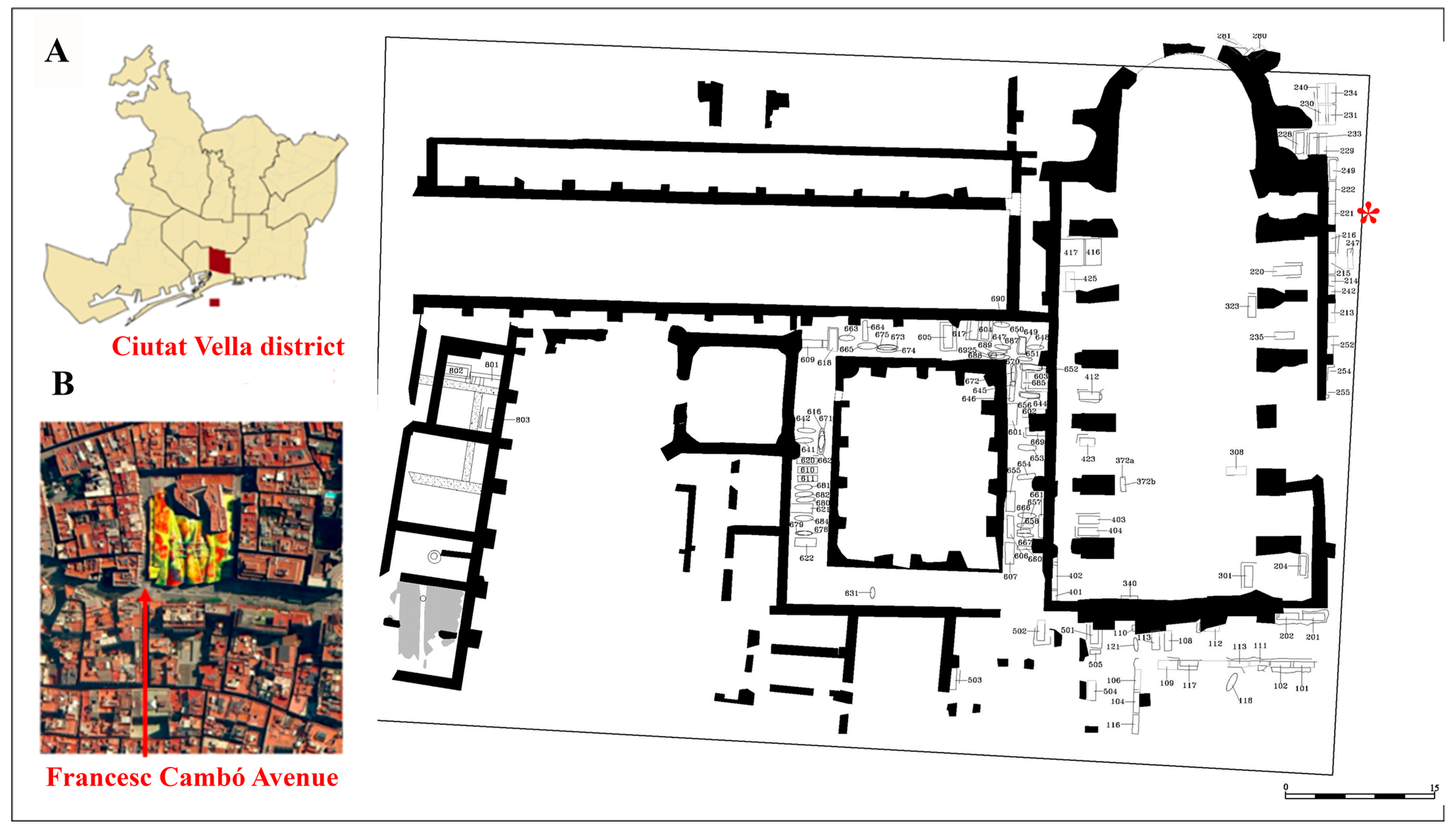
2. Materials and Methods
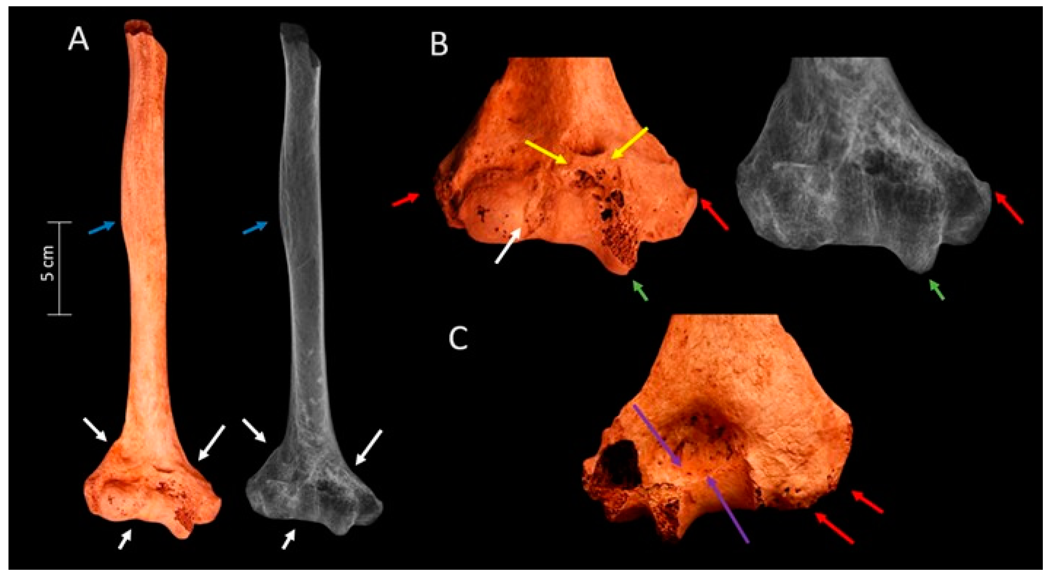
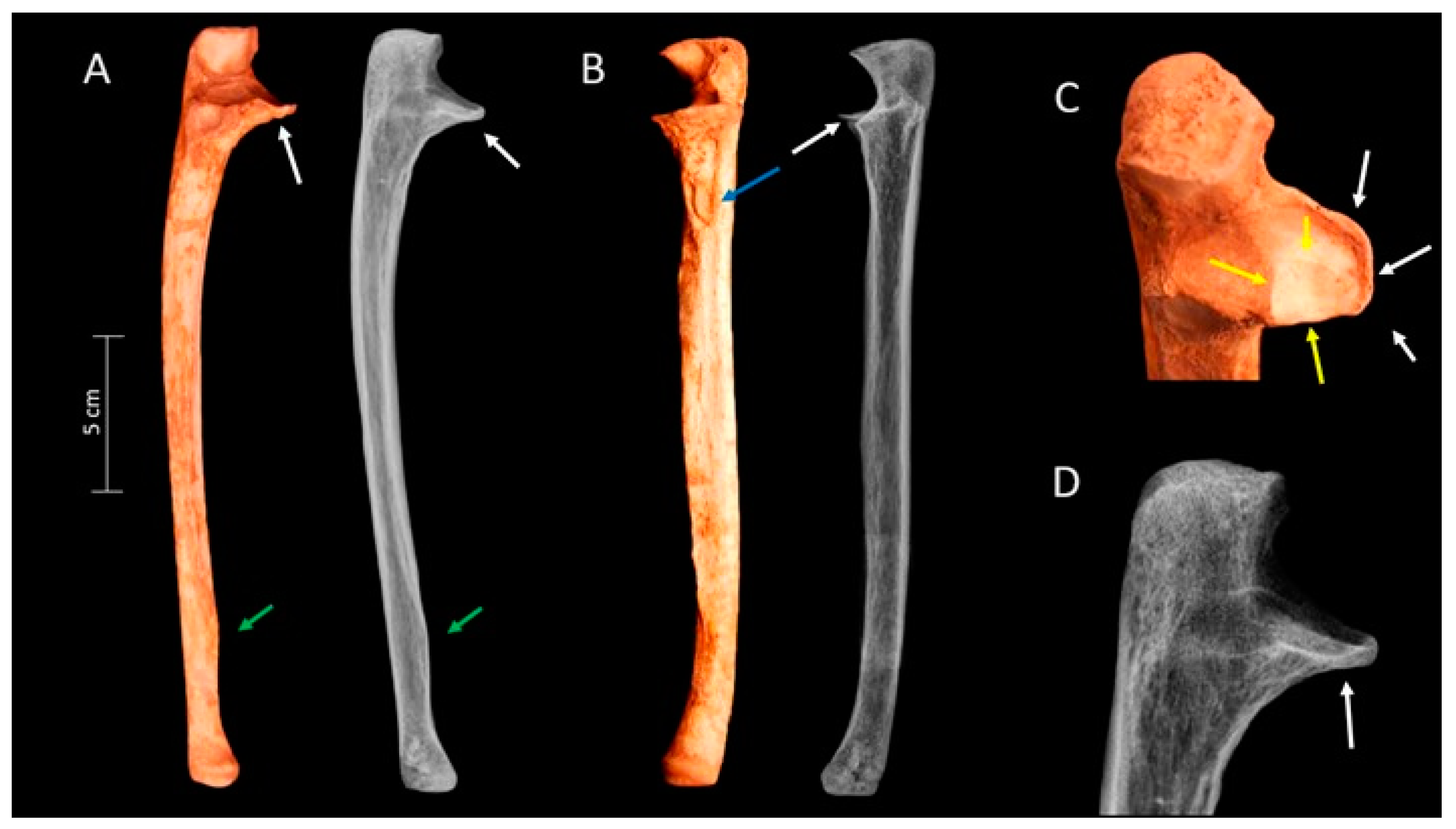
The material under investigation is two articulated bones, consisting of a right humerus (Humerus-009) and a right ulna (Ulna-012), found in anatomical connection and corresponding to the same individual (Figure 2A,B). Both bones are almost complete, except for the absence of the proximal epiphysis in the humerus and the styloid process in the ulna. They were found in the funerary unit (FU) 221 of the Santa Caterina Friary archeological site, specifically in stratigraphic unit 2369 (SU-2369), and they are housed in the Museum of History of the City of Barcelona (MUHBA). Santa Caterina Friary belonged to the Dominican order and was located in the Ciutat Vella district of Barcelona, Spain, serving as a pivotal institution during the medieval and post-medieval periods.
The FU-221 was located outside of the nave church of the friary, between two external buttresses of the Gothic chapels (Figure 1C). Archeologically and anthropologically, it was related to a secular guild community with a profession that required a high demand for physical effort [13,14]. The individuals were exhumed during the archeological interventions carried out between 1999 and 2003 by the company CODEX. Arqueologia i Patrimoni, SCCL [13]. In FU-221, there were two layers of burials belonging to two clearly differentiated historical periods: medieval and modern. The medieval period (13th to 14th centuries) corresponds to not individualized secondary burials, a commingling of bone elements, and is represented by a single stratigraphic unit (SU), SU-2378. The modern period (15th to 16th century) corresponds to successive primary burials and comprises five stratigraphic units (SU): 2374, 2375, 2376, 2377 (corresponding to individual tombs), and SU-2369, which corresponds to a collective burial that the bones are mixed and not individualized [13]. The last stratigraphic unit, SU-2369, is where the humerus and ulna of this study are from. Therefore, in this study, establishing associations between the analyzed humerus and ulna with other skeletal elements, such as the radius, shoulder girdle bones, and carpals, was not achievable due to the commingling of bone elements in SU-2369.
FU-221 contained a minimum of sixty-two individuals, including at least fifteen males, seven females, and five non-adults from the medieval period, as well as at least twelve males, eight females, and fifteen non-adults from the modern period [14]. Differences in robustness, height, health, and physical activity were observed among these individuals and the friars, with the friars being notably shorter and less robust. Additionally, individuals from the medieval and modern periods of FU-221 were also more robust compared to their peninsular contemporaries [14,15]. Notably, individuals from the modern period exhibited lower levels of physical activity compared to their medieval counterparts, possibly due to developments that facilitated less strenuous work during the transition to modernization, with machinery technology likely reducing the physical force requirements and supporting their primary work activities [14].
Anthropological and paleopathological studies were conducted at the Collections Center of the Archaeological Archive of the Museum of History of the City of Barcelona (MU-HBA). The sex and age of the analyzed elements were estimated using discriminant function analysis and the degree of maturation and fusion of epiphyses [16], respectively. Due to the incomplete condition of the bone and pathological alterations in certain anatomical structures, measurements that were not affected by these conditions were employed. Specifically, these included the least circumference of the humeral shaft, the anteroposterior shaft diameter of the ulna, the midshaft circumference, and the transverse shaft diameter [17]. We scored the entheses as activity markers, utilizing the standardized method for the postcranial skeleton proposed by Mariotti et al. [18]. Injuries were classified according to the AO/OTA system (Arbeitsgemeinschaft für Osteosynthesefragen/Orthopedic Trauma Association) [19], which is a clinical tool accepted and used in paleopathology research [3].
Measurements of the humerus and ulnae were obtained to evaluate the extent of the injury. Maximum ulnar length and midshaft diameters of both the humerus and ulna were recorded using an osteometric board and sliding calipers [20,21]. To assess the degree of angulation, measurements of the elbow joint (composed of the humerus and ulna) were also recorded. Elbow joint angularity was measured to assess the degree of angulation using 2D renderings in ImageJ number of software 1.54f [22]. The carrying angle, which is measured in supination between the long axes of the humerus and ulna, and the humeral tangential angle (articular surface angle)—the angle formed between the humerus’s longitudinal axis and the line drawn along the most distal axis of the capitellum and trochlea—were measured [3].
Additionally, plain film radiography was conducted on both the humerus and ulna at the Hospital Clínic in Barcelona. The radiographs were taken in two orthogonal projections—anteroposterior and mediolateral—using Philips Diagnost W medical radiological equipment, set at 50 kilovolts (kV) and 15 milliamperes per second (mAs), with a focus-plate distance of 120 cm. The processing of the radiographs utilized digital radiological MultiSync LCD 18805XW equipment, AGFA, Esplugues de Llobregat, Barcelona, Spain, with adjustments made to brightness and contrast to achieve optimal image quality.

3. Results

The results indicate an adult male. A more precise age estimation was not possible due to a lack of information. The humerus and ulna exhibit robust insertions: the deltoid muscle insertion at the humerus (Figure 3A, blue arrows), and at the ulna, a conspicuous curvature is discernible in the insertion of the pronator quadratus, indicating a pronounced insertion (Figure 4A, green arrows), as well as the brachialis muscle insertion (Figure 4B, blue arrows). When articulated, these two bones showed an elbow with excessive lateral angulation (cubitus valgus) (Figure 2A,B).
The examination of the distal part of the humerus (Figure 3A, white arrows) reveals a healed fracture. The radiographic image depicts a radiolucent “Y”-shaped line dividing the distal humeral epiphysis into three parts—separating the capitellum, trochlea, and medial epicondyle (Figure 3A, white arrows)—corresponding to an AO/OTA type 13 C1.1 fracture (complete articular fracture, articular simple, metaphyseal simple, above the transcondylar axis). Notably, the trochlea is slightly rotated postero-inferiorly, displaying an unusually large and prominent size (Figure 3B, green arrows). A line of bone growth anterior and transversal to the coronoid fossa secondary to post-traumatic arthritis is observed (Figure 3B, yellow arrows). Macroscopic and radiological observations also reveal new bone formation secondary to post-traumatic arthritis around the capitulum and in the medial and lateral condyles (Figure 3B,C red arrows). Finally, at the olecranon fossa, bone remodeling secondary to post-traumatic arthrosis is observed (Figure 3C, purple arrows).
The ulna also exhibits a healed fracture in the coronoid process within the sublime tubercle (Figure 4A–D; white arrows). Radiographically, the presence of a radiopaque area in the coronoid process indicates a transverse deposition of new bone, attributable to the healed fracture in this region (Figure 4D, white arrow). The coronoid process was displaced inferiorly and anteriorly due to the shear fracture. The coronoid facet was splayed widely, and in a lateral view, the trochlear notch was V-shaped. Furthermore, partial eburnation at the coronoid facet is observed (Figure 4C, yellow arrows). These fractures are probably due to anterior trochlear humeral dislocation, provoking a probable O’Driscoll classification type 1 (a coronoid process secondary fracture due to anterior trochlear dislocation impact) [23], which refers to a transverse fracture of the distal apophysis with the involvement of the sublime tubercle.
These humeral and ulnar morphological modifications precipitated biomechanical alterations within the elbow joint. The collective impact of injuries involving both the humerus and ulna significantly contributed to the evident angular deformity in the elbow joint. Additionally, the prominent muscle insertions on both bones are likely the result of overusing the joint due to previous fractures.
Anthropometric measurements, indexes, and angularity measures are detailed in Table 1. These anthropometric data are crucial for understanding the dimensions of these bones. However, due to the variability in the human skeleton and the trauma involved in this case, it is impossible to establish the original anatomical morphology of the bones. Ideally, comparing the left and right sides of the same individual can provide more accurate insights, particularly when comparing injured and non-injured sides. Given these considerations, we present the measurements and indices for both bones as a reference. Notably, the distal humerus is the most affected area in this case, with parameters such as epicondylar breadth, distal articular breadth, olecranon fossa breadth, and minimum trochlear breadth potentially being impacted by the injury. However, the most significant factor to consider is the angularity of the elbow. The carrying angle, measured at 28°, indicates excessive lateral angulation (cubitus valgus), which is a crucial feature in assessing the extent of the injury.

4. Discussion

This article presents a case of a complete articular fracture (AO/ATO type 13C1.1) of the distal humerus, accompanied by a secondary fracture of the coronoid process due to the anterior trochlear dislocation impact (O’Driscoll type 1), resulting in cubitus valgus. These findings were observed in the remains of a robust male from an unidentified guild buried at Santa Caterina Friary (15th to early 16th century).
Paleopathological records indicate varying prevalence rates of humeral fractures, ranging from 0% to 4.4% [3,11,24,25,26,27]. In contemporary clinical settings, distal humeral fractures are infrequent, constituting only 2% of all adult fractures [28,29], with an incidence of less than 10 per 100,000 of the population per year and an almost equal male-to-female ratio [29,30]. Some studies suggest prevalence rates of distal humerus fractures ranging from 0.5% to 7%, comprising 30% of elbow fractures [31,32]. In historical populations such as this one, cubitus valgus as a complication of humeral fracture has rarely been described. This angular deformation of the elbow can be the result of lateral condylar mal-union or non-union in the distal humerus [8,33]. Nevertheless, it can also be caused by the dislocation of the elbow [34,35] and the development of congenital disorders, such as achondroplasia [36]. Achondroplasia is a metaphyseal dysplasia, which modifies the bone epiphyses, giving it a very distinct form. This disorder was discarded because it is not a likely causative factor in the analyzed case.
Clinically, distal humeral fractures are commonly associated with falls in the elderly or high-energy impacts such as traffic accidents in the young [29]. Falls often result in injuries to the forearm and lower leg joints [3,37]. During a fall, individuals instinctively protect themselves by either extending or flexing their arms, leading to fractures of the forearm or elbow [38]. However, due to the incomplete skeleton in this case, the specific mechanism of injury cannot be determined. Indirect impacts on the elbow generate axial forces along the radius, ulna, and distal humerus, resulting in posterior displacement of the joint segments, whereas direct impacts on a flexed elbow cause anterior displacement of the joint segments [39].
The reported fracture in this case suggests a fall onto the right outstretched arm, as evidenced by the posterior rotation of the trochlea and coronal displacement of the ulnar coronoid process. It is probable that this individual’s radius also sustained a transverse fracture of the radial head, although confirmation is difficult due to the absence of the radius element. Consideration was also given to the possibility of concurrent ankle injuries before or after the elbow injury; however, insufficient information precludes further determination.
The timing of the injury during the individual’s lifetime remains uncertain. Fracture healing and remodeling periods vary among individuals, influenced by factors such as stabilization and mobility of the injured limb [40,41]. In adults, fracture remodeling may begin as early as one month post-injury, with full healing typically taking three to five months and possibly extending over years [42]. However, angulation in the coronal plane as seen in cubitus valgus may persist without surgical intervention, potentially worsening with age [43,44]. From a clinical perspective, the average time to union for distal humeral fractures following surgical intervention is 12–14 weeks [45]. In the presented case, fusion and remodeling of the fracture margins with marked angulation indicate that several years have likely passed since the injury, suggesting that some form of care was provided.

Treatment

In the 15th to early 16th century, management of elbow fractures such as the reported type 13-C1.1 likely involved immobilization with splints made from wood, metal, or leather to minimize movement and support healing [46]. Linen or cotton bandages were used to secure the splints and provide additional support. There was a formula for a kind of plaster casing anticipating the modern plaster [46]. Albucasis of Cordoba (936-1013), for humerus fractures, applied plasters of mill-dust and egg-white immediately after reduction provided no swelling or inflammation was present [47]. Herbal remedies, including poultices made from comfrey, arnica, and chamomile, were usually applied for pain relief and healing promotion [46,47,48]. Surgical intervention was rare during this historical period, with severe cases potentially resulting in amputation [46,47,48]. In fact, knowledge in bone and joint anatomy of the shoulder and elbow useful for surgical purposes is not found in medieval sources [47]. This information starts to be evident in the Renaissance with the studies of Leonardo da Vinci, who demonstrated biomechanical knowledge of these anatomical regions [47]. Despite consideration for surgical intervention, the reported case healed adequately, preserving relatively the anatomy of the elbow joint.
Cubitus valgus restricts elbow motion, but forearm rotation is usually unaffected [8,49]. In this case, visible angulation would have been present at the angle of the arm, with limited elbow joint motion. While there may have been some pain, limited functionality may have still been possible, as evidenced by the presence of osteoarthritic changes. The absence of synostosis suggests that, despite potential pain and complications, the individual retained functional use of the joint for daily activities. This resilience is evidenced by the development of musculoskeletal stress markers in the deltoid, brachial, and pronator quadratus muscles, muscles engaged in activities such as throwing, domestic chores, livestock handling, or heavy lifting [50].
Distal humeral fracture may affect the neurovascular supply of the elbow and forearm [51,52], resulting in a quarter of the cases in the damage of the median or ulnar nerve [53]. In addition, the carrying angle augmented increases the probability of ulnar neuropathy [54,55]. The incidence of ulnar neuropathy following non-surgical treatment is high [54,56]. Thus, it is highly probable that this individual would have had visible and functional impairment, including difficulty carrying heavy weights and possible loss of sensation and fine motor skills to the right hand. Limited function in the injured arm requires extra compensation in the uninjured arm.
Currently, treating distal humerus fractures remains challenging due to the complex triangular anatomy. Even with modern techniques, achieving and maintaining anatomically correct reductions during healing is difficult, often resulting in misalignment post-recovery [57,58].
Considering the previous archeological and anthropological information, FU-221 seems to correspond to the burials of a secular guild community with a profession that required a high demand for physical effort [13,14], as indicated by the presence of this distal elbow fracture. This affirmation is also reinforced by the high prevalence of traumatic pathologies, such as Schmorl’s nodes among individuals (49.10% in the medieval series and 34.30% in the modern series), significant disparities in antemortem trauma (31.10% in the medieval series and 9.00% in the modern series) [14], and markers of occupational stress and robustness compared to friars and contemporary individuals from the Santa Caterina site and the other contemporary Mediterranean populations [14,15]. It is reasonable to infer that these guild members of FU-221 engaged in continuous physically demanding activities, with increased risks of accidents related to hazardous tasks or the use of machinery as primary occupational activities. The sustained rigor of these guild activities and increased accident risks are particularly noteworthy, pointing to some type of occupation such as that of metalworkers.
The requirement for affluent individuals to afford burial strongly suggests that these guild members belonged to a financially well-off social class, highlighting their elevated status and potential contributions to the guild’s economic prosperity [59]. In the 13th century, in Barcelona, many artisans were grouped into corporations that, in the 14th century, were transformed into guilds, extending into the modern period of the 16th century. The guilds not only trained new artisans but also defended the economic and professional interests of those who belonged to the same trade. They controlled production levels and set prices. The guilds formed the foundations of medieval and modern society in Barcelona [60]. The members of these guilds gained significant political importance due to the creation of the Consell de Cent (Council of One Hundred), which gave them a political force unmatched in any other city in Spain [60].

5. Limitations

Assessing a differential diagnosis in cases involving commingled remains or poorly preserved skeletons poses unique challenges. Despite these difficulties, numerous studies provide evidence that the meticulous examination of both commingled remains and poorly preserved individualized remains is feasible [61]. Advancements in the field of paleopathology and the application of technological tools aim to enhance the accuracy of diagnoses, even in challenging conditions.
The limitation of this study is the absence of the radius, carpals, shoulder girdle, and entire skeleton. These elements, in general, would have been crucial for providing a better description of the individual, a more precise diagnosis, and more comprehensive information about this individual’s health.

6. Conclusions

This article describes and analyzes the case of a distal humeral fracture in an adult male buried at the Santa Caterina Friary site in Barcelona from funerary unit 221 (15th to early 16th century). The individual exhibited a healed elbow fracture, specifically showing healed distal humerus and proximal ulna fractures, resulting in cubitus valgus (28°). The humerus fracture was classified as type 13C1.1 according to the AO/OTA system. The proximal ulna fracture was classified as O’Driscoll type 1. This is one of the first paleopathological cases from Spain in which a cubitus valgus is described. The identified fracture probably resulted from a fall onto the right outstretched arm, revealing posterior rotation of the trochlea and coronal displacement of the coronoid process. The absence of the radius complicates the confirmation, and the possibility of concurrent ankle injuries is considered. The timing of the injury remains uncertain, and the healing and remodeling process can vary, impacting functional outcomes.
Bioarcheological and paleopathological records contextualize the prevalence of humeral fractures, emphasizing the challenges of identifying and reporting healed fractures in the archeological record. The observed enthesopathies provide clues about the individual’s lifestyle and occupational stress. Treatment in the 15th and early 16th centuries likely involved immobilization with splints and herbal remedies. Despite potential limitations in medical knowledge and technology, the reported case healed without surgical intervention, suggesting that the individual adapted to the injury, although likely experiencing long-term discomfort, probable functional impairment of the arm, and possible ulnar neuropathy.
This case, along with paleopathological observations of other individuals from the guild community in FU-221, underscores the physically demanding nature of their occupation. In summary, this case not only contributes to the understanding of paleopathology but also provides insights into the socio-economic dynamics and challenges faced by guild-affiliated individuals during the medieval period in Barcelona. The integration of archeological, clinical, anthropological, and paleopathological perspectives enriches our knowledge of the complexities of life in the past, emphasizing the resilience and interconnectedness of communities in overcoming physical adversities.

Author Contributions

A.C.: writing—review and editing, original draft, visualization, investigation, formal analysis, differential diagnosis, methodology, conceptualization. X.T.: writing—review and editing, differential diagnosis, and radiological images. L.L.: writing—review and editing, original draft, visualization, supervision, project administration. C.R.: writing—review and editing, original draft, visualization, supervision, differential diagnosis, investigation, conceptualization. All authors have read and agreed to the published version of the manuscript.

Funding

This research was funded by: MONBONES, Ministerio de Ciencia e Innovación (MICINN) Ref: PID2020-118194RJ-I00, SGR Evolució Social, Cultural i Biològica al Pleistocè (StEP) Ref: 2021 SGR. A. C. was funded by Agència de Gestió d’Ajuts Universitaris i de Recerca (AGAUR) grant ref. 2023 FISDU 00247.

Data Availability Statement

The data that support the findings of this study are available from the corresponding author upon reasonable request.

Acknowledgments

The authors would like to thank the Museo de Historia de Barcelona (MUHBA), and particularly Emili Revilla from the Barcelona History Museum for their complete access to the osteological collections.

Conflicts of Interest

The authors declare no conflicts of interest.

References

  1. Lovell, N.C. Analysis and interpretation of skeletal trauma. In Biological Anthropology of the Human Skeleton; Katzenberg, M.A., Saunders, S.R., Eds.; Wiley: Hoboken, NJ, USA, 2008. [Google Scholar]
  2. Bigham-Sadegh, A.; Oryan, A. Basic concepts regarding fracture healing and the current options and future directions in managing bone fractures. Int. Wound J. 2015, 12, 238–247. [Google Scholar] [CrossRef]
  3. Mole, C.; Stynder, D.; Gibbon, V.; Deano, D. Traumatic cubitus valgus consequent of distal humeral fracture: Two case studies from the Holocene Later Stone Age in southern Africa. Int. J. Paleopathol. 2023, 12, 7–15. [Google Scholar] [CrossRef]
  4. Lovell, N.C. Trauma analysis in paleopathology. Yearb. Phys. Anthropol. 1997, 40, 139–170. [Google Scholar] [CrossRef]
  5. Morrey, B.F.; An, K.N. Articular and ligamentous contributions to the stability of the elbow joint. Am. J. Sports Med. 1983, 11, 315–319. [Google Scholar] [CrossRef]
  6. Stroyan, M.; Wilk, K.E. The functional anatomy of the elbow complex. J. Orthop. Sports Phys. Ther. 1993, 17, 279–288. [Google Scholar] [CrossRef]
  7. McCoy, G.F.; Piggot, J. Supracondylar osteotomy for cubitus varus. Value Straight Arm. Position. J. Bone Jt. Surg.—Ser. B 1988, 70, 283–286. [Google Scholar] [CrossRef] [PubMed]
  8. Kozin, S.H. Cubitus Valgus. In Pediatric Elbow Fractures: A Clinical Guide to Management; Abzug, J.M., Herman, M.J., Kozin, S., Eds.; Springer: Berlin/Heidelberg, Germany, 2018; pp. 217–224. [Google Scholar]
  9. Glencross, B.; Stuart-Macadam, P. Radiographic clues to fractures of distal humerus in archaeological remains. Int. J. Osteoarchaeol. 2001, 11, 298–310. [Google Scholar] [CrossRef]
  10. Nagaoka, T.; Kazuhiro, U.; Yuji, S.; Morales, D. Pacopampa: Early Evidence of Violence ata Ceremonial Site in the Northern Peruvian Highlands. PLoS ONE 2017, 12, e0185421. [Google Scholar] [CrossRef]
  11. Djurić, M.P.; Roberts, C.A.; Rakocević, Z.B.; Djonić, D.D.; Lesić, A.R. Fractures in late medieval skeletal populations from Serbia. Am. J. Phys. Anthropol. 2006, 130, 167–178. [Google Scholar] [CrossRef] [PubMed]
  12. Judd, M. Comparison of long bone trauma recording methods. J. Archaeol. Sci. 2002, 29, 1255–1265. [Google Scholar] [CrossRef]
  13. Huertas, J.; Aguelo, J. Memòria de la Intervenció Arqueològica al Solar del al Mercat de Santa Caterina. Barcelona (Barcelonès), Spain, 2006. Unpublished.
  14. Cevallos, A.; Rissech, C.; Huertas, J.; Lloveras Roca, L. Estudio bioantropológico de los restos humanos procedentes de la unidad funeraria (UF) 221 del antiguo convento de Santa Caterina (1243–1836), Barcelona. RODIS J. Mediev. Post-Mediev. Archaeol. 2023, 6, 171–216. [Google Scholar] [CrossRef]
  15. Trujillo, J.; Rissech, C.; Lloveras Ll Huertas, J. El convento de Santa Caterina. Estudio bioantropológico de una muestra de las inhumaciones del claustro gótico y la cabecera del ábside. RODIS J. Mediev. Post-Mediev. Archaeol. 2023, 6, 217–242. [Google Scholar] [CrossRef]
  16. Scheuer, L.; Black, S.; Christie, A. Developmental Juvenile Osteology; Elsevier: Amsterdam, The Netherlands, 2010. [Google Scholar]
  17. Alemán, I.; Botella, M.C.; Ruiz, L. Determinación del sexo en el esqueleto postcraneal. Estudio de una población mediterránea actual. Arch. Español Morfol. 1997, 2, 7–17. [Google Scholar]
  18. Mariotti, V.; Facchini, F.; Belcastro, M.G. The study of entheses: Proposal of a standardised scoring method for twenty-three entheses of the post-cranial skeleton. Coll. Antropol. 2007, 31, 291–313. [Google Scholar] [PubMed]
  19. Meinberg, E.; Agel, J.; Roberts, C.; Karam, M.D.; Kellam, J.F. Fracture and Dislocation Classification Compendium. J. Orthop. Trauma 2018, 32, S1–S120. [Google Scholar] [CrossRef] [PubMed]
  20. Martin, R.; Saller, K. Lehrbuch der Anthropologie; G. Fischer: Sttutgart, Germany, 1957. [Google Scholar]
  21. Moore-Jansen, P.H.; Jantz, R.L. Data Collection Procedures for Forensic Skeletal Material; Report of Investigations No. 48; University of Tennessee: Knoxville, TX, USA, 1989. [Google Scholar]
  22. Schneider, C.A.; Rasband, W.S.; Eliceiri, K.W. NIH Image to ImageJ: 25 years of image analysis. Nat. Methods 2012, 9, 671–675. [Google Scholar] [CrossRef] [PubMed]
  23. O’Driscoll, S.W. Classification and evaluation of recurrent instability of the elbow. Clin. Orthop. Relat. Res. 2000, 370, 34–43. [Google Scholar] [CrossRef] [PubMed]
  24. Jurmain, R. Paleoepidemiolgical patterns of trauma in a prehistoric population from Central California. Am. J. Phys. Anthr. 2001, 115, 13–23. [Google Scholar] [CrossRef]
  25. Judd, M. Trauma in the City of Kerma: Ancient versus modern injury patterns. Int. J. Osteoarchaeol. 2004, 14, 34–51. [Google Scholar] [CrossRef]
  26. Oxenham, M.F.; Walters, I.; Cuong, N.L.; Thuy, N.K. Case studies in ancient trauma: Mid-Holocene through metal periods in northern Viet Nam. In Causes and Effects of Human Variation; Hennenberg, M., Ed.; Australasian Society for Human Biology, University of Adelaide: Adelaide, Australia, 2001; pp. 83–102. [Google Scholar]
  27. Domett, K.M.; Tayles, N. Adult fracture patterns in prehistoric Thailand: A biocultural interpretation. Int. J. Osteoarchaeol. 2006, 16, 185–199. [Google Scholar] [CrossRef]
  28. Jupiter, J.B.; Morrey, B.F. Fractures of the distal humerus in the adult. In The Elbow and its Disorders, 2nd ed.; Morrey, B.F., Ed.; W.B. Saunders: Philadelphia, PA, USA, 1993; pp. 328–366. [Google Scholar]
  29. Robinson, C.M.; Hill, R.M.F.; Jacobs, N.; Dall, G.; Court-Brown, C.M. Adult distal humeral metaphyseal fractures: Epidemiology and results of treatment. J. Orthop. Trauma 2003, 17, 38–47. [Google Scholar] [CrossRef]
  30. Van Staa, T.P.; Dennison, E.M.; Leufkens, H.G.M.; Cooper, C. Epidemiology of fractures in England and Wales. Bone 2001, 29, 517–522. [Google Scholar] [CrossRef] [PubMed]
  31. Wang, X.; Liu, G. A comparison between perpendicular and parallel plating methods for distal humerus fractures. Medicine 2020, 99, 19602. [Google Scholar] [CrossRef]
  32. Galal, S.; Mattar, Y.; Solyman, A.; Ezzat, M. Locking versus non-locking plates in fixation of extra-articular distal humerus fracture: A randomized controlled study. Int. Orthop. 2020, 44, 2761–2767. [Google Scholar] [CrossRef] [PubMed]
  33. Lahoti, O.; Akilapa, O. Not Kidding! Sequalae of elbow trauma in children. J. Clin. Orthop. Trauma 2021, 20, 101471. [Google Scholar] [CrossRef] [PubMed]
  34. Adaş, M.; Bayraktar, M.K.; Tonbul, M.; Uzun, M.; Çakar, M.; Tekin, A.Ç.; Kalkar, I.; Esenyel, M. The role of simple elbow dislocations in cubitus valgus development in children. Int. Orthop. 2014, 38, 797–802. [Google Scholar] [CrossRef] [PubMed]
  35. Kaas, L.; Struijs, P.A.A. Congenital radial head dislocation with a progressive cubitus valgus: A case report. Strateg. Trauma Limb Reconstr. 2012, 7, 39–44. [Google Scholar] [CrossRef] [PubMed]
  36. Nater, C.I.; Theuws, F.C.W.J.; Waters-Rist, A.L. Osteological evidence of achondroplasia in an individual from medieval Reusel, the Netherlands. J. Paleopathol. 2016, 26, 73–83. [Google Scholar]
  37. Verma, S.K.; Willetts, J.L.; Corns, H.L.; Marucci-Wellman, H.R.; Lombardi, D.A.; Courtney, T.K. Falls and fall-related injuries among community-dwelling adults in the United States. PLoS ONE 2016, 11, e0150939. [Google Scholar] [CrossRef]
  38. Galloway, A. The upper extremity. In Broken Bones: Anthropological Analysis of Blunt Force Trauma; Wedel, V.L., Galloway, A., Eds.; Charles C Thomas: Springfield, IL, USA, 2014; pp. 195–244. [Google Scholar]
  39. Stans, A.A. Supracondylar fractures of the elbow in children. In Morrey’s the Elbow and Its Disorders; Morrey, B.F., Sanchez-Sotelo, J., Morrey, M.E., Eds.; Elsevier: Philadelphia, PA, USA, 2018; pp. 253–271. [Google Scholar]
  40. Redfern, R.C.; Roberts, C.A. Trauma. In Ortner’s Identification of Pathological Conditions in Human Skeletal Remains; Buikstra, J., Ed.; Academic Press: London, UK, 2019; pp. 211–284. [Google Scholar]
  41. Foster, A.L.; Moriarty, T.F.; Zalavras, C.; Morgenstern, M.; Jaiprakash, A.; Crawford, R.; Burch, M.A.; Boot, W.; Tetsworth, K.; Miclau, T.; et al. The influence of biomechanical stability on bone healing and fracture-related infection: The legacy of Stephan Perren. Injury 2021, 52, 43–52. [Google Scholar] [CrossRef]
  42. Viero, A.; Obertová, Z.; Cappella, A.; Messina, C.; Sconfienza, L.M.; Sardanelli, F.; Tritella, S.; Montisci, M.; Gregori, D.; Tagliaro, F.; et al. The problem of dating fractures: A retrospective observational study of radiologic features of fracture healing in adults. Forensic Sci. Int. 2021, 329, 111058. [Google Scholar] [CrossRef]
  43. Mann, T.S. Prognosis in supracondylar Fract. J. Bone Jt. Surg. 1963, 45, 516–522. [Google Scholar] [CrossRef]
  44. McIntosh, A.L. Complications of supracondylar fractures of the elbow. In Morrey’s the Elbow and its Disorders; Morrey, B.F., Sanchez-Sotelo, J., Morrey, M.E., Eds.; Elsevier: Philadelphia, PA, USA, 2018; pp. 272–285. [Google Scholar]
  45. Huang, T.L.; Chiu, F.Y.; Chuang, T.Y.; Chen, T.H. The results of open reduction and internal fixation in elderly patients with severe fractures of the distal humerus: A critical analysis of the results. J. Trauma—Inj. Infect. Crit. Care 2005, 58, 62–69. [Google Scholar] [CrossRef]
  46. Browne, E. Arabian Medicine, 1st ed.; Cambridge University: Cambridge, UK, 1921. [Google Scholar]
  47. Brorson, S. Medieval and early modern approaches to fractures of the proximal humerus: An historical review. Minerva Ortop. Traumatol. 2010, 61, 449–462. [Google Scholar]
  48. Ortner, D. Identification of Pathological Conditions in Human Skeletal Remains; Smithsonian University Press: Washington, DC, USA, 2003. [Google Scholar]
  49. Holdsworth, B.J.; Mossad, M.M. Fractures of the adult distal humerus. Elbow function after internal fixation. J. Bone Jt. Surg. 1990, 72, 362–365. [Google Scholar] [CrossRef] [PubMed]
  50. Estévez, M.C. Marcadores de Estrés y Actividad en la Población Guanche de Tenerife; Estudios Prehispánicos; Dirección General de Patrimonio Histórico: Canarias, Spain, 2003; Volume 14. [Google Scholar]
  51. Noaman, H.H. Microsurgical reconstruction of brachial artery injuries in displaced supracondylar fracture humerus in children. Microsurgery 2006, 26, 498–505. [Google Scholar] [CrossRef]
  52. Ring, D.; Jupiter, J.B. Complex fractures of the distal humerus and their complications. J. Shoulder Elb. Surg. 1999, 8, 85–97. [Google Scholar] [CrossRef] [PubMed]
  53. Bégué, T. Articular fractures of the distal humerus. Orthop. Traumatol. Surg. Res. 2014, 100, S55–S63. [Google Scholar] [CrossRef]
  54. Abed, Y.; Nour, K.; Kandil, Y.R.; El-Negery, A. Triple management of cubitus valgus deformity complicating neglected nonunion of fractures of lateral humeral condyle in children: A case series. Int. Orthop. 2018, 42, 375–384. [Google Scholar] [CrossRef]
  55. Chang, C.; Wang, Y.; Chu, C. Increased carrying angle is a risk factor for nontraumatic ulnar neuropathy at the elbow. Clin. Orthop. Relat. Res. 2008, 466, 2190–2195. [Google Scholar] [CrossRef]
  56. Toh, S.; Tsubo, K.; Nishikawa, S.; Inoue, S.; Nakamura, R.; Harata, S. Longstanding nonunion of fractures of the lateral humeral condyle. J. Bone Jt. Surg. 2002, 84, 593–598. [Google Scholar] [CrossRef] [PubMed]
  57. Mehne, D.K.; Jupiter, J.B. Part II—Fractures of the distal humerus. In Skeletal Trauma: Fractures, Dislocations, Ligamentous Injuries; Browner, B.D., Ed.; Saunders (W.B.) Co, Ltd.: Philadelphia, PA, USA, 1992. [Google Scholar]
  58. Dey Hazra, R.; Lill, H.; Jensen, G.; Imrecke, J.; Ellwein, A. Fracture-Pattern-Related Therapy Concepts in Distal Humeral Fractures. Obere Extrem. 2018, 13, 23–32. [Google Scholar] [CrossRef] [PubMed]
  59. Zaldívar, A.M. Patricians’ Embrace of the Dominican Convent of St. Catherine in Thirteenth Century Barcelona. Mediev. Encount. 2012, 18, 174–206. [Google Scholar] [CrossRef]
  60. Ortoll, E. Algunas consideraciones sobre la iglesia de Santa Caterina de Barcelona. Locus Amoenus 1996, 2, 47–63. [Google Scholar] [CrossRef]
  61. Assis, S.; Henderson, C.Y.; Casimiro, S.; Alves Cardoso, F. Is differential diagnosis attainable in disarticulated pathological bone remains? A case-study from a late 19th/early 20th century necropolis from Juncal (Porto de Mós, Portugal). Int. J. Paleopathol. 2018, 20, 26–37. [Google Scholar] [CrossRef]
Figure 1. The location of the Santa Caterina Friary in the northwest of the Ciutat Vella district of the city of Barcelona, Spain (A). The location of the Ribera neighborhood along Francesc Cambó Avenue (B). Location of FU-221, situated between the exterior buttresses of the Gothic chapels (C).
Heritage 07 00196 g001
Figure 2. Anteroposterior view (A) and posterior view (B) of the articulated right elbow (humerus and ulna).
Heritage 07 00196 g002
Figure 3. Humerus-009 from FU-221 and SU-2369. Anterior view of the humerus with the associated radiograph (A). Magnification of the anterior view of the distal humerus with the associated radiograph (B). Magnification of the posterior view of the distal humerus (C). Blue arrows indicate a prominent deltoid muscle insertion. White arrows show remodeling of the fracture at the distal epiphysis. Green arrows indicate post-traumatic arthritis in the trochlea and changes in its size. Yellow arrows highlight osteophytes secondary to post-traumatic arthritis. Red arrows show osteophytes and post-traumatic arthritis at the capitulum, medial, and lateral condyles. Purple arrows indicate post-traumatic arthritis in the olecranon fossa.
Heritage 07 00196 g003
Figure 4. Lateral view of the right ulna with the associated radiograph (A). Anterior view of the right ulna with the associated radiograph (B). Magnification of the proximal epiphysis of the right ulna (C) and its radiographic image (D). White arrows show remodeling of the fracture at the coronoid process of the right ulna. Green arrows show prominent pronator quadratus muscle insertion. Blue arrows indicate a prominent insertion of the brachialis muscle. Yellow arrows indicate an eburnation at the coronoid facet.
Heritage 07 00196 g004
Table 1. Absolute measurements, indices, and elbow angularity values of the humerus and ulna from FU-221 of the Santa Caterina Friary site.
Humerus (Measurement/Index)ValueUlna (Measurement/Index)ValueElbow Angularity
Measure of Angularity Value
Midshaft circumference60.80 mmPhysiological length255.00 mmCarrying angle28°
Maximum midshaft diameter17.70 mmMinimum circumference40.80 mm
Minimum midshaft diameter17.00 mmMidshaft circumference51.20 mm
Epicondylar breadth64.90 mmMaximum anteroposterior diameter13.30 mm
Distal articular breadth42.30 mmMediolateral anteroposterior diameter10.60 mmHumeral tangential angle137°
Olecranon fossa breadth21.90 mmOlecranon anteroposterior diameter27.3 mm
Minimum trochlear breadth16.60 mmOlecranon coronoid process distance24.90 mm
Deltoid V circumference70.30 mmMidshaft index20.08 cm
Robustness index18.04 cm
Disclaimer/Publisher’s Note: The statements, opinions and data contained in all publications are solely those of the individual author(s) and contributor(s) and not of MDPI and/or the editor(s). MDPI and/or the editor(s) disclaim responsibility for any injury to people or property resulting from any ideas, methods, instructions or products referred to in the content.

