Abstract
In this article, the classification of signals arising from the movements of the lower limb of the leg (LLL) based on electromyography (EMG) (walking, sitting, up and down the stairs) was carried out. In the data collection process, 25 athletes aged 15–22 were involved, and two types of data sets (DS-dataset) were formed using FreeEMG and Biosignalsplux devices. Six important time and frequency domain features were extracted from the EMG signals—RMS (Root Mean Square), MAV (Mean Absolute Value), WL (Waveform Length), ZC (Zero Crossing), MDF (Median Frequency), and SSCs (Slope Sign Changes). Several classification algorithms were used to detect and classify movements, including RF (Random Forest), NN (Neural Network), SVM (Support Vector Machine), k-NN (k-Nearest Neighbors), and LR (Logistic Regression) models. Analysis of the experimental results showed that the RF algorithm achieved the highest accuracy of 98.7% when classified with DS collected via the Biosignalsplux device, demonstrating an advantage in terms of performance in motion recognition. The results obtained from the open systems used in signal processing enable real-time monitoring of athletes’ physical condition, which plays a crucial role in accurately and rapidly determining the degree of muscle fatigue and the level of physical stress experienced during training sessions, thereby allowing for more effective control of performance and timely prevention of injuries.
1. Introduction
Surface EMG (sEMG) signal represents neuromuscular activity during potential changes on the skin surface during muscle contraction. Surface EMG signal detection is a non-invasive detection method. It is important in the analysis of sports movements, clinical diagnostics, and rehabilitation. In particular, the most important movements in sports are performed using the muscles of the arms and legs.
sEMG signals allow direct measurement of the electrical activity of muscles during the movements of athletes. This allows for the assessment of muscle activity in real time, directly related to the internal processes of the muscle, unlike other analysis methods, including kinematic and dynamic approaches. Kinematic analyses (e.g., motion trajectory, joint angles, or velocity measurements) evaluate the external result of the movement of muscles, but they cannot detect subtle changes in the initial phase of muscle activity. Dynamic analyses (e.g., force platforms or torque measurements) evaluate the mechanical forces and torques generated by muscles, but do not clearly indicate the source of the change in force, i.e., which muscle or muscle group is activated. It is EMG signals that directly measure the neuromotor activity of muscles, which gives a huge advantage in determining parameters such as fatigue, muscle coordination, and the quality of technique in athletes. Therefore, the EMG method is considered the most accurate and reliable choice for assessing LLL segment movements.
In recent years, extensive research has been conducted on leg movement detection using EMG signals [1,2,3,4,5,6]. These studies are mainly aimed at improving the control capabilities of rehabilitation technologies, smart prostheses, and exoskeleton robotic systems. In particular, various machine learning algorithms (SVM, RF, KNN, TCN—Temporal Convolutional Network) and feature extraction methods (in the time, frequency, time–frequency domains) have been used to classify movement from EMG signals. However, problems such as increasing classification accuracy, ensuring fatigue resistance, and real-time performance efficiency are still relevant. Therefore, approaches in this area and their results are analyzed by studying the existing literature (Table 1).

Table 1.
Literature review on EMG-based LLL segment movement studies.
In a study [1] aimed at assessing the muscle activity of the LLL segment, an experimental method was developed to detect leg movements from EMG signals of human movement. Feature vectors were formed based on time domain features (such as RMS, MAV, and ZC), and based on this data, an SVM classifier was selected to detect five main leg movements. As a result of experiments conducted based on the proposed model, an average accuracy rate of 95.66% was recorded.
The potential of EMG signals is gaining importance in gait analysis and control of rehabilitation exoskeletons. This study evaluated the effectiveness of machine learning algorithms (KNN, RF, SVM) in classifying movements based on EMG signals obtained from 22 participants [2]. As a result of experiments, the RF model with a combination of time and frequency domain features showed the highest result (92%).
Research is underway on smart prosthetic systems based on EMG signals to improve the quality of life of patients with lower limb amputations. In this study, EMG signals from leg muscles were obtained, and time domain features and the CatBoost algorithm were used to classify five movements (level walking, up the stairs, down the stairs, and ramp ascent and descent) [3].
An integrated approach of EEG and EMG signals based on discriminant correlation analysis (DCA) was considered for detecting bilateral LLL segment movements [4]. EEG and EMG signals from 28 healthy participants were combined at the feature level, and five types of classifiers were used to detect movements. The multimodal approach showed a particularly high performance (96.64%) with the linear discriminant analysis (LDA) classifier.
Wang, J. et al. [5] presented a systematic approach to analyze and recognize human lower limb movements based on EMG signals. Four lower limb muscles with low correlation, identified by OpenSim 4.3 and SPSS 28 gait analysis, were selected for data collection using inertial measurement units and EMG sensors under seven slope gradients, five gaits, and four functional movements. The raw EMG signals were smoothed with a Butterworth filter, and time–frequency domain features were extracted to serve as input to a two-hidden-layer BP neural network. Experimental results showed clear periodic patterns in EMG activity across different locomotor tasks, which allows for reliable classification. The proposed method achieved an average recognition accuracy of 86.49% for inclines, 93.76% for walks, and 86.07% for movements.
Inter-subject differences in sEMG signals are a major problem in detecting LLL segment movements in exoskeleton robots. In this regard, a motion detection method based on sEMG signals using non-negative matrix factorization, multiple nonlinear features, Fisher discriminant function, and GA-PSO optimized SVM is proposed [6]. This approach achieved 96.03% accuracy in distinguishing three different movements in eleven healthy and eleven knee pathology participants.
Existing functional electrical stimulation (FES) devices are inconvenient to place and cannot detect the user’s movement intention or muscle fatigue, which limits their application in daily life. A new wearable FES system based on sEMG with electrodes specially woven for the user is an important step in this direction [7]. The proposed deep learning-based parallel model FES system was tested on five participants and was able to detect LLL movements and muscle fatigue with high accuracy (93.33%).
In order to improve human–computer interaction in the control of smart prosthetics, a method for detecting LLL segment movements based on sEMG signals is proposed. To overcome the problem of phase information loss in existing methods, the proposed approach implements S-transform-based energy density analysis and multi-channel synthesis [8]. In this regard, sEMG signals obtained from six muscles of ten participants were analyzed based on four movements, and a detection accuracy of 96% was achieved.
Tu, J. et al. [9] proposed an improved denoising and classification system that combines VMD, an α-based thresholding algorithm, CNN feature extraction, and KELM classification to solve the problem of extracting discriminant features and high-accuracy motion detection from sEMG signals of the lower limbs. The sEMG signals from six lower limb muscles were recorded in five motion modes, pre-processed with notch filtering in the 50 Hz and 10–450 Hz bandpass filtering, and separated into multiple variational mode functions. The enhanced thresholding algorithm was applied to each mode to minimize signal distortion to suppress noise, and then the denoised signal was reconstructed. The cleaned sEMG data were transformed into a matrix form and sent to a CNN to automatically learn deep, task-relevant features, avoiding the limitations of manual feature extraction. KELM was then used for classification, taking advantage of its fast learning rate and strong generalization ability. Experimental evaluation using precision, accuracy, recall, F1 score, ROC curve, and AUC showed that the proposed CNN-KELM architecture consistently outperformed traditional feature-based standalone CNN and KELM models. The method achieved a classification accuracy of 95.90%, which represents an improvement of 18.29% over KELM and 11.86% over CNN.
A novel solution is to use a CNN-Transformer-LSTM (CNN-TL) coupled model based on sEMG data to classify LLL segment movements with greater accuracy [10]. sEMG signals from twenty participants were collected during four movements and analyzed in the time and frequency domains, and the selected features were fed into a neural network. The CNN-TL model achieved 96% accuracy and was 3.76%, 5.92%, and 14.92% higher than CNN, LSTM, and SVM, respectively.
In our previous scientific work [11], an analysis of classification methods was carried out to assess the physical fitness of athletes involved in wrestling based on EMG signals. In this study, the muscle activity of athletes was determined through EMG signals, bioelectric data from eight main muscles were analyzed, and their technical and physical exercises were classified into ten classes. The results showed that the RF algorithm achieved the highest result with an accuracy of 96.97%, proving that this model has high efficiency in processing complex bioelectric signals and minimizing uncertainties. However, these approaches may have some limitations; for example, since the FreeEMG device is a closed system, it cannot provide real-time processing of EMG signals obtained from athletes and obtain results. Second, it limits the assessment of athletes only based on muscle systems, which does not allow the use of data obtained from other biosystems of athletes in the analysis process in the future. Third, cross-subject and cross-session tests are necessary to ensure the reliability and stability of the model. Fourth, considering that athletes experience strong biomechanical loads on the LLL segment of the body during training, it is necessary to analyze the leg segments of athletes. At the same time, due to the stochastic nature of EMG signals, special attention should be paid to windowing and filtration processes in the processing processes.
In this work, the primary goal is to achieve better outcomes by utilizing devices that support the development of real-time monitoring systems. As can be observed from the literature review (Table 1), the majority of existing devices are based on closed platforms designed to first store the acquired files and then process them later, which creates significant limitations for the implementation of a real-time sports system. Whereas the present study seeks to overcome these restrictions by enhancing the performance of an open platform capable of processing signals and thereby ensuring more effective and practical applications in sports monitoring.
Based on these experimental results, this article further expands the research direction and proposes a system aimed at detecting athletes’ LLL segment movements in real time.
The main contributions of this paper are as follows:
- To estimate the LLL segment based on four movements, taking into account the biomechanical loading of the leg segment of the body during training in athletes.
- To prove the superiority of an open signal recording system and select lightweight classification models in order to offer more stable and practical solutions for real-time sports monitoring.
- To conduct cross-subject and cross-session tests to evaluate the reliability and stability of the model.
2. Data Collection Organization
This study used two high-resolution EMG data acquisition devices—FreeEMG (Milan, Italy, BTS Bioengineering S.P.A.) and Biosignalsplux (Lisbon, Portugal, PLUX Wireless Biosignals S.A.)—and compared the performance of different classification algorithms (RF, SVM, k-NN, NN, and LR). This approach systematically analyzes the impact of device parameters and selected classification methods on the motion detection process.
Although research on LLL segment motion detection using EMG signals has increased in recent years, most of the existing work has been conducted on a single device, and the stability and generalization ability between different devices have not been sufficiently evaluated. This is especially important for practical applications of the technology in different sports conditions and real-time monitoring systems. The aim of this study is to systematically compare the accuracy, stability, and generalization of different machine learning algorithms in classifying LLL segment motion based on data from two widely used EMG devices, FreeEMG and Biosignalsplux. The research hypothesis is that one of the two devices will show higher classification accuracy than the other due to its technical characteristics and signal quality.
Figure 1 illustrates the sequence of the research organization process. In the first stage of the process, EMG signals are recorded in real time using two devices, and a data set is formed. In the next stage, the initial signal processing process is performed on the raw data. In this stage, the signals are cleaned of various noise and artifacts, and signal cleaning filtration operations are performed using low-pass filter, high-pass filter, band-pass filter, and notch filters. After the initial processing, a feature extraction stage is performed to identify the most important components of the signal. In the final stage, each leg movement is classified using classification algorithms (SVM, KNN, RF, NN, and LR).
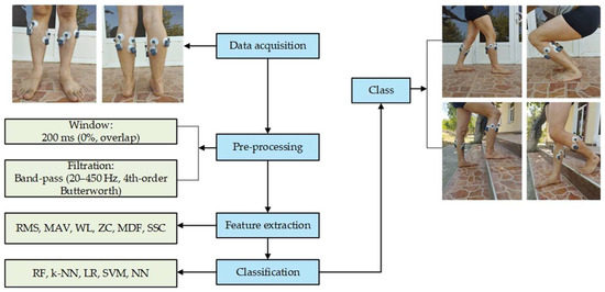
Figure 1.
The process of collecting and classifying EMG signals.
2.1. Devices
Special test-experiments were conducted to organize the DS. During the experiments, the athletes were adjusted taking into account the characteristics of the LLL segment movements.
In this study, two different EMG acquisition systems—the 8-channel BTS FreeEMG 1000 (Figure 2a) (Milan, Italy, BTS Bioengineering S.P.A.) and the 8-channel Biosignalsplux (Figure 2b) (Lisbon, Portugal, PLUX Wireless Biosignals S.A.)—were purposefully selected to evaluate the consistency, generalizability, and robustness of LLL movement classification across varying hardware platforms. This choice was motivated by both their technical distinctions and their practical relevance in different application domains. The technical specifications of the two devices are listed in Table 2.
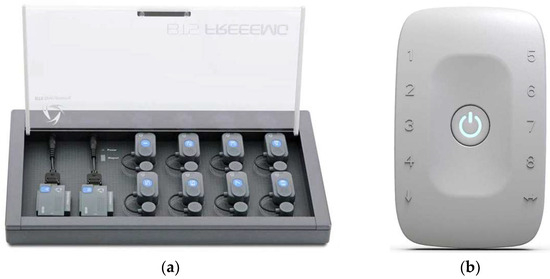
Figure 2.
(a) FreeEMG and (b) Biosignalsplux device.
Table 2.
Comparative analysis of FreeEMG and Biosignalsplux devices.
FreeEMG, widely used in controlled laboratory environments, provides high-fidelity EMG recordings with low latency and Wi-Fi connectivity, which is especially suited for gait analysis and sports biomechanics. On the other hand, Biosignalsplux, known for its modular design and multi-modal capabilities (supporting EMG, ECG, and EEG), is frequently used in wearable and real-time monitoring scenarios due to its Bluetooth-based portability.
By conducting parallel experiments using both systems under identical protocols, we aimed to investigate how device-specific characteristics such as sampling stability, noise resilience, and physical ergonomics may influence classification outcomes. The comparative analysis allows for more robust conclusions and enables us to offer practical recommendations for researchers and practitioners in selecting appropriate EMG hardware for specific use cases in athlete monitoring and rehabilitation.
During the signal recording process, Ag/AgCl (silver chloride) electrodes were used and placed in the innervation zones of the muscles (Figure 3a,b).
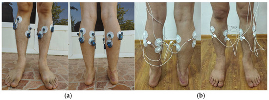
Figure 3.
Electrode placement: (a)—anterior, (b)—posterior.
The electrodes of the BTS FreeEMG and Biosignalsplux devices were selected to target the muscles that were most active during leg movements (Figure 3). Based on the location of the human leg muscles and the correspondence between the muscles and movement, and the following muscles were selected for each of the right and left legs: fibularis anterior, soleus, gastrocnemius lateral, and gastrocnemius medial.
2.2. DS Structure
In this study, the main muscles of the LLL segment were selected, considering that the leg plays an important role in human movement. In addition, four important types of physical exercises that are most often used in the leg were selected: walking, sitting and standing, up the stairs, and down the stairs (Figure 4).
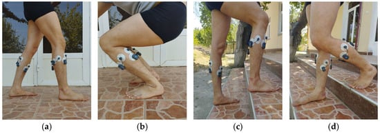
Figure 4.
Leg movement exercises used in the experiment ((a)—walking, (b)—sitting and standing, (c)—up the stairs, (d)—down the stairs).
During the study, a separate DS was created for each device. Each participant repeated the leg movements five times. Each session was held once a week. Fifteen sessions were held in 3 weeks. The volume of the DS is as follows:
15 (repetition) × 4 (class number) × 25 (members) = 1500
The experiment was conducted on 25 students, including 11 girls and 14 boys.
As a sample, the representative segments of EMG signals recorded from the lateral gastrocnemius muscle of the left leg are visually presented in Figure 5. This figure illustrates the time domain variations of the EMG signals corresponding to each movement.
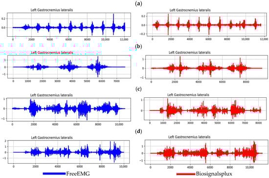
Figure 5.
Visual representation of EMG signals obtained from the left gastrocnemius lateral muscle ((a)—walking, (b)—sitting and standing, (c)—up the stairs, (d)—down the stairs).
3. Feature Extraction and Classification
This section describes the step-by-step process of detecting athletes’ leg movements based on EMG signals, pre-filtering the signals, and forming a set of features necessary for their classification. Characteristic features of movements are extracted, and modern and efficient classification algorithms are used to automatically identify movements based on these features.
As part of the study, analyses were conducted on EMG data sets collected separately using FreeEMG and Biosignalsplux devices. The data collected using each device was processed separately, and the accuracy of the classification models used to classify movements was compared. The experimental results analyzed the effect of the feature set on classification for different devices, as well as the performance of the algorithms, and their advantages and disadvantages were identified.
3.1. Filtration of EMG Signal
Factors (noises) that negatively affect the quality of EMG signals are power supply, motion artifacts, intermuscular interference, signal saturation, and physiological noise. Various filters are used to eliminate these factors. In our study, a 4th-order zero-phase Butterworth band-pass filter was used at the stage of preprocessing of EMG signals. The filter range was chosen to be 20–450 Hz. The lower cutoff frequency (20 Hz) was set to suppress low-frequency noise such as motion artifacts and basal drift. The upper cutoff frequency (450 Hz) reduces high-frequency electrical noise in addition to useful physiological components in EMG signals [13,14]. This range is based on international scientific recommendations that the main energy of electromyographic signals is concentrated in the range of 20–450 Hz. This range ensures the accuracy of features such as ZC, SSC, and MDF and allows for physiologically correct interpretation of muscle activity. After filtering the signals, 200 ms sequential time windows (0% overlap) were used to manipulate the incoming data.
3.2. Feature Extraction
It is not recommended to use raw EMG signals directly in classification algorithms, because these signals are very large and have a diverse nature. Therefore, the feature extraction method is used. Through this process, useful information is extracted from the signal and the data volume is reduced. The feature extraction technique is a necessary step for identifying effective patterns, and its effectiveness increases the accuracy of the classification result [15,16]. Table 3 presents an analysis of the studies conducted on the features of EMG signals.
Table 3.
Analysis of the literature on the properties of the EMG signal.
For efficient classification, the best six features were selected from the EMG signals based on the results of various scientific works.
The RMS feature has been used in many studies, such as Refs. [17,18,19,20,21]. In particular, 95% accuracy was achieved in studies [20,21]. This feature is a key parameter representing the total energy of the signal and provides stable results in classification. MAV is also found in many sources; for example, it was used in Refs. [17,18,22,23,24]. In Ref. [17], 97.44% accuracy was achieved and in Ref. [23], 97% accuracy was achieved. This feature calculates the average power of the signal in a simple and efficient way. WL represents the overall complexity of the signal shape. It was used in Refs. [18,19,21,23], and in Ref. [23] it provided 97% accuracy. This feature provides good discrimination in classification. ZC is the frequency variation of the signal by counting the zero crossing points. This feature was used in Refs. [18,19,22,24]. In particular, it showed 96% accuracy in Ref. [22]. MDF is a frequency domain feature that indicates the spectral midpoint of the signal energy. It was used in Refs. [18,19,23], and in Ref. [23] it achieved 97% accuracy and in Ref. [19] it achieved 93%. SSC represents the variability of the signal shape. This feature was used in Refs. [18,19,22], and in Ref. [22] it achieved 96% accuracy.
The remaining features—STD, VAR, Mean, and Skew—were not used because they showed low accuracy in the analyzed studies.
The six feature extraction models selected above are calculated as follows:
- ➢
- SSCs characterize the frequency-related dynamics of EMG signals by quantifying the number of sign reversals in the signal’s slope within a defined time window [19] and is calculated as follows:
Here:—ith signal sample, N—number of samples, threshold—predefined sensitivity value.
- ➢
- RMS is a widely used time domain feature in electromyographic (EMG) signal processing [25]. RMS effectively reflects muscle contraction intensity and is sensitive to signal amplitude variations, making it valuable for assessing neuromuscular activity. It can be obtained as follows:
- ➢
- MAV reflects the overall magnitude of muscle activation and is often used in real-time EMG-based control systems due to its computational simplicity and responsiveness to muscle contractions [22] and is defined as follows:
- ➢
- WL reflects the complexity and variability of the signal and is sensitive to both amplitude and frequency changes, making it useful for capturing the dynamic characteristics of muscle activity [21] and is calculated as follows:
- ➢
- ZC quantifies the number of instances where the signal amplitude transitions through zero, indicating a change in polarity [22]. It can be obtained as follows:
- ➢
- MDF represents the frequency point within the EMG power spectrum at which the spectrum is partitioned into two regions of equal power [21] and is defined as follows:
Here pj is the amplitude value in the frequency band indexed by j, and M is the total number of frequency components.
The assessment of muscle fatigue for the LLL segment is important in the field of sports. For this, we used the above parameters, which describe the frequency and amplitude of EMG signals in the assessment of muscle fatigue [26]. These features were selected not only because they were useful in improving classification performance in previous studies, but also because they have a physiological basis. RMS, MAV, and WL are effective in assessing the overall intensity and coordination of muscle activity, while ZC and MDF are effective in detecting muscle fatigue and spectral changes. SSC, on the other hand, characterizes rapid dynamic changes in muscle activity. This confirms that the selected features are closely related to muscle physiology and have a positive impact on the classification results.
3.3. Classification
A convolutional neural network (CNN) is a supervised learning model (SLM) that has shown high performance for temporal and visual data analysis. Many studies have used CNN models to achieve high performance [27,28,29]. However, when processing images in the classification process, high-performance GPUs are required.
In order to train and evaluate the classification models, the available dataset was randomly partitioned into training and testing subsets. Specifically, 80% of the dataset was allocated for training and the remaining 20% was used for testing. This division ensures that the model’s performance is evaluated on unseen data, allowing assessment of its generalization capability and detection of potential overfitting.
In this study, five classification algorithms—RF, k-NN, LR, SVM, and NN—were used to classify athletes’ LLL segment movements for both DSs collected from two devices and the classification results were compared. These methods are known to work effectively with features in different structures and to ensure the stability of classification results [11,28,29,30,31].
The confusion matrix serves as the main indicator in the evaluation of classification models. It allows you to visually represent the results of the selected classification algorithm. The evaluation results of the RF model used in this study for both DS are shown in Figure 6 in the form of a confusion matrix, and the values of precision, recall, and f1-score are shown in Table 4.
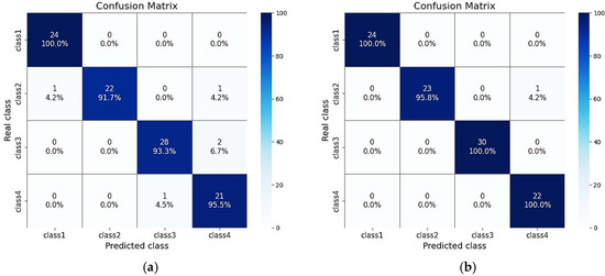
Figure 6.
(a) Confusion matrix for FreeEMG, (b) confusion matrix for Biosignalsplux. class1—walking, class2—sitting and standing, class3—up the stairs, and class4—down the stairs.
Table 4.
RF model classification results.
The evaluation results show that the classification model provided high accuracy in all classes for both devices, but a small number of misclassifications were observed due to similar electromyographic characteristics between some classes.
The highest accuracy in the FreeEMG device was recorded in the “walking” (100%) and “sitting and standing” (91.7%) classes. However, 4.3% of the samples in the “stairs descent” class were misclassified as “stairs climb” class. This is explained by the fact that similar muscle groups are activated in the EMG signals of these two movements. In addition, 4.2% of the samples in the “sit and stand” class were mistaken for the “walking” class, which indicates that muscle activity passes through similar phases between the two movements.
The accuracy in the Biosignalsplux device was even higher, with almost 100% results observed in all classes. Only 4.3% of the samples from the “sitting and standing” class were misclassified into the “stairs descent” class. This is also due to the short-term similar phases of the EMG signal. In general, the confusion matrix analysis shows that the misclassifications are mainly due to the similarities in the activity of muscle groups and short-term phase overlaps between movements. At the same time, the number of misclassifications in the Biosignalsplux device was significantly reduced due to the low signal quality and noise level.
The classification accuracy for the athletes’ LLL segment movement classes was calculated separately using the five selected classification algorithms using both DSs, and a generalized evaluation was performed based on these results.
Experimental tests showed that all classifiers, based on the selected feature set, were able to classify athletes’ footwork with high efficiency. The results differed in accuracy depending on the type of movement, and there were some significant differences between the classifiers (Figure 7).
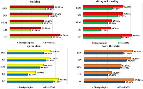
Figure 7.
Classification results.
In addition, in order to more accurately assess the stability of the model in practice, cross-subject and cross-session validation protocols were used in addition to the general random assignment.
In the cross-subject validation process, each participant was selected in turn as a test set, and the remaining participants formed the training set. This approach allows us to assess the level of generalization of the model across different users.
In the cross-session validation process, the model was trained on data recorded in weeks 1 and 2 and tested on data recorded in week 3. This method allows us to determine the stability of the model over time.
In the cross-subject and cross-session validation protocols, the generalization ability of the model was maintained at a high level for both devices.
Cross-Subject and cross-session validation values achieved 89.3% and 91.4% accuracy for the FreeEMG device with the RF algorithm, and 91.2% and 93.5% accuracy for the Biosignalsplux device, respectively (Table 5).
Table 5.
Performance results for both devices in different validation protocols.
The model’s high accuracy in both cross-subject and cross-session protocols confirms that the RF algorithm has high generalization capabilities across different users and different sessions.
The following table (Table 6) compares the results of the studies reviewed with the results achieved in our work. It can be seen from the table that eleven studies used both open and closed systems. Our study, using an open platform system (Biosignalsplux), showed the highest result (98.7%).
Table 6.
Comparative analysis of this work from state-of-the-art sources (“✓”-available, “✕”-not available).
4. Discussion
In this study, data were collected using two different devices, FreeEMG and Biosignalsplux, to detect athletes’ leg movements through EMG signals, and classification was performed using five classification algorithms: RF, k-NN, LR, SVM, and NN. The movements were divided into four types: walking, sitting and standing, up the stairs, and down the stairs.
The analysis of the research results shows that the RF algorithm showed the highest accuracy among all tested classifiers in classifying movements. In both devices, the RF algorithm outperformed the other models in all types of movements. In particular, for walking movements, the FreeEMG device achieved 95% accuracy, and the Biosignalsplux device achieved 98.7%. For sitting and standing movements, the accuracies were 91.4% and 95.8%, for up the stairs 94.3% and 96.6%, and for down the stairs 93% and 97.2%, respectively. These indicators prove the effectiveness of the RF algorithm in identifying differences between complex movements, its stability, and its high adaptability to the characteristics of the EMG signal. When used in conjunction with the Biosignalsplux device, the RF algorithm provided the highest results in all cases.
In addition, the NN and kNN algorithms also showed high accuracy results. For example, in the walking movement, the NN algorithm worked with 93.4% accuracy in FreeEMG and 95.2% in Biosignalsplux. The kNN algorithm has achieved the highest results in other studies for classifying EMG signals [31]. However, in our study, it performed worse than the RF and NN models. In the down the stairs movement, the kNN algorithm achieved 92.4% accuracy in FreeEMG and 96.6% in Biosignalsplux.
At the same time, the results of the SVM and LR algorithms were relatively lower. In the sitting and standing movements, LR showed 82.3% accuracy in the FreeEMG device and 86.7% accuracy in the Biosignalsplux device, while SVM achieved 85.5% and 87.2% accuracy, respectively. It was observed that the SVM and LR algorithms could not perform at the level of powerful models such as RF and NN in cases of complex, dynamic movements or in cases where there is similarity between movements. In some studies, high results were obtained by hybridizing the SVM model with methods such as ReliefF and Chi2 to improve its accuracy [29,32].
The overall results show that the complexity of the movements and the differences in the electromyographic responses of the muscles have a significant impact on the performance of the classifiers. The RF algorithm is an effective solution for determining the level of muscle activity in the process of analyzing EMG signals, providing high accuracy and stable operation with noisy data. Since muscle activity in EMG signals is complex and diverse, the ensemble-based approach of the RF algorithm allows for a deep analysis of this complexity. In addition, the RF model is able to extract important features of the classes in the dataset, which helps to understand the relationship between them more deeply. Therefore, the RF classifier showed superior results compared to other models in all types of movements.
The slightly superior results of the Biosignalsplux device compared to the FreeEMG device are explained by several technical and practical factors. First of all, the stable real-time transmission capability of the Biosignalsplux based on Bluetooth helps to record without reducing the signal quality. In addition, the electrode configuration and ergonomics of the Biosignalsplux allow for more accurate recording of active muscle segments. Therefore, although the RF algorithm performed well in both devices, the model trained on the DS recorded by the Biosignalsplux device showed slightly higher accuracy. This may serve as a benchmark for creating low-cost applications in the field of sports in the future.
Although the kNN model is simple, the results are not stable when there is similarity and noise between EMG signals. In our study, the classification accuracy of the kNN model was lower than that of the RF model, which is due to the close features of some classes of EMG signals (e.g., walking, climbing stairs, and descending). Although the LR model is effective in separating some EMG classes by linear boundaries, the performance of the LR model in our study dataset was inferior to powerful classifiers such as RF. This is due to the complexity of the movements and the uncertainty of muscle activity, and the LR model relies more on the linear relationship of signal features. The SVM model can work well on small datasets, but due to the complex and high-dimensional features of the EMG signals, as well as the similarities between different classes, the performance of the SVM model was limited in our study dataset. The NN model was also tested in our study and showed satisfactory results among the general classifiers. However, due to the inherent complexity of electromyographic signals and the uncertain distribution of muscle activity, the NN model gave inferior results compared to the RF model. In addition, the NN model fully demonstrates its advantages in large-scale and complex data sets.
Table 6 shows that the literature cited in Refs. [2,5,9,10] work on open platforms, that is, on devices where the processing process can be performed in real-time, the best result (96.13%) was achieved in Ref. [10]. However, this result was obtained by dividing the pre-processing part into 1024 (overlapping) windows and using CNN and TL algorithms in a hybrid mode. Models used in a hybrid mode can lead to delays in real-time reaction times. In this regard, in our study, 200 (non-overlapping) windows and simple non-hybrid models were used. This method significantly reduces the real-time processing time compared to hybrid models with overlapping (non-overlapping). In real-time operation mode, one of the most important characteristics is processing time, along with accuracy. Naturally, when using hybrid algorithms, the processing time may increase. The accuracy rate we achieved was 5.80/4.94/2.80/2.57 percentage points higher than that of open-platform studies [2,5,9,10], respectively. It was 3.04/4.70/2.06/2.67/5.37/2.70/1.73/2.20 percentage points higher than that of closed-platform studies [1,3,4,6,7,8,11]. The best result (96.97%) was achieved in the literature [11], which works on a closed platform. While this result is high, the FreeEMG device used in the literature cannot be used in real-time processing mode. In addition, in the literature [11], only the time domain features of the EMG signal are extracted and not divided into windows. This may not necessarily increase the accuracy values, but it may not provide stability. The main result achieved in this paper is that the results of the closed platform FreeEMG, which has achieved the best results in previous studies, and the results of the open platform we chose, Biosignalsplux, were compared. The analysis results show that the 96.50% result in the current study almost repeated the 96.97% result achieved in the study [11]. However, the accuracy of the real-time platform Biosignalsplux, which we chose, increased when the time domain and frequency domain features were used in a hybrid mode and divided into 200 (non-overlapping) windows. In the studies studied above, validation protocols were not performed in most of the studies, which does not ensure the stability of the model. We implemented the validation protocols shown in Table 5 to ensure the accuracy of the model. These studies show that the cross-subject and cross-session results obtained from Biosignalsplux are significantly higher. Biosignalsplux had a cross-subject accuracy of 91.2% and a cross-session accuracy of 93.5%, while the FreeEMG device had a cross-subject accuracy of 89.3% and a cross-session accuracy of 91.4%.
5. Study Limitations and Future Work
The results of this study are based on data from two high-resolution EMG recorders (FreeEMG and Biosignalsplux) and are limited to 25 athletes. Extrapolation of these results to a larger population should be conducted with caution. In addition, since the tests were conducted in a laboratory and controlled environment, the signal quality and classification accuracy may differ slightly in real-world competition or daily activity conditions.
Future work will focus on further improving the generalizability of the model. This will involve recruiting more participants and collecting data from groups of different ages and fitness levels. In addition, future research will focus on developing optimized algorithms that allow real-time processing of EMG signals, integrating deep learning models, and exploring the possibilities of application via mobile platforms.
6. Conclusions
This article presents the recognition of athletes’ LLL segment movements based on EMG signals. A total of 25 athletes participated in the data collection process. This study analyzed previous work on movement recognition using EMG signals. It is worth noting that in this study, unlike other studies, data were collected separately from two different devices, and their classification performance was compared. The Biosignalsplux device provided higher accuracy in movement classification compared to FreeEMG. The Biosignalsplux device consistently outperformed the FreeEMG device for each movement type and classifier, and this difference was especially evident in the cases of walking up and down the stairs.
The EMG signals were filtered to remove various noise and artifacts. In the next stage, several literatures were analyzed, and the RMS, MAV, WL, ZC, MDF, and SSC features of the EMG signal that provide high accuracy in classification were selected, and DSs were created using these features. RF, LR, SVM, NN, and kNN classification algorithms were tested on the basis of EMG signals collected by FreeEMG and Biosignalsplux devices to recognize four types of leg movements of athletes. All algorithms showed an overall accuracy of 82.3% to 98.7%. The RF algorithm achieved the highest result, with an accuracy of up to 97% in the Biosignalsplux device. This result demonstrated the effectiveness of the RF algorithm in identifying differences between movements in the EMG signal.
Based on the experimental results obtained in this study, the combination of the RF algorithm and the Biosignalsplux device demonstrated superior classification accuracy among the tested methods and devices. While these results are promising, the findings are limited to the dataset and movement types considered in this study. Further investigations with larger and more diverse datasets are needed before making generalized recommendations for broader applications.
In addition to the above conclusions, we can say that the accuracy rate we achieved showed a 1.73 percentage point difference compared to the best result in the closed platform study [11]. When calculated compared to the best result in the literature analyzed in the open platform [10], the difference was 2.57 percentage points.
Author Contributions
Methodology, K.Z., S.B., M.T., F.R. and G.B.; software, S.B., M.T. and F.R.; validation, K.Z., S.B. and S.M.; formal analysis, K.Z., G.P., G.S. and S.B.; resources, M.T., F.R., F.A. and G.P.; data curation, K.Z. and S.B.; writing—original draft, K.Z., S.B., F.R. and M.T.; writing—review and editing, K.Z., S.B. and F.A.; supervision, K.Z., S.B. and M.T.; project administration, K.Z. and S.B. All authors have read and agreed to the published version of the manuscript.
Funding
This research received no external funding.
Institutional Review Board Statement
This study did not require ethical approval. Review and ethical approval were waived for this study because it did not involve the administration of tests or experiments on humans, but only measurement campaigns on sEMG signals.
Informed Consent Statement
Not applicable.
Data Availability Statement
The data are not publicly available due to privacy, ethical restrictions, and development.
Conflicts of Interest
The authors declare no conflicts of interest.
References
- Ge, W.; Zhao, J.; Wang, F.; Xu, C.; Yang, Z.; She, J. Experimental Design of Lower-limb Movement Recognition Based on Support Vector Machine. In Proceedings of the 2022 41st Chinese Control Conference (CCC), Hefei, China, 25–27 July 2022; pp. 6493–6497. [Google Scholar] [CrossRef]
- Mitsantisuk, P.; Kiatthaveephong, S.; Autthasan, P.; Wilaiprasitporn, T. MotionXpert: EMG-Based Classification for Optimized Lower-Limb Motion Detection. In Proceedings of the 2024 IEEE-EMBS Conference on Biomedical Engineering and Sciences (IECBES), Penang, Malaysia, 11–13 December 2024; pp. 1–6. [Google Scholar] [CrossRef]
- Arunganesh, K.; Vinoth Krishna, S.; Hamsadhwani, V.; Karthick, P.A.; Kumaravel, S.; Sivakumaran, N. Recognition of Lower Limb Movements from the Time Domain Features of Surface EMG and Catboost Classifier. In Proceedings of the 2024 International Conference on Brain Computer Interface & Healthcare Technologies (iCon-BCIHT), Thiruvananthapuram, India, 19–20 December 2024; pp. 70–73. [Google Scholar] [CrossRef]
- Al-Quraishi, M.S.; Elamvazuthi, I.; Tang, T.B.; Al-Qurishi, M.; Parasuraman, S.; Borboni, A. Multimodal Fusion Approach Based on EEG and EMG Signals for Lower Limb Movement Recognition. IEEE Sens. J. 2021, 21, 27640–27650. [Google Scholar] [CrossRef]
- Wang, J.; Dai, Y.; Si, X. Analysis and Recognition of Human Lower Limb Motions Based on Electromyography (EMG) Signals. Electronics 2021, 10, 2473. [Google Scholar] [CrossRef]
- Tu, P.; Li, J.; Wang, H. Lower Limb Motion Recognition with Improved SVM Based on Surface Electromyography. Sensors 2024, 24, 3097. [Google Scholar] [CrossRef]
- Zhang, W.; Bai, Z.; Yan, P.; Liu, H.; Shao, L. Recognition of Human Lower Limb Motion and Muscle Fatigue Status Using a Wearable FES-sEMG System. Sensors 2024, 24, 2377. [Google Scholar] [CrossRef]
- Li, B.; Xu, G.; Pei, J.; Luo, D.; Li, H.; Du, C.; Zhang, K.; Zhang, S. Lower Limb Motion Recognition Based on Surface Electromyography Decoding Using S-Transform Energy Concentration. Machines 2025, 13, 346. [Google Scholar] [CrossRef]
- Tu, J.; Dai, Z.; Zhao, X.; Huang, Z. Lower limb motion recognition based on surface electromyography. Biomed. Signal Process. Control 2023, 81, 104443. [Google Scholar] [CrossRef]
- Zhou, Z.; Tao, Q.; Su, N.; Liu, J.; Chen, Q.; Li, B. Lower Limb Motion Recognition Based on sEMG and CNN-TL Fusion Model. Sensors 2024, 24, 7087. [Google Scholar] [CrossRef] [PubMed]
- Zohirov, K.; Nasimov, R.; Boykobilov, S.; Sherboboyeva, G.; Temirov, M.; Sattorov, M. An EMG Signal Based Approach for Assessing the Physical Condition of Athletes in Wrestling. In Proceedings of the 8th International Conference on Future Networks & Distributed Systems (ICFNDS’24), Marakech, Morocco, 11–12 December 2024; Association for Computing Machinery: New York, NY, USA, 2025; pp. 602–608. [Google Scholar] [CrossRef]
- Fedorova, L.; Rajtukova, V.; Toth, T.; Zivcak, J. EMG system application in muscle parametrization of the upper extremities. In Proceedings of the 2014 IEEE 12th International Symposium on Applied Machine Intelligence and Informatics (SAMI), Herl’any, Slovakia, 23–25 January 2014; pp. 85–89. [Google Scholar] [CrossRef]
- Ayachi, M.; Seddik, H. Overview of EMG Signal Preprocessing and Classification for Bionic Hand Control. In Proceedings of the 2022 IEEE Information Technologies & Smart Industrial Systems (ITSIS), Paris, France, 15–17 July 2022; pp. 1–6. [Google Scholar] [CrossRef]
- Chand, R.; Tripathi, P.; Mathur, A.; Ray, K.C. FPGA implementation of fast FIR low pass filter for EMG removal from ECG signal. In Proceedings of the 2010 International Conference on Power, Control and Embedded Systems, Allahabad, India, 29 November–1 December 2010. [Google Scholar] [CrossRef]
- Zohirov, K. A New Approach to Determining the Active Potential Limit of an Electromyography Signal. In Proceedings of the 2023 3rd International Conference on Technological Advancements in Computational Sciences (ICTACS), Tashkent, Uzbekistan, 1–3 November 2023; pp. 291–294. [Google Scholar] [CrossRef]
- Pancholi, S.; Joshi, A.M. Electromyography-Based Hand Gesture Recognition System for Upper Limb Amputees. IEEE Sens. Lett. 2019, 3, 1–4. [Google Scholar] [CrossRef]
- Prakash, M.B.; Harish, H.M.; Niranjana Kumara, M. Time Domain Analysis of EMG Signals using KNN and SVM Techniques. WSEAS Trans. Signal Process. 2022, 18, 70–76. [Google Scholar] [CrossRef]
- Abbaspour, S.; Lindén, M.; Gholamhosseini, H.; Naber, A.; Ortiz-Catalan, M. Evaluation of surface EMG-based recognition algorithms for decoding hand movements. Med. Biol. Eng. Comput. 2020, 58, 83–100. [Google Scholar] [CrossRef] [PubMed]
- Altın, C.; Er, O. Comparison of Different Time and Frequency Domain Feature Extraction Methods on Elbow Gesture’s EMG. Eur. J. Interdiscip. Stud. 2016, 2, 35–44. [Google Scholar] [CrossRef][Green Version]
- Rojas-Martínez, M.; Mañanas, M.A.; Alonso, J.F. High-density surface EMG maps from upper-arm and forearm muscles. J. Neuroeng. Rehabil. 2011, 9, 85. [Google Scholar] [CrossRef]
- Qi, J.; Jiang, G.; Li, G.; Sun, Y.; Tao, B. Surface EMG hand gesture recognition system based on PCA and GRNN. Neural Comput. Appl. 2020, 32, 6343–6351. [Google Scholar] [CrossRef]
- Oskoei, M.A.; Hu, H. Support vector machine-based classification scheme for myoelectric control applied to upper limb. IEEE Trans. Biomed. Eng. 2008, 55, 1956–1965. [Google Scholar] [CrossRef] [PubMed]
- Arteaga, M.V.; Castiblanco, J.C.; Mondragon, I.F.; Colorado, J.D.; Alvarado-Rojas, C. Emg-driven hand model based on the classification of individual finger movements. Biomed. Signal Process. Control 2020, 58, 101834. [Google Scholar] [CrossRef]
- Shim, H.-M.; Lee, S. Multi-channel electromyography pattern classification using deep belief networks for enhanced user experience. J. Cent. South Univ. 2015, 22, 1801–1808. [Google Scholar] [CrossRef]
- Raurale, S.; McAllister, J.; del Rincon, J.M. EMG Wrist-Hand Motion Recognition System for Real-Time Embedded Platform. In Proceedings of the 2019 International Conference on Acoustics, Speech, and Signal Processing (ICASSP), Brighton, UK, 12–17 May 2019; Institute of Electrical and Electronics Engineers Inc.: New York, NY, USA, 2019; pp. 1523–1527. [Google Scholar] [CrossRef]
- Kim, H.; Lee, J.; Kim, J. Electromyography-signal-based muscle fatigue assessment for knee rehabilitation monitoring systems. Biomed. Eng. Lett. 2018, 8, 345–353. [Google Scholar] [CrossRef]
- Laganà, F.; Pratticò, D.; Angiulli, G.; Oliva, G.; Pullano, S.A.; Versaci, M.; La Foresta, F. Development of an Integrated System of sEMG Signal Acquisition, Processing, and Analysis with AI Techniques. Signals 2024, 5, 476–493. [Google Scholar] [CrossRef]
- Zohirov, K. Classification of Some Sensitive Motion of Fingers to Create Modern Biointerface. In Proceedings of the 2022 International Conference on Information Science and Communications Technologies (ICISCT), Tashkent, Uzbekistan, 28–30 September 2022; pp. 1–4. [Google Scholar] [CrossRef]
- Pires, N.; Macedo, M.P. A Bimodal EMG/FMG System Using Machine Learning Techniques for Gesture Recognition Optimization. Signals 2025, 6, 8. [Google Scholar] [CrossRef]
- Muguro, J.K.; Laksono, P.W.; Rahmaniar, W.; Njeri, W.; Sasatake, Y.; bin Suhaimi, M.S.A.; Matsushita, K.; Sasaki, M.; Sulowicz, M.; Caesarendra, W. Development of Surface EMG Game Control Interface for Persons with Upper Limb Functional Impairments. Signals 2021, 2, 834–851. [Google Scholar] [CrossRef]
- Miaoulis, D.; Stivaros, I.; Koubias, S. Developing a Novel Muscle Fatigue Index for Wireless sEMG Sensors: Metrics and Regression Models for Real-Time Monitoring. Electronics 2025, 14, 2097. [Google Scholar] [CrossRef]
- Zaim, T.; Abdel-Hadi, S.; Mahmoud, R.; Khandakar, A.; Rakhtala, S.M.; Chowdhury, M.E.H. Machine Learning- and Deep Learning-Based Myoelectric Control System for Upper Limb Rehabilitation Utilizing EEG and EMG Signals: A Systematic Review. Bioengineering 2025, 12, 144. [Google Scholar] [CrossRef] [PubMed]


Disclaimer/Publisher’s Note: The statements, opinions and data contained in all publications are solely those of the individual author(s) and contributor(s) and not of MDPI and/or the editor(s). MDPI and/or the editor(s) disclaim responsibility for any injury to people or property resulting from any ideas, methods, instructions or products referred to in the content. |
© 2025 by the authors. Licensee MDPI, Basel, Switzerland. This article is an open access article distributed under the terms and conditions of the Creative Commons Attribution (CC BY) license (https://creativecommons.org/licenses/by/4.0/).