Review

Interplay Between Diabetes, Obesity and Glioblastoma Multiforme, and the Role of Nanotechnology in Its Treatment

by
Sourav De
1,
Sabyasachi Banerjee
2,
Gourab Dey
1,
Subhasis Banerjee
1,* and
S.K. Ashok Kumar
3,*
1
Department of Pharmaceutical Technology, Eminent College of Pharmaceutical Technology, Kolkata 700126, West Bengal, India
2
Department of Pharmaceutical Chemistry, Gupta College of Technological Sciences, Asansol 713301, West Bengal, India
3
Department of Chemistry, School of Advanced Sciences, Vellore Institute of Technology, Vellore 632014, Tamil Nadu, India
*
Authors to whom correspondence should be addressed.
J. Nanotheranostics 2025, 6(1), 7; https://doi.org/10.3390/jnt6010007
Submission received: 9 December 2024 / Revised: 27 January 2025 / Accepted: 25 February 2025 / Published: 27 February 2025

Abstract

:
A very aggressive and deadly brain cancer, glioblastoma multiforme (GBM) poses formidable obstacles to effective therapy. Despite advancements in conventional therapies like surgery, chemotherapy, and radiation therapy, the prognosis for GBM patients remains poor, with limited survival outcomes. Nanotechnology is gaining popularity as a promising platform for managing GBM, offering targeted drug delivery, improved therapeutic efficacy, and reduced systemic toxicity. This review offers a comprehensive analysis of the current therapeutic approach for GBM using nanotechnology-based interventions. This study explored various nanocarrier (NC) systems like polymeric nanoparticles, liposomes, dendrimers, polymeric micelles, and mesoporous silica nanoparticles for improved precision as well as efficacy in encapsulating and delivering therapeutic agents to GBM tumors. Methods for improving drug delivery into GBM cells are described in this study, including novel delivery modalities such as convection-enhanced delivery, intranasal administration, magnetic hyperthermia, peptide-guided nanoparticles, and immune liposomes. It also explores the influence of diabetes and obesity on GBM prognosis and survival rates, suggesting that managing glucose levels and using metformin may improve patient outcomes. The discussion focuses on the advancements in nanotechnology-enabled GBM therapy, highlighting the challenges and opportunities in implementing these promising technologies in clinical practice. The study highlights the potential of nanotechnology and metabolic modulation in transforming GBM treatment strategies. To further understand how these factors impact GBM patients and develop innovative nanotechnology-based treatments for GBM and diabetes mellitus, more study is necessary.

1. Introduction

Glioblastoma multiforme (GBM) is a prevalent and challenging cancer, accounting for over 60% of malignant brain tumors in adults and children [1]. GBM can occur at any age, but its incidence increases with age, with a significant proportion detected in the sixth and eighth decades of life [2]. The main challenges include scattered infiltration, rapid dissemination, and necrosis, leading to therapeutic complexities [3]. GBM exhibits resistance to programmed cell death through various mechanisms, creating an inherent obstacle to apoptosis [4]. Inhibitors of apoptosis proteins (IAPs), specifically cellular IAP 1 (cIAP1) and X-linked IAP (XIAP), are considered the most efficacious caspase inhibitors [5,6]. The pharmacotherapy of malignant cells faces challenges due to the increased expression of IAPs and the suppression of the second mitochondrial-derived activator of caspases (Smac). Smac mimetics, acting as IAP antagonists, can inhibit IAP activity and impede cancer proliferation during preclinical stages, but their anticancer efficacy in monotherapy has been inconsistent in clinical studies [7]. The complicated structure of IAPs, toxicity to off-target locations, and poor permeability across the BBB are some of the obstacles that smac mimetics encounter when used.
The cause of glioma is unknown, but obesity and diabetes have been linked to common types of cancer, including breast, colorectal, esophageal, pancreatic, ovarian, and kidney cancer [8,9,10,11,12,13,14]. Diabetes mellitus is a chronic disease that involves insulin deficiency and beta cell dysfunction that affects 422 million people globally, causing 1.5 million fatalities annually [15,16,17]. Metabolic syndrome, including obesity, hyperinsulinemia, hyperglycemia, and high-fat diets, are independent risk factors for cancer [18,19,20,21,22]. Studies showed that diabetes and hyperglycemia could increase mortality and morbidity rates in both chronic and acute illnesses [23,24]. These conditions have been found to minimize overall survival rates in patients diagnosed with various types of malignancies, including brain [25], colorectal [26], and breast cancers [27].
Elevated levels of blood glucose have been found to be significantly associated with a lesser probability of developing prostate cancer and glioma [28,29]. Diabetes might be suspected at different stages of its progression, and subsequent medication regimens may impact the association of possible glioma risk. Numerous mechanisms exist that underlie the association between glioma and hyperglycemia. For example, Metformin has been observed to exert inhibitory effects on various cellular processes, including invasion, migration, proliferation, autophagy, apoptosis induction, and differentiation in glioma cells [2,30,31,32,33].
The correlation in concern is particularly contentious among patients diagnosed with GBM, which is the most prevalent and severe form of astrocytoma. The average OS rate for individuals who have undergone surgery is estimated to be between 13 and 15 months [34,35]. The successful completion of multiple surgeries, specifically a second surgery, in conjunction with conventional adjuvant therapy, such as temozolomide (TMZ) chemotherapy and radiation, has been observed to increase OS to 18.5 months or potentially beyond [36]. A correlation exists between a reduced risk of glioma and long-term diabetes mellitus, as indicated by elevated glycated hemoglobin A1c (HbA1c) levels [2,37]. The presence of diabetes stimulates an inflammatory microenvironment that facilitates the proliferation of interleukins, thereby elevating the probability of an unfavorable prognosis, irrespective of the preexisting cancer status of the patient. Interleukins, including interleukin-6 (IL-6), are synthesized and secreted in reaction to an inflammatory context [38]. The elevation of insulin resistance is associated with an elevation in the plasma concentration [39]. The available evidence suggests that the presence of diabetes may exert a direct influence on the development and growth of gliomas [40,41,42]. Concurrently, GBM exhibits an elevated level of IL-6, which promotes angiogenesis, cellular invasion, and diminished survival rate [43]. Elevated interleukin levels give rise to an inflammatory context that amplifies the stimulation of signaling pathways, such as c-Jun N-terminal kinases (JNKs), with JNK1 being particularly prominent in DM and JNK3 in GBM [44,45]. The administration of JNK inhibitors targeting both isoforms in diabetes may lead to a reduction in inflammation and consequently, mitigate the deleterious impact on GBM [46].
Elevated levels of plasma D-glucose are commonly detected in cases of glioblastoma [47] and DM [48]. These disorders are characterized by the dysregulation of D-glucose homeostasis, which leads to increased proliferation and tumorigenesis due to the avid consumption of D-glucose by glioma cells [49,50,51]. The Warburg effect, denoting the heightened dependability of neoplastic cells on glucose, despite the availability of oxygen, is a hallmark feature of cancer [52]. Cancer cells have an inefficient glucose metabolism that does not include oxidative phosphorylation, even when oxygen is present. This discrepancy is known as the Warburg effect [53]. There is currently no novel cancer treatment approach that takes advantage of this potential susceptibility [54]. Research indicates that glioblastoma is a disease characterized by various tumor cell populations, including glioblastoma initiating/propagating cells, which possess stem cell properties [55,56]. No scholarly investigation has been conducted on this specific scenario in glioblastoma stem cells (GSCs) to date. However, it has been observed that mesenchymal GSCs (Mes GSCs) exhibit a higher degree of glycolytic activity compared to proneural GSCs (PN GSCs). The study suggests that the hyperglycemic state commonly observed in diabetes patients may potentially facilitate or promote the growth of Mes GSCs. Studies on Mes GSCs show a higher level of aggressiveness compared to PN GSCs in both in vitro and intracranial xenografts. It is recommended that individuals diagnosed with type 2 DM initiate treatment with metformin [57]. It is noteworthy that metformin exhibits a significantly greater efficacy in diminishing the viability of glioblastoma stem cells (GSCs) as compared to both healthy stem cells and developed GBM cells. Metformin has been found to decrease the capacity of in vitro GBM GSCs to regenerate themselves, as evidenced by their spherogenic activity [31,32].
At present, there exists no identifiable therapeutic target for GBM or DM. Elevated levels of plasma adenosine have been observed to exhibit a positive correlation with increased extracellular blood D-glucose levels, thereby facilitating the activation of adenosine receptors [58]. Elevated adenosine signaling is a complication arising from metabolic changes that result in chronic renal failure among individuals with diabetes mellitus [59]. GBM tumors exhibit elevated levels of extracellular adenosine, particularly in the hypoxic zones that are malignant, identical to DM [60]. The preservation of stem and GSC characteristics is linked to an increased extracellular concentration of adenosine and the activation signaling of adenosine receptors, in addition to chemotherapy resistance, cellular invasion, and proliferation [61]. The potential therapeutic application of adenosine and its associated signaling pathways for the management of diabetes and GBM is worth considering.
Recurrence is still seen in nearly all cases, despite advances in surgical technology and an increased emphasis on obtaining gross total resection (GTR) of GBM. This is relevant even when intraoperative direct electrical stimulation during awake surgery, adjuvant therapy, or the use of new, safe, effective treatments like laser interstitial thermal therapy are applied. Thus, it is crucial to identify novel therapeutic drugs that can enhance the probability of survival for such patients.
In contemporary neuro-oncology, the assessment of GBM heavily relies on genetic alterations and molecular tumor markers, which are deemed indispensable [62]. Currently, prominent genetic mutations, such as O6-methylguanine methyltransferase (MGMT) promoter methylation, which has been linked to a notably improved median survival following treatment with TMZ [63], along with other molecular tumor biomarkers, such as aldehyde dehydrogenase 1A3 (ALDH1A3), epidermal growth factor receptor (EGFR) amplification, and isocitrate dehydrogenase (IDH1/IDH2) isoforms, are the subject of investigation and seemed to be associated with prognosis [62,64]. Studies have demonstrated that metformin could reduce the possibility of tumor development in individuals with diabetes [65]. Additionally, metformin has exhibited targeted antiproliferative effects on glioma cells both in vivo and in vitro [31,32]. Administering standard oral hypoglycemic agents to patients with GBM poses a challenge due to the frequent modifications in diabetes status, glycemic control, and their interrelationships. In addition, individuals diagnosed with GBM have a greater chance of developing hyperglycemia due to the administration of high-dose glucocorticoids. These medications have been demonstrated to elevate plasma glucose levels while impeding glucose transport and are frequently employed to manage peritumoral edema [66,67].

2. Impact of Metabolic Disorders on Glioblastoma Multiforme

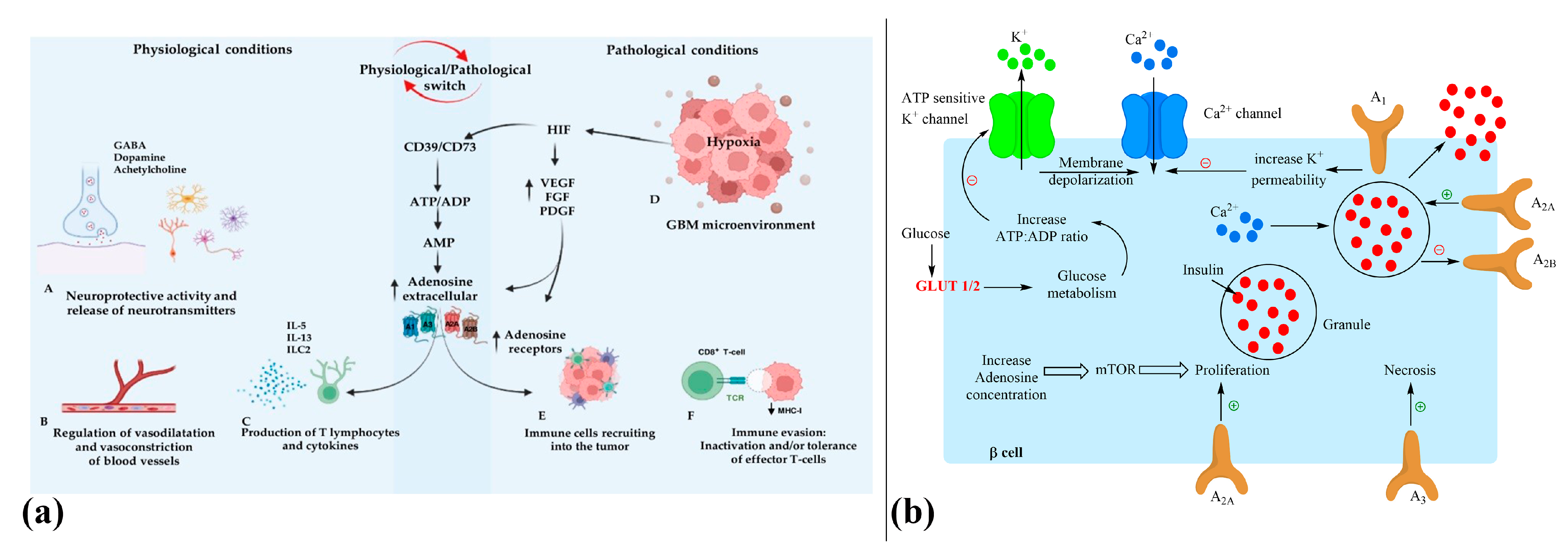
2.1. Role of Diabetes, Glioma Metabolism, and Adenosine Signaling in GBM

The development of type 2 DM is attributed to the degenerative conditions of pancreatic islets, which are exacerbated by insulin resistance caused by obesity [68,69]. Many studies have shown that adenosine has a role in the pathogenesis of type 1 and type 2 diabetes by controlling D-glucose homeostasis [70,71]. Several studies have shown that adenosine signaling plays a role in the normal functioning of the body as well as in the development of diseases, including cancer and DM [72,73,74,75]. Adenosine is essential for many important functions in the central nervous system (CNS), including synaptic plasticity [76], neurotransmitter release [77], and neuroprotection under conditions of hypoxia, ischemia, and oxidative stress [78,79]. Over the years, research on key tumor development signaling pathways in the tumor microenvironment has revealed the significant role of extracellular adenosine [80].
The acceleration of angiogenesis and matrix remodeling by adenosine has been observed to facilitate the advancement and growth of tumors [81,82,83,84,85,86,87,88]. The significance of an ecto-5′-nucleotidase cluster of differentiation 73 (CD73), that transforms adenosine monophosphate (AMP) into adenosine, has been observed in the catabolic activity that generates an adenosine “halo” that is both immunosuppressive and proangiogenic. This phenomenon is essential in the advancement of cancer in glioma and chronic lymphocytic leukemia (CLL) [89,90]. One possible option that operates at two levels in chronic CLL is the adenosinergic axis, which inhibits the establishment of an adenosine protective environment and activates the immune system’s reaction to leukemic cells [91,92,93]. Research indicates that glioma cells show significant resistance to ATP-induced cell death compared to tissue samples from individuals without any medical conditions. The purinergic system plays a crucial role in developing glioma due to the promotion of glioma cell growth by ATP. The CD73 activity in human glioma cell lines, namely U251, U373, U138, and U87, is comparatively higher than that of astrocytes, thus highlighting the crucial role of the adenosine cascade in this ailment [94].
Upon its release into the extracellular space, adenosine can undergo deamination to inosine through the utilization of nucleoside membrane transporters present in cells, which can either be concentrative (CNTs) or equilibrative (ENTs) in nature [95,96]. The nucleoside transporters that are most commonly found in the brain are known as ENTs. These transporters are known to be expressed at high levels in both glial cells and neurons [97]. It is noteworthy that A1AR and ENT1 exhibit selective co-expression in the hippocampus, cortex, and thalamus of the human brain, whereas adenosine A2a receptor and ENT2 do not [98]. Transcriptional modifications involving ENTs and CNTs have been detected in many animal models of type 1 diabetes [99,100].
The correlation between inflammation and insulin resistance is widely acknowledged in the academic literature. Atherosclerosis, myocardial infarction, and stroke are cardiovascular complications that are more likely to occur in people with diabetes [101,102]. The academic literature emphasizes the connection between inflammation and insulin resistance, and diabetes is associated with an increased risk of these complications [101,102].
In addition, it has been observed that macrophages and myeloid-derived suppressor cells (MDSCs) have the ability to hinder adaptive antitumor responses via the expression of distinct metabolites, cytokines, and membrane receptors. Neoplastic cells employ diverse mechanisms to evade the immune response directed against them. The second messenger of adenosine signaling, cAMP, has been found to act as a negative regulator of T-cell immune activity. Consequently, the extracellular tumor microenvironment often contains higher levels of adenosine, an immunosuppressive molecule [103]. The accumulation of adenosine within the inflammatory milieu, a pivotal biomarker of diabetes, results in the inhibition of T-cell response [104]. Neutrophils have been observed to secrete AMP during their transit through an endothelial monolayer. This AMP is subsequently subjected to dephosphorylation by CD73, resulting in the production of adenosine. The nucleoside in consideration binds to the A2AAR receptors found on neutrophils and effectively diminishes their ability to cause tissue damage by decreasing the production of free radicals, release of cytokines, level of leukotriene-B4, and development of adhesion molecules [105]. By activating A2aAR, adenosine limits the infiltration of neutrophils into tissues, thereby decreasing the extent of tissue damage caused by neutrophils [106]. However, monocytes cannot differentiate into macrophages or acquire a dendritic cell-like phenotype when exposed to exogenous adenosine. In hypoxic areas, such as cancerous and inflamed tissues, a new cell type begins to develop as a consequence of dendritic cell differentiation altered by activation of adenosine receptors. In addition, it has been observed that there is an upsurge in adenosine levels in areas of solid tumors that experience hypoxia. This increase in adenosine levels can hinder the capability of cytotoxic immune cells to properly identify tumor cells [107]. The conducive conditions of this phenomenon can facilitate the proliferation and survival of cancerous cells. Consequently, adenosine exerts a local immunosuppressive effect within the confines of the solid tumor microenvironment, thereby impeding the cytotoxic activity of T-cells against non-cancerous cells. Figure 1 illustrates the interaction between diabetes, glioma, and adenosine [108].

2.2. T2DM Clinical Risk to GBM Development

Diabetes increases the risk of tumor development in individuals, but the exact biochemical mechanism behind this increased risk remains unclear [10]. Nevertheless, Barami et al. (2013) [109] and Disney-Hogg et al. [2] have documented the absence of heightened susceptibility to glioma in individuals with T2DM and hyperglycemia. Seliger et al. [12] presented a correlation between diabetes and a decreased occurrence of glioma, namely GBM. The study found a noteworthy reduction in the probability of developing GBM in diabetic males with prolonged diabetes or inadequate glycemic control. This implies that there may be a biological mechanism, which involves the co-occurrence of elevated HbA1c levels and low testosterone levels in conjunction with prolonged diabetes [110]. According to a recent investigation conducted on a population of 2.3 million individuals in Israel [111], prediagnostic diabetes was found to be associated with a heightened occurrence of malignant brain tumors in patients. In a meta-analysis, Zhao et al. [112] explored the link between T2DM and the likelihood of developing any kind of glioma. The findings of the study revealed that type 2 diabetes mellitus significantly lowered the likelihood of gliomas. No statistically significant difference in the incidence of brain tumors was found between those with and without diabetes, according to a meta-analysis of 13 studies carried out by Tong et al. [25].
Diabetes has been associated with the prognosis of GBM in a very complex and controversial manner; thus, several conflicting reports have been found in the literature. These discrepancies reflect several factors affecting study design, population characteristics, and methods of data analysis [113]. For example, outcomes reported in retrospective studies may differ from prospective studies, while differences in sample size and follow-up period add to inconsistency [114]. Furthermore, the heterogeneity of GBM and comorbid conditions associated with diabetes, such as obesity and hypertension, may have an impact on prognosis. Moreover, further genetic and molecular heterogeneity between tumor subtypes, in addition to possible insulin resistance and hyperglycemia effects, can be complicating factors in tumor progressions. These represent complexities whereby, to understand the association of diabetes with prognosis in GBM, detailed research will be required.

2.3. Influence of Diabetes and Obesity on GBM Prognosis and Survival Outcomes

Recently, there has been a notable increase in research focused on understanding how T2DM and elevated blood sugar levels affect clinical outcomes in cancer patients. The occurrence of diabetes and obesity is linked to an increased need for surgical procedures throughout a person’s life. These conditions also pose a risk in several surgical procedures due to heightened rates of postoperative morbidity and mortality [115,116,117,118,119]. Adverse effects such as infections at the surgical site, implantation failure, delay in wound healing, and various medical issues may arise.
Factors that may indicate a longer overall survival (OS) in patients with GBM encompass a younger age, a high level of performance status, a gross total resection (GTR) of the tumor that exceeds 95%, the absence of inflammatory diseases or metabolic disorders, and the successful completing of postoperative radiochemotherapy [120]. The precise connection between T2DM and obesity in patients with GBM remains unresolved [8,119,121,122], including our own assessment, which has revealed unfavorable associations between the impacts of GBM and T2DM on OS. The investigation conducted by Chambless et al. [123] revealed that patients with diabetes exhibited a comparatively inferior median OS of 312 days in contrast to non-diabetic patients who had a median overall survival of 470 days. Additionally, the progression-free survival (PFS) was also lower in individuals with diabetes with a median PFS of 106 days as opposed to non-diabetic patients who had a median PFS of 166 days. The study further established a negative correlation between increased body mass index (BMI) and median PFS. In a similar vein, Welch and Grommes [124] discovered that individuals with diabetes exhibited a decreased overall survival rate (10 months) in comparison to those without diabetes (13.4 months). According to the findings of Siegel et al. [125], there appears to be no relationship between DM and OS. It has been noted that patients categorized as underweight, with a median overall survival of 12.00 months, or those who are obese, with a median overall survival of 13.60 months, tend to have a lower overall survival compared to individuals with a normal weight, who have a median overall survival of 17.50 months. The study observed a cohort of 392 patients diagnosed with GBM, wherein the individuals with elevated BMI exhibited significantly better overall survival rates. The median OS was found to be 13.50 months in individuals with normal BMI, 15.40 months in those who were overweight, and 15.10 months in those who were obese. Despite lacking a comprehensive understanding, the “obesity paradox” has exhibited an increasing prevalence among cancer patients in recent times. Research indicates that individuals suffering from lung cancer, renal cell carcinoma, and melanoma who are classified as obese may have more favorable outcomes in comparison to those who are not obese [126].
T2DM and obesity play significant roles in the progression and treatment resistance of GBM via various interconnected mechanisms. In individuals with diabetes, this led to heightened levels of glucose and insulin resistance, which in turn contributed to increased systemic inflammation and an oxidative storm that could enhance a pro-tumorigenic microenvironment [127]. The processes of hyperinsulinemia and insulin-like growth factor signaling play a crucial role in promoting cell proliferation, survival, and invasion in GBM. Additionally, adipokines linked to obesity, including leptin and adiponectin, contribute to increased inflammation and modify cellular metabolism, which in turn fosters a more aggressive tumor phenotype [128]. The disruption of metabolic pathways, such as glycolysis and the pentose phosphate pathway, is frequently observed in patients with glioblastoma who also suffer from obesity and diabetes. This dysregulation typically leads to a heightened resistance to treatments like chemotherapy and radiotherapy [129]. Moreover, the immune responses that are modified in individuals with obesity and diabetes can undermine the efficacy of immunotherapy, thereby adding complexity to the treatment landscape for GBM. The understanding of the metabolic and inflammatory factors that play a role in the progression and resistance of GBM highlights potential avenues for intervention [130].

2.4. Role of Hyperglycemia in Shaping GBM Patient Outcomes

Previous research by Deng et al. has examined the link between hyperglycemia and cancer patients, finding varying degrees of positive, neutral, or negative associations between T2DM and its associated factors [4]. Elevated levels of blood glucose, commonly known as hyperglycemia, have been identified as a distinct risk factor that can independently contribute to reduced survival rates in various diseases and tumors. Additionally, patients diagnosed with GBM may experience a decrease in OS as a result of hyperglycemia. The abbreviated OS observed in individuals diagnosed with GBM and hyperglycemia has also been noted in those who have undergone complete tumor resection and received adjuvant therapy [131]. Mayer et al. [131] have indicated that patients diagnosed with GBM and who encounter one or multiple instances of uncontrolled blood glucose levels exceeding 10 mM exhibit a reduction in their median overall survival period, from 16.70 to 8.80 months. McGirt and colleagues have reported that a negative correlation exists between prolonged hyperglycemia and OS in patients who have undergone surgical resection. This correlation has been found to be independent of factors such as the severity of the patient’s disability, their diabetic condition, their prolonged use of dexamethasone, and their subsequent treatment modalities [132].
Derr et al. [67] conducted a study wherein patients were categorized into quartiles based on their levels of blood glucose, which varied from 65 to 459 mg/dL. The findings of the study revealed a reduction in the median OS with an increase in blood glucose values. The correlation between elevated levels of blood glucose and reduced OS remained significant even after controlling for factors such as Karnofsky’s performance score, age, and daily glucocorticoid dosage. The study conducted by Stevens et al. [133] on 242 patients with GBM revealed that elevated preoperative glucose level was linked to decreased overall survival, regardless of whether they underwent GTR or not. The findings suggest that strict glucose management may potentially improve outcomes for these patients. The research carried out by Tieu et al. [134] showed that glycemia continued to be a reliable prognostic indicator for survival in patients with GBM who received surgical resection, followed by radiotherapy and temozolomide treatment.
Hagan et al. [135] performed a multivariate analysis and found no statistically significant relationship between hyperglycemia and PFS. Nonetheless, the researchers noted a statistically significant decrease in overall survival among glioblastoma patients whose plasma glucose levels surpassed 112 mg/dL and 180 mg/dL in the preoperative phase (p = 0.01). Decker et al. [136] conducted an investigation into the mechanisms that underlie the association between hyperglycemia and inferior overall survival. Their findings revealed that a median blood glucose level exceeding 167 mg/dL was significantly associated with a rise in severe postoperative complications, while levels surpassing 163 mg/dL were significantly associated with an increase in hospital readmissions within 30 days. Patients diagnosed with GBM who encounter hyperglycemia after undergoing surgery are at a higher risk of experiencing postoperative complications and readmissions, which could potentially hinder the efficacy of their disease’s treatment. According to the research conducted by Welch and Grommes [124], patients who achieved adequate glycemic control, reflected by a median glucose level of 173 mg/dL, experienced an extended median overall survival of 11 months. In comparison, those with glucose levels between 174 and 247 mg/dL had an OS of 9 months, while individuals with a median glucose level of 247 mg/dL had an OS of 8 months. Nonetheless, it is essential to include larger patient groups to enable a more comprehensive analysis of how hyperglycemia and obesity affect individuals diagnosed with GBM. Consideration of improved glucose management is essential for patients with GBM, as it may significantly contribute to better overall survival and outcomes.
Despite the involvement of several potential mechanisms, the precise effects of hyperglycemia on GBM cells remain incompletely comprehended. Hyperglycemia triggers various biochemical pathways that accelerate tumor formation, including stimulation of the epithelial–mesenchymal transition, the elevation of inflammatory cytokine levels, enhanced WNT/catenin signaling, activation of pro-cell survival AKT/mTOR, and increased leptin levels in the blood [137,138,139]. In recent times, there has been an association established between cellular metabolism and certain pathways that have been traditionally linked to tumor cell proliferation, evasion of apoptosis, angiogenesis, and resistance to therapy. One such pathway is the TP53 gene, which encodes for the tumor suppressor protein p53. The p53 gene, frequently mutated in individuals with GBM, facilitates various cellular reactions to hypoxia, oncogene stimulation, and DNA damage [140]. Additionally, it regulates glycolysis and contributes to the maintenance of mitochondrial stability [141]. The hyper-activation of the stress-responsive PI3K/AKT signaling pathway exhibits a strong association with metabolism and induces rapid apoptosis in tumor cells under conditions of low glucose availability [142]. Furthermore, hyperglycemia was found to increase the regulatory expression of the G protein-linked formylpeptide receptor 2 (FPR2). The FPR1 subtype, which is present in both human specimens and xenograft models, is expressed on human GBM cells and has been associated with poorer patient survival. This subtype is a variant of FPR2. Zhou et al. [143] reported that hyperglycemia induces upregulation of FPR1 in human GMB cells, which is associated with the modulation of human GBM development, angiogenesis, and motility. Additionally, it was observed that EGFR expression was increased in diabetic mice, a condition that is recognized to correlate with accelerated tumor progression. The molecular target of FPR may be utilized in the development of novel therapies for glioma.

2.5. Effects of Metformin and Oral Anti-Diabetic Medications on Patients with Glioblastoma Multiforme

A study suggests that medications affecting cellular metabolism could potentially improve existing treatments, as tumor cells heavily rely on glucose [144]. Metformin, glimepiride, repaglinide, and pioglitazone are among the frequently prescribed classes of oral antidiabetic medications [145]. Several observational studies [146,147] have suggested that the use of metformin and thiazolidinediones (PPAR-γ agonists) may have a positive impact on the survival of cancer patients. Conversely, several academic studies have reported that individuals with tumors who were administered sulfonylureas exhibited a reduced OS rate [148].
Metformin is recognized for its role as a growth inhibitor, contributing to the stabilization of glucose levels, improving insulin sensitivity, and lowering circulating insulin levels, all without leading to hypoglycemia [149]. In contemporary times, there has been a surge in curiosity regarding the effects of T2DM and antidiabetic medications on the longevity of individuals afflicted with GBM. The use of metformin has been noted to show a beneficial relationship with enhanced overall survival in patients diagnosed with glioblastoma multiforme. Conversely, the use of sulfonylureas has been linked to an unfavorable prognosis in such individuals. As per the findings of Welch and Grommes, the administration of metformin to patients with glioblastoma multiforme resulted in a superior median OS outcome in comparison to those who were treated with alternative antidiabetic medications such as insulin monotherapy, thiazolidinedione, and sulfonylureas. The utilization of metformin was associated with a demonstrable advantage in terms of survival. The administration of metformin resulted in a median survival time of 10 months among patients, while the use of alternative monotherapies was associated with a survival time of 8 months. Upon examination of the sub-group comprising exclusively of GBM patients with diabetes who underwent surgery, radiation, and chemotherapy, the observed statistically significant difference persisted. Several studies suggest that metformin may augment the cytotoxic impacts of TMZ and/or radiation. The investigation conducted by Soritau et al. [149] revealed that patients who received a combination of TMZ and metformin exhibited superior clinical outcomes compared to those who were administered TMZ alone. This finding suggests that metformin may potentially enhance the therapeutic effects of TMZ. The precise functional and biological implications of metformin on the development of cancer remain unclear. However, it is postulated that the activation of AMP-activated protein kinase and the deactivation of mRNA translation, S6 kinase, and mTOR are among the effects that metformin may exert. Sesen et al. [150] demonstrated that metformin induces decreased apoptosis, proliferation, autophagy, cell cycle arrest, and cellular demise in both in vitro and human glioblastoma multiforme cells. These effects are attributed to a reduction in mitochondrial-dependent ATP generation and consumption of oxygen, alongside an increase in lactate and glycolytic ATP production. Metformin has been shown to potentially induce apoptosis in glioma cells and inhibit their migration, invasion, and proliferation. The outcomes may be influenced by the AMPK/mTOR signaling pathway and oxidative stress, as both are potential contributing factors [150]. Xiao et al. reported that the in vitro proliferation and migration of human GBM cells were significantly reduced by Repaglinide, and in in vivo, the median survival time of mice with orthotopic gliomas was significantly increased. In vivo, Repaglinide was observed to decrease the expression of PD-L1, Beclin-1, and Bcl-2 in glioma tissues, leading to a notable growth in the median survival time of mice with orthotopic gliomas. The findings indicate that repaglinide has the potential to combat cancer through the activation of immunological checkpoints, apoptosis, and autophagy [151].

2.6. Influence of Steroid Therapy on Treatment Outcomes in the Context of Diabetes and Obesity in GBM Patients

Clinical research indicates a positive correlation between elevated blood glucose levels in glioma patients and poor prognosis, making steroid-induced hyperglycemia a significant topic [152,153]. Corticosteroids are administered in a sequential manner to mitigate adverse outcomes and are employed in the postoperative phase to manage cerebral peritumoral edema and reduce intracranial pressure [154]. The precise impact of corticosteroids, both in vivo and in vitro, remains unresolved. Elevated levels of blood glucose have been suggested to potentially enhance the radioresistance of GBM cells through multiple pathways. As a result, the administration of steroids might diminish the efficacy of therapy and decrease overall survival in GBM patients [155]. Dexamethasone’s antiproliferative properties seem to confer safeguards against genotoxic stress induced by radiation and chemotherapy. Pitter et al. [156] proposed investigating alternative pharmaceuticals, including vascular endothelial growth factor antagonists, as a means of mitigating peritumoral edema. Adeberg et al. as well as Welch and Grommes have indicated a stronger association between survival and steroid dependence, with patients exhibiting this reliance demonstrating an unfavorable prognosis (p < 0.001). The study revealed that patients who remained dependent on steroids (85% of the sample) exhibited a significantly shorter median overall survival (9 months) compared to those who eventually discontinued steroid use (17 months). The assessment of corticosteroid treatment lacked standardization and primarily relied on discharge reports and patient records. Consequently, it is possible that patients who received short corticosteroid therapy may have been overlooked, potentially influencing the outcomes. Variations in the duration and dosage of steroid therapy may exist among patients and healthcare facilities, as some institutions may have a tendency to terminate steroid treatment earlier than others. The causative factor behind the reduced OS in patients who remain steroid-dependent is ambiguous. It is uncertain whether the cause is the side effects of steroids or the need for steroid therapy until death in patients with more extensive glioblastoma multiforme located in eloquent areas or those who did not undergo gross total resection, both of which are known to decrease OS. The study conducted by Welch and Grommes revealed a significant association between the dependence on dexamethasone and the tumor burden. Furthermore, their findings indicated that patients who were weaned off dexamethasone exhibited an enhanced 8-month survival rate, which could be attributed to the less aggressive nature of the disease. As per the existing literature, there appears to be no correlation between tumor size and the prognostic implications of hyperglycemia induced by steroid use. The study conducted by Pitter et al. [156] revealed that administering corticosteroids in the early stages of the disease, along with radiotherapy and chemotherapy, is a significant predictor of unfavorable outcomes in three distinct patient groups. The patients who received steroids at the onset of therapy had similar characteristics in terms of age, gender, duration of symptoms, and use of TMZ as those who did not receive steroids. This finding underscores the independent role of corticosteroids in predicting poor outcomes [156]. Additional research is necessary to explore how hyperglycemia associated with glucocorticoids impacts the overall survival of patients diagnosed with glioblastoma multiforme.

3. Ketogenic Diet

The ketogenic diet has been proposed as a supplementary therapeutic approach for various medical conditions such as cancer, diabetes, weight management, and neurological disorders. The ketogenic diet is presently being explored as a complementary treatment due to the heightened reliance of malignant brain tumors on glucose [157]. The ketogenic diet is associated with decreased glucose levels, reduced levels of insulin and insulin-like growth factor (IGF), and restricted intake of carbohydrates. Furthermore, the escalation of ketone levels from the baseline is associated with a reduction in body mass index (BMI) and a decline in vasogenic peritumoral edema. In addition to the initial postulated mechanism of diminishing glucose availability, ketogenic therapy for brain tumors such as glioma may also mitigate inflammation and oxidative stress, among other probable modes of operation [158,159]. There are other metabolic pathways that ketogenic diets influence beyond glucose metabolism, and these changes might influence the results of GBM. This encompasses the regulation of lipid metabolism, which serves as an alternative energy source for tumor cells, potentially diminishing their reliance on glucose [160]. The ketogenic diet has the potential to modify the gut microbiota, which may, in turn, affect immune responses and inflammation—critical elements in the progression of GBM [161]. Ketogenic interventions may offer a comprehensive strategy for managing GBM by influencing systemic inflammation and improving antioxidant defenses, rather than merely restricting glucose intake [162].

4. Biomaterials for Managing GBM in the Context of Diabetes and Obesity

The utilization of natural and polymeric biomaterials is imperative in the advancement of novel drug delivery systems. Biomaterials possess similar physiological parameters, structure, properties, and applications, and have the potential to improve biodegradability. Furthermore, they could be utilized for drug delivery both in vitro and in vivo. The pro-intrusive properties of GBM and DM have been studied through a biophysical and biochemical lens, taking into account the specific microenvironment of the GBM and DM, as well as their impact on a physiologically relevant context. These biomaterials have been thoroughly investigated to gain a better understanding of their properties. Significant progress has been made by researchers and clinicians in the development of improved medication for DM and GBM. Historically, parenteral or oral administration of formulations led to notable toxicity and fast release/degradation within the gastrointestinal tract. In recent decades, extensive research has been conducted on nanomaterials as efficient carriers for administering active pharmacological agents in the treatment of DM and GBM.

5. Nanocarriers Designed for the Delivery of Anticancer Agents

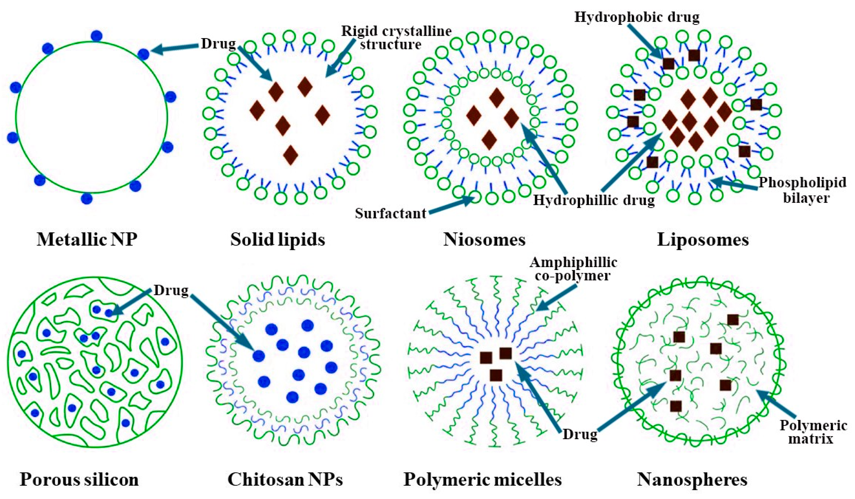
5.1. Nanocarrier Characteristics

Novel technologies and delivery modalities are needed for efficient drug distribution within the brain matrix due to the BBB’s limited molecule passage. Nanocarriers (NCs) and nanotechnology-based drug delivery systems have been identified as potential vehicles for drug delivery across the BBB due to their biosafety properties, extended drug release, higher solubility, enhanced drug bioactivity, ability to penetrate the BBB, and self-assembly (Figure 2) [163,164]. The National Nanotechnology Project designates particles that possess a minimum of one dimension and a size ranging from one to one hundred nanometers as nanoparticles (NPs). NPs possess the ability to traverse small capillaries owing to their diminutive size. The nanoparticles are surrounded by chemotherapeutic agents that are either contained within the matrix or affixed to their surface. In the processes of extravasation and receptor-mediated transcytosis, cells take up NP–drug complexes, which then release the drug into their cytoplasm or designated compartment. After crossing the blood–brain barrier, the gathering of chemotherapeutic agent-loaded nanoparticles at tumor locations is affected by their engagement with tumor cells and the diffusion within the tumor environment. The size, shape, and surface characteristics of the nanoparticles are crucial elements that influence this process [165,166]. NPs comprised of biodegradable materials offer advantages in terms of sustained drug release at targeted locations [167,168]. It has been demonstrated that appropriate surface coatings in the form of ligands can render drug-loaded NPs stable within the bloodstream, while also ensuring their nontoxic and nonimmunogenic properties [169]. NPs that possess ligands on their surfaces have the capability to transport the carrier system to specific targeted areas that possess receptors [170,171]. Transferrin, apolipoprotein (Apo) A, E, and B, as well as specific antibodies present on the surface of NPs, have been identified as potential ligands that can aid in the transportation of drug–NP complexes across the BBB through receptor-mediated endocytosis [170,171,172,173]. In addition, the field of nanotechnology has the potential to mitigate the negative side effects associated with chemotherapeutic drugs that have brief half-lives, through the augmentation of their bioavailability [174].
The influence of size and surface charge on the permeation of the reticuloendothelial system (RES) by NPs is a significant factor to be considered [175]. NPs that possess a positive charge and fall within the size range of 5 to 500 nm are essential for achieving enhanced cellular uptake. Particles with a size of less than 200 nm are particularly suitable and optimal for systemic delivery [175,176]. Particles that are smaller than 5 nanometers are unable to be filtered out by the kidneys. Nanoparticles demonstrate a heightened ability to interact with negatively charged cell membranes, effectively targeting specific biological regions or particular proteins. As a result, positively charged nanoparticles are favored due to their ability to improve stability in circulation within living organisms [176]. The diminutive dimensions of nanoparticles present a complex scenario, as they facilitate the passage through tissue junctions and cellular membranes, thereby allowing access to the cytoplasm of healthy cells. According to the literature, NPs have the potential to cause damage to DNA, RNA, and the mitochondrial structure present in cells, leading to cellular apoptosis [177]. The implementation of surface coatings and other modifications to augment the safety of NPs within the human body is imperative for their utilization in the field of clinical medicine (Figure 3).
Researchers have been studying nanoparticle surface modification for over a decade to prevent harmful interactions between nanomaterials and healthy tissues [177]. Polyethylene glycol (PEG) was initially used as a surface coating to help nanoparticles avoid recognition by the reticuloendothelial system (RES), thereby extending their half-life and persistence in the bloodstream. This is attributed to its hydrophilic outer surface and hydrophobic inner polymeric matrix [178]. Following this, researchers created chitosan PEGylated albumin-coated nanoparticles alongside specific antibodies to enhance the targeting of drugs to the brain via receptor-mediated transporter endocytosis. This method sought to improve the affinity and specificity of the nanoparticles for the targeted tissue [179,180]. Numerous techniques have been employed to administer drugs to the CNS [181,182,183,184,185,186,187,188,189,190]. However, a novel approach has been introduced through the utilization of NPs composed of poly (ethylene glycol)-poly(ῳ-pentadecalactone-co-p-dioxanone). These NPs exhibit a prolonged release of drugs, obviating the need for repeated infusions, thereby enhancing safety and translatability [191].

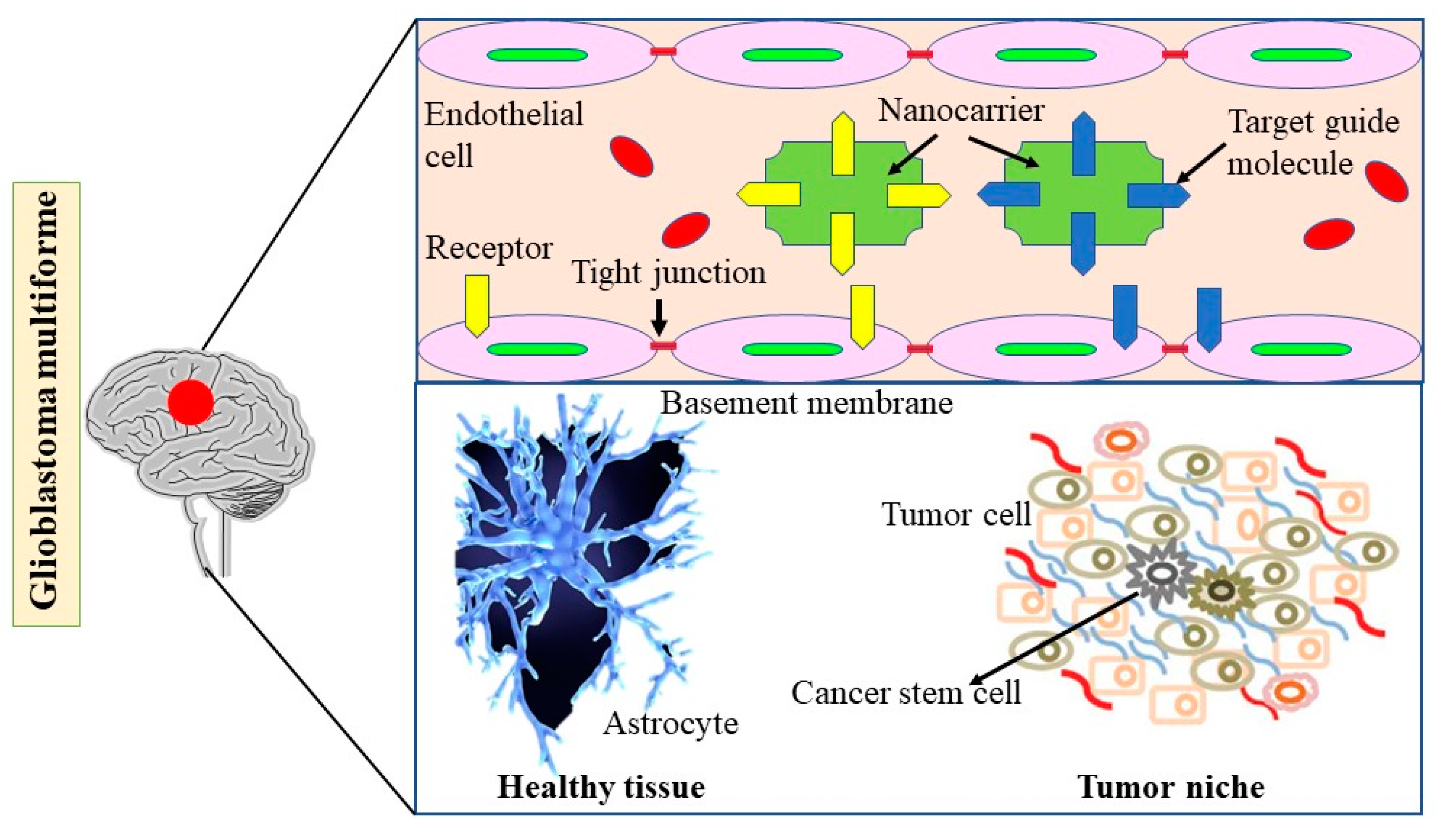
5.2. Nanotechnology-Based Strategies for Enhancing Drug Delivery in GBM: Exploring the Impact of Diabetes and Obesity

The primary impediments to treating GBM are the presence of the BBB, the capacity of the RES to gather and eliminate anticancer medications, and the absence of a specific targeting mechanism that would enable drugs to selectively bind to GSCs. It is imperative to develop distinct delivery system designs and administration methods that are specific to NCs in order to effectively address these challenges. According to a study, the utilization of convection-enhanced delivery (CED) and an intratumor delivery route can enable the retention of nanoformulated drugs in the tumor site for an extended period, an achievement that is unattainable with non-nanoformulated therapies [192]. Additional advantages of utilizing the CED technique encompass the capacity to administer nanoengineered chemotherapeutic agents to GBM cells with a meticulously controlled infusion velocity [62,193], thereby enhancing the efficacy of antineoplastic treatment [193].
Current studies have shown that mice with intracranial gliomas were treated with a newly developed nanoparticle derived from magnetotactic bacteria. The mice were then subjected to an alternating magnetic field or magnetic hyperthermia. The results showed a significant improvement in antitumor activity, with almost complete elimination of the tumors [194,195]. The proposed methodology presents a feasible and distinct strategy for managing invasive malignancies such as gliomas. Nevertheless, achieving complete coverage of the tumor using NPs poses a significant challenge.
An alternative method involves administering anticancer drugs via intranasal injection. The feasibility of direct nose-to-brain transport bypassing the blood–brain barrier has been investigated in mice with glioblastoma multiforme [196]. This was achieved through the use of theranostic polyfunctional gold–iron oxide nanoparticles that were coated with therapeutic miRNAs. Furthermore, the utilization of this nanoformulation enables the systemic administration of TMZ to GBM cells [197]. The intranasal route of drug transmission to the brain presents a number of potential advantages in comparison to the intravenous method. However, it is important to note that intranasal administration has limitations and is primarily in the preclinical phase of investigation [198]. The challenges that need to be addressed include the anatomical constraints of the nasal cavity, low bioavailability of peptides, rapid clearance, and other related factors [198]. The opening of the main barrier of the BBB can be facilitated through the utilization of MRI-guided targeted ultrasound or the administration of bradykinin, thereby enhancing the diffusion of chemotherapeutic drugs across the BBB [198,199]. The utilization of MRI-guided focused ultrasound has been observed to facilitate the penetration of cisplatin-loaded NPs coated with PEG into the brain by inhibiting macrophage uptake. This approach holds promise for crossing both the BBB and blood tumor barrier (BTB) [200]. In order to achieve the objective of reducing and altering the BTB and BBB, it is imperative to employ therapies that specifically target the aquaporin-4 (AET) and tight junction (TJ) proteins [201].
Historically, trials involving inhibitors of multidrug-resistant efflux transporters have yielded predominantly negative results. However, it is possible to reverse the multidrug resistance efflux transporters by utilizing Pgp inhibitors that are enclosed in surfactant-based NPs. This approach can be utilized to augment the drug’s therapeutic effectiveness [202]. In order to promote the dissemination of magnetic anti-GBM drugs to GBM cells, a magnetic field has been utilized [194,195]. Previous challenges encompassed inadequate equipment for generating a sufficiently potent magnetic field in a targeted area and concerns regarding inadvertent impacts on healthy tissue [203,204]. The administration of magnetic MNPs directly into the tumor site has emerged as a feasible approach for treating GBM. This is due to the ability of MNPs to accumulate significantly at the tumor site through the use of a magnetic platform that could be manipulated by an external magnetic field [205]. An additional illustration pertains to the hybrid magnetic nano-vectors that have been recently formulated. These nano-vectors are composed of lipids and are functionalized with angiopep-2. They bring about the death of GBM cells via a conjugation effect of chemotherapy and lysosomal membrane permeabilization, thus exhibiting a synergistic action [206].

5.3. Impact of Diabetes, Obesity, and Nanotechnology on Glioblastoma Multiforme Cells and Glioblastoma Stem Cells

The active targeting strategy for GBM entails the attachment of an agent to nanoparticles, enabling them to selectively engage with receptors or antigens present in GBM cells or glioblastoma stem cells (GSCs) [193,207,208]. GBM cells have been found to express a variety of proteins or receptors, including IL-13 receptor, metalloproteinase-2, Integrin-5, CD133, and CD33, which could potentially serve as targets for NP therapy. In recent years, scholars have conducted research on the precise targeting of GSCs [209]. This is due to the fact that the presence of GSCs is a major contributor to the recurrence of GBM. The utilization of drug delivery systems predicated on nanocrystals (NCs) may result in the expression of multiple receptors or markers as targets by GSCs. GSCs exhibit distinct cellular surface markers such as CD133 and CD15, post-transcriptional factors, transcription factors such as OCT4, and cytoskeletal proteins such as nestin, which may vary depending on their specific anatomical location [210]. Although there are several potential therapeutic approaches for targeting GSCs, the majority of these therapies have demonstrated clinical inefficacy [211]. One recent instance of nanotechnology being employed for GSC targeting is the amalgamation of calf thymus DNA with gold NPs, which enhances the sensitivity of GSCs to radiation [212]. Neurofilaments give rise to NFL-TBS.40-63 and LinTT1 peptides, which specifically target GSCs [213,214]. The utilization of gold nanorods that have been functionalized with a specifically designed peptide has been shown to accurately identify nestin-positive GSCs. This approach has been deemed a promising strategy for the development of a successful nanomedicine for the treatment of recurrent GBM [215]. A drug carrier, consisting of CBP4-coated gold NPs, has been developed to specifically target the cell surface marker CD133 in GSCs [216].

5.4. Current NCs and GBM Treatment Strategies

Nanocarrier-based combination therapies for GBMs offer improved tumor-specific drug delivery, simplified administration, and optimal validation of synergistic drug proportion [53]. NCs can be categorized into three types, namely nanocapsules, NPs, and nanospheres, based on their method of production. Various nanoparticles, including liposomes, solid lipid NPs, polymeric NPs, polymeric micelles, silica, and dendrimers, are frequently utilized in the treatment of GBM. The classification of these particles is dependent on the nature of the colloidal drug carriers utilized in their production.

5.4.1. Liposomes

Liposomes bear a resemblance to cellular membranes as they comprise an outer phospholipid bilayer enveloping an aqueous core. This characteristic enhances the ability of a substance to bind with lipids, facilitating the passage of lipophilic macromolecules through the blood–brain barrier. Liposomal nanoparticles offer a range of benefits, such as ease of production, straightforward encapsulation of various anticancer agents, remarkable biocompatibility, effectiveness, absence of immunogenicity, enhanced solubility of anticancer agents, and availability in the market [217,218]. More than twenty years ago, the development of liposomes began with the aim of integrating radiosensitizers and chemo-therapeutic agents, including doxorubicin, to address different types of drug-resistant cancers [217]. Over the course of the last decade, various liposomal formulation techniques, receptor-mediated transcytosis, and novel conjugated drugs have been investigated as potential treatments for GBMs [218,219,220]. The incorporation of PEG into the surface of the liposome phospholipid bilayer has been reported to enhance the longevity of liposomes in circulation by facilitating the evasion of RES capture [76]. The advancement of novel nanotechnology aimed at targeting tumors may involve the identification of specific receptors or antigens that are overexpressed on GBM cells. In murine models, a significant decrease in tumor growth was observed with the use of interleukin (IL)-13-conjugated liposomes and IL-4 receptor-targeted liposomal doxorubicin, as compared to unconjugated liposomes [219,220]. Over a decade ago, researchers developed anti-EGFR immunoliposomes with the aim of targeting GBM cells that exhibited overexpression of the protein in an animal model. The findings of this study suggested that these immunoliposomes could significantly enhance the efficacy of various anticancer drugs [221]. Although liposomal NPs are extensively employed in the management of GBM, there exist various limitations that require further attention. The impact of liposomal nanoparticles on various brain regions is nonuniform, and their capacity to traverse the BBB is contingent upon the medication they carry or the surface molecules with which they are coated [219,220].

5.4.2. Polymeric Micelles

Polymeric micelles are composed of a structure consisting of a lipophilic polymer core and a lipophobic shell. The design of block co-polymers is a result of their self-assembly, which can be utilized to regulate the rate of controlled release and the effectiveness of incorporating chemotherapeutic drugs [222]. The core–shell designs that possess unique features and are limited in size between 10 and 100 nm have been found to be effective in protecting the core containing the drug from any potential interactions with the complement system and macrophage absorption. This leads to an extended circulation period and a half-life of over 10 h [223,224]. Biodegradable polyesters such as long-chain alkyl derivatives, poly(caprolactone), poly(D,L-lactide), and poly(D,L-lactide-co-glycolide) are frequently utilized as the polymer matrix for container cores [222]. PEG [222,223] is considered the optimal polymer for the production of a shell that effectively inhibits interaction with serum proteins. The primary hindrance to the utilization of polymeric micelles-based treatment for GBM is the absence of targeting agents that could facilitate more GBM-specific accumulation, subsequent to the resolution of the initial constraint of inadequate drug-circulation duration [222]. Currently, further investigations are being carried out with the aim of enhancing the efficacy of the present formulation by specifically targeting receptors that are expressed in GBM cells. The efficacy of 17-AAG as a cancer cell inhibitor is attributed to its ability to destabilize heat shock protein 90 (Hsp90) and related client proteins. To this end, the use of polymeric mixed micelles comprising Pluronic P-123 and F-127 and incorporating 17-allylamino-17-demethoxy geldanamycin (17-AAG) has been proposed as a promising strategy for drug delivery via nanomaterials [224,225]. The attributes of potential delivery mechanisms are outlined as follows: The therapeutic potential of 17-AAG in the management of GBM can be attributed to its targeting proficiency, controlled release kinetics, and elevated drug loading capacity [224]. The formulation of mixed micelles comprising Pluronic P-123 and F-127 loaded with 17-AAG exhibits favorable design characteristics. The transferrin receptor (TfR) is a potential target location that exhibits overexpression in both GBM and BBB cells. Sun et al. [226] successfully generated TfR-PEG polymeric micelles that demonstrated effective BBB penetration. Tumor cells have the ability to rapidly absorb them. Paclitaxel-loaded TfR-PEG polymeric micelles have been found to effectively inhibit the proliferation of U87 GBM cells, while also leading to an increase in the median survival time of nude mice bearing GBM [226]. In another study, Woensel et al. [227] demonstrated that Gal-1-targeting siRNA-loaded chitosan NPs could serve as a promising delivery system for treating GBM. The NPs served to safeguard siRNA from degradation, facilitated its transport through the nasal route into the CNS, and ultimately led to a decrease in Gal-1 expression, which in turn diminished the motility of tumor cells in both murine and human models of glioblastoma. This highlights the promising role of chitosan NPs in the context of siRNA-based therapy for GBM. In an innovation, Alswailem et al. [228] established chitosan NPs as a viable carrier for microRNA-219 delivery to glioblastoma patients. The optimized chitosan NPs demonstrated a notable entrapment efficiency, a sustained release profile, and a targeted action towards GBM cells, leading to a significant reduction in the survival of U87 MG cells while leaving fibroblasts largely unaffected. The findings underscore the promising role of chitosan NPs in facilitating targeted gene delivery for the treatment of GBM.

5.4.3. Dendrimers

Dendrimers, possessing a compact and highly branched scaffold design, are considered to be the smallest molecules with diameters below 12 nm. This unique feature renders them suitable for the transportation and safeguarding of short interfering RNA (siRNA) in the circulatory system [229,230,231]. Dendrimers that are loaded with methotrexate have been found to possess supplementary advantages such as improved drug potency and exceptional efficiency in crossing the BBB [231]. Despite their potential benefits, dendrimers possess several limitations such as rapid clearance by the reticuloendothelial system, toxicity towards healthy tissue due to their proximity to cell membranes, and a significantly inferior ability to regulate release behavior [229,232]. Gold nanoparticles (AuNPs) were entrapped in poly(amidoamine) dendrimers to achieve the condensation of two separate small interfering RNA (siRNA) molecules for the purpose of silencing oncogenes. This is a recent development in the field. The PAMAM-Au dendrimers have been coated with beta-cyclodextrin (β-CD) and have demonstrated a high level of efficacy in transporting siRNA to glioma cells [232,233]. Research indicates that the biocompatibility and endosomal escape of amino acid-functionalized dendrimers may be improved through the involvement of endogenous amino acids. Conversely, phosphate dendrimers characterized by a hydrophobic backbone and a hydrophilic surface demonstrate an enhanced ability to penetrate the blood–brain barrier [234,235]. An illustration of this is the utilization of dendrimers that are functionalized with arginine–glycine–aspartic acid and entrapped with gold NPs. These dendrimers exhibit robust cytocompatibility and remarkably effective transfection and have demonstrated potential efficacy as gene therapy for glioblastomas [234]. The creation of PEPE dendrimers linked with d-glucosamine seeks to improve the delivery of drugs across the blood–brain barrier and enhance the targeting of tumors [231]. The results of the in vitro study indicate that glycosylation of the PEPE dendrimers led to enhanced and rapid accumulation around tumor spheroids. Additionally, the glycosylated PEPE dendrimers were found to be effective in overcoming MTX resistance, as evidenced by their ability to induce cell death in MTX-resistant cells when loaded with methotrexate [231].

5.4.4. Metal Nanoparticles

The use of metal particles has been found to enhance the radio-sensitization of glioblastoma multiforme (GBM) tumor cells. The utilization of metal particles has been observed to enhance the radio-sensitization of glioblastoma multiforme (GBM) tumor cells. In animal models, the administration of metal particles prior to radiation treatment has been reported to result in significant DNA damage to tumor cells [236]. Metallic particles exhibit robust X-ray absorption, synthetic plasticity, and unique electrical characteristics, rendering them appealing contenders for radio sensitization [237]. AuNPs are a suitable material for nanomedicine in the treatment of GBM due to their modifiability, variable diameters, and significant surface-to-volume ratio [238]. The capacity of AuNPs to traverse the BBB is attributed to their precisely controlled dimensions. However, their limited targeting capability poses a constraint on their medical utility [238,239].
Despite the well-regulated size of AuNPs, their limited targeting abilities hinder their utilization in medical contexts [238,239]. The efficacy of aptamer targeting within AuNPs is enhanced by the formation of a covalent bond between gold and sulfur [239]. The identification of aptamer–AuNP complexes as a potential therapeutic agent for GBM treatment has been reported. These complexes have demonstrated significant inhibition of tumor growth in both in vitro and in vivo studies [239]. AuNPs of specific sizes have the potential to mitigate the limited transmembrane penetration of aptamers. NPs can also serve as a vehicle for the delivery of therapeutic gene targets. The delivery of the tumor suppressor miR-100 can be achieved through the use of a novel polyfunctional gold–iron oxide nanoparticle. This NP has been recently developed and has shown promising results in enhancing the sensitivity of glioblastoma multiforme cells to systemically administered temozolomide in murine models. The established biodistribution and clearance processes of various gold and silver nanoformulations containing chemotherapeutic drugs have led to their authorization for clinical studies by the American Food and Drug Administration [240].

5.4.5. Silica Nanoparticles

Silica nanoparticles (SiNPs) are frequently utilized in various medicinal applications due to their notable attributes such as high biocompatibility, substantial surface area for drug loading, stability, and cost-effectiveness [241]. SiNPs have been investigated for their potential applications and safety in various scientific disciplines. The toxicity induced by SiNPs can potentially be mitigated through the careful selection of size, dosage, and cell type [242,243]. This has opened up avenues for scientists to explore the modification of SiNPs through multimodal approaches, with the aim of enhancing their therapeutic utility. The application of synthetic modifications on SiNPs has been observed to mitigate the elevated toxicity levels associated with SiNPs of smaller dimensions [244]. Porous SiNPs (pSiNPs) that have been modified with transferrin are currently a prevalent formulation for the treatment of GBM. These NPs possess desirable characteristics such as high drug-loaded capacity, biocompatibility, and degradability. This has contributed to their popularity in the field [245,246]. Transferrin-functionalized pSiNPs can facilitate sustained drug release at the specific site of action due to the frequent overexpression of transferrin receptors on the BBB and the surface of GBM cells. This approach can be particularly effective for drugs like doxorubicin. The transportation of a significant amount of medication to the GBM can be achieved through the utilization of an external low-power radiofrequency field in conjunction with a multicomponent NP that possesses a fibronectin-targeting ligand-loaded iron oxide core and mesoporous silica shell [247].

5.5. Regulatory Hurdles and Ethical Considerations

Nanotechnology in targeted drug delivery, especially for complex diseases like glioblastoma multiforme, has huge potential to improve therapeutic outcomes. However, translating these innovations into clinical applications poses enormous regulatory and ethical challenges [248]. Regulatory bodies, including the US Food and Drug Administration and the European Medicines Agency, have set up strict guidelines for the approval of nanomedicines with an emphasis on safety, efficacy, and reproducibility [249]. The unique physicochemical properties of nanocarriers in size, shape, and surface modifications call for new testing paradigms; traditional frameworks may not be adequate to address the potential risks such as toxicity, biodistribution, and off-target effects [250]. Moreover, the lack of uniform guidelines on the characterization and evaluation of nanocarriers adds to the extra hurdles that delay the translation from bench to bedside [251].
From an ethical standpoint, nanotechnology presents numerous concerns, including the assurance of patient safety, the affordability of treatments, and equitable access to these therapies for everyone. It is essential to ensure that treatments based on nanotechnology are accessible to a wide range of populations, particularly those in resource-limited environments [252]. Furthermore, it is essential to take into account the long-term environmental toxicity and biodegradability of nanomaterials, as their large-scale production may present ecological risks. It is essential to ensure transparency in clinical trials, obtain informed consent from patients, and uphold a rigorous ethical review process. These measures are crucial not only to safeguard vulnerable populations from exploitation but also to foster trust in emerging therapies [253].
Overcoming these challenges will require a concerted effort among researchers, industry stakeholders, and regulatory bodies. The establishment of global standards for the development and evaluation of nanomedicines, along with comprehensive guidelines for ethical assessment, is likely to facilitate the swift and safe integration of nanotechnology into cancer treatment [254]. Future research should focus on reducing production costs and optimizing scalability for nanocarrier-based systems. This will enable the widespread adoption of these advanced technologies, particularly benefiting large populations, including patients with diabetes and obesity who require more specialized treatment approaches [255].

6. Role of NCs Beta-Cell Function and Their Implications for Diabetes, Obesity, and GBM Treatment

The field of medicine has been significantly impacted by advances in nanotechnology due to the close size proximity of NPs to various biological molecules. Drug-loaded NPs exhibit enhanced therapeutic properties and reduced morbidity, while also facilitating efficient drug delivery to the intended tissue. The field of medicine that involves the use of nanotechnology for therapeutic purposes is commonly referred to as nanomedicine or nanotherapeutics.
Liposomes are diminutive spherical structures composed of nontoxic phospholipids and cholesterol. Niosomes refer to multilamellar vesicles composed of nonionic surfactants. NPs of metallic nature possess the ability to readily associate with diverse biological entities. Nanospheres refer to spherical nanostructures that are typically composed of polymers and serve as matrices. Polymeric micelles are complex structures composed of a core and a shell, formed by the assembly of amphiphilic block copolymers. Chitosan nanoparticles are created by combining chitosan with a polyanion, like tripolyphosphate. Porous silicon nanoparticles are a specific category of hollow nanoparticles made from porous silicon.
Nanotechnology has been utilized in the treatment of various pathological medical conditions such as cancer, Alzheimer’s disease, Parkinson’s disease, diabetes mellitus, and tuberculosis [256]. Nanotechnology has made a noteworthy contribution to diabetes treatment through the identification of novel nanosensors that enable fast, precise, and delicate detection of blood glucose levels [257]. Nanotechnology has enabled the development of potent insulin delivery systems that can directly transport insulin molecules into the bloodstream, bypassing the harsh acidic conditions of the stomach. This presents a viable option to the conventional practice of administering insulin through subcutaneous injections on a daily basis [258]. Furthermore, nanotechnology is employed in the production of biofunctional food and nanodrugs for the management of prediabetes [259].

6.1. Biomolecule-Based Nanomaterials for DM Therapy

The prevalence of diabetes has experienced a significant surge globally, impacting a vast population that has had to adapt to a predominantly inactive way of life. Diabetes is characterized by persistent hyperglycemia, which leads to both microvascular and macrovascular complications including but not limited to nephropathy, retinopathy, neuropathy, stroke, and cardiovascular disease. The projected rise in the population of individuals aged 65 and above is expected to result in a significant increase in the incidence of DM, with estimates suggesting that the number of cases will exceed 643 million by 2030, representing a more than threefold increase from current levels [260,261,262,263,264,265]. Therefore, it is imperative to manage DM pathology efficiently. The utilization of drug delivery systems based on NCs has the potential to be efficacious in the management of diabetes and its associated complications.

6.2. Polymeric NPs for Drug Delivery

Nanoparticles play a significant role in the pharmaceutical field, facilitating the delivery of a range of substances including polypeptides, nucleic acids, synthetic compounds, proteins, and vaccines. Compared to parenteral formulations, the oral delivery of insulin-loaded nanoparticles is often favored because of its ease of administration and enhanced patient adherence. The restricted bioavailability of orally administered insulin encapsulated in nanoparticles can be linked to insufficient absorption, which is often due to proteases that degrade free insulin within the gastrointestinal tract. In order to improve the bioavailability of insulin taken orally, a peptide was employed to aid in cellular penetration and was then encapsulated within mucoadhesive nanoparticles. Polymeric dendrimers represent a fascinating class of nanostructured macromolecules, distinguished by their tree-like architecture. They feature a highly branched outer structure, a central globular core, and reactive end groups that populate their surface. The dendrimer family includes a diverse array of types, such as hybrid dendrimers, liquid crystalline dendrimers, poly(propyleneimine), glycol, peptide, and PAMAM. The prevalent utilization of these compounds in biochemistry can be attributed primarily to their unique physiochemical properties. PAMAM G4 has the potential to mimic hypoglycemic drugs and reduce plasma glucose levels during extended experimentation on diabetic animal models [266,267].
Upon reaching the critical micelle concentration, amphiphilic co-polymers have the ability to undergo self-assembly, resulting in the formation of polymeric micelles that exhibit a core–shell architecture. The outer layer, which is hydrophilic in nature, serves as a protective shell and also facilitates chemical modification through the presence of functional groups. The hydrophobic core could potentially be engineered to facilitate the incorporation of hydrophobic substances within a polymeric matrix. Micelles have been extensively investigated for enhanced pharmacokinetics, biodistribution, and enzymatic degradation. In their study, Fang et al. employed various analytical techniques, including circular dichroism, bis-anilinonaphthalene sulfonates binding assay, turbidity assay, matrix-assisted laser desorption ionization–time of flight mass spectrometry, agarose gel electrophoresis, and thioflavin-T binding assay, to investigate the reconversion of DTT-denatured insulin into its native conformation by PEG-phosphatidylethanolamine (PE) micelles [268]. The results of the study indicate that PEGPE micelles possess a layered structure with a negative charge, which bears resemblance to a nanocage. This structure has the potential to hinder aggregation by disrupting hydrophobic interactions and capturing insulin A and B chains. The nanocage has the potential to facilitate the docking of insulin A and B chains. The utilization of PEG-PE micelles has the potential to function as an artificial chaperone in the process of protein refolding. Moreover, the incorporation of linkable hydrophilic moieties into hydrophobic polymers can facilitate the formation of stable micellar structures [269,270,271].

7. Clinical Trials Exploring the Impact of Diabetes, Obesity, and Nanotechnology on GBM Prognosis and Treatment

Nanotechnology and NC-based therapy have shown potential in treating GBM in both in vivo and in vitro studies (Table 1), but their use in clinical trials remains limited.

8. Limitations

Although the application of nanotechnology in addressing GBM and DM presents several encouraging benefits, it is important to consider certain limitations. These encompass challenges in translating the achievements observed in preclinical applications into tangible clinical outcomes. Initially, GBM presents significant challenges due to its aggressive characteristics, diverse cellular composition, and elevated rates of recurrence, making consistency in treatment and management quite difficult. In addition to these factors, several significant challenges emerge regarding the clinical application of nanomaterials, particularly concerning their bio-compatibility, toxicity, and scalability. Additionally, the lack of adequate long-term data concerning safety and efficacy presents another obstacle to the widespread adoption of this nano-therapy. Despite the progress made in the field of experimental nanodrugs, the substantial costs associated with their development and manufacturing are limiting their accessibility.

9. Conclusions and Future Perspective

This compilation of research articles highlights the complex relationship between metabolism, diabetes, and drug delivery in the context of brain cancer, specifically GBM. Nanotechnology offers potential for targeted drug delivery to GBM cells, but inconsistent findings reveal complex relationships between diabetes and glioma risk, including GBM. Hyperglycemia, obesity, and metabolic syndrome significantly impact cancer risk and patient outcomes, particularly in GBM, posing a significant challenge for clinicians and researchers.
Over the past two decades, there has been a significant rise in the use of nano-composites in the field of biomaterials. NCs like liposomes, polymeric micelles, dendrimers, and metal nanoparticles provide unique advantages in overcoming challenges like the blood–brain barrier and improving drug bioavailability. NCs for DM and GBM pathobiology were developed due to site-specific administration, drug treatment promotion, medication, and patient compliance. The outlook for individuals with GBM is generally unfavorable, and current therapeutic strategies have not shown significant improvements in OS or PFS. Nanoparticles, with their distinctive physical features like size, shape, and surface, effectively encapsulate and transport therapeutic agents to the brain. Nano-therapies effectiveness in preclinical studies has been proven, but their translation into clinical trials for GBM treatment has been limited, highlighting the need for further research and development. The complexity of GBM and challenges in drug delivery highlight the need for ongoing exploration and innovation in nanotechnology to enhance treatment strategies for aggressive brain cancer. The tumor and diabetes exhibit heterogeneity, aggressiveness, and recurrence, complicating the treatment process. The full investigation into the potential use of biomaterials as a tissue engineering strategy for treating glioblastoma/DM remains unexplored. The use of diverse biomaterials and methodologies in invitro models suggests a potential latent technique that could potentially reduce the impact of lethal diseases.
The rapid growth of experimental nanodrugs, particularly for treating GBM and T1DM (nanoformulations of insulin), enables their use against T2DM. The employment should focus on enhancing current treatments by eliminating side effects and utilizing nanotechnology for the development of innovative antidiabetic and anticancer drugs. Advancements in nanotechnology are expected to lead to innovative strategies to combat the increasing prevalence of T2DM and GBM.

Funding

This research received no external funding.

Acknowledgments

Authors are thankful to the Department of Pharmaceutical Technology, Eminent College of Pharmaceutical Technology, Kolkata, West Bengal, 700126; Department of Pharmaceutical Chemistry, Gupta College of Technological Sciences, Asansol; and Department of Chemistry, School of Advanced Sciences, VIT, Vellore for providing resources to write this review.

Conflicts of Interest

The authors declare no conflict of interest.

Abbreviations

A2AARanti-adenosine A2A receptor
Aktprotein kinase B/Akt
ALDH1A3aldehyde dehydrogenase 1A3
AMP adenosine monophosphate
ATP adenosine triphosphate
AuNP gold nanoparticles
BBB blood–brain barrier
CD73cluster of differentiation 73
CED convection-enhanced delivery
cIAP1cellular IAP 1
CLL chronic lymphocytic leukemia
CNS central nervous system
DM diabetes mellitus
EDVDoxEnGeneral delivery vehicle-doxorubicin
EGFRepithelial growth factor receptor
FPR2formylpeptide receptor 2
GBM glioblastoma multiforme
GSC glioblastoma stem cell
GTR gross total resection
HbA1c hemoglobin A1c
Hsp90heat shock protein 90
IAP inhibitors of apoptosis protein
IDH isocitrate dehydrogenase
IGF insulin-like growth factor
ILinterleukin
JNK c-Jun N-terminal kinase
MDSC myeloid-derived suppressor cells
Mes GSC mesenchymal GSC
MGMT O6-methylguanine methyltransferase
MNP magnetic nanoparticle
NC nanocarrier
NP nanoparticle
OS overall survival
PEG polyethylene glycol
PEGPE PEG-phosphatidylethanolamine
PEG-Dox Pegylated liposomal doxorubicin
PN GSC proneural GSC
SiNP silica nanoparticles
siRNA short interfering RNA
Smac second mitochondrial-derived activator of caspases
TMZ temozolomide
T2DM type 2 diabetes mellitus
VEGF vascular endothelial growth factor
XIAPX-linked IAP

References

  1. Kawamura, Y.; Takouda, J.; Yoshimoto, K.; Nakashima, K. New aspects of glioblastoma multiforme revealed by similarities between neural and glioblastoma stem cells. Cell Biol. Toxicol. 2018, 34, 425–440. [Google Scholar] [CrossRef] [PubMed]
  2. Disney-Hogg, L.; Sud, A.; Law, P.J.; Cornish, A.J.; Kinnersley, B.; Ostrom, Q.T.; Labreche, K.; Eckel-Passow, J.E.; Armstrong, G.N.; Claus, E.B. Influence of obesity-related risk factors in the aetiology of glioma. Br. J. Cancer 2018, 118, 1020–1027. [Google Scholar] [CrossRef]
  3. Behl, T.; Sharma, A.; Sharma, L.; Sehgal, A.; Singh, S.; Sharma, N.; Zengin, G.; Bungau, S.; Toma, M.M.; Gitea, D. Current perspective on the natural compounds and drug delivery techniques in glioblastoma multiforme. Cancers 2021, 13, 2765. [Google Scholar] [CrossRef] [PubMed]
  4. Deng, L.; Zhai, X.; Liang, P.; Cui, H. Overcoming TRAIL resistance for glioblastoma treatment. Biomolecules 2021, 11, 572. [Google Scholar] [CrossRef] [PubMed]
  5. Frazzi, R. BIRC3 and BIRC5: Multi-faceted inhibitors in cancer. Cell Biosci. 2021, 11, 1–14. [Google Scholar] [CrossRef]
  6. Opo, F.A.D.M.; Rahman, M.M.; Ahammad, F.; Ahmed, I.; Bhuiyan, M.A.; Asiri, A.M. Structure based pharmacophore modeling, virtual screening, molecular docking and ADMET approaches for identification of natural anti-cancer agents targeting XIAP protein. Sci. Rep. 2021, 11, 4049. [Google Scholar] [CrossRef]
  7. Cetraro, P.; Plaza-Diaz, J.; MacKenzie, A.; Abadía-Molina, F. A review of the current impact of inhibitors of apoptosis proteins and their repression in cancer. Cancers 2022, 14, 1671. [Google Scholar] [CrossRef] [PubMed]
  8. Fukumura, D.; Incio, J.; Shankaraiah, R.C.; Jain, R.K. Obesity and cancer: An angiogenic and inflammatory link. Microcirculation 2016, 23, 191–206. [Google Scholar] [CrossRef]
  9. Edlinger, M.; Strohmaier, S.; Jonsson, H.; Bjørge, T.; Manjer, J.; Borena, W.T.; Häggström, C.; Engeland, A.; Tretli, S.; Concin, H. Blood pressure and other metabolic syndrome factors and risk of brain tumour in the large population-based Me-Can cohort study. J. Hypertens. 2012, 30, 290–296. [Google Scholar] [CrossRef] [PubMed]
  10. Giovannucci, E.; Harlan, D.M.; Archer, M.C.; Bergenstal, R.M.; Gapstur, S.M.; Habel, L.A.; Pollak, M.; Regensteiner, J.G.; Yee, D. Diabetes and cancer: A consensus report. CA A Cancer J. Clin. 2010, 60, 207–221. [Google Scholar] [CrossRef]
  11. Kyrgiou, M.; Kalliala, I.; Markozannes, G.; Gunter, M.J.; Paraskevaidis, E.; Gabra, H.; Martin-Hirsch, P.; Tsilidis, K.K. Adiposity and cancer at major anatomical sites: Umbrella review of the literature. Bmj 2017, 356, j477. [Google Scholar] [CrossRef] [PubMed]
  12. Niedermaier, T.; Behrens, G.; Schmid, D.; Schlecht, I.; Fischer, B.; Leitzmann, M.F. Body mass index, physical activity, and risk of adult meningioma and glioma: A meta-analysis. Neurology 2015, 85, 1342–1350. [Google Scholar] [CrossRef] [PubMed]
  13. Sergentanis, T.N.; Tsivgoulis, G.; Perlepe, C.; Ntanasis-Stathopoulos, I.; Tzanninis, I.-G.; Sergentanis, I.N.; Psaltopoulou, T. Obesity and risk for brain/CNS tumors, gliomas and meningiomas: A meta-analysis. PLoS ONE 2015, 10, e0136974. [Google Scholar] [CrossRef] [PubMed]
  14. Wiedmann, M.; Brunborg, C.; Lindemann, K.; Johannesen, T.; Vatten, L.; Helseth, E.; Zwart, J. Body mass index and the risk of meningioma, glioma and schwannoma in a large prospective cohort study (The HUNT Study). Br. J. Cancer 2013, 109, 289–294. [Google Scholar] [CrossRef] [PubMed]
  15. Danaei, G.; Finucane, M.M.; Lu, Y.; Singh, G.M.; Cowan, M.J.; Paciorek, C.J.; Lin, J.K.; Farzadfar, F.; Khang, Y.-H.; Stevens, G.A. National, regional, and global trends in fasting plasma glucose and diabetes prevalence since 1980: Systematic analysis of health examination surveys and epidemiological studies with 370 country-years and 2· 7 million participants. Lancet 2011, 378, 31–40. [Google Scholar] [CrossRef]
  16. Seliger, C.; Ricci, C.; Meier, C.R.; Bodmer, M.; Jick, S.S.; Bogdahn, U.; Hau, P.; Leitzmann, M.F. Diabetes, use of antidiabetic drugs, and the risk of glioma. Neuro-Oncology 2015, 18, 340–349. [Google Scholar] [CrossRef] [PubMed]
  17. Kaul, K.; Tarr, J.M.; Ahmad, S.I.; Kohner, E.M.; Chibber, R. Introduction to diabetes mellitus. In Diabetes: An Old Disease, a New Insight; Springer: New York, NY, USA, 2013; pp. 1–11. [Google Scholar] [CrossRef]
  18. Heidemann, C.; Boeing, H.; Pischon, T.; Nöthlings, U.; Joost, H.-G.; Schulze, M.B. Association of a diabetes risk score with risk of myocardial infarction, stroke, specific types of cancer, and mortality: A prospective study in the European Prospective Investigation into Cancer and Nutrition (EPIC)-Potsdam cohort. Eur. J. Epidemiol. 2009, 24, 281–288. [Google Scholar] [CrossRef] [PubMed]
  19. Saltzman, B.S.; Doherty, J.A.; Hill, D.A.; Beresford, S.A.; Voigt, L.F.; Chen, C.; Weiss, N.S. Diabetes and endometrial cancer: An evaluation of the modifying effects of other known risk factors. Am. J. Epidemiol. 2008, 167, 607–614. [Google Scholar] [CrossRef]
  20. Ben, Q.; Cai, Q.; Li, Z.; Yuan, Y.; Ning, X.; Deng, S.; Wang, K. The relationship between new-onset diabetes mellitus and pancreatic cancer risk: A case–control study. Eur. J. Cancer 2011, 47, 248–254. [Google Scholar] [CrossRef] [PubMed]
  21. Wang, C.-S.; Yao, W.-J.; Chang, T.-T.; Wang, S.-T.; Chou, P. The impact of type 2 diabetes on the development of hepatocellular carcinoma in different viral hepatitis statuses. Cancer Epidemiol. Biomark. Prev. 2009, 18, 2054–2060. [Google Scholar] [CrossRef] [PubMed]
  22. Campbell, P.T.; Newton, C.C.; Patel, A.V.; Jacobs, E.J.; Gapstur, S.M. Diabetes and cause-specific mortality in a prospective cohort of one million US adults. Diabetes Care 2012, 35, 1835–1844. [Google Scholar] [CrossRef] [PubMed]
  23. Rapone, B.; Corsalini, M.; Converti, I.; Loverro, M.T.; Gnoni, A.; Trerotoli, P.; Ferrara, E. Does periodontal inflammation affect type 1 diabetes in childhood and adolescence? A meta-analysis. Front. Endocrinol. 2020, 11, 278. [Google Scholar] [CrossRef]
  24. Van Den Berghe, G.; Wouters, P.; Weekers, F.; Verwaest, C.; Bruyninckx, F.; Schetz, M.; Vlasselaers, D.; Ferdinande, P.; Lauwers, P.; Bouillon, R. Intensive insulin therapy in critically ill patients. N. Engl. J. Med. 2001, 345, 1359–1367. [Google Scholar] [CrossRef] [PubMed]
  25. Tong, J.J.; Tao, H.; Hui, O.T.; Jian, C. Diabetes mellitus and risk of brain tumors: A meta-analysis. Exp. Ther. Med. 2012, 4, 877–882. [Google Scholar] [CrossRef]
  26. Polednak, A.P. Comorbid diabetes mellitus and risk of death after diagnosis of colorectal cancer: A population-based study. Cancer Detect. Prev. 2006, 30, 466–472. [Google Scholar] [CrossRef]
  27. Yancik, R.; Wesley, M.N.; Ries, L.A.; Havlik, R.J.; Edwards, B.K.; Yates, J.W. Effect of age and comorbidity in postmenopausal breast cancer patients aged 55 years and older. JAMA 2001, 285, 885–892. [Google Scholar] [CrossRef] [PubMed]
  28. Schwartzbaum, J.; Edlinger, M.; Zigmont, V.; Stattin, P.; Rempala, G.A.; Nagel, G.; Hammar, N.; Ulmer, H.; Föger, B.; Walldius, G. Associations between prediagnostic blood glucose levels, diabetes, and glioma. Sci. Rep. 2017, 7, 1436. [Google Scholar] [CrossRef]
  29. Van Hemelrijck, M.; Garmo, H.; Holmberg, L.; Walldius, G.; Jungner, I.; Hammar, N.; Lambe, M. Prostate cancer risk in the Swedish AMORIS study: The interplay among triglycerides, total cholesterol, and glucose. Cancer 2011, 117, 2086–2095. [Google Scholar] [CrossRef]
  30. Gritti, M.; Würth, R.; Angelini, M.; Barbieri, F.; Peretti, M.; Pizzi, E.; Pattarozzi, A.; Carra, E.; Sirito, R.; Daga, A. Metformin repositioning as antitumoral agent: Selective antiproliferative effects in human glioblastoma stem cells, via inhibition of CLIC1-mediated ion current. Oncotarget 2014, 5, 11252. [Google Scholar] [CrossRef]
  31. Sato, A.; Sunayama, J.; Okada, M.; Watanabe, E.; Seino, S.; Shibuya, K.; Suzuki, K.; Narita, Y.; Shibui, S.; Kayama, T. Glioma-initiating cell elimination by metformin activation of FOXO3 via AMPK. Stem Cells Transl. Med. 2012, 1, 811–824. [Google Scholar] [CrossRef] [PubMed]
  32. Würth, R.; Pattarozzi, A.; Gatti, M.; Bajetto, A.; Corsaro, A.; Parodi, A.; Sirito, R.; Massollo, M.; Marini, C.; Zona, G. Metformin selectively affects human glioblastoma tumor-initiating cell viability: A role for metformin-induced inhibition of Akt. Cell Cycle 2013, 12, 145–156. [Google Scholar] [CrossRef]
  33. Yu, Z.; Zhao, G.; Xie, G.; Zhao, L.; Chen, Y.; Yu, H.; Zhang, Z.; Li, C.; Li, Y. Metformin and temozolomide act synergistically to inhibit growth of glioma cells and glioma stem cells in vitro and in vivo. Oncotarget 2015, 6, 32930. [Google Scholar] [CrossRef]
  34. Müller, D.M.; Robe, P.A.; Eijgelaar, R.S.; Witte, M.G.; Visser, M.; de Munck, J.C.; Broekman, M.L.; Seute, T.; Hendrikse, J.; Noske, D.P. Comparing glioblastoma surgery decisions between teams using brain maps of tumor locations, biopsies, and resections. JCO Clin. Cancer Inform. 2019, 2, 1–12. [Google Scholar] [CrossRef]
  35. Bruhn, H.; Strandéus, M.; Milos, P.; Hallbeck, M.; Vrethem, M.; Lind, J. Improved survival of Swedish glioblastoma patients treated according to Stupp. Acta Neurol. Scand. 2018, 138, 332–337. [Google Scholar] [CrossRef] [PubMed]
  36. Montemurro, N.; Perrini, P.; Blanco, M.O.; Vannozzi, R. Second surgery for recurrent glioblastoma: A concise overview of the current literature. Clin. Neurol. Neurosurg. 2016, 142, 60–64. [Google Scholar] [CrossRef]
  37. Schwartzbaum, J.; Jonsson, F.; Ahlbom, A.; Preston-Martin, S.; Malmer, B.; Lönn, S.; Söderberg, K.; Feychting, M. Prior hospitalization for epilepsy, diabetes, and stroke and subsequent glioma and meningioma risk. Cancer Epidemiol. Biomark. Prev. 2005, 14, 643–650. [Google Scholar] [CrossRef]
  38. Mauer, J.; Chaurasia, B.; Goldau, J.; Vogt, M.C.; Ruud, J.; Nguyen, K.D.; Theurich, S.; Hausen, A.C.; Schmitz, J.; Brönneke, H.S. Signaling by IL-6 promotes alternative activation of macrophages to limit endotoxemia and obesity-associated resistance to insulin. Nat. Immunol. 2014, 15, 423–430. [Google Scholar] [CrossRef] [PubMed]
  39. Weiss, R.; Dziura, J.; Burgert, T.S.; Tamborlane, W.V.; Taksali, S.E.; Yeckel, C.W.; Allen, K.; Lopes, M.; Savoye, M.; Morrison, J. Obesity and the metabolic syndrome in children and adolescents. N. Engl. J. Med. 2004, 350, 2362–2374. [Google Scholar] [CrossRef]
  40. Chen, W.; Xia, T.; Wang, D.; Huang, B.; Zhao, P.; Wang, J.; Qu, X.; Li, X. Human astrocytes secrete IL-6 to promote glioma migration and invasion through upregulation of cytomembrane MMP14. Oncotarget 2016, 7, 62425. [Google Scholar] [CrossRef] [PubMed]
  41. Li, R.; Li, G.; Deng, L.; Liu, Q.; Dai, J.; Shen, J.; Zhang, J. IL-6 augments the invasiveness of U87MG human glioblastoma multiforme cells via up-regulation of MMP-2 and fascin-1. Oncol. Rep. 2010, 23, 1553–1559. [Google Scholar] [CrossRef]
  42. Liu, Q.; Li, G.; Li, R.; Shen, J.; He, Q.; Deng, L.; Zhang, C.; Zhang, J. IL-6 promotion of glioblastoma cell invasion and angiogenesis in U251 and T98G cell lines. J. Neuro-Oncol. 2010, 100, 165–176. [Google Scholar] [CrossRef] [PubMed]
  43. Tchirkov, A.; Khalil, T.; Chautard, E.; Mokhtari, K.; Veronese, L.; Irthum, B.; Vago, P.; Kémény, J.; Verrelle, P. Interleukin-6 gene amplification and shortened survival in glioblastoma patients. Br. J. Cancer 2007, 96, 474–476. [Google Scholar] [CrossRef]
  44. Solinas, G.; Becattini, B. JNK at the crossroad of obesity, insulin resistance, and cell stress response. Mol. Metab. 2017, 6, 174–184. [Google Scholar] [CrossRef]
  45. Yeung, Y.; McDonald, K.; Grewal, T.; Munoz, L. Interleukins in glioblastoma pathophysiology: Implications for therapy. Br. J. Pharmacol. 2013, 168, 591–606. [Google Scholar] [CrossRef]
  46. Zeke, A.; Misheva, M.; Reményi, A.; Bogoyevitch, M.A. JNK signaling: Regulation and functions based on complex protein-protein partnerships. Microbiol. Mol. Biol. Rev. 2016, 80, 793–835. [Google Scholar] [CrossRef]
  47. Bielecka-Wajdman, A.M.; Ludyga, T.; Smyk, D.; Smyk, W.; Mularska, M.; Świderek, P.; Majewski, W.; Mullins, C.S.; Linnebacher, M.; Obuchowicz, E. Glucose influences the response of Glioblastoma cells to temozolomide and dexamethasone. Cancer Control 2022, 29, 10732748221075468. [Google Scholar] [CrossRef]
  48. Association, A.D. Diagnosis and classification of diabetes mellitus. Diabetes Care 2010, 33, S62–S69. [Google Scholar] [CrossRef]
  49. Ding, C.-Z.; Guo, X.-F.; Wang, G.-L.; Wang, H.-T.; Xu, G.-H.; Liu, Y.-Y.; Wu, Z.-J.; Chen, Y.-H.; Wang, J.; Wang, W.-G. High glucose contributes to the proliferation and migration of non-small-cell lung cancer cells via GAS5-TRIB3 axis. Biosci. Rep. 2018, 38, BSR20171014. [Google Scholar] [CrossRef]
  50. Klil-Drori, A.J.; Azoulay, L.; Pollak, M.N. Cancer, obesity, diabetes, and antidiabetic drugs: Is the fog clearing? Nat. Rev. Clin. Oncol. 2017, 14, 85–99. [Google Scholar] [CrossRef] [PubMed]
  51. Strickland, M.; Stoll, E.A. Metabolic reprogramming in glioma. Front. Cell Dev. Biol. 2017, 5, 43. [Google Scholar] [CrossRef]
  52. Oppermann, H.; Ding, Y.; Sharma, J.; Berndt Paetz, M.; Meixensberger, J.; Gaunitz, F.; Birkemeyer, C. Metabolic response of glioblastoma cells associated with glucose withdrawal and pyruvate substitution as revealed by GC-MS. Nutr. Metab. 2016, 13, 1–11. [Google Scholar] [CrossRef]
  53. Warburg, O. On respiratory impairment in cancer cells. Science 1956, 124, 269–270. [Google Scholar] [CrossRef]
  54. Xing, F.; Luan, Y.; Cai, J.; Wu, S.; Mai, J.; Gu, J.; Zhang, H.; Li, K.; Lin, Y.; Xiao, X. The anti-Warburg effect elicited by the cAMP-PGC1α pathway drives differentiation of glioblastoma cells into astrocytes. Cell Rep. 2017, 18, 468–481. [Google Scholar] [CrossRef]
  55. Singh, S.K.; Hawkins, C.; Clarke, I.D.; Squire, J.A.; Bayani, J.; Hide, T.; Henkelman, R.M.; Cusimano, M.D.; Dirks, P.B. Identification of human brain tumour initiating cells. Nature 2004, 432, 396–401. [Google Scholar] [CrossRef]
  56. Uribe, D.; Torres, Á.; Rocha, J.D.; Niechi, I.; Oyarzún, C.; Sobrevia, L.; San Martín, R.; Quezada, C. Multidrug resistance in glioblastoma stem-like cells: Role of the hypoxic microenvironment and adenosine signaling. Mol. Asp. Med. 2017, 55, 140–151. [Google Scholar] [CrossRef]
  57. Davidson, J.A.; Sloan, L. Fixed-dose combination of canagliflozin and metformin for the treatment of type 2 diabetes: An overview. Adv. Ther. 2017, 34, 41–59. [Google Scholar] [CrossRef]
  58. Torres, A.; Vargas, Y.; Uribe, D.; Jaramillo, C.; Gleisner, A.; Salazar-Onfray, F.; López, M.N.; Melo, R.; Oyarzún, C.; San Martín, R. Adenosine A3 receptor elicits chemoresistance mediated by multiple resistance-associated protein-1 in human glioblastoma stem-like cells. Oncotarget 2016, 7, 67373. [Google Scholar] [CrossRef] [PubMed]
  59. Oyarzún, C.; Garrido, W.; Alarcón, S.; Yáñez, A.; Sobrevia, L.; Quezada, C.; San Martín, R. Adenosine contribution to normal renal physiology and chronic kidney disease. Mol. Asp. Med. 2017, 55, 75–89. [Google Scholar] [CrossRef]
  60. Yang, L.; Lin, C.; Wang, L.; Guo, H.; Wang, X. Hypoxia and hypoxia-inducible factors in glioblastoma multiforme progression and therapeutic implications. Exp. Cell Res. 2012, 318, 2417–2426. [Google Scholar] [CrossRef] [PubMed]
  61. Perrini, P.; Gambacciani, C.; Weiss, A.; Pasqualetti, F.; Delishaj, D.; Paiar, F.; Morganti, R.; Vannozzi, R.; Lutzemberger, L. Survival outcomes following repeat surgery for recurrent glioblastoma: A single-center retrospective analysis. J. Neuro-Oncol. 2017, 131, 585–591. [Google Scholar] [CrossRef] [PubMed]
  62. Montemurro, N. Glioblastoma multiforme and genetic mutations: The issue is not over yet. An overview of the current literature. J. Neurol. Surg. Part A Cent. Eur. Neurosurg. 2020, 81, 064–070. [Google Scholar] [CrossRef]
  63. Bobola, M.S.; Alnoor, M.; Chen, J.Y.-S.; Kolstoe, D.D.; Silbergeld, D.L.; Rostomily, R.C.; Blank, A.; Chamberlain, M.C.; Silber, J.R. O6-methylguanine-DNA methyltransferase activity is associated with response to alkylating agent therapy and with MGMT promoter methylation in glioblastoma and anaplastic glioma. BBA Clin. 2015, 3, 1–10. [Google Scholar] [CrossRef]
  64. Carr, M.T.; Hochheimer, C.J.; Rock, A.K.; Dincer, A.; Ravindra, L.; Zhang, F.L.; Opalak, C.F.; Poulos, N.; Sima, A.P.; Broaddus, W.C. Comorbid medical conditions as predictors of overall survival in glioblastoma patients. Sci. Rep. 2019, 9, 20018. [Google Scholar] [CrossRef]
  65. Evans, J.M.; Donnelly, L.A.; Emslie-Smith, A.M.; Alessi, D.R.; Morris, A.D. Metformin and reduced risk of cancer in diabetic patients. Bmj 2005, 330, 1304–1305. [Google Scholar] [CrossRef]
  66. Donihi, A.C.; Raval, D.; Saul, M.; Korytkowski, M.T.; DeVita, M.A. Prevalence and predictors of corticosteroid-related hyperglycemia in hospitalized patients. Endocr. Pract. 2006, 12, 358–362. [Google Scholar] [CrossRef]
  67. Derr, R.L.; Ye, X.; Islas, M.U.; Desideri, S.; Saudek, C.D.; Grossman, S.A. Association between hyperglycemia and survival in patients with newly diagnosed glioblastoma. J. Clin. Oncol. 2009, 27, 1082–1086. [Google Scholar] [CrossRef]
  68. Al-Goblan, A.S.; Al-Alfi, M.A.; Khan, M.Z. Mechanism linking diabetes mellitus and obesity. Diabetes Metab. Syndr. Obes. Targets Ther. 2014, 7, 587–591. [Google Scholar] [CrossRef]
  69. Kleinert, M.; Clemmensen, C.; Hofmann, S.M.; Moore, M.C.; Renner, S.; Woods, S.C.; Huypens, P.; Beckers, J.; De Angelis, M.H.; Schürmann, A. Animal models of obesity and diabetes mellitus. Nat. Rev. Endocrinol. 2018, 14, 140–162. [Google Scholar] [CrossRef]
  70. Antonioli, L.; Blandizzi, C.; Csóka, B.; Pacher, P.; Haskó, G. Adenosine signalling in diabetes mellitus—Pathophysiology and therapeutic considerations. Nat. Rev. Endocrinol. 2015, 11, 228–241. [Google Scholar] [CrossRef]
  71. Silva, L.; Subiabre, M.; Araos, J.; Sáez, T.; Salsoso, R.; Pardo, F.; Leiva, A.; San Martín, R.; Toledo, F.; Sobrevia, L. Insulin/adenosine axis linked signalling. Mol. Asp. Med. 2017, 55, 45–61. [Google Scholar] [CrossRef]
  72. Pardo, F.; Villalobos-Labra, R.; Chiarello, D.I.; Salsoso, R.; Toledo, F.; Gutierrez, J.; Leiva, A.; Sobrevia, L. Molecular implications of adenosine in obesity. Mol. Asp. Med. 2017, 55, 90–101. [Google Scholar] [CrossRef]
  73. Saez, T.; De Vos, P.; Sobrevia, L.; Faas, M.M. Is there a role for exosomes in foetoplacental endothelial dysfunction in gestational diabetes mellitus? Placenta 2018, 61, 48–54. [Google Scholar] [CrossRef] [PubMed]
  74. Salsoso, R.; Farias, M.; Gutierrez, J.; Pardo, F.; Chiarello, D.I.; Toledo, F.; Leiva, A.; Mate, A.; Vazquez, C.M.; Sobrevia, L. Adenosine and preeclampsia. Mol. Asp. Med. 2017, 55, 126–139. [Google Scholar] [CrossRef]
  75. Sobrevia, L.; Fredholm, B.B. Adenosine-from molecular mechanisms to pathophysiology. Mol. Asp. Med. 2017, 55, 1–3. [Google Scholar] [CrossRef]
  76. Sebastiao, A.M.; Ribeiro, J.A. Neuromodulation and metamodulation by adenosine: Impact and subtleties upon synaptic plasticity regulation. Brain Res. 2015, 1621, 102–113. [Google Scholar] [CrossRef]
  77. Sperlágh, B.; Sylvester Vizi, E. The role of extracellular adenosine in chemical neurotransmission in the hippocampus and Basal Ganglia: Pharmacological and clinical aspects. Curr. Top. Med. Chem. 2011, 11, 1034–1046. [Google Scholar] [CrossRef] [PubMed]
  78. Dorotea, D.; Cho, A.; Lee, G.; Kwon, G.; Lee, J.; Sahu, P.K.; Jeong, L.S.; Cha, D.R.; Ha, H. Orally active, species-independent novel A3 adenosine receptor antagonist protects against kidney injury in db/db mice. Exp. Mol. Med. 2018, 50, 1–14. [Google Scholar] [CrossRef] [PubMed]
  79. Fredholm, B.B.; IJzerman, A.P.; Jacobson, K.A.; Linden, J.; Müller, C.E. International Union of Basic and Clinical Pharmacology. LXXXI. Nomenclature and classification of adenosine receptors—An update. Pharmacol. Rev. 2011, 63, 1–34. [Google Scholar] [CrossRef] [PubMed]
  80. Sepúlveda, C.; Palomo, I.; Fuentes, E. Role of adenosine A2b receptor overexpression in tumor progression. Life Sci. 2016, 166, 92–99. [Google Scholar] [CrossRef]
  81. Mandapathil, M.; Szczepanski, M.J.; Szajnik, M.; Ren, J.; Lenzner, D.E.; Jackson, E.K.; Gorelik, E.; Lang, S.; Johnson, J.T.; Whiteside, T.L. Increased ectonucleotidase expression and activity in regulatory T cells of patients with head and neck cancer. Clin. Cancer Res. 2009, 15, 6348–6357. [Google Scholar] [CrossRef]
  82. Sitkovsky, M.V.; Kjaergaard, J.; Lukashev, D.; Ohta, A. Hypoxia-adenosinergic immunosuppression: Tumor protection by T regulatory cells and cancerous tissue hypoxia. Clin. Cancer Res. 2008, 14, 5947–5952. [Google Scholar] [CrossRef]
  83. Stagg, J.; Smyth, M. Extracellular adenosine triphosphate and adenosine in cancer. Oncogene 2010, 29, 5346–5358. [Google Scholar] [CrossRef]
  84. Vaupel, P.; Mayer, A. Hypoxia-driven adenosine accumulation: A crucial microenvironmental factor promoting tumor progression. In Proceedings of the Oxygen Transport to Tissue XXXVII; Springer: New York, NY, USA, 2016; pp. 177–183. [Google Scholar]
  85. Young, A.; Mittal, D.; Stagg, J.; Smyth, M.J. Targeting cancer-derived adenosine: New therapeutic approaches. Cancer Discov. 2014, 4, 879–888. [Google Scholar] [CrossRef] [PubMed]
  86. Feoktistov, I.; Biaggioni, I.; Cronstein, B.N. Adenosine receptors in wound healing, fibrosis and angiogenesis. In Adenosine Receptors in Health and Disease; Springer: Berlin/Heidelberg, Germany, 2009; pp. 383–397. [Google Scholar] [CrossRef]
  87. Linden, J. Adenosine metabolism and cancer. Focus on “Adenosine downregulates DPPIV on HT-29 colon cancer cells by stimulating protein tyrosine phosphatases and reducing ERK1/2 activity via a novel pathway”. Am. J. Physiol.-Cell Physiol. 2006, 291, C405–C406. [Google Scholar] [CrossRef] [PubMed]
  88. Novitskiy, S.V.; Ryzhov, S.; Zaynagetdinov, R.; Goldstein, A.E.; Huang, Y.; Tikhomirov, O.Y.; Blackburn, M.R.; Biaggioni, I.; Carbone, D.P.; Feoktistov, I. Adenosine receptors in regulation of dendritic cell differentiation and function. Blood J. Am. Soc. Hematol. 2008, 112, 1822–1831. [Google Scholar] [CrossRef] [PubMed]
  89. Antonioli, L.; Pacher, P.; Vizi, E.S.; Haskó, G. CD39 and CD73 in immunity and inflammation. Trends Mol. Med. 2013, 19, 355–367. [Google Scholar] [CrossRef] [PubMed]
  90. Xu, S.; Shao, Q.-Q.; Sun, J.-T.; Yang, N.; Xie, Q.; Wang, D.-H.; Huang, Q.-B.; Huang, B.; Wang, X.-Y.; Li, X.-G. Synergy between the ectoenzymes CD39 and CD73 contributes to adenosinergic immunosuppression in human malignant gliomas. Neuro-oncology 2013, 15, 1160–1172. [Google Scholar] [CrossRef] [PubMed]
  91. Vaisitti, T.; Arruga, F.; Deaglio, S. Targeting the adenosinergic axis in chronic lymphocytic leukemia: A way to disrupt the tumor niche? Int. J. Mol. Sci. 2018, 19, 1167. [Google Scholar] [CrossRef]
  92. Ledur, P.F.; Villodre, E.S.; Paulus, R.; Cruz, L.A.; Flores, D.G.; Lenz, G. Extracellular ATP reduces tumor sphere growth and cancer stem cell population in glioblastoma cells. Purinergic Signal. 2012, 8, 39–48. [Google Scholar] [CrossRef]
  93. Morrone, F.B.; Horn, A.P.; Stella, J.; Spiller, F.; Sarkis, J.J.; Salbego, C.G.; Lenz, G.; Battastini, A.M.O. Increased resistance of glioma cell lines to extracellular ATP cytotoxicity. J. Neuro-Oncol. 2005, 71, 135–140. [Google Scholar] [CrossRef]
  94. Wink, M.R.; Lenz, G.; Braganhol, E.; Tamajusuku, A.S.; Schwartsmann, G.; Sarkis, J.J.; Battastini, A.M. Altered extracellular ATP, ADP and AMP catabolism in glioma cell lines. Cancer Lett. 2003, 198, 211–218. [Google Scholar] [CrossRef] [PubMed]
  95. Whiteside, T.L. Targeting adenosine in cancer immunotherapy: A review of recent progress. Expert Rev. Anticancer. Ther. 2017, 17, 527–535. [Google Scholar] [CrossRef]
  96. Pastor-Anglada, M.; Pérez-Torras, S. Emerging roles of nucleoside transporters. Front. Pharmacol. 2018, 9, 606. [Google Scholar] [CrossRef]
  97. Parkinson, F.E.; Damaraju, V.L.; Graham, K.; Yao, S.Y.; Baldwin, S.A.; Cass, C.E.; Young, J.D. Molecular biology of nucleoside transporters and their distributions and functions in the brain. Curr. Top. Med. Chem. 2011, 11, 948–972. [Google Scholar] [CrossRef] [PubMed]
  98. Pardo, F.; Arroyo, P.; Salomón, C.; Westermeier, F.; Guzmán-Gutiérrez, E. Gestational Diabetes Mellitus and the Role of Adenosine in the Human Placental En-dothelium and Central Nervous System. J. Diabetes Metab. S 2012, 2. [Google Scholar] [CrossRef]
  99. Pawelczyk, T.; Podgorska, M.; Sakowicz, M. The effect of insulin on expression level of nucleoside transporters in diabetic rats. Mol. Pharmacol. 2003, 63, 81–88. [Google Scholar] [CrossRef] [PubMed]
  100. Podgorska, M.; Kocbuch, K.; Grden, M.; Szulc, A.; Szutowicz, A.; Pawelczyk, T. Different signaling pathways utilized by insulin to regulate the expression of ENT2, CNT1, CNT2 nucleoside transporters in rat cardiac fibroblasts. Arch. Biochem. Biophys. 2007, 464, 344–349. [Google Scholar] [CrossRef]
  101. Esser, N.; Legrand-Poels, S.; Piette, J.; Scheen, A.J.; Paquot, N. Inflammation as a link between obesity, metabolic syndrome and type 2 diabetes. Diabetes Res. Clin. Pract. 2014, 105, 141–150. [Google Scholar] [CrossRef]
  102. Tan, B.K.; Adya, R.; Randeva, H.S. Omentin: A novel link between inflammation, diabesity, and cardiovascular disease. Trends Cardiovasc. Med. 2010, 20, 143–148. [Google Scholar] [CrossRef]
  103. Allard, D.; Turcotte, M.; Stagg, J. Targeting A2 adenosine receptors in cancer. Immunol. Cell Biol. 2017, 95, 333–339. [Google Scholar] [CrossRef]
  104. Liu, C.; Mukienko, Y.; Wu, C.; Zavialov, A. Human adenosine deaminases control the immune cell responses to activation signals by reducing extracellular adenosine concentration. J. Immunol. 2016, 196, 124.163. [Google Scholar] [CrossRef]
  105. Cronstein, B.N. Adenosine, an endogenous anti-inflammatory agent. J. Appl. Physiol. 1994, 76, 5–13. [Google Scholar] [CrossRef] [PubMed]
  106. Thompson, L.F.; Takedachi, M.; Ebisuno, Y.; Tanaka, T.; Miyasaka, M.; Mills, J.H.; Bynoe, M.S. Regulation of leukocyte migration across endothelial barriers by ECTO-5′-nucleotidase-generated adenosine. Nucleosides Nucleotides Nucleic Acids 2008, 27, 755–760. [Google Scholar] [CrossRef]
  107. Merighi, S.; Mirandola, P.; Varani, K.; Gessi, S.; Leung, E.; Baraldi, P.G.; Tabrizi, M.A.; Borea, P.A. A glance at adenosine receptors: Novel target for antitumor therapy. Pharmacol. Ther. 2003, 100, 31–48. [Google Scholar] [CrossRef]
  108. Bova, V.; Filippone, A.; Casili, G.; Lanza, M.; Campolo, M.; Capra, A.P.; Repici, A.; Crupi, L.; Motta, G.; Colarossi, C. Adenosine targeting as a new strategy to decrease glioblastoma aggressiveness. Cancers 2022, 14, 4032. [Google Scholar] [CrossRef]
  109. Barami, K.; Lyon, L.; Conell, C. Type 2 diabetes mellitus and glioblastoma multiforme–assessing risk and survival: Results of a large retrospective study and systematic review of the literature. World Neurosurg. 2017, 106, 300–307. [Google Scholar] [CrossRef]
  110. Rapone, B.; Ferrara, E.; Corsalini, M.; Converti, I.; Grassi, F.R.; Santacroce, L.; Topi, S.; Gnoni, A.; Scacco, S.; Scarano, A. The effect of gaseous ozone therapy in conjunction with periodontal treatment on glycated hemoglobin level in subjects with type 2 diabetes mellitus: An unmasked randomized controlled trial. Int. J. Environ. Res. Public Health 2020, 17, 5467. [Google Scholar] [CrossRef]
  111. Dankner, R.; Boffetta, P.; Balicer, R.D.; Boker, L.K.; Sadeh, M.; Berlin, A.; Olmer, L.; Goldfracht, M.; Freedman, L.S. Time-dependent risk of cancer after a diabetes diagnosis in a cohort of 2.3 million adults. Am. J. Epidemiol. 2016, 183, 1098–1106. [Google Scholar] [CrossRef]
  112. Zhao, L.; Zheng, Z.; Huang, P. Diabetes mellitus and the risk of glioma: A meta-analysis. Oncotarget 2016, 7, 4483. [Google Scholar] [CrossRef] [PubMed]
  113. Chen, W.; Zhang, T.; Zhang, H. Causal relationship between type 2 diabetes and glioblastoma: Bidirectional Mendelian randomization analysis. Sci. Rep. 2024, 14, 16544. [Google Scholar] [CrossRef]
  114. Fogel, D.B. Factors associated with clinical trials that fail and opportunities for improving the likelihood of success: A review. Contemp. Clin. Trials Commun. 2018, 11, 156–164. [Google Scholar] [CrossRef] [PubMed]
  115. Montemurro, N.; Murrone, D.; Romanelli, B.; Ierardi, A. Postoperative textiloma mimicking intracranial rebleeding in a patient with spontaneous hemorrhage: Case report and review of the literature. Case Rep. Neurol. 2020, 12, 7–12. [Google Scholar] [CrossRef] [PubMed]
  116. Akiboye, F.; Rayman, G. Management of hyperglycemia and diabetes in orthopedic surgery. Curr. Diabetes Rep. 2017, 17, 13. [Google Scholar] [CrossRef]
  117. Montemurro, N.; Perrini, P.; Mangini, V.; Galli, M.; Papini, A. The Y-shaped trabecular bone structure in the odontoid process of the axis: A CT scan study in 54 healthy subjects and biomechanical considerations. J. Neurosurg. Spine 2019, 30, 585–592. [Google Scholar] [CrossRef]
  118. Corsalini, M.; Di Venere, D.; Sportelli, P.; Magazzino, D.; Ripa, M.; Cantatore, F.; Cagnetta, C.; De Rinaldis, C.; Montemurro, N.; De Giacomo, A. Evaluation of prosthetic quality and masticatory efficiency in patients with total removable prosthesis: Study of 12 cases. Oral Implantol. 2018, 11, 230–240. [Google Scholar]
  119. Perrini, P.; Gambacciani, C.; Martini, C.; Montemurro, N.; Lepori, P. Anterior cervical corpectomy for cervical spondylotic myelopathy: Reconstruction with expandable cylindrical cage versus iliac crest autograft. A retrospective study. Clin. Neurol. Neurosurg. 2015, 139, 258–263. [Google Scholar] [CrossRef] [PubMed]
  120. Eriksson, M.; Kahari, J.; Vestman, A.; Hallmans, M.; Johansson, M.; Bergenheim, A.T.; Sandström, M. Improved treatment of glioblastoma–changes in survival over two decades at a single regional Centre. Acta Oncol. 2019, 58, 334–341. [Google Scholar] [CrossRef] [PubMed]
  121. Adeberg, S.; Bernhardt, D.; Harrabi, S.B.; Bostel, T.; Mohr, A.; Koelsche, C.; Diehl, C.; Rieken, S.; Debus, J. Metforminbeeinflusst die Progression bei diabetischen Glioblastompatienten. Strahlenther. Und. Onkol. 2015, 191, 928–935. [Google Scholar] [CrossRef]
  122. Yang, T.O.; Cairns, B.J.; Kroll, M.E.; Reeves, G.K.; Green, J.; Beral, V.; Collaborators, M.W.S. Body size in early life and risk of lymphoid malignancies and histological subtypes in adulthood. Int. J. Cancer 2016, 139, 42–49. [Google Scholar] [CrossRef]
  123. Chambless, L.B.; Parker, S.L.; Hassam-Malani, L.; McGirt, M.J.; Thompson, R.C. Type 2 diabetes mellitus and obesity are independent risk factors for poor outcome in patients with high-grade glioma. J. Neuro-Oncol. 2012, 106, 383–389. [Google Scholar] [CrossRef] [PubMed]
  124. Welch, M.R.; Grommes, C. Retrospective analysis of the effects of steroid therapy and antidiabetic medication on survival in diabetic glioblastoma patients. CNS Oncol. 2013, 2, 237–246. [Google Scholar] [CrossRef]
  125. Siegel, E.M.; Nabors, L.B.; Thompson, R.C.; Olson, J.J.; Browning, J.E.; Madden, M.H.; Han, G.; Egan, K.M. Prediagnostic body weight and survival in high grade glioma. J. Neuro-Oncol. 2013, 114, 79–84. [Google Scholar] [CrossRef]
  126. Petrelli, F.; Cortellini, A.; Indini, A.; Tomasello, G.; Ghidini, M.; Nigro, O.; Salati, M.; Dottorini, L.; Iaculli, A.; Varricchio, A. Obesity paradox in patients with cancer: A systematic review and meta-analysis of 6,320,365 patients. MedRxiv 2020. [Google Scholar] [CrossRef]
  127. Vucenik, I.; Jones, L.P.; McLenithan, J.C. Linking obesity, metabolism, and cancer. In Metabolic Syndrome: A Comprehensive Textbook; Springer: Berlin/Heidelberg, Germany, 2024; pp. 603–620. [Google Scholar]
  128. Behrooz, A.B.; Cordani, M.; Fiore, A.; Donadelli, M.; Gordon, J.W.; Klionsky, D.J.; Ghavami, S. The obesity-autophagy-cancer axis: Mechanistic insights and therapeutic perspectives. Semin. Cancer Biol. 2024, 99, 24–44. [Google Scholar] [CrossRef] [PubMed]
  129. Wang, G.; Fu, X.-L.; Wang, J.-J.; Guan, R.; Tang, X.-J. Novel strategies to discover effective drug targets in metabolic and immune therapy for glioblastoma. Curr. Cancer Drug Targets 2017, 17, 17–39. [Google Scholar] [CrossRef]
  130. Lin, H.; Liu, C.; Hu, A.; Zhang, D.; Yang, H.; Mao, Y. Understanding the immunosuppressive microenvironment of glioma: Mechanistic insights and clinical perspectives. J. Hematol. Oncol. 2024, 17, 31. [Google Scholar]
  131. Mayer, A.; Vaupel, P.; Struss, H.-G.; Giese, A.; Stockinger, M.; Schmidberger, H. Strong adverse prognostic impact of hyperglycemic episodes during adjuvant chemoradiotherapy of glioblastoma multiforme. Strahlenther Onkol. 2014, 190, 933–938. [Google Scholar] [CrossRef]
  132. McGirt, M.J.; Chaichana, K.L.; Gathinji, M.; Attenello, F.; Than, K.; Ruiz, A.J.; Olivi, A.; Quiñones-Hinojosa, A. Persistent outpatient hyperglycemia is independently associated with decreased survival after primary resection of malignant brain astrocytomas. Neurosurgery 2008, 63, 286–291. [Google Scholar] [CrossRef]
  133. Stevens, G.; Ahluwalia, M. Elevated preoperative glucose levels and survival in elderly newly diagnosed glioblastoma patients. Neurology 2012, 78, P07.111. [Google Scholar] [CrossRef]
  134. Tieu, M.T.; Lovblom, L.E.; McNamara, M.G.; Mason, W.; Laperriere, N.; Millar, B.-A.; Ménard, C.; Kiehl, T.-R.; Perkins, B.A.; Chung, C. Impact of glycemia on survival of glioblastoma patients treated with radiation and temozolomide. J. Neuro-Oncol. 2015, 124, 119–126. [Google Scholar] [CrossRef]
  135. Hagan, K.; Bhavsar, S.; Arunkumar, R.; Grasu, R.; Dang, A.; Carlson, R.; Cowles, C.; Arnold, B.; Potylchansky, Y.; Rahlfs, T.F. Association between perioperative hyperglycemia and survival in patients with glioblastoma. J. Neurosurg. Anesthesiol. 2017, 29, 21–29. [Google Scholar] [CrossRef]
  136. Decker, M.; Sacks, P.; Abbatematteo, J.; De Leo, E.; Brennan, M.; Rahman, M. The effects of hyperglycemia on outcomes in surgical high-grade glioma patients. Clin. Neurol. Neurosurg. 2019, 179, 9–13. [Google Scholar] [CrossRef]
  137. Bao, Z.; Chen, K.; Krepel, S.; Tang, P.; Gong, W.; Zhang, M.; Liang, W.; Trivett, A.; Zhou, M.; Wang, J.M. High glucose promotes human glioblastoma cell growth by increasing the expression and function of chemoattractant and growth factor receptors. Transl. Oncol. 2019, 12, 1155–1163. [Google Scholar] [CrossRef] [PubMed]
  138. Vasconcelos-Dos-Santos, A.; Loponte, H.; Mantuano, N.; Oliveira, I.; De Paula, I.; Teixeira, L.; De-Freitas-Junior, J.; Gondim, K.; Heise, N.; Mohana-Borges, R. Hyperglycemia exacerbates colon cancer malignancy through hexosamine biosynthetic pathway. Oncogenesis 2017, 6, e306. [Google Scholar] [CrossRef] [PubMed]
  139. Yu, Y.; Bao, Z.; Wang, X.; Gong, W.; Chen, H.; Guan, H.; Le, Y.; Su, S.; Chen, K.; Wang, J.M. The G-protein-coupled chemoattractant receptor Fpr2 exacerbates high glucose-mediated proinflammatory responses of müller glial cells. Front. Immunol. 2017, 8, 1852. [Google Scholar] [CrossRef]
  140. Grommes, C.; Conway, D.S.; Alshekhlee, A.; Barnholtz-Sloan, J.S. Inverse association of PPARγ agonists use and high grade glioma development. J. Neuro-Oncol. 2010, 100, 233–239. [Google Scholar] [CrossRef]
  141. Puzio-Kuter, A.M. The role of p53 in metabolic regulation. Genes Cancer 2011, 2, 385–391. [Google Scholar] [CrossRef] [PubMed]
  142. Yang, C.; Sudderth, J.; Dang, T.; Bachoo, R.G.; McDonald, J.G.; DeBerardinis, R.J. Glioblastoma cells require glutamate dehydrogenase to survive impairments of glucose metabolism or Akt signaling. Cancer Res. 2009, 69, 7986–7993. [Google Scholar] [CrossRef]
  143. Zhou, Y.; Bian, X.; Le, Y.; Gong, W.; Hu, J.; Zhang, X.; Wang, L.; Iribarren, P.; Salcedo, R.; Howard, O.Z. Formylpeptide receptor FPR and the rapid growth of malignant human gliomas. J. Natl. Cancer Inst. 2005, 97, 823–835. [Google Scholar] [CrossRef]
  144. Woolf, E.C.; Scheck, A.C. The ketogenic diet for the treatment of malignant glioma. J. Lipid Res. 2015, 56, 5–10. [Google Scholar] [CrossRef]
  145. Nathan, D.M. Finding new treatments for diabetes—How many, how fast... how good? N. Engl. J. Med. 2007, 356, 437–440. [Google Scholar] [CrossRef] [PubMed]
  146. Zander, T.; Kraus, J.A.; Grommes, C.; Schlegel, U.; Feinstein, D.; Klockgether, T.; Landreth, G.; Koenigsknecht, J.; Heneka, M.T. Induction of apoptosis in human and rat glioma by agonists of the nuclear receptor PPARγ. J. Neurochem. 2002, 81, 1052–1060. [Google Scholar] [CrossRef] [PubMed]
  147. He, X.; Esteva, F.; Ensor, J.; Hortobagyi, G.; Lee, M.-H.; Yeung, S.-C. Metformin and thiazolidinediones are associated with improved breast cancer-specific survival of diabetic women with HER2+ breast cancer. Ann. Oncol. 2012, 23, 1771–1780. [Google Scholar] [CrossRef]
  148. Bowker, S.L.; Majumdar, S.R.; Veugelers, P.; Johnson, J.A. Increased cancer-related mortality for patients with type 2 diabetes who use sulfonylureas or insulin. Diabetes Care 2006, 29, 254–258. [Google Scholar] [CrossRef]
  149. Soritau, O.; Tomuleasa, C.; Aldea, M.; Petrushev, B.; Susman, S.; Gheban, D.; Ioani, H.; Cosis, A.; Brie, I.; Irimie, A. Metformin plus temozolomide-based chemotherapy as adjuvant treatment for WHO grade III and IV malignant gliomas. J. Buon 2011, 16, 282–289. [Google Scholar] [PubMed]
  150. Sesen, J.; Dahan, P.; Scotland, S.J.; Saland, E.; Dang, V.-T.; Lemarié, A.; Tyler, B.M.; Brem, H.; Toulas, C.; Cohen-Jonathan Moyal, E. Metformin inhibits growth of human glioblastoma cells and enhances therapeutic response. PLoS ONE 2015, 10, e0123721. [Google Scholar] [CrossRef]
  151. Xiao, Z.X.; Chen, R.Q.; Hu, D.X.; Xie, X.Q.; Yu, S.B.; Chen, X.Q. Identification of repaglinide as a therapeutic drug for glioblastoma multiforme. Biochem. Biophys. Res. Commun. 2017, 488, 33–39. [Google Scholar] [CrossRef] [PubMed]
  152. Perrini, P.; Montemurro, N. Congenital absence of a cervical spine pedicle. Neurol. India 2016, 64, 189–190. [Google Scholar] [CrossRef]
  153. Montemurro, N.; Ortenzi, V.; Naccarato, G.A.; Perrini, P. Angioleiomyoma of the knee: An uncommon cause of leg pain. A systematic review of the literature. Interdiscip. Neurosurg. 2020, 22, 100877. [Google Scholar] [CrossRef]
  154. Perrini, P.; Montemurro, N.; Iannelli, A. The contribution of Carlo Giacomini (1840–1898): The limbus Giacomini and beyond. Neurosurgery 2013, 72, 475–482. [Google Scholar] [CrossRef]
  155. Klement, R.J.; Champ, C.E. Calories, carbohydrates, and cancer therapy with radiation: Exploiting the five R’s through dietary manipulation. Cancer Metastasis Rev. 2014, 33, 217–229. [Google Scholar] [CrossRef] [PubMed]
  156. Pitter, K.L.; Tamagno, I.; Alikhanyan, K.; Hosni-Ahmed, A.; Pattwell, S.S.; Donnola, S.; Dai, C.; Ozawa, T.; Chang, M.; Chan, T.A. Corticosteroids compromise survival in glioblastoma. Brain 2016, 139, 1458–1471. [Google Scholar] [CrossRef] [PubMed]
  157. Panhans, C.M.; Gresham, G.; Amaral, L.; Hu, J. Exploring the feasibility and effects of a ketogenic diet in patients with CNS malignancies: A retrospective case series. Front. Neurosci. 2020, 14, 390. [Google Scholar] [CrossRef]
  158. Poff, A.; Koutnik, A.P.; Egan, K.M.; Sahebjam, S.; D’Agostino, D.; Kumar, N.B. Targeting the Warburg effect for cancer treatment: Ketogenic diets for management of glioma. Semin. Cancer Biol. 2019, 56, 135–148. [Google Scholar] [CrossRef] [PubMed]
  159. Rapone, B.; Ferrara, E.; Montemurro, N.; Converti, I.; Loverro, M.; Loverro, M.T.; Gnoni, A.; Scacco, S.; Siculella, L.; Corsalini, M. Oral microbiome and preterm birth: Correlation or coincidence? A narrative review. Open Access Maced. J. Med. Sci. 2020, 8, 123–132. [Google Scholar] [CrossRef]
  160. Valerio, J.; Borro, M.; Proietti, E.; Pisciotta, L.; Olarinde, I.O.; Fernandez Gomez, M.; Alvarez Pinzon, A.M. Systematic Review and Clinical Insights: The Role of the Ketogenic Diet in Managing Glioblastoma in Cancer Neuroscience. J. Pers. Med. 2024, 14, 929. [Google Scholar] [CrossRef] [PubMed]
  161. Puig-Saenz, C.; Pearson, J.R.; Thomas, J.E.; McArdle, S.E. A Holistic Approach to Hard-to-Treat Cancers: The Future of Immunotherapy for Glioblastoma, Triple Negative Breast Cancer, and Advanced Prostate Cancer. Biomedicines 2023, 11, 2100. [Google Scholar] [CrossRef]
  162. Dal Bello, S.; Valdemarin, F.; Martinuzzi, D.; Filippi, F.; Gigli, G.L.; Valente, M. Ketogenic diet in the treatment of gliomas and glioblastomas. Nutrients 2022, 14, 3851. [Google Scholar] [CrossRef]
  163. Zhao, M.; van Straten, D.; Broekman, M.L.; Préat, V.; Schiffelers, R.M. Nanocarrier-based drug combination therapy for glioblastoma. Theranostics 2020, 10, 1355. [Google Scholar] [CrossRef]
  164. Liu, D.; Dai, X.; Ye, L.; Wang, H.; Qian, H.; Cheng, H.; Wang, X. Nanotechnology meets glioblastoma multiforme: Emerging therapeutic strategies. Wiley Interdiscip. Rev. Nanomed. Nanobiotechnol. 2023, 15, e1838. [Google Scholar] [CrossRef] [PubMed]
  165. Frellsen, A.F.; Hansen, A.E.; Jølck, R.I.; Kempen, P.J.; Severin, G.W.; Rasmussen, P.H.; Kjær, A.; Jensen, A.T.; Andresen, T.L. Mouse positron emission tomography study of the biodistribution of gold nanoparticles with different surface coatings using embedded copper-64. ACS Nano 2016, 10, 9887–9898. [Google Scholar] [CrossRef]
  166. Guo, Q.-L.; Dai, X.-L.; Yin, M.-Y.; Cheng, H.-W.; Qian, H.-S.; Wang, H.; Zhu, D.-M.; Wang, X.-W. Nanosensitizers for sonodynamic therapy for glioblastoma multiforme: Current progress and future perspectives. Mil. Med. Res. 2022, 9, 26. [Google Scholar] [CrossRef]
  167. Sharma, G.; Sharma, A.R.; Lee, S.-S.; Bhattacharya, M.; Nam, J.-S.; Chakraborty, C. Advances in nanocarriers enabled brain targeted drug delivery across blood brain barrier. Int. J. Pharm. 2019, 559, 360–372. [Google Scholar] [CrossRef]
  168. Zhou, Y.; Peng, Z.; Seven, E.S.; Leblanc, R.M. Crossing the blood-brain barrier with nanoparticles. J. Control. Release 2018, 270, 290–303. [Google Scholar] [CrossRef]
  169. Khongkow, M.; Yata, T.; Boonrungsiman, S.; Ruktanonchai, U.R.; Graham, D.; Namdee, K. Surface modification of gold nanoparticles with neuron-targeted exosome for enhanced blood–brain barrier penetration. Sci. Rep. 2019, 9, 8278. [Google Scholar] [CrossRef]
  170. Abdul Razzak, R.; Florence, G.J.; Gunn-Moore, F.J. Approaches to CNS drug delivery with a focus on transporter-mediated transcytosis. Int. J. Mol. Sci. 2019, 20, 3108. [Google Scholar] [CrossRef]
  171. Jain, A.; Jain, A.; Garg, N.K.; Tyagi, R.K.; Singh, B.; Katare, O.P.; Webster, T.J.; Soni, V. Surface engineered polymeric nanocarriers mediate the delivery of transferrin–methotrexate conjugates for an improved understanding of brain cancer. Acta Biomater. 2015, 24, 140–151. [Google Scholar] [CrossRef] [PubMed]
  172. Gagliardi, M.; Borri, C. Polymer nanoparticles as smart carriers for the enhanced release of therapeutic agents to the CNS. Curr. Pharm. Des. 2017, 23, 393–410. [Google Scholar] [CrossRef]
  173. Song, Q.; Song, H.; Xu, J.; Huang, J.; Hu, M.; Gu, X.; Chen, J.; Zheng, G.; Chen, H.; Gao, X. Biomimetic ApoE-reconstituted high density lipoprotein nanocarrier for blood–brain barrier penetration and amyloid beta-targeting drug delivery. Mol. Pharm. 2016, 13, 3976–3987. [Google Scholar] [CrossRef]
  174. Shi, C.; Guo, D.; Xiao, K.; Wang, X.; Wang, L.; Luo, J. A drug-specific nanocarrier design for efficient anticancer therapy. Nat. Commun. 2015, 6, 7449. [Google Scholar] [CrossRef]
  175. Mohammadinejad, R.; Moosavi, M.A.; Tavakol, S.; Vardar, D.Ö.; Hosseini, A.; Rahmati, M.; Dini, L.; Hussain, S.; Mandegary, A.; Klionsky, D.J. Necrotic, apoptotic and autophagic cell fates triggered by nanoparticles. Autophagy 2019, 15, 4–33. [Google Scholar] [CrossRef] [PubMed]
  176. Jo, D.H.; Kim, J.H.; Lee, T.G.; Kim, J.H. Size, surface charge, and shape determine therapeutic effects of nanoparticles on brain and retinal diseases. Nanomed. Nanotechnol. Biol. Med. 2015, 11, 1603–1611. [Google Scholar] [CrossRef]
  177. Shin, S.W.; Song, I.H.; Um, S.H. Role of physicochemical properties in nanoparticle toxicity. Nanomaterials 2015, 5, 1351–1365. [Google Scholar] [CrossRef] [PubMed]
  178. Kim, J.; Ahn, S.I.; Kim, Y. Nanotherapeutics engineered to cross the blood-brain barrier for advanced drug delivery to the central nervous system. J. Ind. Eng. Chem. 2019, 73, 8–18. [Google Scholar] [CrossRef] [PubMed]
  179. Monsalve, Y.; Tosi, G.; Ruozi, B.; Belletti, D.; Vilella, A.; Zoli, M.; Vandelli, M.A.; Forni, F.; Lopez, B.L.; Sierra, L. PEG-g-chitosan nanoparticles functionalized with the monoclonal antibody OX26 for brain drug targeting. Nanomedicine 2015, 10, 1735–1750. [Google Scholar] [CrossRef]
  180. Cai, Q.; Wang, L.; Deng, G.; Liu, J.; Chen, Q.; Chen, Z. Systemic delivery to central nervous system by engineered PLGA nanoparticles. Am. J. Transl. Res. 2016, 8, 749. [Google Scholar]
  181. Alli, S.; Figueiredo, C.A.; Golbourn, B.; Sabha, N.; Wu, M.Y.; Bondoc, A.; Luck, A.; Coluccia, D.; Maslink, C.; Smith, C. Brainstem blood brain barrier disruption using focused ultrasound: A demonstration of feasibility and enhanced doxorubicin delivery. J. Control. Release 2018, 281, 29–41. [Google Scholar] [CrossRef] [PubMed]
  182. Timbie, K.F.; Afzal, U.; Date, A.; Zhang, C.; Song, J.; Miller, G.W.; Suk, J.S.; Hanes, J.; Price, R.J. MR image-guided delivery of cisplatin-loaded brain-penetrating nanoparticles to invasive glioma with focused ultrasound. J. Control. Release 2017, 263, 120–131. [Google Scholar] [CrossRef] [PubMed]
  183. Lundy, D.J.; Lee, K.-J.; Peng, I.-C.; Hsu, C.-H.; Lin, J.-H.; Chen, K.-H.; Tien, Y.-W.; Hsieh, P.C. Inducing a transient increase in blood–brain barrier permeability for improved liposomal drug therapy of glioblastoma multiforme. Acs Nano 2018, 13, 97–113. [Google Scholar] [CrossRef]
  184. Wen, L.; Tan, Y.; Dai, S.; Zhu, Y.; Meng, T.; Yang, X.; Liu, Y.; Liu, X.; Yuan, H.; Hu, F. VEGF-mediated tight junctions pathological fenestration enhances doxorubicin-loaded glycolipid-like nanoparticles traversing BBB for glioblastoma-targeting therapy. Drug Deliv. 2017, 24, 1843–1855. [Google Scholar] [CrossRef]
  185. Van Tellingen, O.; Yetkin-Arik, B.; De Gooijer, M.; Wesseling, P.; Wurdinger, T.; De Vries, H. Overcoming the blood–brain tumor barrier for effective glioblastoma treatment. Drug Resist. Updates 2015, 19, 1–12. [Google Scholar] [CrossRef] [PubMed]
  186. Salaroglio, I.C.; Abate, C.; Rolando, B.; Battaglia, L.; Gazzano, E.; Colombino, E.; Costamagna, C.; Annovazzi, L.; Mellai, M.; Berardi, F. Validation of thiosemicarbazone compounds as P-Glycoprotein inhibitors in human primary brain–blood barrier and glioblastoma stem cells. Mol. Pharm. 2019, 16, 3361–3373. [Google Scholar] [CrossRef] [PubMed]
  187. Mittapalli, R.K.; Chung, A.H.; Parrish, K.E.; Crabtree, D.; Halvorson, K.G.; Hu, G.; Elmquist, W.F.; Becher, O.J. ABCG2 and ABCB1 limit the efficacy of dasatinib in a PDGF-B–Driven brainstem glioma model. Mol. Cancer Ther. 2016, 15, 819–829. [Google Scholar] [CrossRef]
  188. Brown, C.B.; Jacobs, S.; Johnson, M.P.; Southerland, C.; Threatt, S. Convection-enhanced delivery in the treatment of glioblastoma. Semin. Oncol. Nurs. 2018, 34, 494–500. [Google Scholar] [CrossRef] [PubMed]
  189. Ung, T.H.; Malone, H.; Canoll, P.; Bruce, J.N. Convection-enhanced delivery for glioblastoma: Targeted delivery of antitumor therapeutics. CNS Oncol. 2015, 4, 225–234. [Google Scholar] [CrossRef]
  190. Mehta, A.; Sonabend, A.; Bruce, J. Convection-enhanced delivery. Neurotherapeutics 2017, 14, 358–371. [Google Scholar] [CrossRef]
  191. Chen, E.M.; Quijano, A.R.; Seo, Y.-E.; Jackson, C.; Josowitz, A.D.; Noorbakhsh, S.; Merlettini, A.; Sundaram, R.K.; Focarete, M.L.; Jiang, Z. Biodegradable PEG-poly (ω-pentadecalactone-co-p-dioxanone) nanoparticles for enhanced and sustained drug delivery to treat brain tumors. Biomaterials 2018, 178, 193–203. [Google Scholar] [CrossRef]
  192. Zhang, C.; Nance, E.A.; Mastorakos, P.; Chisholm, J.; Berry, S.; Eberhart, C.; Tyler, B.; Brem, H.; Suk, J.S.; Hanes, J. Convection enhanced delivery of cisplatin-loaded brain penetrating nanoparticles cures malignant glioma in rats. J. Control. Release 2017, 263, 112–119. [Google Scholar] [CrossRef] [PubMed]
  193. Ganipineni, L.P.; Danhier, F.; Préat, V. Drug delivery challenges and future of chemotherapeutic nanomedicine for glioblastoma treatment. J. Control. Release 2018, 281, 42–57. [Google Scholar] [CrossRef]
  194. Alphandéry, E.; Idbaih, A.; Adam, C.; Delattre, J.-Y.; Schmitt, C.; Guyot, F.; Chebbi, I. Development of non-pyrogenic magnetosome minerals coated with poly-l-lysine leading to full disappearance of intracranial U87-Luc glioblastoma in 100% of treated mice using magnetic hyperthermia. Biomaterials 2017, 141, 210–222. [Google Scholar] [CrossRef] [PubMed]
  195. Alphandéry, E.; Idbaih, A.; Adam, C.; Delattre, J.-Y.; Schmitt, C.; Guyot, F.; Chebbi, I. Chains of magnetosomes with controlled endotoxin release and partial tumor occupation induce full destruction of intracranial U87-Luc glioma in mice under the application of an alternating magnetic field. J. Control. Release 2017, 262, 259–272. [Google Scholar] [CrossRef] [PubMed]
  196. Bayoumi, M.; Youshia, J.; Arafa, M.G.; Nasr, M.; Sammour, O.A. Nanocarriers for the treatment of glioblastoma multiforme: A succinct review of conventional and repositioned drugs in the last decade. Arch. Der Pharm. 2024, 357, e2400343. [Google Scholar] [CrossRef]
  197. Sukumar, U.K.; Bose, R.J.; Malhotra, M.; Babikir, H.A.; Afjei, R.; Robinson, E.; Zeng, Y.; Chang, E.; Habte, F.; Sinclair, R. Intranasal delivery of targeted polyfunctional gold–iron oxide nanoparticles loaded with therapeutic microRNAs for combined theranostic multimodality imaging and presensitization of glioblastoma to temozolomide. Biomaterials 2019, 218, 119342. [Google Scholar] [CrossRef]
  198. Bruinsmann, F.A.; Richter Vaz, G.; de Cristo Soares Alves, A.; Aguirre, T.; Raffin Pohlmann, A.; Stanisçuaski Guterres, S.; Sonvico, F. Nasal drug delivery of anticancer drugs for the treatment of glioblastoma: Preclinical and clinical trials. Molecules 2019, 24, 4312. [Google Scholar] [CrossRef] [PubMed]
  199. Parodi, A.; Rudzińska, M.; Deviatkin, A.A.; Soond, S.M.; Baldin, A.V.; Zamyatnin, A.A., Jr. Established and emerging strategies for drug delivery across the blood-brain barrier in brain cancer. Pharmaceutics 2019, 11, 245. [Google Scholar] [CrossRef]
  200. Coluccia, D.; Figueiredo, C.A.; Wu, M.Y.; Riemenschneider, A.N.; Diaz, R.; Luck, A.; Smith, C.; Das, S.; Ackerley, C.; O’Reilly, M. Enhancing glioblastoma treatment using cisplatin-gold-nanoparticle conjugates and targeted delivery with magnetic resonance-guided focused ultrasound. Nanomed. Nanotechnol. Biol. Med. 2018, 14, 1137–1148. [Google Scholar] [CrossRef] [PubMed]
  201. Wang, D.; Wang, C.; Wang, L.; Chen, Y. A comprehensive review in improving delivery of small-molecule chemotherapeutic agents overcoming the blood-brain/brain tumor barriers for glioblastoma treatment. Drug Deliv. 2019, 26, 551–565. [Google Scholar] [CrossRef]
  202. Singh, M.S.; Lamprecht, A. Cargoing P-gp inhibitors via nanoparticle sensitizes tumor cells against doxorubicin. Int. J. Pharm. 2015, 478, 745–752. [Google Scholar] [CrossRef]
  203. Gupta, R.; Sharma, D. Evolution of magnetic hyperthermia for glioblastoma multiforme therapy. ACS Chem. Neurosci. 2019, 10, 1157–1172. [Google Scholar] [CrossRef] [PubMed]
  204. Rego, G.N.; Nucci, M.P.; Mamani, J.B.; Oliveira, F.A.; Marti, L.C.; Filgueiras, I.S.; Ferreira, J.M.; Real, C.C.; Faria, D.d.P.; Espinha, P.L. Therapeutic efficiency of multiple applications of magnetic hyperthermia technique in glioblastoma using aminosilane coated iron oxide nanoparticles: In vitro and in vivo study. Int. J. Mol. Sci. 2020, 21, 958. [Google Scholar] [CrossRef] [PubMed]
  205. Grillone, A.; Battaglini, M.; Moscato, S.; Mattii, L.; de Julián Fernández, C.; Scarpellini, A.; Giorgi, M.; Sinibaldi, E.; Ciofani, G. Nutlin-loaded magnetic solid lipid nanoparticles for targeted glioblastoma treatment. Nanomedicine 2019, 14, 727–752. [Google Scholar] [CrossRef] [PubMed]
  206. Pucci, C.; De Pasquale, D.; Marino, A.; Martinelli, C.; Lauciello, S.; Ciofani, G. Hybrid magnetic nanovectors promote selective glioblastoma cell death through a combined effect of lysosomal membrane permeabilization and chemotherapy. ACS Appl. Mater. Interfaces 2020, 12, 29037–29055. [Google Scholar] [CrossRef]
  207. Agarwal, S.; Muniyandi, P.; Maekawa, T.; Kumar, D.S. Vesicular systems employing natural substances as promising drug candidates for MMP inhibition in glioblastoma: A nanotechnological approach. Int. J. Pharm. 2018, 551, 339–361. [Google Scholar] [CrossRef]
  208. Jiang, Y.; Wang, X.; Liu, X.; Lv, W.; Zhang, H.; Zhang, M.; Li, X.; Xin, H.; Xu, Q. Enhanced antiglioma efficacy of ultrahigh loading capacity paclitaxel prodrug conjugate self-assembled targeted nanoparticles. ACS Appl. Mater. Interfaces 2017, 9, 211–217. [Google Scholar] [CrossRef] [PubMed]
  209. Tan, D.C.; Roth, I.M.; Wickremesekera, A.C.; Davis, P.F.; Kaye, A.H.; Mantamadiotis, T.; Stylli, S.S.; Tan, S.T. Therapeutic targeting of cancer stem cells in human glioblastoma by manipulating the renin-angiotensin system. Cells 2019, 8, 1364. [Google Scholar] [CrossRef] [PubMed]
  210. Kalkan, R. Glioblastoma stem cells as a new therapeutic target for glioblastoma. Clin. Med. Insights Oncol. 2015, 9, CMO-S30271. [Google Scholar] [CrossRef]
  211. Tarasov, V.V.; Svistunov, A.A.; Chubarev, V.N.; Zatsepilova, T.A.; Preferanskaya, N.G.; Stepanova, O.I.; Sokolov, A.V.; Dostdar, S.A.; Minyaeva, N.N.; Neganova, M.E. Feasibility of targeting glioblastoma stem cells: From concept to clinical trials. Curr. Top. Med. Chem. 2019, 19, 2974–2984. [Google Scholar] [CrossRef] [PubMed]
  212. Kunoh, T.; Shimura, T.; Kasai, T.; Matsumoto, S.; Mahmud, H.; Khayrani, A.C.; Seno, M.; Kunoh, H.; Takada, J. Use of DNA-generated gold nanoparticles to radiosensitize and eradicate radioresistant glioma stem cells. Nanotechnology 2018, 30, 055101. [Google Scholar] [CrossRef] [PubMed]
  213. Lépinoux-Chambaud, C.; Eyer, J. The NFL-TBS. 40–63 peptide targets and kills glioblastoma stem cells derived from human patients and also targets nanocapsules into these cells. Int. J. Pharm. 2019, 566, 218–228. [Google Scholar] [CrossRef]
  214. Säälik, P.; Lingasamy, P.; Toome, K.; Mastandrea, I.; Rousso-Noori, L.; Tobi, A.; Simón-Gracia, L.; Hunt, H.; Paiste, P.; Kotamraju, V.R. Peptide-guided nanoparticles for glioblastoma targeting. J. Control. Release 2019, 308, 109–118. [Google Scholar] [CrossRef]
  215. Gonçalves, D.P.; Rodriguez, R.D.; Kurth, T.; Bray, L.J.; Binner, M.; Jungnickel, C.; Gür, F.N.; Poser, S.W.; Schmidt, T.L.; Zahn, D.R. Enhanced targeting of invasive glioblastoma cells by peptide-functionalized gold nanorods in hydrogel-based 3D cultures. Acta Biomater. 2017, 58, 12–25. [Google Scholar] [CrossRef]
  216. Cho, J.-H.; Kim, A.-R.; Kim, S.-H.; Lee, S.-J.; Chung, H.; Yoon, M.-Y. Development of a novel imaging agent using peptide-coated gold nanoparticles toward brain glioma stem cell marker CD133. Acta Biomater. 2017, 47, 182–192. [Google Scholar] [CrossRef] [PubMed]
  217. Glaser, T.; Han, I.; Wu, L.; Zeng, X. Targeted nanotechnology in glioblastoma multiforme. Front. Pharmacol. 2017, 8, 166. [Google Scholar] [CrossRef]
  218. Hosseini, M.; Haji-Fatahaliha, M.; Jadidi-Niaragh, F.; Majidi, J.; Yousefi, M. The use of nanoparticles as a promising therapeutic approach in cancer immunotherapy. Artif. Cells Nanomed. Biotechnol. 2016, 44, 1051–1061. [Google Scholar] [CrossRef]
  219. Madhankumar, A.B.; Slagle-Webb, B.; Wang, X.; Yang, Q.X.; Antonetti, D.A.; Miller, P.A.; Sheehan, J.M.; Connor, J.R. Efficacy of interleukin-13 receptor–targeted liposomal doxorubicin in the intracranial brain tumor model. Mol. Cancer Ther. 2009, 8, 648–654. [Google Scholar] [CrossRef]
  220. Yang, F.-Y.; Wong, T.-T.; Teng, M.-C.; Liu, R.-S.; Lu, M.; Liang, H.-F.; Wei, M.-C. Focused ultrasound and interleukin-4 receptor-targeted liposomal doxorubicin for enhanced targeted drug delivery and antitumor effect in glioblastoma multiforme. J. Control. Release 2012, 160, 652–658. [Google Scholar] [CrossRef]
  221. Limasale, Y.D.P.; Tezcaner, A.; Özen, C.; Keskin, D.; Banerjee, S. Epidermal growth factor receptor-targeted immunoliposomes for delivery of celecoxib to cancer cells. Int. J. Pharm. 2015, 479, 364–373. [Google Scholar] [CrossRef]
  222. Nishiyama, N.; Matsumura, Y.; Kataoka, K. Development of polymeric micelles for targeting intractable cancers. Cancer Sci. 2016, 107, 867–874. [Google Scholar] [CrossRef] [PubMed]
  223. Karim, R.; Palazzo, C.; Evrard, B.; Piel, G. Nanocarriers for the treatment of glioblastoma multiforme: Current state-of-the-art. J. Control. Release 2016, 227, 23–37. [Google Scholar] [CrossRef] [PubMed]
  224. Saxena, V.; Hussain, M.D. Formulation and in vitro evaluation of 17-allyamino-17-demethoxygeldanamycin (17-AAG) loaded polymeric mixed micelles for glioblastoma multiforme. Colloids Surf. B Biointerfaces 2013, 112, 350–355. [Google Scholar] [CrossRef]
  225. Talaei, S.; Mellatyar, H.; Asadi, A.; Akbarzadeh, A.; Sheervalilou, R.; Zarghami, N. Spotlight on 17-AAG as an Hsp90 inhibitor for molecular targeted cancer treatment. Chem. Biol. Drug Des. 2019, 93, 760–786. [Google Scholar] [CrossRef] [PubMed]
  226. Sun, P.; Xiao, Y.; Di, Q.; Ma, W.; Ma, X.; Wang, Q.; Chen, W. Transferrin receptor-targeted PEG-PLA polymeric micelles for chemotherapy against glioblastoma multiforme. Int. J. Nanomed. 2020, 15, 6673–6688. [Google Scholar] [CrossRef] [PubMed]
  227. Van Woensel, M.; Wauthoz, N.; Rosière, R.; Mathieu, V.; Kiss, R.; Lefranc, F.; Steelant, B.; Dilissen, E.; Van Gool, S.W.; Mathivet, T. Development of siRNA-loaded chitosan nanoparticles targeting Galectin-1 for the treatment of glioblastoma multiforme via intranasal administration. J. Control. Release 2016, 227, 71–81. [Google Scholar] [CrossRef] [PubMed]
  228. Alswailem, R.; Alqahtani, F.Y.; Aleanizy, F.S.; Alrfaei, B.M.; Badran, M.; Alqahtani, Q.H.; Abdelhady, H.G.; Alsarra, I. MicroRNA-219 loaded chitosan nanoparticles for treatment of glioblastoma. Artif. Cells Nanomed. Biotechnol. 2022, 50, 198–207. [Google Scholar] [CrossRef] [PubMed]
  229. Stenström, P.; Manzanares, D.; Zhang, Y.; Ceña, V.; Malkoch, M. Evaluation of amino-functional polyester dendrimers based on Bis-MPA as nonviral vectors for siRNA delivery. Molecules 2018, 23, 2028. [Google Scholar] [CrossRef]
  230. Tambe, V.; Thakkar, S.; Raval, N.; Sharma, D.; Kalia, K.; Tekade, R.K. Surface engineered dendrimers in siRNA delivery and gene silencing. Curr. Pharm. Des. 2017, 23, 2952–2975. [Google Scholar] [CrossRef]
  231. Dhanikula, R.S.; Argaw, A.; Bouchard, J.-F.; Hildgen, P. Methotrexate loaded polyether-copolyester dendrimers for the treatment of gliomas: Enhanced efficacy and intratumoral transport capability. Mol. Pharm. 2008, 5, 105–116. [Google Scholar] [CrossRef] [PubMed]
  232. Qiu, J.; Kong, L.; Cao, X.; Li, A.; Wei, P.; Wang, L.; Mignani, S.; Caminade, A.-M.; Majoral, J.-P.; Shi, X. Enhanced delivery of therapeutic siRNA into glioblastoma cells using dendrimer-entrapped gold nanoparticles conjugated with β-cyclodextrin. Nanomaterials 2018, 8, 131. [Google Scholar] [CrossRef]
  233. Ghaffari, M.; Dehghan, G.; Abedi-Gaballu, F.; Kashanian, S.; Baradaran, B.; Dolatabadi, J.E.N.; Losic, D. Surface functionalized dendrimers as controlled-release delivery nanosystems for tumor targeting. Eur. J. Pharm. Sci. 2018, 122, 311–330. [Google Scholar] [CrossRef]
  234. Li, J.; Liang, H.; Liu, J.; Wang, Z. Poly (amidoamine)(PAMAM) dendrimer mediated delivery of drug and pDNA/siRNA for cancer therapy. Int. J. Pharm. 2018, 546, 215–225. [Google Scholar] [CrossRef]
  235. Kong, L.; Wu, Y.; Alves, C.S.; Shi, X. Efficient delivery of therapeutic siRNA into glioblastoma cells using multifunctional dendrimer-entrapped gold nanoparticles. Nanomedicine 2016, 11, 3103–3115. [Google Scholar] [CrossRef] [PubMed]
  236. Bobyk, L.; Edouard, M.; Deman, P.; Vautrin, M.; Pernet-Gallay, K.; Delaroche, J.; Adam, J.-F.; Estève, F.; Ravanat, J.-L.; Elleaume, H. Photoactivation of gold nanoparticles for glioma treatment. Nanomed. Nanotechnol. Biol. Med. 2013, 9, 1089–1097. [Google Scholar] [CrossRef] [PubMed]
  237. Her, S.; Jaffray, D.A.; Allen, C. Gold nanoparticles for applications in cancer radiotherapy: Mechanisms and recent advancements. Adv. Drug Deliv. Rev. 2017, 109, 84–101. [Google Scholar] [CrossRef]
  238. Liu, J.; Peng, Q. Protein-gold nanoparticle interactions and their possible impact on biomedical applications. Acta Biomater. 2017, 55, 13–27. [Google Scholar] [CrossRef]
  239. Peng, L.; Liang, Y.; Zhong, X.; Liang, Z.; Tian, Y.; Li, S.; Liang, J.; Wang, R.; Zhong, Y.; Shi, Y. Aptamer-conjugated gold nanoparticles targeting epidermal growth factor receptor variant III for the treatment of glioblastoma. Int. J. Nanomed. 2020, 23, 1363–1372. [Google Scholar] [CrossRef] [PubMed]
  240. Bourquin, J.; Milosevic, A.; Hauser, D.; Lehner, R.; Blank, F.; Petri-Fink, A.; Rothen-Rutishauser, B. Biodistribution, clearance, and long-term fate of clinically relevant nanomaterials. Adv. Mater. 2018, 30, 1704307. [Google Scholar] [CrossRef]
  241. Krętowski, R.; Kusaczuk, M.; Naumowicz, M.; Kotyńska, J.; Szynaka, B.; Cechowska-Pasko, M. The effects of silica nanoparticles on apoptosis and autophagy of glioblastoma cell lines. Nanomaterials 2017, 7, 230. [Google Scholar] [CrossRef] [PubMed]
  242. Kim, I.-Y.; Joachim, E.; Choi, H.; Kim, K. Toxicity of silica nanoparticles depends on size, dose, and cell type. Nanomed. Nanotechnol. Biol. Med. 2015, 11, 1407–1416. [Google Scholar] [CrossRef]
  243. Yazdimamaghani, M.; Moos, P.J.; Dobrovolskaia, M.A.; Ghandehari, H. Genotoxicity of amorphous silica nanoparticles: Status and prospects. Nanomed. Nanotechnol. Biol. Med. 2019, 16, 106–125. [Google Scholar] [CrossRef] [PubMed]
  244. Dong, X.; Wu, Z.; Li, X.; Xiao, L.; Yang, M.; Li, Y.; Duan, J.; Sun, Z. The size-dependent cytotoxicity of amorphous silica nanoparticles: A systematic review of in vitro studies. Int. J. Nanomed. 2020, 15, 9089–9113. [Google Scholar] [CrossRef] [PubMed]
  245. Luo, M.; Lewik, G.; Ratcliffe, J.C.; Choi, C.H.J.; Mäkilä, E.; Tong, W.Y.; Voelcker, N.H. Systematic evaluation of transferrin-modified porous silicon nanoparticles for targeted delivery of doxorubicin to glioblastoma. ACS Appl. Mater. Interfaces 2019, 11, 33637–33649. [Google Scholar] [CrossRef]
  246. Sheykhzadeh, S.; Luo, M.; Peng, B.; White, J.; Abdalla, Y.; Tang, T.; Mäkilä, E.; Voelcker, N.H.; Tong, W.Y. Transferrin-targeted porous silicon nanoparticles reduce glioblastoma cell migration across tight extracellular space. Sci. Rep. 2020, 10, 2320. [Google Scholar] [CrossRef]
  247. Turan, O.; Bielecki, P.A.; Perera, V.; Lorkowski, M.; Covarrubias, G.; Tong, K.; Yun, A.; Loutrianakis, G.; Raghunathan, S.; Park, Y. Treatment of glioblastoma using multicomponent silica nanoparticles. Adv. Ther. 2019, 2, 1900118. [Google Scholar] [CrossRef] [PubMed]
  248. Ghaznavi, H.; Afzalipour, R.; Khoei, S.; Sargazi, S.; Shirvalilou, S.; Sheervalilou, R. New insights into targeted therapy of glioblastoma using smart nanoparticles. Cancer Cell Int. 2024, 24, 160. [Google Scholar] [CrossRef]
  249. Đorđević, S.; Gonzalez, M.M.; Conejos-Sánchez, I.; Carreira, B.; Pozzi, S.; Acúrcio, R.C.; Satchi-Fainaro, R.; Florindo, H.F.; Vicent, M.J. Current hurdles to the translation of nanomedicines from bench to the clinic. Drug Deliv. Transl. Res. 2022, 12, 500–525. [Google Scholar] [CrossRef]
  250. Khan, I.; Saeed, K.; Khan, I. Nanoparticles: Properties, applications and toxicities. Arab. J. Chem. 2019, 12, 908–931. [Google Scholar] [CrossRef]
  251. Rosenblum, D.; Joshi, N.; Tao, W.; Karp, J.M.; Peer, D. Progress and challenges towards targeted delivery of cancer therapeutics. Nat. Commun. 2018, 9, 1410. [Google Scholar] [CrossRef] [PubMed]
  252. Bawa, R.; Johnson, S. Emerging issues in nanomedicine and ethics. In Nanotechnology & Society: Current and Emerging Ethical Issues; Springer: Berlin/Heidelberg, Germany, 2009; pp. 207–223. [Google Scholar]
  253. Ma, X.; Tian, Y.; Yang, R.; Wang, H.; Allahou, L.W.; Chang, J.; Williams, G.; Knowles, J.C.; Poma, A. Nanotechnology in healthcare, and its safety and environmental risks. J. Nanobiotechnol. 2024, 22, 715. [Google Scholar] [CrossRef]
  254. Wasti, S.; Lee, I.H.; Kim, S.; Lee, J.-H.; Kim, H. Ethical and legal challenges in nanomedical innovations: A scoping review. Front. Genet. 2023, 14, 1163392. [Google Scholar] [CrossRef] [PubMed]
  255. Wu, Y.; Li, X.; Fu, X.; Huang, X.; Zhang, S.; Zhao, N.; Ma, X.; Saiding, Q.; Yang, M.; Tao, W. Innovative Nanotechnology in Drug Delivery Systems for Advanced Treatment of Posterior Segment Ocular Diseases. Adv. Sci. 2024, 11, 2403399. [Google Scholar] [CrossRef]
  256. Nikalje, A.P. Nanotechnology and its applications in medicine. Med. Chem. 2015, 5, 81–89. [Google Scholar] [CrossRef]
  257. DiSanto, R.M.; Subramanian, V.; Gu, Z. Recent advances in nanotechnology for diabetes treatment. Wiley Interdiscip. Rev. Nanomed. Nanobiotechnol. 2015, 7, 548–564. [Google Scholar] [CrossRef] [PubMed]
  258. Gupta, R. Diabetes treatment by nanotechnology. J. Biotechnol. Biomater. 2017, 7, 268. [Google Scholar] [CrossRef]
  259. Miñon-Hernández, D.; Villalobos-Espinosa, J.; Santiago-Roque, I.; González-Herrera, S.L.; Herrera-Meza, S.; Meza-Alvarado, E.; Bello-Pérez, A.; Osorio-Díaz, P.; Chanona-Pérez, J.; Méndez-Méndez, J.V. Biofunctionality of native and nano-structured blue corn starch in prediabetic Wistar rats. CyTA-J. Food 2018, 16, 477–483. [Google Scholar] [CrossRef]
  260. Antwi-Baah, R.; Wang, Y.; Chen, X.; Yu, K. Metal-based nanoparticle magnetic resonance imaging contrast agents: Classifications, issues, and countermeasures toward their clinical translation. Adv. Mater. Interfaces 2022, 9, 2101710. [Google Scholar] [CrossRef]
  261. Su, C.; Liu, Y.; Li, R.; Wu, W.; Fawcett, J.P.; Gu, J. Absorption, distribution, metabolism and excretion of the biomaterials used in Nanocarrier drug delivery systems. Adv. Drug Deliv. Rev. 2019, 143, 97–114. [Google Scholar] [CrossRef] [PubMed]
  262. Kesharwani, P.; Gorain, B.; Low, S.Y.; Tan, S.A.; Ling, E.C.S.; Lim, Y.K.; Chin, C.M.; Lee, P.Y.; Lee, C.M.; Ooi, C.H. Nanotechnology based approaches for anti-diabetic drugs delivery. Diabetes Res. Clin. Pract. 2018, 136, 52–77. [Google Scholar] [CrossRef]
  263. Moros, M.; Mitchell, S.; Grazu, V.; Fuente, J.d.l. The fate of nanocarriers as nanomedicines in vivo: Important considerations and biological barriers to overcome. Curr. Med. Chem. 2013, 20, 2759–2778. [Google Scholar] [CrossRef]
  264. Ding, C.; Li, Z. A review of drug release mechanisms from nanocarrier systems. Mater. Sci. Eng. C 2017, 76, 1440–1453. [Google Scholar] [CrossRef]
  265. Farokhzad, O.C.; Langer, R. Impact of nanotechnology on drug delivery. ACS Nano 2009, 3, 16–20. [Google Scholar] [CrossRef]
  266. Xing, H.; Hwang, K.; Lu, Y. Recent developments of liposomes as nanocarriers for theranostic applications. Theranostics 2016, 6, 1336. [Google Scholar] [CrossRef]
  267. Veiseh, O.; Tang, B.C.; Whitehead, K.A.; Anderson, D.G.; Langer, R. Managing diabetes with nanomedicine: Challenges and opportunities. Nat. Rev. Drug Discov. 2015, 14, 45–57. [Google Scholar] [CrossRef] [PubMed]
  268. Fang, X.; Yang, T.; Wang, L.; Yu, J.; Wei, X.; Zhou, Y.; Wang, C.; Liang, W. Nano-cage-mediated refolding of insulin by PEG-PE micelle. Biomaterials 2016, 77, 139–148. [Google Scholar] [CrossRef] [PubMed]
  269. Fang, X.; Yousaf, M.; Huang, Q.; Yang, Y.; Wang, C. Dual effect of PEG-PE micelle over the oligomerization and fibrillation of human islet amyloid polypeptide. Sci. Rep. 2018, 8, 4463. [Google Scholar] [CrossRef]
  270. Consultation, W. Definition, diagnosis and classification of diabetes mellitus and its complications. Diabet. Med. 1999, 15, 539–553. [Google Scholar]
  271. Group, N.D.D. Classification and diagnosis of diabetes mellitus and other categories of glucose intolerance. Diabetes 1979, 28, 1039–1057. [Google Scholar] [CrossRef]
  272. Maier-Hauff, K.; Ulrich, F.; Nestler, D.; Niehoff, H.; Wust, P.; Thiesen, B.; Orawa, H.; Budach, V.; Jordan, A. Efficacy and safety of intratumoral thermotherapy using magnetic iron-oxide nanoparticles combined with external beam radiotherapy on patients with recurrent glioblastoma multiforme. J. Neuro-Oncol. 2011, 103, 317–324. [Google Scholar] [CrossRef]
  273. Whittle, J.R.; Lickliter, J.D.; Gan, H.K.; Scott, A.M.; Simes, J.; Solomon, B.J.; MacDiarmid, J.A.; Brahmbhatt, H.; Rosenthal, M.A. First in human nanotechnology doxorubicin delivery system to target epidermal growth factor receptors in recurrent glioblastoma. J. Clin. Neurosci. 2015, 22, 1889–1894. [Google Scholar] [CrossRef]
  274. Ren, H.; Boulikas, T.; Söling, A.; Warnke, P.; Rainov, N. Immunogene therapy of recurrent glioblastoma multiforme with a liposomally encapsulated replication-incompetent Semliki forest virus vector carrying the human interleukin-12 gene–a phase I/II clinical protocol. J. Neuro-Oncol. 2003, 64, 147–154. [Google Scholar] [CrossRef]
  275. Menei, P.; Capelle, L.; Guyotat, J.; Fuentes, S.; Assaker, R.; Bataille, B.; François, P.; Dorwling-Carter, D.; Paquis, P.; Bauchet, L. Local and sustained delivery of 5-fluorouracil from biodegradable microspheres for the radiosensitization of malignant glioma: A randomized phase II trial. Neurosurgery 2005, 56, 242–248. [Google Scholar] [CrossRef]
  276. Beier, C.P.; Schmid, C.; Gorlia, T.; Kleinletzenberger, C.; Beier, D.; Grauer, O.; Steinbrecher, A.; Hirschmann, B.; Brawanski, A.; Dietmaier, C. RNOP-09: Pegylated liposomal doxorubicine and prolonged temozolomide in addition to radiotherapy in newly diagnosed glioblastoma-a phase II study. BMC Cancer 2009, 9, 1–10. [Google Scholar] [CrossRef]
  277. Ananda, S.; Nowak, A.K.; Cher, L.; Dowling, A.; Brown, C.; Simes, J.; Rosenthal, M.A.; Neuro-Oncology, C.T.G.f. Phase 2 trial of temozolomide and pegylated liposomal doxorubicin in the treatment of patients with glioblastoma multiforme following concurrent radiotherapy and chemotherapy. J. Clin. Neurosci. 2011, 18, 1444–1448. [Google Scholar] [CrossRef] [PubMed]
Figure 1. (a) Schematic representation illustrating the mechanistic pathway by which adenosine reduces GBM. Reused from Bova et al., 2022, distributed under the terms and conditions of the Creative Commons Attribution (CC BY) license [108]. (b) Illustration depicting the intricate role of adenosine signaling in DM by regulating β-cell function. Glucose is transported into β cells by GLUT-1/2 and undergoes fast metabolism. This metabolic process results in a rise in ATP levels, causing the closure of ATP-sensitive K+ channels and subsequent membrane depolarization. Membrane depolarization stimulates the activation of voltage-gated Ca2+ channels, resulting in elevated levels of intracellular Ca2+. When calcium levels are high, insulin-containing granules merge with the plasma membrane and release insulin into the bloodstream. Activation of A1 adenosine receptors enhances the capacity of potassium ions (K+) to pass through the membrane of the β-cell. This results in the cell being more negatively charged (hyperpolarization) and subsequently inhibits the entry of calcium ions (Ca2+) into the cell. As a consequence, the release of insulin is inhibited. Unlike A1 adenosine receptors, A2A adenosine receptors enhance insulin secretion. Adenosine facilitates the replication of β-cells by stimulating A2A and A2B adenosine receptors and activating the intracellular enzyme mTOR. Activation of the A3 adenosine receptor leads to the death of β-cells by necrosis. Abbreviations: A1, A1 adenosine receptor; A2A, A2A adenosine receptor; A2B, A2B adenosine receptor; A3, A3 adenosine receptor; GLUT-1, glucose transporter type 1; GLUT-2, glucose transporter type 2; mTOR, mammalian target of rapamycin.
Jnt 06 00007 g001
Figure 2. An overview of the various NCs employed for the administration of anti-diabetic agents.
Jnt 06 00007 g002
Figure 3. The BBB and GBM micro-environments. Drug delivery to the tumor microenvironment is restricted by the presence of endothelial cells, tight junctions, and basement membrane. Hence, the blood–brain barrier can be effectively traversed by nanocarriers that are conjugated with target guide molecules and loaded with chemotherapeutic agents.
Jnt 06 00007 g003
Table 1. Application of nanotechnology and nanocarrier-based delivery systems in clinical trials for the prognosis and treatment of GBM in the context of diabetes and obesity.
Brand NameFormulationResultsPatients NoReference
Nano-thermotherapy
Phase II
Thermotherapy and magnetic iron oxide
nanoparticles + reduced dose radiotherapy.
The amalgamation of these elements has been deemed secure and efficacious, resulting in an extended duration of survival on the whole.59[272]
EDV-doxorubicin Phase IEnGenelC delivery vehicle (EDV)-doxorubicin + radiation and oral TMZ.The EnGenelC delivery vehicle (EDV) has been utilized in combination with doxorubicin and radiation, as well as oral TMZ.14[273]
Interleukin-12 Phase I, IIThe utilization of a Semliki Forest virus vector that carries the IL-12 gene, which has been encapsulated in cationic liposomes.The efficient delivery of liposomally encapsulated virus to GBM can be achieved through the utilization of convection-enhanced delivery.Adult patients[274]
5-fluorouracil Phase II5-fluorouracil-releasing microspheres followed by early radiotherapy.The study group exhibited a marginal improvement in overall survival as compared to those who received radiotherapy alone.95[275]
Caelyx, PEG-Dox Phase I, IIPegylated liposomal doxorubicin + prolonged TMZ and radiotherapy.The rate of progression-free survival at the end of 12 months was observed to be 30.2%, while the median overall survival was found to be 17.6 months. The incorporation of PEG-Dox or extended TMZ administration did not yield a significant enhancement.63[276]
PEG-Dox Phase IIThe utilization of TMZ and Pegylated liposomal doxorubicin following radiotherapy and surgery.The rate of progression-free survival at the six-month mark was determined to be 58%, while the median OS was found to be 13.6 months. The co-administration of TMZ and PEG-Dox has not been observed to confer any discernible clinical advantage.40[277]
Abbreviations: TMZ: temozolomide; PEG-Dox: Pegylated liposomal doxorubicin; GBM: glioblastoma multiforme; IL-12: Interleukin-12.
Disclaimer/Publisher’s Note: The statements, opinions and data contained in all publications are solely those of the individual author(s) and contributor(s) and not of MDPI and/or the editor(s). MDPI and/or the editor(s) disclaim responsibility for any injury to people or property resulting from any ideas, methods, instructions or products referred to in the content.

