Review

Advances in Research on Semi-Synthesis, Biotransformation and Biological Activities of Novel Derivatives from Maslinic Acid

1
Medicinal Chemistry and Natural Products Team, Laboratory of Heterocyclic Chemistry, Natural Products and Reactivity (LR11ES39), Faculty of Science of Monastir, University of Monastir, Avenue of Environment, Monastir 5019, Tunisia
2
Laboratoire de Génie Chimique, Université Paul Sabatier, CNRS, INPT, UPS, 31062 Toulouse, France
*
Author to whom correspondence should be addressed.
Chemistry 2024, 6(5), 1146-1188; https://doi.org/10.3390/chemistry6050067
Submission received: 12 July 2024 / Revised: 13 September 2024 / Accepted: 26 September 2024 / Published: 30 September 2024
(This article belongs to the Section Biological and Natural Products)

Abstract

:
Since ancient times, humans have turned to medicinal plants for treating various ailments and curing specific diseases, as these natural plants serve as the primary source of a range of phytochemicals, including triterpenes. Maslinic acid (MA), also known as (2α,3β)-2,3-dihydroxyolean-12-en-28-oic acid, is a pentacyclic triterpene acid present in numerous plants including olive, known for its high safety profile in humans. Recent experimental data increasingly suggests that MA exhibits diverse biological properties and therapeutic effects on various organ diseases, highlighting its significant potential for clinical applications due to its diverse potential pharmacological activities that promote health and resist various diseases, such as hypoglycemic, neuroprotective, anti-tumor, anti-inflammatory, antioxidant and multiple other biological activities. However, the undesirable pharmacokinetic properties of MA, such as high lipophilicity, pose a limitation to its application and development, impacting its bioavailability. Consequently, extensive research spanning decades has focused on structurally modifying MA to overcome these limitations and enhance its pharmacokinetic and therapeutic characteristics, leading to the identification of several potential lead compounds. In this review, we focus on the progress of research in recent years on MA with interest to its chemical and enzymatic modifications as well as the relationships between the modified structures or derivatives and their biological activities.

1. Introduction

The natural world is a rich reservoir of chemical diversity, holding immense potential for the discovery of bioactive natural products. Consistently, it has been found that roughly 60% of recently approved small molecule drugs have roots either in natural compounds or in their derivatives [1,2]. This underscores the indispensable role natural products continue to play in enhancing human health by offering a plethora of valuable compounds like paclitaxel, berberine and artemisinin widely known and utilized in nutritional products, health supplements and medications [3,4,5,6,7]. Triterpenoids are known for their vast range of different natural products, with over 20,000 varieties documented in the Plantae kingdom [4]. These compounds exhibit a diverse array of biological properties such as anti-diabetic, anti-bacterial, anti-cancer, hepatoprotective, hypolipidemic, antiviral and anti-inflammatory properties [5,6,7,8]. Certain pentacyclic triterpenoids with unique structures have garnered significant attention owing to their exceptional biological activities, such as MA [9], oleanolic acid [10], ursolic acid [11], and glycyrrhizic acid [12,13].
MA (Figure 1) is a pentacyclic triterpene acid widely found in a variety of plants including olive pomace [14], Prunella vulgaris [15], Eriobotrya japonica [16], Plinia edulis (Vell.) Sobral fruits [17], fruits of Zizyphus jujuba [18], leaves of Orthosiphon stamineus [19], Hippophae rhamnoides L [20], Coleus tuberosus [21], leaves of Eriobotrya Japonica [22], Ulmus davidiana var. japonica [23], and Lagerstroemia speciosa leaves [24]. It is noteworthy that olive pomace has the highest content (Table 1) of MA (6.6 g/Kg).
MA is also present in olive oil, which serves as the traditional edible oil in Mediterranean coastal regions, known for its positive nutritional and health benefits. Moreover, olive oil is widely utilized in beauty care, further indicating the safety of MA for human consumption. Research reveals that the MA content varies across diverse types of olive oil; for example, extra virgin olive oil ranges from 20 to 98 mg/kg, high acid value olive oil contains 212 to 356 mg/kg of MA, with the highest MA content found in olive residue ranging from 212 to 1485 mg/kg in various regions [25]. Studies have indicated that even at high concentrations, MA does not induce discomfort or fatalities in animal models of disease treatment. In fact, the oral administration of a single dose of 1000 mg/kg of MA to mice did not have any negative effect on various organs [26]; as the same was observed for oleanolic acid, histopathological examination of organs after a high dose (1000 mg/kg) revealed no evidence of significant toxicological changes related to the oral administration of the oleanolic acid [27]. Subsequently, researchers delved into MA’s pharmacological activity and potential molecular mechanisms for treating diseases and evaluated toxicological effects [28], revealing that MA possesses a wide array of pharmacological benefits, such as antiplatelet aggregation [29,30], hepatoprotective [31], anti-inflammatory, cardioprotective [32,33], anti-diabetic, anti-inflammatory, analgesic effects [34], anticancer [35,36,37,38], antimicrobial [39,40] and anti-hyperlipidemic [41,42]. Despite its intriguing properties, the bioavailability of MA is significantly compromised due to its lipophilic core, which imparts unfavorable physicochemical characteristics and subsequently hinders its development as a viable therapeutic agent [43]. To address these limitations, various approaches have been explored, including chemical derivatization [44] and enzymatic modifications that have improved its pharmacokinetic and therapeutic properties. Yan et al. [45] provided an overview of recent research on derivatives generated from MA, emphasizing the biological properties of both MA and its derivatives. However, they have often overlooked the intricate details of the enzymatic modifications carried out on MA and the specific synthetic routes and pathways leading to these derivatives. Thus, this review addresses these gaps by providing an exhaustive examination of the enzymatic transformations such as biotransformations applied to MA, alongside a thorough analysis of the resulting bioactivities. Additionally, we delve deeper into semi-synthesis of different derivatives with greater focus on detailed synthetic routes and pathways for each derivative formed, ultimately elaborating a comprehensive understanding of the function-activity relationships, linking the molecular structures to their biological activities. This detailed insight not only advances the current knowledge of MA derivatives but also paves the way for future research and development in the field. In conclusion, this review can act as a comprehensive reference in both biology and organic chemistry for guiding future therapeutic evolution.

2. Biotransformations of MA (1) and Bioactivities of Derivatives

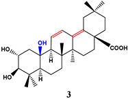
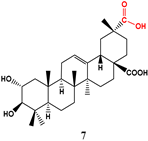
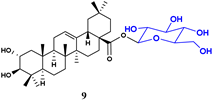
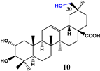
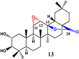
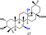
Over the years, many researchers have taken an interest in the enzymatic modification of MA (1). Therefore, biotransformations using a wide range of microorganisms have been investigated over the years, and these are shown in Table 2. Wang et al. [46] performed a study on the biotransformation of MA using five frequently employed microorganisms in food processing, namely Streptomyces griseus, Streptomyces olivaceus, Bacillus subtilis, Penicillium griseofulvum and Bacillus megaterium, with various yields. The anti-inflammatory activity of compounds 29 were assessed against NO production in LPS-induced and HMGB1-induced RAW 264.7 cells (Table 2). In the bioassay, it was observed that all metabolites exhibited non-cytotoxicity, whereas MA demonstrated a mild inhibitory effect on both models (HMGB1: >100 and cell viability: 104.39). In the context of HMGB1-activated inflammation, the majority of metabolites exhibited selective inhibitory effects. Notably, compounds 4, 6, and 8 demonstrated IC50 values of 16.41, 10.77, and 11.52 μM, respectively. The analysis of the function-activity relationship indicated the influence of the substitution site and the number of hydroxyl groups, particularly the hydroxylation of angular methyl groups on the PT skeleton, on the anti-inflammatory potential of these compounds. Results show that compounds hydroxylated at C-15, C-7, and C-1 exhibited a moderate level of inhibition on the HMGB1 model, whereas significant inhibitory effects have been proven with hydroxylation at C-29, C-21 and C-23. Furthermore, when oxidizing the hydroxyl group at C-29 to the carboxyl group, the inhibitory effect diminished, while glycosylation at C-28 did not affect the inhibitory effect. Additionally, compounds with double bond rearrangement did not exhibit a great enhancement in the anti-inflammatory effect. On the other hand, no inhibitory effect was observed in the case of LPS-induced NO release, except for analog 3, which showed moderate inhibition (IC50 = 24.40 μM). In summary, the study of the function-activity relationship of MA (1) transformation products provides valuable insights for the future development of highly effective and selective HMGB1 inhibitors. Another study [47] has shown that the action of Rhizomucor miehei on MA produces a C-30 hydroxylated metabolite 10, an 11α,12α-epoxy, 28,13β-olide metabolite 13, an 11-oxo derivative 12 and an olean-11-en-28,13β-olide derivative 11. An additional study [48] demonstrated that Cunninghamella blakesleeana CGMCC 3.910 catalyzed the MA biotransformation that led to these different metabolites: 2α,3β,7β,13β-tetrahydroxyolean-11-en-28-oic acid 17, 2α,3β,15α-trihydroxyolean-12-en-28-oic acid 15, 2α,3β,7β-trihydroxyolean-12-en-28-oic acid 14, and 2α,3β,7β,15α-tetrahydroxyolean-12-en-28-oic acid 16. Another experiment [49] showed the recombinant expression of three UDP-glucosyltransferases (UGTs), namely YojK, YjiC, and UGT109A3, from B. subtilis. Their capacity to glycosylate MA in vitro was explored with the aim of producing MA-2-O-β-D-glucose.

3. Chemical Modifications of MA (1) and Bioactivities

3.1. Anticancer Derivatives

One of the most notable classes of plant secondary compounds is the pentacyclic triterpenoids, which are recognized for their potent anti-tumor properties and apparent absence of toxicity. Given these favorable characteristics, they are regarded as promising primary compounds for the development of novel multi-targeting anti-cancer agents [50].
MA’s anti-cancer properties have been repeatedly highlighted in numerous recent studies that underscore its potential as a prospect for the creation of anti-cancer medications as well as anti-cancer agents. Seemingly, for the development of more potent anti-cancer treatments, the discovery of novel anti-tumor agents for boosting or reinstating apoptosis in malignant cancer cells is crucial [51]. Considering the substantial cytotoxicity exhibited by MA, structural modifications may facilitate the development of MA derivatives that possess improved IC50 values across a range of cancer cell lines. Furthermore, numerous drug-related properties, such as solubility and bioavailability, can be significantly improved. Several MA analogs exhibiting potent anti-cancer activity are illustrated in Figure 2 and Figure 3, with their synthetic routes detailed in Table 3.
Chouaïb et al. [52] investigated the effects of MA and corresponding triazole analogs on cancer cell lines, specifically human SW480 (Colon) and mouse EMT-6 (Breast). Their research revealed that MA displayed notable anti-proliferative effects on both SW480 cancer cells and EMT-6, leading to cell survival rates of 9% and 5% at 100 μM, respectively. The study also highlighted that the 1.4-regioisomers, particularly compound 20, exhibited superior anti-proliferative activity in comparison with the 1.5-regioisomers. Remarkably, it is worth highlighting that compound 20 demonstrated the highest activity against SW480 and EMT-6, with cell viabilities of 34 and 13% at 100 μM. In the case of the 1.4-regioisomers, derivative 19 showed the most potent activity against both cell lines EMT-6 and SW480, with cell viabilities of 10% and 6% at 30 μM. This observed activity was linked to the attachment of the triazole to the aryl group concerning the triterpene moiety in the cellular environment. In a separate experimental investigation, it was discovered that MA and its analog, compound 21, displayed activity in damaging the cell membrane of tumor cells, particularly A2780 cells. In fact, the IC50 values for MA and its analogs in A2780 cells were 19.5 and 10.6 μM, respectively. It was found that derivative 21 formed crystals with cholesterol in the cell membrane, or in close proximity to it, during cell culture. Subsequently, compound 21 penetrated the cell membrane, interacting with the lipid raft containing cholesterol, leading to alterations in the cell membrane structure, reduction in cell volume, and initiation of apoptosis [53].
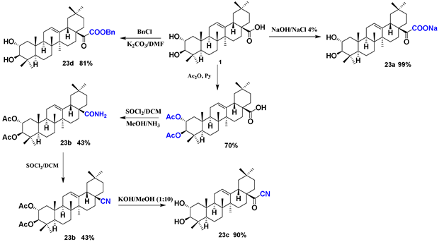
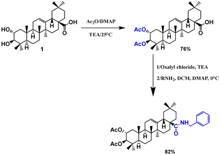
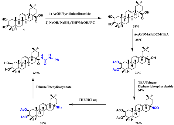
The study by Medina-O’Donnell et al. [54] highlighted the potential of PEG polymer for conjugation of pro-drugs, owing to its great solubility in aqueous environments. In fact, experiments revealed the substantial anti-cancer potential of diamine and PEGylated-diamine derivatives 22 of MA. Within non-tumor HPF cell lines, all MA diamine conjugates demonstrated cell viability levels ranging from 81% to 94%. Interestingly, MA diamine derivatives possessing the longest and shortest diamine chains exhibited the most pronounced cytotoxic effects in tumor cell lines, with IC50 values spanning from 0.76 μM to 1.76 μM. On the other hand, these conjugates demonstrated effectiveness that was 20- and 140-fold greater than the original precursors in B16-F10 cell lines [55]. Parra et al. [51] illustrated that MA and its derivatives possess the capability to hinder the development of B16F10 melanoma cells by triggering apoptosis. MA conversion into the sodium salt derivative 23a underwent a series of steps. Additionally, MA diacetyl analog underwent a transformation into the related amide analog initially using thionyl chloride in DCM, followed by NH3/MeOH, to be transitioned into the nitrile derivative 23b through thionyl chloride/DCM treatment, after which a further deacetylation yielded 23c. Moreover, compound 23d, formed by treating MA through DMF and benzyl chloride, displayed notable anti-tumor properties. The assessment of these following specific compounds for their pro-apoptotic effects at a concentration of 1 μM gives the following values: 23b at 68.62%, 23d at 87.50%, 23c at 78.75% and 23a at 56.67%. Another study emphasized the significant anti-melanoma effects of MA and the corresponding acetylated derivative (EM2, 24) [56]. On the other hand, MA displayed an IC50 equal to 13.7 μM, while compound 24 exhibited stronger toxicity with a lower IC50 value of 1.5 μΜ in 518A2 cells and an IC50 value of 33.8 μM in the nonmalignant mouse fibroblast cell line (or else NiH 3T3). Specifically, in A2780 ovarian cancer cells, the EC50 value of analog 24 was notably reduced to 0.5 μM. Moreover, it is noteworthy that the anti-proliferative activity in A2780 ovarian cancer cells was enhanced (EC50 equal to 0.5 μM for compound 24). This evaluation was suggested to be in relation with the derivatization occurred at MA’s C-28 position [57]. In summary, structural modifications conducted on compound 24 highlighted the importance of the acetyl groups and the presence of (2b, 3b)-configurated centers with the aim of achieving enhanced cytotoxicity along with ideal discrimination between cancerous cells and non-cancerous mouse fibroblasts. Consequently, the substitution of the MA benzylamide function with a phenylurea fragment led to the formation of derivative 25, contributing to the improvement of EC50 values of 0.9 μM (for A2780 ovarian cancer cells) and EC50 > 120 μM for fibroblasts (NIH 3T3), ultimately inducing apoptosis [58]. In another study, Serbian et al. [59] carried out a reaction between MA and benzoyl chlorides that afforded the 3-O-acylated and 2-O-acylated MA analogs. These compounds were biologically screened using the SRB or (sulforhodamine B) assays, and it was shown that cancer cell cytotoxicity increased in comparison with the parent compound MA. Additionally, when treating A2780 cells with MA during 96 h, the EC50 value was equal to 19.5 μΜ. Notably, the EC50 values of compounds 26af consistently remained below 10 μΜ. The acetylation of MA led to acetates 27a. Subsequent reactions involved treating 27a through oxalyl chloride and a reaction with piperazine in a later step, resulting in the formation of amides 27b. Thereafter, violet-colored compounds 27c were formed through a reaction of 27b with rhodamine B. Compound 27c exhibited approximately a 1000-fold higher cytotoxicity compared to the parent compound, and its selectivity for FSi (introduced as EC50 NIH 3T3 compared to EC50 A2780 tumor cell line) was enhanced by 50 times. Remarkably, rhodamine B showed no signs of cytotoxicity at concentrations up to 30 μM. Hence, compound 27c stands out as one of the most potent triterpenoic acid derivatives discovered thus far, exhibiting cytotoxicity in nano-molar concentrations. Its cytotoxic effects are comparable to those of well-known cytotoxic drugs such as paclitaxel or doxorubicin [60]. An interesting study by Vega-Granados et al. [61] highlighted the potential of MA–coumarin conjugates as anti-cancer candidates against three cancer cell lines (B16-F10, HT29, and Hep G2) respectively compared with three non-tumor cell lines (HPF, IEC-18, and WRL68). Their research revealed that the most pronounced cytotoxic activity was exhibited by 27e where the two molecules of coumarin-3-carboxylic acid are coupled through the C-2 and C-3 hydroxy groups of MA. It is noteworthy that compound 27e, with IC50 = 0.6, 1.1, and 0.9 μM, respectively, in two of the three cancer cell lines, was between 110- and 30-fold greater potency than the parent compound MA (1), showing cell viability percentages of approximately 90% observed in all non-tumor cells. Notably, results indicated that compound 27d showed cell viability percentages spanning from 61 to 88% for the non-tumor cells (HPF, IEC-18, and WRL68), at the corresponding IC50 concentration of the respective cancer cell line.

3.2. Anti-Inflammatory Derivatives

Chouaïb et al. [62] reported that MA derivatives conjugated with an isoxazole have been synthesized and assessed for their anti-inflammatory properties. In fact, the synthesis was carried out using various dipolarophiles ae under microwave irradiation in anhydrous DMF (Table 4). The synthesis of isoxazole derivatives 2833 was performed with a regiospecific method in the presence of Et3N and CuI. This approach utilized diverse aromatic hydroximyl chlorides and propargyl-(2,3)-2,3-dihydroxyolean-12-en-28-oate a under microwave irradiation for 5–10 min, achieving yields spanning from 84 to 96% (Scheme 1). The in vitro anti-inflammatory test demonstrated a strong inhibitory effect for all derivatives compared to the parent compound 1 (Table 5). It is noteworthy that among all synthesized analogs, derivative 33, featuring a furfuryl moiety on the isoxazole fragment, exhibited the highest anti-inflammatory activity (% IL-1β production = 4; 100 µM). Notably, compounds 29, 30, 31 and 32 also displayed enhanced activity (% IL-1β production = 33–55; 30–100 µM) in comparison with MA (1) (% IL-1β production = 109, 30 µM). Consequently, this suggests that halogens contribute more significantly to the activity than other substituents, with the observed activity appearing to depend on the nature of the substituents rather than their electronic effects [62].
In a follow-up to their prior research, Chouaïb et al. [52] synthesized MA-triazole conjugates and assessed their anti-inflammatory properties. Bis-alkynes c or d, copper iodide, and triethylamine were added at rt. The appropriate azide was then introduced to the mixture, which was subsequently subjected to microwave irradiation for a few minutes, resulting in the formation of compounds 3437 (Scheme 2) as well as compounds 38-42 (Scheme 3). Moreover, tri-alkyne e and the appropriate azide were dissolved in H2O. To this mixture, copper iodide and trimethylamine were added. The reaction mixture was irradiated with microwaves for 5–10 min to afford compounds 4346 (Scheme 4). Furthermore, the 1.5-regioisomers 4752 were produced through mixing a solution of alkyne a in DMF with Cp*RuCl(PPh3)2 at rt. Then, the appropriate aryl azide was added, and the reaction mixture was subjected to microwave irradiation for a few minutes (Scheme 5). As a result, 1,4-regioisomers 5358 were formed under solvent-free conditions through mixing dipolarophile a, copper(I) iodide and Et3N at rt. Subsequently, the appropriate aryl azide was added to this mixture, and the reaction mixture was subjected to microwave irradiation for a few minutes (Scheme 6). The study revealed that stronger inhibitory properties have been proven for all the formed triazoles compared to their precursor 1. Notably, incorporating triazole moieties into compound 1 significantly enhanced its anti-inflammatory efficacy. Among the bis-1,4-disubstituted triazoles 3437 (Table 6), only compounds 34 and 35 demonstrated relatively significant effects, with % IL-1β production = 71 and 56, respectively, in 100 µM compared with the other derivatives and MA (1). Furthermore, the % IL-1β production observed for the bis-1,4-disubstituted triazoles 3842 (Table 7) indicated that a higher activity is noticed in cases where the aromatic ring possesses electron-donating groups, which is not the case with electron-withdrawing groups or without substitutions that showed moderate activity. Specifically, analogs 41 and 39 showed the best results (% IL-1β production = 52 and 34, respectively; 100 µM). The placement of the triazole at either C-3 (derivatives 3842) or C-2 (derivatives 3437) within MA (1) plays a critical role in determining its effectiveness. In fact, this is evident in instances like 36 and 41 with % IL-1β production equal to 100 and 52, respectively, at 100 µM, in cases where the same aromatic system (m-MePh) is attached to the triazole. In contrast, compounds 46 (featuring a naphthyl fragment on the triazole moiety) and 45 (m-Me), outlined in Table 8, exhibited significant effects (% IL-1β production = 34 and 23, respectively; 30 µM) on reducing PBMCs in comparison with MA and other conjugates. This discovery underscores the significance of both the quantity and potentially the positioning of triazole moieties in enhancing the anti-inflammatory properties of MA (1) [52]. Results outlined in Table 9 showed also that in the series of 1,5-regioisomers, derivatives 50 and 51 demonstrated pronounced activity (%IL-1β production 42 and 46, respectively; 100 µM), while compound 49 with (p-Br) showed the highest inhibition with % IL-1β production value equal to 21 in 100 µM. Additionally, the results depicted in Table 10 indicated that most of the tested 1,4-regioisomers 5358 displayed significant anti-inflammatory effects. Among them, 56 (p-Br) was the most potent derivative showing a % IL-1β production value equal to 21 at 100 µM. Upon comparing the % IL-1β production values of derivatives 4752 with those of derivatives 5358, which share identical substitutions at the triazole ring, it becomes evident that the latter exhibit greater activity (% IL-1β production = 40–91 and 21–82, respectively; 100 µM). Nevertheless, a noteworthy observation emerged during the examination of the regiochemistry in two conjugates from each series, 4752 and 5358, which share the same substituent. This is exemplified in compounds 56 (p-Br substituted) (% IL-1β production = 21; 100 µM) and 49 (p-Br substituted) (% IL-1β production = 61; 100 µM) where activity is reversed due to the regiochemistry. This study highlights the noteworthiness of the triazole’s regiochemistry and the type of aryl group linked to it in influencing the observed activity [52].

3.3. Glycogen Phosphorylase Inhibitors Derivatives

Wen et al. [63] conducted a study wherein they reported a newly synthesized MA series of analogs that were assessed for their enzymatic inhibition properties against rabbit muscle glycogen phosphorylase a (GPa), which is readily available commercially and bears substantial similarity in sequence to the GPa of human liver. Throughout the synthesis, the methyl ester 59 was accompanied by treatment of MA (1) with iodomethane in the presence of K2CO3 in DMF at rt. The same was reported for MA esters 60 and 61, which were produced in good yields by treating 1 with ethyl bromoacetate and N-chloromethylpyrazole. Ester 61 was subsequently transformed into 62 by hydrolysis of 61 with an aqueous solution of 4 N NaOH in THF without modifying the C-28 bond (Scheme 7). The reaction of MA (1) with 1,4-dibromobutane or 1,2-dibromoethane in DMF with K2CO3 at rt produced derivatives 63 and 64 (Scheme 8). Subsequently, heating 63 in refluxing EtOH with Et3N resulted in the formation of compound 65. The reaction of 63 or 64 with corresponding amines afforded conjugates 6671. In fact, stirring a mixture of 63, triethylamine, in DMF with presence of K2CO3 at rt led to 66 (Scheme 8). The reduction of compound 72 with NaBH4 produced compounds 74 and 76. Similarly, the reduction of 73 afforded 75 and 77 (Scheme 9).
To evaluate these analogs, rabbit muscle GPa enzymatic activity was assessed through tracking the phosphate production from glucose-1-phosphate during the synthesis of glycogen (Table 11, Table 12 and Table 13). As a result, it was found that most of the novel analogs formed demonstrated inhibitory effects against rabbit muscle GPa (IC50 values spanning from 7 to 1707 µM), in contrast to the original compound MA (IC50 = 28 µM). Results showed that compound 64 stands out as the most effective GPa inhibitor with IC50 value equal to 7 µM (Table 12). It is noteworthy that the comparison between compounds 63 and 64 highlights the influence of the size of the C-28 side-chain. Compound 64 (IC50 = 7 µM) exhibits significantly higher potency than 63 with IC50 value equal to 50 µM. It is worth highlighting that the enzyme-inhibitory effect is influenced by the size of the C-28 hydrophobic side chains, as the length of the carbon side chain linked to the C-28 carboxyl group was the primary structural distinction among these compounds. In summary, enzyme inhibition is greatly influenced by the tendency for hydrophobic groups at the C-28 position, presenting a hurdle for refining compound 1 and its related triterpenoids for better drug efficacy, considering that drug design usually favors compounds with good water solubility.

3.4. Antibacterial and Antifungal Derivatives

Blanco-Cabra et al. [40] reported that a series of MA C-28 amide conjugates have been synthesized and tested for their in vitro antibacterial efficacy and toxicity properties. First, after activation of the MA carboxyl group with TBTU, the MA-TBTU derivatives were obtained by adding TBTU in the presence of DIEA in anhydrous THF at room temperature. Second, the MA-TBTU derivatives were dissolved in CH2Cl2 and reacted with the corresponding diamine reagents (DAD, HDA, and PDA) in the presence of potassium carbonate, forming both monomers (MA-PDA, MA-HDA, MA-DAD) and dimers (MA-PDA-MA, MA-HDA-MA, MA-DAD-MA). Conversely, the reaction of MA-TBTU derivatives with N,N-dimethyl-1,3-propanediamine exclusively produced MA-DMPA (Scheme 10). The study showed that MA-HDA derivatives possessed enhanced in vitro antimicrobial effectiveness and lower toxicity than the original compound MA (1) on the other side (Table 14). This decrease is evident from a reduction in the minimum inhibitory concentration (MIC) against most Gram-positive bacteria assessed, highlighting their effectiveness, especially against Methicillin-Resistant S. aureus (MRSA) and S. aureus. Additionally, in the Galleria mellonella animal infection model, MA-HDA exhibited heightened in vivo activity [40]. In the context of planktonic bacterial growth, the MA amide derivatives were assessed for their antibacterial effect. The results presented in Table 14 depict the antibacterial efficacy in terms of MIC50 (the minimal inhibitory concentration 50%). As a result, it was proven that among all examined derivatives, MA-HDA was the most potent conjugate showing enhanced activity, consistently keeping or enhancing MA’s antimicrobial properties through most tested strains, with specific significance noted against S. aureus and MRSA (66% reduction in MIC50 with MIC50 values of 25 μM), which represents a substantial improvement compared to the original compound (MA, MIC50 of 0.15 μM). An analogous pattern was noted in case of MA-DAD (reducing MRSA with MIC50 by 66%). It was demonstrated that the in vitro toxicity, quantified in Table 14 as the concentration required to achieve a 50% reduction in cell viability (CC50), shows that the MA-DAD and HDA derivatives exhibited no greater toxicity than their precursor. This is noteworthy, considering its improved antimicrobial activity against certain bacterial strains. Next, these derivatives were evaluated for their efficacy and in vivo toxicity in Galleria mellonella. In fact, G. mellonella represents the toxicity evaluation model (lethal dose) of the newly tested antimicrobial derivatives [64]. The evaluation involved determining the compound dose per kilogram that resulted in a 50% mortality rate in greater wax moth larvae, referred to as LD50 (the lethal dose 50). It is worth noting that selectivity indexes (SI), whether in cells or G. mellonella, underscored the heightened efficacy of the MA-HDA derivative against S. aureus growth in relation with their toxicity (SI = 17.9 or 661). It was notable that the SI calculation and the choice of G. mellonella as a model of an invertebrate animal for in vivo toxicity assessment is pivotal. The selectivity index experiences a substantial boost when calculated using LD50 rather than CC50 (refer to Table 14). In certain instances, in vivo toxicity can markedly differ; for instance, the in vitro toxicity of MA-HDA was enhanced, while its in vivo toxicity diminished compared to the parent compound MA.
In another study, Chouaïb et al. [65] synthesized a series of MA C-2 and C-3 esters that were assessed for their antifungal and antibacterial potential. The condensation of MA (1) with multiple acid chlorides in presence of DMAP led to bis-acylated esters 8589 and mono-acylated esters 9094. The treatment of MA (1) with cyclic anhydrides produced exclusively the bis-esters 9599 (Scheme 11). Results showed that derivatives with sulfur moiety (85 and 90; MIC = 5 μg/mL) and chlorine atoms demonstrated a pronounced antibacterial potential (Table 14). The sulfur triterpenoid 85 exhibited the highest antifungal activity, showing good effectiveness against Trichoderma harzianum, Aspergillus niger, Penicillium italicum, Penicillium digitatum and Aspergillus flavus. However, compound 90 was the most active against Aspergillus niger (MIC = 19 μg/mL). It is noteworthy that the fungus Penicillium italicum had the highest sensitivity to the derivative with sulfur atom 85 (inhibition zone = 24 mm) as outlined in Table 15.

3.5. Derivatives with Herbicide Activity

In their study, Nejma et al. [66] conducted the semi-synthesis of novel hybrid compounds by employing a 1,3-dipolar cycloaddition method that included phthalimide-based azides and propargylated MA 100 (Scheme 12). By employing CuAAC cycloaddition conditions using microwave irradiation, the reaction selectively produced various 1,4-disubstituted triazole derivatives 101107 in different yields, within 5 min as a maximum reaction time. The efficacy of these 1,4-disubstituted triazole compounds in inhibiting seed germination and consequently impeding plant growth was assessed, demonstrating encouraging properties that could act as templates for developing effective and valuable herbicides. Following the synthesis of the MA-based analogs, their impact on regulating seed germination and the initial development phases of Lactuca sativa L. as the target plant was investigated. Results outlined in Table 16 demonstrated notable herbicidal potential, with inhibition reaching up to 100%. Within the series based on MA, MA (1) displayed a minimal inhibition percentage equal to 5.56%. Conversely, MA propargylation demonstrated a contrasting impact. Therefore, this compound can serve as a reference in the development of triazolophthalimide hybrid compounds based on MA. The herbicidal impacts of the synthesized triazoles 101107 (Table 16) exhibited increased efficacy in comparison to propargylated MA 100. These analogs notably hindered total germination, ranging from 91.79 to 100%. Based on the existing literature and the effects of propargylated MA 100, the herbicidal potential of compounds 101107 was primarily attributed to the additional triazolomethyl phthalimide component [67]. Notably, it is crucial to highlight that the presence of compound 105 resulted in 100% complete inhibition of seed germination. In the phthalimide series, as hypothesized previously, the increased inhibitory effect on germination seen in this study is likely caused by the presence of the thiol moiety within the structure of the hybrid compound. In conclusion, MA shows minimal inhibitory effects on Lactuca sativa L. seeds germination in comparison to propargylated MA 100 which appears to enhance their growth. Significantly, the transition in the activity of propargylated MA 100 from promoting to inhibiting germination in the synthesized compounds is clearly attributed to the presence of the methyltriazolo and phthalimide moieties. The significant impacts of these new hybrid molecules mainly stem from the connection between MA and phthalimide components through the triazole viaduct.

3.6. Anti-Diabetic Derivatives

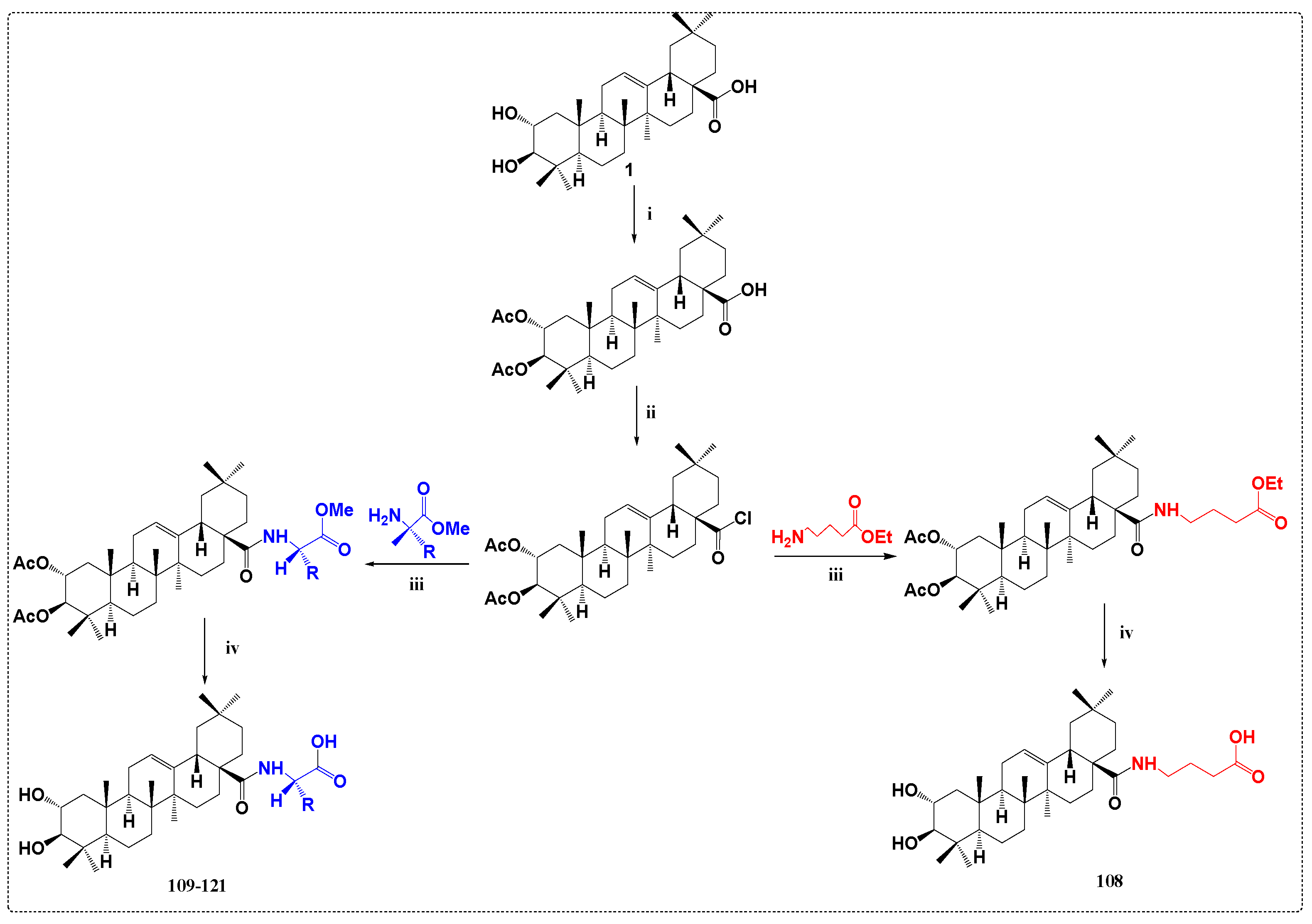
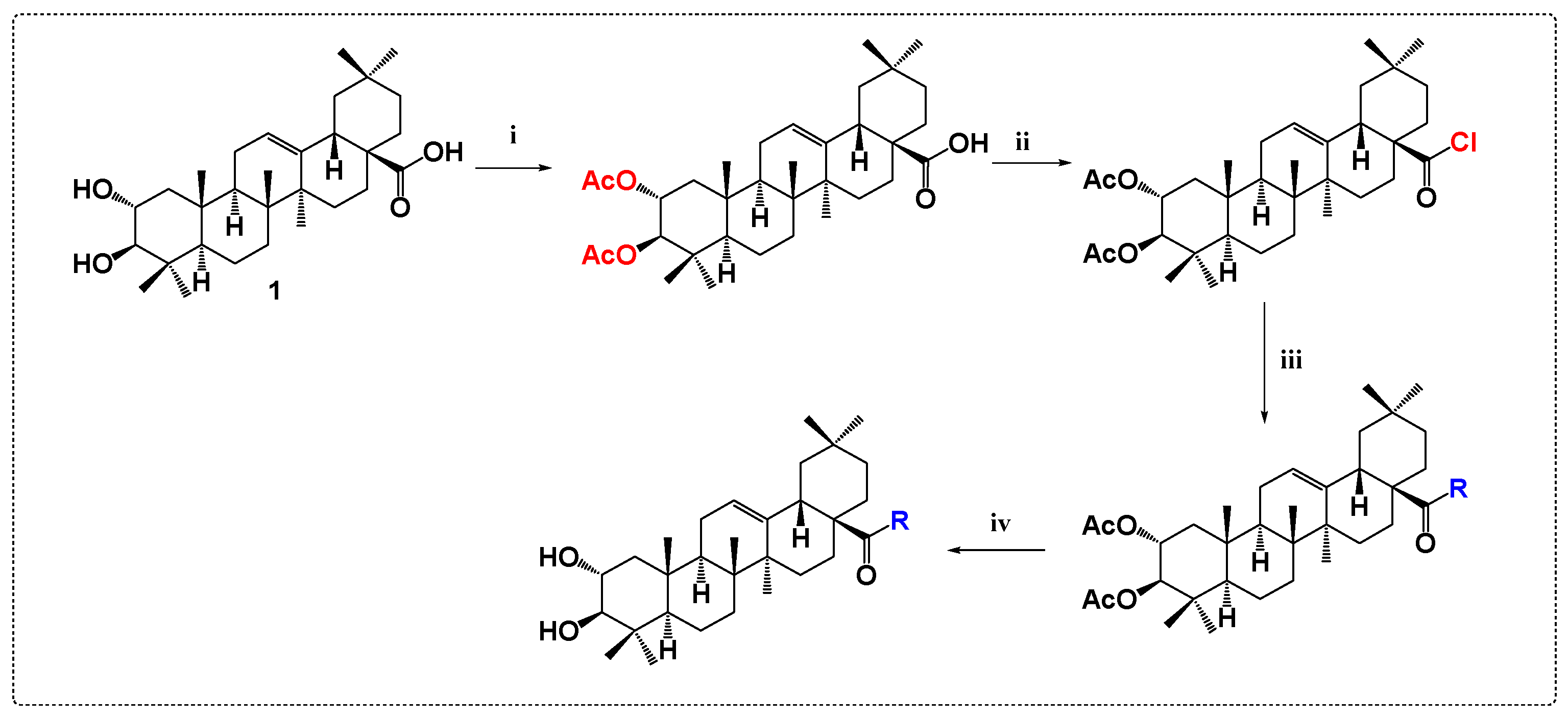
Zeng et al. [68] investigated the synthesis of a series of derivatives coupling MA with amino acids, followed by the in vitro assessment of their inhibitory activity against α-glucosidase. The precursor MA (1) was first converted into the 2α, 3β-O-diacetate by treatment with Ac2O. This intermediate was then reacted with oxalyl chloride and subsequently condensed with the appropriate amino acid methyl/ethyl ester hydrochlorides (like L-valine, γ-aminobutyric acid, glycine). The final products, 108 and 109121, were obtained through a simple hydrolysis (Scheme 13). Results showed that all newly synthesized derivatives exhibited varying α-glucosidase inhibitory effect in comparison to acarbose in cases of using the DMSO or EtOH-H2O system as solvents (Table 17). Specifically, both the derivatives and acarbose demonstrated a reduced inhibitory effect compared to that of the precursor MA (1). Notably, among the presented derivatives, those with a C-28 amide side chain featuring two free carboxyl groups (113: IC50 = 382 μM) displayed higher inhibitory activity than acarbose (IC50 = 484 μM) in both EtOH and DMSO systems, except for derivative 112. Within the ethanol-water system, the majority of derivatives displayed slightly reduced inhibitory effect compared to acarbose. Notably, compounds with only one free carboxyl group on the C-28 amide side chain displayed lower inhibitory potential compared to those featuring two free carboxyl groups on the C-28 amide side chain (IC50 values equal to 608 and 798 μM for 116 and 117, respectively). Interestingly, the lack of a free hydroxyl group on the C-28 amide side chain did not impact the inhibitory effects.
In another study, Huang et al. [69] reported the synthesis of a series of MA derivatives by coupling the piperazine-L-amino acid complex at the C-28 site of the parent compound 1. Firstly, MA (1) was acetylated by reacting with acetic anhydride. This intermediate was then reacted with oxalyl chloride and condensed with piperazines successively in the presence of trimethylamine. After reactions of dehydration and condensation using N-benzyloxycarbonyl-L-amino acid, the target compounds 122131 were obtained by both reduction and hydrolysis reactions (Scheme 14). The final derivatives were tested in vitro for their α-glucosidase inhibitory activity (Table 18). The results indicate that the α-glucosidase inhibitory activity of compound 126 is close to that of the reference acarbose in the EtOH-H2O system. In comparison to the ethanol-water system, the DMSO system yielded lower IC50 values for the derivatives. Among all the synthesized derivatives, compound 126, which includes a piperazine-L-aspartic conjugate, showed a similar α-glucosidase inhibitory effect (IC50 = 591 μM) to acarbose (IC50 = 347 μM). Upon inspecting the molecular structure, the presence of a free carboxyl group on the amino acid’s branched chain in compound 126 was found to be crucial in improving its α-glucosidase inhibitory activity. Furthermore, it was noted that conjugates resulting from the combination of triterpenoid acid (MA) with serine (128 IC50 = 2754 μM) and threonine (129 IC50 = 2579 μM) displayed comparable α-glucosidase inhibitory properties to other derivatives. Notably, these two compounds, which feature an additional hydroxyl group in the side chain, did not lead to an enhancement in α-glucosidase inhibitory activity.
Liu’s research [70] revealed another noteworthy discovery, demonstrating that within the class of pentacyclic tricarboxylic acid-piperazine analogs, a range of C28-modified analogs of MA with saturated nitrogen heterocycle segments (such as 1-deoxynojirimycin or piperazines) was prepared. MA was initially reacted with α,ω-bromoalkanes. The resulting intermediate was then subjected to a nucleophilic substitution reaction with 1-DNJ to produce MA-DNJ analogs 132149 (Scheme 15). Further, MA (1) was acetylated using acetic anhydride. The resulting intermediate was then reacted with oxalyl chloride and piperazines in the presence of Et3N. The final products 140142 were obtained through a subsequent hydrolysis reaction (Scheme 16). This process entailed merging two bioactive components via a covalent link, generating a unique hybrid biological entity following the concept of active splicing. Subsequently, the α-glucosidase inhibitory activity of these synthesized compounds was assessed in vitro. The results revealed that certain analogs like (142: IC50 = 768.5, 133: IC50 = 1468.4 and 141: IC50 = 499.6 μM) exhibited higher inhibitory activity against α-glucosidase in comparison with the precursor MA (IC50 = 2540.6 μM). Specifically, compound 141 (IC50 = 499.6 μM) demonstrated stronger inhibitory activity than acarbose (IC50 = 606 μM). To explore the inhibitory impact of the synthesized analogs on α-glucosidase, all final products were dissolved in DMSO for in vitro inhibition experiments. As outlined in Table 19, most compounds exhibit significant inhibitory effects against α-glucosidase. In the initial series of triterpenic acid-DNJ derivatives (132139), a lower IC50 value than the primary compound MA (1) (IC50 = 2540.6 μM) were observed for compounds 132 (IC50 = 2041.4 μM), 133 (IC50 = 1468.4 μM), and 134 (IC50 = 1718.4 μM) containing an alkyl chain linker (2–4 carbon atoms). However, with the introduction of a longer hydrophobic carbon chain (comprising 4–8 carbon atoms) into the compound structure, the inhibitory activity of the analogs decreased compared to the parent compound MA (1), as observed in compounds like 137 (IC50 = 3068.4 μM) and 135 (IC50 = 3660.4 μM). The combination of pentacyclic dihydroxytriterpenecarboxylic acid MA (1) with piperazines (specifically 140142, part of the second series of derivatives) resulted in compounds where the piperazine component with one free hydroxyl (141: IC50 = 499.6 μM) and amino groups (142: IC50 = 768.5 μM) exhibited notably higher inhibitory activity than MA alone (IC50 = 2540.6 μM) as depicted in Table 20. Particularly noteworthy is that compound 141 displayed superior hypoglycemic activity compared to acarbose (IC50 = 606 μM). These results emphasize that incorporating a free hydroxyl or an amino group into the piperazine substructure of the pentacyclic triterpene acid derivatives can increase the modified compounds’ α-glucosidase inhibition effect, which is the case of 141 compared to 140 and 142 compared to 140.
In another study, Xu et al. [71] investigated the synthesis of a series of compounds by glycosylation of MA with monosaccharides and disaccharides. MA was initially transformed into its benzyl ester. The resulting intermediate was then glycosylated using TMSOTf (cat) at the C(2α)-OH and C(3β)-OH positions. Finally, compounds 143148 were synthesized through transesterification with NaOMe in methanol (Scheme 17). Hence, the solubility and inhibitory activity of α-glucosidase assay of these newly synthesized compounds showed that MA bis-disaccharide glycoside demonstrated higher water solubility and α-glucosidase inhibitory potential than the bis-monosaccharide glycoside (Table 21). It is noteworthy that compound 148 exhibited the highest α-glucosidase inhibitory activity (IC50 = 684 μM). This result highlights the importance of the presence of the lactose group at C-3 and C-2, which helped improve and enhance the water solubility as well as the inhibitory activity.

3.7. Inhibitors of Protein Tyrosine Phosphatase 1B Derivatives

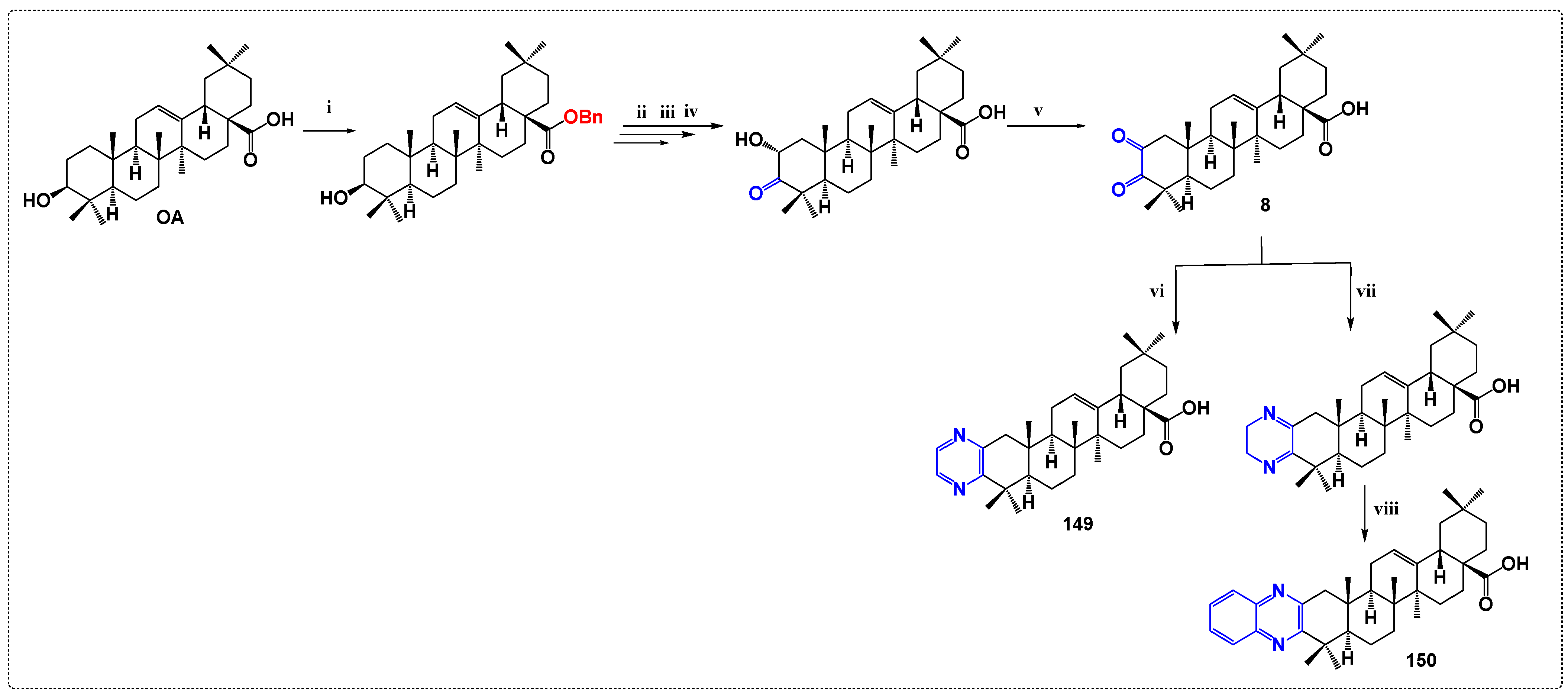
Qui et al. [72] investigated the synthesis of a series of derivatives coupling MA (1) with fused heterocyclic rings (indole, pyrazine, thiazole, pyrimidin, pyrazole and isoxazole) at the C-2 and C-3 positions. The final quinoxaline derivative 149 was afforded by the reaction of the diketone with phenylenediamine and the final product, 150, was then obtained through a dehydro-aromatization reaction with MnO2. In addition, compound 152 was synthesized through an MeI scavenger (Scheme 18). Compounds 153 and 154 were obtained through a similar process as compound 152, with the key difference being that the demethylation of the C-28 methyl ester was carried out in anhydrous DMF but in the presence of LiI, without the use of n-octylamine (Scheme 19). Compounds 160163 were prepared by reacting the pyrazole derivative 159 with acetyl chloride, hexanoyl chloride, nicotinoyl chloride, and isonicotinoyl chloride, respectively (Scheme 20). Further, these newly synthesized derivatives were assessed for their inhibitory activities on PTP1B, TCPTP, and PTPs (Table 22, Table 23 and Table 24). The inhibitory effect of some conjugates was tested on various other PTPs which negatively regulate insulin dephosphorylation, such as src homology phosphatase-1 (SHP-1), leukocyte antigen-related phosphatase (LAR) and src homology phosphatase-2 (SHP-2). Results showed that the inhibition potential of most of the newly synthesized derivatives was increased significantly. It is noteworthy that derivatives 155 and 162 (Table 24) were the most potent PTP1B inhibitors, showing great activity in comparison with the parent compound 1 (IC50 = 0.61 and 0.64 µΜ, respectively). We can highlight that derivative 162 was the most selective compound showing a 6.9-fold preference for PTP1B over TCPTP.

3.8. Derivatives as Inhibitors of Acylcholinesterases

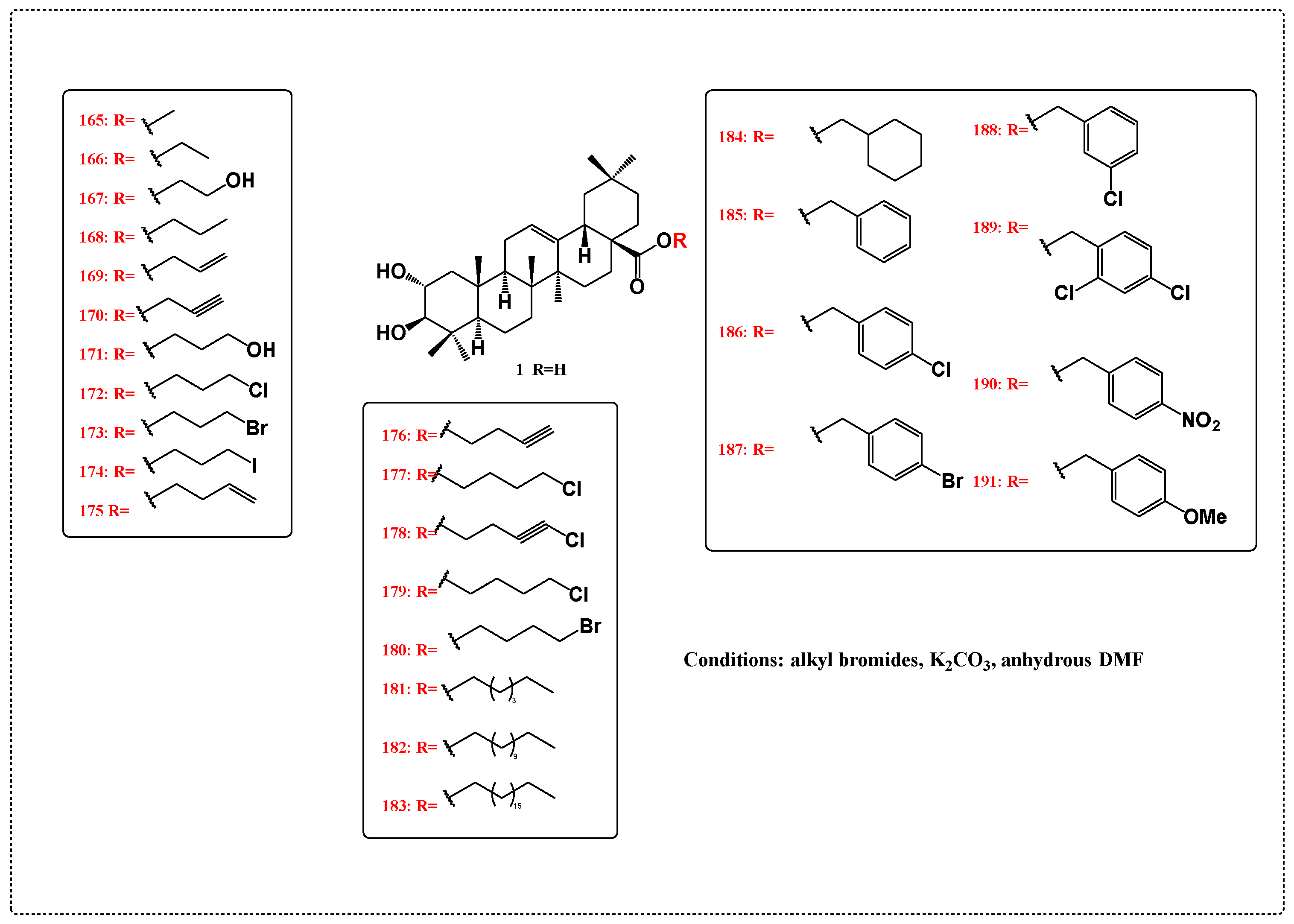
In this study, Schwartz et al. [73] investigated the synthesis of new MA conjugates that were assessed for their potential as cholinesterase inhibitors. The first group of compounds 165191 (Scheme 21), representing MA esters, were synthesized by reacting MA with alkyl bromides in the presence of potassium carbonate in anhydrous DMF. The second group of compounds 192207 (Scheme 22) includes various MA derivatives (192, 193), its methyl esters (194, 195), and amides (196207), featuring either free or acetylated hydroxyl groups at the C-2 and C-3 positions. Acetylations were performed in pyridine using Ac2O, while the sulfamates 194 and 195 were derived from methyl ester 165 and sodium hydride in THF, followed by the addition of sulfamoyl chloride. The synthesis of amides began with 2,3-diacetyl MA, which was first reacted with thionyl chloride, followed by the addition of an amine. The evaluation of these derivatives as cholinesterase inhibitors demonstrated that compound 177 was a marvelous inhibitor for AChE, being the most active analog with an inhibition constant equal to 1.6 μM in comparison with the parent compound MA that showed no inhibition (Table 25).

3.9. Antiviral Derivatives

Parra et al. [74] investigated the synthesis of MA conjugates that comprise amino acids and peptides. Derivatives (208219) were prepared by condensing MA (1) and the corresponding amino acid (α-amino acids: glycine (Gly), L-alanine (Ala) and L-valine (Val)); or λ-aminobutyric acid (GABA), 6-aminohexanoic acid (6AHA) and 11-aminoundecanoic acid (11AUA) with the carboxylic group protected as methyl ester. The condensations were carried out at rt in anhydrous dichloromethane in the presence of N,N’-dicyclohexylcarbodiimide (DCC), N-hydroxybenzotriazole (HOBt), and triethylamine (TEA). Hence, analogs 208213 with the corresponding amino acid methyl ester moiety were obtained. Subsequently, the saponification of compounds 208213 with NaOH/MeOH/THF at room temperature led to 214219 (Scheme 23). In order to find out the anti-HIV properties of these novel compounds, MT-2 cells were infected with the pseudotyped HIV-1 clone pNL4-3 using the VSV envelope, which avoids HIV-1’s normal entrance mechanism into these cells facilitating strong HIV-1 replication. After being integrated into the host firefly, luciferase gene expression is mediated by chromosomes; as a result, the rate of viral replication in infected cells is correlated with luciferase activity. Consequently, 24 h after cellular infection with the VSV-pseudotyped HIV-1 clone, high luciferase activity levels were found. Pretreating MT-2 cells with MA (1) and derivatives 30 min before infection led to a dose-dependent inhibition of the luciferase activity, with compounds 211 and 219 being the most potent inhibitors of HIV-1 (Table 26). The amount of luciferase activity in cell extracts was measured and the results depicted in Table 26 are shown as a percentage of activation when compared to 100% activation in untreated infected cells.
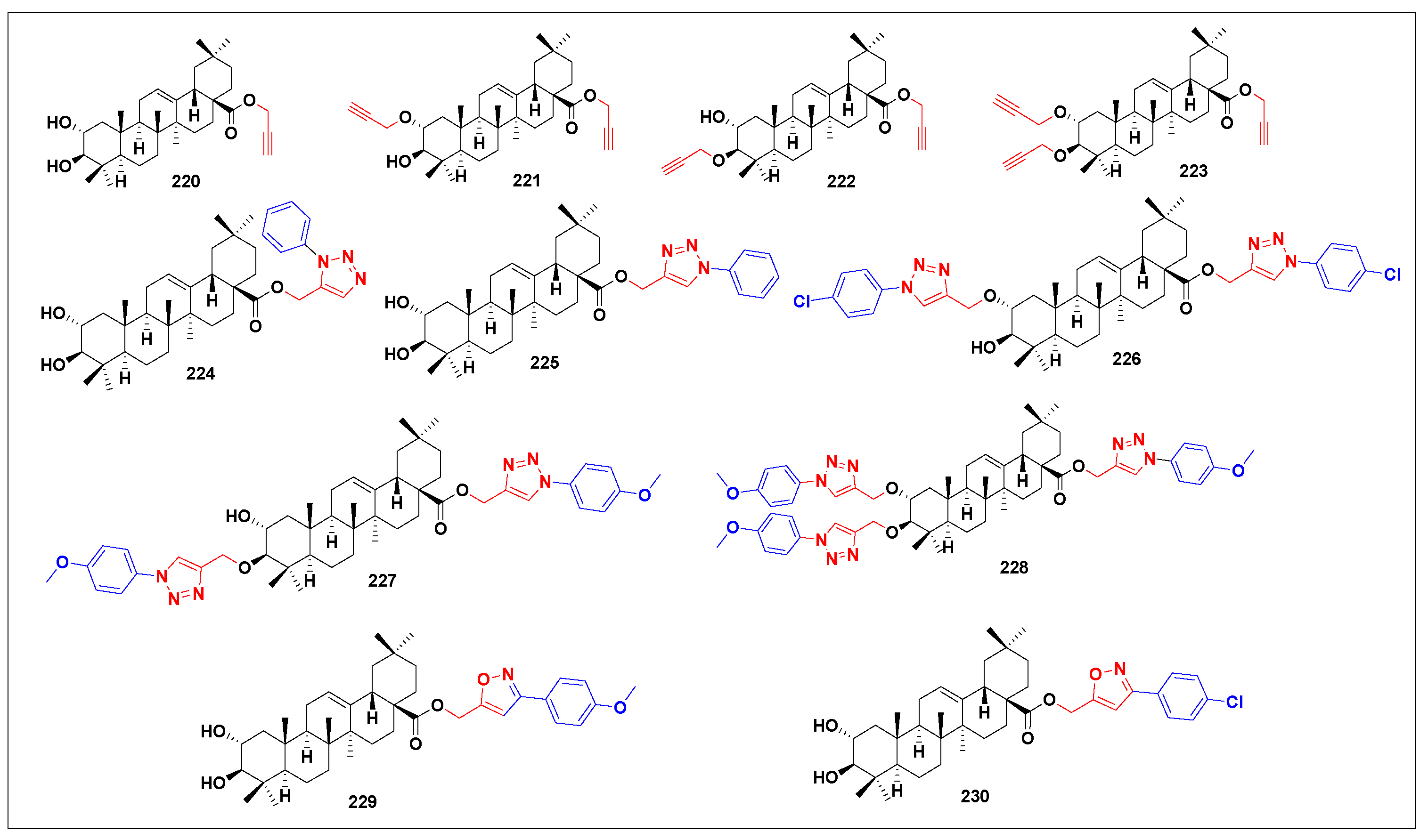
In another study, Soltane et al. [75] screened the inhibitory activity of previously synthesized MA derivatives [52,62] (Figure 4) on highly pathogenic coronaviruses (anti-SARS-CoV-2 activity). Subsequently, half maximal cytotoxic concentration, or “CC50,” was computed for each analog individually in order to determine the appropriate doses at which to characterize the antiviral activity. Out of the evaluated panel conjugates, compound 230 bearing the isoxazole side chain moiety with a p-chlorophenyl substitution showed a promising in vitro activity against SARS-CoV-2 with IC50 value equal to 4.12 μM (Table 27).
In another study, Serra et al. [76] have tested the antiviral activity of compound 231 (Figure 5). The results showed that this conjugate exhibited a modest inhibition, as outlined in Table 28. In fact, to inhibit the cytopathic effect, the number of viruses infecting the cells in multi-well plates was kept low—approximately one virus per three cells. After incubating the cells for two to three days with and without the substances under study (100 µg/mL), morphological alterations in the cells were observed using a light microscope.

3.10. Derivatives Synthesized with No Activity Assessed

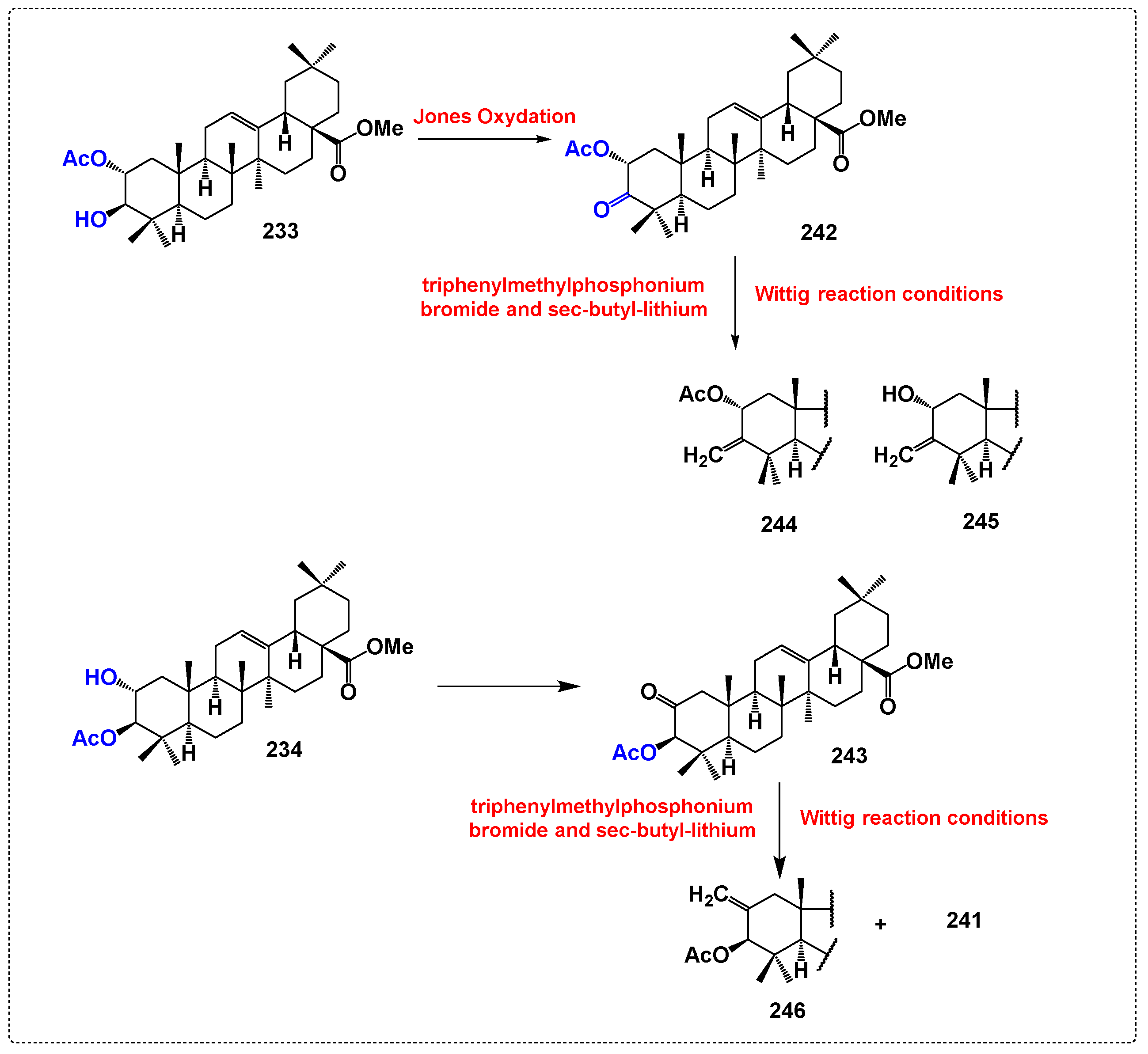
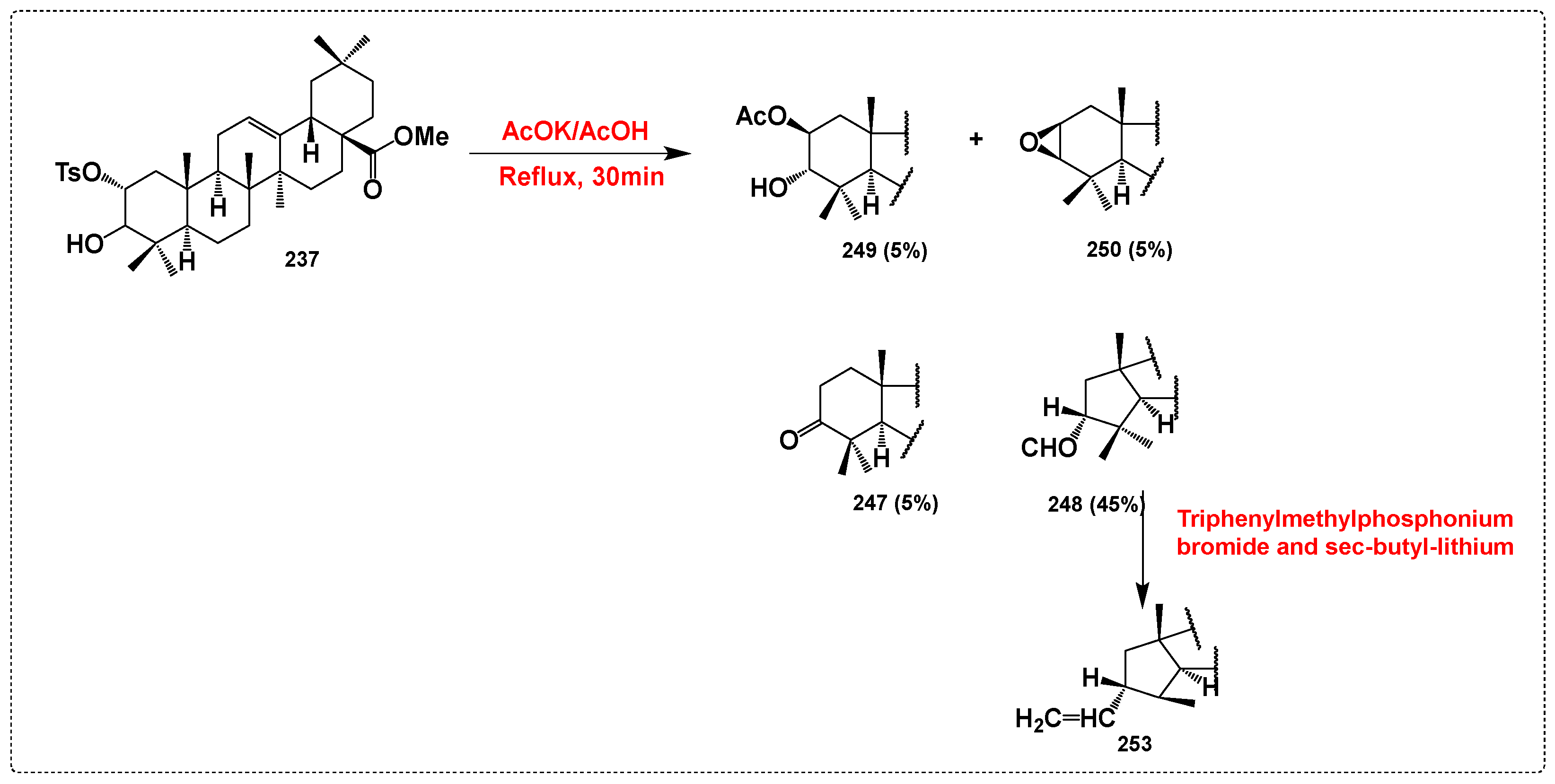
García-Granados et al. [77] have investigated the synthesis of a wide range of derivatives from the MA-A-ring (Figure 6). Various derivatives of methyl maslinate were synthesized using standard reaction procedures (Scheme 24, Scheme 25 and Scheme 26). Among these derivatives, those with good leaving groups in the A-ring of the oleanene skeleton were employed in several rearrangements described below. Jones reagent was utilized to oxidize the hydroxyl group at C-3 in compound 233, producing the 3-oxo derivative 242. Similarly, the hydroxyl group at C-2 in compound 234 was oxidized to yield the 2-oxo derivative 233 (Scheme 24). To synthesize oleanene derivatives with short carbon chains in the A-ring, oxo derivatives 242 and 243 were treated with triphenylmethylphosphonium bromide and sec-butyllithium under Wittig reaction conditions. The Wittig reaction with 3-oxo derivative 242 resulted in the formation of products 241, 244, and 245 (Scheme 24). Compound 239 was treated under reflux for 30 min with AcOH/AcOK, to afford 251 and 252 (Scheme 25). Compound 241, previously obtained from the oxidation of methyl maslinate, was a byproduct of this reaction. Additionally, product 237 was treated under reflux for 30 min with AcOH/AcOK, leading to compounds 232 (25%), 233 (15%), 247 (5%), 248 (45%), and 249 (5%) (Scheme 26).

4. Conclusions

Multiple recent evaluations have consistently shown that derivatives of MA exhibit a range of impressive biological activities. These derivatives have been confirmed to possess significant therapeutic potential, emerging as promising agents in the treatment of multiple diseases, particularly cancer, diabetes, and inflammatory conditions. This comprehensive review delves into and summarizes diverse synthetic pathways, chemical structures, biological properties, and function-activity relationships associated with analogs derived from MA. Structural modifications of MA extend beyond the traditionally targeted four “active” sites, namely C28 –COOH, C2–OH, C12 = C13 double bond and C3–OH. For instance, earlier studies compellingly demonstrated the potent inhibitory effect of MA derivatives against α-glucosidase. Systematic exploration of the structure-activity relationship (SAR) revealed that incorporating piperazines and piperazine-L-amino moieties at C-28, as well as amides at C-28, enhanced inhibitory potency against α-glucosidase. Furthermore, numerous MA derivatives have been synthesized and assessed for their anti-tumor activities. Among them, diamine and PEGylated diamine conjugates showed conspicuous potential. Notably, in tumor cell lines, the MA diamine conjugate with the shortest and longest diamine chains demonstrated the most potent cytotoxic effects, with IC50 values ranging from 0.76 to 1.76 mΜ. It is clearly notable also that regarding the GPa inhibition, compound 64 was the most potent GPa inhibitor with IC50 = 7 µM. Therefore, a distinct inclination towards hydrophobic groups at C-28 is noticeable in enzyme inhibition, posing a challenge for optimizing leads using MA and similar triterpenoids, as drug design typically favors good water solubility. Interestingly, the derivative MA-HDA exhibited enhanced antimicrobial activity, notably reducing MIC50 against MRSA by 66%. Additionally, several MA conjugates have demonstrated increased inhibitory activity on germination of Lactuca sativa L., which was possibly attributed to the thiol group within the hybrid compound structure. Moreover, it was crystal clear that many of the MA derivatives conjugated with triazoles or isoxazoles exhibited potent anti-inflammatory activity (the derivative featuring a furfuryl fragment on the isoxazole moiety demonstrated the most significant anti-inflammatory properties, with a % IL-1β production of 4 at a concentration of 100 µM). Furthermore, MA derivatives have demonstrated exceptional efficacy as protein tyrosine phosphatase 1B inhibitors. Notably, we can highlight that derivative 162 exhibited the highest selectivity (IC50 value of 0.64 μM) with a 6.9-fold preference for PTP1B over T-cell protein tyrosine phosphatase (TCPTP). Additionally, it is noteworthy that compound 177 emerged as an outstanding acetylcholinesterase (AChE) inhibitor, boasting an inhibition constant of 1.68 μM, surpassing the activity of the parent compound MA. Further, in terms of antiviral activity, compounds 211, with an R group of γ-aminobutyric acid (GABA), and 219, with an R group of 11-aminoundecanoic acid (11AUA), were identified as the most potent inhibitors of HIV-1 as they exhibited doses equal to 32.5 and 56.7, respectively, at 10 μM. It is worth highlighting also that compound 230, which incorporates an isoxazole side chain moiety with a p-chlorophenyl substitution, exhibited significant in vitro activity against SARS-CoV-2, with an IC50 value of 4.12 μM. The synthesis and design of structurally and functionally diverse MA analogs have paved the way for compounds that exhibit strong affinity and selectivity for specific molecular therapeutic targets. The literature strongly suggests that MA derivatives hold potential as complementary and alternative therapies for various diseases, warranting further exploration. In summary, the emphasis of this review sheds light on the different synthesis routes of multiple MA derivatives and brings to light their biological activities as well as diving into their structure-activity relationships.
The assessment carried out aims to provide guidance to researchers to target synthesis routes or structures of derivatives with interesting activity to go further, for example, towards in vivo tests and clinical trials.

Author Contributions

Y.T. carried out the practical tasks and drafting of the manuscript. M.Z., H.B.J. and J.B. were involved in the correction of the manuscript, the orientation of the work, and the coordination of the project. All authors have read and agreed to the published version of the manuscript.

Funding

Campus France PHC-UTIQUE PROJECT (reference 22G1202), E-mail: [email protected].

Institutional Review Board Statement

Not applicable.

Informed Consent Statement

Not applicable.

Data Availability Statement

The study did not report any data.

Conflicts of Interest

The authors declare no conflicts of interest.

References

  1. Heard, S.C.; Wu, G.; Winter, J.M. Antifungal Natural Products. Curr. Opin. Biotechnol. 2021, 69, 232–241. [Google Scholar] [CrossRef] [PubMed]
  2. Newman, D.J.; Cragg, G.M. Natural Products as Sources of New Drugs over the Nearly Four Decades from 01/1981 to 09/2019. J. Nat. Prod. 2020, 83, 770–803. [Google Scholar] [CrossRef] [PubMed]
  3. Ma, N.; Zhang, Z.; Liao, F.; Jiang, T.; Tu, Y. The Birth of Artemisinin. Pharmacol. Ther. 2020, 216, 107658. [Google Scholar] [CrossRef] [PubMed]
  4. Nourbakhsh, F.; Lotfalizadeh, M.; Badpeyma, M.; Shakeri, A.; Soheili, V. From Plants to Antimicrobials: Natural Products against Bacterial Membranes. Phytother. Res. 2022, 36, 33–52. [Google Scholar] [CrossRef] [PubMed]
  5. Bishayee, A.; Ahmed, S.; Brankov, N.; Perloff, M. Triterpenoids as potential agents for the chemoprevention and therapy of breast cancer. Front. Biosci. 2011, 16, 980. [Google Scholar] [CrossRef]
  6. Stiti, N.; Triki, S.; Hartmann, M.A. Formation of triterpenoids throughout Olea europaea fruit ontogeny. Lipids 2007, 42, 55–67. [Google Scholar] [CrossRef]
  7. García, A.; Brenes, M.; Dobarganes, M.C.; Romero, C.; Ruiz-Méndez, M.V. Enrichment of pomace olive oil in triterpenic acids during storage of “Alpeorujo” olive paste. Eur. J. Lipid Sci. Technol. 2008, 110, 1136–1141. [Google Scholar] [CrossRef]
  8. Sun, Z.; Li, Z.; Zuo, L.; Wang, Z.; Zhou, L.; Shi, Y.; Kang, J.; Zhu, Z.; Zhang, X. Qualitative and quantitative determination of YiXinShu Tablet using ultra high performance liquid chromatography with Q Exactive hybrid quadrupole orbitrap high-resolution accurate mass spectrometry. J. Sep. Sci. 2017, 40, 4453–4466. [Google Scholar] [CrossRef]
  9. Jing, Z.; Rui, W.; Ruihua, L.; Hao, Y.; Hengtong, F. Review of the Biological Activity of Maslinic Acid. Curr. Drug. Targets. 2021, 22, 1496–1506. [Google Scholar] [CrossRef]
  10. Wang, S.-S.; Zhang, Q.-L.; Chu, P.; Kong, L.-Q.; Li, G.-Z.; Li, Y.-Q.; Yang, L.; Zhao, W.-J.; Guo, X.-H.; Tang, Z.-Y. Synthesis and Antitumor Activity of α,β-Unsaturated Carbonyl Moiety- Containing Oleanolic Acid Derivatives Targeting PI3K/AKT/mTOR Signaling Pathway. Bioorg. Chem. 2020, 101, 104036. [Google Scholar] [CrossRef]
  11. Wan, S.Z.; Liu, C.; Huang, C.K.; Luo, F.Y.; Zhu, X. Ursolic acid improves intestinal damage and bacterial dysbiosis in liver fibrosis mice. Front. Pharmacol. 2019, 10, 1321. [Google Scholar] [CrossRef] [PubMed]
  12. Zhao, Z.; Xiao, Y.; Xu, L.; Liu, Y.; Jiang, G.; Wang, W.; Li, B.; Zhu, T.; Tan, Q.; Tang, L.; et al. Glycyrrhizic Acid Nanoparticles as Antiviral and Anti-Inflammatory Agents for COVID-19 Treatment. ACS Appl. Mater. Interfaces 2021, 13, 20995–21006. [Google Scholar] [CrossRef] [PubMed]
  13. Zheng, W.; Huang, X.; Lai, Y.; Liu, X.; Jiang, Y.; Zhan, S. Glycyrrhizic acid for COVID-19: Findings of targeting pivotal inflammatory pathways triggered by SARS-CoV-2. Front. Pharmacol. 2021, 12, 631206. [Google Scholar] [CrossRef] [PubMed]
  14. Nieto, F.R.; Cobos, E.J.; Entrena, J.M.; Parra, A.; García-Granados, A.; Baeyens, J.M. Antiallodynic and analgesic effects of maslinic acid, a pentacyclic triterpenoid from Olea europaea. J. Nat. Prod. 2013, 76, 737–740. [Google Scholar] [CrossRef] [PubMed]
  15. Lee, I.K.; Kim, D.H.; Lee, S.Y.; Kim, K.R.; Choi, S.U.; Hong, J.K.; Lee, J.H.; Park, Y.H.; Lee, K.R. Triterpenoic Acids of Prunella Vulgaris Var. Lilacina and Their Cytotoxic Activities In Vitro. Arch. Pharm. Res. 2008, 31, 1578–1583. [Google Scholar] [CrossRef]
  16. Ho, H.Y.; Lin, W.C.; Kitanaka, S.; Chang, C.T.; Wu, J.B. Analysis of bioactive triterpenes in Eriobotrya japonica Lindl. by high-performance liquid chromatography. J. Food. Drug. Anal. 2008, 16, 5. [Google Scholar] [CrossRef]
  17. Xu, J.; Liu, T.; Li, Y.; Yuan, C.; Ma, H.; Seeram, N.P.; Liu, F.; Mu, Y.; Huang, X.; Li, L. Hypoglycemic and hypolipidemic effects of triterpenoid-enriched Jamun (Eugenia jambolana Lam.) fruit extract in streptozotocin-induced type 1 diabetic mice. Food Funct. 2018, 9, 3330–3337. [Google Scholar] [CrossRef]
  18. Masullo, M.; Montoro, P.; Autore, G.; Marzocco, S.; Pizza, C.; Piacente, S. Quali-quantitative determination of triterpenic acids of Ziziphus jujuba fruits and evaluation of their capability to interfere in macrophages activation inhibiting NO release and iNOS expression. Food Res. Int. 2015, 77, 109–117. [Google Scholar] [CrossRef]
  19. Hossain, M.A.; Ismail, Z. Isolation and characterization of triterpenes from the leaves of Orthosiphon stamineus. Arab. J. Chem. 2013, 6, 295–298. [Google Scholar] [CrossRef]
  20. Yang, Z.-G.; Li, H.-R.; Wang, L.-Y.; Li, Y.-H.; Lu, S.-G.; Wen, X.-F.; Wang, J.; Daikonya, A.; Kitanaka, S. Triterpenoids from Hippophae rhamnoides L. and their nitric oxide production-inhibitory and DPPH radical-scavenging activities. Chem. Pharm. Bull. 2007, 55, 15–18. [Google Scholar] [CrossRef]
  21. Mooi, L.Y.; Wahab, N.A.; Lajis, N.H.; Ali, A.M. Chemopreventive properties of phytosterols and maslinic acid extracted from Coleus tuberosus in inhibiting the expression of EBV early-antigen in Raji cells. Chem. Biodivers. 2010, 7, 1267–1275. [Google Scholar] [CrossRef] [PubMed]
  22. Kuraoka-Oliveira, Â.M.; Radai, J.A.S.; Leitão, M.M.; Lima Cardoso, C.A.; Silva-Filho, S.E.; Leite Kassuya, C.A. Anti-inflammatory and anti-arthritic activity in extract from the leaves of Eriobotrya japónica. J. Ethnopharmacol. 2020, 249, 112418. [Google Scholar] [CrossRef] [PubMed]
  23. Lee, S.H.; Liu, Q.; Kim, S.B.; Ahn, J.H.; Ahn, M.-J.; Hwang, B.Y.; Lee, M.K. Maslinic acid, a triterpenoid from the root barks of Ulmus davidiana var. japonica, affects the viability of HSC-T6 hepatic stellate cells. Nat. Prod. Sci. 2011, 17, 216–220. [Google Scholar]
  24. Zong, W.; Xia, W.; Cui, B. Determination of Corosolic and Maslinic Acids in Lagerstroemia Speciosa Leaves by TLC/HPLC. Method. Pharm. Chem. J. 2007, 41, 222–224. [Google Scholar] [CrossRef]
  25. Pérez-Camino, M.C.; Cert, A. Quantitative determination of hydroxy pentacyclic triterpene acids in vegetable oils. J. Agric. Food Chem. 1999, 47, 1558–1562. [Google Scholar] [CrossRef]
  26. Sánchez-González, M.; Lozano-Mena, G.; Juan, M.E.; García-Granados, A.; Planas, J.M. Assessment of the safety of maslinic acid, a bioactive compound from Olea europaea L. Mol. Nutr. Food. Res. 2013, 57, 339–346. [Google Scholar] [CrossRef]
  27. Gupta, N.; Singh, A.T. Toxicological Evaluation Of Oleanolic Acid (Pentacyclic Triterpenoid) Extracted From Lantana Camara Roots Following Oral Exposure In Wistar Rats. Asian J. Pharm. Clin. Res. 2024, 17, 5. [Google Scholar] [CrossRef]
  28. Lozano-Mena, G.; Sánchez-González, M.; Juan, M.E.; Planas, J.M. Maslinic acid, a natural phytoalexin-type triterpene from olives—a promising nutraceutical? Molecules 2014, 19, 11538–11559. [Google Scholar] [CrossRef]
  29. Kim, K.; Kim, J.; Baek, M.; Bae, J. Novel Factor Xa Inhibitor, Maslinic Acid, with Antiplatelet Aggregation Activity. Cell. Physiol. 2020, 235, 9445–9456. [Google Scholar] [CrossRef]
  30. Pavel, I.Z.; Csuk, R.; Danciu, C.; Avram, S.; Baderca, F.; Cioca, A.; Moacă, E.-A.; Mihali, C.-V.; Pinzaru, I.; Muntean, D.M. Assessment of the Antiangiogenic and Anti-Inflammatory Properties of a Maslinic Acid Derivative and Its Potentiation Using Zinc Chloride. Int. J. Mol. Sci. 2019, 20, 2828. [Google Scholar] [CrossRef]
  31. Liou, C.J.; Dai, Y.W.; Wang, C.L.; Fang, L.W.; Huang, W.C. Maslinic Acid Protects against Obesity-Induced Nonalcoholic Fatty Liver Disease in Mice through Regulation of the Sirt1/AMPK Signaling Pathway. FASEB. J. 2019, 33, 11791. [Google Scholar] [CrossRef] [PubMed]
  32. Phang, S.W.; Ooi, B.K.; Ahemad, N.; Yap, W.H. Maslinic Acid Suppresses Macrophage Foam Cells Formation: Regulation of Monocyte Recruitment and Macrophage Lipids Homeostasis. Vasc. Pharmacol. 2020, 128, 106675. [Google Scholar] [CrossRef] [PubMed]
  33. Liu, Y.L.; Kong, C.Y.; Song, P.; Zhou, H.; Zhao, X.S.; Tang, Q.Z. Maslinic Acid Protects against Pressure Overload-Induced Cardiac Hypertrophy in Mice. J. Pharmacol. Sci. 2018, 138, 116–122. [Google Scholar] [CrossRef] [PubMed]
  34. Chen, Y.; Yan, D.; Wu, C.; Xuan, J.; Jin, C.; Hu, X.; Bao, G.; Bian, Y.; Hu, Z.; Shen, Z.; et al. Maslinic Acid Prevents IL-1β-induced Inflammatory Response in Osteoarthritis via PI3K/AKT/NF-κB Pathways. J. Cell. Physiol. 2021, 236, 1939–1949. [Google Scholar] [CrossRef] [PubMed]
  35. Siewert, B.; Pianowski, E.; Csuk, R. Esters and Amides of Maslinic Acid Trigger Apoptosis in Human Tumor Cells and Alter Their Mode of Action with Respect to the Substitution Pattern at C-28. Eur. J. Med. Chem. 2013, 70, 259–272. [Google Scholar] [CrossRef]
  36. Gao, H.; Wu, L.; Kuroyanagi, M.; Harada, K.; Kawahara, N.; Nakane, T.; Umehara, K.; Hirasawa, A.; Nakamura, Y. Antitumor-Promoting Constituents from Chaenomeles sinensis K OEHNE and Their Activities in JB6 Mouse Epidermal Cells. Chem. Pharm. Bull. 2003, 51, 1318–1321. [Google Scholar] [CrossRef]
  37. Taniguchi, S.; Imayoshi, Y.; Kobayashi, E.; Takamatsu, Y.; Ito, H.; Hatano, T.; Sakagami, H.; Tokuda, H.; Nishino, H.; Sugita, D.; et al. Production of bioactive triterpenes by Eriobotrya japonica calli. Phytochemistry 2002, 2002 59, 315–323. [Google Scholar] [CrossRef]
  38. Yu, Y.; Wang, J.; Xia, N.; Li, B.; Jiang, X. Maslinic acid potentiates the antitumor activities of gemcitabine in vitro and in vivo by inhibiting NF-κB-mediated survival signaling pathways in human gallbladder cancer cells. Oncol. Rep. 2015, 33, 1683–1690. [Google Scholar] [CrossRef]
  39. Lee, W.; Lee, H.; Lee, T.; Park, E.K.; Bae, J.-S. Inhibitory Functions of Maslinic Acid, a Natural Triterpene, on HMGB1-Mediated Septic Responses. Phytomedicine 2020, 69, 153200. [Google Scholar] [CrossRef]
  40. Blanco-Cabra, N.; Vega-Granados, K.; Moya-Andérico, L.; Vukomanovic, M.; Parra, A.; Álvarez De Cienfuegos, L.; Torrents, E. Novel Oleanolic and Maslinic Acid Derivatives as a Promising Treatment against Bacterial Biofilm in Nosocomial Infections: An in Vitro and in Vivo Study. ACS Infect. Dis. 2019, 5, 1581–1589. [Google Scholar] [CrossRef]
  41. Hung, Y.; Yang, H.; Yin, M. Asiatic Acid and Maslinic Acid Protected Heart via Anti-Glycative and Anti-Coagulatory Activities in Diabetic Mice. Food. Funct. 2015, 6, 2967–2974. [Google Scholar] [CrossRef] [PubMed]
  42. Mkhwanazi, B.N.; Van Heerden, F.R.; Mavondo, G.A.; Mabandla, M.V.; Musabayane, C.T. Triterpene Derivative Improves the Renal Function of Streptozotocin-Induced Diabetic Rats: A Follow-up Study on Maslinic Acid. Ren. Fail. 2019, 41, 547–554. [Google Scholar] [CrossRef] [PubMed]
  43. Qian, X.P.; Zhang, X.H.; Sun, L.N.; Xing, W.F.; Wang, Y.; Sun, S.Y.; Ma, M.Y.; Cheng, Z.P.; Wu, Z.D.; Xing, C.; et al. Corosolic Acid and Its Structural Analogs: A Systematic Review of Their Biological Activities and Underlying Mechanism of Action. Phytomedicine 2021, 91, 153696. [Google Scholar] [CrossRef] [PubMed]
  44. Hodon, J.; Borkova, L.; Pokorny, J.; Kazakova, A.; Urban, M. Design and Synthesis of Pentacyclic Triterpene Conjugates and Their Use in Medicinal Research. Eur. J. Med. Chem. 2019, 182, 111653. [Google Scholar] [CrossRef] [PubMed]
  45. Yan, R.; Liu, L.; Huang, X.; Quan, Z.S.; Shen, Q.K.; Guo, H.Y. Bioactivities and structure-activity relationships of maslinic acid derivatives: A review. Chem. Biodivers. 2024, 21, e202301327. [Google Scholar] [CrossRef] [PubMed]
  46. Wang, J.; Shen, P.; Jiang, X.; Zhu, Y.; Ye, J.; Raj, R.; Xu, S.; Wang, W.; Yu, B.; Zhang, J. Microbial Transformation of Maslinic Acid for Potential Food Supplements against Sterile Inflammation. ACS. Food. Sci. Technol. 2023, 3, 808–815. [Google Scholar] [CrossRef]
  47. Martinez, A.; Rivas, F.; Perojil, A.; Parra, A.; Garcia-Granados, A.; Fernandez-Vivas, A. Biotransformation of Oleanolic and Maslinic Acids by Rhizomucor Miehei. Phytochemistry 2013, 94, 229–237. [Google Scholar] [CrossRef]
  48. Xu, F.; Luan, J.; Guo, F.F.; Li, D.P.; Chu, Z.Y. Microbial Transformation of Maslinic Acid by Cunninghamella Blakesleana. J. Mol. Catal. B Enzym. 2012, 82, 127–130. [Google Scholar] [CrossRef]
  49. Hu, F.; Chen, J.; Zhang, Y.; Sun, Y.; Liu, Y.; Yu, Y.; Xu, K.; Cai, H. Novel Biotransformation of Maslinic Acid to MA-2-O-β-D-Glucoside by UDP-Glycosyltransferases from Bacillus Subtilis. Catalysts 2022, 12, 884. [Google Scholar] [CrossRef]
  50. Falev, D.I.; Kosyakov, D.S.; Ul’yanovskii, N.V.; Ovchinnikov, D.V. Rapid Simultaneous Determination of Pentacyclic Triterpenoids by Mixed-Mode Liquid Chromatography–Tandem Mass Spectrometry. J. Chromatogr. A 2020, 1609, 460458. [Google Scholar] [CrossRef]
  51. Parra, A.; Rivas, F.; Martin-Fonseca, S.; Garcia-Granados, A.; Martinez, A. Maslinic Acid Derivatives Induce Significant Apoptosis in B16f10 Murine Melanoma Cells. Eur. J. Med. Chem. 2011, 46, 5991–6001. [Google Scholar] [CrossRef] [PubMed]
  52. Chouaïb, K.; Delemasure, S.; Dutartre, P.; Jannet, H.B. Microwave-Assisted Synthesis, Anti-Inflammatory and Anti-Proliferative Activities of New Maslinic Acid Derivatives Bearing 1,5- and 1,4-Disubstituted Triazoles. J. Enzyme Inhib. Med. Chem. 2016, 31, 130–147. [Google Scholar] [CrossRef] [PubMed]
  53. Siewert, B.; Csuk, R. Membrane Damaging Activity of a Maslinic Acid Analog. Eur. J. Med. Chem. 2014, 74, 1–6. [Google Scholar] [CrossRef] [PubMed]
  54. Medina-O’Donnell, M.; Rivas, F.; Reyes-Zurita, F.J.; Martinez, A.; Martin-Fonseca, S.; Garcia-Granados, A.; Ferrer-Martín, R.M.; Lupianez, J.A.; Parra, A. Semi-Synthesis and Antiproliferative Evaluation of PEGylated Pentacyclic Triterpenes. Eur. J. Med. Chem. 2016, 118, 64–78. [Google Scholar] [CrossRef] [PubMed]
  55. Medina-O’Donnell, M.; Rivas, F.; Reyes-Zurita, F.J.; Martinez, A.; Lupiañez, J.A.; Parra, A. Diamine and PEGylated-Diamine Conjugates of Triterpenic Acids as Potential Anticancer Agents. Eur. J. Med. Chem. 2018, 148, 325–336. [Google Scholar] [CrossRef] [PubMed]
  56. Pavel, I.Z.; Danciu, C.; Oprean, C.; Dehelean, C.A.; Muntean, D.; Csuk, R.; Muntean, D.M. In Vitro Evaluation of the Antimicrobial Ability and Cytotoxicity on Two Melanoma Cell Lines of a Benzylamide Derivative of Maslinic Acid. Anal. Cell. Pathol. 2016, 2016, 2787623. [Google Scholar] [CrossRef]
  57. Skehan, P.; Storeng, R.; Scudiero, D.; Monks, A.; McMahon, J.; Vistica, D.; Warren, J.T.; Bokesch, H.; Kenney, S.; Boyd, M.R. New Colorimetric Cytotoxicity Assay for Anticancer-Drug Screening. Jnci-J. Natl. Cancer. I 1990, 82, 1107–1112. [Google Scholar] [CrossRef]
  58. Sommerwerk, S.; Heller, L.; Kuhfs, J.; Csuk, R. Urea Derivates of Ursolic, Oleanolic and Maslinic Acid Induce Apoptosis and Are Selective Cytotoxic for Several Human Tumor Cell Lines. Eur. J. Med. Chem. 2016, 119, 1–16. [Google Scholar] [CrossRef]
  59. Serbian, I.; Siewert, B.; AlHarrasi, A.; Csuk, R. 2-O-(2-Chlorobenzoyl) Maslinic Acid Triggers Apoptosis in A2780 Human Ovarian Carcinoma Cells. Eur. J. Med. Chem. 2019, 180, 457–464. [Google Scholar] [CrossRef]
  60. Sommerwerk, S.; Heller, L.; Kerzig, C.; Kramell, A.E.; Csuk, R. Rhodamine B Conjugates of Triterpenoic Acids Are Cytotoxic Mitocans Even at Nanomolar Concentrations. Eur. J. Med. Chem. 2017, 127, 1–9. [Google Scholar] [CrossRef]
  61. Vega-Granados, K.; Medina-O’Donnell, M.; Rivas, F.; Reyes-Zurita, F.J.; Martinez, A.; Alvarez De Cienfuegos, L.; Lupiañez, J.A.; Parra, A. Synthesis and Biological Activity of Triterpene–Coumarin Conjugates. J. Nat. Prod. 2021, 84, 1587–1597. [Google Scholar] [CrossRef] [PubMed]
  62. Chouaïb, K.; Romdhane, A.; Delemasure, S.; Dutartre, P.; Elie, N.; Touboul, D. Regiospecific Synthesis, Anti-Inflammatory and Anticancer Evaluation of Novel 3, 5-Disubstituted Isoxazoles from the Natural Maslinic and Oleanolic Acids. Ind. Crops. Prod. 2016, 85, 287–299. [Google Scholar] [CrossRef]
  63. Wen, X.; Zhang, P.; Liu, J.; Zhang, L.; Wu, X.; Ni, P.; Sun, H. Pentacyclic Triterpenes. Part 2: Synthesis and Biological Evaluation of Maslinic Acid Derivatives as Glycogen Phosphorylase Inhibitors. Bioorg. Med. Chem. Lett. 2006, 16, 722–726. [Google Scholar] [CrossRef] [PubMed]
  64. Ignasiak, K.; Maxwell, A. Galleria Mellonella (Greater Wax Moth) Larvae as a Model for Antibiotic Susceptibility Testing and Acute Toxicity Trials. BMC. Res. Notes 2017, 10, 428. [Google Scholar] [CrossRef] [PubMed]
  65. Chouaïb, K.; Hichri, F.; Nguir, A.; Daami-Remadi, M.; Elie, N.; Touboul, D.; Ben Jannet, H.; Hamza, M.A. Semi-Synthesis of New Antimicrobial Esters from the Natural Oleanolic and Maslinic Acids. Food Chem. 2015, 183, 8–17. [Google Scholar] [CrossRef]
  66. Nejma, A.B.; Znati, M.; Daich, A.; Othman, M.; Lawson, A.M.; Jannet, H.B. Design and Semisynthesis of New Herbicide as 1, 2, 3-Triazole Derivatives of the Natural Maslinic Acid. Steroids 2018, 138, 102–107. [Google Scholar] [CrossRef]
  67. Burke, I.C.; Bell, J.L. Plant Health Management: Herbicides. In Encyclopedia of Agriculture and Food Systems; Van Alfen, N.K., Ed.; Academic Press: Cambridge, MA, USA, 2014; pp. 425–440. [Google Scholar]
  68. Zeng, Z.; Yin, X.; Wang, X.; Yang, W.; Liu, X.; Hong, Y. Synthesis of Water Soluble Pentacyclic Dihydroxyterpene Carboxylic Acid Derivatives Coupled Amino Acids and Their Inhibition Activities on α-Glucosidase. Bioorg. Chem. 2019, 86, 277–287. [Google Scholar] [CrossRef]
  69. Huang, J.; Zang, X.; Yang, W.; Yin, X.; Huang, J.; Wu, S.; Hong, Y. Pentacyclic Triterpene Carboxylic Acids Derivatives Integrated Piperazine-Amino Acid Complexes for α-Glucosidase Inhibition in Vitro. Bioorg. Chem. 2021, 115, 105212. [Google Scholar] [CrossRef]
  70. Liu, X.; Zang, X.; Yin, X.; Yang, W.; Huang, J.; Huang, J.; Yu, C.; Ke, C.; Hong, Y. Semi-Synthesis of C28-Modified Triterpene Acid Derivatives from Maslinic Acid or Corosolic Acid as Potential α-Glucosidase Inhibitors. Bioorg. Chem. 2020, 97, 103694. [Google Scholar] [CrossRef]
  71. Xu, J.; Nie, X.; Hong, Y.; Jiang, Y.; Wu, G.; Yin, X.; Wang, C.; Wang, X. Synthesis of Water Soluble Glycosides of Pentacyclic Dihydroxytriterpene Carboxylic Acids as Inhibitors of α-Glucosidase. Carbohydr. Res. 2016, 424, 42–53. [Google Scholar] [CrossRef]
  72. Qiu, W.W.; Shen, Q.; Yang, F.; Wang, B.; Zou, H.; Li, J.Y.; Li, J.; Tang, J. Synthesis and biological evaluation of heterocyclic ring-substituted maslinic acid derivatives as novel inhibitors of protein tyrosine phosphatase 1B. Bioorg. Med. Chem. Lett. 2009, 19, 6618–6622. [Google Scholar] [CrossRef] [PubMed]
  73. Schwarz, S.; Loesche, A.; Lucas, S.D.; Sommerwerk, S.; Serbian, I.; Siewert, B.; Pianowski, E.; Csuk, R. Converting maslinic acid into an effective inhibitor of acylcholinesterases. Eur. J. Med. Chem. 2015, 103, 103–445. [Google Scholar] [CrossRef] [PubMed]
  74. Parra, A.; Rivas, F.; Lopez, P.E.; Garcia-Granados, A.; Martinez, A.; Albericio, F.; Marquez, N.; Muñoz, E. Solution-and solid-phase synthesis and anti-HIV activity of maslinic acid derivatives containing amino acids and peptides. Bioorg. Med. Chem. 2009, 17, 1139–1145. [Google Scholar] [CrossRef] [PubMed]
  75. Soltane, R.; Chrouda, A.; Mostafa, A.; Al-Karmalawy, A.A.; Chouaïb, K.; Dhahri, A.; Pashameah, R.A.; Alasiri, A.; Kutkat, O.; Shehata, M.; et al. Strong inhibitory activity and action modes of synthetic maslinic acid derivative on highly pathogenic coronaviruses: COVID-19 drug candidate. Pathogens 2021, 10, 623. [Google Scholar] [CrossRef] [PubMed]
  76. Serra, C.; Lampis, G.; Pompei, R.; Pinza, M. Antiviral activity of new triterpenic derivatives. Pharmacol. Res. 1994, 29, 359–366. [Google Scholar] [CrossRef]
  77. Garcia-Granados, A.; Dueñasa, J.; Melguizo, E.; Moliza, J.N.; Parraa, A.; Péreza, F.L.; Dobadob, J.A.; Molina, M. Semi-synthesis of triterpene A-ring derivatives from oleanolic and maslinic acids. Part II. Theoretical and experimental 13C chemical shifts. J. Chem. Res. 2000, 2, 211–212. [Google Scholar] [CrossRef]
Figure 1. Chemical structure of maslinic acid (1).
Chemistry 06 00067 g001
Figure 2. MA derivatives as promising anti-cancer agents.
Chemistry 06 00067 g002
Figure 3. MA analogs as anti-cancer agents.
Chemistry 06 00067 g003
Scheme 1. Route of synthesis to 3,5-Disubstituted isoxazoles 28–33 synthesis.
Chemistry 06 00067 sch001
Scheme 2. CuAAC Synthesis of Bis-1,4-Disubstituted Triazoles 34–37.
Chemistry 06 00067 sch002
Scheme 3. bis-1,4-disubstituted triazoles 38–42 prepared by the Cu(I) catalyzed synthesis.
Chemistry 06 00067 sch003
Scheme 4. Tri-1,4-disubstituted triazoles 43–46 prepared by the Cu(I)-catalyzed synthesis.
Chemistry 06 00067 sch004
Scheme 5. Synthesis of the 1,5-triazolyl and 47–52 conjugates.
Chemistry 06 00067 sch005
Scheme 6. Synthesis of the 1,4-triazolyl 53–58 conjugates.
Chemistry 06 00067 sch006
Scheme 7. Route of synthesis of MA derivatives 59–62. i: RX (ethyl bromoacetate, CH3I or N-chloromethylpyrazole), DMF, K2CO3; ii: 4 N NaOH, THF.
Chemistry 06 00067 sch007
Scheme 8. Synthesis of MA derivatives 63–71. i: 1,4-dibromobutane or 1,2-dibromoethane, K2CO3, DMF ; ii: Et3N, reflux for 65; amine, DMF, K2CO3 for 66–71.
Chemistry 06 00067 sch008
Scheme 9. Synthesis of MA derivatives 72–77. i: DMF, K2CO3, BnCl; ii: CH2Cl2, PCC; iii: H2SO4, mCPBA, MeOH-CH2Cl2 ; iv: for 72: Py, Ac2O, rt; for 73: (tert-Butyldimethylsilyl chloride or TBDMS), imidazole; v: NaBH4, THF.
Chemistry 06 00067 sch009
Scheme 10. Synthesis of pentacyclic dihydroxyterpene carboxylic acid (MA) derivatives 78–84. Reagents and conditions: Diisopropylethylamine (DIEA), THF, CH2Cl2, rt, TBTU (O-(Benzotriazol-1-yl)-N,N,N’,N’-tetramethyluronium tetrafluoroborate), HDA (hexane-1,6-diamine), PDA (propane-1,3-diamine), and DAD (decane-1,10-diamine), K2CO3, DMPA.
Chemistry 06 00067 sch010
Scheme 11. Synthesis of pentacyclic dihydroxyterpene carboxylic acid (MA) derivatives 85–99. i: Chloride acid, Py Anhy, N,N-dimethyl-4-aminopyridine (DMAP), reflux, 4H ; ii: Cyclic anhydrid, DMSO, 40°C, DMAP, 24h.
Chemistry 06 00067 sch011
Scheme 12. Synthesis of MA 1,4-triazolyl derivatives 100–107. i: NaH, 80%; ii: sodium ascorbate, CuSO4.5H2O (condition a); CuI, MW-250 W, 39–56% (condition b).
Chemistry 06 00067 sch012
Scheme 13. Synthesis of MA derivatives 108–121. i: (CH3CO)2O, Pyridine, 18h, rt; ii: (COCl)2, CH2Cl2, rt; iii: amino acid methyl (ethyl) ester hydrochloride, Et3N, CH2Cl2; iv: 4 N NaOH, CH3OH.
Chemistry 06 00067 sch013
Scheme 14. The synthetic pathway of pentacyclic triterpenoid acid (MA) derivatives 122–131. I: Ac2O, Py; ii: oxalyl chloride, Et3N; iii: piperazines, Et3N; iv: EDCI (1-ethyl-3-[3-(dimethylamino)propyl]carbodiimide); v: H2, CH3OH; vi: CH3OH/THF, 4 N NaOH, 25°C.
Chemistry 06 00067 sch014
Scheme 15. Synthesis of derivatives of pentacyclic dihydroxyterpene carboxylic acid 132–139. i: K2CO3, 25°C; ii: K2CO3, DMF, N2 , KI, 50 °C.
Chemistry 06 00067 sch015
Scheme 16. Synthesis of derivatives of pentacyclic dihydroxyterpene carboxylic acid 140–142. i: Ac2O, Py; ii: Et3N, oxalyl chloride; iii: piperazines, Ac2O; iv: 4 N NaOH, CH3OH/THF, rt.
Chemistry 06 00067 sch016
Scheme 17. Synthesis of derivatives of pentacyclic dihydroxyterpene carboxylic acid 143148. i: BnBr, K2CO3, DMF; ii: CH2Cl2, TMSOTf, rt; iii: H2, EtOAc, Pd-C; iv: MeOH, NaOMe.
Chemistry 06 00067 sch017
Scheme 18. Synthesis of MA derivatives 149–150. i: BnCl, DMF, K2CO3, 92%; ii: PCC, DCM, 90%; iii: mCPBA, H2SO4, MeOH-DCM, 80%; iv: H2, Pd/C, MeOH, 98%; v: NaOH, MeOH, 97%; vi: EtOH, phenylenediamine, 56%; vii: EtOH, ethylene diamine, 95%; viii: DMF, MnO2, KOH.
Chemistry 06 00067 sch018
Scheme 19. Synthesis of MA derivatives 151–154. i: PCC, DCM, 85%; ii: TEA, N,N-dimethylformamide dimethyl acetal, toluene, 78%; iii: guanidine hydrochloride, EtOH, NaOEt, 70%; iv: ethanimidamide hydrochloride or methanimidamide, Ac2O, EtOH, NaOEt, R = Me, 74%; R = H, 78%; v: anhydrous DMF, LiI; vi: anhydrous DMF, n-octylamine LiI.
Chemistry 06 00067 sch019
Scheme 20. Synthesis of MA derivatives 155–163. i: TFA, C6H5NHNH2, HOAc; ii: pyridinium tribromide, DCM, HOAc, 82%; iii: anhydrous EtOH, thiourea; iv: MeONa, benzene, HCOOEt, 94%; v: EtOH/H2O, hydroxylamine hydrochloride; vi: EtOH/H2O, phenylhydrazine hydrochloride; vii: EtOH/H2O, hydrazine hydrochloride; viii: anhydrous DMF, py, RCOCl.
Chemistry 06 00067 sch020
Scheme 21. MA structures and corresponding esters 165191.
Chemistry 06 00067 sch021
Scheme 22. Structures of 2,3-substituted MA esters 192195 and amides 196207 (Prg: Propargyl and all: Allyl).
Chemistry 06 00067 sch022
Scheme 23. The synthetic pathway of pentacyclic triterpenoid acid (MA) derivatives 208–219.
Chemistry 06 00067 sch023
Figure 4. MA derivatives 220230 screened for their anti- SARS-CoV-2 activity.
Chemistry 06 00067 g004
Figure 5. MA derivative 231 tested for antiviral activity.
Chemistry 06 00067 g005
Figure 6. MA derivatives.
Chemistry 06 00067 g006
Scheme 24. Reaction pathway for compound 233 and 234.
Chemistry 06 00067 sch024
Scheme 25. Reaction pathway suggested for compound 239.
Chemistry 06 00067 sch025
Scheme 26. Reaction pathway suggested for compound 237.
Chemistry 06 00067 sch026
Table 1. Content of MA in various sources.
SourceContent (g/Kg)Ref.
Olive pomace6.6[14]
Plinia edulis
(Vell.) Sobral fruits
6.5[17]
Fruits of Zizyphus
jujuba
8.65[18]
Leaves of Orthosiphon
stamineus
0.03[19]
Hippophae rhamnoides L0.09[20]
Coleus tuberosus0.043[21]
Leaves of Eriobotrya
japonica
1.7[22]
Ulmus davidiana
var. japonica
0.042[23]
Lagerstroemia speciosa leaves0.53[24]
Eriobotrya japonica0.012[16]
Prunella vulgaris0.007[15]
Table 2. Examples of biotransformations carried out on MA (1) and anti-inflammatory potential of the resulted derivatives.
MicroorganismResulting ProductsYield (%)Anti-Inflammatory Activity: Inhibitory Effects on NO Production in LPS-Induced or HMGB1-Induced RAW 264.7 Cells 1Ref.
IC50 (μM)Cell Viability % (50 μM)
LPSHMGB1
Bacillus megaterium CGMCC 1.1741Chemistry 06 00067 i00128>100 279.2100.38[46]
Chemistry 06 00067 i0021224.404699.07
Streptomyces olivaceus CICC 23628Chemistry 06 00067 i00326>100 216.41104.19
Penicillium griseofulvum CICC 40293Chemistry 06 00067 i0045>100 249.1198.47
Chemistry 06 00067 i00512>100 210.7794.69
Streptomyces griseus ATCC 1Chemistry 06 00067 i00610>100 281.0086.09
Chemistry 06 00067 i00715>100 211.5296.55
Bacillus subtilis ATCC 6633Chemistry 06 00067 i00856>100 2>100 297.84
Rhizomucor mieheiChemistry 06 00067 i00911---[47]
Chemistry 06 00067 i0100.5---
Chemistry 06 00067 i0111---
Chemistry 06 00067 i0120.2---
Cunninghamella blakesleeanaChemistry 06 00067 i01337---[48]
Chemistry 06 00067 i0143---
Chemistry 06 00067 i0153---
Chemistry 06 00067 i0162----
Bacillus subtilis (UDP-glycosyltransferases UGTs : YojK, YjiC, and UGT109A3)Chemistry 06 00067 i01752 (YojK)
40 (YjiC)
39 (UGT109A3)
---[49]
1 DG as a positive control. 2 Results showed NO inhibition.
Table 3. Synthetic routes of derivatives with anti-cancer potential.
DerivativeSynthetic Route
19Chemistry 06 00067 i018
20Chemistry 06 00067 i019
21Chemistry 06 00067 i020
22Chemistry 06 00067 i021
23 (a–d)Chemistry 06 00067 i022
24Chemistry 06 00067 i023
25Chemistry 06 00067 i024
26Chemistry 06 00067 i025
27 (a–c)Chemistry 06 00067 i026
27d, 27eChemistry 06 00067 i027
Table 4. Synthesis of dipolarophiles a-e from MA.
Chemistry 06 00067 i028
DerivativesMethod A (82%) 1(%) 2Method B (99%) 1(%) 2
aNaH (2 eq) 3
Propargyl bromide
(3 eq) 3
2 h
44NaH (4 eq) 3
Propargyl
bromide
(4 eq) 3
8 h
25
b50
c1523
d1120
e731
1 Mixture yield, stemming from MA as a precursor. 2 Isolated yield of compounds (a–e) following column chromatography eluted with PE-EtOAc, stemming from the MA. 3 Compared to the starting material (MA).
Table 5. Effect of MA and derivatives 28–33 on viability and inflammatory cytokine (IL-1β) production in LPS-stimulated PBMCs.
Concentration (µM)% PBMCs 1% IL-1β Production 2Ref.
DMSO 0.33% + LPS 100100[62]
DEXA 3 + LPS 18132
PRED 4 + LPS 706743
ZVAD 5+ LPS 511042
Code
MA 10014Nd 6
3053109
RTime (min)Yield (%)
28Chemistry 06 00067 i0295961008982
3032Nd
29Chemistry 06 00067 i0306901008755
3074Nd
30Chemistry 06 00067 i03169210023Nd
305733
31Chemistry 06 00067 i03288810044Nd
307252
32Chemistry 06 00067 i03359510058Nd
304244
33Chemistry 06 00067 i0341084100784
3085Nd
1 The anti-inflammatory activity of the tested compounds was assessed using LPS-stimulated human peripheral blood mono-nuclear cells (PBMCs) with LPS (lipopolysaccharide). 2 %IL-1β production: indicates the anti-inflammatory activity compared to the starting material (MA). 3 Dexamethasone (DEXA, 1 mM). 4 Prednisolone (PRED, 70 mM). 5 ZVAD (5 mM). 6 Non-determined.
Table 6. Effect of MA and derivatives 34–37 on viability and inflammatory cytokine (IL-1β) production in LPS-stimulated PBMCs.
Concentration (µM)% PBMCs 1% IL-1β Production 2Ref.
DMSO 0.33% + LPS 100100[52]
DEXA 3 + LPS 18132
PRED 4 + LPS 706743
ZVAD 5+ LPS 511042
Code
MA 10014
3053
RTime (min)Yield (%)
34Chemistry 06 00067 i0354961009271
3092Nd 6
35Chemistry 06 00067 i0366941009856
3094Nd
36Chemistry 06 00067 i03769410066100
3093Nd
37Chemistry 06 00067 i0385981009795
3080Nd
1 The anti-inflammatory activity of the tested compounds was assessed using LPS-stimulated human peripheral blood mono-nuclear cells (PBMCs) with LPS (lipopolysaccharide). 2 %IL-1β production: indicates the anti-inflammatory activity compared to the starting material (MA). 3 Dexamethasone (DEXA, 1 mM). 4 Prednisolone (PRED, 70 mM). 5 ZVAD (5 mM). 6 Non-determined.
Table 7. Effect of MA and derivatives 38–42 on viability and inflammatory cytokine (IL-1β) production in LPS-stimulated PBMCs.
Concentration (µM)% PBMCs 1% IL-1β Production 2Ref.
DMSO 0.33% + LPS 100100[52]
DEXA 3 + LPS 18132
PRED 4 + LPS 706743
ZVAD 5+ LPS 511042
Code
MA 10014
3053
RTime (min)Yield (%)
38Chemistry 06 00067 i0394961008882
3082Nd 6
39Chemistry 06 00067 i0404981007434
3088Nd
40Chemistry 06 00067 i0417901007174
3091Nd
41Chemistry 06 00067 i0426961008152
3083Nd
42Chemistry 06 00067 i0436941009591
3093Nd
1 The anti-inflammatory activity of the tested compounds was assessed using LPS-stimulated human peripheral blood mono-nuclear cells (PBMCs) with LPS (lipopolysaccharide). 2 %IL-1β production: indicates the anti-inflammatory activity compared to the starting material (MA). 3 Dexamethasone (DEXA, 1 mM). 4 Prednisolone (PRED, 70 mM). 5 ZVAD (5 mM). 6 Non-determined.
Table 8. Effect of MA and derivatives 43–46 on viability and inflammatory cytokine (IL-1β) production in LPS-stimulated PBMCs.
Concentration (µM)% PBMCs 1% IL-1β Production 2Ref.
DMSO 0.33% + LPS 100100[52]
DEXA 3 + LPS 18132
PRED 4 + LPS 706743
ZVAD 5+ LPS 511042
Code
MA 10014
3053
RTime (min)Yield (%)
43Chemistry 06 00067 i0445981007343
3096Nd
44Chemistry 06 00067 i0458951008347
3085Nd 6
45Chemistry 06 00067 i046109610040Nd
306323
46Chemistry 06 00067 i04789810031Nd
307234
1 The anti-inflammatory activity of the tested compounds was assessed using LPS-stimulated human peripheral blood mono-nuclear cells (PBMCs) with LPS (lipopolysaccharide). 2 %IL-1β production: indicates the anti-inflammatory activity compared to the starting material (MA). 3 Dexamethasone (DEXA, 1 mM). 4 Prednisolone (PRED, 70 mM). 5 ZVAD (5 mM). 6 Non-determined.
Table 9. Effect of MA and derivatives 47–52 on viability and inflammatory cytokine (IL-1β) production in LPS-stimulated PBMCs.
Concentration (µM)% PBMCs 1% IL-1β Production 2Ref.
DMSO 0.33% + LPS 100100[52]
DEXA 3 + LPS 18132
PRED 4 + LPS 706743
ZVAD 5+ LPS 511042
Code
MA 10014
3053
RTime (min)Yield (%)
47Chemistry 06 00067 i0484981008791
3093Nd 6
48Chemistry 06 00067 i0496951007660
3074Nd
49Chemistry 06 00067 i0504961009461
3087Nd
50Chemistry 06 00067 i0518831007542
3093Nd
51Chemistry 06 00067 i0526951005146
3099Nd
52Chemistry 06 00067 i0536961009376
3095Nd
1 The anti-inflammatory activity of the tested compounds was assessed using LPS-stimulated human peripheral blood mono-nuclear cells (PBMCs) with LPS (lipopolysaccharide). 2 %IL-1β production: indicates the anti-inflammatory activity compared to the starting material (MA). 3 Dexamethasone (DEXA, 1 mM). 4 Prednisolone (PRED, 70 mM). 5 ZVAD (5 mM). 6 Non-determined.
Table 10. Effect of MA and derivatives 53–58 on viability and inflammatory cytokine (IL-1β) production in LPS-stimulated PBMCs.
Concentration (µM)% PBMCs 1% IL-1β Production 2Ref.
DMSO 0.33% + LPS 100100[52]
DEXA 3 + LPS 18132
PRED 4 + LPS 706743
ZVAD 5+ LPS 511042
Code
MA 10014
3053
RTime (min)Yield (%)
53Chemistry 06 00067 i0542981009262
3091Nd 6
54Chemistry 06 00067 i0552981007165
3075Nd
55Chemistry 06 00067 i0564941008382
3055Nd
56Chemistry 06 00067 i0574941009221
3074Nd
57Chemistry 06 00067 i0582961008859
3097Nd
58Chemistry 06 00067 i0592951009753
3088Nd
1 The anti-inflammatory activity of the tested compounds was assessed using LPS-stimulated human peripheral blood mono-nuclear cells (PBMCs) with LPS (lipopolysaccharide). 2 %IL-1β production: indicates the anti-inflammatory activity compared to the starting material (MA). 3 Dexamethasone (DEXA, 1 mM). 4 Prednisolone (PRED, 70 mM). 5 ZVAD (5 mM). 6 Non-determined.
Table 11. Effects of MA analogs 59–62 on rabbit muscle GPa (glycogen phosphorylase a).
CodeRYield (%)GPa Inhibition Assay Results
RMGPa 1 IC50 (µM)
59-CH395393
60Chemistry 06 00067 i060-66
61-CH2COOC2H5-19
62-CH2COOH-1651
1 Rabbit muscle glycogen phosphorylase a.
Table 12. Effects of MA analogs 63–71 on rabbit muscle GPa (glycogen phosphorylase a).
CodeRYield (%)GPa Inhibition Assay Results
RMGPa 1 IC50 (µM)
63-Br
n = 2
-50
64-Br
n = 4
-7
65-OH
n = 2
78153
66-N(C2H5)2
n = 2
8251
67Chemistry 06 00067 i061
n = 2
-31
68Chemistry 06 00067 i062
n = 2
-43
69-N(C2H5)2
n = 4
-62
70Chemistry 06 00067 i063
n = 4
-580
1 Rabbit muscle glycogen phosphorylase a.
Table 13. Effects of MA analogs 72–77 on rabbit muscle GPa (glycogen phosphorylase a).
CodeRYield (%)GPa Inhibition Assay Results
RMGPa 1 IC50 (µM)
72-Ac-29
73-TBDMS-Nd 2
74-Ac7580
75-TBDMS741707
76-Ac1463
77-TBDMS19580
1 Rabbit muscle glycogen phosphorylase a. 2 Not determined.
Table 14. Antibacterial activity, in vitro and in vivo toxicity of MA amine derivatives 78–84.
CodeCompoundYield (%)MIC50 (µM)
Gram-PositiveGram-Negative
S. aureusS. aureus MRSAS. epidermidisS. mutansS. faecalisE. coliP. aeruginosaCC50 1 (µM/mL)LD50 2 (µM/mL)
1MA-0.03
20.9/504 4
0.150.05
12.5/302
0.03
20.9/504
0.03
20.9/504
NA 3NA0.660.64
78MA-PDA630.370.37NANANANA0.370.58-
79MA-HDA640.02
17.9/661
0.04
10.7/397
0.03
13.4/496
0.09
5.4/198
0.14NANA0.500.75
80MA-DAD620.03
10.5
0.03
8.4
0.01
21
0.03
8.4
0.03
8.4
NANA0.39-
81MA-DMPA900.130.150.130.150.13NANANC 5-
82MA-PDA MA27NANANANANANANA0.11-
83MA-HDA-MA28NANA100100NANANA0.29-
84MA-DAD-MA30NANA0.04NANANANA0.30-
1 Cytotoxicity (CC50) was evaluated on human alveolar epithelial A459 cells. 2 Lethal doses LD50 were evaluated in Galleria mellonella larvae. 3 No activity. 4 Selectivity index (SI), calculated as CC50/MIC50 and LD50/MIC50 (in bold). 5 NC, non-cytotoxic.
Table 15. Antifungal activity of MA esters derivatives 85–99.
CodeCompoundYield (%)MIC50 (µM) 1
Gram-PositiveGram-NegativeZone of Inhibition (mm)
S. aureusS. faecalisE. coliP. aeruginosaA. flavusA. nigerP. digitatumP. italicumT. harzianum
1MA-15301515NI 29NI139.5
85Chemistry 06 00067 i064595101025913.511.52426
86Chemistry 06 00067 i06548155155NINININI9.5
87Chemistry 06 00067 i066555555NINI15NI9.5
88Chemistry 06 00067 i067455555NININININI
89Chemistry 06 00067 i06852150100300150NININININI
90Chemistry 06 00067 i0693315101015NI1915.51416
91Chemistry 06 00067 i07032100251510NI19.0NINI13
92Chemistry 06 00067 i0713110551512NINININI
93Chemistry 06 00067 i072395555NINININI10
94Chemistry 06 00067 i07328NININININININI13NI
95Chemistry 06 00067 i0748130305050NINININI16
96Chemistry 06 00067 i07588300505090NINI9NI9.5
97Chemistry 06 00067 i0768230030502515NI13NINI
98Chemistry 06 00067 i077765050255019NININI17
99Chemistry 06 00067 i078785010010050NININININI
IMIPENEM-1515255-----
FONG 3-----4028454334
1 MIC (μg/mL): minimum inhibitory concentration, 2 NI: No inhibition zone detected. 3 FONG: Carbendazim 0.5 mg/mL.
Table 16. Effects of 1,4-triazolyl derivatives 100–107 of MA on root length (R L), shoot length (S L) and total inhibition (G T) of Lactuca sativa L.
GT (1)RL (2)SL (3)
Control 01.8998.5798.94
Code
MA 5.5697.8195.70
100 0.00100100
RYield (%)
Condition (a)Condition (b)
101-O365691.7909.010.00
102-OH345596.0003.8504.50
103-OAc325292.4406.8509.08
104Chemistry 06 00067 i079335694.8505.0905.66
105Chemistry 06 00067 i0803456100.0000.000.0
106Chemistry 06 00067 i081314795.7903.002.78
107Chemistry 06 00067 i082293998.2102.2303.02
(1) Total inhibition of Lactuca sativa L. (2) Root length. (3) Shoot length.
Table 17. In vitro evaluation of the inhibitory effect of MA derivatives 108–121 on α-glucosidase in different measurement systems.
CodeRYield (%)IC50 (μM)Ref.
EtOH (5%)/H2ODMSO (5%)
108-70987-[68]
109-CH(CH3)C2H565847-
110-CH(CH3)268NI 1NI
111-CHOHCH365998-
112-CH2CH2COOH62495486
113-CH2COOH64382438
114-H751000-
115-CH2OH681321-
116-CH2CH(CH3)266608-
117-CH2C6H563798-
118-CH369987-
119Chemistry 06 00067 i08371NI-
120-CH2C6H4-p-OH73684-
121-CH2CH2SCH362598-
Acarbose--484447
MA---283
1 NI: No inhibition.
Table 18. In vitro evaluation of the inhibitory effect of MA derivatives 122–131 on α-glucosidase in different measurement systems.
CodeRYield (%)IC50 (μM)Ref.
EtOH (5%)/H2ODMSO (5%)
122-H833638-[69]
123-CH2C6H5864072-
124-CH(CH3)285NI 1NI
125-CH3882754-
126-CH2COOH87591-
127-CH2CH2CH2NH289NINI
128-CH2OH872754-
129-CHOHCH3892579-
130-H7815051505
131-CH37215061193
Acarbose--374493
MA---83
1 NI: No inhibition.
Table 19. In vitro evaluation of the inhibitory effect of MA derivatives 132–139 on α-glucosidase in different measurement systems.
CodeRYield (%)IC50 (μM)Ref.
EtOH (5%)/H2ODMSO (5%)
132n = 252-2041.4[70]
133n = 350-1468.4
134n = 450-1718.4
135n = 550-3660.4
136n = 645-NI 1
137n = 847-3068.4
138n = 1050-/ 2
139n = 1254-/ 2
Acarbose---606
MA---2540.6
1 NI: No inhibition. 2 The derivatives are insoluble in the solvent system used for measurement.
Table 20. In vitro evaluation of the inhibitory effect of MA derivatives 140–142 on α-glucosidase in different measurement systems.
CodeRYield (%)IC50 (μM)Ref.
EtOH (5%)/H2ODMSO (5%)
140Chemistry 06 00067 i08457-NI 1[70]
141Chemistry 06 00067 i08562-499.6
142Chemistry 06 00067 i08666-768.5
Acarbose---606
MA---2540.6
1 NI: No inhibition.
Table 21. In vitro evaluation of the inhibitory effect of MA derivatives 143–148 on α-glucosidase in different measurement systems.
CodeRYield (%)IC50 (μM)Ref.
EtOH (5%)/H2ODMSO (5%)
143Chemistry 06 00067 i08788.37865-[71]
144Chemistry 06 00067 i08886.75362-
145Chemistry 06 00067 i08980.67837-
146Chemistry 06 00067 i09088.2NI 1NI
147Chemistry 06 00067 i09170.9684705
148Chemistry 06 00067 i09280.22940-
MA --103
Acarbose -478449
1 NI: No inhibition.
Table 22. Inhibitory activities of MA derivatives 149–150 on PTP1B, TCPTP and PTPs.
CodeYield (%)IC50 (μM)TCPTP/PTP1B 1
PTP1BTCPTPLAR 2SHP-1 3SHP-2 4
1-5.9319.47>40>40>403.3
149561.435.88>40>40>404.1
150281.798.31>40>40>404.6
1 TCPTP/PTP1B, the ratio of IC50 of TCPTP and PTP1B. 2 LAR: leukocyte antigen-related phosphatase. 3 SHP-1: src homology phosphatase-1. 4 SHP-2: src homology phosphatase-2.
Table 23. Inhibitory activities of MA derivatives 151–154 on PTP1B, TCPTP and PTPs.
CodeYield (%)IC50 (μM)TCPTP/PTP1B 1
PTP1BTCPTPLAR 2SHP-1 3SHP-2 4
151125.44Nd 5----
152781.785.51---3.1
153742.738.19>40>40>403.0
154802.616.50---2.5
1 TCPTP/PTP1B, the ratio of IC50 of TCPTP and PTP1B. 2 LAR: leukocyte antigen-related phosphatase. 3 SHP-1: src homology phosphatase-1. 4 SHP-2: src homology phosphatase-2. 5 Nd: Not determined.
Table 24. Inhibitory activities of MA derivatives 155–163 on PTP1B, TCPTP and PTPs.
CodeYield (%)IC50 (μM)TCPTP/PTP1B 1
PTP1BTCPTPLAR 2SHP-1 3SHP-2 4
155550.611.60---2.6
156631.926.44>40>40>403.4
157852.608.44>40>40>403.3
158651.393.80>40>40>402.7
159801.655.99>40>40>403.6
160821.484.93---3.3
161851.755.56---3.2
162770.644.39---6.9
163800.813.62---4.5
164 5-3.896.24---1.6
1 TCPTP/PTP1B, the ratio of IC50 of TCPTP and PTP1B. 2 LAR: leukocyte antigen-related phosphatase. 3 SHP-1: src homology phosphatase-1. 4 SHP-2: src homology phosphatase-2. 5 Positive control chloroacetimidate.
Table 25. IC50 values in SRB assays, with NiH 3T3 or nonmalignant murine embryonic fibroblasts and Ki (inhibition constants in μM for AChE).
CompoundMA168173174177178181189
IC50 (μM)16.613.421.612.312.933.114.117.3
Ki (AChE)>10016.834.86.011.622.44.782.03
Table 26. Effects of MA and the derivatives 208219 on recombinant virus replication.
CodeDose (10 μM)Dose (25 μM)Ref.
Infection Percentage: Untreated Cells = 100% Infection[74]
MA56.36.7
20875.556.3
20963.68.9
21072.011.1
21132.512.0
21269.636.8
21366.935.6
214162.1130.7
215124.260.9
216102.8121.2
217126.493.8
218146.479.3
21956.721.5
Table 27. Cytotoxic data for the screened MA derivatives 220230.
CodeMA220221222223224225226227228229230
CC5097.3157.290.6208.897.4185.3218.1513.8356.4405.4324.2141.6
IC50 (μM)99.8236.385.2135108131.9134.7116.1158.9136.7218.64.1
Table 28. Observed inhibition of viral cytopathic effects by compound 231.
Virus Inhibition
Polio 1 1HSV1 2HSV2 3VV 4VSV 5SWN 6Rotavirus
±±
1 Polio 1: Poliovirus type Sabin 1. 2 HSV: Herpes simplex virus type 1. 3 HSV2: Herpes simplex virus type 2. 4 VV: Vaccinia virus. 5 VSV: Vesicular stomatitis virus. 6 SWN: Influenza A virus swine strain.
Disclaimer/Publisher’s Note: The statements, opinions and data contained in all publications are solely those of the individual author(s) and contributor(s) and not of MDPI and/or the editor(s). MDPI and/or the editor(s) disclaim responsibility for any injury to people or property resulting from any ideas, methods, instructions or products referred to in the content.