Article

Embryo Morphokinetic Activity Evident in Short Videos of In Vitro Bovine Embryos

1
EmGenisys Inc., Houston, TX 77002, USA
2
Department of Biomedical Engineering, Texas A&M University, College Station, TX 77843, USA
*
Author to whom correspondence should be addressed.
Dairy 2022, 3(4), 849-861; https://doi.org/10.3390/dairy3040058
Submission received: 27 September 2022 / Revised: 9 November 2022 / Accepted: 17 November 2022 / Published: 23 November 2022
(This article belongs to the Section Reproduction)

Abstract

:
Embryo transfer (ET) and in vitro fertilization (IVF) are increasing in use by dairy producers as a means to breed their animals as these assisted reproductive techniques can optimize the genetics of the dairy breed or enable “beef on dairy” programs to increase the profitability of the dairy. Due to the advantages of ET and IVF, it is anticipated that their use will continue to increase despite the status of underwhelmingly low pregnancy outcomes. Pregnancy rates of bovine ET/IVF remain below 56%, with many dairy producers implementing beef on dairy programs reporting pregnancy to be lower than 23%. The inability to objectively evaluate embryo health prior to transfer into a recipient is a contributing factor to this problem as 20% of transferred embryos are inviable at the time of transfer and have little chance of establishing a pregnancy. The objective of this research was to evaluate bovine embryo real-time morphokinetic activity based on 30 s video recordings of day 7.5 morulas and correlate morphokinetic activity to developmental outcomes. Eighty-eight embryos were recorded in standard embryo culture conditions with an SMZ-1000 Stereo zoom microscope and TE-300 Nikon inverted microscope. The difference in the embryo’s morphokinetic activity was measured frame-by-frame and correlated to embryo hatching outcomes. It was found that embryos with lower morphokinetic activity demonstrated higher hatching rates and developmental outcomes, suggesting measurement of embryo morphokinetic activity is a noninvasive and non-subjective method to evaluate embryo competency prior to transfer and can be used to improve the reproductive efficiency and profitability of IVF/ET of dairy cattle.

1. Introduction

In 2050, the global population is expected to reach 9.5 billion people and it is projected that the demand for animal-derived protein will double, resulting in concerns for sustainability and food security [1]. Current agricultural practices are already placing tremendous pressure on Earth’s finite resources. Well-managed cattle production can be advantageous to meeting food security and environmental goals, as beef and dairy products provide nutrient-rich, high-quality protein, and cattle are robust animals, which can adapt to climate change, graze on pastureland generally unsuitable for crop production due to climate, soil and topographic limitations, produce fertilizer, and convert forage into high-quality protein [2]. Historically, the genetic selection of dairy cattle has yielded tremendous benefits regarding the state of food security, environmental sustainability, and economic growth. For example, the economic return from increased milk yield has been the main pillar for continuing the genetic selection for milk yield while also being a key solution to addressing global challenges of ensuring food security and reducing greenhouse gas emissions, as the dilution of maintenance results in both better feed efficiency and reduced methane emissions per kg of milk produced [3,4]. Others have selected to breed animals with genetics that are more adapted to less intensive and suboptimal management practices and harsher environmental conditions such as high temperature and relative humidity, endo- and ectoparasites, higher altitudes, or lower-quality feed, have greater fertility and longevity, and have lower incidences of metabolic diseases, hoof health issues, and reproductive disorders [4,5,6,7,8,9,10,11]. Continued use of genetic selection in the future is expected to continue to select animals that have a better life (in terms of positive welfare, health, and longevity) and are part of an environmentally and economically sustainable production program that can be implemented in both precision farming and low-input farming systems [4].
Embryo transfer (ET) and in vitro fertilization (IVF) can maximize the impact of genetic selection because these breeding methods enable superior animals to have more genetic offspring in a single year than can be achieved in nature. ET and IVF reduce the generation interval required to generate genetic progression to quickly obtain demonstrable advantages of genetic selection. Since ET in cattle was developed for routine use in the 1970s, milk production has more than doubled [12]. ET was initially an experimental procedure but is now a routine breeding strategy in livestock operations as over 2.5 million cattle embryos are transferred in the United States each year [13].
Despite the advantages of ET and IVF, the live birth rate of these methods is typically less than 56% [14]. A contributing factor to this problem is the veterinarian’s or livestock embryologist’s inability to objectively evaluate embryo health prior to transfer, making it common practice to unknowingly transfer low-quality or inviable embryos into recipients. Methods to accurately identify inviable embryos prior to transfer and objectively evaluate embryo health prior to transfer into the recipient are necessary to fulfill the potential of ET and IVF as economically efficient breeding strategies in dairy cattle.
The success of ET or IVF to produce a live calf is dependent on the technician’s ability to select a healthy embryo for transfer into a recipient heifer or cow. This is typically performed with a stereomicroscope at 50× to 100× magnification, with the embryo in a small holding dish [15]. The technician will then evaluate the embryo based on both Embryo Developmental Stage and Quality Grade. A standardized coding system for use in describing the stage of development and quality of the embryo is described in the International Embryo Transfer Society (IETS) manual [15]. While this method has been used for over forty years and remains the most prevalent methods to evaluate embryo health today, it is well understood that the decision as to whether an embryo is worthy of transfer or freezing will rely on the expertise and experience of the person that evaluates the embryos and embryo selection based on a discrete routine assessment of the embryo’s morphology is not always associated with a higher implantation or pregnancy rate [15,16,17]. Novel methods which are objective and can compute information beyond what can be gathered from the morphological analysis can reduce human error and detect signs of embryo life that are not humanly perceptible.
Embryo morphokinetics, or time-specific morphological changes during embryo development providing dynamic information on a fertilized egg, have been well studied in recent years with the use of time-lapse imaging systems which enable undisturbed monitoring of embryos throughout the entire culture period to provide insight into parameters regarding embryo cell dynamics [18]. These time-lapse imaging studies have monitored embryos from fertilization to blastocyst stage and repeatedly shown that cleavage patterns, as well as other morphokinetic phenomena such as developmental arrest, direct cleavage, disordered division, internalization of fragments, and cytoplasmic movement, can indicate an embryos developmental capacity, implantation rate, clinical pregnancy rate, and live birth rate [9,19,20,21,22,23,24,25,26,27,28,29]. In recent years, methods to automate the time-lapse imaging systems through machine learning have been developed to automate tracking of cellular division and auto-classify embryo scoring, which has produced results superior to conventional scoring using morphology [18,30,31,32]. These results provide strong evidence that embryo morphokinetic activity is a key indicator of embryo developmental potential and can be used to objectively evaluate embryo health. Unfortunately, the use of time-lapse imaging systems is often not practical or feasible for use on livestock embryos. Time-lapse imaging systems require several days of undisturbed monitoring of embryos, which inherently excludes in vivo-derived embryos from their scope of use. Additionally, time-lapse imaging systems are expensive which has stifled their adoption by the livestock industry. Practical methods to observe and assess embryo morphokinetic events are needed for the analysis of morphokinetic activity to add value to bovine ET and IVF breeding systems.
Previous data from these researchers have demonstrated that bovine pre-implantation embryos demonstrate morphokinetic activity which can be observed and measured acutely [33]. In these studies, 35 s videos of embryos in culture were recorded and amplified with a video motion magnification filter to make embedded microscopic motion visually perceptible [33]. Measurements of the embryo’s diameter, perivitelline space, and area were recorded at 5 s time intervals and correlated to pregnancy outcomes, demonstrating embryos with average subzonal changes outside of one standard deviation of the mean were less likely to establish pregnancies than embryos with subzonal changes within one standard deviation of the mean, and embryos with less subzonal activity established more pregnancies than embryos with high subzonal activity [33]. These studies demonstrated that embryos display morphokinetic activity in short observatory periods and analysis of the activity can be indicative of the embryo’s ability to implant. These studies present the idea suggesting assessment of morphokinetic activity is likely a reflection of the embryo’s metabolic activity and can serve as an important indicator of embryo viability [33]. Therefore, the objectives of this study aimed to assess real-time bovine embryo morphokinetic activity and correlate outcomes to embryo development and viability.

2. Materials and Methods

2.1. Laboratory Study to Collect Video Data of Bovine Embryos

A total of 380 bovine embryos (n = 380) were produced from the post-mortem harvest of oocytes, a process in which oocytes are aspirated from the ovaries of slaughtered cattle (Simplot Animal Science; Kuna, ID, USA). Oocytes were fertilized in vitro at Texas A&M Reproductive Service Complex and cultured under standard conditions for a total of eight days (Figure 1). Eighty-eight embryos (n = 88) developed into grade 1 or grade 2 morulas or early blastocysts by day six and met the inclusion criteria of this study. On day 7.5 of culture, developed morulas and early blastocysts were graded according to International Embryo Transfer Society (IETS) standards, which classifies embryos based on a number code system for their stage of development (1 to 9) and for their quality (1 to 4) (Figure 1) [15]. Only embryos receiving a quality grade 1 or 2 according to IETS standards were included in this study, as these embryos are considered “excellent” or “good” according to IETS standards and would be eligible for transfer in a field production environment.
Then, 30s videos of embryos in groups of up to five embryos per group were recorded with a Nikon camera mounted into a trinocular port on an SMZ-1000 Stereo Zoom Microscope (Figure 1). After collecting video data with the SMZ-1000 Stereo Zoom Microscope, embryos were immediately placed into individual chambers in a Well of the Well culture dish (Figure 1) (VitaVitro; Shenzhen, China). Additionally, 30 s videos of individual embryos were recorded at 400x magnification with a TE-300 Nikon inverted microscope. After video recording, embryos were returned to standard culture conditions for 24–48 h (Figure 1).
Embryos were reassessed for development at 24 h and 48 h post video collection on the eighth and ninth day of culture and were assigned a stage and quality grade according to IETS standards at each assessment checkpoint. Embryos that received a stage grade of 8 or 9, which demonstrated normal, progressive development by hatching from the zona pellucida were recorded as “viable” (Figure 1). By hatching from the zona pellucida, they showed evidence of advanced development that an incompetent embryo could not accomplish. Embryos that did not advance into the later developmental stages (stage grade 7 or below) on day eight or nine or showed visual evidence of degradation were recorded as “inviable”. These embryos did not demonstrate the advanced development required to indicate competence. All embryos were discarded at the end of the study.

2.2. Graphic Imaging Techniques to Evaluate Videos of Bovine Embryos

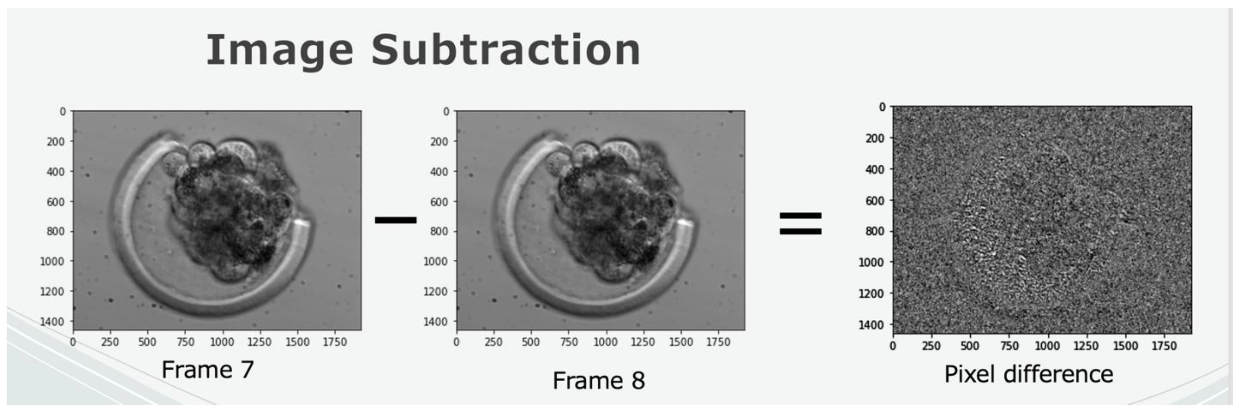
To extrapolate meaningful data from the videos, various graphic imaging techniques were applied. These techniques include object recognition, image subtraction, and contrast boosting. Object recognition is a computer vision technique for identifying objects in images or videos. This allows the computer to recognize and detect individual embryos apart from the background and from each other. Image subtraction is a process in which the digital numeric value of each pixel is subtracted from the value of the corresponding pixel in the previous frame. The output is the absolute difference between pixel values. This technique allows the objective measurement of the morphokinetics of the embryo over time because we can quantify the pixel change.
Each video was recorded at 15 frames per second. Therefore, each 30 s video comprised 450 frames (15 fps × 30 s = 450 total frames). Image subtraction was applied between each frame, creating 449 assessment points per video. Each subtracted frame generated output of the absolute difference between pixel values. Pixels that were different between consecutive frames appeared as white pixels on the screen. For each image subtracted frame, the number of white pixels (change) present in each embryo was counted. This count provided the basis for the objective analysis, as well as evidenced real-time morphokinetic changes within the embryo proper.

2.3. Implementation of Quality Control Methods and Evaluation of Noise

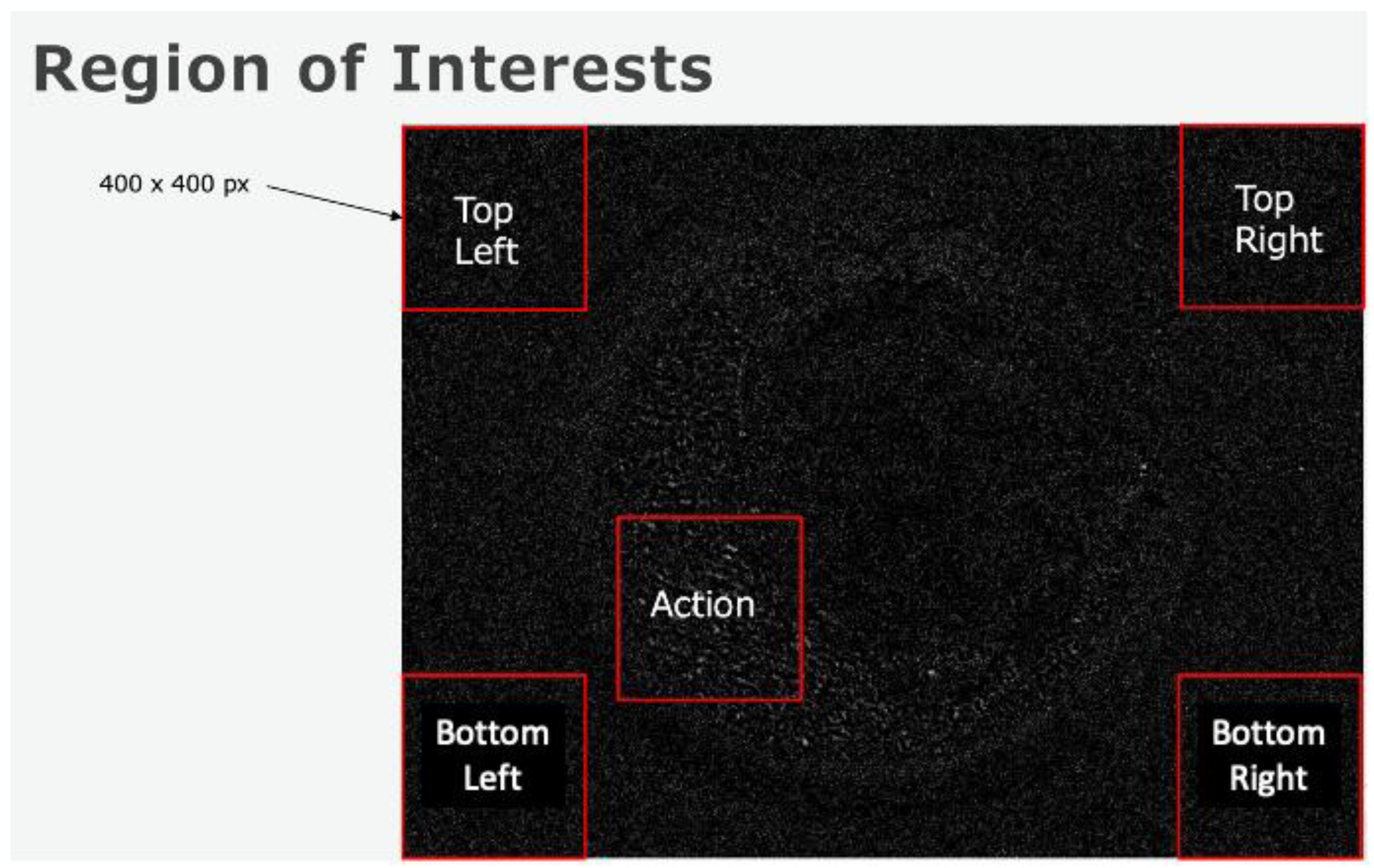
To understand if the captured motion derives from embryonic physiological processes or environmental noise, a series of experiments were performed to compare a motion analysis on regions of interest with live matter and a motion analysis on regions of interest without live matter. In these experiments, 400 × 400-pixel boxes in the four corners of the video, external to the embryo proper were assessed (Figure 2). A fifth 400 × 400-pixel box, including the region of the embryo proper, was also assessed (Figure 2). Comparisons of the count of pixel change over time were compared.

2.4. Identification of Metrics Correlating to Embryo Viability

To determine if video data can capture morphokinetic activity which correlates to embryo viability and development, comparisons were made between the embryos which demonstrated viability by hatching from the zona pellucida (“viable”) on the eighth and ninth day of culture and the embryos which failed to demonstrate viability by stalling in development or showing signs of degradation and decay at the time of assessment (“inviable”). Image recognition and image subtraction was applied to each video recording of embryos. The pixel change was counted between each video frame for each embryo for the entire duration of the video.
As each frame presents an opportunity for a morphokinetic event to occur, it is important to notice that some frames will display more morphokinetic activity (or change) than other frames. Therefore, it is important to study the amount and variability of the change for the duration of the video and not simply just the total count of embryo video pixel change over time. Some embryos displayed a sudden burst of change and then returned to a quiescent state, others displayed a consistent, steady, and rhythmic change throughout the duration of the video, and others displayed very little, flatline, change for the total duration of the video.
To address the variability of the pixel change and express the variability in a single calculation, the frame-by-frame standard deviation of the change of the action for areas housing the embryo proper was plotted for each embryo and compared to the four corner regions of interest which did not contain the embryo proper. In this analysis, the four corner regions of interest served as a means of quality control, as they did not contain live matter and theoretically should not possess any type of activity. Any change within these regions would suggest an external factor was causing motion (i.e., airflow, vibrations, unlevel surface, physical disturbances), inducing pixel changes in the embryos which were not derived from physiological or biological factors. Image subtracted frames showing mean activity above 1 in the four corner regions of interest were omitted from the study.
The cumulative value of the frame-by-frame standard deviation at the end of each video was compared between the viable embryos and the inviable embryos.

3. Statistical Analysis

All data were analyzed using R. The basic analysis was an unpaired Student’s t-test using a p-value of 0.05 for significance.

4. Results

Both the SMZ-1000 stereo zoom scope 90× and TE-300 Nikon inverted microscope 400× produced high-resolution images sufficient to capture real-time embryo morphokinetic activity (Figure 3).
The high magnification and high-resolution images of the TE-300 Nikon inverted microscope capture more data, as each embryo includes more pixels conveying digital information. However, the videos capturing groups of embryos on the SMZ-1000 stereo zoom scope are more practical for use in the dairy industry.
To determine if 30 s videos can capture embryo morphokinetic activity, graphic imaging techniques including background subtraction and contrasting boosting were used to track and quantify frame-by-frame pixel changes (Figure 4 and Figure 5).
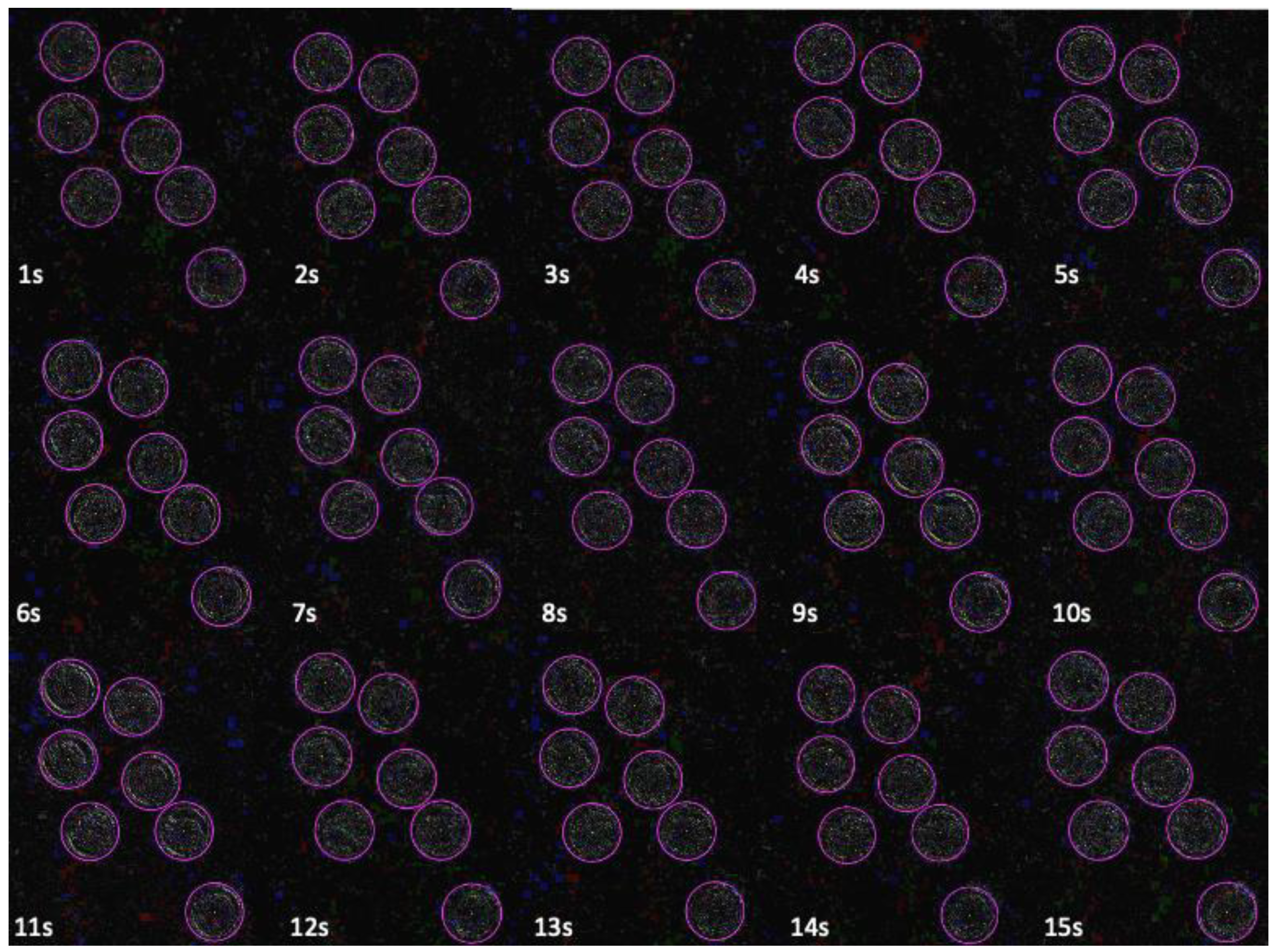
When these techniques were applied to videos of bovine embryos, the embryos’ real-time change can be quantified and perceived visually (Figure 6). The image subtraction emphasizes any frame-by-frame differences. Black pixels represent there was no change in the subsequent frames, whereas white pixel denotes a change in the subsequent frames. The number of white pixels is counted in each subtracted frame to measure and quantify embryo change, but in a visual examination of these videos, slight morphological changes in the embryo proper are apparent. Close evaluation indicates embryos are not identical in each frame, indicating morphological and morphokinetic changes occur, even in short observatory periods (Figure 6).
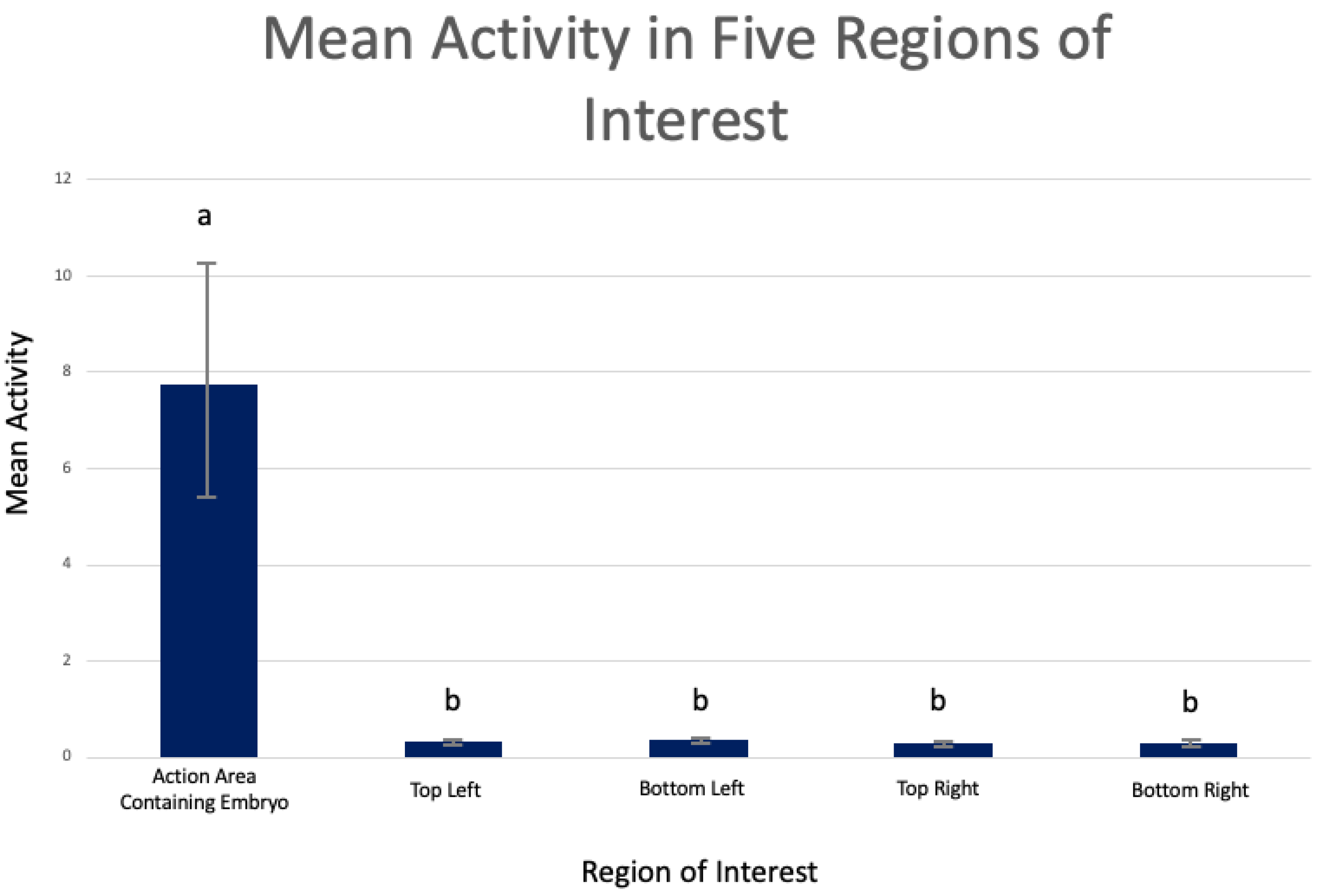
When the 400 × 400-pixel region containing live matter within the embryo proper was compared to the four 400 × 400-pixel regions external to the embryo, it was found that mean changes in the region of interest containing the embryo proper were significantly higher compared to mean changes in the region of interest of each corner region (Figure 7). Additionally, the region of interest containing the embryo proper demonstrated variable and inconsistent mean changes throughout the duration of the video, whereas each of the four corner regions external to the embryo proper demonstrated consistent and invariable mean changes (Figure 7).
For all 88 embryos, the mean pixel changes of the action area containing the embryo proper were significantly higher for the region of interest containing the embryo proper than the four regions of interest which were external to the embryo (p < 0.001) (Figure 8).
Of the 88 recorded embryos, 32 embryos demonstrated viability and normal development by hatching out of the zona pellucida on day 8. Fifty-six embryos did not demonstrate viability by hatching from the zona pellucida. Embryos that hatched had a lower normalized activity and lower range in normalized activity (p < 0.05) (Figure 9). Normalized activity = standard deviation/mean.

5. Discussion

Previous studies from these researchers analyzed individual embryo morphokinetic changes (measures of the embryo’s diameter at 5 s time intervals) recorded together in a single video [33]. In this study, the embryo diameter demonstrated a low correlation in relation to the diameter changes of the other embryos, suggesting embryos in a single video file demonstrated unique, individual growth patterns [33]. It was hypothesized that if the morphokinetic changes were a result of external factors, all embryos would respond uniformly, and display a high correlation in relation to each other. Despite the promise of this preliminary data, it was important to use image subtraction and calculate pixel change frame-by-frame on regions of interest containing the embryo as well as evaluate regions of the video background, external to the embryo. Understanding that the background of the video captures video data of fluid media, which can react to physical disturbances but does not contain any live matter, can provide an excellent means of quality control to ensure morphokinetic activity observed in analyzed regions of the video containing an embryo (live matter) is embryo-derived activity.
Analysis of 30 s videos of bovine embryos in culture demonstrated that embryo morphokinetic activity is present in short videos captured with standard microscopy and camera equipment (Figure 3 and Figure 6). When regions of the video containing the embryo proper were compared to regions of the video external to the embryo proper, significant differences in the pixels’ change over time were observed between the region containing the embryo proper and external regions. While each embryo demonstrated unique frequencies and magnitudes of pixel changes, these data suggest that detectable changes in the pixels are derived from physiological causes in the embryo, rather than environmental noise and disturbances such as light disturbances, external sound vibrations, or airflow and therefore, calculating the pixel changes from image subtracted videos of an embryo can be used to quantify real-time embryo morphokinetic activity in short observatory periods.
While this study demonstrates that embryo real-time morphokinetic activity is present in the video data, the cause of the embryo’s morphokinetic activity is not fully elucidated. The authors hypothesize that observed morphokinetic activity can be a result of mitotic activity such as cellular division or chromosomal separation, metabolic or metabolomic activity, cellular differentiation, cellular migration, apoptotic activity, cellular extrusion, adaptive behaviors as the embryo responds to its environment and external factors or cellular death [33] Observance of these morphokinetic changes through time-lapse imaging systems allowing continuous culture over a span of several days is well understood and enables researchers to screen embryos for altered morphokinetics, abnormal embryo cleavage, or multinucleation and has that demonstrated morphokinetic events are related to blastocyst formation, embryo implantation, and live birth [20,21,22,25,28,29,34,35,36,37,38,39,40,41,42,43,44,45,46,47]. These time-lapse imaging studies provide a foundation for understanding the role embryo morphokinetic activity plays as a key indicator to predict embryo viability and developmental potential. While time-lapse imaging systems require several days of monitoring embryos in a controlled system to create developmental outcome predictions which is very different than the 30 s observation period described in this research, time-lapse imaging systems have provided proof-of-concept that embryonic morphokinetic events can be captured with camera equipment, making it reasonable to believe that real-time assessments can also capture important embryo morphokinetic events which can convey important information pertaining to an embryo’s viability and health status. Therefore, new systems which are less expensive and require less time to monitor embryos for key morphokinetic events could equip dairy embryo transfer practitioners and livestock embryologists with the objective information to make data-driven embryo transfer and breeding decisions.
When image subtraction and contrast boosting techniques were applied to 30 s videos of bovine embryos, visible changes in the embryo’s morphology were apparent (Figure 6). These changes were represented by white pixels denoting that the pixel in the frame was different than the pixel in the previous frame. Black pixels represent no change. When observing the captured images of the image-subtracted frames, one can visibly see slight changes in the contours of the embryos. However, even in the image subtraction images, differences between viable and inviable embryos are not visually obvious. Therefore, computer systems to count each pixel change is necessary to extract meaningful data.
By definition, a pixel is the basic unit of programmable color on a computer image and is the smallest unit of measurement on a computer screen (each individual dot is a pixel). The density of pixels is resolution. The number of pixels present frame-by-frame is constant. However, counting the number of pixels that change, confined within the bounds of the embryo proper, can allow us to objectively quantify embryo morphokinetic activity. Comparing the changed pixels of the viable and inviable embryos can allow us to apply mathematical calculations which are associated with either viable or inviable embryos. In the study comparing the pixel change in the region of interest containing embryos that demonstrated viability by hatching and the embryos which were considered inviable because they did not hatch, it was found viable embryos have significantly lower mean normalized activity and lower range of normalized activity, as denoted by pixel change, over the course of the 30 s video than embryos which are inviable (p < 0.05) (Figure 9). This means viable embryos display less pixel change throughout the duration of the 30 s videos, and for what change the viable embryos do display, frame-by-frame changes are modest. When plotting the frame-by-frame standard deviation of embryos, viable embryos were characterized by consistent, steady, and rhythmic changes whereas inviable embryos were characterized by sudden and abrupt changes in the pattern of the frame-by-frame standard deviation with higher amplitude and larger ranges in standard deviation. While the data showcasing each individual embryo’s standard deviation over time are not shown, the overall cohort of viable embryos shows a lower range in pixel change than the overall cohort of inviable embryos for all 88 embryos included in this study (Figure 9). These characteristics suggest viable embryos demonstrate conservative and consistent morphokinetic activity whereas inviable embryos portray more variable, inconsistent patterns of morphokinetic activity.
Interestingly, these data resemble studies by Leese et al. suggesting the most viable embryos exhibit a quieter metabolism because they are required to expend less energy rectifying damage to the genome, transcriptome, and proteome, less energy sodium pumping, and are less susceptible to burn out due to failure to sustain a fast metabolic pace and respiration rate [48,49]. As the data from this study indicate a similar trend of viable embryos portraying reduced pixel change and activity, the authors of this research hypothesize that 30 s videos of embryos in culture can provide a means to non-invasively monitor embryo metabolism, which can allow researchers to detect the early embryonic onset of stress and deterioration as well as infer continued developmental potential. Proper metabolic function is crucial to the development of embryos as they must properly utilize energy sources to survive and grow. Detection of abnormal metabolic function could be an early indicator that an embryo will not be able to advance to later developmental stages, such as hatching, which is necessary for implantation in the uterus to establish a pregnancy.
While 30 s is a relatively short duration, this length of time is sufficient for the monitoring of some metabolic events as ATP turnover is derived from the sum of glycolytic and oxidative phosphorylation activity and the demand for cellular energy which can happen in the order of tens of seconds [50]. Noninvasive methods to monitor real-time embryonic metabolic activity can provide new insights into understanding pre-implantation bovine embryo health, allow for early detection of embryos that will not survive, and be used to improve pregnancy outcomes of ET and IVF in cattle.

6. Conclusions

Data from these studies suggest 30 s video recordings of in vitro bovine embryos capture real-time embryo morphokinetic activity which can provide key indicators signifying an embryo’s viability status. This method is practical and affordable for use in the dairy industry and can provide key objective data to help embryo transfer practitioners distinguish embryo viability prior to embryo transfer into recipient or cryopreservation.
Additionally, the ability to non-invasively monitor embryo metabolic activity can not only indicate embryo viability, developmental potential, and early signs of embryonic stress but can be used to optimize embryo culture conditions to enhance the growth and development of in vitro embryo production. Further research will develop machine learning models and algorithms to better analyze video data, automate the analysis and create comprehensive solutions for embryo health evaluation.

Author Contributions

All authors were significant contributors to the work and manuscript preparation of this project. C.W. and R.K. developed the original concept and method of the bovine video analysis. C.W. and R.K. were co-PIs of this study and designed the experiment. A.W., J.H. and A.Z. developed the assessment, graphic imaging techniques, and data analysis methods. C.W. developed the original manuscript, and R.K. and A.W. were involved in editing for the final content. All authors have read and agreed to the published version of the manuscript.

Funding

EmGenisys paid Texas A&M University Reproductive Services Complex $6000 to create the embryos and rent the microscopes used in this study.

Institutional Review Board Statement

Not applicable.

Informed Consent Statement

Not applicable.

Data Availability Statement

The data presented in this study are available on request from the corresponding author. The data are not publicly available due to the proprietary nature of the data.

Conflicts of Interest

C.W. and R.K. declare a conflict of interest as both authors are named inventors on patents associated with this work and own stock in EmGenisys, an animal agricultural company which owns the assignments for associated intellectual property. Patent number 11,455,725. A.W., J.H. and A.Z. declare no conflict of interest.

References

  1. Henchion, M.; Hayes, M.; Mullen, A.M.; Fenelon, M.; Tiwari, B. Future Protein Supply and Demand: Strategies and Factors Influencing a Sustainable Equilibrium. Foods 2017, 6, 53. [Google Scholar] [CrossRef] [PubMed] [Green Version]
  2. Capper, J. The environmental impact of beef production in the United States: 1977 compared with 2007. J. Anim. Sci. 2011, 89, 4249–4261. [Google Scholar] [CrossRef] [PubMed] [Green Version]
  3. Capper, J.; Cady, R.; Bauman, D. The environmental impact of dairy production: 1944 compared with 2007. J. Anim. Sci. 2009, 87, 2160–2167. [Google Scholar] [CrossRef] [PubMed]
  4. Brito, L.; Bedere, N.; Douhard, F.; Oliveria, H.; Arnal, M.; Penagaricano, F.; Schnickel, A.; Baes, C.; Miglior, F. Review: Genetic selection of high-yielding dairy cattle toward sustainable farming systems in a rapidly changing world. Animals 2021, 15, 1. [Google Scholar] [CrossRef]
  5. Dillon, P.; Buckley, F.; O’Connor, P.; Hegarty, D.; Rath, M. A comparison of different dairy cow breeds on a seasonal grass-based system of milk production: 1. Milk production, live weight, body condition score and DM intake. Livest. Prod. Sci. 2003, 83, 21–33. [Google Scholar] [CrossRef]
  6. Dillon, P.; Snijders, S.; Buckley, F.; Harris, B.; O’Connor, P.; Mee, J. A comparison of different dairy cow breeds on a seasonal grass-based system of milk production: 2. Reproduction and survival. Livest. Prod. Sci. 2003, 83, 35–42. [Google Scholar] [CrossRef]
  7. Walsh, S.; Buckley, F.; Berry, D.; Rath, M.; Pierce, K.; Byrne, N.; Dillon, P. Effects of breed, feeding system, and parity on udder health and milking characteristics. J. Dairy Sci. 2007, 90, 5767–5779. [Google Scholar] [CrossRef]
  8. Walsh, S.; Buckley, F.; Pierce, K.; Byrne, N.; Patton, J.; Dillon, P. Effects of breed and feeding system on milk production, body weight, body condition score, reproductive performance, and postpartum ovarian function. J. Dairy Sci. 2008, 91, 4401–4413. [Google Scholar] [CrossRef] [Green Version]
  9. Azzarello, A.; Hoest, T.; Hay-Schmidt, A.; Mikkelsen, A.L. Live birth potential of good morphology and vitrified blastocysts presenting abnormal cell divisions. Reprod. Biol. 2017, 17, 144–150. [Google Scholar] [CrossRef]
  10. Bedere, N.; Disenhaus, C.; Ducrocq, V.; Leurent-Colette, S.; Delaby, L. Ability of dairy cows to be inseminated according to breed and genetic merit for production traits under contrasting pasture-based feeding systems. Animal 2017, 11, 826–835. [Google Scholar] [CrossRef]
  11. Bedere, N.; Disenhaus, C.; Ducrocq, V.; Leurent-Colette, S.; Delaby, L. Ability of dairy cows to ensure pregnancy according to breed and genetic merit for production traits under contrasted pasture-based systems. J. Dairy Sci. 2017, 100, 2812–2827. [Google Scholar] [CrossRef] [PubMed] [Green Version]
  12. Potts, B.; Shaughness, M.; Erdman, R.A. The decline in digestive efficiency of US dairy cows from 1970 to 2014. J. Dairy Sci. 2017, 100, 5400–5410. [Google Scholar] [CrossRef] [Green Version]
  13. Wells, C. EmGenisys Pitch Deck. In Luminate NextCorps Accelerator; EmGenisys: Rochester, NY, USA, 2022. [Google Scholar]
  14. Demetrio, D.; Looney, C.; Rees, H.; Werhman, M. 2020 Statistical Information Committee Report (2019 Data). 2020. Annual Report of the AETA Statistical Information Committee. Available online: https://www.aeta.org/docs/2019_Stats.pdf (accessed on 22 September 2022).
  15. Bo, G.; Mapletoft, R. Evaluation and classification of bovine embryos. Anim. Reprod. 2013, 10, 344–348. [Google Scholar]
  16. Wessels, C.; Penrose, L.; Prien, S. Current State of Art Embryo Selection Techniques. Investig. Obstet. Gynecol. Res. 2017, 1, 1–6. [Google Scholar] [CrossRef]
  17. Cruz, M.; Blanca, G.; Garrido, N.; Pedersen, K.; Martínez, M. Embryo Quality, Blastocyst and Ongoing Pregnancy Rates in Oocyte Donation Patients Whose Embryos Were Monitored by Time-lapse Imaging. J. Assist. Reprod. Genet. 2011, 28, 569–573. [Google Scholar] [CrossRef] [Green Version]
  18. Del Gallego, R.; Remohi, J.; Meseguer, M. Time-lapse imaging: State of the art. Biol. Reprod. 2019, 101, 1146–1154. [Google Scholar] [CrossRef]
  19. Zaninovic, N.; Irani, M.; Meseguer, M. Assessment of embryo morphology and developmental dynamics by time-lapse microscopy: Is there a relation to implantation and ploidy? Fertil. Steril. 2017, 108, 722–729. [Google Scholar] [CrossRef] [Green Version]
  20. Athayde Wirka, K.; Chen, A.A.; Conaghan, J.; Ivani, K.; Gvakharia, M.; Behr, B.; Suraj, V.; Tan, L.; Shen, S. Atypical Embryo Phenotypes Identified by Time-Lapse Microscopy: High Prevalence and Association with Embryo Development. Fertil. Steril. 2014, 101, 1637–1648. [Google Scholar] [CrossRef] [PubMed]
  21. Goodman, L.R.; Goldberg, J.; Falcone, T.; Austin, C.; Desai, N. Does the Addition of Time-Lapse Morphokinetics in the Selection of Embryos for Transfer Improve Pregnancy Rates? A Randomized Controlled Trial. Fertil. Steril. 2016, 105, 275–285. [Google Scholar] [CrossRef] [PubMed] [Green Version]
  22. Zhan, Q.; Ye, Z.; Clarke, R.; Rosenwaks, Z.; Zaninovic, N. Direct Unequal Cleavages: Embryo Developmental Competence, Genetic Constitution and Clinical Outcome. PLoS ONE 2016, 11, e0166398. [Google Scholar] [CrossRef] [Green Version]
  23. Desai, N.; Ploskonka, S.; Goodman, L.; Attaran, M.; Goldberg, J.M.; Austin, C.; Falcone, T. Delayed blastulation, multinucleation, and expansion grade are independently associated with live-birth rates in frozen blastocyst transfer cycles. Fertil. Steril. 2016, 106, 1370–1378. [Google Scholar] [CrossRef] [PubMed]
  24. Aguilar, J.; Rubio, I.; Muñoz, E.; Pellicer, A.; Meseguer, M. Study of nucleation status in the second cell cycle of human embryo and its impact on implantation rate. Fertil. Steril. 2016, 106, 291–299. [Google Scholar] [CrossRef] [Green Version]
  25. Rubio, I.; Kuhlmann, R.; Agerholm, I.; Kirk, J.; Herrero, J.; Escribá, M.-J.; Bellver, J.; Meseguer, M. Limited Implantation Success of Direct-Cleaved Human Zygotes: A Time-Lapse Study. Fertil. Steril. 2012, 98, 1458–1463. [Google Scholar] [CrossRef] [PubMed]
  26. Ebner, T.; Höggerl, A.; Oppelt, P.; Radler, E.; Enzelsberger, S.H.; Mayer, R.B.; Petek, E.; Shebl, O. Time-lapse imaging provides further evidence that planar arrangement of blastomeres is highly abnormal. Arch. Gynecol. Obstet. 2017, 296, 1199–1205. [Google Scholar] [CrossRef]
  27. Desch, L.; Bruno, C.; Luu, M.; Barberet, J.; Choux, C.; Lamotte, M.; Schmutz, E.; Sagot, P.; Fauque, P. Embryo multinucleation at the two-cell stage is an independent predictor of intracytoplasmic sperm injection outcomes. Fertil. Steril. 2017, 107, 97–103. [Google Scholar] [CrossRef] [PubMed] [Green Version]
  28. Yang, S.T.; Shi, J.X.; Gong, F.; Zhang, S.P.; Lu, C.F.; Tan, K.; Leng, L.Z.; Hao, M.; He, H.; Gu, Y.F.; et al. Cleavage Pattern Predicts Developmental Potential of Day 3 Human Embryos Produced by IVF. Reprod. Biomed. Online 2015, 30, 625–634. [Google Scholar] [CrossRef] [Green Version]
  29. Barrie, A.; Homburg, R.; McDowell, G.; Brown, J.; Kingsland, C.; Troup, S. Preliminary Investigation of the Prevalence and Implantation Potential of Abnormal Embryonic Phenotypes Assessed Using Time-Lapse Imaging. Reprod. Biomed. 2017, 34, 455–462. [Google Scholar] [CrossRef] [Green Version]
  30. Wong, C.C.; Loewke, K.E.; Bossert, N.L.; Behr, B.; De Jonge, C.J.; Baer, T.M.; Pera, R.A.R. Non-invasive imaging of human embryos before embryonic genome activation predicts development to the blastocyst stage. Nat. Biotechnol. 2010, 28, 1115–1121. [Google Scholar] [CrossRef]
  31. Aparicio-Ruiz, B.; Basile, N.; Pérez Albalá, S.; Bronet, F.; Remohí, J.; Meseguer, M. Automatic time-lapse instrument is superior to single-point morphology observation for selecting viable embryos: Retrospective study in oocyte donation. Fertil. Steril. 2016, 106, 1379–1385.e10. [Google Scholar] [CrossRef] [Green Version]
  32. Yeung, S.; Downing, N.L.; Fei-Fei, L.; Milstein, A. Bedside computer vision—Moving artificial intelligence from driver assistance to patient safety. N. Engl. J. Med. 2018, 378, 1271–1273. [Google Scholar] [CrossRef]
  33. Wells, C.; Killingsworth, R.; Real-time embryo morphokinetic activity can be measured in short observatory periods to provide objective evaluation of embryo health. A Closer Look. Am. Embryo Transf. Assoc. Newsl. Available online: https://www.aeta.org/newsletter/?p=2623 (accessed on 20 September 2022).
  34. Sayed, S.; Reigstad, M.M.; Petersen, B.M.; Schwennicke, A.; Wegner Hausken, J.; Storeng, R. Time-lapse imaging derived morphokinetic variables reveal association with implantation and live birth following in vitro fertilization: A retrospective study using data from transferred human embryos. PLoS ONE 2010, 15, e0242377. [Google Scholar] [CrossRef] [PubMed]
  35. Rienzi, L.; Capalbo, A.; Stoppa, M.; Romano, S.; Maggiulli, R.; Albricci, L.; Sarica, C.; Farcomeni, A.; Vajta, G.; Ubaldi, F.M. No evidence of association between blastocyst aneuploidy and morphokinetic assessment in a selected population of poor-prognosis patients: A longitudinal cohort study. Reprod. Biomed. Online 2015, 30, 57–66. [Google Scholar] [CrossRef] [Green Version]
  36. Sundvall, L.; Ingerslev, H.J.; Breth Knudsen, U.; Kirkegaard, K. Inter- and intra-observer variability of time-lapse annotations. Hum. Reprod. 2013, 28, 3215–3221. [Google Scholar] [CrossRef] [PubMed] [Green Version]
  37. Storr, A.; Venetis, C.A.; Cooke, S.; Kilani, S.; Ledger, W. Inter-observer and intra-observer agreement between embryologists during selection of a single Day 5 embryo for transfer: A multicenter study. Hum. Reprod. 2017, 32, 307–314. [Google Scholar] [CrossRef] [PubMed]
  38. Adolfsson, E.; Andershed, A.N. Morphology vs morphokinetics: A retrospective comparison of inter-observer and intra-observer agreement between embryologists on blastocysts with known implantation outcome. JBRA Assist. Reprod. 2018, 22, 228–237. [Google Scholar] [CrossRef]
  39. Boucret, L.; Tramon, L.; Saulnier, P.; Ferré-L’Hôtellier, V.; Bouet, P.-E.; May-Panloup, P. Change in the Strategy of Embryo Selection with Time-Lapse System Implementation—Impact on Clinical Pregnancy Rates. J. Clin. Med. 2021, 10, 4111. [Google Scholar] [CrossRef]
  40. Yang, S.-H.; Wu, C.-H.; Chen, Y.-C.; Yang, C.-K.; Wu, T.-H.; Chen, P.-C.; Tsai, H.-D. Effect of Morphokinetics and Morphological Dynamics of Cleavage Stage on Embryo Developmental Potential: A Time-Lapse Study. Taiwan J. Obstet. Gynecol. 2018, 57, 76–82. [Google Scholar] [CrossRef]
  41. Desai, N.; Goldberg, J.M.; Austin, C.; Falcone, T. Are Cleavage Anomalies, Multinucleation, or Specific Cell Cycle Kinetics Observed with Time-Lapse Imaging Predictive of Embryo Developmental Capacity or Ploidy? Fertil. Steril. 2018, 109, 665–674. [Google Scholar] [CrossRef] [Green Version]
  42. Liu, Y.; Chapple, V.; Roberts, P.; Matson, P. Prevalence, Consequence, and Significance of Reverse Cleavage by Human Embryos Viewed with the Use of the Embryoscope Time-Lapse Video System. Fertil. Steril. 2014, 102, 295–1300. [Google Scholar] [CrossRef]
  43. Conaghan, J.; Chen, A.A.; Willman, S.P.; Ivani, K.; Chenette, P.E.; Boostanfar, R.; Baker, V.L.; Adamson, G.D.; Abusief, M.E.; Gvakharia, M.; et al. Improving Embryo Selection Using a Computer-Automated Time-Lapse Image Analysis Test plus Day 3 Morphology: Results from a Prospective Multicenter Trial. Fertil. Steril. 2013, 100, 412–419. [Google Scholar] [CrossRef]
  44. Motato, Y.; de los Santos, M.J.; Escriba, M.J.; Ruiz, B.A.; Remohí, J.; Meseguer, M. Morphokinetic Analysis and Embryonic Prediction for Blastocyst Formation through an Integrated Time-Lapse System. Fertil. Steril. 2016, 105, 376–384. [Google Scholar] [CrossRef] [PubMed]
  45. Dal Canto, M.; Coticchio, G.; Mignini Renzini, M.; De Ponti, E.; Novara, P.V.; Brambillasca, F.; Comi, R.; Fadini, R. Cleavage Kinetics Analysis of Human Embryos Predicts Development to Blastocyst and Implantation. Reprod. Biomed. 2012, 25, 474–480. [Google Scholar] [CrossRef]
  46. Cruz, M.; Garrido, N.; Herrero, J.; Pérez-Cano, I.; Muñoz, M.; Meseguer, M. Timing of Cell Division in Human Cleavage-Stage Embryos Is Linked with Blastocyst Formation and Quality. Reprod. Biomed. Online 2012, 25, 371–381. [Google Scholar] [CrossRef]
  47. Carrasco, B.; Arroyo, G.; Gil, Y.; Gómez, M.J.; Rodríguez, I.; Barri, P.N.; Veiga, A.; Boada, M. Selecting Embryos with the Highest Implantation Potential Using Data Mining and Decision Tree Based on Classical Embryo Morphology and Morphokinetics. J. Assist. Reprod. Genet. 2017, 34, 983–990. [Google Scholar] [CrossRef] [PubMed]
  48. Leese, H.J. Quiet please, do not disturb: A hypothesis of embryo metabolism and viability. Bioessay 2002, 24, 845–849. [Google Scholar] [CrossRef]
  49. Leese, H.J.; Sturmey, R.; Baumann, C.G.; McEvoy, T.G. Embryo viability and metabolism: Obeying the quiet rules. Hum. Reprod. 2007, 22, 3047–3050. [Google Scholar] [CrossRef] [PubMed]
  50. Brison, D.; Leese, H. Energy metabolism in late preimplantation rat embryos. J. Reprod. Fert. 1991, 93, 245–251. [Google Scholar] [CrossRef] [PubMed]
Figure 1. Timeline of events to collect embryo developmental outcomes and video recordings.
Dairy 03 00058 g001
Figure 2. Five regions of interest were compared to evaluate the impact of environmental noise on the collected video data. Frame-by-frame changes in 400 × 400 pixel bounding boxes were assessed in regions of interest containing an action area containing live matter of the embryo proper, the top left corner external to the embryo, the top right corner external to the embryo, the bottom left corner external to the embryo, and the bottom right corner external to the embryo. Image collected from TE-300 inverted microscope, 400×.
Dairy 03 00058 g002
Figure 3. Still images of bovine embryos on different microscopy systems. (A) TE-300 Nikon inverted microscope, 400×; (B) SMZ-1000 stereo zoom microscope, 90×; (C) SMZ-1000 stereo zoom microscope, 90× 4 embryos in a group.
Dairy 03 00058 g003
Figure 4. Subsequent frames are subtracted from each other. If there is no difference in pixel color/gradient, the image will appear static without any contours/artifacts or remnants of the image. If there is a difference, a ghostly figure will appear which can be quantified. Image collected from TE-300 inverted microscope, 400×.
Dairy 03 00058 g004
Figure 5. Contrast boosting can make these contours/artifacts/remnants more visibly apparent. Image collected from TE-300 inverted microscope, 400×.
Dairy 03 00058 g005
Figure 6. Image subtraction and contrast boosting images of bovine embryos. Videos, recorded at 15 frames per second, were processed with graphic imaging techniques. Still capture images of the video data at 1 image per second were placed in a schematic to represent detectable changes in the embryos over a 15 s period. Image collected on an SMZ-1000 stereo zoom microscope, 90×.
Dairy 03 00058 g006
Figure 7. Frame-by-frame changes of the five regions of interest of a single embryo. The X axis represents the frame number, and the Y axis represents the standard deviation of the pixel change per frame. This graph represents a video with a single embryo. The blue line represents the “action” area which is the region of interest containing the embryo proper. The yellow line represents the top right corner region external to the embryo, the green line represents the top right corner region external to the embryo, the red line represents the bottom left corner region external to the embryo and the purple line represents the top left corner region external to the embryo.
Dairy 03 00058 g007
Figure 8. Mean activity in the five regions of interest. Activity in the 400 × 400-pixel bounding box containing the action area of the embryo proper was significantly higher than the activity in the four 400 × 400-pixel bounding boxes which were external to the embryo. Bars with different letters a–b represent statistical differences in mean activity (p < 0.001). Bars indicate standard deviation.
Dairy 03 00058 g008
Figure 9. Normalized activity of bovine embryos in relation to developmental outcomes. Normalized activity (standard deviation/mean) was calculated for each of the embryo’s change over time. The X represents the mean of the normalized activity. Bars represent the range of the data and whiskers represent standard deviation (p < 0.05).
Dairy 03 00058 g009
Publisher’s Note: MDPI stays neutral with regard to jurisdictional claims in published maps and institutional affiliations.