Article

Dual Mobility Hip Arthroplasty: Innovative Technological Advances

1
U.O.C. Ortopedia e Traumatologia, Ospedale Maggiore “C.A. Pizzardi”, 40133 Bologna, Italy
2
Clinical Research—Permedica Orthopaedics, 23807 Merate, Italy
*
Author to whom correspondence should be addressed.
Prosthesis 2024, 6(2), 393-400; https://doi.org/10.3390/prosthesis6020029
Submission received: 5 February 2024 / Revised: 4 April 2024 / Accepted: 5 April 2024 / Published: 17 April 2024
(This article belongs to the Special Issue State of Art in Hip, Knee and Shoulder Replacement (Volume 2))

Abstract

:
The use of 3D-printed highly porous titanium acetabular cups in total hip arthroplasty (THA) is increasing. The porosity and mechanical properties of such highly porous titanium structures mimic those of natural cancellous bone, possibly allowing biological implant fixation to be improved. Recently, a 3D-printed highly porous Dual Mobility (DM) monobloc construct fully manufactured using Ti6Al4V alloy, with a titanium–niobium nitride (TiNbN) ceramic coating on the articular side to allow articulation against the mobile liner by improving the titanium vs. polyethylene tribological behavior, was introduced in THA. To the best of our knowledge, this is the first highly porous titanium monobloc DM implant on the market. The reasons for using a Ti alloy highly porous DM are multifarious: to prevent any possible adverse reactions due to the corrosion of Cobalt–Chromium–Molybdenum Alloy (CoCrMo) and Stainless Steel (SS) implants and to improve implant primary and secondary stability, particularly in cases of poor bone quality. Finally, with the introduction of an inner TiNbN ceramic coating surface, it was possible to overcome the poor tribological quality of titanium. Another interesting characteristic is this material’s higher implant radiolucency, which might facilitate the radiographic assessment of cup orientation, which can, in turn, facilitate the detection of any intraprosthetic dislocation (IPD) and the measurement of polyethylene wear, which is very important in the study of the durability of THA.

1. Introduction

The dual mobility (DM) cup was introduced in France in the late nineteen-seventies by the innovative surgeon Gill Bousquet, implementing the concept of the Christiansen system, involving the placement of a large head composed of a derlin piece and an SS cup that had the ability to rotate around the neck axis due to component design [1]. Bousquet’s idea was to allow mobility of the prosthetic femoral head around the space, combining the “low friction” principle of THA popularized by Charnley [2], thanks to the small diameter of the femoral head (22.2 mm) and the Mckee–Farrar concept of using a larger-diameter femoral head to enhance implant stability [3] further than the already-cited Christiansen mobility notion [1]. Since then, several improvements and refinements of the design of the DM device have been made. While numerous hip implant systems are currently available on the market, only two types of materials are currently available for manufacturing the monobloc DM acetabular shell: SS and CoCr [4]. CoCr and SS are suitable materials for articulation against polyethylene; a matter of concern in this regard, however, is related to a possible allergic reaction in biological environments. SS and CoCr are preferred in DM shells because the cup’s articular surface requires proper surface finishing and tribological properties that allow low friction and wear against the mobile polyethylene insert [5]. SS has good mechanical properties but considerably low resistance to corrosion. In biological environments, SS typically exhibits pitting corrosion as a result of anodic polarization. CoCr shows excellent mechanical properties, castability, wear, and corrosion resistance [6]. However, SS and CoCr alloys can both lead to metallosis or periprosthetic adverse biological reactions due to metal ion release and wear byproducts [7]. The usage of CoCr alloys or stainless steel is related to a series of complications, including corrosion-induced toxicity and device degradation, a severe inflammatory response called an aseptic lymphocyte-dominated vasculitis-associated lesion (ALVAL), and metal hypersensitivity [8,9]. Regarding DM THA, a growing number of studies are analyzing the serum metal ion levels in patients with modular DM constructs [10], raising concerns regarding fretting and corrosion at the titanium shell/CoCr insert interface leading to the release of metal ions. Recently, at least two papers have reported on cobalt and chromium levels in patients with monoblock DM cups [11,12]. Titanium alloys have been precluded due to their low hardness and poor tribological performance against polyethylene due to the presence of a thin and unstable superficial passive oxide film that can act in articulation as a third body [13,14]. In an effort to reduce complications and improve implant longevity, a new cementless DM acetabular cup was recently developed and introduced on the market in 2021 [10]. This implant is a monobloc DM cup with a polar-flatted hemispherical profile, a 0° cup opening plane, and 2.5 mm cylindrical equatorial extra coverage. The center of the femoral head has a medial eccentricity from the center of the polyethylene mobile liner (Figure 1).
As with all monoblock DM devices, it lacks the additional screw fixation options available for other conventional metal-backed acetabular shells. Actually, the inner surface is highly polished in order to allow safe mobility of the polyethylene. Recently, several companies introduced the concept of modularity in DM devices (modular DM devices). These modular DM devices have a standard titanium metal-backed shell, which allows an inner metal liner to couple the outer titanium surface with the polyethylene liner. The use of modular prostheses has currently demonstrated satisfactory results, while also adding a series of new complications [15,16,17,18].

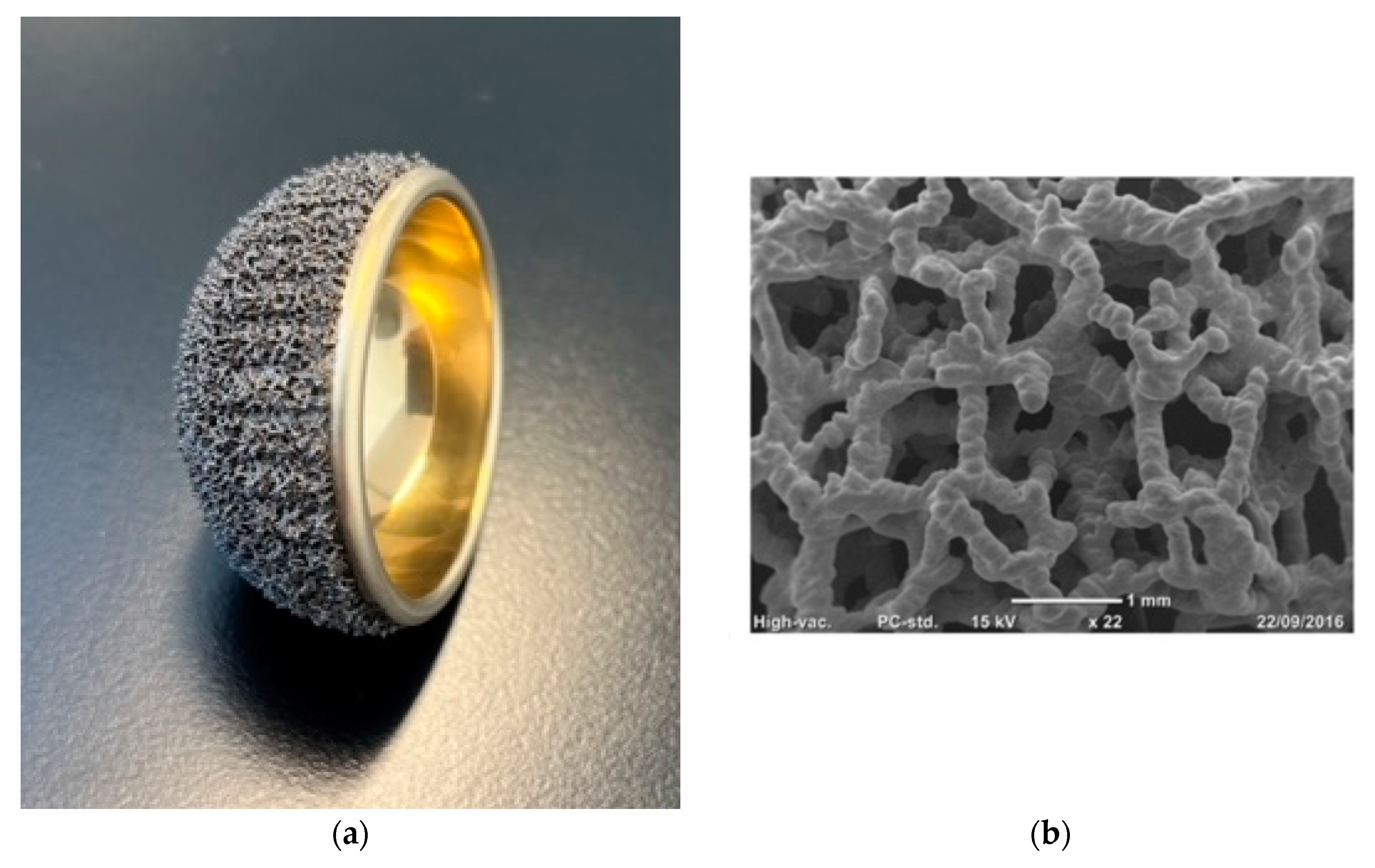
Characteristics and Rationale of the Acorn Traser System

The implant, the Acorn Traser DM cup (Permedica Orthopaedics, Merate, Italy), is a cementless press-fit acetabular cup featuring a highly porous random trabecular titanium structure, commercially named Traser®, manufactured using selective laser-melting technology without providing continuity on the bone-implant side of the cup. The Acorn Traser is a standard device that is mass-produced.
Additive manufacturing (or 3D-printing) is a technology that allows the production of three-dimensional solid objects based on a digital model, using metal powder as a raw material. The selective laser-melting process is conducted in an inert atmosphere (argon) in order to avoid titanium powder oxidation. The solid and porous portions of the cup are built up in one continuous process, thus creating a single piece without interface layers between different portions, as occurs for coatings, and, consequently, without shear forces. The final structure reproduces a highly porous trabecular network, similar to cancellous bone and characterized by an open, fully interconnected, irregular 70% porosity material with a pore size ranging from 0.1 to 2.0 mm (Figure 2).
Highly porous titanium structures are able to improve primary implant stability by virtue of the higher friction at the cup–bone interface and secondary implant stability provided by the optimization of newly formed osseointegrated bone within their pores, leading to higher cup retention than plasma-sprayed porous titanium coatings [6,20]. High porosity was reported to be able to improve local vascularization (in terms of the number and size of vessels), stimulating a better bony ingrowth and reducing the fibrotic peri-implant tissue [21,22]. As a result, a highly porous DM cup could theoretically reduce aseptic cup loosening, mainly in high-risk patients with acetabular poor bone quality. Tantalum acetabular construct cups were the first implants with such properties to be used, whereas titanium and titanium alloy cups with highly porous surfaces were later developed thanks to additive manufacturing. Multiple studies have reported low failure rates and improved survivorship following the use of these porous materials in primary and revision THA [23,24,25].

2. Materials and Methods

We conducted a retrospective cohort study including patients treated using THA in a single tertiary referral center for traumatology from 2021 to 2022.
Inclusion criteria were as follows: male and female adult patients (>18 years old) who were subjected to primary cementless THA using Acorn Traser DM cup (Permedica Orthopaedics) with at least one year of follow-up data.
Patients who underwent THA using different types of acetabular cups, had a severe dysplastic hip or were affected by high-grade obesity, or with a body mass index (BMI) of 40 or higher were excluded.

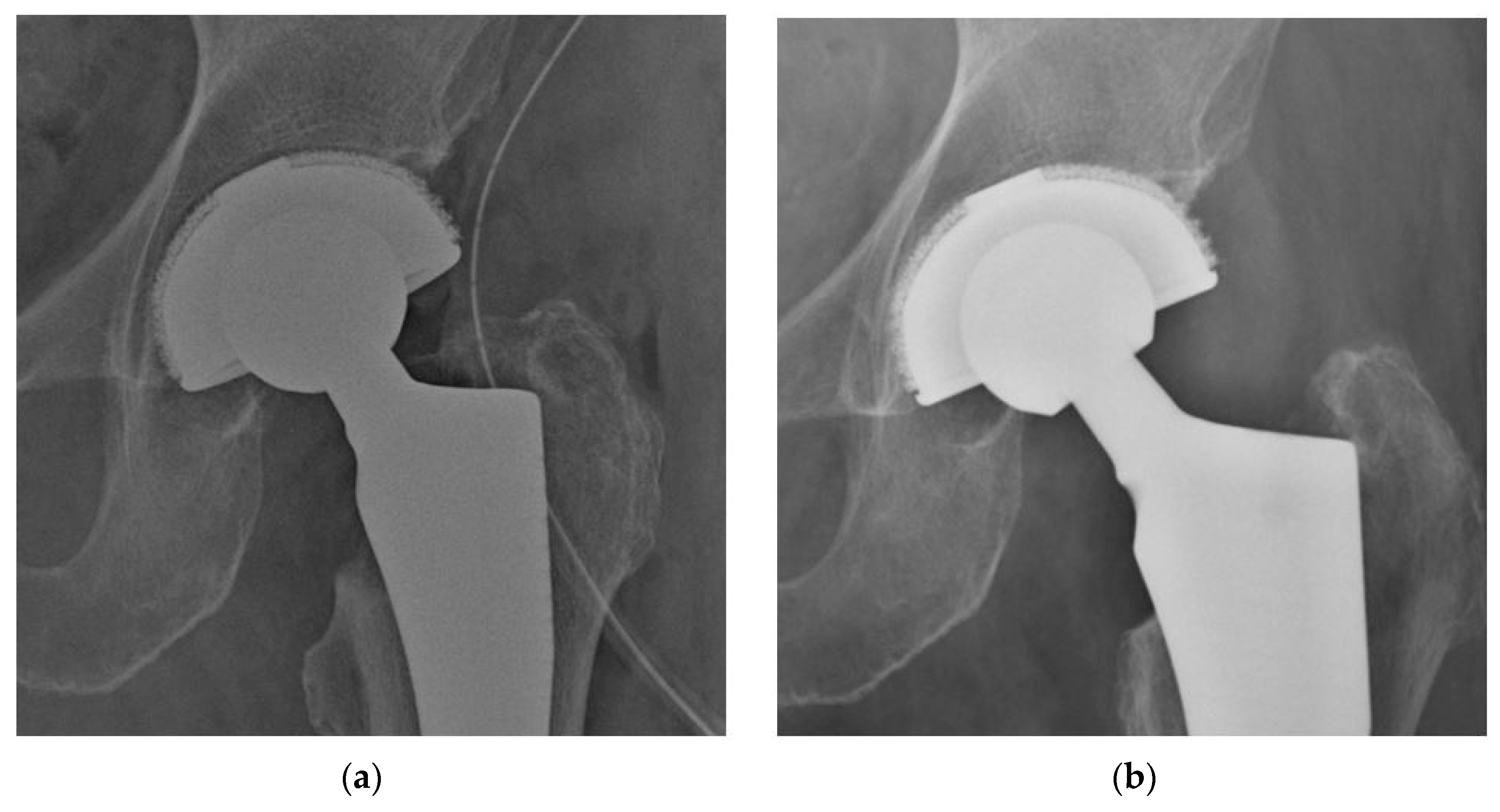
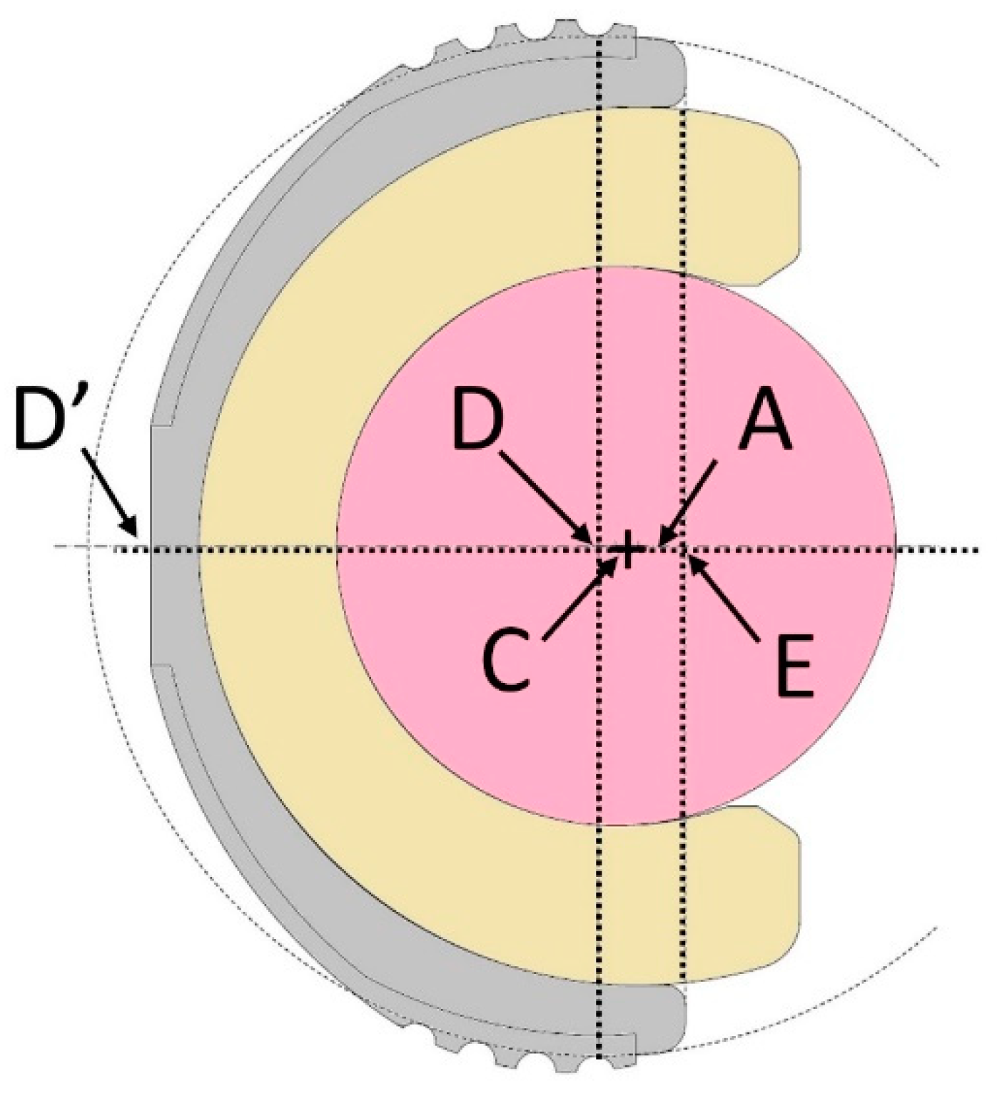
2.1. Patients and Radiographic Evaluation

Demographics and patient characteristics at baseline, including age at operation, sex, BMI, hip side, and reasons for THA, were recorded using medical charts.
All THA procedures were performed by two senior surgeons with more than 20 years of experience using a posterior–lateral approach with hip extrarotator detachment and reinsertion of the piriformis.
Radiographic parameters were collected using plain antero-posterior pelvis X-ray postoperatively and at 12-month follow-up by a single observer using Carestream Vue Pacs (Rochester, NY, USA). Positional parameters, such as cup abduction and anteversion, were measured using the Widmer method [25], while acetabular osseointegration was assessed by adopting the 5 criteria described by Moore et al. [26]. Radiographic cup loosening was defined as >3 mm cup migration or >5° cup rotation [27,28] or a radiolucent line greater than 2 mm in more than one zone outlining the cup [29]. The presence of a polar gap radiographically visible as a radiolucent area behind the acetabular dome was assessed using the first postoperative radiograph and compared with the 12-month radiograph to verify consequent new bone formation.

2.2. Statistical Analysis

Continuous data were expressed as means, whereas categorical and ordinal data were expressed as absolute values and percentages. All analyses were performed with SPSS v. 22.0 (SPSS, Chicago, IL, USA) and Microsoft Excel v. 16.30 (Microsoft Corporation, Redmond, WA, USA).

3. Results

3.1. Patient Population

One hundred seventy-nine hips underwent treatment with THA using the Acorn Traser cup at our institution since January 2021. Fifty patients (50 hips) satisfied all the inclusion criteria and were enrolled in this study. The mean patient age at operation was 75 years (range: 40–86); twenty-nine of these patients were women. Twenty-seven patients underwent THA for a femoral neck fracture, while 56% of patients were treated for degenerative joint pathologies such as osteoarthritis (87%) or avascular necrosis of the femoral head (13%) (Table 1).
Complications were observed in two patients (4%). Both of them underwent THA for a femoral neck fracture and sustained intraoperatively an acetabulum fracture during press-fit implantation. For one patient, stabilization with a one-third tubular plate was needed for a fracture of the posterior wall of the acetabulum. The second patient sustained an intraoperative fracture of the anterior wall. The acetabular components were determined to be stable despite the fracture, and no additional treatments were performed.
No cases of true dislocation, nor of intraprosthetic dislocation, were observed in our cohort.

3.2. Radiographic Evaluation

One patient (2%) showed a significant vertical malposition of the cup.
At the 12-month follow-up, all 50 cups implanted showed good radiographic osseointegration, with no evidence of radiographic lucencies or cup migration.
When looking for polar gaps behind the acetabular dome, areas with no initial bone–cup contact in the immediate postoperative radiograph were completely filled through new bone formation within the first year (Figure 3). No cup or liner revision was performed, nor did radiographic loosening occur at the final follow-up. In particular, no cases of >3 mm cup migration or >5° cup rotation or a radiolucent line greater than 2 mm in more than one zone outlining the cup were observed.

4. Discussion

To date, no cases of metal sensitization after THA with DM cups have been reported in the literature, although its normal constituents are potentially allergenic. It has been estimated that cutaneous allergies to common metals such as nickel, cobalt, and chromium occur in 13%, 2%, and 1% of the general population, respectively. Contextually, concerns exist about the release of cobalt and chromium ions from DM cups in patients receiving either monoblock or contemporary modular CoCr DM constructs [11,12,13,14]. All metals in contact with biological systems corrode. Serum levels of metal ions are significantly increased in patients with normally functioning metal-on-metal hips [30]. Released ions themselves are not immunogenic; they are so called haptens. However, they can activate the immune system by forming complexes with native proteins. Nickel is the most common sensitizer in humans, followed by cobalt and chromium. Therefore, although rare and unpredictable, it is important to account for metal hypersensitivity as a potential etiology for implant failure. Contextually, there are clear recommendations in regard to considering hypoallergenic components in case of a history of a metallic allergy or positive tests, ensuring that all the metal constituents that demonstrate metal hypersensitivity are eliminated from the implant [19,31]. In the field of total knee arthroplasty, a medical history of delayed-type hypersensitivity (DTH) has been found to increase fourfold the likelihood of failure. In this context, titanium implants are frequently used as an alternative prosthesis in primary hip arthroplasty in conjunction with a polyethylene- or ceramic-bearing surface [32]. The inner TiNbN ceramic coating surface can overcome the poor tribological quality of titanium in the case of articulating with polyethylene [33]. The articular surface of the acetabular shell is fully coated by titanium–niobium nitride (TiNbN). This ceramic coating is applied via physical vapor deposition to cover the titanium alloy surface of the cup with a single layer (with an average thickness of 4 microns) to allow articulation with the polyethylene dual mobility liner. The wettability, hardness, and surface roughness (with an Ra of 0.03 microns) of this coating lead to a polyethylene wear rate similar to that against CoCr.
Our study has some limitations. The restricted cohort and the very short follow-up period make the statistical analysis unreliable. Accordingly, any conclusions could be risky or misleading. However, as the evolution of standard cementless metal-backed acetabular cups has mainly involved the cup–bone fixation interface, this implant represents a profound design change in the field of DM devices. A highly porous titanium DM device may further reduce aseptic cup loosening (especially in high-risk patients with suboptimal acetabular bone quality) and might be a landmark for the beginning of a new generation of highly porous. Even if early radiographic results are favorable in terms of signs of osseointegration, further studies, such as prospective case control studies with longer follow-ups, are necessary to outline the reliability of such sockets.
These implants are aimed at young, active patients, particularly patients with metal sensitivity, and studies have shown them to be stable over time [34].
Another interesting characteristic of titanium DM monobloc cups is the higher implant radiolucency that might facilitate radiographic assessment of cup orientation, intraprosthetic dislocation (IPD) evaluation, and measurement of polyethylene linear wear. Therefore, the possible advantages of using 3D-printed highly porous DM monobloc cups fully manufactured using Ti6Al4V alloy with a ceramic coating are multiple.

5. Conclusions

The evolution of traditional cementless metal-backed acetabular cups has mainly involved the cup–bone fixation interface. The development of DMs has progressed in a similar fashion, culminating in the elaboration of a highly porous cup surface, which represents a profound design change from past DM generations and might be a landmark for the beginning of a new generation of highly porous DMs. These preliminary promising results, showing excellent cup osseointegration, establish the basis for future clinical research investigating these results using longer follow-ups as well as examining the advantages and drawbacks of the DM device discussed herein in comparison to traditional DM devices.

Author Contributions

D.T. and S.S. participated in conceptualization, the development of the study design, data collection and curation, and the writing of the original draft. L.S. and F.T. performed formal analysis and participated in writing the original draft; T.D. and L.B. participated in the review and editing of the data and final draft. D.T., G.M. and C.M.L. provided the study materials, patients, and setting. They were involved in oversight and leadership with respect to the research activity and in planning and execution. S.S. was involved in project administration, management, and coordination for the research activity. All authors have read and agreed to the published version of the manuscript.

Funding

This research received no external funding.

Institutional Review Board Statement

Ethical approval was not required by our institution since this is a retrospective study.

Informed Consent Statement

Informed consent was obtained from all subjects involved in the study.

Data Availability Statement

The raw data supporting the conclusions of this article will be made available by the authors on request.

Conflicts of Interest

The authors declare no conflicts of interest.

References

  1. Christiansen, T. A New Hip Prosthesis with Trunnion-Bearing. Acta Chir. Scand. 1969, 135, 43–46. [Google Scholar] [PubMed]
  2. Charnley, J.; Halley, D. Rate of Wear in Total Hip Replacement. Clin. Orthop. Relat. Res. 1975, 112, 170–179. [Google Scholar] [CrossRef]
  3. McKee, G.K.; Watson-Farrar, J. Replacement of Arthritic Hips by the McKee-Farrar Prosthesis. J. Bone Jt. Surg. Br. 1966, 48, 245–259. [Google Scholar] [CrossRef] [PubMed]
  4. Eskildsen, S.M.; Olsson, E.C.; Del Gaizo, D.J. Canted Seating of the Stryker Modular Dual Mobility Liner within a Trident Hemispherical Acetabular Shell. Arthroplast. Today 2016, 2, 19–22. [Google Scholar] [CrossRef] [PubMed]
  5. Massin, P.; Orain, V.; Philippot, R.; Farizon, F.; Fessy, M.H. Fixation Failures of Dual Mobility Cups: A Mid-Term Study of 2601 Hip Replacements. Clin. Orthop. Relat. Res. 2012, 470, 1932–1940. [Google Scholar] [CrossRef]
  6. Hanawa, T. Biocompatibility of Titanium from the Viewpoint of Its Surface. Sci. Technol. Adv. Mater. 2022, 23, 457–472. [Google Scholar] [CrossRef] [PubMed]
  7. Hanawa, T. Overview of Metals and Applications. In Metals for Medical Devices; Niinomi, M., Ed.; Woodhead Publishing: Cambridge, UK, 2019; pp. 3–30. [Google Scholar]
  8. Wu, D.; Bhalekar, R.M.; Marsh, J.S.; Langton, D.J.; Stewart, A.J. Periarticular Metal Hypersensitivity Complications of Hip Bearings Containing Cobalt–Chromium. EFORT Open Rev. 2022, 7, 758–771. [Google Scholar] [CrossRef] [PubMed]
  9. Bogdanova-Bennett, A.; Sagi, A.; Asopa, V.; Field, R.E.; Sochart, D.H. Nickel Hypersensitivity and Skin Patch Testing in Total Hip Replacement Surgery: A Systematic Review. EFORT Open Rev. 2021, 6, 825–838. [Google Scholar] [CrossRef] [PubMed]
  10. Tigani, D.; Banci, L.; Stallone, S.; Melucci, G.; Pieratelli, G.; Castiello, E. Evolution and New Generation of Dual Mobility Cups. Orthopedics 2023, 46, e273–e280. [Google Scholar] [CrossRef]
  11. Matsen Ko, L.J.; Pollag, K.E.; Yoo, J.Y.; Sharkey, P.F. Serum Metal Ion Levels Following Total Hip Arthroplasty With Modular Dual Mobility Components. J. Arthroplasty 2016, 31, 186–189. [Google Scholar] [CrossRef]
  12. Nam, D.; Salih, R.; Nahhas, C.R.; Barrack, R.L.; Nunley, R.M. Is a Modular Dual Mobility Acetabulum a Viable Option for the Young, Active Total Hip Arthroplasty Patient? Bone Jt. J. 2019, 101, 365–371. [Google Scholar] [CrossRef] [PubMed]
  13. Marie-Hardy, L.; O’Laughlin, P.; Bonnin, M.; Ait Si Selmi, T. Are Dual Mobility Cups Associated with Increased Metal Ions in the Blood? Clinical Study of Nickel and Chromium Levels with 29 Months’ Follow-Up. Orthop. Traumatol. Surg. Res. 2018, 104, 1179–1182. [Google Scholar] [CrossRef] [PubMed]
  14. Greenberg, A.; Nocon, A.; De Martino, I.; Mayman, D.J.; Sculco, T.P.; Sculco, P.K. Serum Metal Ions in Contemporary Monoblock and Modular Dual Mobility Articulations. Arthroplast. Today 2021, 12, 51–56. [Google Scholar] [CrossRef] [PubMed]
  15. Jacobs, J.; Skipor, A.; Patterson, L.; Hallab, N.; Paprosky, W.; Black, J.; Galante, J. Metal Release in Patients Who Have Had a Primary Total Hip Arthroplasty. A Prospective, Controlled, Longitudinal Study. J. Bone Jt. Surg. 1998, 80, 1447–1458. [Google Scholar] [CrossRef] [PubMed]
  16. Chalmers, B.P.; Dubin, J.; Westrich, G.H. Modular Dual-Mobility Liner Malseating: A Radiographic Analysis. Arthroplast. Today 2020, 6, 699–703. [Google Scholar] [CrossRef] [PubMed]
  17. Solarino, G.; Simone, F.; Panella, A.; Carlet, A.; Riefoli, F.; Moretti, B. Preliminary Results of Total Hip Arthroplasty in Subjects at Risk for Dislocation Using a Novel Modular Cementless Dual-Mobility Cup. A Single-Center Prospective Study. Prosthesis 2021, 3, 53–60. [Google Scholar] [CrossRef]
  18. Moghnie, A.; Tigani, D.; Consoli, A.; Castiello, E.; Ganci, M.; Amendola, L. Modular Dual Mobility Articulation in Primary and Revision Hip Arthroplasty: Lights and Shadows. J. Orthop. Surg. Res. 2023, 18, 278. [Google Scholar] [CrossRef]
  19. Niki, Y.; Matsumoto, H.; Otani, T.; Yatabe, T.; Kondo, M.; Yoshimine, F.; Toyama, Y. Screening for Symptomatic Metal Sensitivity: A Prospective Study of 92 Patients Undergoing Total Knee Arthroplasty. Biomaterials 2005, 26, 1019–1026. [Google Scholar] [CrossRef] [PubMed]
  20. Dall’Ava, L.; Hothi, H.; Henckel, J.; Di Laura, A.; Tirabosco, R.; Eskelinen, A.; Skinner, J.; Hart, A. Osseointegration of Retrieved 3D-Printed, off-the-Shelf Acetabular Implants. Bone Jt. Res. 2021, 10, 388–400. [Google Scholar] [CrossRef]
  21. Ponader, S.; von Wilmowsky, C.; Widenmayer, M.; Lutz, R.; Heinl, P.; Körner, C.; Singer, R.F.; Nkenke, E.; Neukam, F.W.; Schlegel, K.A. In Vivo Performance of Selective Electron Beam-melted Ti-6Al-4V Structures. J. Biomed. Mater. Res. Part A 2010, 92, 56–62. [Google Scholar] [CrossRef]
  22. Taniguchi, N.; Fujibayashi, S.; Takemoto, M.; Sasaki, K.; Otsuki, B.; Nakamura, T.; Matsushita, T.; Kokubo, T.; Matsuda, S. Effect of Pore Size on Bone Ingrowth into Porous Titanium Implants Fabricated by Additive Manufacturing: An in Vivo Experiment. Mater. Sci. Eng. C 2016, 59, 690–701. [Google Scholar] [CrossRef] [PubMed]
  23. Banerjee, S.; Issa, K.; Kapadia, B.; Pivec, R.; Khanuja, H.; Mont, M. Highly-Porous Metal Option for Primary Cementless Acetabular Fixation. What Is the Evidence? Hip Int. 2013, 23, 509–521. [Google Scholar] [CrossRef] [PubMed]
  24. Massari, L.; Bistolfi, A.; Grillo, P.; Borré, A.; Gigliofiorito, G.; Pari, C.; Francescotto, A.; Tosco, P.; Deledda, D.; Ravera, L.; et al. Periacetabular Bone Densitometry after Total Hip Arthroplasty with Highly Porous Titanium Cups: A 2-Year Follow-up Prospective Study. Hip Int. 2017, 27, 551–557. [Google Scholar] [CrossRef] [PubMed]
  25. Castagnini, F.; Bordini, B.; Stea, S.; Calderoni, P.; Masetti, C.; Busanelli, L. Highly Porous Titanium Cup in Cementless Total Hip Arthroplasty: Registry Results at Eight Years. Int. Orthop. 2019, 43, 1815–1821. [Google Scholar] [CrossRef] [PubMed]
  26. Moore, M.S.; McAuley, J.P.; Young, A.M.; Engh, C.A. Radiographic Signs of Osseointegration in Porous-Coated Acetabular Components. Clin. Orthop. Relat. Res. 2006, 444, 176–183. [Google Scholar] [CrossRef] [PubMed]
  27. Nunn, D.; Freeman, M.A.R.; Hill, P.F.; Evans, S.J.W. The Measurement of Migration of the Acetabular Component of Hip Prostheses. J. Bone Jt. Surg. Br. 1989, 71, 629–631. [Google Scholar] [CrossRef] [PubMed]
  28. Cordero-ampuero, J.; Garcia-Cimbrelo, E.; Munuera, L. Fixation of Cementless Acetabular Cups. A Radiographic 4-8-Year Study of 102 Porous-Coated Components. Acta Orthop. Scand. 1994, 65, 263–266. [Google Scholar] [CrossRef]
  29. Hodgkinson, J.P.; Shelley, P.; Wroblewski, B.M. The Correlation between the Roentgenographic Appearance and Operative Findings at the Bone-Cement Junction of the Socket in Charnley Low Friction Arthroplasties. Clin. Orthop. Relat. Res. 1988, 228, 105–109. [Google Scholar] [CrossRef]
  30. Hartmann, A.; Hannemann, F.; Lützner, J.; Seidler, A.; Drexler, H.; Günther, K.P.; Schmitt, J. Metal Ion Concentrations in Body Fluids after Implantation of Hip Replacements with Metal-on-Metal Bearing-Systematic Review of Clinical and Epidemiological Studies. PLoS ONE 2013, 8, e70359. [Google Scholar] [CrossRef]
  31. Van Der Merwe, J.M. Metal Hypersensitivity in Joint Arthroplasty. J. Am. Acad. Orthop. Surg. Glob. Res. Rev. 2021, 5, e20. [Google Scholar] [CrossRef]
  32. Mesinkovska, N.A.; Tellez, A.; Molina, L.; Honari, G.; Sood, A.; Barsoum, W.; Taylor, J.S. The Effect of Patch Testing on Surgical Practices and Outcomes in Orthopedic Patients with Metal Implants. Arch. Dermatol. 2012, 148, 687–693. [Google Scholar] [CrossRef] [PubMed]
  33. Skjöldebrand, C.; Tipper, J.L.; Hatto, P.; Bryant, M.; Hall, R.M.; Persson, C. Current Status and Future Potential of Wear-Resistant Coatings and Articulating Surfaces for Hip and Knee Implants. Mater. Today Bio 2022, 15, 100270. [Google Scholar] [CrossRef] [PubMed]
  34. Ragone, V.; Canciani, E.; Arosio, M.; Olimpo, M.; Piras, L.A.; von Degerfeld, M.M.; Augusti, D.; D’Ambrosi, R.; Dellavia, C. In Vivo Osseointegration of a Randomized Trabecular Titanium Structure Obtained by an Additive Manufacturing Technique. J. Mater. Sci. Mater. Med. 2020, 31, 1–11. [Google Scholar] [CrossRef] [PubMed]
Figure 1. Cross-sectional drawing of Acorn Traser® DM cup produced by Permedica Orthopaedics. A: Center of mobile polyethylene liner. C: Center of femoral head. D: Center of the ideal spherical cup outer profile. E: Intersection point between the cup opening plane and cup axis. D’: Point of the cup polar apex, corresponding to the intersection between the outer cup profile and the cup axis.
Prosthesis 06 00029 g001
Figure 2. (a) The Acorn Traser® cup (Permedica Orthopaedics) is a cementless dual mobility cup that is additively manufactured using a titanium alloy. The cup has a highly porous trabecular structure (Traser®) on the bone-cup surface and a TiNbN ceramic coating on the articular side to improve tribological behavior of the titanium shell with respect to the mobile liner. (b) Scanning electron micrograph of the Traser structure, which was 3-dimensionally printed via selective laser melting without providing continuity with the shell. Traser has 70% permeable porosity and a mean pore size of 520 μm, allowing osseointegration and bone-ingrowth optimization [19]. (Image courtesy of Permedica Orthopaedics.)
Prosthesis 06 00029 g002
Figure 3. (a) Polar gap visible as a radiolucent area behind the acetabular cup dome in zone 1 and zone 2 in an immediate postoperative radiograph; this polar gap is due to the difference between the cup profile, which is polar-flatted, and the hemispherical profile of the acetabular reamers. (b) Polar gap filled through new bone formation within the first year.
Prosthesis 06 00029 g003
Table 1. Patient characteristics and baseline variables. All results are expressed as crude numbers for dichotomous variables and as mean ± standard deviation for continuous variables.
Patients Baseline Variables
Patients (Male/Female)50 (21/29)
Hips (Left/Right)50 (24/26)
Mean Age at THA 1 (years)75 ± 9.6 (40–86)
BMI * (Kg/m2)22.8 ± 4.1 (18–29)
Cause of THA (trauma/degenerative)27/23
1 Total hip arthroplasty; * body mass index.
Disclaimer/Publisher’s Note: The statements, opinions and data contained in all publications are solely those of the individual author(s) and contributor(s) and not of MDPI and/or the editor(s). MDPI and/or the editor(s) disclaim responsibility for any injury to people or property resulting from any ideas, methods, instructions or products referred to in the content.

