Review

Biofabrication Approaches for Peri-Implantitis Tissue Regeneration: A Focus on Bioprinting Methods

by
Dobromira Shopova
1,*,
Anna Mihaylova
2,
Antoniya Yaneva
3,
Desislava Bakova
2 and
Mariana Dimova-Gabrovska
4
1
Department of Prosthetic Dentistry, Faculty of Dental Medicine, Medical University-Plovdiv, 4000 Plovdiv, Bulgaria
2
Department of Healthcare Management, Faculty of Public Health, Medical University-Plovdiv, 4000 Plovdiv, Bulgaria
3
Department of Medical Informatics, Biostatistics and eLearning, Faculty of Public Health, Medical University-Plovdiv, 4000 Plovdiv, Bulgaria
4
Department of Prosthetic Dentistry, Faculty of Dental Medicine, Medical University-Sofia, 1431 Sofia, Bulgaria
*
Author to whom correspondence should be addressed.
Prosthesis 2024, 6(2), 372-392; https://doi.org/10.3390/prosthesis6020028
Submission received: 21 February 2024 / Revised: 30 March 2024 / Accepted: 10 April 2024 / Published: 16 April 2024

Abstract

:
Dental implant utilization has emerged as a contemporary strategy for rectifying dental arch anomalies. However, the effective management of potential complications is paramount. Peri-implantitis, characterized by inflammation and bone resorption around dental implants, resembles periodontitis but specifically affects implant sites. Restoring lost peri-implant tissues poses a multifaceted challenge, with bioprinting methods showing promise as a viable solution. Three-dimensional bioprinting represents a forefront advancement in tissue engineering, traditionally focusing on scaffolds, cells, and signaling pathways. This systematic review aims to aggregate and synthesize data concerning bioprinting’s application in peri-implantitis treatment. Adhering to PRISMA guidelines, the review conducted an extensive literature search across PubMed, Scopus, Google Scholar, and ScienceDirect. Importantly, the search timeframe was not limited, reflecting the scarcity of available information on the subject. Bioprinting advancements offer auspicious avenues for refining treatment modalities, prompting clinicians to explore optimal solutions for establishing ideal anatomical conditions. In essence, this systematic review underscores 3D bioprinting’s potential in peri-implantitis management, highlighting its pivotal role in contemporary dental medicine and its capacity to reshape clinical approaches toward achieving optimal outcomes.

1. Introduction

The field of regenerative dentistry faces numerous challenges, prompting exploration into innovative technologies like three-dimensional (3D) bioprinting. This cutting-edge approach enables the fabrication of intricate 3D tissue constructs tailored to address the complexities of dental restoration and function [1]. Three-dimensional bioprinting represents an evolution of traditional 3D printing methods, specifically tailored for tissue engineering applications. It involves the utilization of bio-ink formulations containing living cells and biomaterials, which can be precisely deposited to create organized tissue structures [2,3]. These techniques hold promise for the repair and replacement of damaged or diseased human tissues and organs, offering spatial control over cell distribution and complex architectural design [4,5]. Recent advancements have led to the development of droplet-based, extrusion-based, and laser-assisted bioprinters, each catering to specific requirements such as resolution, cell viability, and density. Additionally, a variety of bio-inks, derived from natural or synthetic biomaterials, have been formulated for successful tissue regeneration [6,7]. These bio-inks, crucial for printability, exhibit properties before, during, and after gelation that influence structural resolution, shape fidelity, and cell survival [8,9]. Dental tissues, including dentin, gingiva, periodontal ligament, and alveolar bone, possess diverse mechanical and biological characteristics critical for their functionality. Consequently, restorative dentistry faces significant hurdles in regenerating damaged or missing dental tissues. The ideal solution lies in biomimetic bioconstructs capable of seamlessly integrating with native tissues and restoring their functions. However, creating such bioconstructs is inherently challenging due to the intricate nature and diversity of dental tissues. Innovative strategies leveraging 3D bioprinting hold promise in overcoming these challenges and revolutionizing the field of regenerative dentistry [10].
The prevalence of dental implant usage has surged in recent decades, accompanied by a corresponding increase in associated complications. Many of these complications are detectable through post-surgical imaging techniques [11,12]. Understanding the risk factors and predisposing conditions for peri-implantitis offers crucial insights into the disease’s pathophysiology, essential for devising preventive measures and rational treatment approaches [11]. Complications related to dental implants typically fall into three primary categories: biomechanical overload, infection or inflammation, and other etiological factors [12]. Mechanical issues with implant components, such as screw or implant fractures, can arise due to excessive occlusal forces [13].
The incidence of peri-implant diseases (PIDs) is on the uptick, with PIDs primarily categorized as peri-implant mucositis (PIM) and peri-implantitis (PI) based on clinical presentations [14]. Biofilm formation is a natural occurrence on all non-shedding hard surfaces within a fluid environment, including teeth and oral implants. When confronted with bacterial challenges, the host mounts a defense response, leading to inflammation of adjacent soft tissues. In the dento-gingival unit, this inflammation manifests as gingivitis, while in the implant-mucosal unit, it is termed “mucositis”. Prolonged accumulation of dental plaque can transition “mucositis” into “peri-implantitis”, resulting in bone loss around the implant despite the implant remaining osseointegrated and clinically stable. Therefore, implant mobility serves as a specific but insensitive diagnostic indicator of “peri-implantitis” [15]. Early implant failure commonly arises from factors such as insufficient primary stability, surgical trauma, and infection. Infections during the early stages of implant placement can have more profound repercussions than those occurring later, as they can disrupt the initial bone healing process. Conversely, late implant failure is predominantly linked to issues such as occlusal overload and peri-implantitis. Risk factors including suboptimal implant design and improper prosthetic constructions contribute significantly to implant complications and eventual failure [16].
Various patient-specific factors influence the process of implant osseointegration. Notably, smokers exhibit a significantly higher rate of implant osseointegration failure. Smoking impedes osseointegration and negatively impacts oral hygiene maintenance around implants, thereby increasing the susceptibility to peri-implantitis [17,18,19]. Peri-implantitis, which can affect up to 56% of implant sites, may lead to implant loss without comprehensive prevention and treatment strategies. Regular check-ups incorporating risk factor assessment and mitigation, such as addressing smoking habits, systemic diseases, and periodontitis, are crucial preventive measures [20]. Interestingly, bruxism appears to be an improbable risk factor for biological complications surrounding dental implants. However, some indications suggest its potential role in mechanical complications related to implants [21].
Treatment options for peri-implantitis encompass both surgical and non-surgical (conservative) approaches. Non-surgical therapy, particularly mechanical methods, has shown efficacy in treating peri-implant mucositis lesions. The incorporation of antimicrobial mouth rinses has further enhanced outcomes for mucositis lesions. However, non-surgical therapy has demonstrated limited effectiveness in addressing peri-implantitis lesions. Although the adjunctive use of chlorhexidine had restricted impacts on clinical and microbiological parameters, the application of antibiotics, whether locally or systemically, led to reduced bleeding on probing and probing depths. Laser therapy has exhibited some minor beneficial effects in peri-implantitis treatment, yet additional research is warranted to comprehensively evaluate its efficacy. Rigorous randomized-controlled studies are imperative to assess various models of non-surgical therapy for both peri-implant mucositis and peri-implantitis [22,23]. Various methods for implant decontamination are employed, ranging from self-performed cleaning techniques to professionally delivered treatments, such as laser therapy, photodynamic therapy, supra-/sub-mucosal mechanical debridement, and air-abrasive devices [24,25]. Surgical intervention for peri-implantitis is typically reserved for cases characterized by significant pocket formation (exceeding 5 mm) and bone loss once the acute infection has been resolved and proper oral hygiene has been established [26]. Access surgery, as investigated in one study, demonstrated resolution in 58% of lesions. However, no single method of surface decontamination (involving chemical agents, air abrasives, and lasers) has been conclusively superior. Additionally, regenerative techniques like bone graft procedures, with or without barrier membranes, have been reported with varying degrees of success [27].
This article presents a systematic review aimed at investigating the potential applications of bioprinting in peri-implantitis treatment.

2. Materials and Methods

The methodology adhered to established guidelines, notably the Preferred Reporting Items for Systematic Reviews and Meta-Analyses (PRISMA), ensuring the rigor and transparency of the review process [28].

2.1. Study Selection

A comprehensive literature search was conducted across multiple databases, including PubMed, Scopus, Google Scholar, and Science Direct. Various keywords were utilized, encompassing terms such as ((3D bioprinting) OR (bioprinting)) AND ((dentistry) OR (regenerative dentistry)) AND ((stem cells) OR (bone engineering) OR (tissue engineering)) AND peri-implantitis. Articles eligible for inclusion in the review had to meet specific criteria. Included were reviews, systematic reviews, or meta-analyses, as well as full-text articles. Exclusion criteria encompassed abstracts, short communications, patents, policy-related documents, and case reports. Language restrictions were applied, considering only articles published in English. Given the limited volume of available information on the topic, the review did not impose a time limit.

2.2. Analysis

To ensure uniformity in data extraction and analysis, a data extraction form was devised using Microsoft Office Excel 2010. Articles sourced from the databases were systematically organized in an Excel spreadsheet, with duplicate entries eliminated. Given the relatively recent emergence of studies concerning bioprinting in peri-implantitis treatment, there were a limited number of experimental and prospective studies meeting all specified inclusion and exclusion criteria.

3. Results

The initial search process identified 143 articles based on their titles across the 4 selected databases, published between 2019 and 2023. Following the removal of duplicate entries, 68 unique studies remained. Upon abstract review, 23 articles were excluded due to inadequate data or differing study methodologies. Consequently, 52 full-text articles met the inclusion criteria for this systematic review. Figure 1, depicted in the PRISMA flow chart, visually outlines the study selection process, from initial article identification to final study inclusion. This graphical representation offers a concise overview of the study selection trajectory. Notably, within the scientific dental literature using the specified keywords, no data were found concerning critical aspects of bioprinted bone graft development, particularly in the realms of vascularization and immune response. To comprehensively address these aspects, an additional component has been incorporated into this review.
Regenerating tissues around dental implants, particularly bone regeneration, is regarded as a relatively tractable process compared to other tissues [29]. The alveolar bones, which serve to support and anchor teeth within their sockets, predominantly comprise trabecular bone. This type of bone is composed of approximately 65% mineralized tissue, 30% organic matrix (predominantly collagen type I and noncollagenous proteins such as osteopontin, osteonectin, and sialoprotein), around 15% water, and approximately 5% cells, primarily osteoclasts, osteoblasts, and osteocytes [30].
Regarding the utilization of bioprinting methods in peri-implantitis, several key insights emerge.

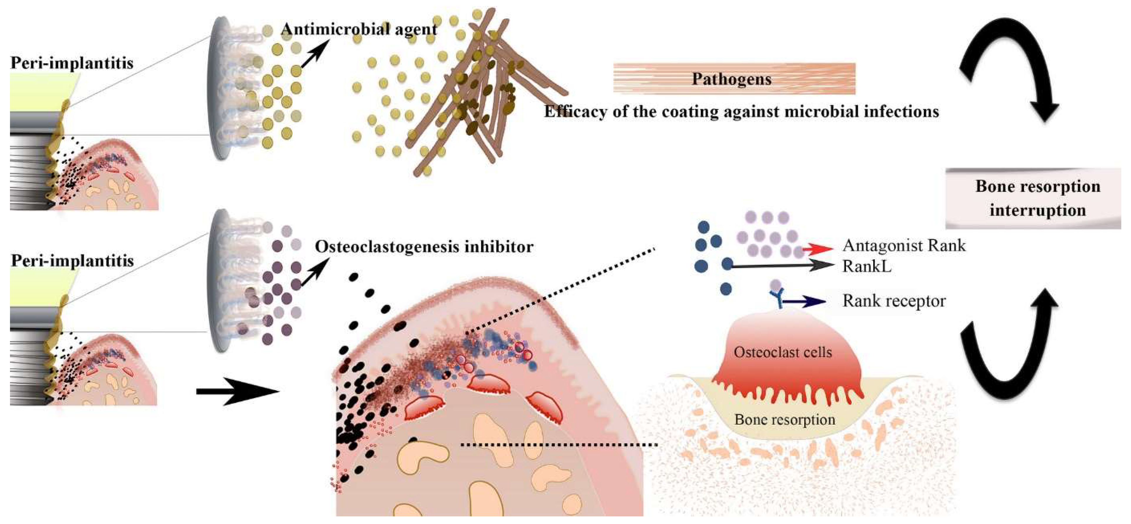
3.1. Implant Coating

In the realm of peri-implantitis prevention, researchers have delved into implant coating strategies. Hydrogels have been employed as coatings on implant surfaces to deter bacterial colonization without provoking inflammatory responses. For instance, when hydrogels were applied as coatings on titanium implants, whether unloaded or loaded with 2% vancomycin, no significant effects on bone apposition volume or timing in the vicinity of the implant site were observed. Notably, these coatings did not incite any inflammatory reactions in vivo [31]. Moreover, Min et al. demonstrated the development of a self-assembled, polymer-based conformal coating using a water-based layer-by-layer (LbL) approach. This coating serves a dual purpose as a biomimetic implant surface, providing controlled and sustained release of antibiotics followed by active release of growth factors for orthopedic implant applications. This multilayered coating comprises two components: a base osteoinductive portion containing bone morphogenetic protein-2 (rhBMP-2) and an antibacterial component containing gentamicin [32]. Physicochemical modifications of dental implants have been explored as a means to mitigate the adhesion of microorganisms, although complete prevention of peri-implantitis may not be achievable through these measures alone. Biomaterials have been utilized as carrier coatings for antimicrobial agents, supporting both the prevention and treatment of peri-implant mucositis and peri-implantitis. A layer-by-layer (LbL) hydrogel coating operates on the basis of electrostatic interactions between polyelectrolytes possessing opposing charges. This design enables the controlled release of antibacterial substances in response to the acidic environment generated by bacterial metabolic processes in peri-implantitis, Figure 2 [33].
Tao et al. introduced an innovative approach for creating an antibacterial coating on titanium (Ti) implants with pH-responsive properties to counteract the acidification of the local microenvironment induced by bacteria. This coating was synthesized by incorporating antibacterial drug-loaded nanoparticles (NPs) into gelatin and chitosan multilayers. It demonstrated remarkable antibacterial efficacy and contributed to enhanced osseointegration of the implants [34]. In another method, chitosan/plate-like hydroxyapatite (P-HAP) bilayers were successfully assembled on the titanium surface using the layer-by-layer technique. These modified surfaces exhibited compatibility with cells and significantly reduced the number of viable bacteria, as measured in colony-forming units, by a factor of 50 compared to plain titanium without the bilayer. However, it was observed that the bilayer modification did not affect osteoblast cell differentiation, despite its capacity to induce biomineralization [35]. A hydrogel precursor, capable of rapid crosslinking using commercially available dental curing systems, was developed to produce a hydrogel adhesive with the ability to adhere to both soft tissues (gingiva) and hard tissues (dental implants/bone). This engineered adhesive possesses high adhesion, mechanical stability, cytocompatibility, antimicrobial properties, biodegradability, and promotes bone regeneration. Overall, this antimicrobial hydrogel adhesive offers a minimally invasive platform for developing more effective therapeutic strategies against peri-implant diseases (PIDs) [36]. In a separate investigation, an anti-inflammatory drug, dexamethasone (DE), was incorporated into a hyaluronic acid (HA)-chitosan (CT) composite hydrogel system intended for peri-implantitis repair. The hydrogel featured uniform and spacious pores, approximately 160 µm in diameter on average, facilitating cell growth [37]. Additionally, Diniz et al. engineered a silver lactate-containing alginate hydrogel scaffold encapsulating gingival mesenchymal stem cells (GMSCs). This scaffold exhibited antimicrobial properties and enhanced the osteogenic differentiation of GMSCs in vitro. These innovative strategies hold promise for addressing peri-implant diseases and improving the success rates of dental implant procedures [38].

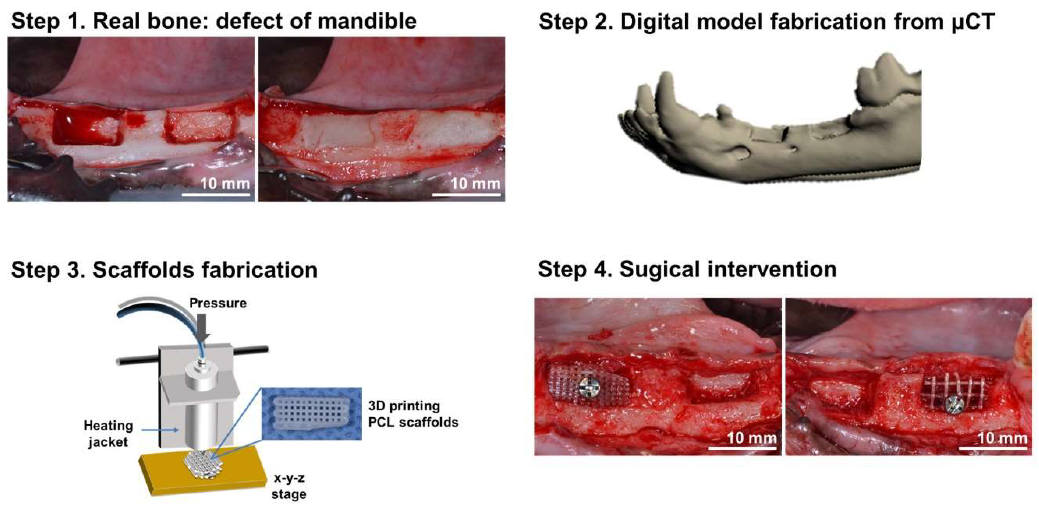
3.2. Bioprinted Scaffolds

Tissue regeneration entails a complex process, emphasizing the importance of scaffolds that closely mimic the natural extracellular matrix. In the context of peri-implantitis, bioprinting technology presents the opportunity to fabricate patient-specific scaffolds precisely tailored to fit around the implant. These scaffolds are typically composed of biocompatible materials, such as hydrogels or biodegradable polymers. Hydrogels, owing to their structural resemblance to the extracellular matrix, have gained significant popularity as scaffold materials in tissue engineering. They offer promising biomaterials in modern dental medicine due to their 3D network structures, high water content, excellent biocompatibility, and various bioactive properties. Hydrogels serve diverse purposes, including drug delivery platforms, antimicrobial materials, tissue regeneration scaffolds, and biosensors [39,40]. Natural and synthetic materials can be utilized to fabricate hydrogel scaffolds for tissue engineering. Examples of naturally derived polymers include agarose, alginate, collagen, chitosan, gelatin, hyaluronic acid, fibrin, and the decellularized extracellular matrix (dECM). Synthetic polymers like PLGA, PLA, and PCL are also extensively employed in dental tissue engineering, offering customizable mechanical properties, degradation rates, and biocompatibility. They can be processed into various forms, such as films, fibers, and porous scaffolds, using techniques like electrospinning, solvent casting, and 3D printing. Synthetic polymer scaffolds have demonstrated efficacy in regenerating various dental tissues, including dentin, periodontal ligaments, and alveolar bone [14,41,42,43].
Hydrogel-forming polymers are particularly suitable for use in bio-ink formulations due to their compatibility with cells, tunable flow behavior, responsiveness to stimuli, and ease of functionalization for 3D bioprinting. The polymer network within hydrogels facilitates crucial aspects of tissue development, including cell migration, adhesion, and proliferation, essential for functional tissue formation [44,45,46]. Additionally, hydrogel networks serve as effective drug delivery platforms owing to their high water retention capacity, enabling the incorporation of drugs or nanoparticles to confer antimicrobial properties and enhance regenerative potential. However, maintaining biocompatibility is crucial to avoid adverse effects [47,48].
Martin et al. introduced compelling research utilizing 3D printing to fabricate PLA scaffolds multifunctionalized with collagen (Col), minocycline (MH), and bioinspired citrate-hydroxyapatite nanoparticles (cHA). These scaffolds, referred to as PLA-Col-MH-cHA scaffolds, closely emulate native bone architecture with uniform macroporous structures, favorable wettability, and excellent compressive strength. Incorporating MH resulted in an antibiotic release profile conducive to local drug delivery therapy, effectively combating Staphylococcus aureus, a common pathogen associated with bone infections. Furthermore, human mesenchymal stem cells (hMSCs) demonstrated enhanced adhesion, proliferation, and osteogenesis-related gene expression when in contact with these scaffolds, particularly those incorporating cHA. The combination of cHA and MH elicited a synergistic effect, promoting increased osteogenic activity. These findings, coupled with their antibiofilm properties, suggest the suitability of these 3D-printed PLA-Col-MH-cHA scaffolds for future application in bone repair. By addressing both bone repair and infection prevention, these scaffolds offer an integrated approach to enhance the success rate of implanted bone devices. Furthermore, the utilization of 3D printing for bioinspired materials holds promise in constructing customized scaffolds incorporating multifunctional biocompatible compounds like collagen and hydroxyapatite. For instance, a novel 3D-printed polylactic acid (PLA) scaffold, functionalized with bioinspired surface coatings, demonstrates potential in enhancing the success rate of bone-implanted devices [49]. These surface coatings, composed of collagen, minocycline, and citrate-hydroxyapatite nanoparticles, aid in reducing bacterial biofilm formation. The scaffold not only provides 3D structural support with adjustable degradation rates but also facilitates drug release to promote cellular infiltration and mineralization. Additionally, the emerging field of 4D bioprinting offers opportunities to fabricate dynamic 3D-patterned biological architectures capable of changing shapes in response to various stimuli. This technology also supports the functional maturation of printed cell-laden constructs over time, offering unprecedented potential for bone tissue engineering. Tailoring the shape memory properties of printed structures to personalized bone defect repair needs, coupled with functional maturation processes, can promote the osteogenic differentiation of stem cells [50].
In the domain of bone tissue engineering (BTE), chitosan-based materials have gained prominence due to their low immunogenicity, biodegradability, bioresorbability, cost-effectiveness, and economic feasibility [51]. To address challenges associated with invasive surgeries required for clinical applications of implanted hydrogels, there is a rising demand for injectable hydrogels capable of non-invasively filling irregularly shaped defects [52,53,54]. Zhao et al. developed a composite hydrogel, CMCh-ACP, by combining carboxymethyl chitosan (CMCh) and amorphous calcium phosphate (ACP). These pH-responsive injectable hydrogels exhibit excellent biocompatibility, promoting cell adhesion, proliferation, upregulation of bone-related markers, and new bone regeneration [55]. Innovative hydrogel formulations also include electroactive hydrogels, enabling precise control over swelling, degradation rates, thermal properties, and conductivity, thus facilitating on-demand drug release through electrical stimuli [56]. Moreover, composite scaffolds containing embedded moxifloxacin hydrochloride exhibit sustained release of antibiotic drugs over several days, stimulating cell differentiation and osteoblast proliferation, thereby benefiting bone tissue formation in animal models [57]. Thermo-responsive composite hydrogels composed of chitosan, gelatin, and bioactive glass nanoparticles (nBG) effectively bond with living tissues, enhance osteoblast proliferation, and upregulate angiogenesis-related gene expressions, thereby contributing to bone regeneration [58].
In tissue engineering, the bioprinting of cell aggregates in scaffold-free spheroid structures has gained prominence. These spheroid structures can be strategically positioned to create complex tissue-engineered constructs, offering innovative possibilities for tissue engineering applications [59,60,61].

3.3. Cell-Seeded Constructs

Bioprinting technology enables precise control over cell placement within scaffolds, facilitating the strategic positioning of mesenchymal stem cells (MSCs) or other relevant cell types. MSCs exhibit the potential to differentiate into various tissue types crucial for regeneration, including bone, periodontal ligament, and soft tissue [62,63,64,65].
Identifying materials that effectively support bone regeneration is a significant concern in this field. Almansoori et al. proposed the use of a composite scaffold composed of poly(ε) caprolactone and β-tricalcium phosphate (PCL-TCP). This composite scaffold, chosen for its desirable attributes such as biocompatibility, bioresorbability, rigidity, and osteoconductivity, is well suited for guided bone regeneration. Additionally, the incorporation of mesenchymal stem cells (MSCs) and platelet-rich plasma (PRP) was anticipated to enhance the bone regeneration process. Results from the study demonstrated the compatibility of the PCL-TCP scaffold with bone regeneration, particularly in cases involving bone defects adjacent to dental implants. Furthermore, the inclusion of MSCs and PRP optimized bone regeneration by influencing scaffold replacement rates, the height of regenerated bone, and implant stability [66]. PCL scaffolds have exhibited excellent in vitro results and hold promise for various dental applications and regenerative therapies [67,68].
In another investigation, Park et al. examined the application of 3D-printed PCL scaffolds for alveolar bone augmentation utilizing a beagle defect model. Alveolar bone defects were intentionally created in this animal model, and wax was applied to maintain the defect volume during scaffold production. Computed tomography images of the animal were employed to generate a model of the defective mandible. Customized scaffolds were meticulously designed using computer-aided design (CAD) software and subsequently fabricated from PCL using 3D-bioprinting techniques. These tailored scaffolds were then implanted into the pre-formed defects, followed by a healing period of 3 months for assessment. This research illustrates the potential of 3D-printed scaffolds in alveolar bone augmentation and presents promising prospects for clinical applications in the dental field, Figure 3 [69].
Biomimetic scaffolds composed of chitosan and gelatin (CS/Gel) have garnered significant attention in tissue engineering across various tissue types. However, there exists a notable gap in our understanding regarding the potential of combining CS/Gel scaffolds with oral cells, particularly dental pulp stem cells (DPSCs), for the creation of customized constructs aimed at alveolar and orofacial bone reconstruction. These findings present a promising approach for the reconstruction of orofacial bone tissue [70].
Stem cells isolated from human exfoliated deciduous teeth (SHED) represent a prominent candidate due to their critical role in tissue engineering and regenerative medicine. Nakajima et al. conducted a study to elucidate the bone regeneration capabilities of SHED compared to human dental pulp stem cells (hDPSCs) and bone marrow mesenchymal stem cells (hBMSCs). Twelve weeks post-transplantation, the degree of bone regeneration achieved with SHED was nearly equivalent to that observed with hDPSCs and hBMSCs. The ratio of new bone formation relative to the pre-existing bone defect did not significantly differ among the SHED, hDPSCs, and hBMSCs groups. Histological evaluation revealed that SHED produced the largest amount of osteoid and widely distributed collagen fibers compared to the hDPSCs and hBMSCs groups. Thus, the transplantation of SHED demonstrated a sufficient capacity for bone defect repair and regeneration [71].
In the context of bone marrow-derived mesenchymal stem cell (BMMSC) maintenance, it is common practice to discard suspended cells. However, these suspended cells were found to possess osteogenic potential similar to that of anchorage-dependent BMMSCs. When transferred and cultured on an extracellular matrix (ECM)-coated culture plate, the suspended cells exhibited higher colony-forming unit-fibroblast (CFU-f) formation and similar proliferation capabilities compared to BMMSCs. Furthermore, these cells demonstrated the ability to undergo osteogenic, adipogenic, and chondrogenic differentiation. When assessing attachment, survival, and proliferation on titanium implant discs, the suspended cells exhibited levels similar to or higher than those of BMMSCs. Both the suspended cells and BMMSCs displayed enhanced bone formation ability in the upper and lower canals of implants relative to controls in a rabbit tibia model with double-canaled implants. This study underscores that suspended cells obtained after primary BMMSC isolation possess bone regeneration capacity akin to BMMSCs, not only in vitro but also in vivo. The ECM was found to be valuable for propagating MSCs for cell-based bone regeneration, suggesting that suspended cells could be valuable tools for bone regeneration following implant surgery. These findings open up new possibilities for harnessing the regenerative potential of suspended cells in the context of bone tissue engineering [72].
Recent research has shed light on the role of Tribbles homolog 3 (Trb3), a member of the Tribbles family of pseudokinases, in regulating the differentiation fate of mesenchymal stem cells (MSCs) and its implications for bone formation. Abnormal lineage commitment of MSCs in the bone marrow can lead to aberrant bone formation, characterized by reduced osteogenic potential and increased adipogenic potential. While major transcription factors associated with lineage differentiation have been identified, the molecular switch determining MSC fate and its role in skeletal regeneration remain elusive, impeding the development of effective therapies. In a recent study, researchers investigated how Trb3 reciprocally regulates MSC differentiation into osteoblasts and adipocytes and elucidated the underlying mechanisms. They found that Trb3 promotes MSC commitment to the osteoblastic lineage while suppressing adipocyte differentiation. Mechanistically, Trb3 influences MSC fate determination through the BMP/Smad and Wnt/β-catenin signaling pathways. Importantly, in vivo experiments utilizing a novel scaffold named gelatin-conjugated caffeic acid-coated apatite/PLGA (GelCA-PLGA) for local Trb3 delivery demonstrated robust bone regeneration and inhibition of fat-filled cyst formation in rodent models with non-healing mandibular defects. These findings underscore the potential of Trb3-based therapeutic strategies to enhance osteoblastogenesis over adipogenesis, thus improving skeletal regeneration and offering promise for treating bone-loss diseases. The scaffold-mediated local gene transfer approach presents a unique avenue for targeting specific therapeutic genes related to lineage commitment in clinical bone treatments. This research paves the way for advancements in bone regeneration treatments and provides insights into potential therapies for bone-loss diseases, offering exciting possibilities for future clinical applications [73].

3.4. Growth Factors and Bioactive Molecules

The integration of growth factors and bioactive molecules into bioprinted constructs holds significant promise for enhancing tissue regeneration, particularly in the context of bone repair [74,75,76]. Bone morphogenetic proteins (BMPs) and vascular endothelial growth factor (VEGF) are among the key factors utilized for their ability to stimulate osteogenesis and angiogenesis, respectively, which are crucial processes in bone healing and regeneration [77].
BMPs, a group of growth factors with diverse roles in tissue development and regeneration, are effective in promoting osteogenesis. By precisely controlling their spatial distribution within bioprinted scaffolds, bioprinting technology optimizes their therapeutic effects. Strategies involving BMPs focus on enhancing BMP receptor binding affinity, upregulating BMP receptor expression, and inhibiting BMP antagonists, thus promoting osteogenic activity [78].
Incorporating genetically modified or pre-treated mesenchymal stem cells (MSCs) into scaffolds represents another approach to enhance tissue regeneration. For instance, hydrogels containing fibrin and plasmonic gold nanoparticles combined with MSCs expressing BMP-2 have shown promising results in facilitating the formation of new mineralized bone tissue [79]. Recombinant human bone morphogenetic protein-2 (rhBMP-2) has demonstrated potential in maxillofacial surgery due to its osteoinductive properties. However, challenges such as identifying an ideal carrier, addressing the high cost of rhBMP-2, and optimizing dosing regimens remain. Despite its potential, rhBMP-2 has yet to become the standard in clinical practice, mainly due to safety and cost concerns [80].
Bone tissue remodeling, orchestrated by osteoclasts and osteoblasts, is regulated by various local and systemic factors, including growth factors and cytokines. Osteocytes, acting as mechanosensors, play a pivotal role in regulating and coordinating the bone remodeling process. This intricate process is under the influence of both local factors like growth factors and cytokines, as well as systemic factors such as calcitonin and estrogens. Together, these factors contribute to maintaining bone homeostasis. Differentiation of bone cells can be guided and facilitated in several ways, including the direct transplantation of stem cells to the site of a bone defect, where they can respond to signals from the local microenvironment. Biomaterials can deliver signals that promote bone regeneration, such as calcium phosphate-based materials and controlled-release osteogenic growth factors, integrated into osteogenic scaffolds [81,82].
In a specific study, a collagen scaffold loaded with Erythropoietin (EPO) significantly enhanced osseous regeneration in alveolar defects compared to bone xenograft or a collagen membrane alone. Further analysis revealed that EPO modulated extracellular matrix protein expression through the vascular endothelial growth factor (VEGF) pathway, even in the absence of blood vessels. This underscores the effectiveness of EPO-impregnated collagen scaffolds for bone regeneration, facilitating rapid extracellular matrix production and osseoinduction near new capillaries via VEGF signaling [83].
Gene delivery techniques offer promising avenues for promoting tissue regeneration, particularly in the context of bone tissue engineering. Malek-Khatabi et al. utilized microfluidic-assisted synthesis to create plasmid DNA (pDNA)-based chitosan nanocomplex platforms encoding human BMP-2 (Bone Morphogenetic Protein 2). These nanocomplexes were then immobilized on a nanofibrous PCL scaffold functionalized with metalloprotease-sensitive peptides. In a rat calvarial defect model, implantation of mesenchymal stem cells (MSCs) into these loaded PCL membranes significantly increased regenerated bone volume and induced the formation of denser bone-like structures [84].
However, despite the therapeutic potential of BMP-2, its clinical use is accompanied by challenges and concerns. The increased utilization of BMP-2 has led to a well-documented side effect profile, including postoperative inflammation, ectopic bone formation, osteoclast-mediated bone resorption, and inappropriate adipogenesis. Serious adverse effects such as tumor growth, hypercalcemia, and jaw osteonecrosis have also been reported [85]. Typically, BMP-2 is combined solely with collagen as a carrier material in clinical applications [86]. To address these concerns and optimize the therapeutic potential of BMP-2, researchers are exploring innovative approaches to better control its presentation. Inspired by the natural cellular environment, material surfaces are being engineered to provide both physical and chemical cues that regulate BMP-2 activity. These efforts aim to mitigate side effects and enhance therapeutic efficacy, representing ongoing advancements in bone tissue engineering and regenerative medicine [87].

3.5. Vascularization

Vascularization is a critical aspect of tissue engineering, ensuring that newly formed tissues receive the necessary nutrients and oxygen for survival and regeneration. Bioprinting technology offers a promising solution by enabling the creation of vascular networks within tissue scaffolds, overcoming the diffusion limit for cell survival in engineered tissues [88,89]. To achieve the correct shape of the tissue construct, clinical imaging data are first translated into a computer model of the anatomical defect. This model is then converted into a program that controls the motion of the bioprinter nozzles, which dispense cells to precise locations. The incorporation of microchannels into these tissue constructs facilitates the diffusion of nutrients to the printed cells, overcoming the diffusion limit of 100–200 μm for cell survival in engineered tissues [90,91,92]. The ultimate goal is to create vessels that closely resemble the endogenous vascular system in terms of biocompatibility, physiological flow rates, and the ability to withstand systemic pressure changes [93,94].
A fibrin-based bioink has been employed to facilitate the development of a stable primitive vascular network in vitro. Establishing microvessels within bioprinted tissues in vitro before implantation has been shown to enhance vascularization once the tissue is implanted in vivo [95]. For instance, a study by Kang et al. reported that 3D-bioprinted constructs containing human amniotic fluid stem cells (hAFSCs) exhibited robust vascularized bone tissue formation throughout the implants, including at the central portion. In contrast, non-treated and scaffold-only constructs showed limited vascularization and minimal bone tissue formation at the periphery of the implant. The study demonstrated the successful generation of mature and vascularized bone tissue [96]. Another study by Kuss et al. involved the 3D bioprinting of stromal vascular fraction-derived cells (SVFCs) within hydrogel bio-inks and conditioning the constructs in either normoxia or hypoxia. Short-term hypoxic conditioning was found to promote microvessel formation in vitro and in vivo, as well as integration with existing host vasculature. It did not, however, affect the osteogenic differentiation of SVFCs. These findings highlight the potential of short-term hypoxia and 3D bioprinting for generating prevascularized 3D-bioprinted bone constructs [97]. Furthermore, Anada et al. developed a two-step digital light processing technique to fabricate a bone-mimetic 3D hydrogel construct comprising octacalcium phosphate (OCP), spheroids of human umbilical vein endothelial cells (HUVEC), and gelatin methacrylate (GelMA) hydrogels. This bone-mimetic construct consists of a peripheral OCP-containing GelMA ring mimicking the cortical shell and a central GelMA ring containing HUVEC spheroids to mimic the bone marrow space. OCP, evenly embedded in GelMA, stimulates the osteoblastic differentiation of mesenchymal stem cells. The study showed that the concentration of GelMA modulates the formation of capillary-like structures originating from the HUVEC spheroids within the construct [98].
Finally, it is important to note that bone morphogenetic proteins (BMPs) play a role in regulating angiogenesis and the secretion of vascular endothelial growth factor (VEGF), which is a key regulator of physiological angiogenesis during embryogenesis, skeletal growth, and reproductive functions [99,100].

3.6. Immunomodulation

Peri-implantitis involves an inflammatory response, and bioprinted constructs can be strategically designed to include immunomodulatory factors that help regulate the immune response. This approach aims to reduce inflammation and promote tissue healing. However, it is important to recognize that 3D bioprinting introduces the concept of bioinks containing biomaterials that may trigger immune responses in the body. The foreign body response (FBR) is a complex process involving various cell types such as B-cells, dendritic cells, macrophages, natural killer cells, neutrophils, and T-cells, along with molecular signals like antibodies (Abs), cytokines, and reactive radical species. Typically, biomaterials are shielded by fibrous encapsulation, a process regulated by molecular signals. Therefore, assessing the immune response against biomaterials used in 3D-printed organs is crucial to mitigate tissue rejection following transplantation [101,102,103]. The field of regenerative medicine faces challenges related to the host response to biomaterial implantation, despite promising technologies for recovering damaged organs and tissues. The success of implants and tissue regeneration is determined by cellular and molecular events at the interface between the foreign body and the host, which involves both innate and adaptive immune responses. To prevent adverse events, the current state of the art suggests the use of immunomodulatory biomaterials and their knowledge-based application to reduce neutrophil activation, optimize macrophage polarization from M1 to M2, shift the balance from Th1 to Th2 lymphocytes, and induce regulatory T cells [104,105]. Recent developments in biomaterials and our understanding of inflammatory and fibrotic pathologies suggest several biological solutions in the context of neural interfaces and the foreign body response (FBR). These solutions include modifications of the interface surface, such as organic and synthetic coatings; the use of specific drugs or molecular biology tools to target the microenvironment around the interface; and the development of bioengineered scaffolds to reduce immune responses and promote integration with surrounding tissue [106]. For instance, research by Hotchkiss et al. demonstrated how macrophage responses to material surface properties can influence the adaptive immune response by altering T-helper cell populations and stem cell recruitment. Surface modifications applied to titanium implants to increase surface roughness and wettability were shown to polarize the adaptive immune response toward a Th2, pro-wound healing phenotype. This led to faster resolution of inflammation and increased stem cell recruitment around rough hydrophilic implants with the presence of macrophages [107]. In the context of biomaterial interactions, the recruitment of platelets and monocytes is followed by monocyte differentiation into macrophages, which adhere to biomaterials along with platelets and monocytes. Subsequently, macrophages activate to form giant cells and, along with monocytes and platelets, secrete cytokines and chemokines to recruit fibroblasts or mesenchymal stem cells (MSCs). Finally, fibroblasts or MSCs contribute to the formation of lymphoblastic tissues surrounding the biomaterial surface and mediate the development of capillary beds and collagen accumulation [108,109].
The host immune system can significantly impact mesenchymal stem cell (MSC)-mediated bone tissue regeneration. However, the therapeutic potential of hydrogel biomaterials in modulating the interaction between MSCs and T-lymphocytes is not well understood. Recent research has shown that encapsulating MSCs in hydrogel has a notable effect on this interaction when used for implantation. The hydrogel encapsulation hinders the penetration of pro-inflammatory cells and/or cytokines, leading to improved viability of the encapsulated MSCs. This counteracts the detrimental effects of the host’s pro-inflammatory T-lymphocytes, which can reduce MSC viability by activating the CASPASE-3 and CASPASE-8-associated proapoptotic cascade, ultimately leading to MSC apoptosis. To further support the rescue of engrafted MSCs from the host immune system’s assault, the hydrogel can be loaded with the anti-inflammatory drug indomethacin. This additional step regulates the local microenvironment and prevents pro-inflammatory cytokine-induced apoptosis. These findings suggest that encapsulating hydrogel has the potential to modulate the interaction between MSCs and the host immune cells, ultimately influencing the fate of implanted MSCs and enhancing tissue regeneration [110].
Apart from the use of innovative biomaterials, pre-implantation assessment of nutrient and vitamin levels can also be crucial, especially for patients with reduced or impaired bone metabolism, in order to prevent postoperative complications. Vitamin D deficiency, for example, increases the risk of wound infections and may delay wound healing. Research has demonstrated a correlation between low vitamin D levels and early implant loss [111]. Vitamin C is also essential, as it is required for the formation and cross-linking of collagen, a critical component of wound tissue. Given the key role of vitamin D in bone metabolism and the anti-inflammatory potential of vitamin C, it is clear that an adequate vitamin supply can positively influence tissue regeneration and implant healing [112,113].
Table 1 presents a synthesis of the scientific literature, highlighting the most significant directions in peri-implantitis tissue regeneration. This table likely provides a summary of key findings and research directions in the field, making it a valuable reference for readers looking for an overview of the current state of research in this area.

4. Discussion

Tissue engineering is indeed a rapidly advancing field that combines various elements like materials, living cells, and growth factors to facilitate the repair or regeneration of damaged or lost tissues [114].
In the context of bioprinting, developing suitable polymeric materials is a significant challenge. These materials need to possess the right rheological properties (flow behavior), biocompatibility, bioactivity, and mechanical strength. The complexity arises from the need to ensure that the biomaterial can maintain its structural integrity during the printing process and provide a supportive environment for cell viability, proliferation, and differentiation [42,62]. For instance, 3D printing using bioinspired materials, such as collagen and hydroxyapatite, has emerged as a powerful technique for constructing customized scaffolds with multifunctional biocompatible properties. An example includes 3D-printed poly(lactic acid) (PLA) scaffolds with bioinspired surface coatings, which have shown promise in improving the success rates of bone-implanted devices [49].
While chitosan-based hydrogels hold potential as alternatives for bone defect repair in clinical applications due to their beneficial properties, they also have some limitations. One significant limitation is the standardization of factors like molecular weight distribution, raw material resources, and commercial production. High molecular weight chitosan may lead to potential inflammation when used as a bone graft material. Additionally, the source of chitosan, whether from cell walls or seafood, can impact reliability and reproducibility. Furthermore, chitosan-based hydrogels for bone scaffolds are still in the early stages of experimental research, with limited studies and insufficient clinical utility. Future research should focus on establishing uniform standards and developing advanced technologies to expand potential clinical applications. There is also a need for improvement in the loading capacity and controlled release of bioactive molecules within chitosan-based hydrogels to enhance their clinical utility upon implantation. Clinical research efforts are also exploring the combination of chitosan-based hydrogels with different biofabrication techniques like electrospinning, microspheres, and 3D printing to create multifaceted and multilayered bone grafts for bone regeneration [51].
Enriching scaffolds with biomolecules like bone morphogenetic proteins (BMPs) is a recognized approach to enhance bone regeneration [77]. Growth factors, including BMPs, have the potential to significantly improve the effectiveness and speed of bone regeneration and repair in tissue engineering applications related to bones [82].
Unseeded scaffolds, meaning scaffolds without cells, have limited regenerative potential. To improve the bone regenerative potential of scaffolds enriched with mesenchymal stem cells (MSCs), researchers often combine them with biomolecules like BMPs or modify biomaterial features, such as pore dimensions. Many studies employ composite scaffolds or biomaterials with surface modifications in combination with MSCs to enhance tissue regeneration, especially in bone repair scenarios [74].
Vascularization is a crucial factor in the bone formation and regeneration process. Developing functional vasculature to enhance the survival and integration of tissue-engineered bone substitutes remains a significant challenge [88]. By integrating tissue engineering, material science, and genetic engineering, it may be possible to create structures that stimulate early vascularization, restore blood flow, and prevent cell death within scaffolds. This approach could accelerate the clinical translation of scaffolds for repairing large bone defects [115]. Additionally, other recently introduced compounds have been demonstrated to have a significant influence on the oral environment. The use of lysates [116] and postbiotics [117] can modify Clinical and Microbiological Parameters in periodontal patients, so these products should also be considered in future trials as adjuvants to bioprinted frameworks to manage peri-implantitis tissue regeneration.
In the context of dental tissue engineering and 3D bioprinting, significant progress has been made in pre-clinical research. The process typically involves 3D modeling of dental defects, isolation and differentiation of stem cells into dental tissue-specific cells, bioink preparation, bioprinting of the desired structure, and any necessary architectural reconfiguration or chemical functionalization before implantation [118].
Looking ahead, the future of bioprinting in dental tissue engineering is likely to focus on personalized medicine. Dental tissues can exhibit diverse injury and damage geometries, varying from patient to patient. Therefore, the combination of bioprinting technologies with advanced imaging systems can enable the creation of patient-specific dental constructs tailored to individual anatomical and functional requirements [42].
A major challenge in dental implantology is ensuring that new material concepts not only guarantee cost-effective and automated production processes but also maintain implant compatibility while fulfilling defect-filling functionality [113].

5. Conclusions and Future Perspectives

Bioprinting holds significant promise for regenerating peri-implant tissues in cases of peri-implantitis, offering several advantages such as real-time monitoring, customization to patient needs, and adaptability to evolving conditions. However, it is important to recognize that bioprinting for peri-implantitis treatment is still in the experimental phase. Further refinement of techniques and long-term clinical studies are needed to establish its efficacy and safety. Collaboration among researchers, clinicians, and bioengineers is crucial to tailor bioprinting methods specifically for peri-implantitis treatment. Thorough research and preclinical trials are essential before bioprinting can be clinically applied for peri-implantitis. Regulatory approvals from health authorities will also be necessary to validate its use in patient care. The translation of bioprinting from research to clinical application is a complex journey requiring careful development and validation processes. While it represents an exciting frontier in regenerative medicine, patience and commitment are needed to ensure the best outcomes for patients with peri-implantitis.

6. Limitations

One limitation of the present article is that the scope of analysis was constrained due to insufficient data from existing studies and clinical trials. The heterogeneity in study designs, as well as the materials and bioprinting techniques applied, make it challenging to draw generalized conclusions or recommendations. Such variations limited the ability to formulate definitive conclusions or endorse specific bioprinting techniques and methodologies over others. The influence of subjective factors could significantly skew results. A critical gap in the literature is the absence of comprehensive scientific evidence of bioprinting methods regarding the long-term outcomes of peri-implantitis treatments. Furthermore, the rapid pace of innovation in bioprinting technologies and methods might mean that very recent studies have not been included.

Author Contributions

Conceptualization, A.M., A.Y. and D.B.; methodology, D.S. and M.D.-G.; software, A.M.; validation, D.S. and D.B.; formal analysis, A.M. and A.Y.; investigation, D.B., A.M. and M.D.-G.; resources, D.S. and D.B.; data curation, D.S. and A.M.; writing—original draft preparation, D.S. and D.B.; writing—review and editing, A.M. and A.Y.; visualization, A.Y. and D.S.; supervision, D.B. and A.Y.; project administration, A.Y. and M.D.-G.; funding acquisition, A.M. All authors have read and agreed to the published version of the manuscript.

Funding

This research was funded by the Medical University of Plovdiv, Bulgaria, grant number PMD 01/2022.

Institutional Review Board Statement

Not applicable.

Informed Consent Statement

Not applicable.

Data Availability Statement

Not applicable.

Conflicts of Interest

The authors declare no conflicts of interest.

References

  1. Mohd, N.; Razali, M.; Ghazali, M.J.; Abu Kasim, N.H. Current Advances of Three-Dimensional Bioprinting Application in Dentistry: A Scoping Review. Materials 2022, 15, 6398. [Google Scholar] [CrossRef]
  2. Sigaux, N.; Pourchet, L.; Breton, P.; Brosset, S.; Louvrier, A.; Marquette, C.A. 3D Bioprinting: Principles, fantasies and prospects. J. Stomatol. Oral Maxillofac. Surg. 2019, 120, 128–132. [Google Scholar] [CrossRef]
  3. Melchels, F.P.; Domingos, M.A.; Klein, T.J.; Malda, J.; Bartolo, P.J.; Hutmacher, D.W. Additive manufacturing of tissues and organs. Prog. Polym. Sci. 2012, 37, 1079–1104. [Google Scholar] [CrossRef]
  4. Jang, J.; Yi, H.G.; Cho, D.W. 3D printed tissue models: Present and future. ACS Biomater. Sci. Eng. 2016, 2, 1722–1731. [Google Scholar] [CrossRef]
  5. Guvendiren, M.; Molde, J.; Soares, R.M.; Kohn, J. Designing biomaterials for 3D printing. ACS Biomater. Sci. Eng. 2016, 2, 1679–1693. [Google Scholar] [CrossRef]
  6. Yu, J.; Park, S.A.; Kim, W.D.; Ha, T.; Xin, Y.-Z.; Lee, J.; Lee, D. Current Advances in 3D Bioprinting Technology and Its Applications for Tissue Engineering. Polymers 2020, 12, 2958. [Google Scholar] [CrossRef]
  7. Gaharwar, A.K.; Arpanaei, A.; Andresen, T.L.; Dolatshahi-Pirouz, A. 3D biomaterial microarrays for regenerative medicine: Current state-of-the-art, emerging directions and future trends. Adv. Mater. 2016, 28, 771–781. [Google Scholar] [CrossRef]
  8. Hölzl, K.; Lin, S.; Tytgat, L.; Van Vlierberghe, S.; Gu, L.; Ovsianikov, A. Bioink properties before, during and after 3D bioprinting. Biofabrication 2016, 8, 032002. [Google Scholar] [CrossRef]
  9. Askari, M.; Naniz, M.A.; Kouhi, M.; Saberi, A.; Zolfagharian, A.; Bodaghi, M. Recent progress in extrusion 3D bioprinting of hydrogel biomaterials for tissue regeneration: A comprehensive review with focus on advanced fabrication techniques. Biomater. Sci. 2021, 9, 535–573. [Google Scholar] [CrossRef]
  10. Jazayeri, H.E.; Fahmy, M.D.; Razavi, M.; Stein, B.E.; Nowman, A.; Masri, R.M.; Tayebi, L. Dental applications of natural-origin polymers in hard and soft tissue engineering. J. Prosthodont. 2016, 25, 510–517. [Google Scholar] [CrossRef]
  11. Robertson, K.; Shahbazian, T.; MacLeod, S. Treatment of peri-implantitis and the failing implant. Dent. Clin. 2015, 59, 329–343. [Google Scholar] [CrossRef]
  12. Liaw, K.; Delfini, R.H.; Abrahams, J.J. Dental implant complications. Semin. Ultrasound CT MRI 2015, 36, 427–433. [Google Scholar] [CrossRef]
  13. Schwarz, M.S. Mechanical complications of dental implants. Clin. Oral Implant. Res. 2000, 11, 156–158. [Google Scholar] [CrossRef]
  14. Mistry, S.; Pal, K.; Shete, O.; Sawant, D. Hydrogels in Dentistry–Applications and Advances. J. Prosthodont. Dent. Mater. 2022, 3, 13–25. [Google Scholar]
  15. Lang, N.P.; Wilson, T.G.; Corbet, E.F. Biological complications with dental implants: Their prevention, diagnosis and treatment Note. Clin. Oral Implant. Res. 2000, 11, 146–155. [Google Scholar] [CrossRef]
  16. Sakka, S.; Baroudi, K.; Nassani, M.Z. Factors associated with early and late failure of dental implants. J. Investig. Clin. Dent. 2012, 3, 258–261. [Google Scholar] [CrossRef]
  17. Schwartz-Arad, D.; Samet, N.; Samet, N.; Mamlider, A. Smoking and complications of endosseous dental implants. J. Periodontol. 2002, 73, 153–157. [Google Scholar] [CrossRef]
  18. McDermott, N.E.; Chuang, S.K.; Woo, V.V.; Dodson, T.B. Complications of dental implants: Identification, frequency, and associated risk factors. Int. J. Oral Maxillofac. Implant. 2003, 18, 846. [Google Scholar] [CrossRef]
  19. Kasat, V.; Ladda, R. Smoking and dental implants. J. Int. Soc. Prev. Community Dent. 2012, 2, 38. [Google Scholar] [CrossRef]
  20. Smeets, R.; Henningsen, A.; Jung, O.; Heiland, M.; Hammächer, C.; Stein, J.M. Definition, etiology, prevention and treatment of peri-implantitis—A review. Head Face Med. 2014, 10, 34. [Google Scholar] [CrossRef]
  21. Manfredini, D.; Poggio, C.E.; Lobbezoo, F. Is bruxism a risk factor for dental implants? A systematic review of the literature. Clin. Implant Dent. Relat. Res. 2014, 16, 460–469. [Google Scholar] [CrossRef]
  22. Renvert, S.; Roos-Jansåker, A.M.; Claffey, N. Non-surgical treatment of peri-implant mucositis and peri-implantitis: A literature review. J. Clin. Periodontol. 2008, 35, 305–315. [Google Scholar] [CrossRef]
  23. Sahm, N.; Becker, J.; Santel, T.; Schwarz, F. Non-surgical treatment of peri-implantitis using an air-abrasive device or mechanical debridement and local application of chlorhexidine: A prospective, randomized, controlled clinical study. J. Clin. Periodontol. 2011, 38, 872–878. [Google Scholar] [CrossRef]
  24. Del Amo, F.S.L.; Yu, S.H.; Wang, H.L. Non-surgical therapy for peri-implant diseases: A systematic review. J. Oral Maxillofac. Res. 2016, 7, e13. [Google Scholar]
  25. Choe, R.; Balhaddad, A.A.; Fisher, J.P.; Melo, M.A.S.; Huang, H.C. Photodynamic therapy for biomodulation and disinfection in implant dentistry: Is it feasible and effective? Photochem. Photobiol. 2021, 97, 916–929. [Google Scholar] [CrossRef]
  26. Schou, S.; Berglundh, T.; Lang, N.P. Surgical treatment of peri-implantitis. Int. J. Oral Maxillofac. Implant. 2004, 19, 140. [Google Scholar]
  27. Claffey, N.; Clarke, E.; Polyzois, I.; Renvert, S. Surgical treatment of peri-implantitis. J. Clin. Periodontol. 2008, 35, 316–332. [Google Scholar] [CrossRef]
  28. Sarkis-Onofre, R.; Catalá-López, F.; Aromataris, E.; Lockwood, C. How to properly use the PRISMA Statement. Syst. Rev. 2021, 10, 117. [Google Scholar] [CrossRef]
  29. Mancini, L.; Romandini, M.; Fratini, A.; Americo, L.M.; Panda, S.; Marchetti, E. Biomaterials for Periodontal and Peri-Implant Regeneration. Materials 2021, 14, 3319. [Google Scholar] [CrossRef]
  30. Ostrovidov, S.; Ramalingam, M.; Bae, H.; Orive, G.; Fujie, T.; Shi, X.; Kaji, H. Bioprinting and biomaterials for dental alveolar tissue regeneration. Front. Bioeng. Biotechnol. 2023, 11, 991821. [Google Scholar] [CrossRef]
  31. Boot, W.; Gawlitta, D.; Nikkels, P.G.J.; Pouran, B.; van Rijen, M.H.P.; Dhert, W.J.A.; Vogely, C.H. Hyaluronic Acid-Based Hydrogel Coating Does Not Affect Bone Apposition at the Implant Surface in a Rabbit Model. Clin. Orthop. Relat. Res. 2017, 475, 1911–1919. [Google Scholar] [CrossRef]
  32. Min, J.; Braatz, R.D.; Hammond, P.T. Tunable staged release of therapeutics from layer-by-layer coatings with clay interlayer barrier. Biomaterials 2014, 35, 2507–2517. [Google Scholar] [CrossRef]
  33. de Avila, E.D.; van Oirschot, B.A.; van den Beucken, J.J. Biomaterial-based possibilities for managing peri-implantitis. J. Periodontal Res. 2020, 55, 165–173. [Google Scholar] [CrossRef]
  34. Tao, B.; Zhao, W.; Lin, C.; Yuan, Z.; He, Y.; Lu, L.; Chen, M.; Ding, Y.; Yang, Y.; Xia, Z.; et al. Surface modification of titanium implants by ZIF-8@ Levo/LBL coating for inhibition of bacterial-associated infection and enhancement of in vivo osseointegration. Chem. Eng. J. 2020, 390, 124621. [Google Scholar] [CrossRef]
  35. Govindharajulu, J.P.; Chen, X.; Li, Y.; Rodriguez-Cabello, J.C.; Battacharya, M.; Aparicio, C. Chitosan-Recombinamer Layer-by-Layer Coatings for Multifunctional Implants. Int. J. Mol. Sci. 2017, 18, 369. [Google Scholar] [CrossRef]
  36. Sani, E.S.; Lara, R.P.; Aldawood, Z.; Bassir, S.H.; Nguyen, D.; Kantarci, A.; Intini, G.; Annabi, N. An antimicrobial dental light curable bioadhesive hydrogel for treatment of peri-implant diseases. Matter 2019, 1, 926–944. [Google Scholar] [CrossRef]
  37. Zhou, Z.; Zhang, Q.; Wang, Y. Preparation and characterization of antibacterial and anti-inflammatory hyaluronic acid-chitosan-dexamethasone hydrogels for peri-implantitis repair. J. Biomater. Appl. 2022, 36, 1141–1150. [Google Scholar] [CrossRef]
  38. Diniz, I.M.; Chen, C.; Ansari, S.; Zadeh, H.H.; Moshaverinia, M.; Chee, D.; Marques, M.M.; Shi, S.; Moshaverinia, A. Gingival mesenchymal stem cell (GMSC) Delivery system based on RGD-coupled alginate hydrogel with antimicrobial properties: A novel treatment modality for peri-implantitis. J. Prosthodont. 2016, 25, 105–115. [Google Scholar] [CrossRef]
  39. Chen, W.; Zhang, C.; Peng, S.; Lin, Y.; Ye, Z. Hydrogels in dental medicine. Adv. Ther. 2023, 7, 2300128. [Google Scholar] [CrossRef]
  40. Ayala-Ham, A.; López-Gutierrez, J.; Bermúdez, M.; Aguilar-Medina, M.; Sarmiento-Sánchez, J.I.; López-Camarillo, C.; Sanchez-Schmitz, G.; Ramos-Payan, R. Hydrogel-based scaffolds in oral tissue engineering. Front. Mater. 2021, 8, 708945. [Google Scholar] [CrossRef]
  41. Yang, J.; Sun, X.; Zhang, Y.; Chen, Y. The application of natural polymer–based hydrogels in tissue engineering. In Hydrogels Based on Natural Polymers; Elsevier: Amsterdam, The Netherlands, 2020; pp. 273–307. [Google Scholar]
  42. Kim, S.; Hwangbo, H.; Chae, S.; Lee, H. Biopolymers and Their Application in Bioprinting Processes for Dental Tissue Engineering. Pharmaceutics 2023, 15, 2118. [Google Scholar] [CrossRef] [PubMed]
  43. Sordi, M.B.; Cruz, A.; Fredel, M.C.; Magini, R.; Sharpe, P.T. Three-dimensional bioactive hydrogel-based scaffolds for bone regeneration in implant dentistry. Mater. Sci. Eng. 2021, 124, 112055. [Google Scholar] [CrossRef] [PubMed]
  44. Dorishetty, P.; Dutta, N.K.; Choudhury, N.R. Bioprintable tough hydrogels for tissue engineering applications. Adv. Colloid Interface Sci. 2020, 281, 102163. [Google Scholar] [CrossRef] [PubMed]
  45. Advincula, R.C.; Dizon, J.R.C.; Caldona, E.B.; Viers, R.A.; Siacor, F.D.C.; Maalihan, R.D.; Espera, A.H. On the progress of 3D-printed hydrogels for tissue engineering. MRS Commun. 2021, 11, 539–553. [Google Scholar] [CrossRef] [PubMed]
  46. Xu, F.; Dawson, C.; Lamb, M.; Mueller, E.; Stefanek, E.; Akbari, M.; Hoare, T. Hydrogels for tissue engineering: Addressing key design needs toward clinical translation. Front. Bioeng. Biotechnol. 2022, 10, 849831. [Google Scholar] [CrossRef]
  47. Atila, D.; Kumaravel, V. Advances in Antimicrobial Hydrogels for Dental Tissue Engineering: Regenerative Strategies for Endodontics and Periodontics. Biomater. Sci. 2023, 11, 6711–6747. [Google Scholar] [CrossRef] [PubMed]
  48. Farzin, A.; Bahrami, N.; Mohamadnia, A.; Mousavi, S.; Gholami, M.; Ai, J.; Moayeri, R.S. Scaffolds in dental tissue engineering: A review. Arch. Neurosci. 2020, 7, e97014. [Google Scholar] [CrossRef]
  49. Martin, V.; Ribeiro, I.A.; Alves, M.M.; Gonçalves, L.; Claudio, R.A.; Grenho, L.; Fernandes, M.H.; Gomes, P.; Santos, C.F.; Bettencourt, A.F. Engineering a multifunctional 3D-printed PLA-collagen-minocycline-nanoHydroxyapatite scaffold with combined antimicrobial and osteogenic effects for bone regeneration. Mater. Sci. Eng. 2019, 101, 15–26. [Google Scholar] [CrossRef] [PubMed]
  50. Wan, Z.; Zhang, P.; Liu, Y.; Lv, L.; Zhou, Y. Four-dimensional bioprinting: Current developments and applications in bone tissue engineering. Acta Biomater. 2020, 101, 26–42. [Google Scholar] [CrossRef]
  51. Tang, G.; Tan, Z.; Zeng, W.; Wang, X.; Shi, C.; Liu, Y.; He, H.; Chen, R.; Ye, X. Recent advances of chitosan-based injectable hydrogels for bone and dental tissue regeneration. Front. Bioeng. Biotechnol. 2020, 8, 587658. [Google Scholar] [CrossRef]
  52. Li, D.; Zhou, J.; Zhang, M.; Ma, Y.; Yang, Y.; Han, X.; Wang, X. Long-term delivery of alendronate through an injectable tetra-PEG hydrogel to promote osteoporosis therapy. Biomater. Sci. 2020, 8, 3138–3146. [Google Scholar] [CrossRef] [PubMed]
  53. Lavanya, K.; Chandran, S.V.; Balagangadharan, K.; Selvamurugan, N.J.M.S. Temperature-and pH-responsive chitosan-based injectable hydrogels for bone tissue engineering. Mater. Sci. Eng. 2020, 111, 110862. [Google Scholar] [CrossRef] [PubMed]
  54. Zhang, Y.; Chen, M.; Tian, J.; Gu, P.; Cao, H.; Fan, X.; Zhang, W. In situ bone regeneration enabled by a biodegradable hybrid double-network hydrogel. Biomater. Sci. 2019, 7, 3266–3276. [Google Scholar] [CrossRef] [PubMed]
  55. Zhao, C.; Qazvini, N.T.; Sadati, M.; Zeng, Z.; Huang, S.; De La Lastra, A.L.; Zhang, L.; Feng, Y.; Liu, W.; Huang, B.; et al. A pH-triggered, self-assembled, and bioprintable hybrid hydrogel scaffold for mesenchymal stem cell based bone tissue engineering. ACS Appl. Mater. Interfaces 2019, 11, 8749–8762. [Google Scholar] [CrossRef] [PubMed]
  56. Bagheri, B.; Zarrintaj, P.; Surwase, S.S.; Baheiraei, N.; Saeb, M.R.; Mozafari, M.; Kim, Y.C.; Park, O.O. Self-gelling electroactive hydrogels based on chitosan–aniline oligomers/agarose for neural tissue engineering with on-demand drug release. Colloids Surf. B Biointerfaces 2019, 184, 110549. [Google Scholar] [CrossRef] [PubMed]
  57. Radwan, N.H.; Nasr, M.; Ishak, R.A.; Abdeltawab, N.F.; Awad, G.A. Chitosan-calcium phosphate composite scaffolds for control of post-operative osteomyelitis: Fabrication, characterization, and in vitro–in vivo evaluation. Carbohydr. Polym. 2020, 244, 116482. [Google Scholar] [CrossRef]
  58. Moreira, C.D.; Carvalho, S.M.; Florentino, R.M.; França, A.; Okano, B.S.; Rezende, C.M.; Mansur, H.S.; Pereira, M.M. Injectable chitosan/gelatin/bioactive glass nanocomposite hydrogels for potential bone regeneration: In vitro and in vivo analyses. Int. J. Biol. Macromol. 2019, 132, 811–821. [Google Scholar] [CrossRef]
  59. Norotte, C.; Marga, F.S.; Niklason, L.E.; Forgacs, G. Scaffold-free vascular tissue engineering using bioprinting. Biomaterials 2009, 30, 5910–5917. [Google Scholar] [CrossRef]
  60. Tan, Y.; Richards, D.J.; Trusk, T.C.; Visconti, R.P.; Yost, M.J.; Kindy, M.S.; Drake, C.J.; Argraves, W.S.; Markwald, R.R.; Mei, Y. 3D printing facilitated scaffold-free tissue unit fabrication. Biofabrication 2014, 6, 024111. [Google Scholar] [CrossRef]
  61. Tao, O.; Kort-Mascort, J.; Lin, Y.; Pham, H.M.; Charbonneau, A.M.; ElKashty, O.A.; Kinsella, J.M.; Tran, S.D. The Applications of 3D Printing for Craniofacial Tissue Engineering. Micromachines 2019, 10, 480. [Google Scholar] [CrossRef]
  62. Proksch, S.; Galler, K.M. Scaffold materials and dental stem cells in dental tissue regeneration. Curr. Oral Health Rep. 2018, 5, 304–316. [Google Scholar] [CrossRef]
  63. Zakrzewski, W.; Dobrzyński, M.; Szymonowicz, M.; Rybak, Z. Stem cells: Past, present, and future. Stem Cell Res. Ther. 2019, 10, 68. [Google Scholar] [CrossRef]
  64. Trovato, L.; Naro, F.; D’Aiuto, F.; Moreno, F. Promoting tissue repair by micrograft stem cells delivery. Stem Cells Int. 2020, 2020, 2195318. [Google Scholar] [CrossRef] [PubMed]
  65. Ercal, P.; Pekozer, G.G.; Kose, G.T. Dental stem cells in bone tissue engineering: Current overview and challenges. In Cell Biology and Translational Medicine, Volume 3: Stem Cells, Bio-Materials and Tissue Engineering; Springer: Cham, Switzerland, 2018; pp. 113–127. [Google Scholar]
  66. Almansoori, A.A.; Kwon, O.J.; Nam, J.H.; Seo, Y.K.; Song, H.R.; Lee, J.H. Mesenchymal stem cells and platelet-rich plasma-impregnated polycaprolactone-β tricalcium phosphate bio-scaffold enhanced bone regeneration around dental implants. Int. J. Implant Dent. 2021, 7, 35. [Google Scholar] [CrossRef] [PubMed]
  67. Park, S.A.; Lee, S.H.; Kim, W.D. Fabrication of porous polycaprolactone/hydroxyapatite (PCL/HA) blend scaffolds using a 3D plotting system for bone tissue engineering. Bioprocess Biosyst. Eng. 2011, 34, 505–513. [Google Scholar] [CrossRef]
  68. Arefin, A.M.; Khatri, N.R.; Kulkarni, N.; Egan, P.F. Polymer 3D printing review: Materials, process, and design strategies for medical applications. Polymers 2021, 13, 1499. [Google Scholar] [CrossRef]
  69. Park, S.A.; Lee, H.-J.; Kim, K.-S.; Lee, S.J.; Lee, J.-T.; Kim, S.-Y.; Chang, N.-H.; Park, S.-Y. In Vivo Evaluation of 3D-Printed Polycaprolactone Scaffold Implantation Combined with β-TCP Powder for Alveolar Bone Augmentation in a Beagle Defect Model. Materials 2018, 11, 238. [Google Scholar] [CrossRef]
  70. Bakopoulou, A.; Georgopoulou, A.; Grivas, I.; Bekiari, C.; Prymak, O.; Loza, Κ.; Epple, M.; Papadopoulos, G.C.; Koidis, P.; Chatzinikolaidou, Μ. Dental pulp stem cells in chitosan/gelatin scaffolds for enhanced orofacial bone regeneration. Dent. Mater. 2019, 35, 310–327. [Google Scholar] [CrossRef]
  71. Nakajima, K.; Kunimatsu, R.; Ando, K.; Ando, T.; Hayashi, Y.; Kihara, T.; Hiraki, T.; Tsuka, Y.; Abe, T.; Kaku, M.; et al. Comparison of the bone regeneration ability between stem cells from human exfoliated deciduous teeth, human dental pulp stem cells and human bone marrow mesenchymal stem cells. Biochem. Biophys. Res. Commun. 2018, 497, 876–882. [Google Scholar] [CrossRef]
  72. Zheng, R.C.; Park, Y.K.; Cho, J.J.; Kim, S.K.; Heo, S.J.; Koak, J.Y.; Lee, J.H. Bone regeneration at dental implant sites with suspended stem cells. J. Dent. Res. 2014, 93, 1005–1013. [Google Scholar] [CrossRef]
  73. Fan, J.; Lee, C.S.; Kim, S.; Zhang, X.; Pi-Anfruns, J.; Guo, M.; Chen, C.; Rahnama, M.; Li, J.; Wu, B.M.; et al. Trb3 controls mesenchymal stem cell lineage fate and enhances bone regeneration by scaffold-mediated local gene delivery. Biomaterials 2021, 264, 120445. [Google Scholar] [CrossRef] [PubMed]
  74. Gugliandolo, A.; Fonticoli, L.; Trubiani, O.; Rajan, T.S.; Marconi, G.D.; Bramanti, P.; Mazzon, E.; Pizzicannella, J.; Diomede, F. Oral Bone Tissue Regeneration: Mesenchymal Stem Cells, Secretome, and Biomaterials. Int. J. Mol. Sci. 2021, 22, 5236. [Google Scholar] [CrossRef]
  75. Cheng, Z.A.; Alba-Perez, A.; Gonzalez-Garcia, C.; Donnelly, H.; Llopis-Hernandez, V.; Jayawarna, V.; Childs, P.; Shields, D.W.; Cantini, M.; Ruiz-Cantu, L.; et al. Nanoscale coatings for ultralow dose BMP-2-driven regeneration of critical-sized bone defects. Adv. Sci. 2019, 6, 1800361. [Google Scholar] [CrossRef] [PubMed]
  76. Freeman, F.E.; Pitacco, P.; van Dommelen, L.H.; Nulty, J.; Browe, D.C.; Shin, J.Y.; Alsberg, E.; Kelly, D.J. 3D bioprinting spatiotemporally defined patterns of growth factors to tightly control tissue regeneration. Sci. Adv. 2020, 6, eabb5093. [Google Scholar] [CrossRef] [PubMed]
  77. Danesh-Meyer, M.J. Tissue engineering in periodontics and implantology using rhBMP-2. Ann. R. Australas. Coll. Dent. Surg. 2000, 15, 144–149. [Google Scholar] [PubMed]
  78. Huang, B.; Yuan, Y.; Liu, C. Biomaterial-guided immobilization and osteoactivity of bone morphogenetic protein-2. Appl. Mater. Today 2020, 19, 100599. [Google Scholar] [CrossRef]
  79. Sanchez-Casanova, S.; Martin-Saavedra, F.M.; Escudero-Duch, C.; Uceda, M.I.F.; Prieto, M.; Arruebo, M.; Acebo, P.; Fabiilli, M.L.; Franceschi, R.T.; Vilaboa, N. Local delivery of bone morphogenetic protein-2 from near infrared-responsive hydrogels for bone tissue regeneration. Biomaterials 2020, 241, 119909. [Google Scholar] [CrossRef] [PubMed]
  80. On, S.W.; Park, S.Y.; Yi, S.M.; Park, I.Y.; Byun, S.H.; Yang, B.E. Current Status of Recombinant Human Bone Morphogenetic Protein-2 (rhBMP-2) in Maxillofacial Surgery: Should It Be Continued? Bioengineering 2023, 10, 1005. [Google Scholar] [CrossRef]
  81. Florencio-Silva, R.; Sasso, G.R.D.S.; Sasso-Cerri, E.; Simões, M.J.; Cerri, P.S. Biology of bone tissue: Structure, function, and factors that influence bone cells. BioMed Res. Int. 2015, 2015, 421746. [Google Scholar] [CrossRef]
  82. Vimalraj, S.; Saravanan, S. Tooth-derived stem cells integrated biomaterials for bone and dental tissue engineering. Cell Tissue Res. 2023, 394, 245–255. [Google Scholar] [CrossRef]
  83. Pandya, M.; Saxon, M.; Bozanich, J.; Tillberg, C.; Luan, X.; Diekwisch, T.G. The glycoprotein/cytokine erythropoietin promotes rapid alveolar ridge regeneration in vivo by promoting new bone extracellular matrix deposition in conjunction with coupled angiogenesis/osteogenesis. Int. J. Mol. Sci. 2021, 22, 2788. [Google Scholar] [CrossRef] [PubMed]
  84. Malek-Khatabi, A.; Javar, H.A.; Dashtimoghadam, E.; Ansari, S.; Hasani-Sadrabadi, M.M.; Moshaverinia, A. In situ bone tissue engineering using gene delivery nanocomplexes. Acta Biomater. 2020, 108, 326–336. [Google Scholar] [CrossRef] [PubMed]
  85. James, A.W.; LaChaud, G.; Shen, J.; Asatrian, G.; Nguyen, V.; Zhang, X.; Ting, K.; Soo, C. A review of the clinical side effects of bone morphogenetic protein-2. Tissue Eng. Part B Rev. 2016, 22, 284–297. [Google Scholar] [CrossRef] [PubMed]
  86. Halloran, D.; Durbano, H.W.; Nohe, A. Bone Morphogenetic Protein-2 in Development and Bone Homeostasis. J. Dev. Biol. 2020, 8, 19. [Google Scholar] [CrossRef] [PubMed]
  87. Migliorini, E.; Valat, A.; Picart, C.; Cavalcanti-Adam, E.A. Tuning cellular responses to BMP-2 with material surfaces. Cytokine Growth Factor Rev. 2016, 27, 43–54. [Google Scholar] [CrossRef] [PubMed]
  88. Chen, E.P.; Toksoy, Z.; Davis, B.A.; Geibel, J.P. 3D Bioprinting of Vascularized Tissues for in vitro and in vivo Applications. Front. Bioeng. Biotechnol. 2021, 9, 664188. [Google Scholar] [CrossRef] [PubMed]
  89. Xing, F.; Xiang, Z.; Rommens, P.M.; Ritz, U. 3D Bioprinting for Vascularized Tissue-Engineered Bone Fabrication. Materials 2020, 13, 2278. [Google Scholar] [CrossRef] [PubMed]
  90. Rahimnejad, M.; Nasrollahi Boroujeni, N.; Jahangiri, S.; Rabiee, N.; Rabiee, M.; Makvandi, P.; Akhavan, O.; Varma, R.S. Prevascularized micro-/nano-sized spheroid/bead aggregates for vascular tissue engineering. Nano-Micro Lett. 2021, 13, 182. [Google Scholar] [CrossRef]
  91. Simunovic, F.; Finkenzeller, G. Vascularization Strategies in Bone Tissue Engineering. Cells 2021, 10, 1749. [Google Scholar] [CrossRef]
  92. Liu, Y.; Chan, J.K.Y.; Teoh, S.-H. Review of vascularised bone tissue-engineering strategies with a focus on co-culture systems. J. Tissue Eng. Regen. Med. 2012, 9, 85–105. [Google Scholar] [CrossRef]
  93. Tomasina, C.; Bodet, T.; Mota, C.; Moroni, L.; Camarero-Espinosa, S. Bioprinting Vasculature: Materials, Cells and Emergent Techniques. Materials 2019, 12, 2701. [Google Scholar] [CrossRef] [PubMed]
  94. Schöneberg, J.; De Lorenzi, F.; Theek, B.; Blaeser, A.; Rommel, D.; Kuehne, A.J.C.; Kießling, F.; Fischer, H. Engineering biofunctional in vitro vessel models using a multilayer bioprinting technique. Sci. Rep. 2018, 8, 10430. [Google Scholar] [CrossRef] [PubMed]
  95. Nulty, J.; Freeman, F.E.; Browe, D.C.; Burdis, R.; Ahern, D.P.; Pitacco, P.; Bin Lee, Y.; Alsberg, E.; Kelly, D.J. 3D bioprinting of prevascularised implants for the repair of critically-sized bone defects. Acta Biomater. 2021, 126, 154–169. [Google Scholar] [CrossRef] [PubMed]
  96. Kang, H.W.; Lee, S.J.; Ko, I.K.; Kengla, C.; Yoo, J.J.; Atala, A. A 3D bioprinting system to produce human-scale tissue constructs with structural integrity. Nat. Biotechnol. 2016, 34, 312–319. [Google Scholar] [CrossRef] [PubMed]
  97. Kuss, M.A.; Harms, R.; Wu, S.; Wang, Y.; Untrauer, J.B.; Carlson, M.A.; Duan, B. Short-term hypoxic preconditioning promotes prevascularization in 3D bioprinted bone constructs with stromal vascular fraction derived cells. RSC Adv. 2017, 7, 29312–29320. [Google Scholar] [CrossRef] [PubMed]
  98. Anada, T.; Pan, C.-C.; Stahl, A.M.; Mori, S.; Fukuda, J.; Suzuki, O.; Yang, Y. Vascularized Bone-Mimetic Hydrogel Constructs by 3D Bioprinting to Promote Osteogenesis and Angiogenesis. Int. J. Mol. Sci. 2019, 20, 1096. [Google Scholar] [CrossRef] [PubMed]
  99. Fiedler, J.; Röderer, G.; Günther, K.-P.; Brenner, R.E. BMP-2, BMP-4, and PDGF-bb stimulate chemotactic migration of primary human mesenchymal progenitor cells. J. Cell. Biochem. 2002, 87, 305–312. [Google Scholar] [CrossRef] [PubMed]
  100. Ferrara, N.; Gerber, H.-P.; LeCouter, J. The biology of VEGF and its receptors. Nat. Med. 2003, 9, 669–676. [Google Scholar] [CrossRef]
  101. Elalouf, A. Immune response against the biomaterials used in 3D bioprinting of organs. Transpl. Immunol. 2021, 69, 101446. [Google Scholar] [CrossRef]
  102. Chung, L.; Maestas Jr, D.R.; Housseau, F.; Elisseeff, J.H. Key players in the immune response to biomaterial scaffolds for regenerative medicine. Adv. Drug Deliv. Rev. 2017, 114, 184–192. [Google Scholar] [CrossRef]
  103. Anderson, J.M.; Rodriguez, A.; Chang, D.T. Foreign body reaction to biomaterials. Semin. Immunol. 2008, 20, 86–100. [Google Scholar] [CrossRef] [PubMed]
  104. Mariani, E.; Lisignoli, G.; Borzì, R.M.; Pulsatelli, L. Biomaterials: Foreign Bodies or Tuners for the Immune Response? Int. J. Mol. Sci. 2019, 20, 636. [Google Scholar] [CrossRef] [PubMed]
  105. Sridharan, R.; Cameron, A.R.; Kelly, D.J.; Kearney, C.J.; O’Brien, F.J. Biomaterial based modulation of macrophage polarization: A review and suggested design principles. Mater. Today 2015, 18, 313–325. [Google Scholar] [CrossRef]
  106. Lotti, F.; Ranieri, F.; Vadalà, G.; Zollo, L.; Di Pino, G. Invasive intraneural interfaces: Foreign body reaction issues. Front. Neurosci. 2017, 11, 497. [Google Scholar] [CrossRef]
  107. Hotchkiss, K.M.; Clark, N.M.; Olivares-Navarrete, R. Macrophage response to hydrophilic biomaterials regulates MSC recruitment and T-helper cell populations. Biomaterials 2018, 182, 202–215. [Google Scholar] [CrossRef]
  108. Yu, T.; Tutwiler, V.J.; Spiller, K. The role of macrophages in the foreign body response to implanted biomaterials. In Biomaterials in Regenerative Medicine and the Immune System; Springer International Publishing: Cham, Switzerland, 2015; pp. 17–34. [Google Scholar]
  109. Franz, S.; Rammelt, S.; Scharnweber, D.; Simon, J.C. Immune responses to implants—A review of the implications for the design of immunomodulatory biomaterials. Biomaterials 2011, 32, 6692–6709. [Google Scholar] [CrossRef]
  110. Moshaverinia, A.; Chen, C.; Xu, X.; Ansari, S.; Zadeh, H.H.; Schricker, S.R.; Paine, M.L.; Moradian-Oldak, J.; Khademhosseini, A.; Snead, M.L.; et al. Regulation of the Stem Cell–Host Immune System Interplay Using Hydrogel Coencapsulation System with an Anti-Inflammatory Drug. Adv. Funct. Mater. 2015, 25, 2296–2307. [Google Scholar] [CrossRef] [PubMed]
  111. Fretwurst, T.; Grunert, S.; Woelber, J.P.; Nelson, K.; Semper-Hogg, W. Vitamin D deficiency in early implant failure: Two case reports. Int. J. Implant Dent. 2016, 2, 24. [Google Scholar] [CrossRef] [PubMed]
  112. Bashutski, J.D.; Eber, R.M.; Kinney, J.S.; Benavides, E.; Maitra, S.; Braun, T.M.; Giannobile, W.; McCauley, L.K. The impact of vitamin D status on periodontal surgery outcomes. J. Dent. Res. 2011, 90, 1007–1012. [Google Scholar] [CrossRef]
  113. Smeets, R.; Henningsen, A.; Zernial, O.; Stürmer, E.; Fiedler, I.; Schäfer, S.; Gosau, M.; Gaudin, R.; Stolzer, C.; Reinelt, A.; et al. New and innovative biomaterials, techniques and therapy concepts: Biologization in maxillofacial surgery, oral surgery and dentistry is in full swing. PRF, PRGF, PRP, blood plasma-stabilized augmentations, supplementation of micronutrients and vitamins–what opportunities do such “biological” approaches actually offer? We introduce them here. GMS Interdiscip. Plast. Reconstr. Surg. DGPW 2022, 11, Doc05. [Google Scholar]
  114. Eltom, A.; Zhong, G.; Muhammad, A. Scaffold techniques and designs in tissue engineering functions and purposes: A review. Adv. Mater. Sci. Eng. 2019, 2019, 3429527. [Google Scholar] [CrossRef]
  115. Shahabipour, F.; Ashammakhi, N.; Oskuee, R.K.; Bonakdar, S.; Hoffman, T.; Shokrgozar, M.A.; Khademhosseini, A. Key components of engineering vascularized 3-dimensional bioprinted bone constructs. Transl. Res. 2020, 216, 57–76. [Google Scholar] [CrossRef]
  116. Vale, G.C.; Mayer, M.P.A. Effect of probiotic Lactobacillus rhamnosus by-products on gingival epithelial cells challenged with Porphyromonas gingivalis. Arch. Oral Biol. 2021, 128, 105174. [Google Scholar] [CrossRef] [PubMed]
  117. Butera, A.; Maiorani, C.; Gallo, S.; Pascadopoli, M.; Venugopal, A.; Marya, A.; Scribante, A. Evaluation of Adjuvant Systems in Non-Surgical Peri-Implant Treatment: A Literature Review. Healthcare 2022, 10, 886. [Google Scholar] [CrossRef] [PubMed]
  118. Mohd, N.; Razali, M.; Fauzi, M.B.; Abu Kasim, N.H. In Vitro and In Vivo Biological Assessments of 3D-Bioprinted Scaffolds for Dental Applications. Int. J. Mol. Sci. 2023, 24, 12881. [Google Scholar] [CrossRef]
Figure 1. PRISMA flow chart for the selection process of the articles.
Prosthesis 06 00028 g001
Figure 2. The proposed antimicrobial mechanism for peri-implantitis treatment involves the application of a biomaterial coating onto a titanium (Ti) substrate. This coating is engineered to release a sustained, high dose of antimicrobial substances into the affected implant sites. The primary objective is to establish a protective barrier on the implant surface that not only impedes bacterial colonization but also actively combats microbial infections [33].
Prosthesis 06 00028 g002
Figure 3. Schematic illustration of the preparation of 3D-printed PCL scaffolds for alveolar bone augmentation in a beagle defect model [69].
Prosthesis 06 00028 g003
Table 1. Significant directions in dental tissue bioprinting.
Key Benefit/TopicArea of Application/SignificanceReferences
Implant coatingAntibioticsBoot et al., 2017 [31]
De Avila et al., 2020 [33]
Tao et al., 2020 [34]
Antibiotics and rhBMP-2Min et al., 2014 [32]
Chitosan Govindharajulu et al., 2017 [35]
Chitosan and dexamethasonZhou et al., 2022 [37]
Silver lactate hydrogelDiniz et al., 2016 [38]
Hydrogel precursorSani et al., 2019 [36]
Bioprinted ScaffoldsHydrogelsChen et al., 2023 [39]
Ayala-Ham et al., 2021 [40]
Yang et al., 2020 [41]
Kim et al., 2023 [42]
Sordi et al., 2021 [43]
Mistry et al., 2019 [14]
Dorishetty et al., 2020 [44]
Advincula et al., 2021 [45]
Xu et al., 2022 [46]
Atila et al., 2023 [47]
Farzin et al., 2020 [48]
PLA/collagen Martin et al., 2019 [49]
Collagen/monocyclineWan et al., 2020 [50]
Composite hydrogel/moxifloxacinRadwan et al., 2020 [57]
ChitosanTang et al., 2020 [51]
Bagheri et al., 2019 [56]
Chitosan/calcium phosphateZhao et al., 2019 [55]
Chitosan/gelatinBakopoulou et al., 2019 [70]
Thermo-responsive composite hydrogelMoreira et al., 2019 [58]
Poly(ε) caprolactone (PCL) scaffoldPark et al., 2011 [67]
Arefin et al., 2021 [68]
PCL-TCP (β-tricalcium phosphate) scaffold Almansoori et al., 2021 [66]
Park et al., 2018 [69]
Injectable hydrogelsLi et al., 2020 [52]
Lavanya et al., 2020 [53]
Zhang et al., 2019 [54]
Scaffold-free cell aggregatesNorrote et al., 2009 [59]
Tan et al., 2014 [60]
Tao et al., 2019 [61]
Cell-Seeded ConstructsMesenchymal stem cells (MSCs)Proksch et al., 2018 [62]
Zakrzewski et al., 2019 [63]
Trovato et al., 2020 [64]
Ercal et al., 2018 [65]
Mesenchymal stem cells (MSCs) and platelet-rich plasma (PRP)Almansoori et al., 2021 [66]
Dental pulp stem cells (DPSCs)Bakopoulou et al., 2019 [70]
Human exfoliated deciduous teeth (SHED)/bone marrow mesenchymal stem cells (hBMSCs)Nakajima et al., 2018 [71]
Bone marrow-derived mesenchymal stem cells (BMMSCs)Zheng et al., 2014 [72]
Mesenchymal stem cells (MSCs)/tribbles homolog 3 (Trb3)Fan et al., 2021 [73]
Growth Factors and Bioactive MoleculesVascular endothelial growth factor (VEGF)Pandya et al., 2021 [83]
Bone morphogenetic proteins (BMPs)Gugliandolo et al., 2021 [74]
Cheng et al., 2019 [75]
Freeman et al., 2020 [76]
Danesh-Meyer et al., 2000 [77]
Huang et al., 2020 [78]
Sanchez-Casanova et al., 2020 [79]
On et al., 2023 [80]
Malek-Khatabi et al., 2020 [84]
Bone morphogenetic proteins (BMPs)—side effects James et al., 2016 [85]
Halloran et al., 2020 [86]
Migliorini et al., 2016 [87]
VascularizationCreate vascular networks within the scaffoldChen et al., 2021 [88]
Xing et al., 2020 [89]
Rahimnejad et al., 2021 [90]
Simunovic et al., 2021 [91]
Liu et al., 2012 [92]
Tomasina et al., 2019 [93]
Schöneberg et al., 2018 [94]
Nulty et al., 2021 [95]
Kang et al., 2016 [96]
Kuss et al., 2017 [97]
Adana et al., 2019 [98]
Bone morphogenetic proteins (BMPs) and vascular endothelial growth factor (VEGF)Fiedler et al., 2002 [99]
Ferrara et al., 2003 [100]
ImmunomodulationForeign body response (FBR)Elalouf et al., 2021 [101]
Chung et al., 2017 [102]
Anderson et al., 2008 [103]
Mariani et al., 2019 [104]
Sridharan et al., 2015 [105]
Yu et al., 2015 [108]
Franz et al., 2011 [109]
Modifications of the interface surface Hotchkiss et al., 2018 [107]
Lotti et al., 2017 [106]
Anti-inflammatory drugMoshaverinia et al., 2015 [110]
Vitamins deficiency (C, D)Fretwurst et al., 2016 [111]
Bashutski et al., 2011 [112]
Smeets et al., 2022 [113]
Disclaimer/Publisher’s Note: The statements, opinions and data contained in all publications are solely those of the individual author(s) and contributor(s) and not of MDPI and/or the editor(s). MDPI and/or the editor(s) disclaim responsibility for any injury to people or property resulting from any ideas, methods, instructions or products referred to in the content.

