Article

Biomechanical Factors in the Prognosis of Implants: A Clinical Study

Department of Surgery, University of Salamanca, 37007 Salamanca, Spain
*
Authors to whom correspondence should be addressed.
Prosthesis 2024, 6(4), 896-912; https://doi.org/10.3390/prosthesis6040065
Submission received: 2 May 2024 / Revised: 30 July 2024 / Accepted: 5 August 2024 / Published: 9 August 2024
(This article belongs to the Collection Oral Implantology: Current Aspects and Future Perspectives)

Abstract

:
This study aims to assess the impact of occlusal loadings on peri-implant bone loss by measuring the cantilever of each implant and evaluating the occlusal pattern using PRESCALE® and to examine the relationship between occlusal overload and peri-implant bone loss, including marginal bone loss (MBL) and biomechanical factors, in a sample of 41 patients with a total of 135 implants. In addition, this study examines the influence of occlusal overload among patient groups with no marginal bone loss, unilateral bone loss, and bilateral bone loss. The PRESCALE® quantifies the occlusal area and load along the dental arch. The analysis of variance (ANOVA) was used for comparing quantitative variables between groups and the Pearson correlation coefficient (r) was applied to analyze linear relationships between quantitative variables. The results of occlusal distribution and pressure range were presented using the PRESCALE®. It was found that the mesial cantilever was statistically significantly greater (p < 0.05) for the bilateral bone loss group. Additionally, MBL was significantly proportional to occlusal loading in the left anterior sector (r = 0.47; p < 0.01) and to follow-up time (r = 0.29; p < 0.01), though it was also proportional to implant diameter and length (r = 0.27 and r = 0.20). The presence of a wider cantilever and excess occlusal loading appears to be associated with increased bone loss.

1. Introduction

Biomechanical factors play a crucial role in the prognosis of dental implants [1,2]. By measuring the cantilever of each implant and assessing occlusal parameters, the impact of occlusal forces on peri-implant bone loss can be studied [3]. This work aims to assess the impact of biomechanical factors on MBL by quantifying cantilevers and occlusal loads.
Current knowledge regarding the effect of occlusion on peri-implant health is limited and the level of evidence is low [4]. Peri-implant diseases encompass inflammatory processes in the tissues surrounding osseointegrated implants due to the presence of a biofilm in susceptible individuals. Depending on their severity, these peri-implant diseases can be classified into three types: mucositis, peri-implantitis, and implant failure [5].
Peri-implantitis is a pathological condition associated with bacterial plaque formation in the tissues surrounding dental implants, characterized by inflammation of the peri-implant mucosa with subsequent progressive loss of supporting bone [6,7]. Biomechanics is defined as the application of mechanical engineering (statics, dynamics, materials strength, and stress analysis) to solving biological problems [8]. It belongs to the field of dentistry because the teeth and jaw perform biomechanical activities during mastication [8]. Another definition of biomechanics found in the literature, published by Branemark, describes it as mechanics applied to biology, with mechanics itself emerging as a response of bodies to forces or displacements [9]. Other authors describe biomechanics as the study of the structure, function, and movement of biological systems, which can be observed from the level of the cell to that of the whole organism [10].
In oral implantology, biomechanical aspects primarily refer to the loads applied on the implant or through prosthetic restoration, typically during mastication [11]. Bone–implant contact can be influenced by biomechanical factors, and a key factor is micromotion [1,12]. Boedeker et al. (2011) [1] and Becker et al. (2011) [12] define micromotion as a subclinical movement, not larger than 150 µm between the implant and the bone, and less present in the mandible compared to the upper jaw. These authors explain that preventing micromotion is essential to avoid the formation of fibrous tissue around the implant, and it should not exceed 150 µm as it can compromise osseointegration and the functional load of implants [1,13]. Therefore, micromotions in the range of 50 to 150 µm are currently considered acceptable, and an insertion torque greater than 32 N is recommended [1,12]. For reference, normal human dentition can exert substantial forces [8], with axial components ranging from 200 to 2440 N [8] and lateral forces on the order of 30 N [8].
The primary cause of dental implant loss can be considered biological in nature, but mechanical complications (implant fracture, prosthetic screw fracture, or abutment fracture) are almost as important as the biological causes [2]. Failure can also result from incorrect prosthetic design (cantilever > 15 mm, extreme reconstruction length, splinting of teeth and implants, patient anatomy, and function) [14,15]. Regarding cantilever length, Shackleton et al. (1994) [15] observed in their study that mandibular cantilevers greater than 15 mm and maxillary cantilevers greater than 10–12 mm generated more problems in implants and prostheses, concluding that shorter cantilevers are more biomechanically favorable, especially when the number of implants is limited [15]. These authors explained that any compressive force applied to the cantilever is doubled at the far end of the extension, becoming a tensile force. In contrast, a resultant compressive force is generated in the abutment closest to the cantilever (which acts as a point of support) [15]. The magnitude of the loads supported by the implants varies based on cantilever length, the number of implants, their location, and the antagonist [15].
The biomechanical principles involved in oral implantology are as important as the clinical aspects [2]. Biomechanics is engaged in the conception of a new implant prototype (studying the need for specific modifications to existing designs), the production and testing (in vitro and in vivo) of the implant, and all clinical stages (planning, insertion, loading, and maintenance) [2]. In summary, a reduced cusp inclination, shallow occlusal anatomy, and wide grooves and pits could be beneficial for implant prosthetics and could help avoid potential implant overloading due to positional changes, which would require periodic evaluation and occlusal adjustments [16,17].
It is important to note that occlusal overload can lead to mechanical complications in dental implants and prostheses, such as the loosening and/or fracture of screws, prosthetic fracture, or implant fracture, which can compromise their longevity, as stated by Schwarz in 2000 [18].
In addition, some authors, such as Chambrone et al. (2010) [19] and Kozlovsky et al. (2007) [20], argue that occlusal overload or excessive occlusal forces, whether axial or non-axial, in the absence of plaque, do not lead to peri-implantitis. In fact, they might even lead to increased bone density. However, in the presence of bacterial plaque, occlusal overload can increase the pre-existing gingival inflammation around the implants, resulting in pocket depths greater than 5 mm and contributing to peri-implant disease or failure. Therefore, occlusal overload cannot be considered an initiating factor for peri-implantitis. However, if an implant already presents this condition, occlusal overload can be a contributing factor to its progression [19,20]. Occlusal overload, large cantilevers, excessive premature contacts, parafunctional activities, or inadequate occlusal designs can limit implant longevity [21].
To date, the correlation between MBL and overload induced by cantilevers remains unclear. Nevertheless, it is worth noting that a specifically long cantilever can exert a greater force on the implant prosthesis, depending on its position and direction, which can lead to the overloading of the supporting implants [14,15]. With respect to cantilever length, a clinical study by Shackleton et al. in 1994 [22] showed that a shorter cantilever length is more favorable for the success of implant-supported prostheses, especially when supported by a smaller number of implants. Other authors, such as Khorshid et al. (2023) [23], concluded that the use of implants placed with a cantilever length in a 1:3 ratio provides predictable results if clinical guidelines are followed and appropriate prosthetic restorations are provided. It can also be concluded that the 1:2 ratio significantly induced more critical bone loss in the implant group, which could significantly reduce long-term implant success and survival. These authors defined what cantilever length is appropriate for a given patient circumstance and, in addition, other aspects such as patient age, gender, and opposing dentition must be considered [23].
This study aimed to analyze the association of biomechanical factors (occlusal loads, cantilevers extension, etc.) with implants, after controlling for other sociodemographic, behavioral, and clinical variables.

2. Materials and Methods

This is a retrospective study including 41 patients with a total of 135 implants. No sample size calculation was performed before the consecutive recruitment began. The University of Salamanca gave the approval to this project (RUSAL_201500006834). All patients were informed before their inclusion in this study, were provided with an information document describing the procedure, and their complete medical history was collected. Informed consent was obtained and a data collection form was completed from the initial visit to the final phase of the procedure.
The inclusion criteria were as follows: adults (>18 years of age) with one or more implants and without implant mobility who had undergone cone beam computed tomography (CBCT) and/or periapical radiography examinations since the implant insertion to date and who attended regular check-ups. Patients attended follow-up visits with maintenance check-ups at one month, six months, and twelve months after implant placement until November 2021. More than 95% of the implants were internal connection implants (SPI, Alpha-Bio®, Petah Tikva, Israel), placed at the crestal level (equicrestal position) so that the postoperative assessment of the bone implant contact (BIC) would allow for the estimation of bone loss.
The exclusion criteria were as follows: patients who refused to consent to inclusion in this study; patients with general health conditions contraindicating implant surgery; pregnant women; uncooperative patients; and patients without implants or implant-supported prostheses.
Regarding the follow-up and data collection protocol, the patient’s age, gender, and predisposing factors (history of periodontal disease, smoking, diabetes, and other systemic diseases) were collected. Parameters related to the implant (number, position, diameter, length, placement date, connection type, presence or absence of regeneration), prosthetic parameters (design, extension, interface, cantilever), clinical parameters (plaque index, bleeding on probing, marginal bone loss, symptoms, use of a bite guard, maintenance), biological complications (presence or absence of mobility, infection or pain), and mechanical or prosthetic complications (porcelain fracture, screw loosening) were recorded, along with the time of occurrence since placement.
Patients also received information, oral hygiene instructions, and motivation during each visit.
All measurements were performed by a single examiner.
To study the effect of occlusal loading on peri-implant bone loss, the mesial and distal cantilevers of each restoration, measured in microns, were calculated in the 41 patients. Calibrated panoramic radiographs and intraoral periapical radiographs using parallelism technique and parallelization rings were used. CS Imaging® (Carestream Dental LLC, Atlanta, GA, USA) and VistaSoft Dental® (DÜRR DENTAL, Bietigheim-Bissingen, Germany) software 3.0.20 were employed for the development and measurement of the mesial and distal cantilever of each radiograph.
For the measurement of both cantilevers, the previous radiograph (Figure 1), edited for measurement purposes according to the process described below, was taken as a reference. Following this image, the intraoral clinical picture of the crown on implant corresponding to that radiograph can be observed (Figure 2).
First, two points, mesial and distal, were marked at the boundary of the implant platform, and tracing a vertical to the marginal ridge of the crown supporting the implant, two other points, mesial and distal, were marked (Figure 3). The mesial and distal ends of the restoration were then marked (Figure 4).
Subsequently, the mesial and distal marking points were joined by lines at the boundary between the implant platform and the marginal ridge of the crown and the mesial and distal ends of the restoration (Figure 5).
The mesial cantilever measurement was calculated as the distance perpendicular to the two vertical lines in red on the left side (Figure 6) [1]. This cantilever was calculated by measuring the distance from the edge of the implant platform to the marginal ridge of the crown it supports in microns, with the help of parallelization rings. The blue arrow corresponds to the distance measured perpendicular to the axis defined by the two red lines and is intended to provide a visual approximation of the order of magnitude. The distal cantilever (Figure 6) [2] was calculated in the same way as the mesial cantilever.
The following is an example of a mesial and distal cantilever measurement (in mm) obtained using VistaSoft Dental® software (DÜRR DENTAL, Bietigheim-Bissingen, Germany) (Figure 7). Considering that the software shows results in millimeters (mm) and that, in this study, we worked in micrometers (µm), the following conversion factor will be used: 1 mm = 1000 µm.
To calculate MBL, standardized orthopantomographs and the usual radiograph viewer software were used as protocols, along with periapical radiographs using a parallel technique. Where necessary, cone beam computed tomography was performed to establish a correct diagnosis and treatment plan.
First, it was calibrated with the Parameters function of the VistaSoft Dental® software (DÜRR DENTAL, Bietigheim-Bissingen, Germany). The measurements of each calibrated radiograph for peri-implant bone loss were made by measuring the vertical distance from the implant neck to the BIC area, both mesially and distally, in microns (Figure 8), which allowed for the extrapolation of the loss from the BIC to the implant platform.
To study the effect of occlusal loading on peri-implant bone loss, each patient’s bite registrations were recorded on Medium MS PS 270 × 200 mm colorimetric sheets (PRESCALE® Fujifilm, Holdings Corporation, Tokio, Japan), which are 150 µm thick pressure-sensitive film sheets consisting of three layers of polyethylene terephthalate (PET), a developer layer, and a microcapsule layer [24]. The sheets were cut to the size of the patient’s arch, 6 × 8 cm, 7 × 8 cm, or a square of approximately 8 × 8 cm, with the shiny side facing the occlusal surface of the lower jaw and the satin side of the colorimetric sheet in contact with the occlusal surfaces of the upper jaw. Maximum intercuspal pressure was applied for three seconds to obtain an impression whose color intensity and distribution corresponded to the applied pressure and the contact area. The microcapsules release a dye in response to an applied pressure ranging from 10 to 120 MPa. During compression, the microcapsules collapse, leading to the mixing of the color former and the developing agent inside, resulting in a red color. These bite registration sheets were scanned, calibrated, and analyzed using PRESCALE® (Fujifilm, Tokyo, Japan) software FPD-8010E to determine the degree of coloration and the area of each occlusal contact point, excluding areas of red coloration that had probably been generated by forces other than occlusal contact (Figure 9).
The forces for the entire arch and by areas (anterior right, anterior left, posterior right, and posterior left) were determined, and the software was calibrated to identify areas of contact above 10 N, total pressure area (mm2), total mean pressure in Mpa, maximum total pressure in Mpa, and total load in N.
Once the analysis process of each of the colorimetric sheets was completed, they were carefully packaged to prevent exposure to light or pressure-induced deformation.
The data collected were stored in a database and subsequently processed statistically using SPSS v.21 (Statistical Package for Social Sciences). For the description of nominal and ordinal sample data, frequency distribution and percentage (n, %) were used. For quantitative data, the mean and standard deviation ( X - , SD) were used. To analyze the effect of different variables on patient groups based on MBL (none = 5 patients; unilateral = 10 patients or bilateral = 26 patients), the chi-squared (χ2) test was applied for nominal variables and analysis of variance (ANOVA) was used for quantitative variables. Data normality was previously checked with either Kolmogorov–Smirnov or Shapiro–Wilk depending on subsample size. The Pearson correlation coefficient (r) was also applied to analyze the linear relationship between quantitative variables. A p-value < 0.05 was set to declare an association statistically significant.

3. Results

The sample was primarily composed of young adults under 45 years old (43.9%), with a mean age of 51.1 ± 13.0 years. The age of the study participants ranged from 29 to 75 years, with a fairly even gender distribution (53.7% male). Most participants were in good general health (87.8%) and usually underwent dental check-ups (82.9%) but did not usually use occlusal splints (92.7%). A total of 75.6% of the patients were non-smokers, and 85.4% did not have periodontal disease.
A total of 54.8% of the implants were located in the upper jaw, and 85.1% were positioned in posterior areas. Most implants were internal connection implants (96.3%), with a predominance of standard-length implants (72.6%), implants used to support fixed prostheses (92.6%), and multiple restorations (63.7%). In 86.7% of the cases, bone regeneration was not performed.
The magnitudes of mesial and distal cantilevers were 1650.9 ± 1530.4 and 1407.0 ± 1426.3 µm, respectively.
In terms of peri-implant health and MBL, just over half of the implants showed no bleeding on probing (57%) and were symptom-free (98.5%). The mean MBL after 47.6 ± 21.0 months of follow-up was 1063.2 ± 1257.5 µm. Specifically, mean mesial bone loss was 1014.0 ± 1306.2 µm and mean distal bone loss was 1112.4 ± 1355.4 µm.
There were no complications in 75.6% of the cases. Screw loosening was observed in 23.0% of the cases, while screw loosening and/or ceramic fracture occurred rarely (1.5%). The number of events per subject was 0.3 ± 0.6, and the mean time to the first complication was 14.6 ± 12.9 months, with an annual complication rate of 0.1 ± 0.3.
The mean occlusal load on the arch was 718.0 ± 402.0 N distributed over an area of 67.8 mm2, with a maximum pressure of 38.2 ± 4.3 Mpa. The mean occlusal contact area in the anterior sector ranged from 9.3 ± 6.8 mm2 on the right side to 10.2 ± 16.8 mm2 on the left side. The maximum pressure in the anterior sector was 30.5 ± 7.3 on the left side and 32.1 ± 6.3 Mpa on the right side. The mean occlusal contact area in the posterior sector ranged from 23.6 ± 20.0 mm2 on the right side to 23.1 ± 25.1 mm2 on the left side.
Regarding posterior occlusal load, it was 260.2 ± 204.6 N on the right side, and similar values were found on the left side, with 240.2 ± 210.5 N. The anterior zone received a mean occlusal load ranging from 107.2 ± 77.7 N on the right side to 121.4 ± 165.6 N on the left side.
No statistically significant differences were found in relation to age after analysis of variance (F = 2.18; p = 0.1). However, it was observed that the group with no MBL had a higher mean age (61.0 ± 14.0 years) than the group with unilateral MBL (46.6 ± 13.4 years). Although there were 80% female participants in the group with no MBL, compared to 40% in the unilateral bone loss group, the χ2 result showed no statistically significant difference in gender distribution. This could be due to the small sample size of the subgroups created for the patient-dependent variables. Mesial cantilever length was significantly greater for the bilateral bone loss group (1833.5 ± 1531.4 µm) compared to the group with no MBL (1029.5 ± 968.6 µm) (F = 2.77; p < 0.05) (Table 1).
Implant diameter was similar for all bone loss groups, as was implant length. PRESCALE® occlusal factors were compared by ANOVA. A trend toward a larger contact area was observed for the group with no MBL (84.2 ± 62.4 mm2) compared to the groups with unilateral (55.0 ± 26.7 mm2) and bilateral (69.5 ± 49.7 mm2) MBL.
A greater tendency to record higher mean pressure was observed in the anterior left sector of the group with no MBL (11.9 ± 1.2 Mpa) compared to those with bilateral bone loss (9.5 ± 2.5 Mpa). The mean pressure in the right and left posterior sectors showed no significant differences, with slightly higher values on the left side of the group with unilateral bone loss (11.9 ± 2.8 Mpa) compared to the right side (10.6 ± 3.0 Mpa). The total occlusal load varied between the unilateral bone loss group (586.4 ± 245.0 N mm2) and that with no MBL (908.0 ± 574.1 N), although no statistically significant differences were found (F = 1.12; p = 0.34). The posterior occlusal load on the right side was similar to that on the left side in groups with no MBL and with bilateral bone loss.
Contrary to expectations, the group with no MBL had almost twice the number of prosthetic complications (0.5 ± 0.8) compared to the group with unilateral bone loss (0.2 ± 0.4). Despite the lack of significant differences, the annual complication rate was twice as high in the group with no bone loss (0.2 ± 0.5 events/year) compared to the groups with bone loss (0.1 ± 0.3 events/year).
The prevalence of periodontal disease (n = 6; 14.6%), diabetes (n = 2; 4.9%), and other systemic diseases (n = 5; 12.2%) was similar for the groups based on MBL. Approximately 40% of the explored implants bled on probing, with no differences between groups, and around 60% of the explored implants had visible plaque on examination, also with no differences between groups.
MBL was significantly associated with anterior left sector occlusal load (r = 0.47; p < 0.01) and follow-up time (r = 0.29; p < 0.01), as well as with implant diameter (r = 0.27) and length (r = 0.20). Therefore, higher occlusal load in the left anterior sector, longer follow-up, larger diameter, and greater length led to increased MBL according to the Pearson coefficient, indicating a linear association (Table 2).
In light of the results, it appears that the extent of single or multiple prosthetic extensions does not significantly affect the degree of bone loss between the groups. It is observed that approximately 95% of the implants explored in this study had an internal connection (SPI®, Alpha-Bio Tec., Petah Tikva, Israel). Therefore, it is concluded that there is no statistically significant evidence.
The variables affecting MBL are also analyzed in Table 3 below.
As shown in Table 3, the differences between groups were not significant. Around 40% of the explored implants bled on probing, with no differences between groups. Approximately 60% of the explored implants had visible bacterial plaque upon examination, with no differences between groups. It was also noted that the prevalence of periodontal disease (n = 6; 14.6%), diabetes (n = 2; 4.9%), and other systemic diseases (n = 5; 12.2%) was similar among the groups based on marginal bone loss. All patients with implants without bone loss or with unilateral bone loss were asymptomatic, while 2.3% of implants with bilateral bone loss had symptoms. Additionally, it can be seen that all patients without marginal bone loss attended regular maintenance visits (n = 5; 100%), followed by the group with unilateral bone loss (n = 9; 90%), and lastly, the group with bilateral bone loss (n = 20; 76.9%) had the lowest regularity in maintenance.

4. Discussion

This clinical research study aimed to analyze various factors attributed to both the subject and the rehabilitation technique involved in MBL. Some of the findings did not reach the level of statistical significance endorsed by the literature because the study design was not specifically focused on assessing a particular objective. An innovative aspect introduced in this clinical study is the analysis of whether occlusal loads play a role in peri-implant bone loss. The methods used to address the proposed investigation include the following:
  • Measurement of peri-implant health: History of periodontal disease, bleeding on probing, increased probing depth, orthopantomography, and standardized intraoral periapical radiographs, among other parameters, have been employed to evaluate the levels of supporting bone around the implants [25,26,27,28].
    Additionally, it should be considered that emerging adjuvant preventive treatments such as Ozone [29] and photobiomodulation [30] can have a significant influence on the oral environment and they could also have an impact on the long-term prognosis of dental implants. Therefore, future studies are needed to mix biomechanical considerations with biological variables and preventive treatments to obtain a complete overview.
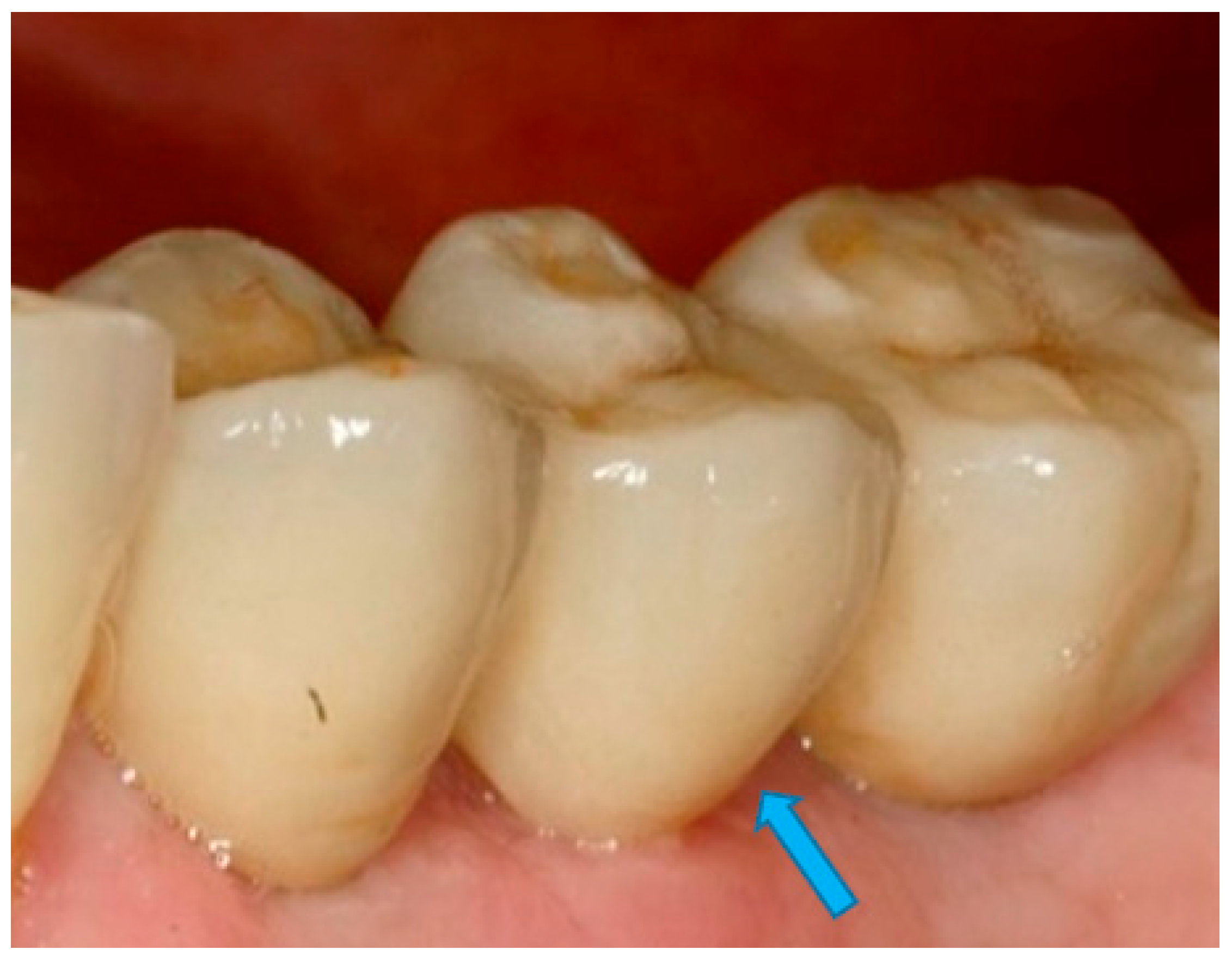
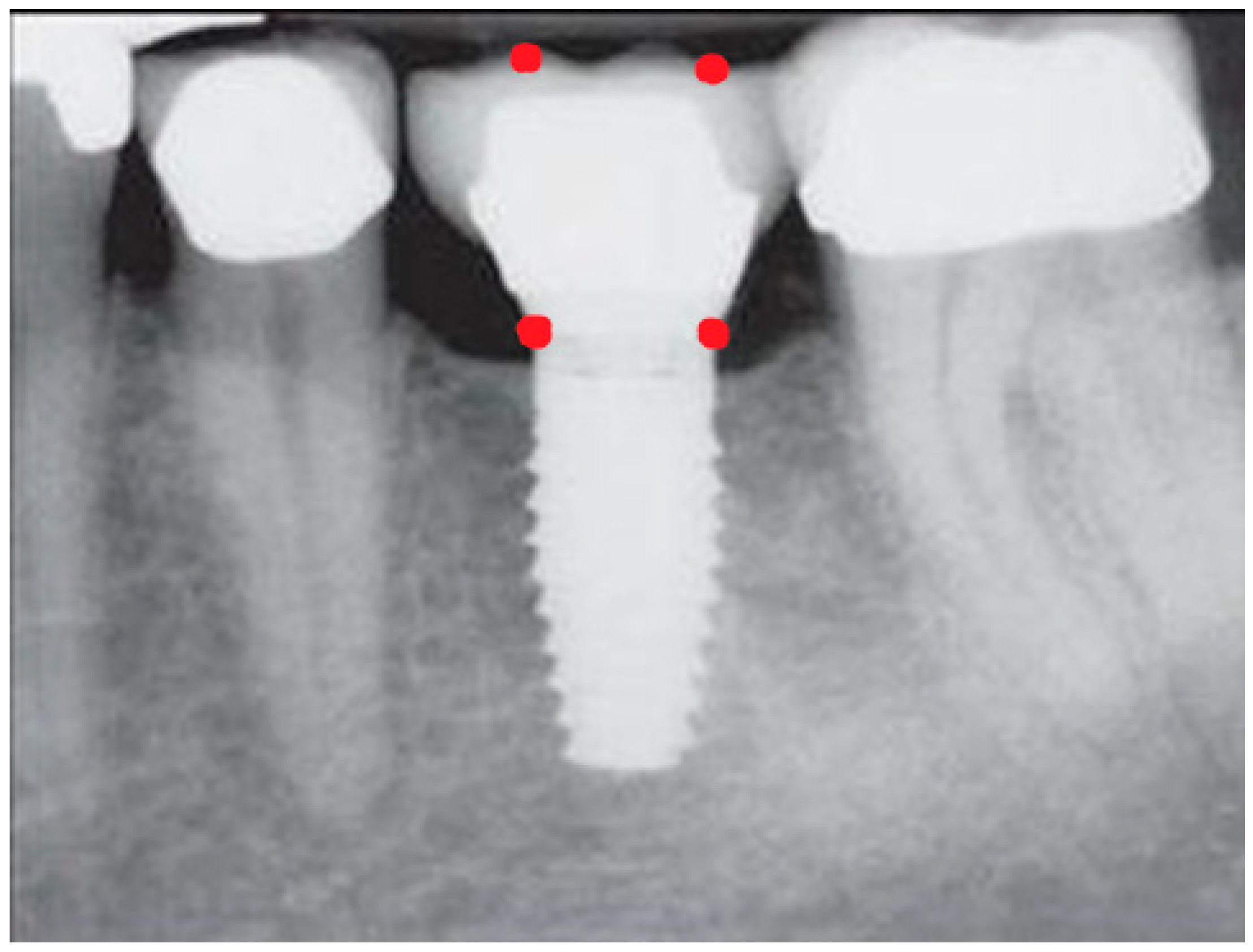
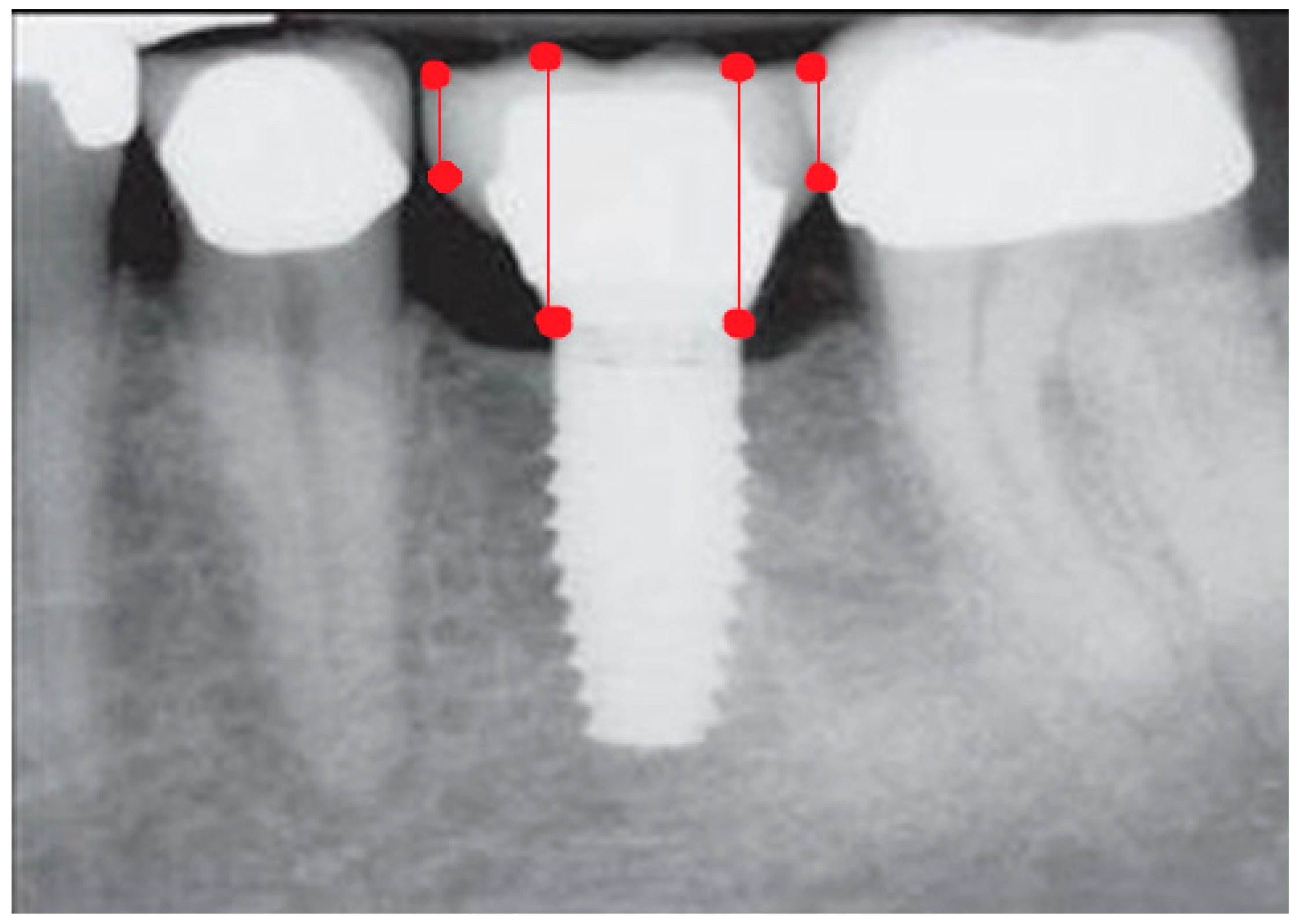
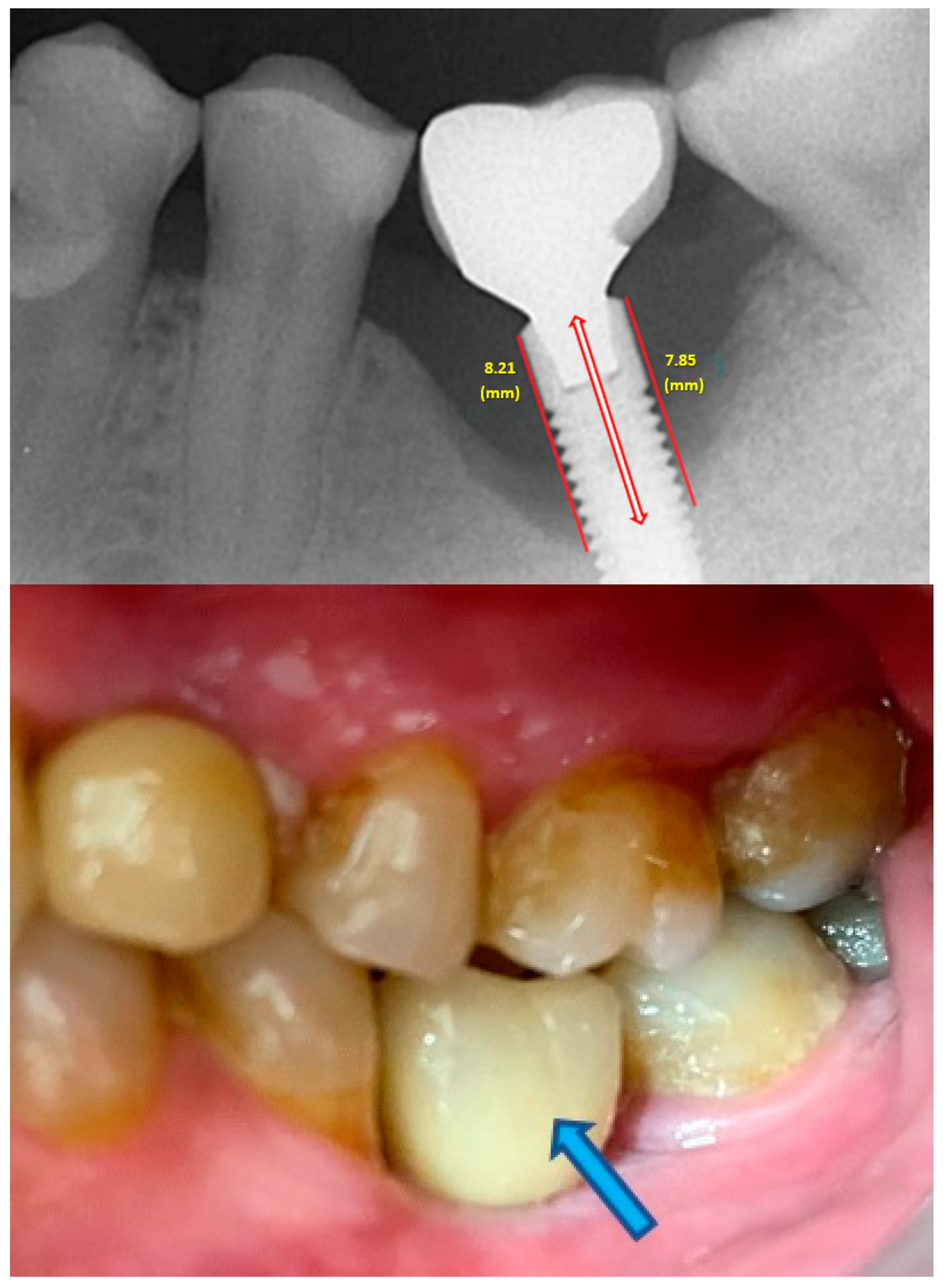
  • Measurement of mesial and distal bone loss: Clinically, an increase in the probing depth of the peri-implant pockets, both mesial and distal, was recorded using a calibrated probe with a force of 0.25 N. Radiographically, mesial and distal peri-implant bone height loss was measured using properly performed radiological projections. In fewer than five patients, statistically significant findings in cases of bone loss were difficult to achieve.
  • Measurement of the mesial and distal cantilever: The mesial cantilever was significantly greater for the bilateral bone loss group (1833.5 ± 1531.4 µm) compared to the group without MBL (1029.5 ± 968.6 µm) (F = 2.77; p < 0.05). Concerning the effect of the mesial versus distal cantilever, although the mesial cantilever appears to be more favorable, the only article found in the literature regarding this finding is a study by Romeo et al. (2003), which showed that this effect is not always consistent, although the difference is minimal [31].
  • Measurement of occlusal load using the PRESCALE® (Fujifilm, Japan) is the standard parameter to objectively evaluate masticatory function. This system is supported by several publications as it is reproducible, accurate, objective, internationally scientifically validated, easy to use, and does not increase the vertical dimension [32,33,34,35,36]. While it may not reveal the sequence and timing of dental contact, making it difficult to identify each individual tooth, PRESCALE® effectively integrated the occlusal analysis and provided the required information for this study. In this clinical study, it is evident that there is a higher occlusal load in the left anterior region, and there is a linear association between this load and MBL (r = 0.47; p < 0.01).
This might be related to the theory of the preferred chewing side. Previous studies have shown that most people usually chew more on one side, known as the preferred chewing side [37,38]. A unilateral chewing pattern is defined when the number of chewing cycles on one side is approximately 30% higher than the number of chewing cycles performed on the opposite side [37,38,39].
In conclusion, we propose the “Monterian theory of marginal bone loss”, pioneered by Prof. Dr. Javier Montero Martín, which supports the aforementioned theory. It states that most of us are right-handed in equal conditions, both in everyday activities and in the act of mastication. As a result, there is more wear on the right side compared to the left. Therefore, with less wear on the left side, there may be more occlusal contact on that side, leading to an asymmetrical bite that acts as an occlusal disruptor. The increased load on the left anterior side could be associated with greater MBL, even if the implant is not placed in that area. With an assisted bite such as that obtained using PRESCALE®, this situation can occur, leading to more contact on one side and thus reflecting a greater imbalance between the right and left sides.
In this research study, no statistically significant differences were found because the sample size was inconsistent between external and internal connection-type implants, with less than 5% of the implants having an external connection. Peñarrocha-Diago et al. (2013) conducted a prospective study radiographically comparing external connection implants with internal connection implants and concluded that external connection implants showed greater bone loss [40]. Similarly, Koo et al. (2012) observed that internal connection implants had significantly less peri-implant bone loss than external connection implants [41].
Unitary or multiple prosthetic extensions had no significant effect on the degree of bone loss in the groups in this study. However, a non-significant trend was observed for the bilateral bone loss group, where there was a higher prevalence of multiple prostheses (68.6%) compared to the group without MBL (52.6%).
In this study, all individual crowns and fixed rehabilitations were screw-retained, so no significant differences were observed because the sample size was uneven between the two variables.
In this research study, 82.2% of the restorations presented a mesial cantilever and 75.6% had a distal cantilever. The mesial cantilever was significantly larger (p < 0.05) in the bilateral bone loss group. Different studies in humans have found a positive effect of cantilevers on peri-implant bone loss, but others have been inconclusive and provided little objective evidence to support a cause-and-effect relationship. Further studies are needed to support this association.
Similar to the variables related to the prosthesis retention type in this study, all rehabilitations were directly attached to the implant; therefore, statistically significant evidence cannot be established under these conditions. The use of intermediate abutments in complete rehabilitations has a lower risk of peri-implant pathologies [42]. Likewise, Montero J. (2021) supports the use of transmucosal abutments at a minimum height of 2 mm to minimize MBL around implants and soft tissues [43].
In this study, 54.8% of the implants were placed in the upper jaw and 85.1% in the posterior areas. Peñarrocha M (2004) [44], Brånemark (1985) [45], and Wennström J (2004) [46] found significantly greater peri-implant bone loss in the posterior region of the upper jaw. However, there is no consensus in the literature, and some authors, such as Toljanic JA (1999), reported similar magnitudes of peri-implant bone loss in both jaws [47].
Standard length was predominant (72.6%). A linear association was observed between length and diameter with MBL, meaning that greater width and length lead to more peri-implant bone loss.
This research study demonstrates that MBL is significantly proportional to occlusal load in the left anterior sector (r = 0.47; p < 0.01). Occlusal overload can result from factors such as poor prosthetic design, inadequate implant position and dimensions, or parafunctional habits, among others. However, no statistically significant association was found in this study due to the different sample sizes of the groups. For occlusal analysis, elements such as PRESCALE® are very accurate for estimating bite force (N), occlusal contact area (mm2), and bite pressure (Mpa). Both occlusal analysis and peri-implant tissue health seem to be the most predictive variables for controlling occlusal overload. Regarding occlusion, a mutually protected occlusal scheme is advised whenever possible to avoid non-axial loading of the implants and shallow cusp inclinations.
Regarding complications, 23% of them consisted of screw loosening and, exceptionally (1.5%), screw loosening and/or porcelain fracture. Zandim-Bareclos DL et al. (2019) suggested that clinical signs of occlusal overload, such as pillar fractures, loss of retention, or chipping, were more frequently observed in MBL sites (27 out of 98 implants), implying a relationship between occlusal overload and MBL. However, further evidence is required to confirm the influence of occlusal overload on the onset and progression of MBL [48].
According to this research study, there is no statistically significant association between age and peri-implant bone loss (p = 0.127). It is worth noting that no study that reports an association between these two parameters has been published.
No statistically significant differences were found in gender distribution in this study. Therefore, as Bilhan H (2011) [49] and Peñarrocha M (2004) [44] also found, there is no association between gender and peri-implant bone loss.
A little over half of the implants showed no bleeding on probing (57%) and no symptoms (98.5%), with only 2.3% of implants with bilateral bone loss showing symptoms. Schwarz (2022) [50], in their recent publication, reported that healthy peri-implant tissues have become synonymous with implant success. This is determined clinically by the absence of erythema, bleeding on probing, inflammation, and suppuration, without considering a specific range of probing depths [51].
In this clinical study, 14.6% of the patients had a history of periodontal disease. Schou (2006) [49] and Karoussis (2003) [52] state that there is strong evidence from longitudinal and cross-sectional studies that a history of periodontitis constitutes a risk factor for MBL.
In this investigation, 24.4% of the patients were smokers. As explained by Levin L et al. (2008), smoking is a primary systemic risk factor associated with detrimental effects on peri-implant tissues [53] and can potentially result in a 3- to 5-fold increase in the risk of MBL [52].
A total of 4.9% of the patients in this study had diabetes. The success rate of dental implants in the general population at five years is estimated at 95%, while in patients with diabetes, it is 85% [54].
A millimeter-calibrated plastic periodontal probe with a force of approximately 0.25 N was used to probe the 135 implants. It is important to establish clinical values of probing depth immediately after implant and prosthesis placement, according to Lindhe and Meyle (2008) [55].
In all cases in this study, digital radiography was used for measuring mesial and distal bone loss, as well as cantilever length, providing high quality and allowing us to make measurements with greater precision, equal to or less than 0.1 mm [56].
It is noteworthy that all patients without MBL attended regular check-up visits (n = 5; 100%), whereas the group with bilateral bone loss (n = 20; 76.9%) exhibited lower regularity in check-ups. A history of periodontal disease, smoking, and poor oral hygiene are risk factors [57], and it is advisable to establish baseline clinical and radiographic parameters at the time of prosthesis placement (Renvert, 2019). This suggests that with longer follow-up periods, there is a greater degree of MBL (p < 0.01), which serves as an indicator of the degree of linear association.
Despite scientific evidence implicating various factors, such as prosthesis design, implant, periodontal disease, smoking, complications, or maintenance [5,52,53,55,58,59], in peri-implant bone loss, no significant differences were found in this study, possibly due to the following limitations:
This study has a retrospective design, with a convenience sample of patients from a single healthcare center; therefore, not all treated patients were able to participate, potentially excluding those who experienced greater bone loss or implant failure.
Additionally, this was not a randomized clinical study, suggesting that not all types of patients (in terms of risk factors) received the same treatment.
Furthermore, relevant variables related to calcium metabolism, muscle strength, or parafunctional habits were not collected, potentially excluding these predictive variables.
Future research in this line of investigation should consider randomized, multicenter studies that longitudinally collect clinical, behavioral, and occlusal predictive variables to obtain unbiased results.
Numerous publications question the role of occlusal overload in osteointegration loss [4,25,60,61,62]. Therefore, further studies are needed to understand the mechanisms by which occlusion can affect dental implants and peri-implant conditions.
The present study’s main limitations include the diversity of implants and the limited sample size, among others. We have presented the results of interest based on our research and noted that the displayed results should be interpreted with caution.

5. Conclusions

Considering the limitations of this study, it can be concluded that there are no significant sociodemographic or behavioral factors associated with MBL. From a biomechanical point of view, a wider cantilever and excessive occlusal loading appear to be associated with greater bone loss. In addition to this, future studies should focus on reducing the variables related to implants.

Author Contributions

J.M. led this research study and analyzed and interpreted the statistical patient data. P.C. performed the data collection and was a major contributor to the writing of the manuscript. All authors have read and agreed to the published version of the manuscript.

Funding

This research received no external funding.

Institutional Review Board Statement

The study was conducted according to the guidelines of the Declaration of Helsinki, and approved by the Institutional Review Board of University of Salamanca (RUSAL_201500006834).

Informed Consent Statement

Informed consent was obtained from all subjects involved in this study.

Data Availability Statement

The datasets generated and analyzed during the present study are not publicly available due to internal university regulations but are available from the corresponding author upon reasonable request.

Acknowledgments

This study was conducted within the Research Group Advances in Oral Health of the Department of Surgery of the University of Salamanca, led by Javier Montero. This research did not receive any specific grant from funding agencies in the public, commercial, or not-for-profit sectors.

Conflicts of Interest

The authors declare no conflicts of interest.

References

  1. Boedeker, D.; Dyer, J.; Kraut, R. Clinical outcome of immediately loaded maxillary implants: A 2-year retrospective study. J. Oral. Maxillofac. Surg. 2011, 69, 1335–1343. [Google Scholar] [CrossRef] [PubMed]
  2. Sugiura, T.; Yamamoto, K.; Horita, S.; Murakami, K.; Tsutsumi, S.; Kirita, T. Effects of implant tilting and the loading direction on the displacement and micromotion of immediately loaded implants: An in vitro experiment and finite element analysis. J. Periodontal Implant Sci. 2017, 47, 251–262. [Google Scholar] [CrossRef]
  3. Delgado-Ruiz, R.A.; Calvo-Guirado, J.L.; Romanos, G.E. Effects of occlusal forces on the peri-implant-bone interface stability. Periodontology 2000 2019, 81, 179–193. [Google Scholar] [CrossRef] [PubMed]
  4. Tonetti, M.S.; Schmid, J. Pathogenesis of implant failures. Periodontology 2000 1994, 4, 127–138. [Google Scholar] [CrossRef]
  5. Caton, J.G.; Armitage, G.; Berglundh, T.; Chapple, I.L.C.; Jepsen, S.; Kornman, K.S.; Mealey, B.L.; Papapanou, P.N.; Sanz, M.; Tonetti, M.S. A new classification scheme for periodontal and peri-implant diseases and conditions—Introduction and key changes from the 1999 classification. J. Clin. Periodontol. 2018, 45 (Suppl. S20), S1–S8. [Google Scholar] [CrossRef]
  6. Berglundh, T.; Armitage, G.; Araujo, M.G.; Avila-Ortiz, G.; Blanco, J.; Camargo, P.M.; Chen, S.; Cochran, D.; Derks, J.; Figuero, E.; et al. Peri-implant diseases and conditions: Consensus report of workgroup 4 of the 2017 World Workshop on the Classification of Periodontal and Peri-Implant Diseases and Conditions. J. Clin. Periodontol. 2018, 45 (Suppl. S20), 286–291. [Google Scholar] [CrossRef]
  7. Araujo, M.G.; Lindhe, J. Peri-implant health. J. Clin. Periodontol. 2018, 45 (Suppl. S20), 230–236. [Google Scholar] [CrossRef] [PubMed]
  8. Manea, A.; Bran, S.; Dinu, C.; Rotaru, H.; Barbur, I.; Crisan, B.; Armencea, G.; Onisor, F.; Lazar, M.; Ostas, D.; et al. Principles of biomechanics in oral implantology. Med. Pharm. Rep. 2019, 92 (Suppl. S3), S14–S19. [Google Scholar] [CrossRef] [PubMed]
  9. Brunski, J.B. Biomaterial and biomechanics in dental implant design. Int. J. Oral. Maxillofac. Implants 1988, 3, 85–97. [Google Scholar]
  10. Brånemark, P.I. Osseointegration and its experimental background. J. Prosthet. Dent. 1983, 50, 399–410. [Google Scholar] [CrossRef]
  11. Duyck, J.; Naert, I.E.; Van Oosterwyck, H.; Van der Sloten, J.; De Cooman, M.; Lievens, S.; Puers, B. Biomechanics of oral implants: A review of the literature. Technol. Health Care 1997, 5, 253–273. [Google Scholar] [CrossRef] [PubMed]
  12. Becker, C.M.; Wilson, T.G., Jr.; Jensen, O.T. Minimum criteria for immediate provisionalization of single-tooth dental implants in extraction sites: A 1-year retrospective study of 100 consecutive cases. J. Oral. Maxillofac. Surg. 2011, 69, 491–497. [Google Scholar] [CrossRef] [PubMed]
  13. Sohn, B.S.; Heo, S.J.; Koak, J.Y.; Kim, S.K.; Lee, S.Y. Strain of implants depending on occlusion types in mandibular implant-supported fixed prostheses. J. Adv. Prosthodont. 2011, 3, 1–9. [Google Scholar] [CrossRef]
  14. Pjetursson, B.E.; Thoma, D.; Jung, R.; Zwahlen, M.; Zembic, A. A systematic review of the survival and complication rates of implant-supported fixed dental prostheses (FDPs) after a mean observation period of at least 5 years. Clin. Oral. Implants Res. 2012, 23 (Suppl. S6), 22–38. [Google Scholar] [CrossRef]
  15. Shackleton, J.L.; Carr, L.; Slabbert, J.C.; Becker, P.J. Survival of fixed implant-supported prostheses related to cantilever lengths. J. Prosthet. Dent. 1994, 71, 23–26. [Google Scholar] [CrossRef] [PubMed]
  16. Sheridan, R.A.; Decker, A.M.; Plonka, A.B.; Wang, H.L. The Role of Occlusion in Implant Therapy: A Comprehensive Updated Review. Implant. Dent. 2016, 25, 829–838. [Google Scholar] [CrossRef] [PubMed]
  17. Bertolini, M.M.; Del Bel Cury, A.A.; Pizzoloto, L.; Acapa, I.R.H.; Shibli, J.A.; Bordin, D. Does traumatic occlusal forces lead to peri-implant bone loss? A systematic review. Braz. Oral. Res. 2019, 33 (Suppl. S1), e069. [Google Scholar] [CrossRef] [PubMed]
  18. Schwarz, M.S. Mechanical complications of dental implants. Clin. Oral. Implants Res. 2000, 11 (Suppl. S1), 156–158. [Google Scholar] [CrossRef]
  19. Chambrone, L.; Chambrone, L.A.; Lima, L.A. Effects of occlusal overload on peri-implant tissue health: A systematic review of animal-model studies. J. Periodontol. 2010, 81, 1367–1378. [Google Scholar] [CrossRef]
  20. Kozlovsky, A.; Tal, H.; Laufer, B.Z.; Leshem, R.; Rohrer, M.D.; Weinreb, M.; Artzi, Z. Impact of implant overloading on the peri-implant bone in inflamed and non-inflamed peri-implant mucosa. Clin. Oral. Implants Res. 2007, 18, 601–610. [Google Scholar] [CrossRef]
  21. Kourtis, S.G.; Sotiriadou, S.; Voliotis, S.; Challas, A. Private practice results of dental implants. Part I: Survival and evaluation of risk factors--Part II: Surgical and prosthetic complications. Implant Dent. 2004, 13, 373–385. [Google Scholar] [CrossRef] [PubMed]
  22. Becker, C.M. Cantilever fixed prostheses utilizing dental implants: A 10-year retrospective analysis. Quintessence Int. 2004, 35, 437–441. [Google Scholar] [PubMed]
  23. Khorshid, H.E.; Issa, N.O.; Ekram, A.M. Effect of implant diameter and cantilever length on the marginal bone height changes and stability of implants supporting screw retained prostheses: A randomized double blinded control trial. J. Adv. Prosthodont. 2023, 15, 101–113. [Google Scholar] [CrossRef] [PubMed]
  24. Iwasaki, M.; Maeda, I.; Kokubo, Y.; Tanaka, Y.; Ueno, T.; Ohara, Y.; Motokawa, K.; Hayakawa, M.; Shirobe, M.; Edahiro, A.; et al. Standard Values and Concurrent Validity of a Newly Developed Occlusal Force-Measuring Device among Community-Dwelling Older Adults: The Otassha Study. Int. J. Environ. Res. Public Health 2022, 19, 5588. [Google Scholar] [CrossRef] [PubMed]
  25. Schwarz, F.; Derks, J.; Monje, A.; Wang, H.L. Peri-implantitis. J. Clin. Periodontol. 2018, 45 (Suppl. S20), 246–266. [Google Scholar] [CrossRef] [PubMed]
  26. Heitz-Mayfield, L.J. Peri-implant diseases: Diagnosis and risk indicators. J. Clin. Periodontol. 2008, 35 (Suppl. S8), 292–304. [Google Scholar] [CrossRef] [PubMed]
  27. Sánchez-Gárces, M.A.; Gay-Escoda, C. Periimplantitis. Med. Oral Patol. Oral Cir. Bucal 2004, 9 (Suppl. S69–S74), 63–69. [Google Scholar]
  28. Renvert, S.; Persson, G.R.; Pirih, F.Q.; Camargo, P.M. Peri-implant health, peri-implant mucositis, and peri-implantitis: Case definitions and diagnostic considerations. J. Periodontol. 2018, 89 (Suppl. S1), S304–S312. [Google Scholar] [CrossRef]
  29. Scribante, A.; Gallo, S.; Pascadopoli, M.; Frani, M.; Butera, A. Ozonized gels vs chlorhexidine in non-surgical periodontal treatment: A randomized clinical trial. Oral Dis. 2023, 1–8. [Google Scholar] [CrossRef]
  30. Elbay, M.; Elbay, Ü.Ş.; Kaya, E.; Kalkan, Ö.P. Effects of photobiomodulation with different application parameters on injection pain in children: A randomized clinical trial. J. Clin. Pediatr. Dent. 2023, 47, 54–62. [Google Scholar]
  31. Romeo, E.; Lops, D.; Margutti, E.; Ghisolfi, M.; Chiapasco, M.; Vogel, G. Implant-supported fixed cantilever prostheses in partially edentulous arches. A seven-year prospective study. Clin. Oral Implants Res. 2003, 14, 303–311. [Google Scholar] [CrossRef]
  32. Shiga, H.; Komino, M.; Yokoyama, M.; Sano, M.; Arakawa, I.; Nakajima, K.; Fujii, S. Relationship between age and occlusal force in adults with natural dentition. Odontology 2023, 111, 487–492. [Google Scholar] [CrossRef] [PubMed]
  33. Okiyama, S.; Ikebe, K.; Nokubi, T. Association between masticatory performance and maximal occlusal force in young men. J. Oral. Rehabil. 2003, 30, 278–282. [Google Scholar] [CrossRef] [PubMed]
  34. Horibe, Y.; Matsuo, K.; Ikebe, K.; Minakuchi, S.; Sato, Y.; Sakurai, K.; Ueda, T. Relationship between two pressure-sensitive films for testing reduced occlusal force in diagnostic criteria for oral hypofunction. Gerodontology 2022, 39, 3–9. [Google Scholar] [CrossRef] [PubMed]
  35. Wang, Q.; Zhao, Z.; Zhou, M.; Zheng, X.; An, N.; Niu, L.; Tay, F.R.; Ma, C.; Wang, F. In vivo evaluation of the reliability and validity of three digital occlusion analysis methods. J. Dent. 2022, 127, 104355. [Google Scholar] [CrossRef] [PubMed]
  36. Huang, Y.F.; Wang, C.M.; Shieh, W.Y.; Liao, Y.F.; Hong, H.H.; Chang, C.T. The correlation between two occlusal analyzers for the measurement of bite force. BMC Oral Health 2022, 22, 472. [Google Scholar] [CrossRef] [PubMed]
  37. Jiang, H.; Liu, H.; Liu, G.; Jin, Z.; Wang, L.; Ma, J.; Li, H. Analysis of brain activity involved in chewing-side preference during chewing: An fMRI study. J. Oral Rehabil. 2015, 42, 27–33. [Google Scholar] [CrossRef] [PubMed]
  38. Barcellos, D.C.; Gonçalves, S.E.; da Silva, M.A.; Batista, G.R.; Pleffken, P.R.; Pucci, C.R.; Borges, A.B.; Rocha Gomes Torres, C. Prevalence of chewing side preference in the deciduous, mixed and permanent dentitions. J. Contemp. Dent. Pract. 2011, 12, 339–342. [Google Scholar] [CrossRef] [PubMed]
  39. Athab Abduljabbar, Z.; Svensson, K.G.; Hjalmarsson, L.; Franke Stenport, V.; Eliasson, A. Chewing side preference and laterality in patients treated with unilateral posterior implant-supported fixed partial prostheses. J. Oral Rehabil. 2022, 49, 1080–1086. [Google Scholar] [CrossRef]
  40. Peñarrocha-Diago, M.A.; Flichy-Fernández, A.J.; Alonso-González, R.; Peñarrocha-Oltra, D.; Balaguer-Martínez, J.; Peñarrocha-Diago, M. Influence of implant neck design and implant-abutment connection type on peri-implant health. Radiological study. Clin. Oral Implants Res. 2013, 24, 1192–1200. [Google Scholar] [CrossRef]
  41. Koo, K.T.; Lee, E.J.; Kim, J.Y.; Seol, Y.J.; Han, J.S.; Kim, T.I.; Lee, Y.M.; Ku, Y.; Wikesjö, U.M.; Rhyu, I.C. The effect of internal versus external abutment connection modes on crestal bone changes around dental implants: A radiographic analysis. J. Periodontol. 2012, 83, 1104–1109. [Google Scholar] [CrossRef] [PubMed]
  42. Koutouzis, T.; Gholami, F.; Reynolds, J.; Lundgren, T.; Kotsakis, G.A. Abutment Disconnection/Reconnection Affects Peri-implant Marginal Bone Levels: A Meta-Analysis. Int. J. Oral Maxillofac. Implants 2017, 32, 575–581. [Google Scholar] [CrossRef] [PubMed]
  43. Montero, J. A Review of the Major Prosthetic Factors Influencing the Prognosis of Implant Prosthodontics. J. Clin. Med. 2021, 10, 816. [Google Scholar] [CrossRef] [PubMed]
  44. Peñarrocha, M.; Palomar, M.; Sanchis, J.M.; Guarinos, J.; Balaguer, J. Radiologic study of marginal bone loss around 108 dental implants and its relationship to smoking, implant location, and morphology. Int. J. Oral Maxillofac. Implants 2004, 19, 861–867. [Google Scholar] [PubMed]
  45. Brånemark, P.I.; Zarb, G.A.; Albrektsson, T. Tissue-Integrated Prostheses: Osseointegration in Clinical Dentistry; Quintessence: Chicago, IL, USA, 1985; pp. 111–197. [Google Scholar]
  46. Wennström, J.; Zurdo, J.; Karlsson, S.; Ekestubbe, A.; Gröndahl, K.; Lindhe, J. Bone level change at implant-supported fixed partial dentures with and without cantilever extension after 5 years in function. J. Clin. Periodontol. 2004, 31, 1077–1083. [Google Scholar] [CrossRef] [PubMed]
  47. Toljanic, J.A.; Banakis, M.L.; Willes, L.A.; Graham, L. Soft tissue exposure of endosseous implants between stage I and stage II surgery as a potential indicator of early crestal bone loss. Int. J. Oral Maxillofac. Implants 1999, 14, 436–441. [Google Scholar] [PubMed]
  48. Zandim-Barcelos, D.L.; Carvalho, G.G.; Sapata, V.M.; Villar, C.C.; Hämmerle, C.; Romito, G.A. Implant-based factor as possible risk for peri-implantitis. Braz. Oral Res. 2019, 33 (Suppl. S1), e067. [Google Scholar] [CrossRef] [PubMed]
  49. Bilhan, H.; Mumcu, E.; Geçkili, O.; Atalay, B. The evaluation of the success of immediately placed single implants: A retrospective clinical study. Implant Dent. 2011, 20, 215–225. [Google Scholar] [CrossRef] [PubMed]
  50. Schwarz, F.; Ramanauskaite, A. It is all about peri-implant tissue health. Periodontology 2000 2022, 88, 9–12. [Google Scholar] [CrossRef]
  51. Schou, S.; Holmstrup, P.; Worthington, H.V.; Esposito, M. Outcome of implant therapy in patients with previous tooth loss due to periodontitis. Clin. Oral Implants Res. 2006, 17 (Suppl. S2), 104–123. [Google Scholar] [CrossRef]
  52. Karoussis, I.K.; Salvi, G.E.; Heitz-Mayfield, L.J.; Brägger, U.; Hämmerle, C.H.; Lang, N.P. Long-term implant prognosis in patients with and without a history of chronic periodontitis: A 10-year prospective cohort study of the ITI Dental Implant System. Clin. Oral Implants Res. 2003, 14, 329–339. [Google Scholar] [CrossRef] [PubMed]
  53. Levin, L.; Hertzberg, R.; Har-Nes, S.; Schwartz-Arad, D. Long-term marginal bone loss around single dental implants affected by current and past smoking habits. Implant Dent. 2008, 17, 422–429. [Google Scholar] [CrossRef] [PubMed]
  54. Wagner, J.; Spille, J.H.; Wiltfang, J.; Naujokat, H. Systematic review on diabetes mellitus and dental implants: An update. Int. J. Implant Dent. 2022, 8, 1. [Google Scholar] [CrossRef] [PubMed]
  55. Lindhe, J.; Meyle, J.; Group D of European Workshop on Periodontology. Peri-implant diseases: Consensus Report of the Sixth European Workshop on Periodontology. J. Clin. Periodontol. 2008, 35 (Suppl. S8), 282–285. [Google Scholar] [CrossRef] [PubMed]
  56. De Bruyn, H.; Vandeweghe, S.; Ruyffelaert, C.; Cosyn, J.; Sennerby, L. Radiographic evaluation of modern oral implants with emphasis on crestal bone level and relevance to peri-implant health. Periodontology 2000 2013, 62, 256–270. [Google Scholar] [CrossRef] [PubMed]
  57. Renvert, S.; Hirooka, H.; Polyzois, I.; Kelekis-Cholakis, A.; Wang, H.L.; Working Group 3. Diagnosis and non-surgical treatment of peri-implant diseases and maintenance care of patients with dental implants-Consensus report of working group 3. Int. Dent. J. 2019, 69 (Suppl. S2), 12–17. [Google Scholar] [CrossRef] [PubMed]
  58. Lang, N.P.; Berglundh, T.; Working Group 4 of Seventh European Workshop on Periodontology. Periimplant diseases: Where are we now?--Consensus of the Seventh European Workshop on Periodontology. J. Clin. Periodontol. 2011, 38 (Suppl. S11), 178–181. [Google Scholar] [CrossRef] [PubMed]
  59. Lindquist, L.W.; Carlsson, G.E.; Jemt, T. A prospective 15-year follow-up study of mandibular fixed prostheses supported by osseointegrated implants. Clinical results and marginal bone loss. Clin. Oral Implants Res. 1996, 7, 329–336. [Google Scholar] [CrossRef] [PubMed]
  60. Serino, G.; Ström, C. Peri-implantitis in partially edentulous patients: Association with inadequate plaque control. Clin. Oral Implants Res. 2009, 20, 169–174. [Google Scholar] [CrossRef]
  61. Heitz-Mayfield, L.J.; Schmid, B.; Weigel, C.; Gerber, S.; Bosshardt, D.D.; Jönsson, J.; Lang, N.P.; Jönsson, J. Does excessive occlusal load affect osseointegration? An experimental study in the dog. Clin. Oral Implants Res. 2004, 15, 259–268. [Google Scholar] [CrossRef]
  62. Lang, N.P.; Wilson, T.G.; Corbet, E.F. Biological complications with dental implants: Their prevention, diagnosis and treatment. Clin. Oral Implants Res. 2000, 11 (Suppl. S1), 146–155. [Google Scholar] [CrossRef] [PubMed]
Figure 1. Representative periapical radiograph of implant and crown on implant in 3.6 using VistaSoft Dental® (DÜRR DENTAL, Bietigheim-Bissingen, Germany) for cantilever measurement.
Prosthesis 06 00065 g001
Figure 2. Representative clinical picture of screw-retained crown on implant placed in position 3.6. Direct view.
Prosthesis 06 00065 g002
Figure 3. Representative periapical radiograph of implant and crown on implant in 3.6 using VistaSoft Dental® software (DÜRR DENTAL, Bietigheim-Bissingen, Germany) with mesial and distal marking points at boundary of implant platform and marginal ridge of crown.
Prosthesis 06 00065 g003
Figure 4. Representative periapical radiograph of implant and crown on implant in 3.6 using VistaSoft Dental® software (DÜRR DENTAL, Bietigheim-Bissingen, Germany) with mesial and distal marking points at boundary of implant platform and marginal ridge of crown and mesial and distal ends of restoration.
Prosthesis 06 00065 g004
Figure 5. Representative periapical radiograph of implant and crown on implant in 3.6 using VistaSoft Dental® software (DÜRR DENTAL, Bietigheim-Bissingen, Germany) with vertical lines joining mesial and distal marking points at boundary of implant platform and marginal ridge of crown and mesial and distal ends of restoration.
Prosthesis 06 00065 g005
Figure 6. Representative periapical radiograph of measurement points for mesial and distal cantilever.
Prosthesis 06 00065 g006
Figure 7. Representative periapical radiograph of mesial and distal cantilever as example of measurement obtained with VistaSoft Dental® software (DÜRR DENTAL, Bietigheim-Bissingen, Germany).
Prosthesis 06 00065 g007
Figure 8. Representative periapical radiograph of measurement points for mesial and distal peri-implant bone loss on implant in position 3.6 using VistaSoft Dental® software (DÜRR DENTAL, Bietigheim-Bissingen, Germany) and its clinical appearance.
Prosthesis 06 00065 g008
Figure 9. A PRESCALE® (Fujifilm, Japan) colorimetric sheet in white. On the left, Medium MS PS 270 × 200 mm; on the right, examples of colorimetric sheets with bite registrations of six different patients.
Prosthesis 06 00065 g009
Table 1. Comparison of the morphology of placed implants and cantilevers by analysis of variance (ANOVA).
NO MBL
(n = 5 Patients with a Total of 19 Implants)
UNILATERAL MBL
(n = 10 Patients with a Total of 30 Implants)
BILATERAL MBL
(n = 26 Patients with a Total of 86 Implants)
p-Value
Clinical ProfileMeanSDMeanSDMeanSD
Mesial cantilever (µm)1029.5968.61521.01149.41833.51531.4F = 2.77; p < 0.05
Distal cantilever (µm)1645.81341.51592.72148.91483.11563.9F = 0.09; p = 0.90
Implant diameter (mm)3.800.53.900.53.80.4F = 0.16; p = 0.85
Implant length (mm)10.01.210.71.410.61.7F = 1.33; p = 0.268
Follow-up (months)45.118.845.119.648.922.0F = 0.50; p = 0.60
Table 2. Correlation of mesial and distal MBL considering clinical variables in the study sample (n = 135 implants).
CLINICAL VARIABLES (Implant-Related)MBL (p-Value)
Implant diameterr = 0.27 (p = 0.001)
Implant lengthr = 0.20 (p = 0.021)
Follow-up timer = 0.29 (p = 0.001)
Occlusal load in the anterior regionr = 0.47 (p = 0.002)
Table 3. Clinical description of the peri-implant health variables in the study sample: disease prevalence, symptomatology, maintenance, plaque index, and bleeding on probing. Comparison using the χ2 test.
NO MBL
(n = 5 with 19 Implants)
UNILATERAL MBL
(n = 10 Patients with 30 Implants)
BILATERAL MBL
(n = 26 Patients with 86 Implants)
COMPARISON
p-Value
CLINICAL
VARIABLES
n%n%n%%
Periodontal disease120.0110.0415.4χ2 = 0.30; p = 0.861
Diabetes000027.7χ2 = 1.21; p = 0.545
Other diseases120.000415.4χ2 = 1.92; p = 0.383
SYMPTOMSn%n%n%
Asymptomatic19100.030100.08497.7χ2 = 1.16; p = 0.56
Symptomatic00.000.022.3
BEHAVIORAL
VARIABLE
n%n%n%
Regular maintenance visits5100.0990.02076.9χ2 = 2.04; p = 0.36
PLAQUE INDEXn%n%n%
No plaque842.11240.03439.5
χ2 = 5.18; p = 0.52

 
1/3 plaque842.11550.03338.4
2/3 plaque210.513.31618.6
>2/3 plaque15.326.733.5
BLEEDING ON PROBINGn%n%n%
No1263.21860.04754.7
χ2 = 0.60; p = 0.74
Yes736.81240.03945.3
Disclaimer/Publisher’s Note: The statements, opinions and data contained in all publications are solely those of the individual author(s) and contributor(s) and not of MDPI and/or the editor(s). MDPI and/or the editor(s) disclaim responsibility for any injury to people or property resulting from any ideas, methods, instructions or products referred to in the content.

