Article

Survival of Chairside Posterior Single Crowns Made from InCoris TZI Zirconia—A Retrospective Analysis up to 10 Years

1
Department of Prosthodontics, Preclinical Education and Dental Materials Science, University of Bonn, 53111 Bonn, Germany
2
Department of Reconstructive Dentistry and Gerodontology, School of Dental Medicine, University of Bern, 3012 Bern, Switzerland
*
Author to whom correspondence should be addressed.
These authors contributed equally to this work.
Prosthesis 2024, 6(5), 1118-1132; https://doi.org/10.3390/prosthesis6050081
Submission received: 7 August 2024 / Revised: 4 September 2024 / Accepted: 9 September 2024 / Published: 11 September 2024
(This article belongs to the Special Issue Advancements in Zirconia Dental Restorations)

Abstract

:
Objective: The aim of the present study was to evaluate the long-term clinical survival and success of chairside-fabricated single-tooth monolithic zirconia restorations on posterior teeth using the speed sintering process. Materials and Methods: Between 2012 and 2022, 250 single-tooth crowns were fabricated for 193 patients using the CEREC® chairside workflow. Restorations were fabricated from monolithic 3Y-TZP zirconia (InCoris TZI, Dentsply Sirona©, Bensheim, Germany) as full-contour crowns. The same clinician performed all procedures. Luting was performed using self-adhesive resin-based cements or glass ionomer cement. Retrospective analysis was conducted, defining survival as crowns still in function regardless of any interventions, and success as crowns that remained functional without the need for intervention. Statistical analysis was performed using Kaplan–Meier analysis, considering “refabrication” and “intervention” as endpoints. Results: Of the 250 crowns, a total of 162 (64.8%) crowns showed success. Over the whole observation period, 44 crowns (17.6%) required refabrication, and 88 (35.2%) required intervention. Mean survival without refabrication was 7.43 years, with a 5- and 7.5-year survival of 86.9% and 76.6%. The mean survival without intervention was 6.5 years, with a 5- and 7.5-year survival of 70.8% and 59.9%. Conclusions: Under appropriate technical conditions, chairside-fabricated 3Y-TZP zirconia single-tooth crowns represent a viable fabrication method. Neither the cementation mode nor the crown position—whether on premolars or molars—significantly impacted the survival rates.

1. Introduction

With the development and introduction of computer-aided design and manufacturing (CAD-CAM), the fabrication of dental prostheses has been standardized, ensuring consistently high quality and faster production. Significant progress has been made, particularly in the production of ceramic dental restorations, as the processing of zirconia (ZrO2) as a dental material has been facilitated and simplified. Consequently, zirconia-based restorations are gradually replacing metal–ceramic restorations, which have long been regarded as the gold standard. Initially used solely as a framework material, zirconia is now employed as a fully anatomical monolithic restoration in the posterior region. This allows for aesthetically pleasing results with good biocompatibility [1], while the excellent mechanical properties [2] enable its use not only for single-tooth restorations in the posterior region but also for fixed dental prostheses (FDPs) [3].
Initially, zirconia could only be manufactured in a laboratory setting. Today, both labside and chairside fabrication are available. The advantage of the chairside workflow lies in the production of dental restorations in a single session [4], resulting in significant time savings and eliminating the need for a dental laboratory and temporary restorations. A pioneer of this approach is the CEREC® system by Dentsply Sirona© (Bensheim, Germany), which enables the production of single-tooth restorations and short-span FDPs. Although some researchers have previously expressed concerns regarding the precision of CEREC® restoration margins, subsequent studies have confirmed that clinically durable restorations can be produced with the latest technological advancements in the CEREC® system [4,5,6,7]. Initially, common materials for this system were feldspathic ceramics, later followed by lithium disilicate ceramics and zirconia-reinforced lithium disilicate. Due to the high strength and associated durability of zirconia, it has also been introduced as a monolithic chairside material. Given its high opacity and the inability to perform ceramic veneering chairside, the material is primarily indicated for posterior restorations, with 3 mol% yttrium oxide-stabilized tetragonal zirconia (3Y-TZP) being commonly used due to its high flexural strength (≈1000 MPa) [8].
However, data in the literature regarding the long-term survival and success rates of monolithic chairside-fabricated zirconia single crowns are insufficient. When considering the available studies on the long-term survival of CEREC® restorations, results comparable to those of noble metal alloy restorations are reported [7]. Survival rates of 94.7% after five years and 85.7% after ten years are cited, although these figures pertain to feldspathic ceramic restorations produced with the CEREC® 1 system. A review by Fasbinder [5] confirms these data, indicating survival rates of 97% (after five years) and 90% (after ten years). In a systematic review and meta-analysis from 2021, Mazza et al. [9] concluded that monolithic zirconia single crowns and FDPs represent a promising form of restoration, exhibiting high survival rates and low complication rates.
Unlike the aforementioned glass–ceramic materials, zirconia conventionally requires a time-consuming sintering process (up to 12 h) [10,11,12] to complete the restoration, necessitating an additional treatment session and, thus, not aligning with the chairside workflow. The introduction of speed (60 to 120 min) and high-speed sintering processes (10 min) has significantly reduced the overall manufacturing time [10,11] and greatly simplified chairside fabrication. Therefore, the alteration of mechanical or optical properties of monolithic zirconia is of clinical interest [10,11]. Sintering zirconia is considered a critical step in ceramic processing and is the focus of much research [13,14,15]. Previous investigations have shown that changes in the sintering process directly affect the density, microstructure, grain size, and phase composition, potentially impacting the mechanical and optical properties of zirconia ceramics [16]. Conventional sintering of Y-TZP ceramics typically employs slow and stable heating and cooling rates (up to 20 °C/min) to a sintering temperature of 1400–1500 °C, with a dwell time of several hours [10]. In contrast, speed sintering has been proposed as a potential strategy to achieve a finer microstructure by preventing grain growth through rapid heating while maintaining high material density [17]. Kaizer et al. [10] reported that higher sintering temperatures combined with shorter sintering times indeed lead to smaller grain size and higher translucency. Ersoy et al. [18] conducted rapid sintering of their samples by sintering in a preheated furnace at 1580 °C for 10 min, significantly improving the flexural strength of zirconia ceramics. However, the main drawbacks of conventional speed sintering have been associated with inhomogeneous densification or the so-called shell densification of 3Y-TZP, negatively impacting the ceramic’s microstructure and leading to mechanical defects [19,20]. Overall, the literature suggests that speed sintering has no negative impact on the mechanical properties of zirconia. In particular, flexural strength and mechanical stability are not affected [21,22].
The aim of the present investigation was to retrospectively analyze the long-term survival rates of chairside fabricated monolithic CEREC® single crowns made of 3Y-TZP zirconia that were fabricated using the speed sintering process and inserted in a dental practice.

2. Materials and Methods

2.1. Study Design

In this single-center study, 193 patients (117 women, 76 men) and 250 restorations were retrospectively analyzed. Initially, 226 patients (288 restorations) were treated; however, 38 (38 restorations) were lost to follow-up as they did not return for further check-ups or treatment appointments.
Ethical approval for this research was obtained from the Ethics Committee of the Medical Faculty of the University of Bonn under the reference number 386/23-EP. Between September 2012 and December 2022, patients were prosthetically treated by a dentist in private practice with monolithic and chairside-fabricated single crowns made of 3Y-TZP zirconia. Restorations were fabricated using the CEREC® system (Chairside Economical Restoration of Esthetic Ceramics) from Dentsply Sirona© (Bensheim, Germany) and blocks of InCoris TZI (Dentsply Sirona©, Bensheim, Germany). Data were extracted and evaluated from patient records, which were available in both the practice management software (Dampsoft©, Damp, Germany) and analog form. These records provided comprehensive details on all treatment-relevant aspects, as well as the survival and success of the restorations. The standardized and structured documentation included detailed information on anamnesis, dental findings, therapy decisions, treatment courses, photo documentation, and digitized laboratory orders (insourcing). All retrospectively included examinations correspond to clinical routine and a standardized dental examination, as well as periodontal recall appointments.
Only patients treated with chairside single crowns made of InCoris TZI (Dentsply Sirona©, Bensheim, Germany) were included. Additionally, only crowns that were fabricated using the CEREC® system and inserted during the same treatment session were considered. The inclusion criteria were as follows.
(1) Chairside-based, monolithic, full-contour single crowns, (2) data acquisition using the CEREC® Omnicam, (3) fabrication of the restoration using the CEREC® MC XL Premium system, (4) treatment of natural abutment teeth, (5) definitive insertion, (6) restoration material: 3Y-TZP InCoris TZI, and (7) at least one follow-up appointment after insertion.
The exclusion criteria were as follows.
(1) Restorations made from other ceramic materials (e.g., glass ceramics), (2) splinted restorations, (3) bridge constructions, (4) implant crowns, (5) provisionally inserted restorations, and (6) no follow-up appointment after insertion.
The relevant patient data for the survival analysis were extracted from the practice management software (Dampsoft© GmbH, Damp, Germany) as well as from the patient records. In characterizing complications, several distinctions were made. These included a loss of retention that necessitated re-cementation, ceramic fractures with or without repair (such as those repaired with composite), and periodontal issues. Additionally, complications related to endodontic treatments, caries or secondary caries, and tooth fractures were recorded. The analysis also accounted for extractions, cases requiring prosthetic re-planning or new planning, and instances where new fabrication was necessary.

2.2. Statistical Analysis

The collected patient data were analyzed using the statistical software SPSS (Statistical Package for the Social Sciences, Version: 29.0.1.0 (171), IBM, Armonk, NY, USA). The data were described using mean values, standard deviations (SDs), and percentages. The interval survival rate of the single crowns was calculated based on the information regarding the period of failure, and the cumulative survival rate was calculated over the maximal reported follow-up period using a life-table survival analysis. The primary focus was on the survival rate of the single crowns. The time-related Kaplan–Meier analysis was used to calculate the survival probability. Two endpoints were defined for this calculation:
  • Duration until replacement (time in years until the end of the functional period);
  • Duration until the first intervention (time in years).
If none of the endpoints occurred, the single crown was still in situ at the time of the last documented patient visit, and the observation interval was censored. Kaplan–Meier curves are commonly used to analyze time-to-event data. In addition to the Kaplan–Meier analysis, the log-rank test concept and the hazard function were applied. The level of statistical significance was set at p < 0.05.

2.3. Fabrication and Insertion
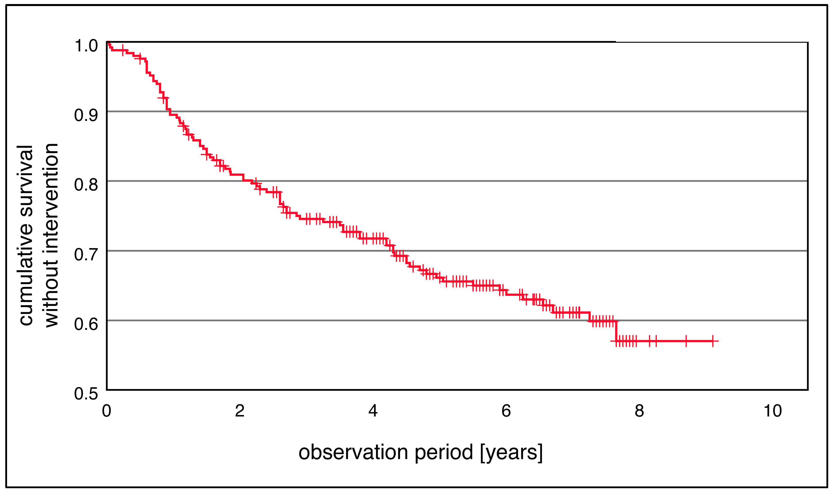
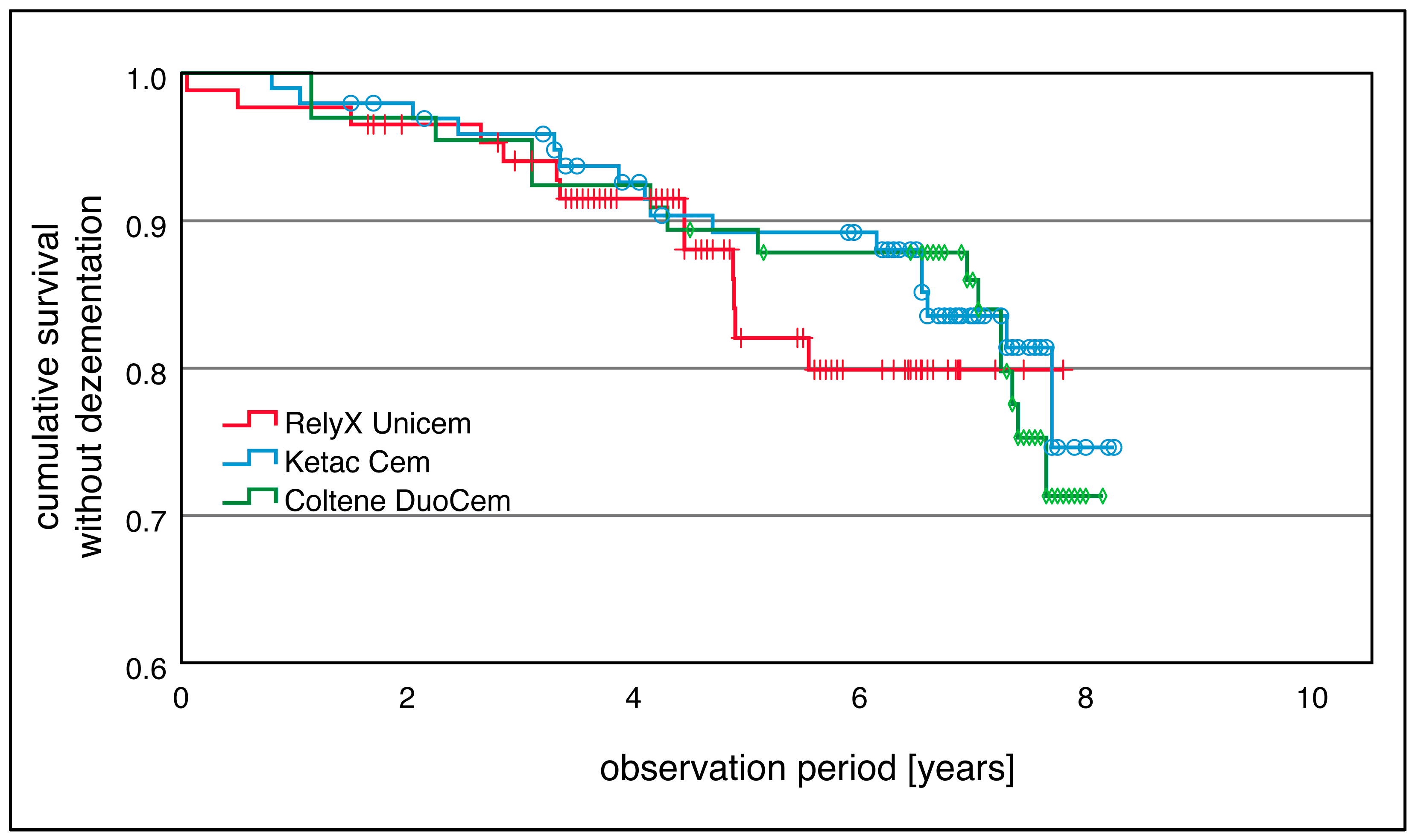
After a thorough medical history and clinical examination, the suitability of the patients for the proposed treatment was assessed. All patients whose retrospective data were used for this study were routinely informed about the procedure, possible associated risks, and potential treatment alternatives before prosthetic treatment, and they provided their consent for the treatment. Before treatment, all relevant tooth-related parameters were recorded to ensure the value of the tooth to be treated. This included a digital two-dimensional X-ray (dental film), measuring pocket depths (4-point measurement: mesial and distal, vestibular, and oral), and conducting bleeding on probing. Additionally, the percussion and vitality of the teeth were examined.
The teeth were prepared according to the guidelines for a full-ceramic crown using a high-speed handpiece (Expertmatic Lux E25 L, KaVo©, Biberach, Germany) and a fine rounded shoulder diamond bur (FG 4307 N broad, Intensiv SA, Collina d’Oro, Switzerland). Care was taken to achieve a uniform circumferential and occlusal substance removal of 1 mm, with the preparation margin always in an epi- or supragingival position. For hemostasis before performing the intraoral scan, an ammonium chloride-based paste (Expasyl®, Acteon®, Merignac, France) was applied. After a two-minute setting time, it was carefully rinsed off with an air–water spray.
After thorough drying of the tooth stump, the intraoral scan was performed using the CEREC® Omnicam (Dentsply Sirona©, Bensheim, Germany). The manufacturer’s specified scanning path was followed, and a powder-free scan was performed. Additionally, the opposing quadrant and the bite were scanned. Subsequently, the three-dimensional model was aligned, the preparation margin defined, and the insertion axis set.
Each restoration was generated in the design mode using “Biogeneric Individual” and optimized by the clinician if necessary. It was crucial to control and maintain the minimum material thickness. All 250 zirconia crowns were fabricated using the CEREC® MC XL Premium (Model-No.: 621100D3355, Serial-No.: 00184, Dentsply Sirona©, Bensheim, Germany) and CEREC® Software SW 4.2/4.3. The CEREC® MC XL Shaper 25 RZ and the CEREC® MC XL Finisher 10 were used as milling tools. The restorations were made from industrially manufactured zirconia blocks, CEREC® InCoris TZI Mono L (20 × 19 × 15.5) (Dentsply Sirona©, Bensheim, Germany). The milling time varied between 10 and 15 min.
The finished milled restoration was cleaned with a steam jet device and then subjected to a drying cycle at 120 °C for 10 min (inFire HTC Speed sintering furnace, Dentsply Sirona©, Bensheim, Germany). The now open-pored restoration was receptive to the color oxides DD Basic Shade Color Liquid (Dental Direkt, Spenge, Germany), allowing for color customization. The inFire HTC Speed sintering furnace (Dentsply Sirona©, Bensheim, Germany) was used for sintering the restorations. The sintering time per single crown, including the cooling phase, was approximately 30 min. The crowns were finally individualized with ceramic stains (Vita Akzent Plus, Vita Zahnfabrik, Bad Säckingen, Germany). The bond between the ceramic stains and the zirconia crown was achieved using a vacuum ceramic furnace (Multimat Cube, Dentsply Sirona©, Bensheim, Germany), which took about another ten minutes.
After completing the fabrication of the restoration, a sandblasting treatment with aluminum oxide was performed before the trial fit, with a pressure of no more than 1 bar and a particle size of 40 µ. After verifying the fit of the crown and the occlusion, minor adjustments were made if necessary. Following a final check of the crown’s fit, the crown lumen was cleaned with sodium hypochlorite (5% NaOCl) to remove any saliva residues. All 250 restorations were placed under relative isolation. Depending on the indication, the zirconia crowns were cemented with different luting agents. The luting agents RelyX™ Unicem 2 (3M, St. Paul, MN, USA), Ketac™ Cem (3M™, St. Paul, MN, USA), or Coltène DuoCem® (Coltène® Holding, Altstätten, Switzerland) were used. The luting agent was applied directly into the crown and then fixed on the prepared tooth stump with finger pressure. Depending on the setting time or light curing, excess cement was carefully removed, ensuring the crown remained in its correct position. Finally, the bite in static and dynamic occlusion was checked again.

3. Results

In this retrospective study, a total of 250 single-tooth crowns on natural abutment teeth were recorded and evaluated in 193 patients (117 females, 61%; 76 males, 39%) over an observation period from 2012 to 2022. Of the total number of restorations, 53% were placed in the upper jaw and 47% in the lower jaw. Additionally, 51% of the crowns were on premolars, while 49% were on molars.
The mean observation period was 5.9 ± 2.1 years, with the longest documented observation period being 8.25 years. Patients had a mean age of 46.99 ± 12.9 years at the time of crown placement.
One hundred sixty-two (64.8%) crowns were recorded as successful and have been incorporated without incidents since insertion. The mean survival without refabrication was 7.43 years. The 5-year and the 7.5-year survival rates were 86.9% and 76.6%, respectively (Figure 1). During the entire observation period, 44 out of 250 single-tooth crowns (17.6%) required refabrication, while 88 out of 250 crowns (35.2%) required treatment (Figure 2). The most common reasons for refabrication were extractions (n = 9, 3.6%), prosthetic re-planning (n = 8, 3.2%), and severe ceramic fractures (n = 6, 2.4%) (Table 1). Out of 250 single-tooth crowns, 88 (35.2%) required intervention (Figure 2). The mean survival without intervention was 6.5 years. The 5-year and 7.5-year survival rates without intervention were 70.8% and 59.9%, respectively. Interventions often involved endodontic complications (n = 15, 6.0%), periodontal issues (n = 14, 5.6%), retention losses (n = 13, 5.2%), and ceramic fractures that could be repaired (n = 12, 4.8%) (e.g., with composite) (Table 1). Analysis of factors such as patient age, gender, jaw location, and specific location within the jaw showed no significant variation in survival time. Neither the log-rank test (p > 0.05) nor the hazard function revealed significant differences.
A total of 17 restorations were lost due to carious, endodontic, and periodontal complications, as well as extractions or prosthetic re-planning that resulted in the loss of the restoration. However, these complications do not necessarily indicate a direct correlation with the quality of the restoration itself but are more likely associated with the proper assessment of the tooth and the prosthetic planning before crowning. If these restorations were excluded, the survival rate without refabrication would be 93.6% after 5 years and 83.2% after 7.5 years. Complication rates would decrease from 17.6% to 10.8%, and the rate of first interventions would reduce from 35.2% to 28.4%.
Generally, single-tooth crowns in the premolar region had a similar survival to those in the molar region (Table 2 and Figure 3). It was noted that the mean survival without intervention for crowns cemented with RelyX™ Unicem 2, Ketac™ Cem, or Coltène DuoCem® was 5.6 years, 6.2 years, and 6.4 years, respectively.
The type of cement used for fixation did not show a significant difference in survival (p > 0.05) in the log-rank test (Table 3 and Figure 4). The mean survival was approximately 7.4 years for crowns cemented with Coltène DuoCem®, 7.5 years for Ketac™ Cem, and 7 years for RelyX™ Unicem 2. Crowns cemented with Ketac™ Cem had the highest survival without intervention.

4. Discussion

The CAD/CAM fabrication of dental restorations has established itself in restorative dentistry and prosthetics in recent years, allowing for more efficient dental technology. The production of dental restorations from alternative modern materials, such as monolithic zirconium dioxide, has been made possible by this technology. The use of yttrium-stabilized zirconia (YSZ) has since been considered for the production of fully anatomical monolithic restorations as single-piece FDPs [23]. Advantages include reduced tooth preparation [24,25] and the possibility of omitting the veneering of zirconia frameworks, which is the main reason for the failure of veneered zirconia crowns [26]. Clinical data show significantly reduced technical complication rates in the posterior region compared to veneered zirconia restorations, regardless of the zirconia generation used [27,28,29]. Since the introduction of translucent zirconia, this form of restoration has become increasingly popular [30,31].
Various modifications of translucent zirconia ceramics are now available. The translucency is primarily caused by the different proportions of alumina and yttrium. The first generation of zirconia exhibited a flexural strength of 1000 to 1200 MPa with a 3 mol% yttrium oxide and low translucency [30]. This property enables the successful use of this generation as framework material [32,33]. In the second generation, the alumina content was reduced, resulting in higher translucency while the mechanical properties remained nearly unchanged [34]. Clinical studies on monolithic single-tooth crowns and FDPs made from second-generation zirconia also show a significant reduction in technical complication rates compared to veneered zirconia restorations [27,28,35]. These studies refer exclusively to labside-fabricated restorations. No clinical data are available in the literature regarding chairside-fabricated monolithic zirconia single-tooth restorations and short FDPs. Due to the comparatively long sintering times, this workflow was long considered impractical [36,37]. Only through the development of so-called speed sintering processes in combination with translucent zirconia blocks does the chairside processing of zirconia crowns and FDPs present an alternative [11,30,36,38].
In the present study, chairside-fabricated single-tooth crowns made from presintered 3Y-TZP zirconia (InCoris TZI, Dentsply Sirona©, Bensheim, Germany) were retrospectively examined over a period of ten years (2012–2022). The fabrication was carried out in a single treatment session by one practitioner using the established CEREC® system (Dentsply Sirona©, Bensheim, Germany). Additionally, the restorations were completed in a speed sintering furnace (inFire HTC Speed, Dentsply Sirona©, Bensheim, Germany).
One hundred sixty-two (64.8%) of the 250 crowns were successful throughout the entire observation period. A percentage of 17.6% (n = 44) needed to be replaced, and 35.2% (n = 88) required dental intervention. The mean survival time until replacement was 7.43 years, with a 5-year survival rate of 86.9% and a 7.5-year survival rate of 76.6%. The mean time until the first intervention was necessary was 6.5 years, with rates of 70.8% after 5 years and 59.9% after 7.5 years.
Due to the lack of comparable studies in the literature, the results of the present study were compared with the survival rates of chairside-fabricated lithium disilicate and labside-fabricated monolithic zirconia single-tooth crowns. These materials represent the benchmark for the survival of chairside zirconia single-tooth crowns. Data from Rauch et al. [39] show a 10-year survival rate of 83.5%. This study employed a prospective design and also used the CEREC® system (Dentsply Sirona©, Bensheim, Germany), investigating lithium disilicate single-tooth crowns in the posterior region (IPS e.max CAD, Ivoclar Vivadent, Schaan, Liechtenstein). The sample size, with 34 restorations, is adequate for a prospective study but small compared to the 250 restorations examined in our retrospective study. No fractures of the restorations were observed during the observation period, contrasting with our study where 6 irreparable and 4 reparable fractures necessitated crown replacement, and 12 reparable fractures required repair fillings. Considering the significantly higher flexural strength of zirconia (≈1000–1200 MPa) [30] compared to lithium disilicate (≈360 MPa) [40], this observation is unusual. However, a limitation of this study is that the patient records did not include information on whether the minimum material thickness was maintained or if patients with bruxism were treated. These factors may have led to material failure and, consequently, to fractures. Due to the retrospective study design, a fractographic analysis for further evaluations could not be conducted to clarify this issue.
Hammoudi et al. [41] reported a 99.1% cumulative survival for monolithic translucent zirconia single-tooth crowns after 6 years, with no fractures observed, even in patients with significant tooth wear. Valenti et al. [42] found similar results, reporting a 99.1% cumulative survival after 7 years for 621 monolithic zirconia single-tooth crowns with a chamfer margin, with only one fracture observed. Waldecker et al. [43] conducted a prospective observational study in which they examined labside monolithic zirconia crowns. Restorations showed a 5-year success of 87% success and a 93.1% complication-free survival. They also found no ceramic fractures after 5 years.
These findings align with the results of Sun et al. [44] and Johansson et al. [45], who confirmed in their in vitro studies that the fracture resistance of monolithic zirconia single crowns is significantly higher than that of monolithic lithium disilicate crowns, veneered zirconia crowns, and metal–ceramic crowns.
Overall, the survival rate of 86.9% after 5 years and 76.6% after 7 years for crowns in this study aligns well with the cumulative survival rates for single-tooth crowns reported by Hawthan et al. [46]. They reported values of 89.9% and 80.9% after 5 and 10 years, respectively. Sailer et al. [26] found better 5-year survival rates for single-tooth restorations made from different materials in their systematic review. The rates varied between 92.1% for veneered zirconia and 96.6% for densely sintered alumina. Since this review evaluated studies on veneered zirconia, these data are not directly comparable with the present results. However, when considering the calculated framework fractures for zirconia as a comparative value for monolithic restorations, a very low rate of 0.4% is reported. This also aligns with the aforementioned studies and contrasts with our findings (2.4%).
In addition, this study has a retrospective design. Unlike in a prospective study, no case selection with defined inclusion and exclusion criteria was conducted before the patients were provided with single-tooth restorations. Therefore, lower survival rates were expected. All restorations were planned, manufactured, and integrated in a routine practice setting. This scenario differs from a prospective study or a university setting, where it is theoretically more likely that technique-sensitive steps might be carried out incorrectly or the minimum material thickness might be maximally exploited. The minimum material thickness plays a crucial role in the fracture resistance of restorations. With the CEREC® system, the practitioner has a tool that warns if material thickness is not maintained due to underpreparation. According to the manufacturer, the minimum material thickness for the zirconia used in this study is at least 0.8–1.0 mm circumferentially and ≥ 1.0 mm occlusally. Whether these values were ultimately adhered to by the practitioner or if adjustments to the intaglio surface of the crowns led to undersized thickness cannot be assessed retrospectively. As a limitation of the study, it cannot be evaluated whether this might have caused the six fractures that occurred. Since the aforementioned studies report significantly better fracture rates and exclusively investigated labside restorations [26,41,42], it can be assumed that labside fabrication provides a more controlled workflow.
The sintering process, especially the sintering time and temperature, can influence the grain structure and, therefore, is related to the mechanical and optical properties of zirconia. Chairside fabrication requires the significantly faster speed sintering process to enable the production and integration of restorations in a single session. The in vitro studies by Cokic et al. [21] and Jerman et al. [22] describe speed or high-speed sintering as a viable alternative to the conventional method. Other studies report similar mechanical values for zirconia with conventional and speed sintering [47,48,49,50,51]. However, Cokic et al. noted that mechanical reliability is still reduced with speed sintering, even though hardness, fracture toughness, and Weibull characteristic strength reached optimal levels. They suggest that the reduction in mechanical reliability might be due to the high heating or cooling rates during speed sintering, as the strength of brittle materials is generally determined by the size of the major flaw [52]. A thermal shock caused by extremely rapid cooling can generate high tensile stresses on the surface and compressive stresses inside the ceramic material [53]. If these tensile surface stresses exceed the material’s strength, surface microcracks can form, leading to material failure. Nevertheless, speed sintering seems suitable for the rapid chairside fabrication of monolithic 3Y-TZP crowns, although there is an increased risk of fracture, even if the overall survival rates fall within the cumulative survival range for single-tooth crowns [46].
Additionally, the different wear behaviors of variously sintered zirconia types should be considered, as sintering conditions play a crucial role in the microstructure and physical properties of zirconia [8,16]. The wear behavior of materials can be influenced by the material type, microstructure, and physical properties [54,55,56]. Kaizer et al. [10] investigated the wear behavior of the InCoris TZI used in this study under different sintering protocols (speed, super speed, and long-term) and described a greater degree of surface pitting on the quickly and super-quickly sintered zirconia. This pitting was also associated with increased wear of the antagonistic surfaces, greater volume loss, and deeper wear grooves. Additionally, they noted the formation of partial cone cracks within the zirconia, originating from the occlusal wear facets, which extended deep into the restoration and suggested a susceptibility to sliding contact fractures. This observation was found exclusively in the quickly and super-quickly sintered zirconia.
The present study is limited in its assessment of the wear of the restoration surfaces and their antagonists, as these parameters could not be recorded. However, the increased fracture rates observed in this study could also be related to these findings. The restorations in this study, like those in Kaizer et al. [10], received only a glaze firing rather than a high-gloss polish. Once this layer wears off, the wear properties of the respective sintered zirconia become evident.
In this study, carious, endodontic, and periodontal complications related to the restorations, as well as extractions or prosthetic re-planning as new fabrication or loss of the restoration, were also counted. A total of 17 restorations were lost due to these reasons (9 extractions and 8 prosthetic re-planning). This resulted in slightly lower survival rates, although restorations showed no failures. Since this aspect cannot be categorically excluded, these apparent failures were counted. If these 17 restorations were excluded, survival without refabrication would be 93.6% after 5 years and 83.2% after 7.5 years. Additionally, complication rates would drop from 17.6% to 10.8%, and the rate of first interventions would decrease from 35.2% to 28.4%. These values are closer to the survival rates reported by Hammoudi et al. [41] and Valenti et al. [42], although still approximately 5% lower.
The type of cementation also showed no statistical differences. The survival rates were 7.4 years for crowns cemented with Coltène DuoCem®, 7.5 years for Ketac™ Cem, and 7 years for RelyX™ Unicem 2. Crowns cemented with Ketac™ Cem had the longest lifespan. Overall, the loss of retention in this study was reported at 5.2%. Sailer et al. [26] reported a similar value of 4.7% (after 5 years) for zirconia single crowns. In their review, debonding was most frequently observed in zirconia restorations. Studies have shown that the pretreatment and cleaning of zirconia significantly influence bonding quality [57,58,59,60,61]. The manufacturer recommends pretreating InCoris TZI zirconia with a maximum of 50 µm aluminum oxide and <2.5 bar. Additionally, the literature suggests using a phosphate monomer-containing adhesive resin [62]. The use of special cleaning solutions such as Ivoclean (Ivoclar Vivadent, Schaan, Liechtenstein) or Katana™ Cleaner (KURARAY NORITAKE©, Hattersheim, Germany) has shown significantly better results compared to phosphoric acid or alcohol [63]. In this study, the restorations were sandblasted with 40 µm aluminum oxide at 1 bar and then cleaned with sodium hypochlorite (5% NaOCl). The self-adhesive resin cements used contained the recommended MDP molecule (10-methacryloyloxydecyl dihydrogen phosphate) [64,65,66]. Cleaning of the restoration with NaOCl is also described as beneficial [61]. However, Tian et al. demonstrated that using special cleaning solutions generated significantly better shear bond strengths than NaOCl [67]. Although NaOCl is known for its antibacterial properties, proteolytic and dissolving capacity, and debridement effects [68,69,70], it might alter the composition of dentin, thereby affecting its interaction with adhesive resins used for bonding restorative materials to treated dentin [71]. Numerous studies have shown that NaOCl decreases the bond strength between dentin and adhesive cements [72,73,74], potentially influencing the radical polymerization due to the presence of free oxygen [75,76]. In the present study, although only the internal surfaces of the restorations were cleaned and not the dentin surfaces of the teeth to be restored, residual oxygen on the intaglio surface could have potentially interfered with the proper polymerization of the adhesive resin. This factor and the fact that only relative isolation was used may explain the 5.2% debonding rate.
Furthermore, no difference in survival rates between conventionally cemented (glass ionomer cement) and adhesively bonded crowns was observed. Unlike composite cements, using glass ionomer cement for cementation does not require absolute isolation [77]. In practice, particularly with subgingival preparation margins, this cement appears advantageous. The good biocompatibility [78] and the quick application of the cement, along with similarly good survival rates compared to self-adhesive cements, support this cementation method for chairside monolithic zirconia crowns.

5. Conclusions

Considering the survival rates of the reviewed 3Y-TZP restorations, it can be concluded that chairside-based monolithic zirconia restorations present a viable alternative to glass ceramic CAD/CAM restorations and conventional labside-based restorations. Under the limitations of the study, it may be concluded that while these restorations show promising survival rates, caution is advised when making general clinical recommendations due to the lack of clinical, evidence-based data and the complexity of fabricating such restorations chairside.

Author Contributions

Conceptualization, M.S., H.S. and J.J.N.; methodology, M.S. and J.J.N.; software, L.S. and J.J.N.; validation, L.S., M.S. and N.E.; formal analysis, N.E. and L.S.; investigation, J.J.N. and M.S.; data curation, J.J.N. and M.S.; writing—original draft preparation, L.S. and J.J.N.; writing—review and editing, M.S. and L.S.; visualization, J.J.N. and L.S.; supervision, D.K. and M.S.; project administration, M.S. and D.K. All authors have read and agreed to the published version of the manuscript.

Funding

This research received no external funding.

Institutional Review Board Statement

The study was conducted in accordance with the Declaration of Helsinki, and approved by the Ethics Committee of t the University of Bonn (386/23-EP).

Informed Consent Statement

Not applicable.

Data Availability Statement

The original contributions presented in the study are included in the article, further inquiries can be directed to the corresponding author.

Conflicts of Interest

The authors declare no conflicts of interest.

References

  1. Piconi, C.; Maccauro, G. Zirconia as a ceramic biomaterial. Biomaterials 1999, 20, 1–25. [Google Scholar] [CrossRef] [PubMed]
  2. Filser, F.; Kocher, P.; Weibel, F.; Luthy, H.; Scharer, P.; Gauckler, L.J. Reliability and strength of all-ceramic dental restorations fabricated by direct ceramic machining (DCM). Int. J. Comput. Dent. 2001, 4, 89–106. [Google Scholar] [PubMed]
  3. Luthy, H.; Filser, F.; Loeffel, O.; Schumacher, M.; Gauckler, L.J.; Hammerle, C.H. Strength and reliability of four-unit all-ceramic posterior bridges. Dent. Mater. 2005, 21, 930–937. [Google Scholar] [CrossRef] [PubMed]
  4. Sannino, G.; Germano, F.; Arcuri, L.; Bigelli, E.; Arcuri, C.; Barlattani, A. CEREC CAD/CAM Chairside System. Oral Implant. 2014, 7, 57–70. [Google Scholar]
  5. Fasbinder, D.J. Clinical performance of chairside CAD/CAM restorations. J. Am. Dent. Assoc. 2006, 137 (Suppl. S1), 22S–31S. [Google Scholar] [CrossRef]
  6. Schneider, O. Long-term Cerec 3D chairside bridges milled from IPS e.max CAD LT blocks. Int. J. Comput. Dent. 2016, 19, 239–255. [Google Scholar]
  7. Zimmer, S.; Gohlich, O.; Ruttermann, S.; Lang, H.; Raab, W.H.; Barthel, C.R. Long-term survival of Cerec restorations: A 10-year study. Oper. Dent. 2008, 33, 484–487. [Google Scholar] [CrossRef]
  8. Stawarczyk, B.; Ozcan, M.; Hallmann, L.; Ender, A.; Mehl, A.; Hammerlet, C.H. The effect of zirconia sintering temperature on flexural strength, grain size, and contrast ratio. Clin. Oral. Investig. 2013, 17, 269–274. [Google Scholar] [CrossRef]
  9. Mazza, L.C.; Lemos, C.A.A.; Pesqueira, A.A.; Pellizzer, E.P. Survival and complications of monolithic ceramic for tooth-supported fixed dental prostheses: A systematic review and meta-analysis. J. Prosthet. Dent. 2022, 128, 566–574. [Google Scholar] [CrossRef]
  10. Kaizer, M.R.; Gierthmuehlen, P.C.; Dos Santos, M.B.; Cava, S.S.; Zhang, Y. Speed sintering translucent zirconia for chairside one-visit dental restorations: Optical, mechanical, and wear characteristics. Ceram. Int. 2017, 43, 10999–11005. [Google Scholar] [CrossRef]
  11. Jansen, J.U.; Lumkemann, N.; Letz, I.; Pfefferle, R.; Sener, B.; Stawarczyk, B. Impact of high-speed sintering on translucency, phase content, grain sizes, and flexural strength of 3Y-TZP and 4Y-TZP zirconia materials. J. Prosthet. Dent. 2019, 122, 396–403. [Google Scholar] [CrossRef] [PubMed]
  12. Wiedhahn, K.; Fritzsche, G.; Wiedhahn, C.; Schenk, O. Zirconia crowns—The new standard for single-visit dentistry? Int. J. Comput. Dent. 2016, 19, 9–26. [Google Scholar]
  13. Inokoshi, M.; Zhang, F.; De Munck, J.; Minakuchi, S.; Naert, I.; Vleugels, J.; Van Meerbeek, B.; Vanmeensel, K. Influence of sintering conditions on low-temperature degradation of dental zirconia. Dent. Mater. 2014, 30, 669–678. [Google Scholar] [CrossRef] [PubMed]
  14. Ebeid, K.; Wille, S.; Hamdy, A.; Salah, T.; El-Etreby, A.; Kern, M. Effect of changes in sintering parameters on monolithic translucent zirconia. Dent. Mater. 2014, 30, e419–e424. [Google Scholar] [CrossRef]
  15. Sen, N.; Sermet, I.B.; Cinar, S. Effect of coloring and sintering on the translucency and biaxial strength of monolithic zirconia. J. Prosthet. Dent. 2018, 119, 308.E1–308.E7. [Google Scholar] [CrossRef]
  16. Kim, M.J.; Ahn, J.S.; Kim, J.H.; Kim, H.Y.; Kim, W.C. Effects of the sintering conditions of dental zirconia ceramics on the grain size and translucency. J. Adv. Prosthodont. 2013, 5, 161–166. [Google Scholar] [CrossRef] [PubMed]
  17. Gómez, S.Y.; da Silva, A.L.; Gouvêa, D.; Castro, R.H.R.; Hotza, D. Nanocrystalline yttria-doped zirconia sintered by fast firing. Mater. Lett. 2016, 166, 196–200. [Google Scholar] [CrossRef]
  18. Ersoy, N.M.; Aydogdu, H.M.; Degirmenci, B.U.; Cokuk, N.; Sevimay, M. The effects of sintering temperature and duration on the flexural strength and grain size of zirconia. Acta Biomater. Odontol. Scand. 2015, 1, 43–50. [Google Scholar] [CrossRef]
  19. Lawson, N.C.; Maharishi, A. Strength and translucency of zirconia after high-speed sintering. J. Esthet. Restor. Dent. 2020, 32, 219–225. [Google Scholar] [CrossRef]
  20. Chen, D.-J.; Mayo, M.J. Rapid Rate Sintering of Nanocrystalline ZrO2−3 mol% Y2O3. J. Am. Ceram. Soc. 1996, 79, 906–912. [Google Scholar] [CrossRef]
  21. Cokic, S.M.; Vleugels, J.; Van Meerbeek, B.; Camargo, B.; Willems, E.; Li, M.; Zhang, F. Mechanical properties, aging stability and translucency of speed-sintered zirconia for chairside restorations. Dent. Mater. 2020, 36, 959–972. [Google Scholar] [CrossRef] [PubMed]
  22. Jerman, E.; Wiedenmann, F.; Eichberger, M.; Reichert, A.; Stawarczyk, B. Effect of high-speed sintering on the flexural strength of hydrothermal and thermo-mechanically aged zirconia materials. Dent. Mater. 2020, 36, 1144–1150. [Google Scholar] [CrossRef] [PubMed]
  23. Stawarczyk, B.; Keul, C.; Eichberger, M.; Figge, D.; Edelhoff, D.; Lumkemann, N. Three generations of zirconia: From veneered to monolithic. Part II. Quintessence Int. 2017, 48, 441–450. [Google Scholar] [CrossRef] [PubMed]
  24. Nakamura, K.; Harada, A.; Inagaki, R.; Kanno, T.; Niwano, Y.; Milleding, P.; Ortengren, U. Fracture resistance of monolithic zirconia molar crowns with reduced thickness. Acta Odontol. Scand. 2015, 73, 602–608. [Google Scholar] [CrossRef]
  25. Tang, Z.; Zhao, X.; Wang, H.; Liu, B. Clinical evaluation of monolithic zirconia crowns for posterior teeth restorations. Med. 2019, 98, e17385. [Google Scholar] [CrossRef]
  26. Sailer, I.; Makarov, N.A.; Thoma, D.S.; Zwahlen, M.; Pjetursson, B.E. All-ceramic or metal-ceramic tooth-supported fixed dental prostheses (FDPs)? A systematic review of the survival and complication rates. Part I: Single crowns (SCs). Dent. Mater. 2015, 31, 603–623. [Google Scholar] [CrossRef] [PubMed]
  27. Pjetursson, B.E.; Sailer, I.; Latyshev, A.; Rabel, K.; Kohal, R.J.; Karasan, D. A systematic review and meta-analysis evaluating the survival, the failure, and the complication rates of veneered and monolithic all-ceramic implant-supported single crowns. Clin. Oral. Implant. Res. 2021, 32 (Suppl. S21), 254–288. [Google Scholar] [CrossRef]
  28. Leitao, C.; Fernandes, G.V.O.; Azevedo, L.P.P.; Araujo, F.M.; Donato, H.; Correia, A.R.M. Clinical performance of monolithic CAD/CAM tooth-supported zirconia restorations: Systematic review and meta-analysis. J. Prosthodont. Res. 2022, 66, 374–384. [Google Scholar] [CrossRef]
  29. Sola-Ruiz, M.F.; Baixauli-Lopez, M.; Roig-Vanaclocha, A.; Amengual-Lorenzo, J.; Agustin-Panadero, R. Prospective study of monolithic zirconia crowns: Clinical behavior and survival rate at a 5-year follow-up. J. Prosthodont. Res. 2021, 65, 284–290. [Google Scholar] [CrossRef]
  30. Sulaiman, T.A. Materials in digital dentistry-A review. J. Esthet. Restor. Dent. 2020, 32, 171–181. [Google Scholar] [CrossRef]
  31. Kontonasaki, E.; Rigos, A.E.; Ilia, C.; Istantsos, T. Monolithic Zirconia: An Update to Current Knowledge. Optical Properties, Wear, and Clinical Performance. Dent. J. 2019, 7, 90. [Google Scholar] [CrossRef] [PubMed]
  32. Sax, C.; Hammerle, C.H.; Sailer, I. 10-year clinical outcomes of fixed dental prostheses with zirconia frameworks. Int. J. Comput. Dent. 2011, 14, 183–202. [Google Scholar] [PubMed]
  33. Lops, D.; Mosca, D.; Casentini, P.; Ghisolfi, M.; Romeo, E. Prognosis of zirconia ceramic fixed partial dentures: A 7-year prospective study. Int. J. Prosthodont. 2012, 25, 21–23. [Google Scholar] [PubMed]
  34. Kontonasaki, E.; Giasimakopoulos, P.; Rigos, A.E. Strength and aging resistance of monolithic zirconia: An update to current knowledge. Jpn. Dent. Sci. Rev. 2020, 56, 1–23. [Google Scholar] [CrossRef] [PubMed]
  35. Zarone, F.; Di Mauro, M.I.; Ausiello, P.; Ruggiero, G.; Sorrentino, R. Current status on lithium disilicate and zirconia: A narrative review. BMC Oral Health 2019, 19, 134. [Google Scholar] [CrossRef]
  36. Blatz, M.B.; Conejo, J. The Current State of Chairside Digital Dentistry and Materials. Dent. Clin. North Am. 2019, 63, 175–197. [Google Scholar] [CrossRef]
  37. Baroudi, K.; Ibraheem, S.N. Assessment of Chair-side Computer-Aided Design and Computer-Aided Manufacturing Restorations: A Review of the Literature. J. Int. Oral Health 2015, 7, 96–104. [Google Scholar]
  38. Mayinger, F.; Pfefferle, R.; Reichert, A.; Stawarczyk, B. Impact of High-Speed Sintering of Three-Unit 3Y-TZP and 4Y-TZP Fixed Dental Prostheses on Fracture Load With and Without Artificial Aging. Int. J. Prosthodont. 2021, 34, 47–53. [Google Scholar] [CrossRef]
  39. Rauch, A.; Reich, S.; Dalchau, L.; Schierz, O. Clinical survival of chair-side generated monolithic lithium disilicate crowns:10-year results. Clin. Oral Investig. 2018, 22, 1763–1769. [Google Scholar] [CrossRef]
  40. Wiedhahn, K. From blue to white: New high-strength material for Cerec--IPS e.max CAD LT. Int. J. Comput. Dent. 2007, 10, 79–91. [Google Scholar]
  41. Hammoudi, W.; Trulsson, M.; Svensson, P.; Smedberg, J.I. Long-term results of a randomized clinical trial of 2 types of ceramic crowns in participants with extensive tooth wear. J. Prosthet. Dent. 2022, 127, 248–257. [Google Scholar] [CrossRef] [PubMed]
  42. Valenti, M.; Valenti, A.; Schmitz, J.H.; Cortellini, D.; Canale, A. Survival analysis up to 7 years of 621 zirconia monolithic single crowns with feather-edge margins fabricated with a cast-free workflow starting from intraoral scans: A multicentric retrospective study. J. Prosthet. Dent. 2023, 129, 76–82. [Google Scholar] [CrossRef] [PubMed]
  43. Waldecker, M.; Behnisch, R.; Rammelsberg, P.; Bomicke, W. Five-year clinical performance of monolithic and partially veneered zirconia single crowns-a prospective observational study. J. Prosthodont. Res. 2022, 66, 339–345. [Google Scholar] [CrossRef] [PubMed]
  44. Sun, T.; Zhou, S.; Lai, R.; Liu, R.; Ma, S.; Zhou, Z.; Longquan, S. Load-bearing capacity and the recommended thickness of dental monolithic zirconia single crowns. J. Mech. Behav. Biomed. Mater. 2014, 35, 93–101. [Google Scholar] [CrossRef]
  45. Johansson, C.; Kmet, G.; Rivera, J.; Larsson, C.; Vult Von Steyern, P. Fracture strength of monolithic all-ceramic crowns made of high translucent yttrium oxide-stabilized zirconium dioxide compared to porcelain-veneered crowns and lithium disilicate crowns. Acta Odontol. Scand. 2014, 72, 145–153. [Google Scholar] [CrossRef]
  46. Hawthan, M.; Chrcanovic, B.R.; Larsson, C. Retrospective clinical study of tooth-supported single crowns: A multifactor analysis. Eur. J. Oral. Sci. 2022, 130, e12871. [Google Scholar] [CrossRef]
  47. Michailova, M.; Elsayed, A.; Fabel, G.; Edelhoff, D.; Zylla, I.M.; Stawarczyk, B. Comparison between novel strength-gradient and color-gradient multilayered zirconia using conventional and high-speed sintering. J. Mech. Behav. Biomed. Mater. 2020, 111, 103977. [Google Scholar] [CrossRef]
  48. Lumkemann, N.; Stawarczyk, B. Impact of hydrothermal aging on the light transmittance and flexural strength of colored yttria-stabilized zirconia materials of different formulations. J. Prosthet. Dent. 2021, 125, 518–526. [Google Scholar] [CrossRef]
  49. Elisa Kauling, A.; Guth, J.F.; Erdelt, K.; Edelhoff, D.; Keul, C. Influence of speed sintering on the fit and fracture strength of 3-unit monolithic zirconia fixed partial dentures. J. Prosthet. Dent. 2020, 124, 380–386. [Google Scholar] [CrossRef]
  50. Nakamura, T.; Nakano, Y.; Usami, H.; Okamura, S.; Wakabayashi, K.; Yatani, H. In vitro investigation of fracture load and aging resistance of high-speed sintered monolithic tooth-borne zirconia crowns. J. Prosthodont. Res. 2020, 64, 182–187. [Google Scholar] [CrossRef]
  51. Soult, M.D.; Lien, W.; Savett, D.A.; Gallardo, F.F.; Vandewalle, K.S. Effect of high-speed sintering on the properties of a zirconia material. Gen. Dent. 2019, 67, 30–34. [Google Scholar] [PubMed]
  52. Quinn, J.B.; Quinn, G.D. A practical and systematic review of Weibull statistics for reporting strengths of dental materials. Dent. Mater. 2010, 26, 135–147. [Google Scholar] [CrossRef]
  53. Adolfsson, E.; Shen, J.Z. Advanced Ceramics for Dentistry: Chapter 17. Defect Minimization in Prosthetic Ceramics; Elsevier Inc.: Amsterdam, The Netherlands, 2013. [Google Scholar]
  54. Heintze, S.D.; Cavalleri, A.; Forjanic, M.; Zellweger, G.; Rousson, V. Wear of ceramic and antagonist—A systematic evaluation of influencing factors in vitro. Dent. Mater. 2008, 24, 433–449. [Google Scholar] [CrossRef]
  55. Park, J.H.; Park, S.; Lee, K.; Yun, K.D.; Lim, H.P. Antagonist wear of three CAD/CAM anatomic contour zirconia ceramics. J. Prosthet. Dent. 2014, 111, 20–29. [Google Scholar] [CrossRef]
  56. Beuer, F.; Stimmelmayr, M.; Gueth, J.F.; Edelhoff, D.; Naumann, M. In vitro performance of full-contour zirconia single crowns. Dent. Mater. 2012, 28, 449–456. [Google Scholar] [CrossRef]
  57. May, L.G.; Passos, S.P.; Capelli, D.B.; Ozcan, M.; Bottino, M.A.; Valandro, L.F. Effect of silica coating combined to a MDP-based primer on the resin bond to Y-TZP ceramic. J. Biomed. Mater. Res. B Appl. Biomater. 2010, 95, 69–74. [Google Scholar] [CrossRef] [PubMed]
  58. Passos, S.P.; May, L.G.; Barca, D.C.; Ozcan, M.; Bottino, M.A.; Valandro, L.F. Adhesive quality of self-adhesive and conventional adhesive resin cement to Y-TZP ceramic before and after aging conditions. Oper. Dent. 2010, 35, 689–696. [Google Scholar] [CrossRef] [PubMed]
  59. Ozcan, M.; Kerkdijk, S.; Valandro, L.F. Comparison of resin cement adhesion to Y-TZP ceramic following manufacturers’ instructions of the cements only. Clin. Oral Investig. 2008, 12, 279–282. [Google Scholar] [CrossRef]
  60. Ozcan, M.; Cura, C.; Valandro, L.F. Early bond strength of two resin cements to Y-TZP ceramic using MPS or MPS/4-META silanes. Odontol 2011, 99, 62–67. [Google Scholar] [CrossRef]
  61. Thammajaruk, P.; Guazzato, M.; Naorungroj, S. Cleaning methods of contaminated zirconia: A systematic review and meta-analysis. Dent. Mater. 2023, 39, 235–245. [Google Scholar] [CrossRef]
  62. Quigley, N.P.; Loo, D.S.S.; Choy, C.; Ha, W.N. Clinical efficacy of methods for bonding to zirconia: A systematic review. J. Prosthet. Dent. 2021, 125, 231–240. [Google Scholar] [CrossRef] [PubMed]
  63. Awad, M.M.; Alhalabi, F.; Alzahrani, K.M.; Almutiri, M.; Alqanawi, F.; Albdiri, L.; Alshehri, A.; Alrahlah, A.; Ahmed, M.H. 10-Methacryloyloxydecyl Dihydrogen Phosphate (10-MDP)-Containing Cleaner Improves Bond Strength to Contaminated Monolithic Zirconia: An In-Vitro Study. Mater. 2022, 15, 1023. [Google Scholar] [CrossRef] [PubMed]
  64. Amaral, R.; Ozcan, M.; Bottino, M.A.; Valandro, L.F. Microtensile bond strength of a resin cement to glass infiltrated zirconia-reinforced ceramic: The effect of surface conditioning. Dent. Mater. 2006, 22, 283–290. [Google Scholar] [CrossRef] [PubMed]
  65. D’Amario, M.; Campidoglio, M.; Morresi, A.L.; Luciani, L.; Marchetti, E.; Baldi, M. Effect of thermocycling on the bond strength between dual-cured resin cements and zirconium-oxide ceramics. J. Oral Sci. 2010, 52, 425–430. [Google Scholar] [CrossRef]
  66. Ozcan, M.; Vallittu, P.K. Effect of surface conditioning methods on the bond strength of luting cement to ceramics. Dent. Mater. 2003, 19, 725–731. [Google Scholar] [CrossRef]
  67. Tian, F.; Londono, J.; Villalobos, V.; Pan, Y.; Ho, H.X.; Eshera, R.; Sidow, S.J.; Bergeron, B.E.; Wang, X.; Tay, F.R. Effectiveness of different cleaning measures on the bonding of resin cement to saliva-contaminated or blood-contaminated zirconia. J. Dent. 2022, 120, 104084. [Google Scholar] [CrossRef]
  68. Del Carpio-Perochena, A.; Monteiro Bramante, C.; Hungaro Duarte, M.; Bombarda de Andrade, F.; Zardin Graeff, M.; Marciano da Silva, M.; Cavalini Cavenago, B.; Lucas Fernandes, S. Effect of Temperature, Concentration and Contact Time of Sodium Hypochlorite on the Treatment and Revitalization of Oral Biofilms. J. Dent. Res. Dent. Clin. Dent. Prospect. 2015, 9, 209–215. [Google Scholar] [CrossRef]
  69. Taneja, S.; Mishra, N.; Malik, S. Comparative evaluation of human pulp tissue dissolution by different concentrations of chlorine dioxide, calcium hypochlorite and sodium hypochlorite: An in vitro study. J. Conserv. Dent. 2014, 17, 541–545. [Google Scholar] [CrossRef]
  70. Abou-Rass, M.; Oglesby, S.W. The effects of temperature, concentration, and tissue type on the solvent ability of sodium hypochlorite. J. Endod. 1981, 7, 376–377. [Google Scholar] [CrossRef]
  71. Abuhaimed, T.S.; Abou Neel, E.A. Sodium Hypochlorite Irrigation and Its Effect on Bond Strength to Dentin. BioMed Res. Int. 2017, 2017, 1930360. [Google Scholar] [CrossRef]
  72. Santos, J.N.; Carrilho, M.R.; De Goes, M.F.; Zaia, A.A.; Gomes, B.P.; Souza-Filho, F.J.; Ferraz, C.C. Effect of chemical irrigants on the bond strength of a self-etching adhesive to pulp chamber dentin. J. Endod. 2006, 32, 1088–1090. [Google Scholar] [CrossRef] [PubMed]
  73. Morris, M.D.; Lee, K.W.; Agee, K.A.; Bouillaguet, S.; Pashley, D.H. Effects of sodium hypochlorite and RC-prep on bond strengths of resin cement to endodontic surfaces. J. Endod. 2001, 27, 753–757. [Google Scholar] [CrossRef] [PubMed]
  74. Abou Neel, E.A.; Knowles, J.C.; Bozec, L. Effect of sodium hypochlorite on adhesive charactersitics of dentin: A systematic review of laboratory-based testing. Int. J. Adhes. Adhes. 2019, 95, 102419. [Google Scholar] [CrossRef]
  75. Cecchin, D.; Farina, A.P.; Bedran-Russo, A.K. Efficacy of Natural Collagen Crosslinkers on the Compromised Adhesive Bond Strength to NaOCl-treated Pulp Chamber Dentin. J. Adhes. Dent. 2018, 20, 365–369. [Google Scholar] [CrossRef] [PubMed]
  76. Lai, S.C.; Mak, Y.F.; Cheung, G.S.; Osorio, R.; Toledano, M.; Carvalho, R.M.; Tay, F.R.; Pashley, D.H. Reversal of compromised bonding to oxidized etched dentin. J. Dent. Res. 2001, 80, 1919–1924. [Google Scholar] [CrossRef]
  77. Curtis, S.R.; Richards, M.W.; Meiers, J.C. Early erosion of glass-ionomer cement at crown margins. Int. J. Prosthodont. 1993, 6, 553–557. [Google Scholar]
  78. Tuygunov, N.; Zakaria, M.N.; Yahya, N.A.; Abdul Aziz, A.; Cahyanto, A. Efficacy and bone-contact biocompatibility of glass ionomer cement as a biomaterial for bone regeneration: A systematic review. J. Mech. Behav. Biomed. Mater. 2023, 146, 106099. [Google Scholar] [CrossRef]
Figure 1. Cumulative survival of all examined single crowns (event: replacement, n = 250, Kaplan–Meier). Tick marks indicate censored subjects.
Prosthesis 06 00081 g001
Figure 2. Cumulative survival of all single crowns (event: first intervention, n = 250, Kaplan–Meier). Tick marks indicate censored subjects.
Prosthesis 06 00081 g002
Figure 3. Cumulative survival of the single-tooth crowns differentiated by “location within the jaw” (endpoint: remake, n = 250, Kaplan–Meier). Circle and tick marks indicate censored subjects.
Prosthesis 06 00081 g003
Figure 4. Cumulative survival of the single-tooth crowns differentiated by “type of cementation material” (endpoint: remake, n = 250, Kaplan–Meier). Circle and tick marks indicate censored subjects.
Prosthesis 06 00081 g004
Table 1. Events causing replacement of the crowns and first interventions, including number and percentage of affected restorations.
EventReplacement or LossFirst Intervention
NumberPercentageNumberPercentage
Loss of retention31.2%135.2%
Irreparable fracture62.4%62.4%
Reparable fracture41.6%124.8%
Periodontal complications20.8%145.6%
Endodontic complications52%156.0%
Caries or secondary caries31.2%72.8%
Tooth fracture41.6%41.6%
Extraction93.6%93.6%
Prosthetic re-planning83.2%83.2%
Total4417.6%8835.2%
Table 2. Mean survival times (in years) including 95% confidence intervals and standard errors based on the location within the jaw. SEM = standard error of mean.
Type of ToothMean
NumberPercentageCensoredEstimatorSEM95% Confidence Interval
Lower BoundUpper Bound
Molar12349%997.3410.1836.9817.700
Premolar12751%1077.4750.1537.1767.775
Total250100%2067.4250.1207.1917.659
Table 3. Mean survival times (in years) including 95% confidence intervals and standard errors by type of cementation. SEM = standard error of mean.
CementMean
NumberPercentageCensoredEstimatorSEM95% Confidence Interval
Lower BoundUpper Bound
RelyX™ Unicem8634%736.9750.2126.5597.391
Ketac™ Cem9839%827.5290.1787.1817.878
Coltène DuoCem®6626%517.3930.2136.9757.810
Total250100%2067.4250.1207.1917.659
Disclaimer/Publisher’s Note: The statements, opinions and data contained in all publications are solely those of the individual author(s) and contributor(s) and not of MDPI and/or the editor(s). MDPI and/or the editor(s) disclaim responsibility for any injury to people or property resulting from any ideas, methods, instructions or products referred to in the content.

