Review

A Scoping Review on Accuracy and Acceptance of 3D-Printed Removable Partial Dentures

Department of Prosthetic Dentistry, College of Dentistry, Jazan University, Jizan 45142, Saudi Arabia
Prosthesis 2025, 7(1), 16; https://doi.org/10.3390/prosthesis7010016
Submission received: 11 September 2024 / Revised: 18 January 2025 / Accepted: 24 January 2025 / Published: 5 February 2025
(This article belongs to the Section Prosthodontics)

Abstract

:
This scoping review aims to provide comprehensive evidence on methods used to assess the accuracy and acceptance of three-dimensional (3D)-printed removable partial dentures (RPDs). An electronic search of English language literature from January 2014 to 2024 was performed on five databases, PubMed, Scopus, Web of Science, EMBASE, and Google Scholar, using MesH terms. The parameter of interest was extracted and presented in tabular form. Of 1025 retrieved studies, 35 studies were included in the final analysis. Most studies were laboratory-based, and clinical trials were conducted between 2018 and 2022 without a control group. The studies included the use of the stone model or duplication model as a reference, as well as the direct 3D printing method and polished frame for detecting the accuracy of fit. The assessment method was divided into two categories: (1) qualitative (visual and tactile method) and (2) quantitative assessment, which includes optical and computerized methods for assessing the accuracy of fit. Dentist perception and patient-related outcomes were evaluated to measure the acceptance of 3D-printed RPDs. In conclusion, patients’ satisfaction and dentists’ acceptance of digitally printed RPDs were greater than those of conventional ones. The quantitative method (mainly computerized) provides a more accurate and precise assessment to evaluate the accuracy of fit. It allows clinicians to detect minute changes that cannot be inspected with visual and optical methods.

1. Introduction

Removable partial denture (RPD) adaptation is critical in evaluating the success rate. Ill-fitting RPD is the most common complaint of denture wearers. Components like clasps, rest, and connectors play a crucial role in enhancing the functionality of RPDs. These components help to maintain the stability of RPDs and maintain periodontal health. Studies have reported that conventionally designed cobalt chromium RPDs are ill-fitting dentures and cause oral health issues such as periodontal pockets, caries, denture stomatitis, temporomandibular disorder, and ulcers [1,2]. Another cause of ill-fitting dentures is dimensional changes during fabrication, mainly during the casting process [3,4]. Studies have reported that around 76% of patients wearing a conventionally prepared RPD report dissatisfaction [2,5]. A well-fitted RPD reduces the chances of oral health damage and improves patient-related outcomes.
In this era of digital dentistry, the fabrication of RPDs from conventional to digital fabrication has evolved rapidly. Theoretically, combining additive (3D printing) and subtractive (computer-aided design and computer-aided manufacturing, CAD/CAM) technology surpasses the fabrication outcome of conventional techniques. Moreover, digital fabrication requires fewer steps and reduces the chances for errors during fabrication [6,7,8,9]. Previous studies reporting the accuracy of RPDs fabricated by CAD/CAM milling and three-dimensional (3D) printing resulted in comparable and clinically accepted findings [8,10,11]. Furthermore, 3D printing requires less fabrication time and material wastage than conventional and milling techniques [12,13,14]. With the advancement in 3D printing technologies, the utilization of biocompatible materials like resins and metals has become possible [15]. Since the fabrication of digitally printed RPDs has recently increased, it is vital to understand the accuracy of fit and acceptance of these dentures.
Various methods have been reported in the literature to assess the accuracy and fit of digitally printed RPDs. The most conventional and clinically used assessment methods are visual and tactile, and a mouth mirror and dental explorer are used [1]. For the extraoral evaluation of fit, the accuracy of a denture on the master cast is considered a successful adaptation [16]. Although these assessments are acceptable in clinical settings, they also have several limitations. Researchers are concerned about the clinically acceptable gap between the material and the corresponding tissue. Several studies have been reported in the literature to assess the gaps, and they have reported that a range between 50 μm and 310 μm is considered clinically acceptable. Gaps of more than 310 μm are considered misfits [17,18,19,20]. A study evaluated the internal fit of conventionally prepared RPDs and reported that around 78% of RPDs do not have optimal contact with the rest seat [4]. This chair-side assessment for internal fit is only acceptable with conventionally prepared RPDs [21]; however, this assessment is inconsistent and does not provide a three-dimensional evaluation.
In the 3D printing technology, the trueness is referred to as the closest resemblance of a 3D-printed model to the reference model, indicating the accuracy of design [1]. Furthermore, the trueness of the 3D printing model is an essential parameter to evaluate the model’s accuracy. It also indicates the deviation from a 3D-printed model to originally designed data that cannot be assessed with visual assessment [14,22,23,24].
In the last decade, numerous studies have been published on the clinical and laboratory evaluation of the accuracy of 3D-printed RPD models [11,14,15,16,18,19,22,25,26,27,28]. All these studies have varied findings, where few studies suggest 3D-printed RPDs are more accurate than conventional and CAD/CAM-milled. In contrast, other studies found no significant difference in the fit of 3D-printed and CAD/CAM-milled prostheses. A few reviews have also been published to check the accuracy of the fit of 3D-printed RPDs, and conflicting findings have been reported [2,29].
With the advent of technologies, it is essential to understand the views and challenges dental practitioners face in adopting 3D-printed prostheses. In their comparative studies, Chobe et al. [30] and Urumova [31] provided an insight into the global perspective of adopting 3D printing technologies. These researchers marked various parameters, such as awareness levels, integration of technology in the curriculum, and comparative perspective, to acknowledge the implementation of 3D printing technology. Multiple studies have focused on patient-related outcomes and experience with digital technology, such as reduced chair-side time, fewer patient appointments, and cost effectiveness [3,32,33]. Understanding the impact of 3D printer RPDs on patient satisfaction and treatment efficacy is a need at this time.
Literature suggests a lack of studies focusing on the methods utilized to check the fit accuracy of 3D-printed RPDs, the treatment efficacy, and patients’ satisfaction with this novel technique. Therefore, this scoping review aims to summarize the current methods utilized to assess the fit of 3D-printed RPDs, treatment efficacy, and dentist perception of this new technique. The main objective of this scoping review is to identify and extract evidence on techniques utilized to evaluate the fit of 3D-printed RPDs. The second objective is to systematically assess dentists’ perception of 3D printing techniques and barriers associated with the implementation.

2. Materials and Methods

The author conducted this review in compliance with PRISMA-ScR [34] (preferred reporting items for systematic review and meta-analysis extension for scoping review) (Supplementary Materials Table S1) to answer the following question: “What is known in the existing literature about the accuracy and acceptance of 3D-printed removable partial dentures?” The PCC format for this research question was P (population) patients wearing RPDs constructed by 3D printer; C (content) clinical and laboratory methods to check the accuracy of fit for RPDs and dentists’ perception; and C (context) clinical setting. Protocol for this scoping review has been register with open science framework with DOI: https://doi.org/10.17605/OSF.IO/6RQMS.

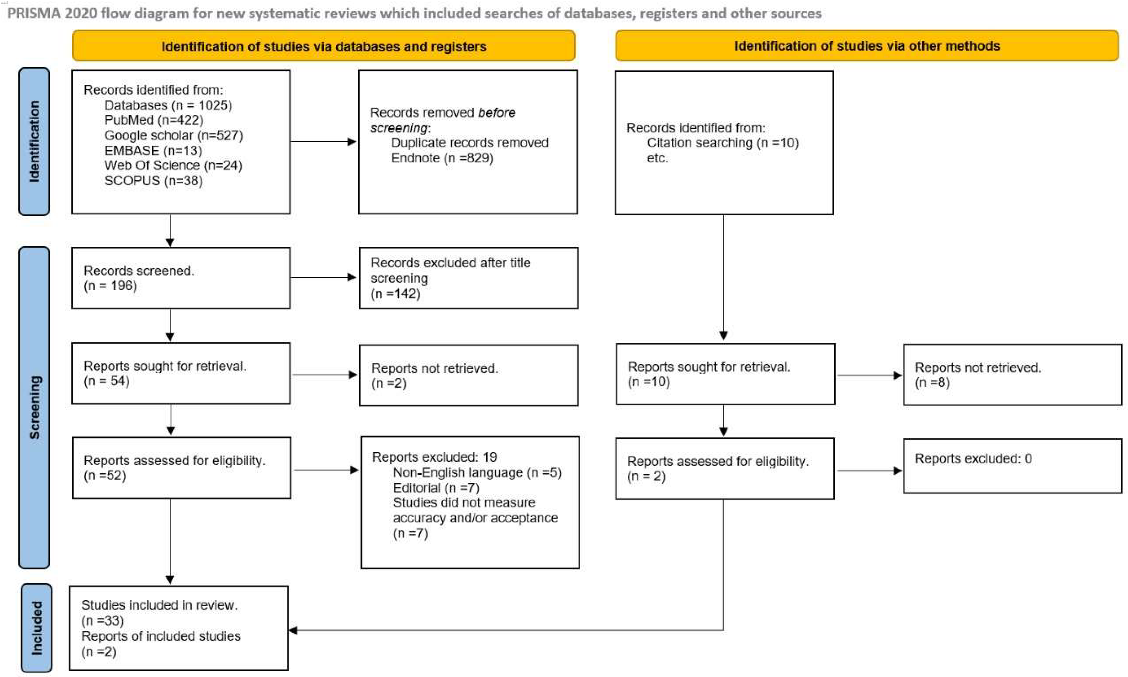
2.1. Search Strategy

The author searched five electronic databases for publications between January 2014 and January 2024: Web of Science, Medline/PubMed, Google Scholar, EMBASE, and SCOPUS. The following relevant key terms used were “3D-printed RPD”, “Rapid prototyping”, “Three-dimensional printing”, “Additive manufacturing”, “Dental polymers”, “Dental framework”, “Removable partial denture”, “PEEK denture”, “PMMA denture”, “Dentists’ perception”, and “Laboratory studies”. The complete list of MesH terms and accessed date are noted in Supplementary Materials Table S1.
The reviewer retrieved data, entered it into the Endnote file, and removed duplicates, followed by data analysis and synthesis. Furthermore, full texts of potential articles were downloaded for further screening.

2.2. Inclusion and Exclusion Criteria

The articles included in this scoping review followed strict inclusion criteria like in vitro studies (experimental, laboratory-based); in vivo studies, clinical trials, and comparative studies of 3D-printed removable partial dentures; clinical case reports, dentists’ perception of acceptance; articles published in English language and in peer-reviewed journals in past ten years. Studies on subtractive and conventional techniques for RPD fabrication, review articles, conference proceedings, and editorials were excluded.

2.3. Data Extraction and Clinical Outcomes

The parameter of interest was extracted from each study and entered into an Excel sheet. These parameters were as follows: author, country, year of study, study design, Kennedy’s classification of edentulism, control group, 3D printer utilized, source of reference for RPD design, and primary outcome. Additionally, in the second table, the parameters analyzed were the type and the method of assessment, the area of measurement, and the materials utilized for RPD fabrication. The primary outcome for each study was presented to analyze the accuracy of the RPD fabricated by 3D printing. Finally, in the third table, the parameters measured were the perception of dentists and prosthodontists on clinical performance and long-term prognosis on acceptance of 3D-printed RPDs.

3. Results

A total of 1025 articles were retrieved from an initial search on the five electronic databases. Of these, 829 were excluded as duplicates, yielding 196 articles screened with the abstract and titles. One hundred forty-two articles were excluded as irrelevant, two reports could not be retrieved, and fifty-two were extracted and included for full-text screening. During the full-text screening, nineteen articles were excluded for reasons such as non-English language, year of publication, not evaluating accuracy and acceptance of 3D-printed RPDs, and studies on crowns and veneers. A manual search of the bibliography included two articles. Finally, 35 articles were included in this scoping review and proceeded with qualitative analysis (Figure 1). Litmaps were evaluated to determine the relationship between the included studies and to thoroughly examine the literature related to the review question (Figure 2).
The results of the study were divided into three parts: (1) basic characteristics of the included studies, (2) comparison of methods utilized for assessment of accuracy of fit of 3D-printed RPDs, and (3) dentists’ perception of acceptance of 3D-printed RPDs. The themes and sub-themes were developed and analyzed in the result sections and are discussed in further sections.

3.1. Characteristics of Included Studies

Table 1 and Table 2 demonstrate the basic characteristics of the included studies. Among the 35 included studies, the maximum was published in 2019, followed by 2018, and only one was published in 2016. This trend shows the importance of 3D printing technologies in dentistry after 2017. Of the included studies, 12 were qualitative (without control), and 21 were quantitative (with control). Seventeen studies were of laboratory design [6,10,11,13,24,32,35,36,37,38,39,40], nine were clinical case reports [17,18,19,25,26,41,42,43,44], three were randomized control trials [28,45,46], and two were experimental studies [47,48] concentrating on the accuracy and acceptance of 3D-printed RPDs in dentistry. Only two studies were cross-sectional and demonstrated dental clinicians’ knowledge, attitude, and awareness regarding the acceptance of 3D-printed dentures [30,31]. Twenty-six studies utilized Kennedy’s classification for edentulism, with a dominance of Kennedy’s class I followed by classes II and III, with or without modifications. However, three studies used simulation of the first molar [27,28,39], while one study utilized the dentate model [45], and two studies did not mention any classification [19,36].
Twelve studies included conventional RPDs; four studies used the stone model [22,32,40,48]; one study used replica models as a control group [19]; and three studies used intraoral scanners [17,44,49]. In comparison, the remaining twelve studies assessed accuracy without a control group.
Regarding the types of printing, twenty studies utilized direct printing techniques like stereolithography, digital light prototyping, selective laser melting, etc. Three studies used indirect printing [28,50,51], including direct printing of metal or resin framework and casting pattern in conventional techniques, and three studies used both direct and indirect printing techniques [14,27,38]. As the primary outcome of this scoping review, almost all included studies demonstrated the accuracy of 3D-printed RPDs. Twelve studies without a control group reported better accuracy and fit of digitally manufactured RPDs, while one stated no significant difference [28]. However, four studies with the control group (lost wax technique) reported better accuracy with 3D-printed RPDs [6,10,47,52]. In comparison, three studies reported a lesser fit for 3D-printed RPDs than the conventional technique, but the authors suggested a clinically acceptable fit for 3D-printed RPDs [17,26,53].
Table 1. Basic characteristics of the included studies (quantitative studies).
Sr No.Author/Year/CountryStudy DesignKennedy’s ClassificationControl GroupType of 3D PrinterOutcome
1Anan and Saadi 2015 [47]; SyriaIn vitro study (Experimental)Mandibular class III modification 1Traditional techniqueLight curing model techniqueFit accuracy of 3D-printed RPDs was better than traditional
2Lee et al., 2017 [19]; South KoreaClinical reportMaxillary and mandibular class IReplica techniqueDigital light projection (DLP)The RPD constructed with the replica technique has varied results
3Ye et al., 2017 [46]; ChinaRandomized control trialMandibular class I modification 1Lost wax- techniqueStereolithography (SLA)The single design RPD prepared with a 3D printer was more accurately fit than those with the traditional technique
4Arnold et al., 2018 [6]; GermanyLaboratory studyMaxillary class III modification 2Lost wax techniqueDirect and indirect 3D printing techniquesThe 3D-printed RPD has less fit
5Soltanzadeh et al., 2018 [50]; USALaboratory studyMaxillary class III modification 1Lost wax techniqueIndirect printingThe conventional RPD was more accurate
6Torii et al., 2018 [28]; JapanExperimental studyFirst molar simulation Lost wax techniqueindirect printing (CAD)No significant difference was reported in the accuracy of fit within both techniques
7Bajunaid et al., 2019 [10]; Saudi ArabiaLaboratory studyMandibular class IIILost wax techniqueDirect CAD printers3D-printed RPDs are more accurate
8Chen et al., 2019 [35]; ChinaLaboratory studyAll classesLost wax techniqueDirect CAD printing technique3D-printed RPDs are less fitting but clinically acceptable
9Negm, Aboutaleb, and Alam-Eldein 2019 [22]; EgyptLaboratory studyMaxillary class IStone cast modelStereolithography (SLA)RPDs prepared by 3D printers have less accuracy
10Carneiro Pereira et al., 2019 [49]; BrazilClinical reportMandibular class III modification 1Intra-oral scanner (Patients mouth)Direct and indirect printing techniquesThe 3D-printed RPDs have an acceptable clinical fit
11Tregemen et al., 2019 [48]; South CarolinaClinical trialMandibular and maxillary class I, II, and IIIStone cast modelSelective laser melting3D-printed RPDs have a better fit
12Hayama et al., 2019 [17]; ChinaClinical reportMandibular class IIntraoral scanners Direct digital technique3D-printed RPD have an acceptable fit
13Oka et al., 2019 [13]; JapanLaboratory studyMandibular and maxillary class I, II, and IIILost wax techniqueDirect CAD printing technique3D-printed RPD have an acceptable fit
14Wu et al., 2020 [44]; ChinaClinical reportMandibular class I modification 1Intraoral scannersSelective laser melting3D-printed RPD have an acceptable fit
15Xie et al., 2020 [39]; ChinaLaboratory studyFirst molar simulationLost wax techniqueDirect CAD printing3D-printed RPDs have better fit
16Yoon et al., 2021 [40]; ChinaLaboratory studyMaxillary class IStone castDirect 3D printingAcceptable fit
17Hussein and Hussein 2022 [11]; Saudi ArabiaLaboratory studyMaxillary class III and Mandibular class 1Lost wax techniqueDigital light processingNo effect on the accuracy of fit
18Saadaldin et al., 2022 [36]; EgyptLaboratory StudyNot mentionedLost wax techniqueSelective laser melting techniqueBetter accuracy fit of SLM-prepared RPDs
19Rokshad et al., 2022 [24]; GermanyLaboratory studyMaxillary class III modification 1Convectional techniqueDigital light processingAcceptable fit
20Peng et al., 2022 [14]; ChinaLaboratory studyMandibular class II modification 2Lost wax techniqueDirect and indirect 3D printing3D-printed RPDs have a better fit
21Grymak et al., 2023 [32]; New ZealandLaboratory studyMaxillary class III modification 1Stone castSelective laser melting3D-printed RPDs have better acceptance
Table 2. Basic characteristics of included studies (qualitative studies).
Sr No.Author/Year/CountryStudy DesignKennedy’s ClassificationType of 3D PrinterOutcome
1.Kattadiyil et al., 2014 [26]; USAClinical reportMaxillary class IIIStereolithographicThe finished prosthesis was successfully placed and used by the patient
2.Husain Omran 2014 [54]; Saudi ArabiaCase reportMaxillary class IDigital light prototypingWell-fitted silicone framework
3.Lee and Lee 2015 [18]; South KoreaCase reportMaxillary class IStereolithographicFit accuracy is satisfactory
4.Mansour et al., 2016 [43]; USAClinical reportMaxillary class I modification 1Stereolithography (SLA)Fit of RPD prepared by rapid prototyping was highly satisfactory
5.Batalha and Araújo 2017 [41]; BrazilClinical reportMandibular class I modification 1Digital light projection(DLP)The accuracy of fit was satisfactory
6.Hu, Pei, and Wen 2017 [42]; ChinaClinical reportMaxillary class IStereolithography (SLA)The 3D-printed RPD is the best alternative to conservative RPD, and the accuracy of fit is acceptable
7.Gan et al., 2018 [45]; ChinaRandomized control trialsDentateSelective laser melting (SLM)RPDs designed with 3D printers have an acceptable fit
8.Katreva et al., 2018 [25]; BulgariaClinical case reportMandibular class IStereolithography (SLA)3D-printed RPD appears to be more precise and accurate
9.Tasaka et al., 2019 [51]; JapanLaboratory studyMandibular class II modification 1Indirect printing RPD fabricated by 3D printer has an acceptable fit
10.Takahashi et al., 2020 [38]; USALaboratory studyFirst molar simulationDirect and indirect printingAcceptable fit
11.Tasaka et al., 2020 [27]; JapanLaboratory studyMandibular class II modification 2Direct and indirect printing3D-printed RPDs have a better fit
12.Cabrita et al., 2021 [55]; USAClinical reportMandibular class ISelective laser melting3D-printed RPD have an acceptable fit

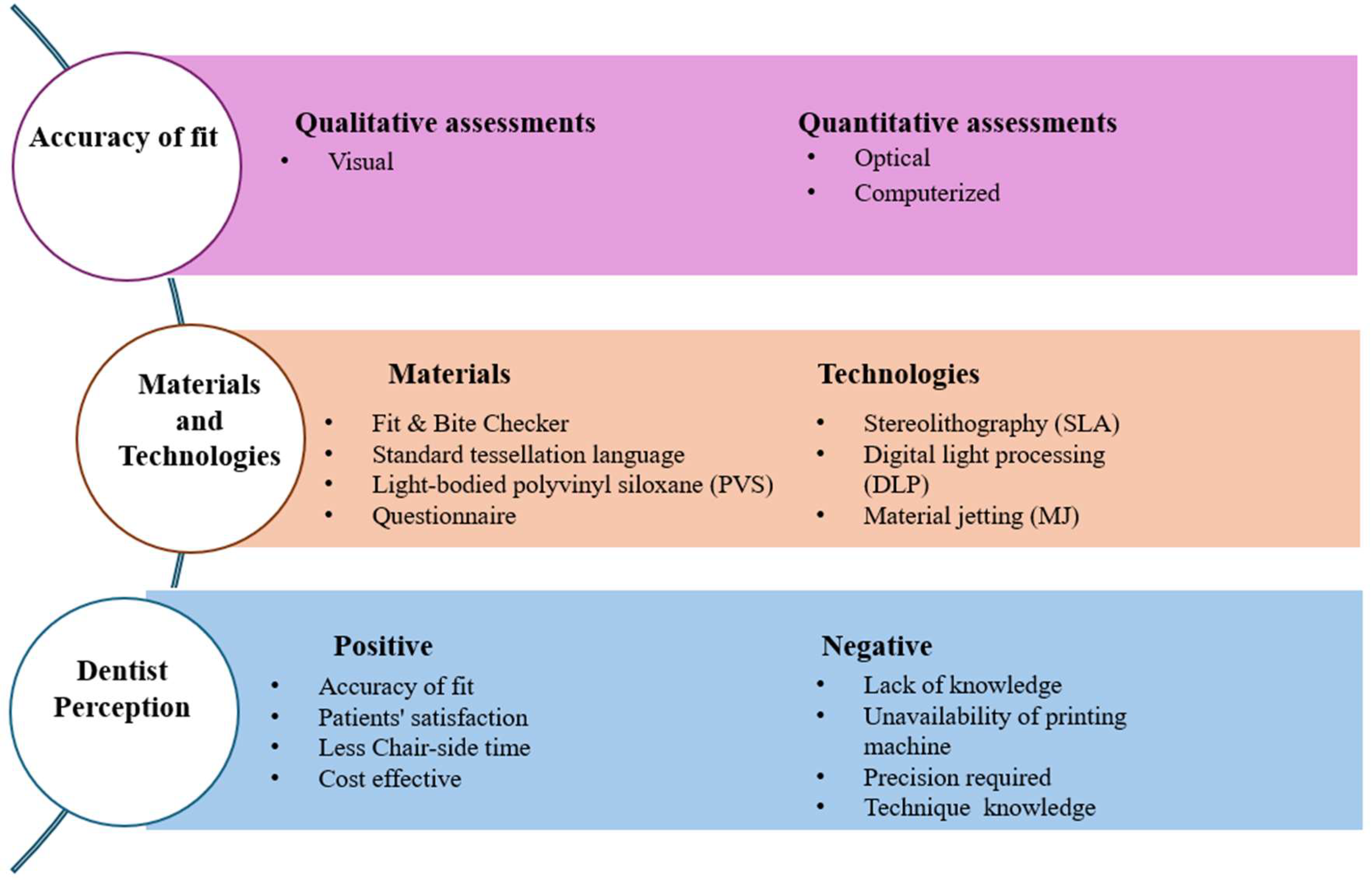
3.2. Comparison of the Method Utilized for Assessment of Fit of 3D-Printed RPDs

The method of assessment is divided into two categories: qualitative and quantitative. The qualitative method for detecting the accuracy of fit is visual inspection, which is usually done in clinical settings and does not require any numerical data. At the same time, the quantitative assessment for analyzing the accuracy is divided into two categories: optical inspection (microscopes) and computer software (Table 3). Quantitative methods usually require the control group to confirm the findings, while qualitative methods involve dentist experience and perception. A comparison between these two assessment methods and their sub-themes will be discussed further.

3.3. Qualitative Assessment Method (Visual Method)

The qualitative assessment method involves dentists’ perception and experience and relies on materials, manufacturing techniques, and gap registration. Eight studies utilized qualitative methods, such as direct inspection (naked eyes, mouth mirror, and probe) or tactile sensation, to determine the overall adaptation of 3D-printed RPDs [26,41,42,44,46,48,49,55]. Of these, three studies demonstrated both qualitative and quantitative methods to check the accuracy of fit of 3D-printed RPDs [40,43,46].
Seven studies used silicon material as a registration material to fill the gap between the fitting surfaces of RPDs and the cast model for measuring the accuracy of fit [6,19,32,38,41,42,43]. Some studies used silicone to demonstrate the accuracy of fit of RPD clasps prepared by 3D printers and conventional RPDs. Two studies used polyvinyl siloxane as a registration material to assess the accuracy of fit of RPD prepared by 3D printers [36,55]. These studies used dial calipers to measure the gaps between the clasp assembly and rest seats. The method to analyze the accuracy of fit with the help of a caliper is most used in clinics. However, using this method, it is not easy to measure the thickness accurately due to the elasticity of the silicone materials. Secondly, placing the material has a chance for distortion, resulting in inaccurate findings. Therefore, the results of the studies utilizing silicone material to register the gap could be erroneous. The same points apply to the studies that evaluated visual and tactile sensation to evaluate the accuracy of fit.

3.4. Quantitative Assessment Method (Computerized and Optical)

This method is divided into two categories: optical and computerized. In the optical method, the accuracy of fit was measured in two dimensions, mainly vertical and horizontal. This method demonstrates the accuracy of fit by analyzing gaps between the RPD components and internal discrepancies between the underlying tissue. Eight studies used optical methods to demonstrate the accuracy of fit [11,22,27,35,39,40,43,44], while one study used both visual and optical methods [28]. Of these, five studies utilized a stereomicroscope [18,19,27,45,46], two studies used profile projectors [28,51], and one study used a light microscope [6]. In the optical method, various components of RPDs are studied, like clasps, proximal plates, and major connectors, to check the accuracy of fit. Optical measurement is performed with or without the help of the registration method. Among the studies included, four studies utilized light-bodied polyvinyl siloxane (PVS) [6,10,35,44], three studies used fit and bite checkers [17,53,55], while two studies did not use any registration materials [39,46].
A study used a light microscope at 560 magnification to determine the accuracy of fit of RPDs prepared by different CAD/CAM systems and compared them to conventionally prepared RPDs [6]. The RPDs produced by various CAD/CAM techniques were indirect prototyping with the lost wax technique, indirect milling (wax milling with the conventional method), and direct milling with resin polymers. These prepared models were placed on the clasp model, and the accuracy of 12 prepared canine clasps of each group was determined at three vertical and three horizontal areas. This study determined that vertical and horizontal distances are essential concepts in measuring accuracy [6]. Using a digital microscope at 15X in micrometers, another study compared the accuracy of fit of 15 RPDs fabricated using the stereolithography technique to 15 RPDs fabricated by the conventional method using light-bodied polyvinyl siloxane (PVS) [32]. Silicone registration materials measured the fit in this study, and a total of 348 images were used to evaluate the accuracy of fit [32].
Furthermore, one study compared 30 RPDs fabricated by the conventional method and the light-curing method from three different angles and concluded that there were clinically acceptable marginal gaps in both methods. The authors of this study also commented that the framework’s accuracy could differ according to the time required for fabrication and the material utilized [44]. In another study with 24 machine-milled casts, it was reported that the accuracy of fit was better for the maxillary major connector than the mandibular connector or control group [46].
The computerized assessment method mainly depends on standard tessellation language files (STL files) for the reference and development of the output of RPD. Eleven studies used the computerized method for assessment. The measurement of accuracy was performed in three dimensions, including the gap distance (n = 4), average deviation (n = 2), trueness (n = 3), and adaptation (n = 3). A study by Rokshad et al. used two measurement types (adaptation and gap distance) [24]. All the studies used different software to assess the accuracy of fit; for example, eight studies used Geomagic software, two used GOM Inspect [27,51], and one did not mention the name of the software [13]. Additionally, a conceptual map was developed from the qualitative and quantitative assessments (Figure 3).

Dentist’s Perception of Acceptance of 3D-Printed RPDs

Since 3D-printed RPDs are most used in modern dentistry, there are limited data available on dentists’ perceptions of the acceptance of 3D-printed RPDs. Only two studies (Table 4) have been published to check dentists’ perception [30,31], of which only one study has checked long-term prognosis [31]. Dentists who participated in both studies suggested that patients accept 3D-printed RPDs well and that they are cost effective. However, dentists believe there is a considerable gap in the knowledge of using 3D-printed RPDs, and this topic should be incorporated in the dental curriculum. Regarding patients’ satisfaction, both studies mentioned that due to less chair-side time and proper retention, the 3D RPDs were accepted by the patients.

4. Discussion

This scoping review aimed to provide comprehensive evidence on methods used to assess the accuracy and acceptance of 3D-printed RPDs in clinical practice. Three primary criteria were checked for accessing the accuracy of RPD: (1) all the rest should be adequately seated on their rest seats, (2) all the rigid elements should touch the teeth, and (3) major connectors should not impinge the underlining soft tissue or palate and the visible space should not exceed 1 mm. Initially, visual and tactile examinations were used in the clinical setting. This technique solely depends on clinicians’ experience, which may vary accordingly. Unfortunately, a visual inspection will not provide a proper quantitative assessment. Moreover, adequate dentist training and RPD calibration are required to obtain accurate and acceptable results.
The optical assessment method employs optical instruments such as light microscopes or stereomicroscopes to achieve more precise fit measurements than visual inspection. This assessment can be conducted with or without registration materials that capture the gap between the removable partial denture (RPD) framework and the opposing surface. Various materials have been utilized, including polyvinyl siloxane (PVS) [10,45,46], Fit & Bite Checker [19,27,28], and radiopaque fit testing material [13]. However, optical assessment has certain limitations, such as the challenge of precisely identifying measurement sites and the inability to comprehensively evaluate the framework’s overall fit. Additionally, the accuracy of the space measured by assessing the thickness of the silicone registration materials may be compromised due to potential distortion or tearing of the material upon removal from the mouth [56]. Some studies have combined visual and optical methods for accurate assessment results.
The advent of digital technology has led to the development of computer-based software that improves the accuracy of accessing RPD fitting by optimizing the alignment of the RPD framework with the opposing surface at various sites. This technology generates a virtual color map, where different colors represent varying degrees of fit. The use of color mapping allows clinicians to detect over-pressed or misfit areas across more than 500 points [10,21,35]. This method significantly increases the number of comparison points compared to traditional optical and visual methods [56,57,58]. Additionally, the superimposition of a 3D-fabricated RPD with the original CAD data provides both qualitative and quantitative insights into dimensional discrepancies, trueness, and the accuracy of the printing process.
For evaluation of the overall accuracy of 3D-printed RPDs, it is advisable to use virtual superimposition of the original CAD design with the 3D-printed RPD via specialized surface-matching software, recording the average deviation. This deviation can be quantified as minimum, maximum, and average values. Notably, only one study [48] utilized the root mean square (RMS) to assess the trueness of 3D-printed RPDs. RMS is an effective metric for comparing prediction errors within different models or configurations of a particular variable. However, it cannot be used to compare different variables due to its scale dependence [17]. Most studies in this review reported low average deviation, indicating high trueness in 3D-printed RPDs, suggesting that the accuracy of these devices is promising and clinically acceptable. The high trueness of 3D-printed RPDs may be attributed to reduced laboratory errors and decreased inter-operator variability [10,14,27]. Data from recent studies indicate that over the past three years, software-based methods for assessing RPD fit have produced more accurate and reliable results, both in laboratory settings and clinically. Oka et al. (2016) introduced a novel method for testing dental prostheses by combining the silicone replica technique with micro-computed tomography (μCT) [13]. This approach can be used during the try-in stage, as it provides non-invasive and accurate fit evaluation. Despite its potential, this method has not been employed in the included studies for assessing RPD framework fit, likely due to the high cost of CT scans.
The studies in this scoping review present mixed results regarding the fit and accuracy of RPDs fabricated using 3D printing techniques. While some studies report promising outcomes, others indicate poorer fit and more significant dimensional discrepancies in 3D-printed frameworks. These varying results may be due to the different protocols employed during the 3D-printing process, including variations in light intensity, printing direction, layer thickness, and number, the amount of supporting material used, and post-processing heat treatments [39,45].
Differences in polishing procedures may also influence the outcomes [10]. It has been reported that the polishing procedure can significantly impact fit accuracy, particularly on the intaglio surface of the framework’s rest component [57]. Al Motardi (2020) suggested meticulous finishing and polishing may enhance fit precision, while excessive finishing could lead to unnecessary metal removal from the internal surface [2]. Brudvik and Reimers (1992) observed an average metal loss of 127 μm from the surface of a Co-Cr framework after finishing and polishing [59]. Careful sandblasting and polishing are essential to minimize their effects on the intimate fit of the RPD framework [57]. The findings of this review revealed inconsistent results regarding the fit of 3D-printed RPDs when different finishing and polishing protocols were applied. No study has identified the optimal polishing protocol for 3D-printed RPD frameworks. Therefore, further research is needed to evaluate the impact of polishing on the dimensional accuracy of digitally fabricated frameworks.
The current scoping review has also focused on dentists’ perceptions and patient-reported outcomes for assessing the long-term prognosis of digitally fabricated RPDs. Only two cross-sectional studies have been conducted to report the perception of dentists. These studies have shown that dentists and patients were highly satisfied with digitally fabricated dentures over conventional ones [30,31]. The main reason for this could be better fit and retention, less chair-side time, and a lighter framework than conventional ones. Further clinical studies are required to evaluate dentist perception of acceptance of 3D-printed RPDs.
One of the strengths of this scoping review is the high quality of the included studies, mainly clinical trials and crossover studies. These designs are advantageous as they require a small sample size and complex statistics. Hence, they provide accurate results when compared with other study designs. This scoping review also summarized dentist’s perceptions and patient-reported outcomes for digitally fabricated RPDs.
This scoping review has a few limitations; the articles included were in English and could not reach a definitive conclusion due to the restriction of published resources. Even though the accuracy of fit yielded a mixed result among the included studies, almost all reported a clinically acceptable range. However, further studies with a larger sample size and randomized control trials are required to support the findings of this review.

5. Conclusions

Based on the findings of this scoping review, evidence suggests that digitally fabricated RPDs are more acceptable and accurate than conventional ones. However, due to the scarcity of articles on this topic, the generalization of the result is not possible. Visual, optical, and computerized methods perform the accuracy of fit of digitally fabricated RPDs. This review reported that the automated method is more reliable than optical and visual. It can detect minute misfit and dimensional changes and can provide precise measurements.

Supplementary Materials

The following supporting information can be downloaded at: https://www.mdpi.com/article/10.3390/prosthesis7010016/s1, Table S1: Keywords and Subject Headings Used During the Search.

Funding

This research received no external funding.

Acknowledgments

The author would like to thank Priyanka Porwal for helping in searching for articles and assisting in data extraction.

Conflicts of Interest

The author declares no conflicts of interest.

References

  1. ISO 5725-1:1994; Accuracy (Trueness and Precision) of Measurement Methods and Results — Part 1: General Principles and Definitions. International Organization for Standardization: Geneva, Switzerland, 1994.
  2. Al Mortadi, N.; Alzoubi, K.H.; Williams, R. A Scoping Review on the Accuracy of Fit of Removable Partial Dentures in a Developing Digital Context. Clin. Cosmet. Investig. Dent. 2020, 12, 551–562. [Google Scholar] [CrossRef]
  3. Almufleh, B.; Emami, E.; Alageel, O.; de Melo, F.; Seng, F.; Caron, E.; Nader, S.A.; Al-Hashedi, A.; Albuquerque, R.; Feine, J.; et al. Patient satisfaction with laser-sintered removable partial dentures: A crossover pilot clinical trial. J. Prosthet. Dent. 2018, 119, 560–567.e561. [Google Scholar] [CrossRef] [PubMed]
  4. Dunham, D.; Brudvik, J.S.; Morris, W.J.; Plummer, K.D.; Cameron, S.M. A clinical investigation of the fit of removable partial dental prosthesis clasp assemblies. J. Prosthet. Dent. 2006, 95, 323–326. [Google Scholar] [CrossRef] [PubMed]
  5. Eggbeer, D.; Bibb, R.; Williams, R. The computer-aided design and rapid prototyping fabrication of removable partial denture frameworks. Proc. Inst. Mech. Eng. H 2005, 219, 195–202. [Google Scholar] [CrossRef]
  6. Arnold, C.; Hey, J.; Schweyen, R.; Setz, J.M. Accuracy of CAD-CAM-fabricated removable partial dentures. J. Prosthet. Dent. 2018, 119, 586–592. [Google Scholar] [CrossRef]
  7. Bibb, R.; Eggbeer, D.; Williams, R. Rapid manufacture of removable partial denture frameworks. Rapid Prototyp. J. 2006, 12, 95–99. [Google Scholar] [CrossRef]
  8. Campbell, S.D.; Cooper, L.; Craddock, H.; Hyde, T.P.; Nattress, B.; Pavitt, S.H.; Seymour, D.W. Removable partial dentures: The clinical need for innovation. J. Prosthet. Dent. 2017, 118, 273–280. [Google Scholar] [CrossRef] [PubMed]
  9. Williams, R.J.; Bibb, R.; Eggbeer, D.; Collis, J. Use of CAD/CAM technology to fabricate a removable partial denture framework. J. Prosthet. Dent. 2006, 96, 96–99. [Google Scholar] [CrossRef] [PubMed]
  10. Bajunaid, S.O.; Altwaim, B.; Alhassan, M.; Alammari, R. The Fit Accuracy of Removable Partial Denture Metal Frameworks Using Conventional and 3D Printed Techniques: An In Vitro Study. J. Contemp. Dent. Pract. 2019, 20, 476–481. [Google Scholar] [CrossRef]
  11. Hussein, M.O.; Hussein, L.A. Trueness of 3D printed partial denture frameworks: Build orientations and support structure density parameters. J. Adv. Prosthodont. 2022, 14, 150. [Google Scholar] [CrossRef]
  12. Alharbi, N.; Osman, R.B.; Wismeijer, D. Factors Influencing the Dimensional Accuracy of 3D-Printed Full-Coverage Dental Restorations Using Stereolithography Technology. Int. J. Prosthodont. 2016, 29, 503–510. [Google Scholar] [CrossRef] [PubMed]
  13. Oka, Y.; Sasaki, J.; Wakabayashi, K.; Nakano, Y.; Okamura, S.Y.; Nakamura, T.; Imazato, S.; Yatani, H. Fabrication of a radiopaque fit-testing material to evaluate the three-dimensional accuracy of dental prostheses. Dent. Mater. 2016, 32, 921–928. [Google Scholar] [CrossRef]
  14. Peng, P.W.; Hsu, C.Y.; Huang, H.Y.; Chao, J.C.; Lee, W.F. Trueness of removable partial denture frameworks additively manufactured with selective laser melting. J. Prosthet. Dent. 2022, 127, 122–127. [Google Scholar] [CrossRef] [PubMed]
  15. Torabi, K.; Farjood, E.; Hamedani, S. Rapid Prototyping Technologies and their Applications in Prosthodontics, a Review of Literature. J. Dent. 2015, 16, 1–9. [Google Scholar]
  16. Lang, L.A.; Tulunoglu, I. A critically appraised topic review of computer-aided design/computer-aided machining of removable partial denture frameworks. Dent. Clin. N. Am. 2014, 58, 247–255. [Google Scholar] [CrossRef]
  17. Hayama, H.; Fueki, K.; Wadachi, J.; Wakabayashi, N. Trueness and precision of digital impressions obtained using an intraoral scanner with different head size in the partially edentulous mandible. J. Prosthodont. Res. 2018, 62, 347–352. [Google Scholar] [CrossRef]
  18. Lee, J.H. Completely digital approach to fabricating a crown under an existing partial removable dental prosthesis by using an intraoral digital scanner in a single appointment. J. Prosthet. Dent. 2016, 115, 668–671. [Google Scholar] [CrossRef]
  19. Lee, J.W.; Park, J.M.; Park, E.J.; Heo, S.J.; Koak, J.Y.; Kim, S.K. Accuracy of a digital removable partial denture fabricated by casting a rapid prototyped pattern: A clinical study. J. Prosthet. Dent. 2017, 118, 468–474. [Google Scholar] [CrossRef]
  20. Mamoun, J.S. The path of placement of a removable partial denture: A microscope-based approach to survey and design. J. Adv. Prosthodont. 2015, 7, 76–84. [Google Scholar] [CrossRef] [PubMed]
  21. Baig, M.R.; Tan, K.B.; Nicholls, J.I. Evaluation of the marginal fit of a zirconia ceramic computer-aided machined (CAM) crown system. J. Prosthet. Dent. 2010, 104, 216–227. [Google Scholar] [CrossRef]
  22. Negm, E.E.; Aboutaleb, F.A.; Alam-Eldein, A.M. Virtual Evaluation of the Accuracy of Fit and Trueness in Maxillary Poly(etheretherketone) Removable Partial Denture Frameworks Fabricated by Direct and Indirect CAD/CAM Techniques. J. Prosthodont. 2019, 28, 804–810. [Google Scholar] [CrossRef] [PubMed]
  23. Preshaw, P.M.; Walls, A.W.; Jakubovics, N.S.; Moynihan, P.J.; Jepson, N.J.; Loewy, Z. Association of removable partial denture use with oral and systemic health. J. Dent. 2011, 39, 711–719. [Google Scholar] [CrossRef] [PubMed]
  24. Rokhshad, R.; Tehrani, A.M.; Nahidi, R.; Zarbakhsh, A. Fit of removable partial denture frameworks fabricated from 3D printed patterns versus the conventional method: An in vitro comparison. J. Prosthet. Dent. 2024, 131, 1144–1149. [Google Scholar] [CrossRef]
  25. Katreva, I.; Dikova, T.; Tonchev, T. 3D printing–an alternative of conventional crown fabrication: A case report. J. IMAB–Annu. Proceeding Sci. Pap. 2018, 24, 2048–2054. [Google Scholar] [CrossRef]
  26. Kattadiyil, M.T.; Mursic, Z.; AlRumaih, H.; Goodacre, C.J. Intraoral scanning of hard and soft tissues for partial removable dental prosthesis fabrication. J. Prosthet. Dent. 2014, 112, 444–448. [Google Scholar] [CrossRef] [PubMed]
  27. Tasaka, A.; Shimizu, T.; Kato, Y.; Okano, H.; Ida, Y.; Higuchi, S.; Yamashita, S. Accuracy of removable partial denture framework fabricated by casting with a 3D printed pattern and selective laser sintering. J. Prosthodont. Res. 2020, 64, 224–230. [Google Scholar] [CrossRef] [PubMed]
  28. Torii, M.; Nakata, T.; Takahashi, K.; Kawamura, N.; Shimpo, H.; Ohkubo, C. Fitness and retentive force of cobalt-chromium alloy clasps fabricated with repeated laser sintering and milling. J. Prosthodont. Res. 2018, 62, 342–346. [Google Scholar] [CrossRef]
  29. Mai, H.Y.; Mai, H.N.; Kim, H.J.; Lee, J.; Lee, D.H. Accuracy of Removable Partial Denture Metal Frameworks Fabricated by Computer-Aided Design/Computer-Aided Manufacturing Method: A Systematic Review and Meta-Analysis. J. Evid. Based Dent. Pract. 2022, 22, 101681. [Google Scholar] [CrossRef] [PubMed]
  30. Chobe, A.; Sushma, R.; Shashikiran, N.; Kore, A.R.; Shivakumar, K.; Vande, A. Study to Assess the Knowledge, Attitude and Practices of Additive Manufacturing Technology by Dental Practitioners in Dentistry across India-A Survey. Journal for ReAttach Ther. Dev. Divers. 2023, 6, 759–766. [Google Scholar]
  31. Urumova, M. Comparative study of retention of telescopic crowns fabricated by 3D printing (in vitro study). J. Int. Dent. Med. Res. 2023, 16, 13–19. [Google Scholar]
  32. Grymak, A.; Badarneh, A.; Ma, S.; Choi, J.J.E. Effect of various printing parameters on the accuracy (trueness and precision) of 3D-printed partial denture framework. J. Mech. Behav. Biomed. Mater. 2023, 140, 105688. [Google Scholar] [CrossRef]
  33. Azari, A.; Nikzad, S. The evolution of rapid prototyping in dentistry: A review. Rapid Prototyp. J. 2009, 15, 216–225. [Google Scholar] [CrossRef]
  34. Tricco, A.C.; Lillie, E.; Zarin, W.; O’Brien, K.K.; Colquhoun, H.; Levac, D.; Moher, D.; Peters, M.D.J.; Horsley, T.; Weeks, L.; et al. PRISMA Extension for Scoping Reviews (PRISMA-ScR): Checklist and Explanation. Ann. Intern. Med. 2018, 169, 467–473. [Google Scholar] [CrossRef] [PubMed]
  35. Chen, H.; Li, H.; Zhao, Y.; Zhang, X.; Wang, Y.; Lyu, P. Adaptation of removable partial denture frameworks fabricated by selective laser melting. J. Prosthet. Dent. 2019, 122, 316–324. [Google Scholar] [CrossRef]
  36. Saadaldin, S.A.; Rizkalla, A.S.; Eldwakhly, E.A.; Soliman, M.; Aldegheishem, A. Assessment of the fitness of removable partial denture frameworks manufactured using additive manufacturing/selective laser melting. Mater. Express 2022, 12, 735–742. [Google Scholar] [CrossRef]
  37. Soltanzadeh, P.; Su, J.-M.; Habibabadi, S.R.; Kattadiyil, M.T. Obturator fabrication incorporating computer-aided design and 3-dimensional printing technology: A clinical report. J. Prosthet. Dent. 2019, 121, 694–697. [Google Scholar] [CrossRef]
  38. Takahashi, K.; Torii, M.; Nakata, T.; Kawamura, N.; Shimpo, H.; Ohkubo, C. Fitness accuracy and retentive forces of additive manufactured titanium clasp. J. Prosthodont. Res. 2020, 64, 468–477. [Google Scholar] [CrossRef]
  39. Xie, W.; Zheng, M.; Wang, J.; Li, X. The effect of build orientation on the microstructure and properties of selective laser melting Ti-6Al-4V for removable partial denture clasps. J. Prosthet. Dent. 2020, 123, 163–172. [Google Scholar] [CrossRef]
  40. Yoon, J.-m.; Liu, Y.; Liu, Y.; Sun, Y.; Ye, H.; Zhou, Y. The accuracy of a novel 3D digital evaluation method of intraoral fitness for removable partial dentures. Comput. Biol. Med. 2022, 144, 105348. [Google Scholar] [CrossRef] [PubMed]
  41. Batalha, A.; Araújo, R.M. Development of removable partial dentures by using additive manufacture and casting processes. Arch. Mater. Sci. Eng. 2017, 87, 33–40. [Google Scholar] [CrossRef]
  42. Hu, F.; Pei, Z.; Wen, Y. Using Intraoral Scanning Technology for Three-Dimensional Printing of Kennedy Class I Removable Partial Denture Metal Framework: A Clinical Report. J. Prosthodont. 2019, 28, e473–e476. [Google Scholar] [CrossRef] [PubMed]
  43. Mansour, M.; Sanchez, E.; Machado, C. The Use of Digital Impressions to Fabricate Tooth-Supported Partial Removable Dental Prostheses: A Clinical Report. J. Prosthodont. 2016, 25, 495–497. [Google Scholar] [CrossRef] [PubMed]
  44. Wu, J.; Li, Y.; Zhang, Y. Use of intraoral scanning and 3-dimensional printing in the fabrication of a removable partial denture for a patient with limited mouth opening. J. Am. Dent. Assoc. 2017, 148, 338–341. [Google Scholar] [CrossRef]
  45. Gan, N.; Ruan, Y.; Sun, J.; Xiong, Y.; Jiao, T. Comparison of Adaptation between the Major Connectors Fabricated from Intraoral Digital Impressions and Extraoral Digital Impressions. Sci. Rep. 2018, 8, 529. [Google Scholar] [CrossRef] [PubMed]
  46. Ye, H.; Ning, J.; Li, M.; Niu, L.; Yang, J.; Sun, Y.; Zhou, Y. Preliminary Clinical Application of Removable Partial Denture Frameworks Fabricated Using Computer-Aided Design and Rapid Prototyping Techniques. Int. J. Prosthodont. 2017, 30, 348–353. [Google Scholar] [CrossRef]
  47. Anan, M.T.M.; Al-Saadi, M.H. Fit accuracy of metal partial removable dental prosthesis frameworks fabricated by traditional or light curing modeling material technique: An in vitro study. Saudi Dent. J. 2015, 27, 149–154. [Google Scholar] [CrossRef]
  48. Tregerman, I.; Renne, W.; Kelly, A.; Wilson, D. Evaluation of removable partial denture frameworks fabricated using 3 different techniques. J. Prosthet. Dent. 2019, 122, 390–395. [Google Scholar] [CrossRef]
  49. Carneiro Pereira, A.L.; Martins de Aquino, L.M.; Carvalho Porto de Freitas, R.F.; Soares Paiva Tôrres, A.C.; da Fonte Porto Carreiro, A. CAD/CAM-fabricated removable partial dentures: A case report. Int. J. Comput. Dent. 2019, 22, 371–379. [Google Scholar]
  50. Soltanzadeh, P.; Suprono, M.S.; Kattadiyil, M.T.; Goodacre, C.; Gregorius, W. An In Vitro Investigation of Accuracy and Fit of Conventional and CAD/CAM Removable Partial Denture Frameworks. J. Prosthodont. 2019, 28, 547–555. [Google Scholar] [CrossRef]
  51. Tasaka, A.; Kato, Y.; Odaka, K.; Matsunaga, S.; Goto, T.K.; Abe, S.; Yamashita, S. Accuracy of Clasps Fabricated with Three Different CAD/CAM Technologies: Casting, Milling, and Selective Laser Sintering. Int. J. Prosthodont. 2019, 32, 526–529. [Google Scholar] [CrossRef] [PubMed]
  52. Chen, G.X.; Zeng, X.Y.; Wang, Z.M.; Guan, K.; Peng, C.W. Fabrication of removable partial denture framework by selective laser melting. Adv. Mater. Res. 2011, 317, 174–178. [Google Scholar] [CrossRef]
  53. Hussein, M.O.; Hussein, L.A. Optimization of digital light processing three-dimensional printing of the removable partial denture frameworks; the role of building angle and support structure diameter. Materials 2022, 15, 2316. [Google Scholar] [CrossRef] [PubMed]
  54. Hussein, M.O.; Hussein, L.A. Novel 3D modeling technique of removable partial denture framework manufactured by 3D printing technology. Int. J. Adv. Res. 2014, 9, 686–694. [Google Scholar]
  55. Cabrita, J.P.; Mendes, T.A.; Martins, J.P.; Lopes, L. Removable partial denture metal framework manufactured by selective laser melting technology—A clinical report. Rev. Port. De Estomatol. Med. Dentária E Cir. Maxilofac. 2021, 62, 109–113. [Google Scholar] [CrossRef]
  56. Ahmed, N.; Abbasi, M.S.; Haider, S.; Ahmed, N.; Habib, S.R.; Altamash, S.; Zafar, M.S.; Alam, M.K. Fit Accuracy of Removable Partial Denture Frameworks Fabricated with CAD/CAM, Rapid Prototyping, and Conventional Techniques: A Systematic Review. Biomed. Res. Int. 2021, 2021, 3194433. [Google Scholar] [CrossRef]
  57. Rudd, R.W.; Rudd, K.D. A review of 243 errors possible during the fabrication of a removable partial denture: Part I. J. Prosthet. Dent. 2001, 86, 251–261. [Google Scholar] [CrossRef]
  58. Wang, C.; Shi, Y.-F.; Xie, P.-J.; Wu, J.-H. Accuracy of digital complete dentures: A systematic review of in vitro studies. J. Prosthet. Dent. 2021, 125, 249–256. [Google Scholar] [CrossRef]
  59. Brudvik, J.S.; Reimers, D. The tooth-removable partial denture interface. J. Prosthet. Dent. 1992, 68, 924–927. [Google Scholar] [CrossRef]
Figure 1. PRISMA flowchart for inclusion of studies 2020 [34].
Prosthesis 07 00016 g001
Figure 2. LitMap analysis for included studies, refs. [6,10,11,13,14,16,18,19,22,25,26,27,28,29].
Prosthesis 07 00016 g002
Figure 3. Conceptual map of results analyzed in the current scoping review.
Prosthesis 07 00016 g003
Table 3. Method of assessment for checking the accuracy of fit.
AuthorMethod of AssessmentType of MeasurementArea of Measurement
Kattadiyil et al., 2014 [26]Visual inspection (mouth mirror and probe)AdaptationOverall
Hussain Orman et al., 2014 [54]Optical (Microscope)Accuracy and adaptation Overall
Batalha and Araújo 2017 [41]Visual inspection (mouth mirror and probe)AdaptationOverall
Hu, Pei, and Wen 2017 [42]Visual inspection (mouth mirror and probe)AdaptationOverall
Lee et al., 2017 [19]Optical (Stereomicroscope)Internal discrepancyAll components
Ye et al., 2017 [46]Visual inspection (mouth mirror and probe)AdaptationOverall
Batalha and Araújo 2017 [26]Optical (Stereomicroscope)Gap distanceDifferent sections
Arnold et al., 2018 [6]Optical (Light microscope)Gap distanceClasp
Gan et al., 2018 [45]Optical (Stereomicroscope)Gap distanceAll components
Soltanzadeh et al., 2018 [50]Computerized (Geomagic)Gap distanceAll components
Torii et al., 2018 [28]Optical (Profile projector)Gap distanceRest, 3-point clasp
Bajunaid et al., 2019 [10]Optical (Digital microscope)Gap distancesRest
Chen et al., 2019 [35]Computerized (Geomagic NX image)Gap distanceOverall
Negm, Aboutaleb, and Alam-Eldein 2019 [22]Computerized (Geomagic)Gap distanceOverall
Oka et al., 2019 [13]Computerized Adaptation Overall
Soltanzadeh et al., 2018 [35]Computerized (Geomagic)TruenessAll components
Carneiro Pereira et al., 2019 [49]Visual inspection (mouth mirror and probe)AdaptationOverall
Tasaka et al., 2019 [51]Computerized (GOM Inspect)Average deviationAll components
Tregemen et al., 2019 [48]Visual inspection (mouth mirror and probe)AdaptationOverall
Takahashi et al., 2020 [38]Optical (Profile projector)Gap distanceClasp
Tasaka et al., 2020 [27]Computerized (GOM Inspect)Average deviationClasp
Wu et al., 2020 [44]Visual inspection (mouth mirror and probe)AdaptationOverall
Xie et al., 2020 [39]Optical (Stereomicroscope)Gap distance3-point clasp
Cabrita et al., 2021 [55]Visual inspection (mouth mirror and probe)AdaptationOverall
Peng et al., 2022 [14]Computerized (Geomagic)TruenessOverall
Rokshad et al., 2022 [24]Computerized (Geomagic Control X)Adaptation and Gap measurement All areas
Grymak et al., 2023 [32]Computerized (Geomagic Control X)AdaptationOverall
Hussain and Hussain 2022 [11]Computerized (Geomagic Control X)TruenessOverall
Table 4. Perception of dentists on acceptance of 3D-printed RPDs.
AuthorsStudy DesignDentist PerceptionLong-Term Prognosis
Urumova et al., 2021 [30]; BulgariaQuestionnaireCost effective and reduces chair-side timingMore clinical studies are required for the analysis of long-term prognosis
Chobe et al., 2023 [31]; IndiaQuestionnaireCost effective and easily acceptableNot mentioned
Disclaimer/Publisher’s Note: The statements, opinions and data contained in all publications are solely those of the individual author(s) and contributor(s) and not of MDPI and/or the editor(s). MDPI and/or the editor(s) disclaim responsibility for any injury to people or property resulting from any ideas, methods, instructions or products referred to in the content.

