Review

Use of Right Ventricular Assist Device Post-Left Ventricular Assist Device Placement

by
Shannon Parness
*,
Tori E. Hester
,
Harish Pandyaram
,
Panagiotis Tasoudis
and
Aurelie E. Merlo
Division of Cardiothoracic Surgery, Department of Surgery, School of Medicine, University of North Carolina at Chapel Hill, Chapel Hill, NC 27599, USA
*
Author to whom correspondence should be addressed.
Submission received: 7 March 2025 / Revised: 23 March 2025 / Accepted: 28 March 2025 / Published: 29 March 2025

Abstract

:
Right heart failure (RHF) is a common manifestation after left ventricular assist device (LVAD) placement and is associated with a high mortality rate. Historically, RV failure requiring an RVAD at the time of LVAD implantation has been associated with an especially high mortality. However, more recently, some studies have shown reasonable outcomes after LVAD implantation even when an RVAD is required, especially if RV failure is recognized early and treated with RV mechanical support. This article analyzes the current trends and studies investigating the use of RVAD placement post-LVAD implantation with an emphasis on the newest devices and treatment paradigms.

1. Introduction

As the use of left ventricular assist devices (LVADs) expands due to improving technology and improving outcomes, so too is the awareness of common complications after LVAD implantation. One of the most difficult complications to treat is right ventricular failure (RVF), which contributes to significant morbidity (especially renal failure) and mortality [1,2,3,4,5,6,7]. Due to the lack of a uniform definition of RVF post-LVAD placement, the incidence level varies per study, with some quoting a rate as high as 50% [1,5], but all acknowledge the increased mortality rate associated with this disease process [1,2,3,4,5]. One definition of RVF used by many studies is the need for an RVAD placement or the use of IV inotropes for more than 14 days post-LVAD placement [2]. The etiology of RVF post-LVAD likely has many factors. Studies list the use of cardiopulmonary bypass, positioning of the outflow graft over the RV, RV ischemia from hypotension, and septal shift toward the LV after LVAD placement as possibilities [4]. James and Smith explain that, because the interventricular septum assists with the contractile power of the RV, if the LVAD placement shifts the septum toward the left, the ejection power of the RV greatly decreases, leading to RVF [7]. Also, a European study found that the etiology of heart failure (HF) leading to LVAD placement can be an important predictor of RVF. They demonstrated that patients with non-ischemic HF undergoing LVAD placement are at a higher risk for developing RVF than those with an etiology of ischemic HF [8].
Grant et al. attempted to create a protocol to determine the likelihood of RVF post-LVAD placement. They found that reduced RV free-wall longitudinal strain prior to LVAD placement had a significant increase in RVF postprocedure; therefore, they determined this could be used as a predictive measure [2]. Determining whether the RV can handle LVAD placement prior to surgery is very important as mortality rates increase with RVF. This allows for the discussion of the biventricular assist device (BVAD) placement to happen early and prevent another procedure soon after LVAD placement. One study found that patients with a planned BVAD placement survived to discharge significantly more than those with a delayed BVAD, defined as LVAD followed by RVAD placement [3]. However, more recent RVADs allow for percutaneous placement so that a resternotomy does not need to take place, improving patient outcomes [9,10,11]. Also, risk assessments are now available to help predict who may be at a higher risk of RVF post-LVAD placement. For example, Fitzpatrick et al. created a risk assessment to determine whether patients would likely need BVAD support [12]. Matthews et al. also created a risk assessment to see whether patients were at high or low risk of RVF prior to LVAD placement [13]. Finally, the EUROMACS right-sided heart failure score was recently created to assess the risk of RVF prior to LVAD placement [14,15]. Overall, RVF is a prominent issue for patients needing to receive LVADs. This article will discuss what is currently known regarding RVADs and what research still needs to be carried out.

2. Right Ventricular Assist Device Placement

2.1. History

The use of pumps to support a failing heart first came about in the 1930s and 1940s [16]. In 1934, DeBakey developed the first roller pump, which was incorporated into the first successful cardiopulmonary bypass machine by Gibbon [16,17,18]. By the 1960s, left-sided heart bypasses using cannulas were being performed and intra-aortic balloon counter-pulsation technology had been developed [16,19,20]. While these strategies were designed to support a failing LV, it was thought they could be used to support the right side of the heart [16]. The first reports of using a balloon counter-pulsation for RV support were published in 1980 in which the balloon was sewn into a graft in the pulmonary artery (PA) [16,21]. By the 1960s, many different devices had been developed to assist the LV, and subsequent RV failure following the implementation of these devices was a common finding [16]. The first RVADs consisted of roller pumps, centrifugal pumps, and pneumatic sac-type pumps [16]. Additionally, the first RVADs consisted of valves with inflow and outflow cannulas and were largely pulsatile in nature [22]. By the 1990s, centrifugal pumps had been demonstrated to be superior to pulsatile pumps for the RV across multiple hemodynamic measures [23]. As development has progressed for these devices, second- and third-generation surgically implanted RVADs use rotational kinetic energy through roto-dynamic pumps to promote circulation [22]. Currently, surgically deployed RVADs via thoracotomy or open sternotomy employ extracorporeal centrifugal pumps with cannulation from the RA to PA [22]. RVADs that are percutaneously inserted are comparatively recent developments in the field [22].

2.2. Availability

While not solely referring to RVADs, there has been a large increase in the use of mechanical circulatory devices within the last 20 years [24]. According to an analysis of the Nationwide Inpatient Sample, the use of non-percutaneous, non-durable circulatory assist devices increased by 1511% from the years 2007 to 2011 [24]. For these same years, there was a 101% increase in the use of percutaneous circulatory assist devices [24]. While this demonstrates an increase in mechanical circulatory support devices, important limitations to the widespread use of RVADs remain.
There are currently no long-term Food and Drug Administration (FDA)-approved RVADs [7]. Most ventricular assist devices placed in the RV for long-term support were used off-label such as the HeartWare Ventricular Assist Device in a left- and right-atrial configuration [7,25]. An alternative strategy for permanent RV support is the use of durable, modified BVADs [7]. Additionally, the biventricular placement of two HeartMate 3 devices has been successful for those patients requiring permanent RV support [26]. Currently, the SynCardia Total Artificial Heart (TAH) is the only FDA-approved TAH system in the United States for bridge to transplant therapy [27]. BiVACOR is a TAH that is not currently FDA approved but is going through an Early Feasibility Study (EFS). Recently, the FDA approved an expansion of this study after this device was successfully implanted in five patients [28]. Finally, CARMAT is another non-FDA-approved TAH that is currently undergoing EFS in the United States as well [29].

3. Current Use

3.1. Percutaneously Implanted Devices

Intra-aortic balloon pumps (IABPs), for example the Arrow AC3 Optimus (Teleflex, Wayne, PA, USA), can indirectly enhance RV function by decreasing RV filling pressure through offloading the LV and by increasing right coronary perfusion [30]. IABPs can be useful in LV failure from myocardial infarction (MI) or cardiomyopathy but have not been shown to be effective in acute RV failure [31,32]. In addition, they are not very useful in patients with a durable LVAD due to decreased pulsatility.
The Impella RP is a microaxial pump that is inserted via the femoral vein that transports blood from the right atrium (RA)/inferior vena cava (IVC) to the PA up to 4–5 L/min [31]. It is most effectively used in RV failure from RV infarction or durable LVAD insertion [31]. It is approved for up to 14 day use in patients with acute right heart failure following LVAD insertion, heart transplant, MI, or open-heart surgery [33]. Its main advantages include the percutaneous insertion mechanism, the flow rate, and the single access site, while its main drawbacks include the femoral access requirement and risk of thrombosis with minimal anticoagulation [31]. As per the manufacturer, important contraindications include pulmonic and tricuspid regurgitation, stenosis, and the existence of a mechanical valve [33]. More recently, the Impella RP Flex has been designed. This is an Impella RP that can be inserted through the internal jugular vein, avoiding femoral access and allowing the patient to ambulate.
The ProtekDuo RVAD (CadiacAssist, Pittsburgh, PA, USA) is an extracorporeal device that utilizes centrifugal flow to pump blood from the RA/RV to the PA [22,31]. It has a flow rate of 4–5 L/min, and its cannula must be connected to an extracorporeal centrifugal pump to serve as an RVAD [31]. It is inserted percutaneously, most commonly through the internal jugular vein (IJV) under fluoroscopic or ultrasound guidance [34]. The key advantages of this device are the percutaneous insertion, the flow rate, and its single-site access [31]. Disadvantages to consider include the elevated risk of superior vena cava syndrome associated with a larger canula size [31] and the association with an increased mean PA pressure [22]. This device works best for acute RV failure for LVAD insertion [31]. It is approved for use for up to 6 days by the FDA [11].

3.2. Surgically Implanted Devices

The Surgical CentriMag RVAD (Abbott, Abbott Park, IL, USA) is a surgically implanted extracorporeal device that pumps blood from the RA/RV/IVC/superior vena cava (SVC) to the PA [31]. As per the manufacturer, it is approved for 30-day RV support [35]. It pumps blood up to 7 L/min and boasts a low rate of red blood cell damage, but its major drawback is the surgical insertion strategy [31]. Interestingly, some centers have inserted CentriMag devices percutaneously using a novel method for biventricular support [36]. One report demonstrated the percutaneous insertion and removal of an RVAD system using a ProtekDuo cannula in the IJV, withdrawing blood from the RA and returning it to the PA by a CentriMag system [37]. The CentriMag is most commonly used as a ventricular assist device rather than as a component of an ECMO circuit [38]. The complications of this device include bleeding, thrombosis, infection, hemolysis, renal complications, and neurologic complications [38].
Veno-arterial extracorporeal membrane oxygenation (VA-ECMO) can be surgically or percutaneously inserted and transports blood from the RA/SVC/IVC to the aorta through centrifugal flow. The Novalung system (Fresenius Medical Care, Bad Homburg vor der Höhe, Germany) is FDA approved for long-term use for more than 6 h [39]. It is a biventricular system with a flow rate of 3–5 L/min. A main advantage is that the VA-ECMO can be emergently inserted at the patient’s bedside [31]. The disadvantages include increased LV afterload and elevated risk of limb ischemia [31,40,41]. Importantly, the VA-ECMO is distinguished from typical ventricular assist devices because of the parallel circuit involved, unlike conventional ventricular assist devices [31]. Furthermore, a VA-ECMO used simultaneously with an LVAD presents a challenging case to manage due to lower flow rates and difficulty managing both the LV and RV with an ECMO system. The LVAD offloading the LV in instances of concurrent use of VA-ECMO has been described [42], and an LVAD, in this scenario, is better used as a vent for the LV [43].
The HeartMate 3 is a surgically implantable LVAD that may be used off-brand as an RVAD. When implanted on the right side, it pumps blood from the RA/RV to the PA at a rate of 4–6 L/min. When it is implanted in this configuration, it is sometimes called the “Heartmate 6”. One of the main advantages is that it is fully implantable, meaning patients can be discharged with the device [31,44]. The lower risk of thrombosis associated with the HeartMate 3 makes it an attractive RVAD option to reduce the risk of thromboembolic events in these patients [31,44,45].

3.3. Alternative Strategies

Following LVAD insertion, some centers practice delayed sternal closure when inserting LVADs [46,47]. An open chest following LVAD insertion offers hemodynamic benefits for the RV due to the additional space an open chest offers [47]. Yanagida et al. concluded that patients with delayed sternal closure experienced more postoperative bleeding, prolonged time to extubation, and extended inotropic support compared to those who underwent immediate closure [47]. These researchers also found no significant reduction in coagulopathy, cardiac tamponade, or RVF for patients who underwent delayed sternal closure [47]. Conversely, Giraldo-Grueso et al. found delayed sternal closure following LVAD insertion to be beneficial for RVF [48]. Delayed sternal closure has been associated with an increased risk for pneumonia postoperatively but not with sepsis, soft tissue infections, or deep tissue infections [49]. However, prolonged delayed sternal closure with a median duration of up to 12 days is linked to increased risk for pneumonia, bacteremia, and sepsis in the postoperative period even with longer prophylactic antibiotic use [50].

4. Indications for Right Ventricular Assist Device Placement

4.1. Right Ventricular Failure

An RVF diagnosis is based on clinical signs, including increased central venous pressure, jugular venous distention, and peripheral edema [51]. In patients who have a Swan–Ganz catheter, hemodynamic changes such as a reduction in PA pulsatility or an increase in central venous pressure (CVP) can be determined readily [52]. Echocardiography is the initial approach in the assessment of RV morphology and function. Patients with RV dysfunction may require the use of an RVAD as a bridge to recovery or, in some cases, as a bridge to heart transplant. The use of RVADs is associated with several drawbacks. A consequence of RVAD placement may be perioperative bleeding, which increases pulmonary strain on the RV, thereby worsening the RVF [53]. Other adverse events of RVAD implantation also include infection, valve thrombosis, malignant arrhythmia, and the requirement for reintubation due to respiratory complications [54].
In order to medically manage RVF, reducing the pressure in the pulmonary vessels is warranted to unload the RV. Nitric oxide is a potent vasodilator that is used to help decrease the pressure within the right heart as well as to improve oxygenation [55]. Other ways to assist the right heart with medical management include relieving the excess fluid, obtaining a stable heart rhythm, and increasing contractility [55].

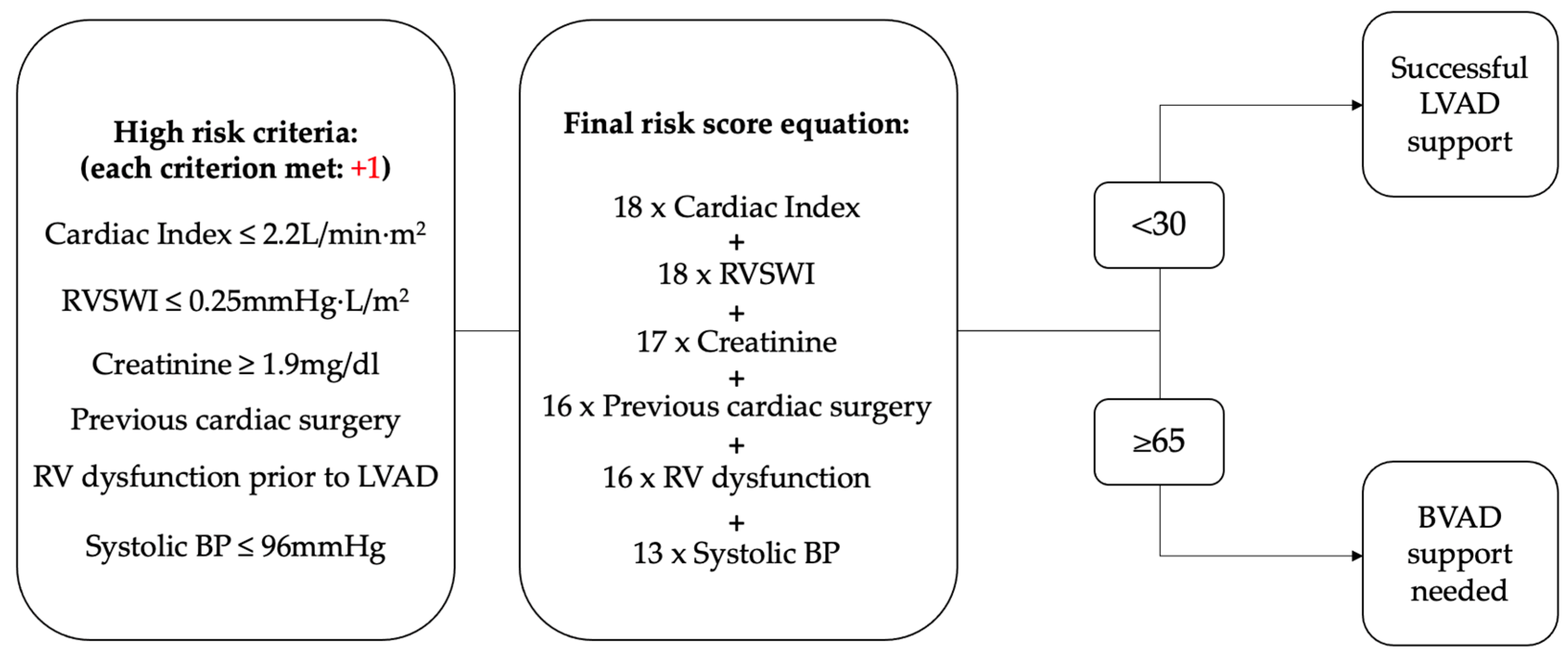
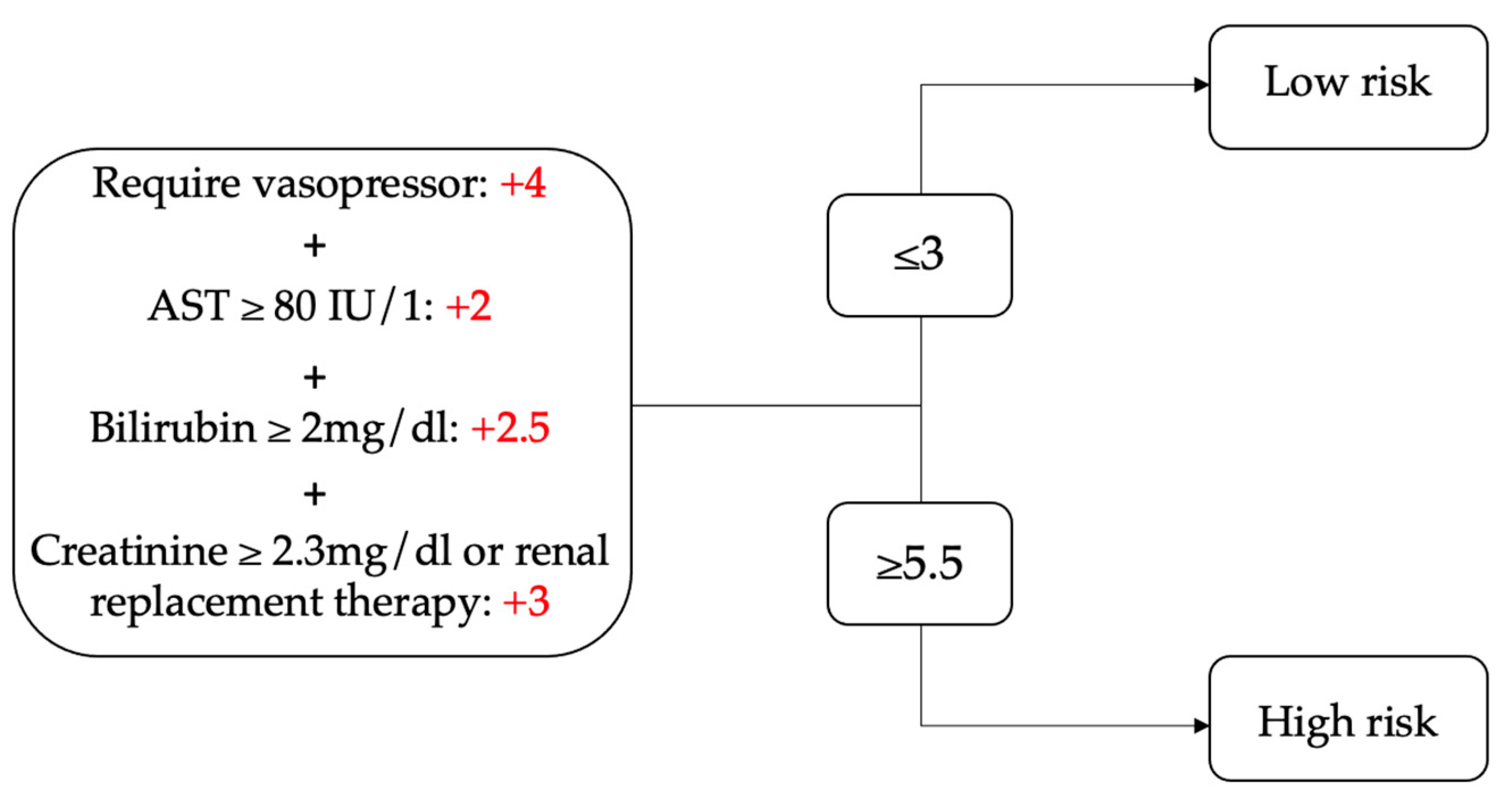
4.2. Scoring Assessments

There are two notable scoring assessments to determine who needs an RVAD. In the Fitzpatrick et al. scoring system, points are weighted for a series of indications indicative of RV dysfunction, such as low cardiac index, RV stroke work index, and elevated creatinine levels. The scores add up to 98, with high scores (≥65) being suggestive of a high likelihood of requiring RVAD, while lower scores (<30) suggest successful LVAD placement without RV support, as shown in Figure 1 [12]. Likewise, Matthews et al. defined RV dysfunction as the requirement of mechanical RV support, continuous use of inotropes for more than 14 days, and hospital discharge on inotropes [13]. The Matthews scoring system identifies indicators of RV dysfunction, such as the use of vasopressors, elevated liver enzymes, and impaired kidney function [13]. A score above 5.5 indicates a significantly higher risk of RVF, while a score below 3.0 suggests reduced risk, as shown in Figure 2 [13]. Other predictors of RVF include the Pulmonary Pulsatility Index (PAPi). A threshold below 1.85 indicates an increased likelihood of RVF in patients with an LVAD [22]. Finally, the EUROMACS score attempts to predict the likelihood of RVF prior to LVAD implantation using five variables: RA/Pulmonary Capillary Wedge Pressure (PCWP), hemoglobin, use of inotropes, INTERMACS class level, and evidence of severe RV dysfunction. Patients with a score of ≤2 are at a low risk for RHF, while those >2 are at intermediate–high risk [56]. Soliman et al. found that this score outperformed other known risk scores in predicting RHF post-LVAD implantation [15]; however, Shah et al. stated this score poorly predicted RHF outcomes post-LVAD placement [14].

5. Outcomes After Right Ventricular Assist Device Placement

5.1. Overview

The placement of an RVAD inherently means a higher-acuity disease state. Without a common definition of RVF, it is difficult for physicians to make the decision to change from medical to surgical management of a failing RV. Currently, the options available for temporary RVAD placement include the ProtekDuo, Impella RP, TandemHeart, and CentriMag [57]. Temporary devices that are commercially available as RVADs are only meant to allow for the right heart to recover and then must be removed. Some studies are looking into the technique used for RVAD placement, specifically a surgical versus a percutaneous technique. Furthermore, studies are investigating the venous location, the femoral vein versus the IJV, for RVAD placement to determine which has better outcomes. Overall, multiple studies are attempting to discern which option is best for those requiring further cardiac intervention.

5.2. Timing

As mentioned above, without a clear definition of RVF, it can be difficult to determine when it is the right time to place an RVAD. However, once medical management no longer provides symptomatic relief, surgical intervention is warranted. Salna et al. mentioned that their decision threshold to implant an RVAD decreased over time, and their center has a protocol for RVAD placement in acute RVF situations [9]. In 2013, Koji et al. investigated the effects of the timing of RVAD insertion, comparing concomitant RVAD placement during the LVAD procedure with a delayed RVAD insertion. They found that there was no difference in outcomes between a BVAD approach and a delayed RVAD insertion approach [58]. However, a study in 2016 concluded that delayed RVAD placement was associated with increased morbidity [59].
RVAD placement is performed with the hope of RV recovery and subsequent removal of the device. Schmack et al. had 10 of their 11 patients successfully weaned off their RVAD [11]. However, other studies have not had such high weaning rates [60,61], and a systematic review found survival to weaning to be 40% to 100% [57]. Even though RVADs are meant for temporary placement, some are retained for longer. For example, Abdelshafy et al. found that the duration of RVAD placement ranged between five hours and 400 days in their systematic review [57]. The timing of RVAD placement is very important, and without a clear definition of RVF post-LVAD placement, this decision is left to the physician’s best judgement.

5.3. Survival

Many studies looking into survival rates post-RVAD placement have few patients and are retrospective in their designs. However, survival rates are increasing in this patient population. For example, Salna et al. found that the 1-year survival rate in their cohort of 27 patients who received an RVAD post-LVAD between 2016 and 2019 was 81% [9]. Similarly, Anderson et al. found that the 180-day survival rate in their cohort of 18 patients receiving an RVAD post-LVAD between 2013 and 2014 was 77.8% [62]. However, a systematic review completed in 2022 shows the wide variability in survival rates between studies. Looking at 30-day survival, the survival rate encompassing 31 different studies between 2011 and 2020 ranged between 46% and 100% [57]. The most common adverse events include bleeding, pump thrombosis, acute kidney injury, stroke, hemolysis, and device malfunction [9,57,62,63]. However, one study found no RVAD-related complications after placement [11].
Factors that might affect survival in this cohort include how the RVAD is placed—surgically or percutaneously. Coromilas et al., in 2018, compared the outcomes of surgical RVAD (sRVAD) placement versus percutaneous RVAD (pRVAD) placement. They found the 30-day survival rate was 84.2% and 66.7% for the pRVAD and sRVAD groups, respectively, but this difference was not significant [10]. The benefits listed for a percutaneous approach include no redo sternotomy, easy removal of the RVAD device, and placement into the IJV, which allows for early mobility of the patient [9,10,11]. The goal is to offload the RV in order to provide hemodynamic stability, and both surgical and percutaneous approaches are successful in achieving this [10]. Patients requiring an RVAD placement post-LVAD are more critically ill and require intervention when medical therapies no longer work. Therefore, it is important to establish protocols on when to surgically intervene so that patient outcomes are maximized.

6. Conclusions

RHF post-LVAD placement is difficult to assess and still does not hold a uniform definition. Therefore, determining who needs an RVAD is very difficult and is often left up to care teams to decide what is best for their patient. There has been success implanting RVADs post-LVAD for those who were decompensating and needed right heart assistance. These cases are classically higher acuity with sicker patients who just underwent surgery. Acknowledging this point is necessary when outcomes are analyzed in this patient population. LVAD technology has greatly enhanced the ability for heart failure patients to be treated without the immediate need for a heart transplant. Dysfunction of the right heart is a known consequence of this treatment, yet the same dedication to furthering technology for RVADs has not occurred. Starting with creating a uniform definition of RHF to allow physicians to recognize classic symptoms and having known recommendations for management is necessary. As many studies have recognized the use of inotropes for more than 14 days as a way to identify RVF post-LVAD placement, it is reasonable to begin to consider this a formal definition of RVF; however, official guidelines still have to be created. Also, advancements in RVAD technology with research to analyze the risks and outcomes are the next step in furthering the knowledge needed for the treatment of this patient population. When a patient is suffering from RHF post-LVAD placement that cannot be controlled with medical management, RVAD placement is called for and has decent results. Therefore, enhancing our knowledge in this field is essential in order to provide patients with the best outcomes.

Author Contributions

Conceptualization, S.P., P.T. and A.E.M.; methodology, S.P., P.T. and A.E.M.; software, S.P.; validation, S.P., P.T. and A.E.M.; formal analysis, S.P., T.E.H. and H.P.; investigation, S.P., T.E.H. and H.P.; resources, S.P., T.E.H. and H.P.; data curation, S.P., T.E.H. and H.P.; writing—original draft preparation, S.P., T.E.H. and H.P.; writing—review and editing, S.P., T.E.H., H.P., P.T. and A.E.M.; visualization, S.P.; supervision, S.P., P.T. and A.E.M.; project administration, S.P.; funding acquisition, N/A. All authors have read and agreed to the published version of the manuscript.

Funding

This research received no external funding.

Institutional Review Board Statement

Not applicable.

Informed Consent Statement

Not applicable.

Data Availability Statement

The original contributions presented in this study are included in the article; further inquiries can be directed to the corresponding author.

Conflicts of Interest

The authors declare no conflicts of interest.

References

  1. Hamdan, R.; Charif, F.; Kadri, Z. Right ventricle failure in patients treated with left ventricular assist device. Ann. Cardiol. D’angéiologie 2020, 69, 51–54. [Google Scholar]
  2. Grant, A.D.; Smedira, N.G.; Starling, R.C.; Marwick, T.H. Independent and incremental role of quantitative right ventricular evaluation for the prediction of right ventricular failure after left ventricular assist device implantation. J. Am. Coll. Cardiol. 2012, 60, 521–528. [Google Scholar] [CrossRef]
  3. Fitzpatrick, J.R.; Frederick, J.R., 3rd; Hiesinger, W.; Hsu, V.M.; McCormick, R.C.; Kozin, E.D.; Laporte, C.M.; O’Hara, M.; Howell, E.; Doughtery, D.; et al. Early planned institution of biventricular mechanical circulatory support results in improved outcomes compared with delayed conversion of a left ventricular assist device to a biventricular assist device. J. Thorac. Cardiovasc. Surg. 2009, 137, 971–977. [Google Scholar] [PubMed]
  4. Cogswell, R.; John, R.; Shaffer, A. Right Ventricular Failure After Left Ventricular Assist Device. Cardiol. Clin. 2020, 38, 219–225. [Google Scholar] [CrossRef] [PubMed]
  5. Pagani, F.D. Right Heart Failure After Left Ventricular Assist Device Placement: Medical and Surgical Management Considerations. Cardiol. Clin. 2020, 38, 227–238. [Google Scholar]
  6. Turner, K.R. Right Ventricular Failure After Left Ventricular Assist Device Placement-The Beginning of the End or Just Another Challenge? J. Cardiothorac. Vasc. Anesth. 2019, 33, 1105–1121. [Google Scholar]
  7. James, L.; Smith, D.E. Supporting the “forgotten” ventricle: The evolution of percutaneous RVADs. Front. Cardiovasc. Med. 2022, 9, 1008499. [Google Scholar]
  8. Logstrup, B.B.; Nemec, P.; Schoenrath, F.; Gummert, J.; Pya, Y.; Potapov, E.; Netuka, I.; Ramjankhan, F.; Parner, E.T.; De By, T.; et al. Heart failure etiology and risk of right heart failure in adult left ventricular assist device support: The European Registry for Patients with Mechanical Circulatory Support (EUROMACS). Scand. Cardiovasc. J. 2020, 54, 306–314. [Google Scholar]
  9. Salna, M.; Garan, A.R.; Kirtane, A.J.; Karmpaliotis, D.; Green, P.; Takayama, H.; Sanchez, J.; Kurlansky, P.; Yuzefpolskaya, M.; Colombo, P.C.; et al. Novel percutaneous dual-lumen cannula-based right ventricular assist device provides effective support for refractory right ventricular failure after left ventricular assist device implantation. Interact. Cardiovasc. Thorac. Surg. 2020, 30, 499–506. [Google Scholar]
  10. Coromilas, E.J.; Takeda, K.; Ando, M.; Cevasco, M.; Green, P.; Karmpaliotis, D.; Kirtane, A.; Topkara, V.K.; Yuzefpolskaya, M.; Takayama, H.; et al. Comparison of Percutaneous and Surgical Right Ventricular Assist Device Support After Durable Left Ventricular Assist Device Insertion. J. Card. Fail. 2019, 25, 105–113. [Google Scholar] [CrossRef]
  11. Schmack, B.; Farag, M.; Kremer, J.; Grossekettler, L.; Brcic, A.; Raake, P.W.; Kreusser, M.M.; Goldwasser, R.; Popov, A.F.; Mansur, A.; et al. Results of concomitant groin-free percutaneous temporary RVAD support using a centrifugal pump with a double-lumen jugular venous cannula in LVAD patients. J. Thorac. Dis. 2019, 11 (Suppl. 6), S913–S920. [Google Scholar] [CrossRef] [PubMed]
  12. Fitzpatrick, J.R.; Frederick, J.R., 3rd; Hsu, V.M.; Kozin, E.D.; O’Hara, M.L.; Howell, E.; Dougherty, D.; McCormick, R.C.; Laporte, C.A.; Cohen, J.E.; et al. Risk score derived from pre-operative data analysis predicts the need for biventricular mechanical circulatory support. J. Heart Lung Transpl. 2008, 27, 1286–1292. [Google Scholar] [CrossRef] [PubMed]
  13. Matthews, J.C.; Koelling, T.M.; Pagani, F.D.; Aaronson, K.D. The right ventricular failure risk score a pre-operative tool for assessing the risk of right ventricular failure in left ventricular assist device candidates. J. Am. Coll. Cardiol. 2008, 51, 2163–2172. [Google Scholar] [CrossRef]
  14. Shah, H.; Murray, T.; Schultz, J.; John, R.; Martin, C.M.; Thenappan, T.; Cogswell, R. External assessment of the EUROMACS right-sided heart failure risk score. Sci. Rep. 2021, 11, 16064. [Google Scholar] [CrossRef]
  15. Soliman, O.I.I.; Akin, S.; Muslem, R.; Boersma, E.; Manintveld, O.C.; Krabatsch, T.; Gummert, J.F.; de By, T.M.M.H.; Bogers, A.J.J.C.; Zijlstra, F.; et al. Derivation and Validation of a Novel Right-Sided Heart Failure Model After Implantation of Continuous Flow Left Ventricular Assist Devices: The EUROMACS (European Registry for Patients with Mechanical Circulatory Support) Right-Sided Heart Failure Risk Score. Circulation 2018, 137, 891–906. [Google Scholar]
  16. Higgins, R.S.; Elefteriades, J.A. Right ventricular assist devices and the surgical treatment of right ventricular failure. Cardiol. Clin. 1992, 10, 185–192. [Google Scholar] [CrossRef]
  17. DeBakey, M.E. A Simple Continuous-Flow Blood Transfusion Instrument. New Orleans Med. Surg. J. 1934, 87, 386–389. [Google Scholar]
  18. Gibbon, J.H., Jr. Application of a mechanical heart and lung apparatus to cardiac surgery. Minn. Med. 1954, 37, 171–185. [Google Scholar]
  19. Dennis, C.; Hall, D.P.; Moreno, J.R.; Senning, A. Left atrial cannulation without thoracotomy for total left heart bypass. Acta Chir. Scand. 1962, 123, 267–279. [Google Scholar]
  20. Kantrowitz, A.; Tjønneland, S.; Freed, P.S.; Phillips, S.J.; Butner, A.N.; Sherman, J.L. Initial Clinical Experience With Intraaortic Balloon Pumping in Cardiogenic Shock. J. Am. Med. Assoc. 1968, 203, 113–118. [Google Scholar] [CrossRef]
  21. Miller, D.C.; Moreno-Cabral, R.J.; Stinson, E.B.; Shinn, J.A.; Shumway, N.E. Pulmonary artery balloon counterpulsation for acute right ventricular failure. J. Thorac. Cardiovasc. Surg. 1980, 80, 760–763. [Google Scholar] [CrossRef] [PubMed]
  22. Kapur, N.K.; Esposito, M.L.; Bader, Y.; Morine, K.J.; Kiernan, M.S.; Pham, D.T.; Burkhoff, D. Mechanical Circulatory Support Devices for Acute Right Ventricular Failure. Circulation 2017, 136, 314–326. [Google Scholar] [CrossRef]
  23. Taylor, A.J.; Edwards, F.H.; Macon, M.G.; Worley, B.; Graeber, G.M. A comparative evaluation of pulmonary artery balloon counterpulsation and a centrifugal flow pump in an experimental model of right ventricular infarction. J. Extra Corpor. Technol. 1990, 22, 85–90. [Google Scholar] [CrossRef]
  24. Stretch, R.; Sauer, C.M.; Yuh, D.D.; Bonde, P. National trends in the utilization of short-term mechanical circulatory support: Incidence, outcomes, and cost analysis. J. Am. Coll. Cardiol. 2014, 64, 1407–1415. [Google Scholar] [CrossRef]
  25. Tran, H.A.; Pollema, T.L.; Silva Enciso, J.; Greenberg, B.H.; Barnard, D.D.; Adler, E.D.; Pretorius, V.G. Durable Biventricular Support Using Right Atrial Placement of the HeartWare HVAD. ASAIO J. 2018, 64, 323–327. [Google Scholar] [CrossRef] [PubMed]
  26. Potapov, E.V.; Kukucka, M.; Falk, V.; Krabatsch, T. Biventricular support using 2 HeartMate 3 pumps. J. Heart Lung Transpl. 2016, 35, 1268–1270. [Google Scholar] [CrossRef]
  27. Essandoh, M.M.; Kumar, N. Total artificial heart system. Int. Anesth. Clin. 2022, 60, 39–45. [Google Scholar] [CrossRef]
  28. Summers, D. BiVACOR Total Artificial Heart Successfully Implanted in Five Patients as Part of FDA Early Feasibility Study; FDA Greenlights Expansion of the EFS; Business Wire: San Francisco, CA, USA, 2024. [Google Scholar]
  29. Carmat Receives Fda Approval to Use the New Version of Its Artificial Heart in the US Early Feasibility Study (Efs). Available online: https://www.carmatsa.com/en/press-release/carmat-receives-fda-approval-use-new-version-artificial-heart-us-early-feasibility-study-efs/ (accessed on 21 March 2025).
  30. Sultan, I.; Kilic, A. Short-Term Circulatory and Right Ventricle Support in Cardiogenic Shock: Extracorporeal Membrane Oxygenation, Tandem Heart, CentriMag, and Impella. Heart Fail. Clin. 2018, 14, 579–583. [Google Scholar] [CrossRef]
  31. DeFilippis, E.M.; Topkara, V.K.; Kirtane, A.J.; Takeda, K.; Naka, Y.; Garan, A.R. Mechanical Circulatory Support for Right Ventricular Failure. Card. Fail. Rev. 2022, 8, e14. [Google Scholar] [CrossRef]
  32. Vanden Eynden, F.; Mets, G.; De Somer, F.; Bouchez, S.; Bove, T. Is there a place for intra-aortic balloon counterpulsation support in acute right ventricular failure by pressure-overload? Int. J. Cardiol. 2015, 197, 227–234. [Google Scholar] [CrossRef]
  33. Explore the Minimally Invasive Impella RP® with SmartAssist®Heart Pump for Right-Sided Heart Recovery. Available online: https://www.abiomed.com/en-us/products-and-services/impella/impella-rp-with-smartassist (accessed on 21 March 2025).
  34. John, K.J.; Nabzdyk, C.G.S.; Chweich, H.; Mishra, A.K.; Lal, A. ProtekDuo percutaneous ventricular support system-physiology and clinical applications. Ann. Transl. Med. 2024, 12, 14. [Google Scholar]
  35. CentriMag Acute Mechanical Circulatory Support Information. Available online: https://www.cardiovascular.abbott/us/en/hcp/products/heart-failure/mechanical-circulatory-support/centrimag-acute-circulatory-support-system/about.html (accessed on 21 March 2025).
  36. Takeda, K.; Garan, A.R.; Ando, M.; Han, J.; Topkara, V.K.; Kurlansky, P.; Yuzefpolskaya, M.; Farr, M.A.; Colombo, P.C.; Naka, Y.; et al. Minimally invasive CentriMag ventricular assist device support integrated with extracorporeal membrane oxygenation in cardiogenic shock patients: A comparison with conventional CentriMag biventricular support configuration. Eur. J. Cardiothorac. Surg. 2017, 52, 1055–1061. [Google Scholar] [PubMed]
  37. Kazui, T.; Tran, P.L.; Echeverria, A.; Jerman, C.F.; Iwanski, J.; Kim, S.S.; Smith, R.G.; Khalpey, Z.I. Minimally invasive approach for percutaneous CentriMag right ventricular assist device support using a single PROTEKDuo Cannula. J. Cardiothorac. Surg. 2016, 11, 123. [Google Scholar] [PubMed]
  38. Borisenko, O.; Wylie, G.; Payne, J.; Bjessmo, S.; Smith, J.; Yonan, N.; Firmin, R. Thoratec CentriMag for temporary treatment of refractory cardiogenic shock or severe cardiopulmonary insufficiency: A systematic literature review and meta-analysis of observational studies. ASAIO J. 2014, 60, 487–497. [Google Scholar]
  39. The Novalung System. Available online: https://freseniusmedicalcare.com/en-us/products/novalung-ecmo-therapy/#:~:text=Fresenius%20Medical%20Care’s%20Product%20offering,for%20more%20than%206%20hours (accessed on 28 March 2025).
  40. Bavaria, J.E.; Ratcliffe, M.B.; Gupta, K.B.; Wenger, R.K.; Bogen, D.K.; Edmunds, L.H. Changes in left ventricular systolic wall stress during biventricular circulatory assistance. Ann. Thorac. Surg. 1988, 45, 526–532. [Google Scholar] [CrossRef] [PubMed]
  41. Cheng, R.; Hachamovitch, R.; Kittleson, M.; Patel, J.; Arabia, F.; Moriguchi, J.; Esmailian, F.; Azarbal, B. Complications of extracorporeal membrane oxygenation for treatment of cardiogenic shock and cardiac arrest: A meta-analysis of 1,866 adult patients. Ann. Thorac. Surg. 2014, 97, 610–616. [Google Scholar]
  42. Shudo, Y.; Wang, H.; Ha, R.V.; Hayes, A.D.; Woo, Y.J. Heart transplant after profoundly extended ambulatory central venoarterial extracorporeal membrane oxygenation. J. Thorac. Cardiovasc. Surg. 2018, 156, e7–e9. [Google Scholar] [CrossRef]
  43. Hess, N.R.; Hickey, G.W.; Murray, H.N.; Fowler, J.A.; Kaczorowski, D.J. Ambulatory simultaneous venoarterial extracorporeal membrane oxygenation and temporary percutaneous left ventricular assist device bridge to heart transplantation. JTCVS Tech. 2022, 13, 131–134. [Google Scholar]
  44. Ricklefs, M.; Hanke, J.S.; Dogan, G.; Chatterjee, A.; Feldmann, C.; Deniz, E.; Korte, W.; Kirchhoff, F.; Haverich, A.; Schmitto, J.D. Successful HeartMate 3 implantation in isolated right heart failure-first in man experience of right heart configuration. J. Thorac. Dis. 2018, 10 (Suppl. 15), S1834–S1837. [Google Scholar] [CrossRef]
  45. Mehra, M.R.; Goldstein, D.J.; Uriel, N.; Cleveland, J.C.; Yuzefpolskaya, M.; Salerno, C.; Walsh, M.N.; Milano, C.A.; Patel, C.B.; Ewald, G.A.; et al. Two-Year Outcomes with a Magnetically Levitated Cardiac Pump in Heart Failure. N. Engl. J. Med. 2018, 378, 1386–1395. [Google Scholar]
  46. Balasubramanian, V.; Bhama, J.K. Technique for “open sternal” chest closure in patients with assist devices and transplant recipients. JTCVS Tech. 2020, 2, 77–79. [Google Scholar] [CrossRef]
  47. Yanagida, R.; Rajagopalan, N.; Davenport, D.L.; Tribble, T.A.; Bradley, M.A.; Hoopes, C.W. Delayed sternal closure does not reduce complications associated with coagulopathy and right ventricular failure after left ventricular assist device implantation. J. Artif. Organs 2018, 21, 46–51. [Google Scholar]
  48. Giraldo-Grueso, M.; Desai, S.V.; Webre, K.; Bansal, A.G.; Parrino, P.; Bansal, A. Does Open Chest After LVAD Surgery Affect Blood Transfusion Rates and PRA. J. Heart Lung Transplant. 2022, 41, S487. [Google Scholar] [CrossRef]
  49. Li, M.; Mazzeffi, M.A.; Gammie, J.S.; Banoub, M.; Pazhani, Y.; Herr, D.; Madathil, R.; Pousatis, S.; Bathula, A. Characterization of Postoperative Infection Risk in Cardiac Surgery Patients With Delayed Sternal Closure. J. Cardiothorac. Vasc. Anesth. 2020, 34, 1238–1243. [Google Scholar] [PubMed]
  50. Wong, J.K.; Joshi, D.J.; Melvin, A.L.; Aquina, C.T.; Archibald, W.J.; Lidder, A.K.; Probst, C.P.; Massey, H.T.; Hicks, G.L.; Knight, P.A. Early and late outcomes with prolonged open chest management after cardiac surgery. J. Thorac. Cardiovasc. Surg. 2017, 154, 915–924. [Google Scholar] [PubMed]
  51. Arrigo, M.; Huber, L.C.; Winnik, S.; Mikulicic, F.; Guidetti, F.; Frank, M.; Flammer, A.J.; Ruschitzka, F. Right Ventricular Failure: Pathophysiology, Diagnosis and Treatment. Card. Fail. Rev. 2019, 5, 140–146. [Google Scholar] [PubMed]
  52. Chatterjee, K. The Swan-Ganz catheters: Past, present, and future. A viewpoint. Circulation 2009, 119, 147–152. [Google Scholar] [CrossRef]
  53. Rodenas-Alesina, E.; Brahmbhatt, D.H.; Rao, V.; Salvatori, M.; Billia, F. Prediction, prevention, and management of right ventricular failure after left ventricular assist device implantation: A comprehensive review. Front. Cardiovasc. Med. 2022, 9, 1040251. [Google Scholar]
  54. Takeda, K.; Naka, Y.; Yang, J.A.; Uriel, N.; Colombo, P.C.; Jorde, U.P.; Takayama, H. Outcome of unplanned right ventricular assist device support for severe right heart failure after implantable left ventricular assist device insertion. J. Heart Lung Transpl. 2014, 33, 141–148. [Google Scholar]
  55. Ende, J.; Wilbring, M.; Ende, G.; Koch, T. The Diagnosis and Treatment of Postoperative Right Heart Failure. Dtsch. Arztebl. Int. 2022, 119, 514–524. [Google Scholar]
  56. Soliman, O. EUROMACS-RHF Score. MD Calc. Available online: https://www.mdcalc.com/calc/10477/euromacs-rhf-score (accessed on 21 March 2025).
  57. Abdelshafy, M.; Caliskan, K.; Guven, G.; Elkoumy, A.; Elsherbini, H.; Elzomor, H.; Tenekecioglu, E.; Akin, S.; Soliman, O. Temporary Right-Ventricular Assist Devices: A Systematic Review. J. Clin. Med. 2022, 11, 613. [Google Scholar] [CrossRef] [PubMed]
  58. Takeda, K.; Naka, Y.; Yang, J.A.; Uriel, N.; Colombo, P.C.; Jorde, U.P.; Takayama, H. Timing of temporary right ventricular assist device insertion for severe right heart failure after left ventricular assist device implantation. ASAIO J. 2013, 59, 564–569. [Google Scholar] [CrossRef] [PubMed]
  59. Shehab, S.; Macdonald, P.S.; Keogh, A.M.; Kotlyar, E.; Jabbour, A.; Robson, D.; Newton, P.J.; Rao, S.; Wang, L.; Allida, S.; et al. Long-term biventricular HeartWare ventricular assist device support—Case series of right atrial and right ventricular implantation outcomes. J. Heart Lung Transpl. 2016, 35, 466–473. [Google Scholar] [CrossRef]
  60. Ravichandran, A.K.; Baran, D.A.; Stelling, K.; Cowger, J.A.; Salerno, C.T. Outcomes with the Tandem Protek Duo Dual-Lumen Percutaneous Right Ventricular Assist Device. ASAIO J. 2018, 64, 570–572. [Google Scholar] [CrossRef] [PubMed]
  61. McGiffin, D.; Kure, C.; McLean, J.; Marasco, S.; Bergin, P.; Hare, J.L.; Leet, A.; Patel, H.; Zimmet, A.; Rix, J.; et al. The results of a single-center experience with HeartMate 3 in a biventricular configuration. J. Heart Lung Transpl. 2021, 40, 193–200. [Google Scholar] [CrossRef]
  62. Anderson, M.B.; Goldstein, J.; Milano, C.; Morris, L.D.; Kormos, R.L.; Bhama, J.; Kapur, N.K.; Bansal, A.; Garcia, J.; Baker, J.N.; et al. Benefits of a novel percutaneous ventricular assist device for right heart failure: The prospective RECOVER RIGHT study of the Impella RP device. J. Heart Lung Transpl. 2015, 34, 1549–1560. [Google Scholar]
  63. Potapov, E.; Starck, C.; Falk, V.; Eulert-Grehn, J.-J. Mechanical circulatory support: Technical tips for the implantation of a right ventricular assist device. JTCVS Open 2021, 8, 37–40. [Google Scholar] [CrossRef]
Figure 1. Adaptation of Fitzpatrick et al. scoring system for likelihood of requiring biventricular support. Acronyms: RVSWI: right ventricular stroke work index, RV: right ventricle, BP: blood pressure, LVAD: left ventricular support device, BVAD: biventricular support device.
Hearts 06 00009 g001
Figure 2. Adaptation of Matthews et al. scoring system for right ventricular failure risk post-LVAD placement. Acronyms: AST: aspartate aminotransferase.
Hearts 06 00009 g002
Disclaimer/Publisher’s Note: The statements, opinions and data contained in all publications are solely those of the individual author(s) and contributor(s) and not of MDPI and/or the editor(s). MDPI and/or the editor(s) disclaim responsibility for any injury to people or property resulting from any ideas, methods, instructions or products referred to in the content.

