Article

Antenatal Secondhand Smoke (SHS) Exposure and the Receptor for Advanced Glycation End-Products (RAGE)

Lung and Placenta Research Laboratory, Department of Cell Biology and Physiology, Brigham Young University, Provo, UT 84602, USA
*
Author to whom correspondence should be addressed.
Reprod. Med. 2024, 5(1), 1-11; https://doi.org/10.3390/reprodmed5010001
Submission received: 6 October 2023 / Revised: 25 January 2024 / Accepted: 26 January 2024 / Published: 30 January 2024

Abstract

:
Exposure to secondhand smoke (SHS) during fetal development results in negative postnatal effects, including altered organ development, changes in metabolism, and increased risk of respiratory illness. Previously, we found the induction of intrauterine growth restriction (IUGR) dependent on the expression of the receptor for advanced glycation end-products (RAGE) in mice treated with SHS. Furthermore, antenatal SHS exposure increases RAGE expression in the fetal lung. Our objective was to determine the postnatal effects of antenatal SHS treatment in 4- and 12-week-old offspring. Pregnant animals were treated with SHS via a nose-only delivery system (Scireq Scientific, Montreal, Canada) for 4 days (embryonic day 14.5 through 18.5), and offspring were evaluated at 4 or 12 weeks of age. Animal and organ weights were measured, and lungs were histologically characterized. Blood pressure and heart rates were obtained, and RAGE protein expression was determined in the lungs of control and treated animals. We observed the following: (1) significant decreases in animal, liver, and heart weights at 4 weeks of age; (2) increased blood pressure in 4-week-old animals; and (3) increased RAGE expression in the lungs of the 4-week-old animals. Our results suggest an improvement in these metrics by 12 weeks postnatally such that measures were not different regardless of RA or SHS exposure. Increased RAGE expression in lungs from 4-week-old mice antenatally treated with SHS suggests a possible role for this important smoke-mediated receptor in establishing adult disease following IUGR pregnancies.

1. Introduction

Cigarette smoke is one of the major contributors to health problems that lead to preventable deaths [1]. It is well established that smoking and exposure to secondhand smoke (SHS) culminate in detrimental health outcomes. Smoke exposure during pregnancy has been linked with several obstetric complications and increased risk for both the mother and fetus. Smoking cigarettes throughout pregnancy may be the single most important avoidable cause of adverse pregnancy outcomes [2]. There are several complications associated with smoking during pregnancy, including altered placental development, intrauterine growth restriction [3,4], preterm birth, low fetal birthweight, and stillbirth [3,4,5,6,7]. Maternal exposure to cigarette smoking has been linked to delayed development of kidneys and the heart, alterations in neurologic development, increased risk of high blood pressure, obesity, and increased respiratory illness [8,9,10,11,12,13,14,15,16,17]. Furthermore, antenatal smoke exposure continues to influence health during postnatal development and is linked to the onset of diabetes and hypertension into adulthood [18]. Although significant studies are available that focus on the consequences of direct smoking during pregnancy, studies detailing the effects of SHS during pregnancy are more limited. The precise effects of maternal SHS exposure on a developing fetus remain under evaluation. It is known that SHS exposure contributes to around 1.2 million deaths annually. Such exposure has also been linked to sickle cell disease and other lung complications in adults. In children, SHS is known to affect lung function, increase lung inflammation, and increase the severity of cystic fibrosis [19,20]. We previously demonstrated in our laboratory that exposure to SHS during pregnancy induced the development of IUGR in pregnant mice [2]. IUGR impacts fetal and neonatal morbidity and mortality, and studies have reported long-term sequelae of complications, including adult hypertension, heart disease, stroke, and diabetes [2]. These observations were associated with increased expression of the receptor for advanced glycation end-products (RAGE) in the lung and placenta of exposed mothers [2,21].
RAGE is a member of the immunoglobulin superfamily that recognizes a host of ligands [22,23,24]. In adults, RAGE is expressed at low levels in most tissues, with the exception of the lung, where RAGE expression is much more pronounced [25]. RAGE is highly expressed during embryonic development and is activated by early growth responses [26]. It has been proposed that RAGE involvement may play a role in the structural maintenance of the lung through adult life [27]. In response to ligand binding, RAGE mediates a signaling cascade that leads to the activation of a host of pro-inflammatory mediators [28,29]. In fact, RAGE signaling is implicated in the pathogenesis of diseases, including Alzheimer’s, diabetes, atherosclerosis, COPD, and diverse rheumatological disorders [30,31,32,33]. Studies have shown that RAGE overexpression during embryogenesis is lethal as a result of pulmonary hypoplasia [34]. Thus, RAGE expression must be intimately regulated during development, and altered fetal RAGE expression in response to antenatal SHS exposure may be a mechanism through which fetal development is impacted.
This study specifically sought to clarify the effects of antenatal SHS treatment on offspring after 4 or 12 weeks of postnatal life. As a general theme, antenatal smoke exposure remained a causal influence on several metrics evaluated after 4 weeks of life. Alternatively, 12 weeks after birth was sufficient time for deleterious hindrances to correct themselves.

2. Methods

2.1. Animal Housing and Tissue Collection

All animal work was performed in accordance with protocols approved by the Institutional Committee for the Care and Use of Animals (IACUC) at Brigham Young University. C57 Black 6 (C57BL/6) mice were obtained from Charles River Laboratories (Wilmington, MA, USA), housed in standard plastic cages with enrichment, maintained in a 12-h light/dark cycle, and provided access to food and water ad libitum. Timed pregnancies were performed, and the identification of a vaginal plug confirmed embryonic (E) day. Pregnant mice experienced normal gestational development until randomized mice were exposed to either room air (RA; n = 8) or SHS (n = 8) for 4 days, from E14.5–E18.5. Litter size averaged around 8–9 pups. After birth, pups remained with the dam until postnatal day (PN) 21. Necropsies were performed at either 4 weeks or 12 weeks of age. On the date of necropsy, animals were anesthetized with avertin (2.5% in 0.015 mL) and euthanized by exsanguination. Heart and kidney weights relative to body weight were determined, and tissues from the heart, kidneys, and right lung were snap-frozen in liquid nitrogen for protein analysis or mitochondrial respiration assessment. The left lung was inflation-fixed with 4% paraformaldehyde for histological analysis, as outlined previously [1].

2.2. Secondhand Smoke (SHS) Exposure

SHS treatment was conducted as previously described by our laboratory [2]. Mice (n = 8) were exposed to SHS generated from 3R4F research cigarettes obtained from the Kentucky Tobacco Research and Development Center, University of Kentucky, via a nose-only exposure system (InExpose System; Scireq, Montreal, QC, Canada). This system produces a 10 s computer-controlled puff of primary smoke every minute that is subsequently cleared from the apparatus, preventing primary smoke exposure. Side-stream smoke was separately generated with a dedicated pump and delivered without mixing with primary smoke. Animals were accordingly exposed to SHS from six cigarettes during a 10-min period. This procedure was repeated each day, from E14.5 to E18.5. There were no deaths observed throughout these experiments. Control mice (n = 8) were exposed to room air (RA) only.

2.3. Blood Pressure and Heart Rate

A CODA monitor system (CODA tail-cuff blood pressure system; Kent Scientific Corporation; Torrington, CT, USA) was used to measure blood pressure and heart rates as previously performed in our laboratory [35]. During measurements, animals were restrained for 5 min by a clear column crafted by Kent Scientific. Heart rates and blood pressure were determined in RA controls and treated mice (SHS).

2.4. Western Blotting

Western blot analysis was performed to determine RAGE protein expression in lung lysates from RA control or SHS animals, as previously described by our laboratory [35]. Briefly, the protein was isolated from lung tissue using RIPA lysis buffer with protease and phosphatase inhibitors. Lung protein lysates (30 mg) were separated using a Mini PROTEANV TGXTM precast gel (Bio-Rad Laboratories, Hercules, CA, USA), followed by transfer to a nitrocellulose membrane. Membranes were incubated with RAGE (1:250 R&D, Minneapolis, MN, USA) or β-actin (1:500 Santa Cruz Biotechnology, Santa Cruz, CA, USA) antibodies overnight at 4 °C. Fluorescent secondary antibodies (LICOR, Lincoln, NE, USA) were applied at 1:3000 and incubated at room temperature for one hour. Membranes were then imaged with a LICOR Odyessy CLx imaging system (Lincoln, NE, USA) and analyzed with Image Studio software 5.5 (LICOR, Lincoln, NE, USA).

2.5. Histology

The left lungs from at least 4 animals were fixed with 4% PFA, processed with a series of ethanol washes, and embedded in paraffin. Then, 5 µm sections were deparaffinized and stained with hematoxylin and eosin to permit observation of general lung morphology. The mean linear intercept (MLI) was determined as outlined [1] by analyzing 10 or more pictures of lung parenchyma containing smaller airways in each section with an Image J program. Slides were imaged with a BX61 compound microscope.

2.6. Immunofluorescence (IF)

IF was performed on paraffin-embedded lung samples. In summary, slides were de-waxed and blocked for 1 h. This was followed by incubation overnight with a mouse primary RAGE antibody (R&D Systems, Minneapolis, MN, USA; Cat# mAb1179, 1:300). For detecting fluorescence, slides were incubated for an hour with a donkey anti-mouse Texas Red (TX) (Santa Cruz Biotechnology, Santa Cruz, CA, USA, 1:5000) secondary antibody. Immunofluorescence was detected using a BX6 microscope.

2.7. Lung Mitochondrial Respiration Analysis

To characterize mitochondrial respiration, lung tissues were collected at the time of necropsy, and high-resolution O2 consumption was determined at 37 °C using the Oroboros O2K Oxygraph (Innsbruck, Austria) with MiR05 respiration buffer. RA control and treated samples were tested to determine electron flow through complex I and to determine basal oxygen consumption [glutamate + malate (GM)]. Following this step, adenosine diphosphate (ADP) (2.5 mM) was added to determine oxidative phosphorylation capacity (GMD). Succinate was then added (GMDS) for complex I + II electron flow into the Q-junction.

2.8. Statistics

Differences in animal and organ weights, blood pressure, and RAGE protein expression were determined between RA control and SHS-treated animals using a Mann–Whitney test. GraphPad Prism software 10.1.2 (GraphPad; Santa Clara, CA, USA) was used for statistical analysis. Differences are shown as mean ± SE, with significant differences between groups noted at p < 0.05.

3. Results

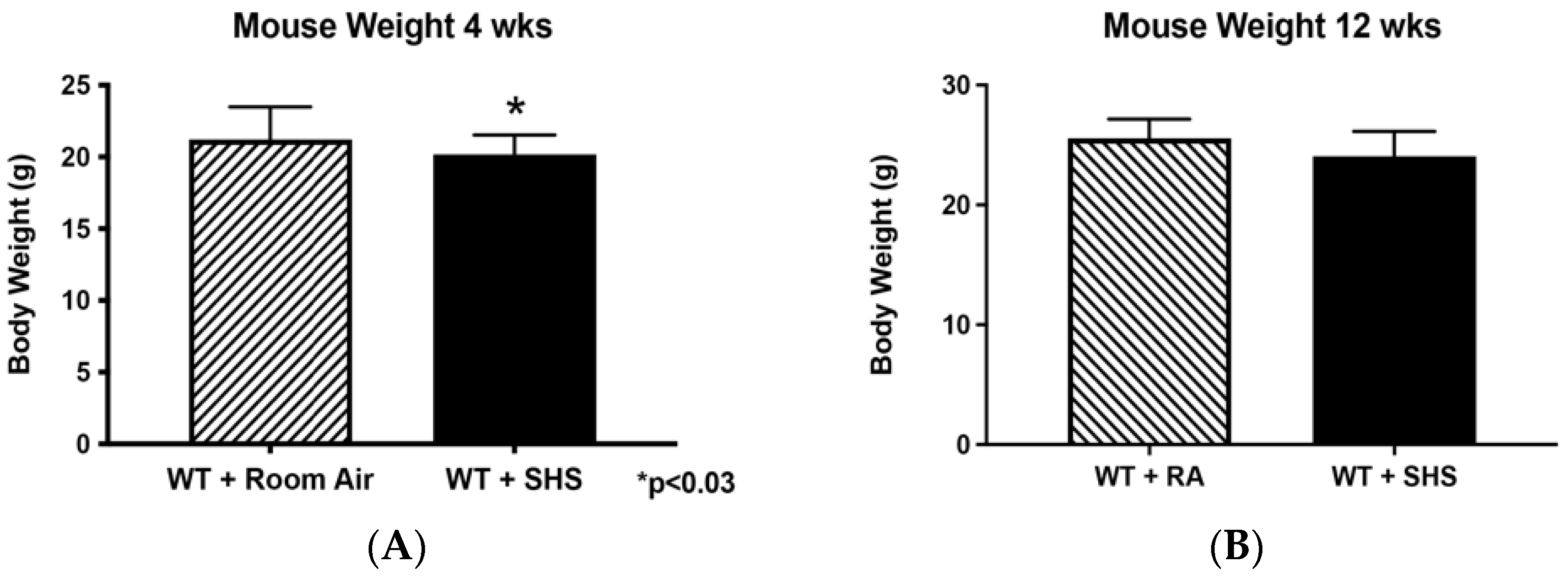
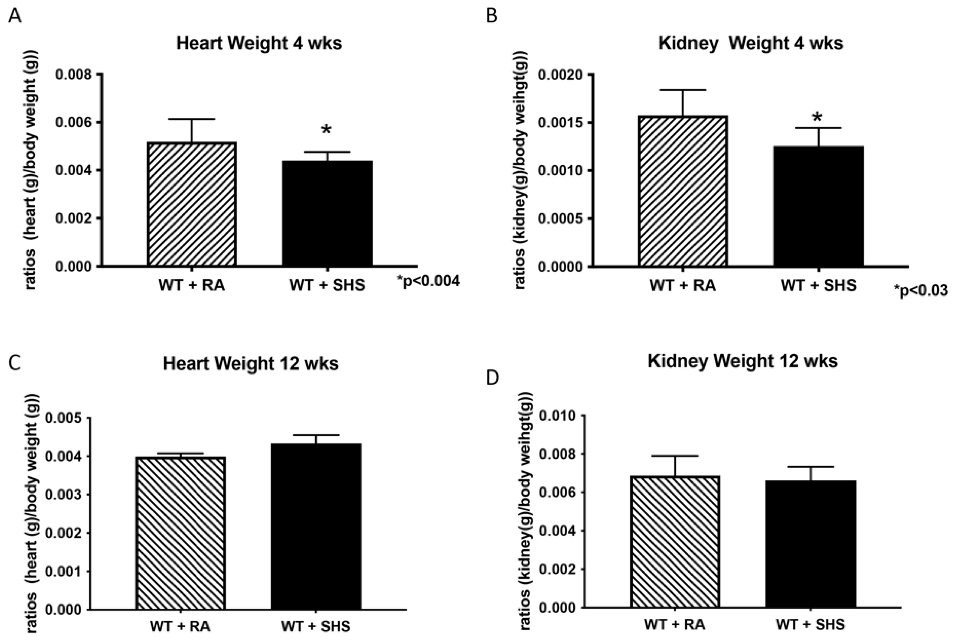
3.1. Body and Organ Weights

Previously, we demonstrated reduced fetal weight (n = 12) following antenatal exposure in an SHS-induced mode of IUGR [2]. In the current study, we still observed reduced weight postnatally in mice 4 weeks of age after antenatal exposure to SHS (Figure 1A, 1.0-fold; p < 0.03). This difference in animal weights when comparing antenatal RA and SHS was not detected when the mice reached 12 weeks of age (Figure 1B). As previously mentioned, maternal exposure to cigarette smoke affects the development of the heart and kidneys [8,9,12]. Offspring showed decreased heart (1.2-fold; p < 0.004) and kidney (1.3-fold; p < 0.03) weights at 4 weeks of age (Figure 2A,B); however, these weights were not different at 12 weeks of age (Figure 2C,D).

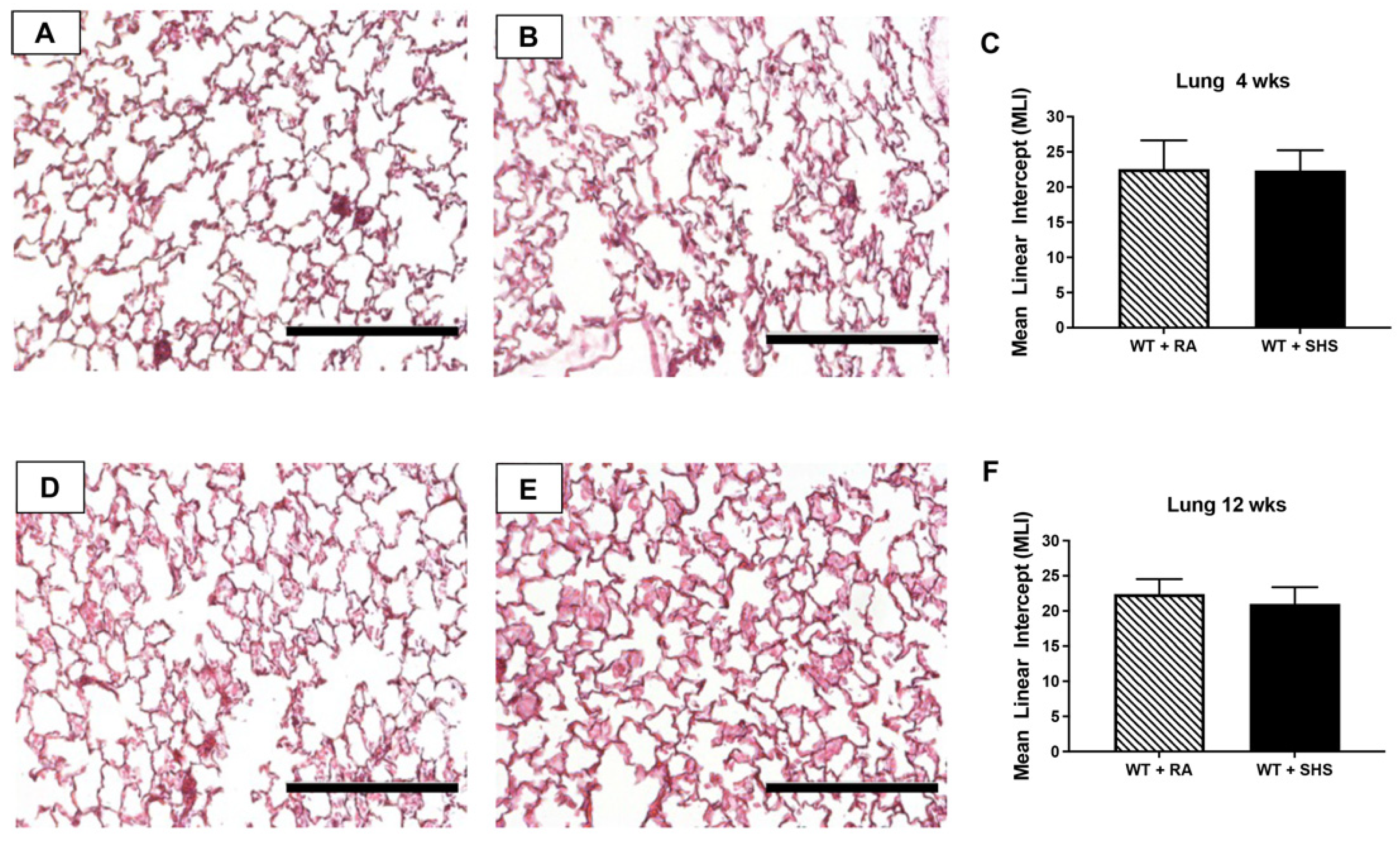
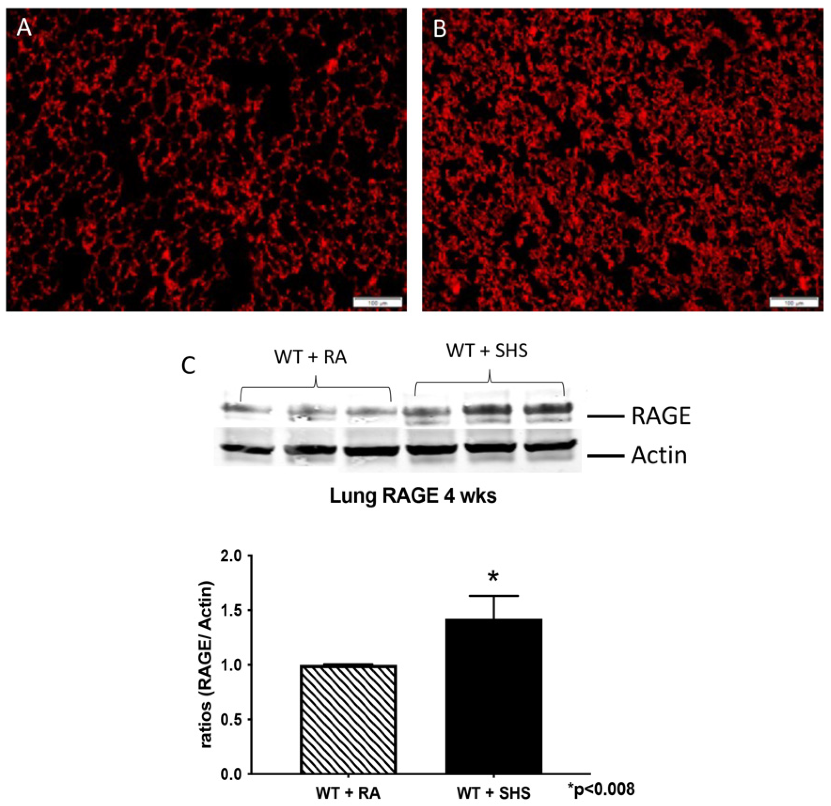
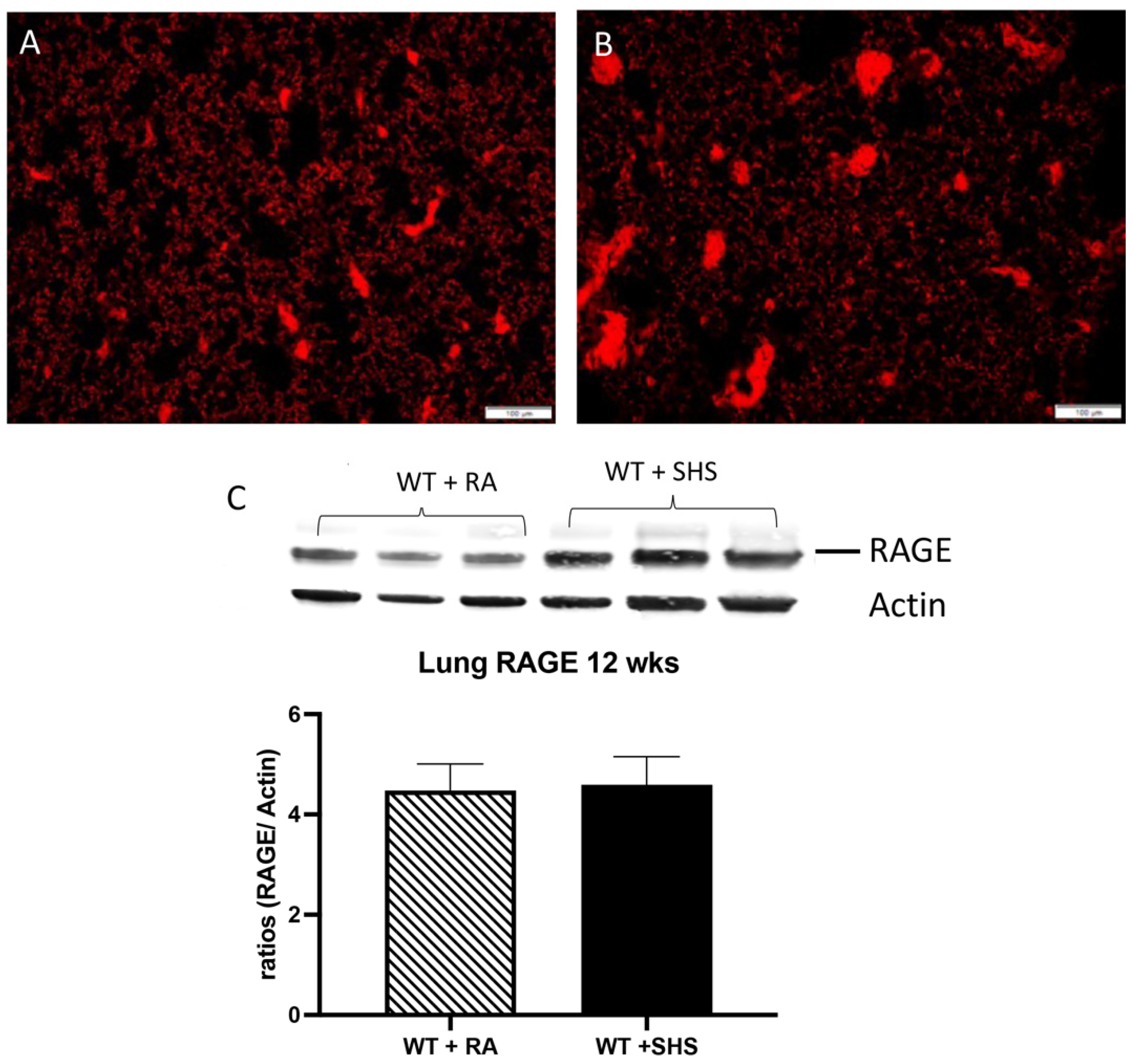
3.2. Pulmonary Structure and RAGE Expression

Assessing mean linear intercepts (MLI) provides an estimate of the area available for gas exchange in the lung. Characteristic histology of the lung from mice (n = 8) at either 4 or 12 weeks of age is shown in Figure 3A,B,D,E. There were no general alterations in lung histology or notable differences in MLI in lungs from offspring at 4 or 12 weeks of age following antenatal exposure to SHS (Figure 3C,F). Previous studies in our lab have shown that antenatal SHS exposure produced an increase in fetal lung expression of RAGE [21]. We, therefore, sought to determine RAGE expression in the lungs of offspring at 4 and 12 weeks of age. Representative RAGE immunofluorescence at 4 weeks of age is shown in Figure 4A,B. Furthermore, RAGE Western blotting revealed significantly more RAGE expression in lungs 4 weeks after antenatal SHS exposure compared to the RA controls (Figure 4C, 1.4-fold; p < 0.03). We discovered no differences in RAGE immunofluorescence (Figure 5A,B) or Western blotting (Figure 5C) in the lungs of 12-week-old mice, regardless of exposure.

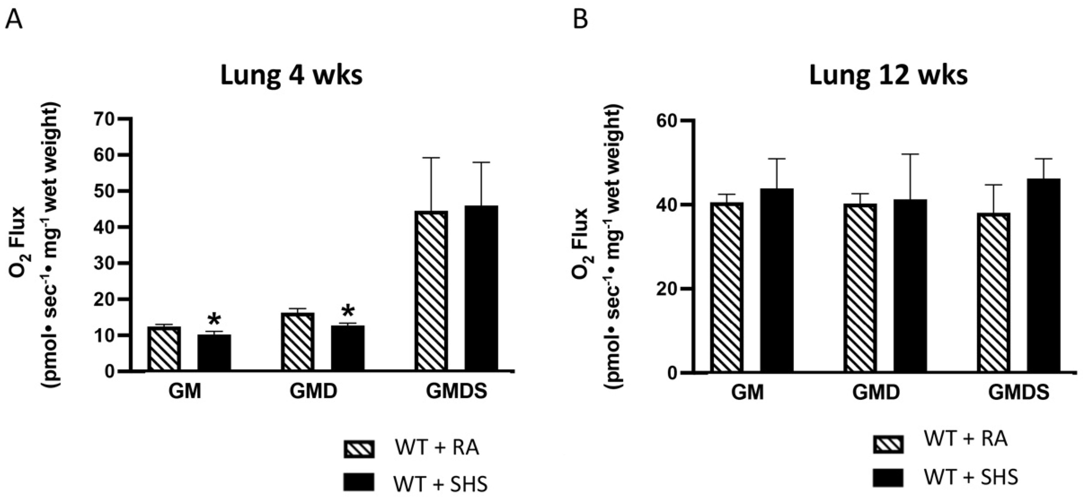
3.3. Lung Mitochondrial Respiration Analysis

The observation that lung weights were decreased in the 4-week-old mice led us to investigate the potential for altered cellular function in terms of mitochondrial respiration (n = 8). Oxygen flux was determined during the exposure conditions of multiple substrates (see Section 2 for details). We discovered a significant reduction in mitochondrial respiration in antenatal SHS-exposed mice at 4 weeks of age compared to RA controls, but no differences were detected in the 12-week-old animals (Figure 6A,B). Despite the difference in respiration rates observed in the 4-week-old animals, respiratory control ratios (RCR), a general indicator of mitochondrial function, revealed no apparent differences in the functionality or overall health of the mitochondria.

3.4. Blood Pressure and Heart Rate

Heart rates were determined at 4 and 12 weeks of age. Interestingly, heart rates were not affected at either point studied. In addition, we measured the systolic and diastolic blood pressures (n = 8) at the time of necropsy. There was a significant increase in systolic (1.3-fold; p < 0.0002) and diastolic (1.3-fold; p < 0.0004) blood pressures in the 4-week-old animals (Figure 7A,B). At 12 weeks, there was no significant difference in blood pressure between groups (Figure 7C,D).

4. Discussion

Preterm birth (PTB) is associated with up to 70% of neonatal deaths and leads to an increased incidence of cerebral palsy, neurological defects, and pulmonary disorders in the neonate [36]. There is an association between the development of Intrauterine Growth Restriction (IUGR) and PTB [37]. Studies have shown that there is a risk of up to 44% of PTB in IUGR pregnancies [38]. IUGR is a significant complication that affects up to 12% of all pregnancies. This disease is characterized by birth weight (BW) below the 10th percentile of the usual gestational age [39]. In addition, several studies reported long-term sequelae of IUGR complications, including adult hypertension, heart disease, stroke, and diabetes. Interestingly, many studies have shown that although newborn infants are smaller in size and have decreased body weight compared to non-IUGR infants, they seem to “catch up” in weight and size during their infancy [40,41,42]. In previous studies, we showed decreased fetal weight when pregnant mice were treated with SHS [19]. This decreased body weight was still observed postnatally in the 4-week-old mice, which correlates to approximately 3 years of age in humans [43]. Further, the concept that babies born during IUGR “catch up” was realized in the current study when we observed no weight differences in mice by 12 weeks of age, which corresponds to approximately 9 years of age in humans [43]. These findings support the observation of others who argue for the recovery of the infant’s weight. Because kidney and heart development, often manifested as altered gross organ weight, is affected in infants born of smoking mothers, we next wanted to investigate whether the weight of these organs contributed to decreased body weight observed at 4 weeks. Both heart and kidney weights were decreased at 4 weeks, but no differences were seen at 12 weeks of age. These weight discrepancies suggest that there is a potential developmental delay in the organ-to-body weight ratios for both the heart and kidneys, as we observed in the 4-week-old mice. Again, the fact that no differences were observed in these organs when comparing controls and antenatal exposed animals at 12 weeks suggests that this metric could be part of the “catch up” experienced with the low-birthweight infants.
After embryonic development, RAGE remains notably expressed in lung tissue and only minimally expressed in most other tissues. At 4 weeks of age, mice that antenatally encountered SHS still expressed significantly more RAGE in pulmonary tissues when compared to RA controls. This was interesting, as these expression data confirmed our previous observations and showed that even after 4 weeks, the initial antenatal SHS stimulus was sufficient to maintain elevated RAGE expression. Furthermore, if RAGE expression remained higher than controls, such a discovery would implicate ongoing inflammatory RAGE-mediated signaling. By extension, the observation that RAGE expression waned to the same levels as RA controls at 12 weeks of age portends inflammatory RAGE signaling becomes diminished during the “catch-up” process experienced by intrauterine growth-restricted offspring. Abundant research has shown that RAGE up-regulation leads to inflammatory outcomes that include decreased cell turnover and pulmonary simplification [27,44,45]. We, therefore, assessed overall lung morphology via screening mean linear intercepts to assess lung simplification at 4 weeks and 12 weeks of age. Because we observed no significant differences between treated and control animals regardless of age, we can correctly assume that SHS-induced lung compromise did not affect histological architecture in the parenchyma.
We were interested in the observation that increased RAGE expression at 4 weeks of age correlated with decreased mitochondrial respiration, suggesting a possible connection between elevated pro-inflammatory RAGE expression and disrupted mitochondrial status. Compromised respiration efficiency has previously been noted in the context of tobacco smoke exposure, and decreased respiration is detrimental to cellular energetics [30,46]. Previous studies have also shown increased blood pressure in offspring of mothers who smoked prenatally. Such disparities in blood pressure, even at 4 weeks after exposure, suggest a role for cigarette smoke in cardiovascular health during adolescence [47,48]. We specifically observed increased systolic and diastolic blood pressure in the 4-week-old animals exposed to SHS prenatally, and pressure differences were absent by 12 weeks. These results suggest that antenatal SHS exposure is enough to maintain a postnatal increase in blood pressure up to 4 weeks of age that is nearly normalized in the “catching up” of the animals by 12 weeks of age. It is important to note that although we did not determine SHS components for these studies, previously published data show that SHS contains high levels of PAHs, tobacco-specific nitrosamines (TSNA), aromatic amines, aza-arenes, carbon monoxide, nicotine, ammonia, pyridine, benzene, toluene, and other harmful substances [49].
In general, our results demonstrated that maternal SHS exposure is sufficient to affect fetal development and that notable adverse effects last up to 4-weeks of age. Although it seems that neonates “catch up” by 12 weeks of age, the effects observed at 4 weeks could be, in part, involved in the development of long-term sequelae of adult hypertension, heart disease, stroke, and diabetes observed in infants affected by IUGR.

Author Contributions

K.L.C., J.A. and P.R.R. contributed to the experimental design; K.L.C., K.M.H., E.T.C. and B.M.S. maintained animals, performed exposures, and assisted with surgical procedures; immunoblotting was performed by K.L.C. and K.T.; data analysis was performed by K.M.H. with the help of B.T.B., P.R.R. and J.A. J.A. and K.M.H. assisted with data collection and interpretation. This manuscript was written primarily by K.L.C. and J.A. All authors have read and agreed to the published version of the manuscript.

Funding

This work was supported by grants from the National Institutes of Health (1R15HL152257; P.R.R. and J.A.A.) and the Flight Attendant’s Medical Research Institute (FAMRI CIA150085; P.R.R. and J.A.A.).

Institutional Review Board Statement

All experimental animal studies were approved by the Institutional Animal Care and Use Committee (IACUC) at Brigham Young University, and all methods were carried out in accordance with relevant animal guidelines and regulations.

Data Availability Statement

All data are presented within the article.

Acknowledgments

Much appreciation is extended to a team of exceptional undergraduate students in the Lung and Placenta Laboratory at Brigham Young University.

Conflicts of Interest

The authors declare no conflict of interest.

References

  1. Abraham, M.; Alramadhan, S.; Iniguez, C.; Duijts, L.; Jaddoe, V.W.; Den Dekker, H.T.; Crozier, S.; Godfrey, K.M.; Hindmarsh, P.; Vik, T.; et al. A systematic review of maternal smoking during pregnancy and fetal measurements with meta-analysis. PLoS ONE 2017, 12, e0170946. [Google Scholar] [CrossRef]
  2. Lewis, J.B.; Mejia, C.; Jordan, C.; Monson, T.D.; Bodine, J.S.; Dunaway, T.M.; Egbert, K.M.; Lewis, A.L.; Wright, T.J.; Ogden, K.C.; et al. Inhibition of the receptor for advanced glycation end-products (RAGE) protects from secondhand smoke (SHS)-induced intrauterine growth restriction IUGR in mice. Cell Tissue Res. 2017, 370, 513–521. [Google Scholar] [CrossRef]
  3. Andres, R.L.; Day, M.C. Perinatal complications associated with maternal tobacco use. Semin. Neonatol. 2000, 5, 231–241. [Google Scholar] [CrossRef] [PubMed]
  4. Reeves, S.; Bernstein, I. Effects of maternal tobacco-smoke exposure on fetal growth and neonatal size. Expert Rev. Obstet. Gynecol. 2008, 3, 719–730. [Google Scholar] [CrossRef] [PubMed]
  5. Windham, G.C.; Hopkins, B.; Fenster, L.; Swan, S.H. Prenatal active or passive tobacco smoke exposure and the risk of preterm delivery or low birth weight. Epidemiology 2000, 11, 427–433. [Google Scholar] [CrossRef]
  6. Badlissi, D.; Guillemette, A.; Fadin, A. Prematurite et petit poids ala naissance: Les effets du tabagisme actif et passif durant la grossesse. [Prematurity and low birth weight: Effects of active and passive smoking during pregnancy]. Can. J. Public Health 2001, 92, 272–275. [Google Scholar] [CrossRef] [PubMed]
  7. Salihu, H.M.; Sharma, P.P.; Getahun, D.; Hedayatzadeh, M.; Peters, S.; Kirby, R.S.; Alio, A.P.; Gaafer-Ahmed, H. Prenatal tobacco use and risk of stillbirth: A case-control and bidirectional case-crossover study. Nicotine Tob. Res. 2008, 10, 159–166. [Google Scholar] [CrossRef]
  8. Taal, H.R.; Geelhoed, J.J.M.; Steegers, E.A.P.; Hofman, A.; Moll, H.A.; Lequin, M.; van der Heijden, A.J.; Jaddoe, V.W.V. Maternal smoking during pregnancy and kidney volume in the offspring: The Generation R Study. Pediatr. Nephrol. 2011, 26, 1275–1283. [Google Scholar] [CrossRef]
  9. Anblagan, D.; Jones, N.W.; Costigan, C.; Parker, A.J.J.; Allcock, K.; Aleong, R.; Coyne, L.H.; Deshpande, R.; Raine-Fenning, N.; Bugg, G.; et al. Maternal smoking during pregnancy and fetal organ growth: A magnetic resonance imaging study. PLoS ONE 2013, 8, e67223. [Google Scholar] [CrossRef]
  10. Block, D.B.; Mesquita, F.F.; de Lima, I.P.; Boer, P.A.; Gontijo, J.A. Fetal kidney programming by maternal smoking exposure: Effects on kidney structure, blood pressure and urinary sodium excretion in adult offspring. Nephron 2015, 129, 283–292. [Google Scholar] [CrossRef]
  11. Edstedt Bonamy, A.K.; Bengtsson, J.; Nagy, Z.; De Keyzer, H.; Norman, M. Preterm birth and maternal smoking in pregnancy are strong risk factors for aortic narrowing in adolescence. Acta Paediatr. 2008, 97, 1080–1085. [Google Scholar] [CrossRef]
  12. Malik, S.; Cleves, M.A.; Honein, M.A.; Romitti, P.A.; Botto, L.D.; Yang, S.; Hobbs, C.A.; the National Birth Defects Prevention Study. Maternal smoking and congenital heart defects. Pediatrics 2008, 121, e810–e816. [Google Scholar] [CrossRef]
  13. Gutvirtz, G.; Wainstock, T.; Landau, D.; Sheiner, E. Maternal smoking during pregnancy and long-term neurological morbidity of the offspring. Addict. Behav. 2019, 88, 86–91. [Google Scholar] [CrossRef] [PubMed]
  14. Sowan, N.A.; Stember, M.L. Effect of maternal prenatal smoking on infant growth and development of obesity. J. Perinat. Educ. 2000, 9, 22–29. [Google Scholar] [CrossRef] [PubMed]
  15. Li, L.; Peters, H.; Gama, A.; Carvalhal, M.I.M.; Nogueira, H.G.M.; Rosado-Marques, V.; Padez, C. Maternal smoking in pregnancy association with childhood adiposity and blood pressure. Pediatr. Obes. 2016, 11, 202–209. [Google Scholar] [CrossRef] [PubMed]
  16. McEvoy, C.T.; Spindel, E.R. Pulmonary Effects of Maternal Smoking on the Fetus and Child: Effects on Lung Development, Respiratory Morbidities, and Life Long Lung Health. Paediatr. Respir. Rev. 2017, 21, 27–33. [Google Scholar] [CrossRef] [PubMed]
  17. Spindel, E.R.; McEvoy, C.T. The Role of Nicotine in the Effects of Maternal Smoking during Pregnancy on Lung Development and Childhood Respiratory Disease. Implications for Dangers of E-Cigarettes. Am. J. Respir. Crit. Care Med. 2016, 193, 486–494. [Google Scholar] [CrossRef]
  18. Falkner, B. Maternal and gestational influences on childhood blood pressure. Pediatr. Nephrol. 2020, 35, 1409–1418. [Google Scholar] [CrossRef]
  19. Yang, J.; Hashemi, S.; Han, W.; Song, Y.; Lim, Y. Exposure and Risk Assessment of Second- and Third-Hand Tobacco Smoke Using Urinary Cotinine Levels in South Korea. Int. J. Environ. Res. Public Health 2022, 19, 3746. [Google Scholar] [CrossRef]
  20. Yang, J.; Hashemi, S.; Kim, T.; Park, J.; Park, M.; Han, W.; Park, D.; Lim, Y. Risk assessment and estimation of controlling safe distance for exposure to particulate matter from outdoor secondhand tobacco smoke. Air Qual. Atmos. Health 2023, 17, 139–154. [Google Scholar] [CrossRef]
  21. Winden, D.R.; Barton, D.B.; Betteridge, B.C.; Bodine, J.S.; Jones, C.M.; Rogers, G.D.; Chavarria, M.; Wright, A.J.; Jergensen, Z.R.; Jimenez, F.R.; et al. Antenatal exposure of maternal secondhand smoke (SHS) increases fetal lung expression of RAGE and induces RAGE-mediated pulmonary inflammation. Respir. Res. 2014, 15, 129. [Google Scholar] [CrossRef] [PubMed]
  22. Taguchi, A.; Blood, D.C.; del Toro, G.; Canet, A.; Lee, D.C.; Qu, W.; Tanji, N.; Lu, Y.; Lalla, E.; Fu, C.; et al. Blockade of RAGE-amphoterin signalling suppresses tumour growth and metastases. Nature 2000, 405, 354–360. [Google Scholar] [CrossRef] [PubMed]
  23. Hofmann, M.A.; Drury, S.; Fu, C.; Qu, W.; Taguchi, A.; Lu, Y.; Avila, C.; Kambham, N.; Bierhaus, A.; Nawroth, P.; et al. RAGE mediates a novel proinflammatory axis: A central cell surface receptor for S100/calgranulin polypeptides. Cell 1999, 97, 889–901. [Google Scholar] [CrossRef] [PubMed]
  24. Schmidt, A.M.; Sahagan, B.; Nelson, R.B.; Selmer, J.; Rothlein, R.; Bell, J.M. The role of RAGE in amyloid-beta peptide-mediated pathology in Alzheimer’s disease. Curr. Opin. Investig. Drugs 2009, 10, 672–680. [Google Scholar] [PubMed]
  25. Demling, N.; Ehrhardt, C.; Kasper, M.; Laue, M.; Knels, L.; Rieber, E.P. Promotion of cell adherence and spreading: A novel function of RAGE, the highly selective differentiation marker of human alveolar epithelial type I cells. Cell Tissue Res. 2006, 323, 475–488. [Google Scholar] [CrossRef] [PubMed]
  26. Reynolds, P.R.; Kasteler, S.D.; Cosio, M.G.; Sturrock, A.; Huecksteadt, T.; Hoidal, J.R. RAGE: Developmental expression and positive feedback regulation by Egr-1 during cigarette smoke exposure in pulmonary epithelial cells. Am. J. Physiol. Lung Cell. Mol. Physiol. 2008, 294, L1094–L1101. [Google Scholar] [CrossRef]
  27. Wolf, L.; Herr, C.; Niederstrasser, J.; Beisswenger, C.; Bals, R. Receptor for advanced glycation endproducts (RAGE) maintains pulmonary structure and regulates the response to cigarette smoke. PLoS ONE 2017, 12, e0180092. [Google Scholar] [CrossRef]
  28. Sparvero, L.J.; Asafu-Adjei, D.; Kang, R.; Tang, D.; Amin, N.; Im, J.; Rutledge, R.; Lin, B.; Amoscato, A.A.; Zeh, H.J.; et al. RAGE (Receptor for Advanced Glycation Endproducts), RAGE ligands, and their role in cancer and inflammation. J. Transl. Med. 2009, 7, 17. [Google Scholar] [CrossRef]
  29. Robinson, A.B.; Stogsdill, J.A.; Lewis, J.B.; Wood, T.T.; Reynolds, P.R. RAGE and tobacco smoke: Insights into modeling chronic obstructive pulmonary disease. Front. Physiol. 2012, 3, 301. [Google Scholar] [CrossRef]
  30. Nelson, M.B.; Swensen, A.C.; Winden, D.R.; Bodine, J.S.; Bikman, B.T.; Reynolds, P.R. Cardiomyocyte mitochondrial respiration is reduced by receptor for advanced glycation end-product signaling in a ceramide-dependent manner. Am. J. Physiol. Heart Circ. Physiol. 2015, 309, H63–H69. [Google Scholar] [CrossRef]
  31. Lubitz, I.; Ricny, J.; Atrakchi-Baranes, D.; Shemesh, C.; Kravitz, E.; Liraz-Zaltsman, S.; Maksin-Matveev, A.; Cooper, I.; Leibowitz, A.; Uribarri, J.; et al. High dietary advanced glycation end products are associated with poorer spatial learning and accelerated Abeta deposition in an Alzheimer mouse model. Aging Cell 2016, 15, 309–316. [Google Scholar] [CrossRef]
  32. Belmokhtar, K.; Robert, T.; Ortillon, J.; Braconnier, A.; Vuiblet, V.; Boulagnon-Rombi, C.; Diebold, M.D.; Pietrement, C.; Schmidt, A.M.; Rieu, P.; et al. Signaling of Serum Amyloid A Through Receptor for Advanced Glycation End Products as a Possible Mechanism for Uremia-Related Atherosclerosis. Arter. Thromb. Vasc. Biol. 2016, 36, 800–809. [Google Scholar] [CrossRef]
  33. Reynolds, P.R.; Kasteler, S.D.; Schmitt, R.E.; Hoidal, J.R. Receptor for advanced glycation end-products signals through Ras during tobacco smoke-induced pulmonary inflammation. Am. J. Respir. Cell Mol. Biol. 2011, 45, 411–418. [Google Scholar] [CrossRef]
  34. Reynolds, P.R.; Stogsdill, J.A.; Stogsdill, M.P.; Heimann, N.B. Up-regulation of receptors for advanced glycation end-products by alveolar epithelium influences cytodifferentiation and causes severe lung hypoplasia. Am. J. Respir. Cell Mol. Biol. 2011, 45, 1195–1202. [Google Scholar] [CrossRef] [PubMed]
  35. Hirschi, K.M.; Tsai, K.Y.F.; Davis, T.; Clark, J.C.; Knowlton, M.N.; Bikman, B.T.; Reynolds, P.R.; A Arroyo, J. Growth arrest-specific protein-6/AXL signaling induces preeclampsia in ratsdagger. Biol. Reprod. 2020, 102, 199–210. [Google Scholar] [CrossRef] [PubMed]
  36. Challis, J.R.; Lockwood, C.J.; Myatt, L.; Norman, J.E.; Strauss, J.F., 3rd; Petraglia, F. Inflammation and pregnancy. Reprod. Sci. 2009, 16, 206–215. [Google Scholar] [CrossRef] [PubMed]
  37. Pilling, E.L.; Elder, C.J.; Gibson, A.T. Growth patterns in the growth-retarded premature infant. Best Pract. Res. Clin. Endocrinol. Metab. 2008, 22, 447–462. [Google Scholar] [CrossRef] [PubMed]
  38. Delpisheh, A.; Brabin, L.; Attia, E.; Brabin, B.J. Pregnancy late in life: A hospital-based study of birth outcomes. J. Women’s Health 2008, 17, 965–970. [Google Scholar] [CrossRef] [PubMed]
  39. Mandruzzato, G.; Antsaklis, A.; Botet, F.; Chervenak, F.A.; Figueras, F.; Grunebaum, A.; Puerto, B.; Skupski, D.; Stanojevic, M. Intrauterine restriction (IUGR). J. Perinat. Med. 2008, 36, 277–281. [Google Scholar] [CrossRef] [PubMed]
  40. Shi, H.; Yang, X.; Wu, D.; Wang, X.; Li, T.; Liu, H.; Guo, C.; Wang, J.; Hu, X.; Yu, G.; et al. Insights into infancy weight gain patterns for term small-for-gestational-age babies. Nutr. J. 2018, 17, 97. [Google Scholar] [CrossRef]
  41. Crume, T.L.; Scherzinger, A.; Stamm, E.; McDuffie, R.; Bischoff, K.J.; Hamman, R.F.; Dabelea, D. The long-term impact of intrauterine growth restriction in a diverse U.S. cohort of children: The EPOCH study. Obesity 2014, 22, 608–615. [Google Scholar] [CrossRef]
  42. Chakraborty, S.; Joseph, D.V.; Bankart, M.J.; Petersen, S.A.; Wailoo, M.P. Fetal growth restriction: Relation to growth and obesity at the age of 9 years. Arch. Dis. Child.-Fetal Neonatal Ed. 2007, 92, F479–F483. [Google Scholar] [CrossRef] [PubMed]
  43. Dutta, S.; Sengupta, P. Men and mice: Relating their ages. Life Sci. 2016, 152, 244–248. [Google Scholar] [CrossRef] [PubMed]
  44. Sharma, A.; Kaur, S.; Sarkar, M.; Sarin, B.C.; Changotra, H. The AGE-RAGE Axis and RAGE Genetics in Chronic Obstructive Pulmonary Disease. Clin. Rev. Allergy Immunol. 2021, 60, 244–258. [Google Scholar] [CrossRef] [PubMed]
  45. Yonchuk, J.G.; Silverman, E.K.; Bowler, R.P.; Agustí, A.; Lomas, D.A.; Miller, B.E.; Tal-Singer, R.; Mayer, R.J. Circulating soluble receptor for advanced glycation end products (sRAGE) as a biomarker of emphysema and the RAGE axis in the lung. Am. J. Respir. Crit. Care Med. 2015, 192, 785–792. [Google Scholar] [CrossRef] [PubMed]
  46. Maremanda, K.P.; Sundar, I.K.; Rahman, I. Protective role of mesenchymal stem cells and mesenchymal stem cell-derived exosomes in cigarette smoke-induced mitochondrial dysfunction in mice. Toxicol. Appl. Pharmacol. 2019, 385, 114788. [Google Scholar] [CrossRef] [PubMed]
  47. Lugg, S.T.; Scott, A.; Parekh, D.; Naidu, B.; Thickett, D.R. Cigarette smoke exposure and alveolar macrophages: Mechanisms for lung disease. Thorax 2022, 77, 94–101. [Google Scholar] [CrossRef]
  48. Cohen, G.; Vella, S.; Jeffery, H.; Lagercrantz, H.; Katz-Salamon, M. Cardiovascular stress hyperreactivity in babies of smokers and in babies born preterm. Circulation 2008, 118, 1848–1853. [Google Scholar] [CrossRef]
  49. Parada-Ricart, E.; Luque, V.; Zaragoza, M.; Ferre, N.; Closa-Monasterolo, R.; Koletzko, B.; Grote, V.; Gruszfeld, D.; Verduci, E.; Xhonneux, A. Effect of maternal smoking during pregnancy on child blood pressure in a European cohort. Sci. Rep. 2022, 12, 17308. [Google Scholar] [CrossRef]
Figure 1. Body weights of 4- and 12-week-old animals exposed to antenatal secondhand smoke (SHS). A significant decrease in body weights (1.0-fold; p < 0.03) was observed in the 4-week-old animals when compared to room air (RA) controls (A). No significant differences in body weights were observed when comparing 12-week animals to RA controls (B). * Statistically different from control (p < 0.05).
Reprodmed 05 00001 g001
Figure 2. Heart and kidney weights in 4- and 12-week-old animals exposed to antenatal secondhand smoke (SHS). A significant decrease in heart ((A), 1.2-fold; p < 0.004) and kidney weights ((B), 1.3-fold; p < 0.03) was observed in the 4-week-old animals when compared to room air (RA) controls. No significant differences in heart (C) or kidney weights (D) were observed when comparing 12-week animals to RA controls. * Statistically different from control (p < 0.05).
Reprodmed 05 00001 g002
Figure 3. General lung morphology in 4- and 12-week-old animals exposed to antenatal secondhand smoke (SHS). Histological sections of mouse lungs revealed indistinguishable differences when comparing samples from 4-week-old mice exposed to RA (A) and those exposed to SHS (B). Mean linear intercepts revealed no significant differences between the 2 groups of 4-week-old mice (C). Similarly, H&E staining of lung sections revealed no morphological disturbances in lungs from 12-week-old mice exposed to RA (D) or SHS (E), which was confirmed by quantifying mean linear intercepts (F). Scale bars represent 200 μm using a 20× objective.
Reprodmed 05 00001 g003
Figure 4. RAGE expression in the lungs of 4-week animals exposed to antenatal secondhand smoke (SHS). RAGE immunofluorescence was qualitatively assessed in lung sections (scale bars represent 200 μm using a 20× objective) from mice exposed to RA (A) or SHS (B). Immunoblotting for RAGE (C) revealed a significant increase in RAGE expression in lungs from 4-week-old mice exposed to antenatal SHS compared to 4-week-old RA controls. * Statistically different from control (p < 0.05).
Reprodmed 05 00001 g004
Figure 5. RAGE expression in the lungs of 12-week animals exposed to antenatal secondhand smoke (SHS). RAGE immunofluorescence was qualitatively assessed in lung sections. Scale bars represent 200 μm using a 20× objective from mice exposed to RA (A) or SHS (B). Immunoblotting for RAGE (C) revealed a significant increase in RAGE expression in lungs from 12-week-old mice exposed to antenatal SHS compared to 12-week-old RA controls. * Statistically different from control (p < 0.05).
Reprodmed 05 00001 g005
Figure 6. Mitochondrial respiration in lungs from 4- and 12-week-old animals exposed to antenatal secondhand smoke (SHS). To measure mitochondrial respiration, cells were treated with GM, Glutamate (10 mM) + Malate (2 mM), GMD: + ADP (2.5 mM), GMDs, and + Succinate (10 mM). Mitochondrial respiration was significantly decreased in the 4-week-old animals when compared to RA controls (A). No differences in mitochondrial activity were observed in the lungs of 12-week-old animals (B). * Statistically different from control (p < 0.05).
Reprodmed 05 00001 g006
Figure 7. Systolic and diastolic blood pressure in the 4- and 12-week-old animals exposed to antenatal secondhand smoke (SHS). A significant increase in systolic and diastolic blood pressure was observed in the 4-week-old animals exposed to SHS as compared to room air controls (A,B). No differences in blood pressure were observed in 12-week-old animals when compared to controls (C,D). * Statistically different from control (p < 0.05).
Reprodmed 05 00001 g007
Disclaimer/Publisher’s Note: The statements, opinions and data contained in all publications are solely those of the individual author(s) and contributor(s) and not of MDPI and/or the editor(s). MDPI and/or the editor(s) disclaim responsibility for any injury to people or property resulting from any ideas, methods, instructions or products referred to in the content.

