Abstract
Precision medicine is mainly based on reliable and noninvasive biomarkers. The aim of this review was to describe the newest biomarkers in the field of kidney transplantation and kidney rejection, one of the most common and severe complications. The standard tools used to identify acute rejection largely result in errors and have many drawbacks. In recent years, new and reliable biomarkers have been identified. These methods avoid risks, are noninvasive, and are able to detect rejection even in cases in which acute rejection is clinically asymptomatic and not otherwise identifiable, which is a frequent occurrence. In recent years, several biomarkers have been identified. Very recently, new relevant biomarkers with high positive predictive value and low negative predictive value have been identified. These are the donor-derived cell-free DNA found in the recipient, the gene expression profile of the donor found in the recipient, and the urinary cytokines that are modified in the graft tissue. The aim of this study was to identify the most recent findings in the literature on this topic and to describe the utility and possible limitations of such new biomarkers for kidney rejection.
1. Introduction and Definitions
Transplant medicine is slowly transforming from “evidence-based medicine” to “precision medicine”. This is due to several factors and novel technologies, among which biomarkers have a relevant role [1]. For a long time, several papers have described the role of biomarkers in renal transplantation [2].
Immunosuppression is a cornerstone in transplantation, but this therapy has to balance insufficient drug therapy leading to acute or chronic rejection and excessive drug therapy leading to infections or malignancies. We have been delivering our immunosuppressive treatment with indiscriminate monitoring tools for decades in the past, waiting for either clinically evident rejection to occur or clinically evident infection or malignancy to develop. The standard-of-care management used for monitoring clinical events is shown in Table 1. These methods have several deficiencies or drawbacks.

Table 1.
Standard-of-care management to monitoring clinical events and their drawbacks.
Serum creatinine is a lagging and nonspecific marker of injury as is urine standard testing.
The screening and monitoring of donor specific antibodies (DSAs) do not consider that not all DSAs are overtly pathogenic and that there are many unknown non-HLA antibodies.
Drug level monitoring is helpful but nonspecific in the absence of other relevant signs.
Renal biopsy has always been considered the gold standard but is expensive and not without risks and complications. In addition, it is subject to sampling errors and interpreter variability. Finally, the histologic assessment has several limitations, principally if it is conducted in nonexpert centers.
Therefore, there is a need for noninvasive biomarkers such as diagnostic biomarkers, prognostic biomarkers, monitoring biomarkers, and pharmacodynamics/response biomarkers, as shown in Table 2 [3,4,5,6,7,8,9,10,11].
Table 2.
Overview of biomarker subtypes and assessment of kidney allografts.
In the context of a transplant, before any disease activity is detected, risk/susceptibility biomarkers will facilitate the identification of high-risk patients who require closer follow-up examinations, which are typically performed using noninvasive diagnostic biomarkers. After a disease is diagnosed, a prognostic biomarker estimates the severity of the disease and the chance of spontaneous resolution. A prognostic biomarker should be able to identify patients who need treatment and patients who will experience spontaneous disease resolution. If a patient with disease has a poor outcome, research on the most appropriate therapy will be based not only on diagnosis and prognosis, but also on predictive biomarkers and safety/pharmacodynamics/response biomarkers and monitoring biomarkers, ideally noninvasive [1].
In this study, several evolving biomarkers in kidney transplantation were treated as donor-derived cell-free DNA (dd-cf DNA), blood gene expression profiles (Trugraf study), and urine biomarkers (CXCL9, gene expression profiles).
In conclusion, we highlight the limits and pitfalls of traditional methods of monitoring a kidney transplant because the majority are either nonspecific or invasive. A biomarker, in addition to being noninvasive, should help in diagnosing, prognosis, monitoring a kidney transplant, and predicting the response to the treatment. To date, new, relevant biomarkers such as dd-cf DNA, gene expression profile, and new urine biomarkers will be discussed.
2. Donor-Derived Cell-Free DNA (dd-cfDNA)
This biomarker is based on the fact that allograft cell injury leads to an increase in dd-cfDNA in the bloodstream of the recipient [12]. It is a reliable marker of endothelial cell injury and can be elevated in rejection, infection, and drug-induced kidney injury [13,14,15,16]. It should be considered that there is a possible release of recipient-derived cfDNA by recipient immunologic effector cells activated during rejection [15].
In addition, urinary cfDNA (so-called transrenal DNA) is important. These molecules cross the kidney barrier and appear in the urine [17]. They reflect an increased burden of tissue injury and apoptosis [16]. They may be both donor derived and recipient derived.
Overall, there are three clinically available assays:
Allosure (Care Dex)
Prospera (Natera)
TRAC (Virecor Eurofils)
Several questions remain in understanding the significance of dd-cfDNA in kidney transplant recipients.
There is ongoing debate on whether relative or absolute quantification of dd-cfDNA is more reliable in detecting acute transplant injury.
In a recent study from Osmadodja et al. [18], 22 kidney transplant patients underwent dd-cfDNA measurement either as a percentage or as an absolute marker. The study concluded that relative dd-cfDNA alone can lead to false-negative and false-positive results. The use of both absolute and relative dd-cfDNA is better for ensuring better reliability and interindividual comparability.
In a different study, Graver et al. [19] stated that the potential benefits and pitfalls of dd-cfDNA are as shown in Table 3. In the same study, the authors highlighted that the release of dd-cfDNA by allografts into the bloodstream is dependent on allograft health and that a level < 0.5% is present in kidney transplant recipients without allograft injury. On the other hand, the modification of dd-cfDNA is likely due to injury or rejection. Biopsy is still required to confirm the pathological diagnosis.
Table 3.
Benefits and pitfalls of the use of dd-cfDNA.
In a study from Sigdel et al. [20] from the UCSF, dd-cfDNA was assessed via massively multiplex PCR in 193 kidney transplant patients. All the patients were biopsy matched: 38 had active rejection, 72 had borderline rejection, 82 had a stable allograft, and 25 had different types of injuries. The dd-cfDNA analyzed by single nucleotide polymorphism (SNP) differentiated patients with active rejection from patients affected by all other conditions (p < 0.0001) with high sensitivity (88.7%) and high specificity (77.6%) (Table 4). In this study, dd-cfDNA was not able to differentiate antibody-mediated rejection (ABMR) from T-cell-mediated rejection (TCMR) (p = 0.855). This is in contrast with the study of Bloom et al. [21]. In that study, dd-cfDNA levels were greater in ABMR patients (both chronic and acute) than in TCMR patients (2.9% versus 1.2%). This study of Circulating Donor-Derived Cell-Free DNA in Blood for Diagnosing Acute Rejection in Kidney Transplant Recipients (DART) highlights that plasma levels of dd-cfDNA can be used to discriminate the pathogenesis of active rejection.
Table 4.
dd_cfDNA and diagnosis of rejection.
Two recent meta-analyses using existing data documented the relevance of dd-cfDNA in the diagnosis of kidney rejection [22,23].
The first analyzed seven studies [24,25,26,27,28] used the “Meta-analysis of Observational Studies in Epidemiology” (MOOSE) guidelines [29].
The median dd-cfDNA level was significantly greater in patients with ABMR than in stable patients, while patients with TCMR did not have a different median dd-cfDNA than did stable patients. Similar results were reported in another meta-analysis [23], which analyzed nine studies.
In conclusion, dd-cfDNA can be a helpful marker for the diagnosis of ABMR in patients with suspected renal dysfunction, but not for patients with TCMR. An explanation for this finding is that ABMR results in microvascular injury [30], with the release of free DNA after endothelial damage, while TCMR is essentially an interstitial injury that is rarely associated with endovasculitis [31].
Indeed, donor-derived cell-free DNA is released during acute rejection from the cell graft (principally tubuli). In the case of ABMR, free DNA comes from the endothelium, the main target of antibodies.
dd-cfDNA may also serve as a prognostic tool. Although several previously cited studies have shown that high levels of dd-cfDNA are diagnostic for ABMR rejection and do not always distinguish TCMR rejection from stable kidneys, a recent important study by Stites et al. [32] revealed that high levels of dd-cfDNA indicate that patients with TCMR1A and borderline allograft rejection are at elevated risk of graft injury. The impact of any acute rejection on the risk of late allograft failure has also been documented by other studies [33,34]. The risk for a poor outcome in kidney recipients with early posttransplant donor-specific anti-HLA antibodies and high dd-cfDNA levels was also documented by Cooper et al. [35].
In a study by Stites [32], 79 patients with TCMR1A or borderline rejection were evaluated. Rejections were evaluated with kidney biopsies, for cause or for surveillance. The patients were stratified by dd-cfDNA >0.5% or dd-cfDNA < 0.5%. The % change in the estimated glomerular filtration rate (eGFR) was measured 3–6 months after the evaluation of dd-cfDNA levels and kidney biopsies. A decrease in the eGFR, the presence of DSAs, and the recurrence of rejection were statistically significant in patients with dd-cfDNA < 0.5% compared with patients with dd-cfDNA > 0.5%. The results are shown in Table 5.
Table 5.
Patients with dd-cfDNA > 0.5 were at increased risk of recurrent rejection, DSA detection, and eGFR decline over the following 3–6 months.
It is important to state that several of the mentioned studies use kidney biopsy (also for protocol), not as a noninvasive way, but only to validate noninvasive biomarkers.
dd-cfDNA is also important for determining the clinical outcomes of a transplant, as determined by monitoring kidney allografts via a longitudinal surveillance study. The study is called ADMIRAL and was reported in a paper by Bu et al. [36].
The study reports the data of 1094 patients from seven transplant centers. All the patients received a single adult kidney; most of the kidneys were from deceased donors. The control of dd-cfDNA lasted 3 years post-transplant. In particular, an analysis of de novo DSAs, eGFR trajectories, and allograft rejections was performed. Two previous studies [37,38] reported that a decrease in the eGFR is superior to that of other surrogate measures of long-term kidney transplant outcomes. The ADMIRAL study confirmed that persistently elevated dd-cfDNA (above 0.5%) predicted a >25% decrease in eGFR over 3 years. Similarly, dd-cfDNA values > 0.5% were associated with a nearly 3-fold increase in the risk of developing de novo donor-specific antibodies (DSAs). Finally, significant elevations in dd-cfDNA were observed during rejection ahead of changes in serum creatinine. In conclusion, the ADMIRAL study demonstrated the broad utility of dd-cfDNA as a leading indicator ahead of clinical presentations of allograft injury, the formation of dnDSA, eGFR decline, and subclinical rejection.
With respect to dd-cfDNA, we have already highlighted the importance of diagnosing subclinical rejection by biomarkers in the absence of clinical signs.
Several studies have documented the importance of early recognition of the occurrence of inflammation and subclinical rejection.
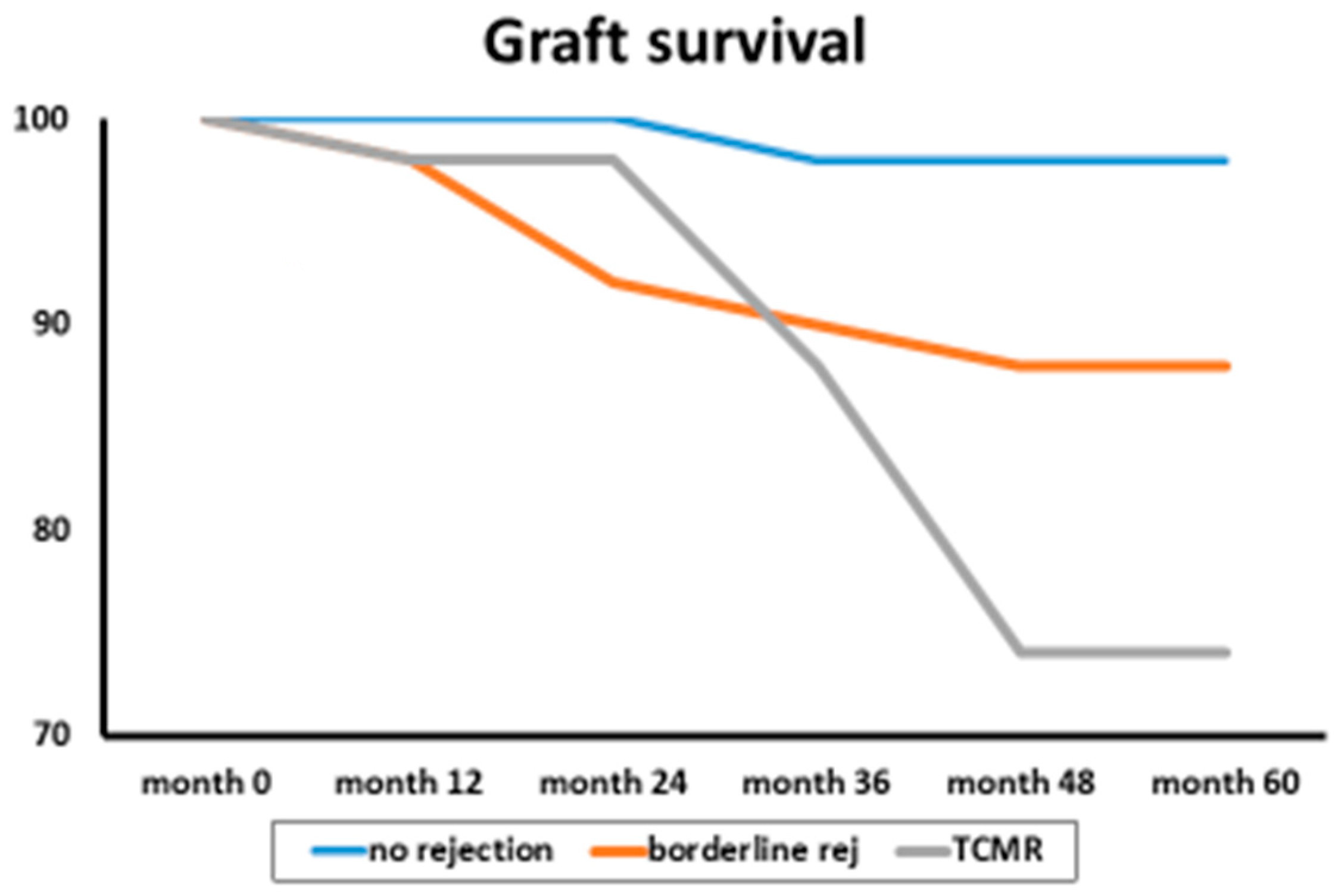
Nankivell et al. [39] evaluated the clinical and pathological significance of borderline T-cell-mediated rejection in 551 renal transplant recipients compared with that in normal controls and acute TCMRs.
The group of patients was followed for 60 months, and borderline rejection was associated with renal dysfunction, acute tubular necrosis, and chronic tubular atrophy. Additionally, patients with borderline rejection were associated with reduced graft survival (Figure 1).
Figure 1.
Graft survival according to rejection type.
Similarly, subclinical inflammation phenotypes and worse long-term renal outcomes have been observed in some groups of patients, such as pediatric kidney transplant recipients [40] and kidney transplant recipients treated with a rapid steroid withdrawal protocol [41]. Hence, new biomarkers in addition to dd-cfDNA are important.
In conclusion, dd-cf DNA has potential benefits, but also pitfalls such as the presence of both donor and recipient DNA. In addition, dd-cf DNA often does not distinguish TCMR and may be elevated in pathologies different from acute rejection. Their importance is highlighted by two recent meta-analyses [22,23]. In the ADMIRAL study [36], dd-cf DNA is important in determining the clinical outcome of a graft.
Several studies have recognized the importance of gene profiles in the diagnosis of early inflammation and subclinical rejection after kidney transplantation.
3. Gene Expression Profiles as Biomarkers
Fridewald et al. [42] documented the development and clinical validity of a novel blood-based molecular biomarker for the diagnosis of subclinical acute rejection following kidney transplantation.
Several previous studies have shown that gene expression profiles (GEPs) in both urine and blood can be used to detect kidney transplant rejection. The CTOT-04 study [4] revealed that a 3-gene signature in urine samples was able to detect kidney rejection. Similarly, the CTOT-01 study [43] revealed that the presence of the CXCL9 protein in the urine was indicative of rejection. In the CTOT-08 study [42], Fridewald et al. demonstrated the clinical validity of the clinical significance of both the clinical phenotype (CP) and the GEP of subclinical acute rejection within the first 12 months on the composite clinical endpoint. The same study documented the development of de novo DNA by 24 months in these subjects. In particular, in the CTOT-08 study, the gene expression profile consisted of 120 genes whose expression was upregulated and downregulated, and these genes were specifically chosen to distinguish stable normal biopsy tissue from subclinical rejection tissue.
In a different study, Zhang et al. [44] examined 191 kidney transplant patients from the prospective Genomic of Chronic Allograft Rejection (GoCAR) study [45] who underwent surveillance biopsies for more than two years and identified patients with subclinical or borderline acute cellular rejection (ACR) at three months (ACR-3) post-transplant. These patients subsequently had worse outcomes, with a decrease in renal function and decreased graft survival. Using RNA sequencing analysis, the authors identified a 17-gene signature (Table 6). This TREx assay based on the 17-gene set achieved a PPV of 0.79 and an NPV of 0.98 for subclinical ACR diagnosis. This set was validated and represents a peripheral blood gene expression signature to diagnose subclinical acute rejection and to risk stratify kidney transplant recipients.
Table 6.
17-gene set for 3-month ACR diagnosis.
An important blood gene expression classifier in the field of kidney transplantation is the TruGraf. It represents a novel molecular biomarker for managing kidney transplant recipients with stable renal function [46]. The TruGraf algorithm is a DNA microarray-based gene expression algorithm that analyzes the gene expression profiles of 120 genes. The original study was designed to avoid surveillance biopsies and was validated in multiple cohorts. Simultaneous blood tests and clinical assessments were performed on 192 patients from seven transplant centers [46]. The results of the TruGraf blood test were compared with the clinical phenotypes of 99 kidney transplant recipients with stable renal function and biopsy-confirmed phenotypes and 63 kidney transplant patients with stable renal function but with per-protocol biopsy documenting subclinical rejection; the accuracy was 74% in patients without rejection and 80% in patients with rejection. The overall negative predictive value was 89%, and the positive predictive value was 48% with a sensitivity of 71% and a specificity of 75%. The conclusion of the study was in favor of a clinical decision without unnecessary surveillance biopsies based on an accurate gene expression profile (GEP).
Based on the TruGraf method, Heilman et al. [47] conducted a prospective study in which peripheral blood was obtained at five time points in the first year post-transplant to obtain GEP. Overall, 240 kidney transplant patients were enrolled and stratified into three groups according to the absence or presence of one or more GEPs. The presence of multiple GEPs correlated with poorer histological aspects, lower eGFRs, and greater death-censoring graft loss.
We have described the relevance of dd-cfDNA in diagnosing subclinical rejection and the gene expression profile. A question is whether blood gene expression assays and donor-derived cell-free DNA may be used together to diagnose subclinical rejection. Park et al. [48] recently answered this question.
In that study, the authors enrolled 208 subjects for a total of 428 biopsy samples. The study was a post hoc analysis of the clinical Trial in Organ Transplantation 08. Surveillance biopsies were performed from month 2 to month 6 post-transplant and at months 12 and 24.
Patients were simultaneously subjected to a gene expression profile assay (TruGraf method), donor-derived cfDNA, and combined tests. With respect to the diagnosis of subclinical rejection, the authors reported a PPV of 47% with the gene profile, of 56% with dd-cfDNA, and of 81% with the combined tests.
The NPV was 82% with the gene profile, 84% with dd-cfDNA, and 88% with the combined tests. The area under the receiver operating characteristic curve (AUROC) values was similar for each method. Overall, the GEP was better at detecting cellular rejection, and dd-cfDNA was better at detecting antibody rejection.
The combination of gene expression profile assay with donor-derived cfDNA offers higher sensitive results in diagnosing subclinical rejection. Notably, when cases were separated on the basis of rejection type, the gene expression profile was significantly better at detecting cellular rejection (area under the receiver operating curve, 0.80 versus 0.62; p = 0.001), whereas the donor-derived cfDNA was significantly better at detecting antibody-mediated rejection.
In conclusion, donor-derived cell-free DNA and gene expression profiles provide a less invasive monitoring strategy for subclinical rejection with different detection methods for antibody and cell-mediated rejection.
Among the several methods to have a correct and available gene expression profile, the TruGraf assay has proven to be the best. TruGraf algorithm is a DNA microarray-based gene expression algorithm analyzing 120 genes. In association with dd-cf DNA, it is able to diagnose subclinical rejection [48]. In addition, the repetition of TruGraf several times after transplantation is able to give an available graft prognosis.
4. Urinary RNA Profile for the Diagnosis of Rejection
Independent of the information on clinical or subclinical rejection that can be drawn from blood, urine may also allow for the rejection of a kidney graft via a noninvasive method.
Li et al. [49] compared urine specimens from 22 kidney grafts with biopsy-confirmed acute rejection with 63 grafts without biopsy-confirmed acute rejection and reported increased levels of perforin mRNA and granzyme B mRNA in patients with biopsy-confirmed acute rejection. Both mRNAs encode cytotoxic proteins.
The authors divided the patients into four groups (acute rejection, stable graft function, other pathological findings not related to rejection, and chronic rejection). The levels of perforin mRNA were significantly greater in patients with acute rejection than in patients with stable renal function (p < 0.001), patients with other findings (p < 0.001), and patients with chronic rejection (p = 0.03).
This was not the case for granzyme B mRNA levels, which were not able to distinguish patients who experienced acute rejection from patients who experienced chronic rejection (p = 0.12).
The authors concluded that the urinary mRNA levels of perforin and granzyme B are useful noninvasive tools for the diagnosis of acute rejection, but the levels of granzyme B mRNA were not able to distinguish acute rejection from chronic rejection.
We have cited the study of Suthanthiran et al. [4]. One year later, the same authors published another more extensive study on the urinary mRNA profile and acute rejection in kidney transplant recipients [50]. Overall, a total of 4300 urine samples were collected from 485 patients for urinary-cell messenger RNA (mRNA) analysis repeatedly after transplantation and at the time of kidney allograft biopsy. Urinary mRNA was examined for CD3 ε perforin, granzyme B, interferon-inducible protein 10 (IP-10), CXCR3, CD103, transforming growth factor β (TGF β), and proteinase inhibitor 9. The patients were divided into three groups (acute cellular rejection, no rejection, and stable function in patients who did not receive renal biopsy). The mRNA levels of CD ε perforin, granzyme B, and IP-10 but not those of CXCR3 (p = 0.06), CD103 (p = 0.13), TGFβ (p = 0.11), or proteinase inhibitor 9 (p = 0.38) differed significantly among the three groups (p <= 0.001).
The authors concluded that CD3 ε mRNA, IP-10 mRNA, and 18S rRNA levels in urinary cells appear to be diagnostic of acute rejection in kidney allografts.
The last Suthanthiran study on the relevance of urinary chemokins as noninvasive diagnostic method for kidney transplant acute rejection is recent and has been published on 2021 [51].
In another study [52], messenger RNA (mRNA) for FOXP3, a specification and functional factor for regulatory T lymphocytes, and mRNA for CD25, CD3εperforin, and 18S ribosomal RNA (rRNA) were examined in urine specimens from patients who experienced acute rejection, chronic allograft nephropathy, and a normal renal biopsy. In particular, the study examined the relationship between these mRNA levels and acute rejection, rejection reversal, and graft failure. The mRNA levels of all the factors studied were significantly greater for acute rejection than for chronic rejection and stable graft function (p < 0.001). FOXP3 mRNA was the only factor related to reversibility. Patients with higher FOXP3 mRNA levels had a lower possibility of a reversible rejection. Similarly, patients with higher FOXP3 mRNA levels had a greater risk of graft loss. In contrast, the mRNA levels of CD25, CD3εperforin, and 18S ribosomal RNA (rRNA) were not related to rejection reversibility or risk of graft loss.
Hricik et al. [43] enrolled 280 kidney transplant patients, principally living donors, to evaluate and compare urinary biomarkers useful for a sure diagnosis of acute rejection. At the time of urine collection, all the patients underwent a for-cause renal biopsy. Urinary samples were examined for sediment RNA already known to be elevated for acute rejection, such as CCR1, CCR5, CXCR3, CCL5, CXCL9 (a cytokine induced by interferon gamma), CXCL10, IL-8, perforin, and granzyme B [53,54,55].
Through correlation analysis, many of these substances were found to be highly correlated and independent. CXCL9 and CXCL10 exhibited greater significance in diagnosing acute rejection. CXCL10 was more abundant in patients with acute rejection and in patients with infections. Therefore, in the author’s opinion, CXCL9 is the protein with the greatest significance in diagnosing acute rejection and distinguishing acute rejection from other renal injuries. In detail, the area under the curve (AUC) of CXCL9 for acute rejection diagnosis was 0.856, the PPV was 67.6%, and the NPV was 92%. The addition of CXCL10 did not modify the results.
However, the utility of the use of urinary cytokines as noninvasive biomarkers for the diagnosis of acute rejection has been discussed by other studies, which have revealed several controversies. In a retrospective French study [56] involving 329 transplanted patients, the utility of urinary CXCL9 and CXCL10 as noninvasive methods for diagnosing allograft rejection was investigated. All the patients included in this retrospective study had allograft biopsy specimens, concomitant urine samples, and blood research for BKV infection. Indeed, the study revealed that a similar increase in these urinary cytokines is found in acute rejection as well as BKV infection and urinary tract infection, which are frequently observed in kidney transplant subjects.
Similarly, a recent study [57] evaluated the utility of urinary CXCL10 for monitoring the renal allografts. In the present study, the patients were divided into two arms: the intervention arm (120 patients) and the control arm (121 patients). In both arms, urine for detecting CXCL10 was collected at months 1, 3, and 6. If elevated, renal biopsy was performed, and the subsequent treatment for rejection occurred in the intervention arm. In the control arm, the results were concealed. At 1 year, the death-censored graft loss, the acute rejection, the presence of de novo DSA, and the presence of an eGFR < 25 mL/min. were evaluated. Considering the combined endpoint at 1 year, the intervention arm and the control arm did not differ (51% vs. 49%) and the study could not demonstrate the beneficial effect of urine CXCL10 monitoring.
On the other hand, another study from Hricik et al. for the Clinical Trials in Organ Transplantation-09 Consortium [58] documented the usefulness of cytokines and other tools in predicting high-risk patients. This study aimed to analyze the safety of Tacrolimus withdrawal in immune-quiescent kidney transplant recipients. Overall, 21 patients were randomized, 14 in the tacrolimus withdrawal group and 7 in the control group. The study was terminated prematurely because of the high risk of acute rejection in the tacrolimus withdrawal group. High mismatches pretransplant, donor-reactive IFN-γ ELISPOT assay, and high levels of urinary CXCL9 were all predictive of the development of acute rejection or/and the development of DSAs in the tacrolimus withdrawal arm.
Concerning the origin of many urinary biomarkers, the main source are immuno-logical activated cells. Indeed, many urinary biomarkers are induced by the activation of immunological cells. Indeed, perforin, secreted by cytotoxic effector cells, causes cell death by knocking holes in target-cell membranes. Granzyme B induces DNA fragmentation and cell death by activating caspase 3.
FOXP3 has been examined in many studies analyzing the possible role of Tregs as a potential biomarker for immunologic monitoring in acute T-cell-mediated rejection.
Urinary RNA profile is important in the diagnosis of rejection. Different authors have studied several mRNA. Even if the usefulness of some of these cytokines such as CXCL9 and CXCL10 has been discussed to make a diagnosis of rejection, others such as perforin, granzyme B, and the interferon inducible protein 10 appear to be the most reliable in this setting.
The main challenges in finding new biomarkers are as follows:
- The rapidly evolving field of biomarker-informed precision medicine will be led astray if clinicians do not continually inform the data to turn it into useful information that can help patients
- This principle is not new, but the allure of a new and “easy” test result that gives us “all the answers” is seductive
- We must remember that our value to the patient remains being the link between the stream of data and the clinical reality
- Lastly, we are dealing with complex patients with complicated intersections of disease states that are in near-constant flux
- These are the main challenges in finding new biomarkers. To date, dd-cf DNA, gene expression profile, and some urinary mRNA substances are good diagnostic and prognostic biomarkers, while we are still waiting for new reliable monitoring and treatment response biomarkers.
5. Conclusions
In conclusion, multiple new noninvasive and invasive biomarkers are changing the paradigms of rejection diagnosis and immunosuppression management.
Further refinement of the proper context of use and interpretation of these tests will shape patient care as transplantation moves further into the field of personalized medicine.
The development of well-designed, interventional clinical trials using these tools is the next logical step.
Author Contributions
M.S., A.R. and G.R. contributed equally to the manuscript; M.S. designed the study. G.R. collected the data from the literature; M.S. and A.R. analyzed the collected data; M.S., A.R. and G.R. wrote the manuscript. All authors have read and agreed to the published version of the manuscript.
Funding
This research received no external funding.
Conflicts of Interest
Maurizio Salvadori, Alberto Rosati and Giuseppina Rosso do not have any conflicts of interest in relation with the manuscript.
References
- Naesens, M.; Anglicheau, D. Precision Transplant Medicine: Biomarkers to the Rescue. J. Am. Soc. Nephrol. 2018, 29, 24–34. [Google Scholar] [CrossRef] [PubMed]
- Salvadori, M.; Tsalouchos, A. Microbiota, renal disease and renal transplantation. World J. Transplant. 2021, 11, 16–36. [Google Scholar] [CrossRef]
- Anglicheau, D.; Naesens, M.; Essig, M.; Gwinner, W.; Marquet, P. Establishing Biomarkers in Transplant Medicine: A Critical Review of Current Approaches. Transplantation 2016, 100, 2024–2038. [Google Scholar] [CrossRef] [PubMed]
- Anglicheau, D.; Muthukumar, T.; Hummel, A.; Ding, R.; Sharma, V.K.; Dadhania, D.; Seshan, S.V.; Schwartz, J.E.; Suthanthiran, M. Discovery and Validation of a Molecular Signature for the Noninvasive Diagnosis of Human Renal Allograft Fibrosis. Transplantation 2012, 93, 1136–1146. [Google Scholar] [CrossRef]
- Roedder, S.; Sigdel, T.; Salomonis, N.; Hsieh, S.; Dai, H.; Bestard, O.; Metes, D.; Zeevi, A.; Gritsch, A.; Cheeseman, J.; et al. The kSORT assay to detect renal transplant patients at high risk for acute rejection: Results of the multicenter AART study. PLoS Med. 2014, 11, e1001759. [Google Scholar] [CrossRef] [PubMed]
- Kurian, S.M.; Williams, A.N.; Gelbart, T.; Campbell, D.; Mondala, T.S.; Head, S.R.; Horvath, S.; Gaber, L.; Thompson, R.; Whisenant, T.; et al. Molecular classifiers for acute kidney transplant rejection in peripheral blood by whole genome gene expression profiling. Am. J. Transplant. 2014, 14, 1164–1172. [Google Scholar] [CrossRef]
- Loupy, A.; Lefaucheur, C.; Vernerey, D.; Prugger, C.; Duong van Huyen, J.P.; Mooney, N.; Suberbielle, C.; Frémeaux-Bacchi, V.; Méjean, A.; Desgrandchamps, F.; et al. Complement-binding anti-HLA antibodies and kidney-allograft survival. N. Engl. J. Med. 2013, 369, 1215–1226. [Google Scholar] [CrossRef] [PubMed]
- Sicard, A.; Ducreux, S.; Rabeyrin, M.; Couzi, L.; McGregor, B.; Badet, L.; Scoazec, J.Y.; Bachelet, T.; Lepreux, S.; Visentin, J.; et al. Detection of C3d-binding donor-specific anti-HLA antibodies at diagnosis of humoral rejection predicts renal graft loss. J. Am. Soc. Nephrol. 2015, 26, 457–467. [Google Scholar] [CrossRef]
- Einecke, G.; Reeve, J.; Sis, B.; Mengel, M.; Hidalgo, L.; Famulski, K.S.; Matas, A.; Kasiske, B.; Kaplan, B.; Halloran, P.F. A molecular classifier for predicting future graft loss in late kidney transplant biopsies. J. Clin. Investig. 2010, 120, 1862–1872. [Google Scholar] [CrossRef]
- Loupy, A.; Lefaucheur, C.; Vernerey, D.; Chang, J.; Hidalgo, L.G.; Beuscart, T.; Verine, J.; Aubert, O.; Dubleumortier, S.; Duong van Huyen, J.P.; et al. Molecular microscope strategy to improve risk stratification in early antibody-mediated kidney allograft rejection. J. Am. Soc. Nephrol. 2014, 25, 2267–2277. [Google Scholar] [CrossRef]
- O’Connell, P.J.; Zhang, W.; Menon, M.C.; Yi, Z.; Schröppel, B.; Gallon, L.; Luan, Y.; Rosales, I.A.; Ge, Y.; Losic, B.; et al. Biopsy transcriptome expression profiling to identify kidney transplants at risk of chronic injury: A multicentre, prospective study. Lancet 2016, 388, 983–993. [Google Scholar] [CrossRef] [PubMed]
- Gielis, E.M.; Ledeganck, K.J.; De Winter, B.Y.; Del Favero, J.; Bosmans, J.L.; Claas, F.H.; Abramowicz, D.; Eikmans, M. Cell-Free DNA: An Upcoming Biomarker in Transplantation. Am. J. Transplant. 2015, 15, 2541–2551. [Google Scholar] [CrossRef] [PubMed]
- Beck, J.; Bierau, S.; Balzer, S.; Andag, R.; Kanzow, P.; Schmitz, J.; Gaedcke, J.; Moerer, O.; Slotta, J.E.; Walson, P.; et al. Digital droplet PCR for rapid quantification of donor DNA in the circulation of transplant recipients as a potential universal biomarker of graft injury. Clin. Chem. 2013, 59, 1732–1741. [Google Scholar] [CrossRef] [PubMed]
- Zhang, J.; Tong, K.L.; Li, P.K.; Chan, A.Y.; Yeung, C.K.; Pang, C.C.; Wong, T.Y.; Lee, K.C.; Lo, Y.M. Presence of donor- and recipient-derived DNA in cell-free urine samples of renal transplantation recipients: Urinary DNA chimerism. Clin. Chem. 1999, 45, 1741–1746. [Google Scholar] [CrossRef] [PubMed]
- García Moreira, V.; Prieto García, B.; Baltar Martín, J.M.; Ortega Suárez, F.; Alvarez, F.V. Cell-free DNA as a noninvasive acute rejection marker in renal transplantation. Clin. Chem. 2009, 55, 1958–1966. [Google Scholar] [CrossRef] [PubMed]
- Sigdel, T.K.; Vitalone, M.J.; Tran, T.Q.; Dai, H.; Hsieh, S.C.; Salvatierra, O.; Sarwal, M.M. A rapid noninvasive assay for the detection of renal transplant injury. Transplantation 2013, 96, 97–101. [Google Scholar] [CrossRef] [PubMed]
- Botezatu, I.; Serdyuk, O.; Potapova, G.; Shelepov, V.; Alechina, R.; Molyaka, Y.; Ananév, V.; Bazin, I.; Garin, A.; Narimanov, M.; et al. Genetic analysis of DNA excreted in urine: A new approach for detecting specific genomic DNA sequences from cells dying in an organism. Clin. Chem. 2000, 46, 1078–1084. [Google Scholar] [CrossRef]
- Osmanodja, B.; Akifova, A.; Budde, K.; Choi, M.; Oellerich, M.; Schütz, E.; Beck, J. Absolute or Relative Quantification of Donor-derived Cell-free DNA in Kidney Transplant Recipients: Case Series. Transplant. Direct. 2021, 7, e778. [Google Scholar] [CrossRef] [PubMed]
- Graver, A.S.; Lee, D.; Power, D.A.; Whitlam, J.B. Understanding Donor-derived Cell-free DNA in Kidney Transplantation: An Overview and Case-based Guide for Clinicians. Transplantation 2023, 107, 1675–1686. [Google Scholar] [CrossRef]
- Sigdel, T.K.; Archila, F.A.; Constantin, T.; Prins, S.A.; Liberto, J.; Damm, I.; Towfighi, P.; Navarro, S.; Kirkizlar, E.; Demko, Z.P.; et al. Optimizing Detection of Kidney Transplant Injury by Assessment of Donor-Derived Cell-Free DNA via Massively Multiplex PCR. J. Clin. Med. 2018, 8, 19. [Google Scholar] [CrossRef]
- Bloom, R.D.; Bromberg, J.S.; Poggio, E.D.; Bunnapradist, S.; Langone, A.J.; Sood, P.; Matas, A.J.; Mehta, S.; Mannon, R.B.; Sharfuddin, A.; et al. Circulating Donor-Derived Cell-Free DNA in Blood for Diagnosing Active Rejection in Kidney Transplant Recipients (DART) Study Investigators. Cell-Free DNA and Active Rejection in Kidney Allografts. J. Am. Soc. Nephrol. 2017, 28, 2221–2232. [Google Scholar] [CrossRef] [PubMed]
- Wijtvliet, V.P.W.M.; Plaeke, P.; Abrams, S.; Hens, N.; Gielis, E.M.; Hellemans, R.; Massart, A.; Hesselink, D.A.; De Winter, B.Y.; Abramowicz, D.; et al. Donor-derived cell-free DNA as a biomarker for rejection after kidney transplantation: A systematic review and meta-analysis. Transpl. Int. 2020, 33, 1626–1642. [Google Scholar] [CrossRef] [PubMed]
- Xiao, H.; Gao, F.; Pang, Q.; Xia, Q.; Zeng, X.; Peng, J.; Fan, L.; Liu, J.; Wang, Z.; Li, H. Diagnostic Accuracy of Donor-derived Cell-free DNA in Renal-allograft Rejection: A Meta-analysis. Transplantation 2021, 105, 1303–1310. [Google Scholar] [CrossRef] [PubMed]
- Oellerich, M.; Shipkova, M.; Asendorf, T.; Walson, P.D.; Schauerte, V.; Mettenmeyer, N.; Kabakchiev, M.; Hasche, G.; Gröne, H.J.; Friede, T.; et al. Absolute quantification of donor-derived cell-free DNA as a marker of rejection and graft injury in kidney transplantation: Results from a prospective observational study. Am. J. Transplant. 2019, 19, 3087–3099. [Google Scholar] [CrossRef] [PubMed]
- Whitlam, J.B.; Ling, L.; Skene, A.; Kanellis, J.; Ierino, F.L.; Slater, H.R.; Bruno, D.L.; Power, D.A. Diagnostic application of kidney allograft-derived absolute cell-free DNA levels during transplant dysfunction. Am. J. Transplant. 2019, 19, 1037–1049. [Google Scholar] [CrossRef]
- Zhang, H.; Zheng, C.; Li, X.; Fu, Q.; Li, J.; Su, Q.; Zeng, L.; Liu, Z.; Wang, J.; Huang, H.; et al. Diagnostic Performance of Donor-Derived Plasma Cell-Free DNA Fraction for Antibody-Mediated Rejection in Post Renal Transplant Recipients: A Prospective Observational Study. Front. Immunol. 2020, 11, 342. [Google Scholar] [CrossRef] [PubMed]
- Bromberg, J.S.; Brennan, D.C.; Poggio, E.; Bunnapradist, S.; Langone, A.; Sood, P.; Matas, A.J.; Mannon, R.B.; Mehta, S.; Sharfuddin, A.; et al. Biological Variation of Donor-Derived Cell-Free DNA in Renal Transplant Recipients: Clinical Implications. J. Appl. Lab. Med. 2017, 2, 309–321. [Google Scholar] [CrossRef] [PubMed]
- Huang, E.; Sethi, S.; Peng, A.; Najjar, R.; Mirocha, J.; Haas, M.; Vo, A.; Jordan, S.C. Early clinical experience using donor-derived cell-free DNA to detect rejection in kidney transplant recipients. Am. J. Transplant. 2019, 19, 1663–1670. [Google Scholar] [CrossRef] [PubMed]
- Stroup, D.F.; Berlin, J.A.; Morton, S.C.; Olkin, I.; Williamson, G.D.; Rennie, D.; Moher, D.; Becker, B.J.; Sipe, T.A.; Thacker, S.B. Meta-analysis of observational studies in epidemiology: A proposal for reporting. Meta-analysis Of Observational Studies in Epidemiology (MOOSE) group. JAMA 2000, 283, 2008–2012. [Google Scholar] [CrossRef]
- Lefaucheur, C.; Loupy, A. Antibody-Mediated Rejection of Solid-Organ Allografts. N. Engl. J. Med. 2018, 379, 2580–2582. [Google Scholar]
- Haas, M.; Loupy, A.; Lefaucheur, C.; Roufosse, C.; Glotz, D.; Seron, D.; Nankivell, B.J.; Halloran, P.F.; Colvin, R.B.; Akalin, E.; et al. The Banff 2017 Kidney Meeting Report: Revised diagnostic criteria for chronic active T cell-mediated rejection, antibody-mediated rejection, and prospects for integrative endpoints for next-generation clinical trials. Am. J. Transplant. 2018, 18, 293–307. [Google Scholar] [CrossRef] [PubMed]
- Stites, E.; Kumar, D.; Olaitan, O.; John Swanson, S.; Leca, N.; Weir, M.; Bromberg, J.; Melancon, J.; Agha, I.; Fattah, H.; et al. High levels of dd-cfDNA identify patients with TCMR 1A and borderline allograft rejection at elevated risk of graft injury. Am. J. Transplant. 2020, 20, 2491–2498. [Google Scholar] [CrossRef] [PubMed]
- Pallardó Mateu, L.M.; Sancho Calabuig, A.; Capdevila Plaza, L.; Franco Esteve, A. Acute rejection and late renal transplant failure: Risk factors and prognosis. Nephrol. Dial. Transplant. 2004, 19 (Suppl. S3), iii38–iii42. [Google Scholar] [CrossRef] [PubMed]
- Jevnikar, A.M.; Mannon, R.B. Late kidney allograft loss: What we know about it, and what we can do about it. Clin. J. Am. Soc. Nephrol. 2008, 3 (Suppl. S2), S56–S67. [Google Scholar] [CrossRef] [PubMed]
- Cooper, J.E.; Gralla, K.; Chan, K. Clinical significance of post kidney transplant de novo DSA in otherwise stable grafts. Clin. Transpl. 2011, 35, 359–364. [Google Scholar]
- Bu, L.; Gupta, G.; Pai, A.; Anand, S.; Stites, E.; Moinuddin, I.; Bowers, V.; Jain, P.; Axelrod, D.A.; Weir, M.R.; et al. Clinical outcomes from the Assessing Donor-derived cell-free DNA Monitoring Insights of kidney Allografts with Longitudinal surveillance (ADMIRAL) study. Kidney Int. 2022, 101, 793–803. [Google Scholar] [CrossRef] [PubMed]
- Clayton, P.A.; Lim, W.H.; Wong, G.; Chadban, S.J. Relationship between eGFR Decline and Hard Outcomes after Kidney Transplants. J. Am. Soc. Nephrol. 2016, 27, 3440–3446. [Google Scholar] [CrossRef] [PubMed]
- Faddoul, G.; Nadkarni, G.N.; Bridges, N.D.; Goebel, J.; Hricik, D.E.; Formica, R.; Menon, M.C.; Morrison, Y.; Murphy, B.; Newell, K.; et al. Analysis of Biomarkers Within the Initial 2 Years Posttransplant and 5-Year Kidney Transplant Outcomes: Results From Clinical Trials in Organ Transplantation-17. Transplantation 2018, 102, 673–680. [Google Scholar] [CrossRef] [PubMed]
- Nankivell, B.J.; Agrawal, N.; Sharma, A.; Taverniti, A.; P’Ng, C.H.; Shingde, M.; Wong, G.; Chapman, J.R. The clinical and pathological significance of borderline T cell-mediated rejection. Am. J. Transplant. 2019, 19, 1452–1463. [Google Scholar] [CrossRef]
- Seifert, M.E.; Yanik, M.V.; Feig, D.I.; Hauptfeld-Dolejsek, V.; Mroczek-Musulman, E.C.; Kelly, D.R.; Rosenblum, F.; Mannon, R.B. Subclinical inflammation phenotypes and long-term outcomes after pediatric kidney transplantation. Am. J. Transplant. 2018, 18, 2189–2199. [Google Scholar] [CrossRef]
- Mehta, R.; Bhusal, S.; Randhawa, P.; Sood, P.; Cherukuri, A.; Wu, C.; Puttarajappa, C.; Hoffman, W.; Shah, N.; Mangiola, M.; et al. Short-term adverse effects of early subclinical allograft inflammation in kidney transplant recipients with a rapid steroid withdrawal protocol. Am. J. Transplant. 2018, 18, 1710–1717. [Google Scholar] [CrossRef] [PubMed]
- Friedewald, J.J.; Kurian, S.M.; Heilman, R.L.; Whisenant, T.C.; Poggio, E.D.; Marsh, C.; Baliga, P.; Odim, J.; Brown, M.M.; Ikle, D.N.; et al. Development and clinical validity of a novel blood-based molecular biomarker for subclinical acute rejection following kidney transplant. Am. J. Transplant. 2019, 19, 98–109. [Google Scholar] [CrossRef] [PubMed]
- Hricik, D.E.; Nickerson, P.; Formica, R.N.; Poggio, E.D.; Rush, D.; Newell, K.A.; Goebel, J.; Gibson, I.W.; Fairchild, R.L.; Riggs, M.; et al. CTOT-01 consortium.Multicenter validation of urinary CXCL9 as a risk-stratifying biomarker for kidney transplant injury. Am. J. Transplant. 2013, 13, 2634–2644. [Google Scholar] [CrossRef] [PubMed]
- Zhang, W.; Yi, Z.; Keung, K.L.; Shang, H.; Wei, C.; Cravedi, P.; Sun, Z.; Xi, C.; Woytovich, C.; Farouk, S.; et al. A Peripheral Blood Gene Expression Signature to Diagnose Subclinical Acute Rejection. J. Am. Soc. Nephrol. 2019, 30, 1481–1494. [Google Scholar] [CrossRef] [PubMed]
- Yazdani, S.; Naesens, M. Foretelling Graft Outcome by Molecular Evaluation of Renal Allograft Biopsies: The GoCAR Study. Transplantation 2017, 101, 5–7. [Google Scholar] [CrossRef] [PubMed]
- Marsh, C.L.; Kurian, S.M.; Rice, J.C.; Whisenant, T.C.; David, J.; Rose, S.; Schieve, C.; Lee, D.; Case, J.; Barrick, B.; et al. Application of TruGraf v1: A Novel Molecular Biomarker for Managing Kidney Transplant Recipients With Stable Renal Function. Transplant. Proc. 2019, 51, 722–728. [Google Scholar] [CrossRef] [PubMed]
- Heilman, R.L.; Fleming, J.N.; Mai, M.; Smith, B.; Park, W.D.; Holman, J.; Stegall, M.D. Multiple abnormal peripheral blood gene expression assay results are correlated with subsequent graft loss after kidney transplantation. Clin. Transplant. 2023, 37, e14987. [Google Scholar] [CrossRef] [PubMed]
- Park, S.; Guo, K.; Heilman, R.L.; Poggio, E.D.; Taber, D.J.; Marsh, C.L.; Kurian, S.M.; Kleiboeker, S.; Weems, J.; Holman, J.; et al. Combining Blood Gene Expression and Cellfree DNA to Diagnose Subclinical Rejection in Kidney Transplant Recipients. Clin. J. Am. Soc. Nephrol. 2021, 16, 1539–1551. [Google Scholar] [CrossRef]
- Li, B.; Hartono, C.; Ding, R.; Sharma, V.K.; Ramaswamy, R.; Qian, B.; Serur, D.; Mouradian, J.; Schwartz, J.E.; Suthanthiran, M. Noninvasive diagnosis of renal-allograft rejection by measurement of messenger RNA for perforin and granzyme B in urine. N. Engl. J. Med. 2001, 344, 947–954. [Google Scholar] [CrossRef]
- Suthanthiran, M.; Schwartz, J.E.; Ding, R.; Abecassis, M.; Dadhania, D.; Samstein, B.; Knechtle, S.J.; Friedewald, J.; Becker, Y.T.; Sharma, V.K.; et al. Urinary-cell mRNA profile and acute cellular rejection in kidney allografts. N. Engl. J. Med. 2013, 369, 20–31. [Google Scholar] [CrossRef]
- Lubetzky, M.L.; Salinas, T.; Schwartz, J.E.; Suthanthiran, M. Urinary Cell mRNA Profiles Predictive of Human Kidney Allograft Status. Clin. J. Am. Soc. Nephrol. 2021, 16, 1565–1577. [Google Scholar] [CrossRef] [PubMed]
- Muthukumar, T.; Dadhania, D.; Ding, R.; Snopkowski, C.; Naqvi, R.; Lee, J.B.; Hartono, C.; Li, B.; Sharma, V.K.; Seshan, S.V.; et al. Messenger RNA for FOXP3 in the urine of renal-allograft recipients. N. Engl. J. Med. 2005, 35, 2342–2351. [Google Scholar] [CrossRef] [PubMed]
- Ho, J.; Wiebe, C.; Gibson, I.W.; Rush, D.N.; Nickerson, P.W. Immune monitoring of kidney allografts. Am. J. Kidney Dis. 2012, 60, 629–640. [Google Scholar] [CrossRef] [PubMed]
- Panzer, U.; Reinking, R.R.; Steinmetz, O.M.; Zahner, G.; Sudbeck, U.; Fehr, S.; Pfalzer, B.; Schneider, A.; Thaiss, F.; Mack, M.; et al. CXCR3 and CCR5 positive T-cell recruitment in acute human renal allograft rejection. Transplantation 2004, 78, 1341–1350. [Google Scholar] [CrossRef]
- Qin, S.; Rottman, J.B.; Myers, P.; Kassam, N.; Weinblatt, M.; Loetscher, M.; Koch, A.E.; Moser, B.; Mackay, C.R. The chemokine receptors CXCR3 and CCR5 mark subsets of T cells associated with certain inflammatory reactions. J. Clin. Investig. 1998, 101, 746–754. [Google Scholar] [CrossRef] [PubMed]
- Tinel, C.; Devresse, A.; Vermorel, A.; Sauvaget, V.; Marx, D.; Avettand-Fenoel, V.; Amrouche, L.; Timsit, M.O.; Snanoudj, R.; Caillard, S.; et al. Development and validation of an optimized integrative model using urinary chemokines for noninvasive diagnosis of acute allograft rejection. Am. J. Transplant. 2020, 20, 3462–3476. [Google Scholar] [CrossRef]
- Hirt-Minkowski, P.; Handschin, J.; Stampf, S.; Hopfer, H.; Menter, T.; Senn, L.; Hönger, G.; Wehmeier, C.; Amico, P.; Steiger, J.; et al. Randomized Trial to Assess the Clinical Utility of Renal Allograft Monitoring by Urine CXCL10 Chemokine. J. Am. Soc. Nephrol. 2023, 34, 1456–1469. [Google Scholar] [CrossRef]
- Hricik, D.E.; Formica, R.N.; Nickerson, P.; Rush, D.; Fairchild, R.L.; Poggio, E.D.; Gibson, I.W.; Wiebe, C.; Tinckam, K.; Bunnapradist, S.; et al. Adverse Outcomes of Tacrolimus Withdrawal in Immune-Quiescent Kidney Transplant Recipients. J. Am. Soc. Nephrol. 2015, 26, 3114–3122. [Google Scholar] [CrossRef]

Disclaimer/Publisher’s Note: The statements, opinions and data contained in all publications are solely those of the individual author(s) and contributor(s) and not of MDPI and/or the editor(s). MDPI and/or the editor(s) disclaim responsibility for any injury to people or property resulting from any ideas, methods, instructions or products referred to in the content. |
© 2024 by the authors. Licensee MDPI, Basel, Switzerland. This article is an open access article distributed under the terms and conditions of the Creative Commons Attribution (CC BY) license (https://creativecommons.org/licenses/by/4.0/).