Review

Adult Presentation of X-Linked Hypophosphatemia

1
Division of Nephrology and Endocrinology, The University of Tokyo Hospital, Tokyo 113-8655, Japan
2
Osteoporosis Center, The University of Tokyo Hospital, Tokyo 113-8655, Japan
Endocrines 2022, 3(3), 375-390; https://doi.org/10.3390/endocrines3030030
Submission received: 18 May 2022 / Revised: 21 June 2022 / Accepted: 24 June 2022 / Published: 1 July 2022
(This article belongs to the Special Issue Update on X-linked Hypophosphatemia)

Abstract

:
Adult X-linked hypophosphatemia (XLH) patients present with specific symptoms, including enthesopathies (e.g., ossification of longitudinal ligaments (OPLL), osteophytes around large joints, and enthesopathy in the Achilles tendons), early osteoarthritis, the development of severe secondary and tertiary hyperparathyroidism (SHPT/THPT), and the subsequent progression of chronic kidney disease (CKD). In addition, these patients exhibit the typical phenotypes of osteomalacia, such as pseudofracture and fracture in weight-bearing bones, odontitis, and tooth abscesses. The mechanism underlying enthesopathy development is unknown; however, a common underlying mechanism among XLH and autosomal recessive hypophosphatemic rickets (ARHR1/2) due to mutations in PHEX, DMP1, and ENPP1 is assumed. Clarification of the pathogenesis and drug discovery for this complication is an urgent issue, as many adult XLH patients suffer subsequent debilitating nervous symptoms or impingement syndrome, and existing treatments are ineffective. Severe SHPT and THPT are associated with conventional therapy, including active vitamin D and phosphate supplementation, and complicated and careful adjustment of dosages by experienced clinicians is required to avoid SHPT/THPT. Burosumab is a very effective therapy without risk for the development of SHPT/THPT. However, indications for this drug should be carefully considered, along with cost-effectiveness, guidelines or recommendations, and the health care system of each country.

1. Introduction

X-linked hypophosphatemia (XLH) is a genetic disease caused by inactivating mutations of the phosphate-regulating endopeptidase gene (PHEX). Symptoms that develop during childhood are typical rachitic phenotypes, including leg deformity, short stature, odontitis, tooth abscesses, and craniosynostosis. In contrast, adult XLH patients present variable symptoms, including complications resulting from its treatment, such as enthesopathy, early osteoarthritis, secondary/tertiary hyperparathyroidism (SHPT/THPT), and chronic kidney disease (CKD), in addition to characteristic phenotypes for osteomalacia, such as pseudofracture and fracture in weight-bearing bones [1,2]. Some clinicians believe that treatment could be terminated after epiphyseal closure even among patients with severe XLH; however, adult XLH patients are currently recognized to be at risk of pseudofractures and fractures in weight-bearing bones due to low turnover even with normal to high bone mineral density (BMD) [3,4]. The mechanism for the development of enthesopathy in adults with XLH is unknown, and even among adult XLH patients treated with active vitamin D and phosphate supplementation or burosumab, remarkable enthesopathies sometimes develop, and these patients experience significant difficulty in activities of daily life (ADL) and lower quality of life (QOL) with nervous symptoms due to ossification of the posterior longitudinal ligament (OPLL), ossification of the ligamentum flavum (OLF), and other ossification-related nerve entrapment, or impingement syndromes in the hip, and the development of osteoarthritis [4].
Treatment for adult XLH with active vitamin D and inorganic phosphate is very difficult, as well as for child patients with XLH, because excess of this treatment is associated with the development of SHPT/THPT and subsequent nephrotic diabetes insipidus, prerenal renal failure, postrenal renal failure, and the progression of CKD [5]. Therefore, the transition of XLH patients to specialized clinicians is vital, especially when patients are treated with conventional therapy, although there are a limited number of specialists for bone and mineral disorders in adults. Burosumab is an anti-fibroblast growth factor (FGF) 23 humanized monoclonal antibody developed by a pharmaceutical company in collaboration with our laboratory, which led to the improvement of phosphate metabolism in children with XLH and adults with XLH up to 96 weeks in clinical studies [6,7,8,9,10,11]. A beneficial effect of burosumab on pseudofracture and fracture healing was confirmed in the clinical studies, and, theoretically, it is protective against the progression of CKD, as burosumab enables patients to withdraw phosphate preparation, although its effect on the prevention or improvement of enthesopathy and early osteoarthritis is unknown.
In this review article about the adult presentation of XLH, the estimated underlying mechanisms for the development of adult XLH-specific problems, including enthesopathy, early osteoarthritis, SHPT/THPT, and CKD, are illustrated, and the recommended indication and selection of pharmaceuticals and detailed adjustment procedure of dosage according to previous publications and personal experience are introduced.

2. Diagnosis of Adult XLH

Mild cases of undiagnosed XLH are infrequently suspected among adults with pseudofractures and fractures in weight-bearing bones or remarkable enthesopathies in the spinal ligament, around the hip joints, and in the Achilles tendons, with concomitant chronic hypophosphatemia. To accelerate an accurate diagnosis, appropriate treatment and prevention of additional pseudofracture, fracture, and dental disorders, clinicians should suspect low turnover disorders such as FGF23-related hypophosphatemia, including XLH and tumor-induced osteomalacia, Fanconi syndrome, vitamin D deficient osteomalacia, mild vitamin D dependent rickets (e.g., heterozygous mutation in CYP3A4), mild hypophosphatasia (e.g., heterozygous mutation in ALPL), mild osteogenesis imperfecta (e.g., Sillence type I), and mild osteopetrosis (e.g., dominant negative type of heterozygous mutation in CLCN7). These ailments should be suspected among adult patients who develop pseudofractures and fractures in weight-bearing bones (costa, pelvis, femoral head subchondral fragility fracture, diaphyses of femur/tibia/fibula, calcaneus, and metatarsal) spontaneously, with low-power trauma, with relatively short-term use of anti-resorptive reagents for osteoporosis (e.g., ≤5 years), among adult patients who develop odontitis, tooth abscesses, necrosis of the jaw spontaneously, or with relatively short-term use of anti-resorptive reagents for osteoporosis.
In Japan, once chronic hypophosphatemia with the relevant symptoms of rickets/osteomalacia is recognized, measurement of serum intact fibroblast growth factor (FGF) 23 (Determinar CL FGF23; Minaris Medical, Tokyo, Japan) is encouraged to determine the etiology of hypophosphatemia, with a cutoff value of 30 pg/mL to discriminate FGF23-related hypophosphatemic rickets/osteomalacia and others, which was revealed to possess high sensitivity and specificity [12,13,14]. Among adult patients with osteomalacia accompanied by FGF23 values of 30 pg/mL or more, diagnosis of XLH is strongly supported by the presence of one or more of the following symptoms: mildly short stature, leg deformity (genu varum, genu vulgus), enthesopathy, or X-linked inheritance of rickets/osteomalacia. However, even among these cases, genetic diagnosis of PHEX mutation is recommended, if available, in an effort to make an accurate diagnosis [15]. In the cases of adult-onset FGF23-related hypophosphatemic osteomalacia without any of the symptoms above, the possibilities of tumor-induced osteomalacia, intravenous infusion of iron preparation-induced osteomalacia, and alcohol-induced FGF23-related hypophosphatemic osteomalacia should be explored [16,17,18].

3. Symptoms of Adult XLH

3.1. Pseudofracture and Fracture

As stated above, some clinicians used to believe that treatment could be terminated after epiphyseal closure, probably due to the normal or relatively high bone mineral density detected in the majority of adults with mild XLH [3,4]. Normal-to-high BMD observed in patients with mild XLH might stem from suppressed osteoclastic function and is probably due to the excessive ossification caused for the same reason XLH patients tend to develop enthesopathy [4]. However, the risk of pseudofractures and fractures in weight-bearing bones (costa, pelvis, femoral head subchondral fragility fracture, diaphyses of femur/tibia/fibula, calcaneus, and metatarsal) is usually not correlated with BMD and is strongly associated with low turnover of bone (e.g., also shown in other hypophosphatemic rickets/otsteomalacia, hypophosphatasia, osteogenesis imperfecta, osteopetrosis, and long-term use of anti-osteoclastic reagents), as accumulated micro bone cracks with delayed healing in weight-bearing bones eventually lead to pseudofracture and fracture. Therefore, the treatment for hypophosphatemia should be continued among adult XLH patients with typical rachitic phenotypes, including short stature, leg deformity, or a past history of surgical correction of leg deformity, with continuously elevated bone-specific alkaline phosphatase (BAP) in the absence of treatment [1,2]. Figure 1a,b show typical femoral pseudofractures developed after years of treatment cessation and surgically treated fractures in the diaphysis of the femur and bilateral tibiae developed under conventional therapy (Figure 1). To detect tiny pseudofractures, X-rays in multiple directions, bone scintigraphy (99mTc-methylene diphosphonate/hydroxymethylene diphosphonate), and T2-weighted fat-suppressed magnetic resonance imaging (MRI) are beneficial.
To prevent the development of pseudofracture and fracture, long periods of strenuous exercise and labor (e.g., long period of walking, stomping exercise for osteoporosis, or weightbearing exercise/labor) should be avoided in adult XLH patients with uncontrolled chronic hypophosphatemia manifested by elevated BAP, and patients should be encouraged to visit their attending clinician immediately when warning pain is recognized in the diaphysis of the femur/tibia/fibula. Adult XLH patients with new pseudofracture or fracture should be treated with conventional therapy or burosumab. It is recommended that patients who developed pseudofracture or fracture under treatment with conventional therapy consider changing the treatment to burosumab. When a surgical procedure is unavoidable for the developed pseudofracture/fracture, medical treatment should be provided for 1 to 3 months before surgery to prevent the loosening of prosthesis or the development of additional pseudofracture/fracture around prosthesis, unless urgent surgery is required [2].
Please note that anti-resorptive reagents (bisphosphonates and denosumab) are contraindicated for patients with uncontrolled low bone turnover disorders, including adult XLH, as these reagents aggravate low bone turnover and increase the risk of developing pseudofracture and fracture in weight-bearing bones, tooth abscesses, and necrosis of the jaw. If coexisting osteoporosis is suspected in postmenopausal females with XLH or older patients with XLH, anti-osteoporotic treatment should not be initiated until osteomalacia improvement by normalization of BAP with conventional therapy or burosumab is confirmed.
Based on retrospective case reviews, conventional therapy with active vitamin D and phosphate preparation is suggested to be beneficial to prevent and heal pseudofractures and fractures in weight-bearing bones; however, this speculation has not been proven, as there are no prospective trials. On the other hand, burosumab treatment improved pseudofractures and fractures in adult XLH patients in clinical trials [7,9,19].

3.2. Muscle Weakness

In 2013, Veilleux et al. reported that muscle force in the lower extremities in 13 XLH patients (6 to 60 years old) was significantly decreased despite normal muscle size in comparison with age- and sex-matched control participants [20]. Clinicians tend to ascribe this muscle weakness in XLH patients to the direct effect of hypophosphatemia on myocytes. However, the observed muscle weakness was also explained by bone pain due to osteomalacia and inefficient transduction of muscle contraction due to leg deformity or impingement syndrome stemming from enthesopathy. Of note, rapidly induced hypophosphatemia is not associated with muscle weakness [21]. The effect of conventional therapy on muscle weakness is inconclusive as there is no prospective trial, and burosumab was associated with an increased distance of 6 min in walk tests, although this might be associated with the improvement of pseudofractures and fractures [11]. Further clinical studies are warranted to clarify whether there is a direct effect of hypophosphatemia on myocytes.

3.3. Dental Health

Uncontrolled rickets/osteomalacia is evidently associated with a high risk of periodontitis, odontitis, and tooth abscesses in children and adults [22,23]. The underlying mechanism for this might be the accumulation of microcracks with delayed healing in the enamel and dentin that penetrate from the pit on the surface to the pulp, similar to how pseudofractures and fractures develop in weight-bearing bones. Orthodontic treatment sometimes results in the loss of permanent teeth in XLH patients with uncontrolled osteomalacia. Early intervention with conventional treatment was reported to have a prophylactic effect on the development of these dental diseases, although this finding was not discovered in a prospective trial [24,25]. In the post hoc analysis of a 64-week, open-label, randomized controlled study with 61 children aged 1 to 12 years with XLH, dental abscesses occurred in 3 of 12 (25%) younger (<5 years) children with conventional therapy, while 0 of 20 (0%) younger children from the burosumab group developed dental abscesses. However, in older (5–12 years) children with XLH, dental abscesses presented more frequently with burosumab than with conventional therapy (9/29 (31%) vs. 2/32 (6%)) [26]. Based on the results of this study, burosumab seems to possess weaker protective effects or, at least, not more intense protective effects, against the development of dental abscesses compared to conventional therapy; however, a longer duration study is needed.
The effect of burosumab on dental health in XLH patients has not yet been reported.

3.4. Enthesopathy

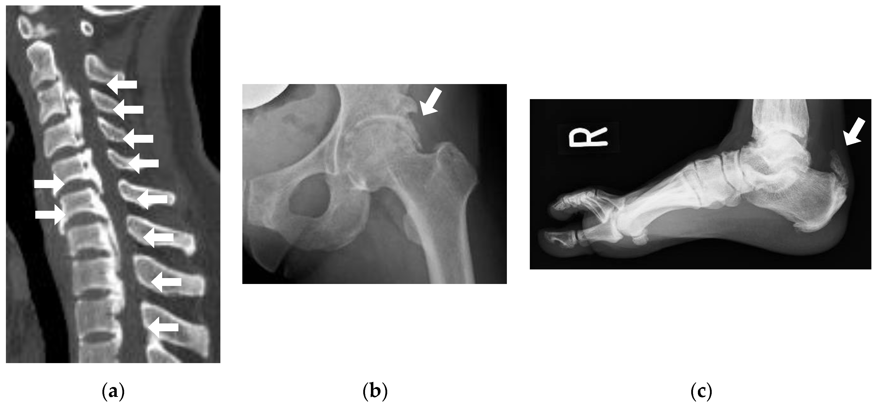
We examined the prevalence of enthesopathies in 25 adult XLH patients (18 to 72 y) and revealed a high prevalence of OPLL (32%), osteophytes around the hip joints equivalent to a Kellgren–Lawrence grade of 2 and more (96%), and enthesopathies in the Achilles tendon (72%), which explained that XLH is an obvious genetic condition in which patients are prone to developing enthesopathies [4]. Normal to high BMD in adult XLH patients was also reported in the same article and was attributed to the same osteogenic nature of XLH [4]. In some adult XLH patients, neurological symptoms due to OPLL and limited range of motion (ROM) in the hip and intervertebral joints severely lowered ADL and QOL [27]. Figure 2a–c are typical images of OPLL, osteophytes around the hip joint, and enthesopathy in the Achilles tendon present in adult XLH patients (Figure 2).
Currently, the precise mechanism for the development of enthesopathy in adult XLH patients (typically over 30 years old) is unknown. It has been recognized that enthesopathy is also frequently present in patients with other inherited FGF23-related hypophosphatemic rickets, including autosomal recessive hypophosphatemic rickets 1 and 2 caused by homozygous mutations in DMP1 and ENPP1, respectively, although other types of inherited or acquired FGF23-related hypophosphatemia are not associated with the development of enthesopathy [28,29]. Recently, we reported that haploinsufficiency of ectonucleotide pyrophosphatase/phosphodiesterase (ENPP1) with heterozygous or compound heterozygous mutations of ENPP1 is also associated with milder phenotypes of enthesopathy, manifested by OPLL and diffuse idiopathic skeletal hyperostosis (DISH) [30]. This means that the enthesopathy present in patients with XLH and ARHR1/2 is not a consequence of chronic hypophosphatemia or high levels of serum FGF23, and there is a common mechanism to develop enthesopathy among XLH and ARHR1/2. Therefore, unfortunately, conventional treatment did not exert a prophylactic effect on the development of enthesopathy or improve enthesopathy [25]. In my view, burosumab is suspected to be ineffective in the prevention or improvement of enthesopathy, as other conditions with highly elevated FGF23, such as advanced CKD patients on renal replacement therapy, are not associated with increased risk for enthesopathy, although this finding will not be conclusive until the results of a long-term study with burosumab are reported. Severe neurologic symptoms, such as paraparesis due to OPLL, are an indication for surgical decompression of the spinal cord.
ENPP1 is a membranous enzyme that metabolizes adenosine triphosphate (ATP) into adenosine monophosphate (AMP) and inorganic pyrophosphate (PPi). PPi was identified to antagonize the formation of hydroxyapatite. Thus, lowering plasma PPi is the candidate mechanism for the development of enthesopathy in patients with homozygous, compound heterozygous, or heterozygous ENPP1 mutations [31,32]. Furthermore, Maulding et al. reported that low plasma PPi levels were identified in Hyp mice, the model mouse for XLH [33]. Therefore, one possible explanation for this tendency to develop enthesopathy in adult XLH patients is the involvement of lowered PPi, albeit the PPi levels in XLH patients have not been reported.

3.5. Osteoarthritis

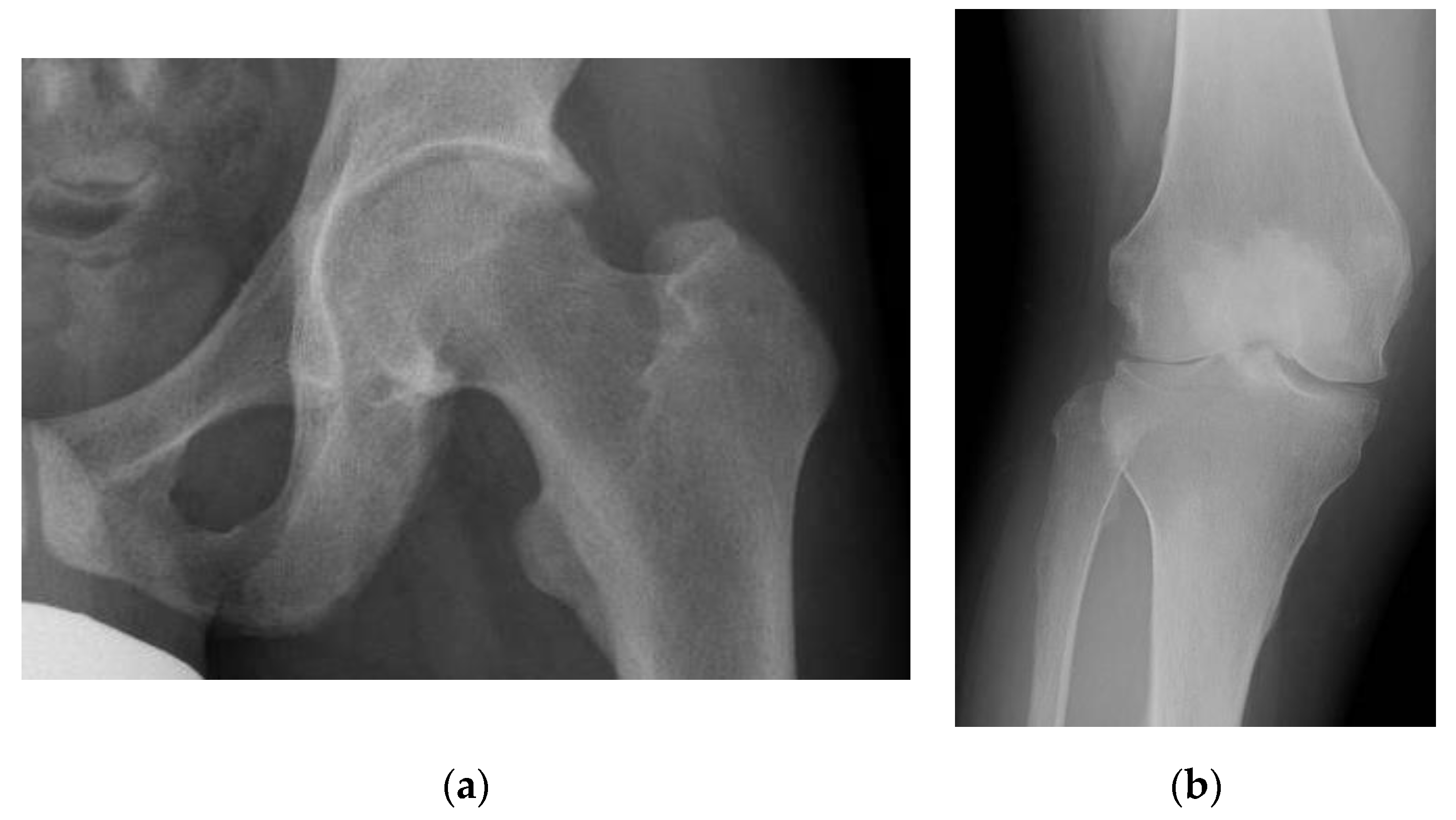
Early osteoarthritis is the other debilitating symptom commonly recognized among adult XLH patients. Development of early osteoarthritis is partly due to abnormal mechanical loading stemming from leg deformity, although it is not fully explained by this [34]. Existence of enthesopathy and excessive ossification in adults with XLH might be associated with the development of early osteoarthritis. We reported the prevalence of hip osteoarthritis (Kellgren–Lawrence grade ≥2, 3) to be 96% and 88%, respectively, and the prevalence of knee osteoarthritis (Kellgren–Lawrence grade ≥2, 3) to be 68% and 36%, which is markedly higher than the general population [4]. Figure 3a,b are typical images of osteoarthritis in the hip and knee joints presented in a 29-year-old male and 30-year-old female with XLH, respectively (Figure 3). As in the case with enthesopathy, conventional therapy did not improve or prevent the development of osteoarthritis in the adults with XLH, while the effect of burosumab on this complication is indecisive.

3.6. SHPT/THPT

XLH patients tend to develop SHPT (hyperparathyroidism with the value of calcium less than the middle of the reference range) due to low 1,25(OH)2D stemming from excessive action of FGF23 and exogenous supplementation of inorganic phosphate. SHPT is an undesired phenotype in XLH patients because hyperparathyroidism stimulates osteoclastic activity, leading to bone loss in addition to rickets/osteomalacia, and accelerates hypophosphatemia as parathyroid hormone (PTH) decreases the expression of sodium–phosphate cotransporter independent of FGF23. Additionally, we recently reported that PTH directly stimulates the transcription of FGF23 in an osteocytic cell line [35]. Furthermore, a handful of XLH patients who already suffer from SHPT develop THPT (hyperparathyroidism with the value of calcium greater than or equal to the middle of the reference range), which is an equivalent clinical condition to primary hyperparathyroidism (PHPT) that causes hypercalciuria and nephrotic diabetes insipidus, leading to repetitive episodic prerenal and postrenal renal failure and progression of CKD.
In 2019, DeLacey et al. reported that among 84 patients with XLH (40 adults, 44 children), 83.3% had SHPT or THPT, and THPT developed in 16.7% of patients [5]. Seventy-five percent (6/8) of the patients who underwent parathyroidectomy for THPT experienced persistent or recurrent THPT, which explains why other parathyroid cells are also ready to convert into autonomous PTH-producing cells in XLH patients who once developed THPT [5]. We are now analyzing the relationship between the highest or cumulative dose of phosphate supplementation and the development of SHPT or THPT in a large cohort of XLH patients in Japan and Korea [36]. To prevent the development of severe SHPT and THPT, the dosage of conventional treatment with active vitamin D and phosphate supplementation should be adjusted with great care by experienced clinicians, and the change in treatment to burosumab should be considered for XLH patients who develop severe SHPT or THPT, as burosumab is not supposed to be associated with the development of severe SHPT and THPT as it enables patients to be freed from phosphate preparation. Once THPT is developed, immediate cessation of conventional therapy and initiation of an allosteric modulator of the calcium sensing receptor should be considered until parathyroidectomy conduction occurs, and treatment for hypophosphatemia should be resumed with burosumab afterward. Detailed guidance for conventional therapy recommended by the author is described in “Section 6.2. Conventional therapy (active vitamin D and phosphate supplementation).”

3.7. CKD

Some adult XLH patients with conventional treatment experience CKD progression uncommon for their age or for those with other medical conditions, and very rarely, patients need renal replacement therapy. The main reason for the progression of CKD in patients with adult XLH is described in “Section 3.5. SHPT/THPT” above; that is, the main reason is hypercalciuria and nephrotic diabetes insipidus due to conventional therapy, leading to repetitive episodic prerenal and postrenal renal failure, which often follows development of THPT. In fact, in the article by DeLacey et al. introduced in the previous section and including 84 XLH patients, nephrocalcinosis and CKD G3 and over (eGFR < 60 mL/min/1.73 m2) were more prevalent in patients with THPT than in patients without THPT (60% vs. 18.6% and 35.7% vs. 1.5%, respectively) [5]. Therefore, to prevent the progression of CKD, adequate handling of severe SHPT and THPT by experienced clinicians is necessary, and appropriate water consumption and intake of salt should be recommended among XLH patients treated with conventional therapy, especially in those with febrile and gastrointestinal disorders or who labor and spend leisure time in the hot sun over a long period. Please note that the dosage of active vitamin D and phosphate supplementation needs to be decreased as CKD progresses; otherwise, a persistent excessive dose of conventional therapy might be associated with the development of THPT and further progression of CKD. The correlation between the highest or cumulative dose of phosphate supplementation and active vitamin D and the progression of CKD to stage G3 and over is also under examination in a large cohort of XLH patients in Japan and Korea [37]. The use of burosumab is thought to be protective against the progression of CKD, as patients can disregard phosphate preparations; however, its protectiveness remains unclear until the result of a long-term study is reported.

3.8. Hypertension and Left Ventricular Hypertrophy

A high prevalence of elevated blood pressure (27.3%, 6/22) unmatched to age has been reported in a small cohort of adult XLH patients, and this characteristic could be the consequence of the progression of CKD. A cohort study with a larger number of adult XLH patients is necessary to address this issue [38]. The effects of conventional therapy and burosumab on the prevention of development and progression of hypertension and left ventricular hypertrophy are unknown.

3.9. Hearing Loss

The prevalence of hearing loss among XLH patients varies from 16% to 76% depending on the population or method of evaluation [39]. In 25 adult XLH patients with a median age of 39 (range 18–60) years from our hospital, 8 (32%) patients presented with hearing impairment. Seven cases had a sensorineural pattern, and one case had both sensorineural and conductive patterns [4]. The precise mechanism of hearing loss in XLH patients has not been clarified; however, temporal bone malformation due to osteomalacia and endolymphatic hydrops stemming from hypophosphatemia have been suggested to be the causes [38]. There has been no report suggesting a beneficial effect of conventional therapy or burosumab to prevent or improve hearing impairment in adults with XLH.

4. QOL of Adult XLH

There are several reports about the QOL of adult XLH patients [39,40,41]. In 2019, Skrinar et al. reported that among 232 adult patients with XLH, 97% had bone or joint pain/stiffness, 44% had a history of fractures, 46% had osteophytes, 27% had enthesopathy in the Achilles tendon, and 19% had spinal stenosis. In addition, the mean scores for age-specific patient-reported outcomes (PROs) evaluating pain, stiffness, and physical function were worse than those of the control population [39]. In 2020, Seefried et al. conducted a systematic literature review including 91 articles and 44 congress abstracts, revealing that XLH had a substantial and wide-ranging negative impact on health-related quality of life (HRQOL), particularly relating to physical function and pain measured by the 36-item Short-Form Health Survey (SF-36), Western Ontario and McMaster Universities Osteoarthritis Index (WOMAC), and Brief Pain Inventory (BPI) [40]. We evaluated the HRQOL in Japanese and Korean patients with XLH, including 32 adult patients; this investigation revealed that among adult XLH patients, 59.4% had bone pain, 65.6% had joint pain, 9.5% had CKD G3 or higher, 15.6% had nephrocalcinosis, 15.6% had SHPT or THPT, and 6.3% underwent parathyroidectomy [41]. In our study, the SF-36, WOMAC, and BPI also revealed lower QOL in adult XLH patients [41]. In fact, it is hard to distinguish if musculoskeletal pain is due to enthesopathy, osteoarthritis, bone pain, or pain from myalgias among adult patients with XLH. However, given the higher prevalence of reported joint pain (65.6%) than that of bone pain (59.4%) observed in our study, (1) early initiation of pharmaceutical intervention to prevent leg deformity and (2) exploration of the mechanism for the development of enthesopathy and early osteoarthritis to develop new treatment options to conquer these debilitating complications are important to further improve the QOL among adult XLH patients.

5. Transition of XLH Patients

Clinical follow-up of XLH patients should be transferred from pediatricians to adult endocrinologists or rheumatologists when patients are approximately 18 to 20 years of age to encourage independence from their caregivers and facilitate the care of other adult-specific medical problems. At the time of transition, patients need to be educated or re-educated about XLH, including the genetic information. XLH patients should be followed by experienced clinicians, as stated above, in the institutions where multidisciplinary care, which involves physical and occupational therapy, dental care, pain clinics, etc., can be offered. For more information about the transition of XLH, please refer to the well-organized mini-review by Dahir et al. [42]. Most importantly, XLH patients should be referred to experienced clinicians, although there are too few endocrinologists, rheumatologists, and medical geneticists worldwide specializing in bone metabolic disorders and skeletal dysplasia to meet demand. Therefore, the need for an increased number of endocrinologists, rheumatologists, and medical geneticists who are educated about bone metabolic disorders and skeletal dysplasia is one of the urgent issues to resolve in this field.

6. Treatment of Adult XLH

6.1. Indication and Selection of Treatment

The indication and selection of conventional treatment and burosumab among adult XLH patients might be influenced by the health care system of each nation. In Japan, I suggest the indication and selection of pharmaceutical treatment in adult XLH patients as described in Table 1; this information reflects my personal opinion based on my own experience with adult XLH patients (Table 1). The severity of the symptoms among adult XLH patients varies widely even between family members sharing the same mutation, and treatment for hypophosphatemia is not necessarily required among patients with mild symptoms defined by a normal value of BAP, stature ≥−1.0 SD, lack of genu varum and genu valgus, and no history of pseudofracture/bone pain, fracture in weight-bearing bones, or odontitis/tooth abscesses without treatment. In contrast, adult XLH patients with severe phenotypes should be treated with burosumab, which is more effective than conventional therapy. In my opinion, severe phenotypes are defined by short stature (<−2.0 SD), deviation of mechanical axis of the leg into zone 3 or greater [43], history of corrective surgery on the leg, history of pseudofracture/bone pain in weight-bearing bones, odontitis, tooth abscesses more than twice without any treatment, more than once under conventional therapy, or more than once but requiring surgery, and history of fracture in weight-bearing bone. The indication for conventional treatment is between the indications for observation and burosumab. Additionally, I recommend changing the treatment approach to include burosumab for adult XLH patients who develop uncontrolled SHPT or THPT with conventional treatment and patients with moderate symptoms (between mild and severe symptoms) and eGFR < 45 mL/min/1.73 m2 to prevent further progression of CKD.
Please note that enthesopathy cannot be improved by therapeutically targeting the increase in serum phosphate. Supplementation of natural vitamin D preparation (ergocalciferol, cholecalciferol) or encouraged intake of natural vitamin D from diet targeting serum 25OHD of 30 ng/mL and higher is recommended in XLH patients to avoid detriment to the bone due to vitamin D deficiency.

6.2. Conventional Therapy (Active Vitamin D and Phosphate Supplementation)

Treatment of XLH patients with conventional therapy is the tricky part of patient management, as inadequate and careless dosage adjustment results in the development of SHPT/THPT and consequent irreversible progression of CKD. Thus, clinicians should always prioritize prevention of the development of severe SHPT and THPT as long as patients are treated with conventional therapy.
In Japan, a powdered preparation is used, containing 330 mg of monobasic sodium phosphate monohydrate (NaH2PO4/H2O) and 119 mg of dibasic sodium phosphate anhydrous (Na2HPO4) in a packet, which is equivalent to 100 mg of inorganic phosphate. Other formulae of phosphate preparations (e.g., K-Phos, neutraphos) are also used to treat XLH patients [1]. Relatively frequent adverse events associated with phosphate preparation include nephrocalcinosis, abdominal pain, nausea, vomiting, and diarrhea, among others, in addition to the aforementioned SHPT, THPT, acute kidney injury, and progression of CKD. Active vitamin D-associated adverse events include nephrocalcinosis, hypercalciuria, thirst, appetite loss, abdominal pain, nausea, vomiting, diarrhea, and constipation, among others, in addition to acute kidney injury and progression of CKD.
The range of daily dosages of conventional therapy described in the consensus statement introduced by specialists from European countries (calcitriol: 0.50 to 0.75 μg, alfacalcidol: 0.75 to 1.5 μg, phosphate supplementation: 750 to 1600 mg) appears very appropriate from the viewpoint of preventing severe SHPT and THPT [2]. Calcitriol should be taken twice daily due to its shorter half-life compared to alfacalcidol, which is taken once daily. Active vitamin D should precede phosphate supplementation by approximately one week, and treatment with phosphate supplementation should always be accompanied by active vitamin D to prevent the development of severe SHPT and THPT [1,2]. Importantly, the serum phosphate level peaks approximately 1.5 h after the intake of phosphate supplementation and goes back to the trough value 2 to 3 h after intake [44]. Thus, phosphate supplementation should be taken four or more times daily to maximize the antihypophosphatemic effect and minimize the risk of developing SHPT/THPT.
I propose laboratory tests, including serum phosphate, calcium, albumin, intact PTH, creatinine, and BAP, with blood samples drawn 1 to 2 h after the intake of phosphate supplementation to detect the peak value of serum phosphate and intact PTH because these values are associated with bone mineralization and risk for the development of severe SHPT/THPT. These effects and risks cannot be inferred from the trough value of serum phosphate and intact PTH.
In our facility, conventional therapy usually starts with 1.0 μg of alfacalcidol, and 1 to 2 weeks later, phosphate supplementation is initiated with 200 mg four times daily. In the dose-adjusting phase, laboratory data are followed every one to four weeks, and phosphate supplementation is adjusted by 100 mg increments until the peak phosphate level stably settles within the lower 50% of the reference range. Most importantly, we do not attempt to adjust conventional therapy to increase trough phosphate levels within the reference range, as this is strongly associated with the development of SHPT/THPT and consequent progression of CKD [1]. Additionally, in adult XLH patients with eGFR < 45 mL/min/1.73 m2, lower dosages of active vitamin D and phosphate are required to maintain the peak serum phosphate level in the target range, and often only a small dose of active vitamin D (e.g., 0.25 to 0.50 μg of alfacalcidol) or no medication is required among adult XLH patients with eGFR < 30 mL/min/1.73 m2, although the change in treatment to burosumab should be recommended to prevent the further progression of CKD in case phosphate supplementation is still required (e.g., uncontrolled BAP) in patients with eGFR < 45 mL/min/1.73 m2. In the maintenance phase, laboratory data are followed every 3 to 6 months, and normalization of BAP is the ideal goal of treatment, as it directly reflects the ossification status of the bone. However, in treatment-naive XLH patients with active osteomalacia, BAP initially increases in response to treatment up to 3 to 6 months, indicating the recommencement of mineralization, and then decreases to the basal value approximately 6 to 12 months after the initiation of treatment. Thus, dose adjustment of conventional therapy targeting normalization of BAP should be considered after 12 to 18 months. Changing treatment to burosumab should be considered in patients with uncontrolled BAP.
Once again, the prioritized agenda alongside conventional treatment is the prevention of severe SHPT and THPT; therefore, the dosage of phosphate should be immediately decreased by 100 mg or more for a period of time once overadjustment of peak phosphate (e.g., within the upper 25% of the reference range or more) or severe SHPT (e.g., peak intact PTH ≥ double the upper limit of the reference range) is observed. Then, if an increase in phosphate supplementation is required in patients who have developed severe SHPT, escalation of active vitamin D by 0.25 μg for calcitriol and 0.25 to 0.5 μg for alfacalcidol should precede an increase in phosphate supplementation by 1 to 2 weeks. In patients who develop THPT (hyperparathyroidism with the value of calcium > the middle of the reference range), conventional therapy should immediately be terminated to prevent the progression of CKD; in addition, patients should try their best to avoid dehydration, and immediate initiation of an allosteric modulator of calcium sensing receptors (e.g., cinacalcet, evocalcet) is recommended until parathyroidectomy is performed after examination with ultrasound and 99mTc-sestamibi (MIBI) scintigraphy. In cases with THPT, all recognizable parathyroid glands by ultrasound and MIBI scintigraphy should be removed, as persistenting THPT and recurrence of THPT after parathyroidectomy occur very frequently (75%) among XLH patients [5]. In patients who developed THPT with contraindication for surgery (e.g., severe cardiopulmonary disorder, oldest-old patients), allosteric modulation of calcium sensing receptors should be continued, and in these patients, additional anti-osteoporotic treatment should be considered after the surrogate marker of osteomalacia (e.g., BAP) is well-controlled, because increased intact PTH persists with allosteric modulation of the calcium sensing receptor [45]. It is recommended to change treatment to burosumab in XLH patients with uncontrolled severe SHPT undergoing conventional therapy or who have developed THPT. Supplementation of active vitamin D or replacement therapy with recombinant 1-84 PTH is required in addition to treatment of hypophosphatemia in patients in whom all parathyroid glands have been removed due to THPT. Please refer to Table 2 for guidance for conventional treatment for adults with XLH (Table 2).

6.3. Burosumab

Burosumab has been reported to be associated with improvement in persistent pseudofractures and fractures; the results of PROs in adult XLH patients show that most of them were treated with conventional treatment beforehand [7,9,11,19]. Consequently, the initiation of or change to burosumab should be considered among adult XLH patients with severe symptoms (Table 1). In addition, given that burosumab repairs dysfunctional phosphate metabolism in patients with XLH to a physiologically corrected state by counteracting excess action of FGF23, burosumab should not be associated with the development of SHPT/THPT and subsequent progression of CKD. Therefore, initiation of burosumab should also be considered in patients with CKD (e.g., eGFR < 45 mL/min/1.73 m2), and a change in treatment from conventional therapy to burosumab should be considered among adult XLH patients with uncontrolled severe SHPT or a history of the development of THPT (Table 1). However, medical economic efficacy should always be considered independently for this kind of expensive orphan drug according to the guidelines/recommendations or the health care system of each country [2].
The initial dosage of burosumab for adult XLH patients is decided worldwide to be 1.0 mg/kg body weight (maximum dose of 90 mg) by subcutaneous injection every four weeks, and concomitant use of active vitamin D and phosphate supplementation are not recommended or are prohibited unless other coexisting medical conditions require active vitamin D (e.g., hypoparathyroidism after parathyroidectomy of all parathyroid glands). In the initial phase, the response to burosumab should be confirmed 1 to 2 weeks after the injection (peak phosphate), as the phosphate level returns to baseline four weeks after the last injection (trough phosphate) in a considerable number of patients. Trough phosphate level should be controlled within the lower half of the reference range, and dosage of burosumab should be decreased by 0.2 to 0.3 mg/kg in patients with trough phosphate levels within the higher half of the reference range. Burosumab should be suspended until phosphate values fall below the normal range in the case of phosphate levels over the reference range; treatment may be restarted at approximately half the initial starting dose. Adverse effects tightly associated with the use of burosumab are injection site reactions, and there are no reports of the development of antagonizing antibodies [7,9,11,19].
When no response or only a trivial response is observed at 1 to 2 weeks after injection (peak phosphate), coexisting vitamin D deficiency should be examined by measuring 25OHD. If the patient develops uncontrolled severe SHPT with conventional therapy, and the treatment is changed to burosumab, the response to burosumab (peak phosphate) might improve after 3 to 6 injections, as intact PTH decreases because stimulation of FGF23 transcription by PTH is alleviated [35]. THPT is also associated with a diminished response to burosumab, and immediate initiation of allosteric modulators of calcium sensing receptors (e.g., cinacalcet, evocalcet) and subsequent parathyroidectomy are recommended along with careful prevention of dehydration. If no response or trivial response to burosumab continues after these problems are ruled out or adequately addressed, changing the treatment to conventional therapy should be considered. Please refer to Table 3 for guidance for burosumab treatment for adults with XLH (Table 3).

7. Remaining Problems and Future Research Topics in Adult XLH Patients

The etiology for the development of enthesopathy and early osteoarthritis needs to be elucidated to develop treatment options, as these complications remarkably debilitate ADL and QOL in a large number of adult XLH patients. Associations between the development of severe SHPT or THPT, progression of CKD, the highest dosage, cumulative dosage of phosphate supplementation, and dosage of active vitamin D needs to be elucidated to create more detailed guidelines for conventional therapy. The mode of administration and dosage of burosumab should be reconsidered, as some of adult XLH patients are obviously undertreated with the current dosage (maximum 1.0 mg/kg body weight) and mode of administration (once every four weeks). The effects of burosumab on oral health and the development or progression of osteoarthritis and enthesopathy have been inconclusive, and we are awaiting the results of long-term observational studies. The impact of burosumab on pregnancy and fetal development must also be clarified.

8. Conclusions

The development of burosumab has been a game changer in the treatment of adult XLH patients, as conventional therapy is associated with undertreatment of osteomalacia, the risk of the developing severe SHPT or THPT, and the progression of CKD. However, there are problems remaining to be addressed, specifically among adults with XLH; of these, the complications of debilitating enthesopathy and early osteoarthritis warrant special attention and must be addressed urgently.

Funding

This research received no external funding.

Institutional Review Board Statement

Not applicable.

Informed Consent Statement

Not applicable.

Data Availability Statement

Not applicable.

Conflicts of Interest

N.I. receives research support from Kyowa Kirin Co. Ltd. (Tokyo, Japan).

References

  1. Carpenter, T.O.; Imel, E.A.; Holm, I.A.; Jan de Beur, S.M.; Insogna, K.L. A clinician’s guide to X-linked hypophosphatemia. J. Bone Miner. Res. 2011, 26, 1381–1388. [Google Scholar] [CrossRef] [PubMed] [Green Version]
  2. Haffner, D.; Emma, F.; Eastwood, D.M.; Duplan, M.B.; Bacchetta, J.; Schnabel, D.; Wicart, P.; Bockenhauer, D.; Santos, F.; Levtchenko, E.; et al. Clinical practice recommendations for the diagnosis and management of X-linked hypophosphataemia. Nat. Rev. Nephrol. 2019, 15, 435–455. [Google Scholar] [CrossRef] [PubMed] [Green Version]
  3. Cheung, M.; Roschger, P.; Klaushofer, K.; Veilleux, L.N.; Roughley, P.; Glorieux, F.H.; Rauch, F. Cortical and trabecular bone density in X-linked hypophosphatemic rickets. J. Clin. Endocrinol. Metab. 2013, 98, E954–E961. [Google Scholar] [CrossRef] [PubMed] [Green Version]
  4. Kato, H.; Koga, M.; Kinoshita, Y.; Taniguchi, Y.; Kobayashi, H.; Fukumoto, S.; Nangaku, M.; Makita, N.; Ito, N. Incidence of Complications in 25 Adult Patients with X-linked Hypophosphatemia. J. Clin. Endocrinol. Metab. 2021, 106, e3682–e3692. [Google Scholar] [CrossRef]
  5. DeLacey, S.; Liu, Z.; Broyles, A.; El-Azab, S.A.; Guandique, C.F.; James, B.C.; Imel, E.A. Hyperparathyroidism and parathyroidectomy in X-linked hypophosphatemia patients. Bone 2019, 127, 386–392. [Google Scholar] [CrossRef] [PubMed]
  6. Aono, Y.; Yamazaki, Y.; Yasutake, J.; Kawata, T.; Hasegawa, H.; Urakawa, I.; Fujita, T.; Wada, M.; Yamashita, T.; Fukumoto, S.; et al. Therapeutic effects of anti-FGF23 antibodies in hypophosphatemic rickets/osteomalacia. J. Bone Miner. Res. 2009, 24, 1879–1888. [Google Scholar] [CrossRef] [PubMed]
  7. Insogna, K.L.; Briot, K.; Imel, E.A.; Kamenický, P.; Ruppe, M.D.; Portale, A.A.; Weber, T.; Pitukcheewanont, P.; Cheong, H.I.; Jan de Beur, S.; et al. A Randomized, Double-Blind, Placebo-Controlled, Phase 3 Trial Evaluating the Efficacy of Burosumab, an Anti-FGF23 Antibody, in Adults with X-Linked Hypophosphatemia: Week 24 Primary Analysis. J. Bone Miner. Res. 2018, 33, 1383–1393. [Google Scholar] [CrossRef] [Green Version]
  8. Carpenter, T.O.; Whyte, M.P.; Imel, E.A.; Boot, A.M.; Högler, W.; Linglart, A.; Padidela, R.; Van’t Hoff, W.; Mao, M.; Chen, C.Y.; et al. Burosumab Therapy in Children with X-Linked Hypophosphatemia. N. Engl. J. Med. 2018, 378, 1987–1998. [Google Scholar] [CrossRef] [Green Version]
  9. Portale, A.A.; Carpenter, T.O.; Brandi, M.L.; Briot, K.; Cheong, H.I.; Cohen-Solal, M.; Crowley, R.; Jan De Beur, S.; Eastell, R.; Imanishi, Y.; et al. Continued Beneficial Effects of Burosumab in Adults with X-Linked Hypophosphatemia: Results from a 24-Week Treatment Continuation Period after a 24-Week Double-Blind Placebo-Controlled Period. Calcif. Tissue Res. 2019, 105, 271–284. [Google Scholar] [CrossRef] [Green Version]
  10. Imel, E.A.; Glorieux, F.H.; Whyte, M.P.; Munns, C.F.; Ward, L.M.; Nilsson, O.; Simmons, J.H.; Padidela, R.; Namba, N.; Cheong, H.I.; et al. Burosumab versus conventional therapy in children with X-linked hypophosphataemia: A randomised, active-controlled, open-label, phase 3 trial. Lancet 2019, 393, 2416–2427. [Google Scholar] [CrossRef]
  11. Briot, K.; Portale, A.A.; Brandi, M.L.; Carpenter, T.O.; Cheong, H.I.; Cohen-Solal, M.; Crowley, R.K.; Eastell, R.; Imanishi, Y.; Ing, S.; et al. Burosumab treatment in adults with X-linked hypophosphataemia: 96-week patient-reported outcomes and ambulatory function from a randomised phase 3 trial and open-label extension. RMD Open 2021, 7, e001714. [Google Scholar] [CrossRef] [PubMed]
  12. Fukumoto, S.; Ozono, K.; Michigami, T.; Minagawa, M.; Okazaki, R.; Sugimoto, T.; Takeuchi, Y.; Matsumoto, T. Pathogenesis and diagnostic criteria for rickets and osteomalacia—Proposal by an expert panel supported by the Ministry of Health, Labour and Welfare, Japan, the Japanese Society for Bone and Mineral Research, and the Japan Endocrine Society. J. Bone Miner. Metab. 2015, 33, 467–473. [Google Scholar] [CrossRef] [PubMed]
  13. Ito, N.; Kubota, T.; Kitanaka, S.; Fujiwara, I.; Adachi, M.; Takeuchi, Y.; Yamagami, H.; Kimura, T.; Shinoda, T.; Minagawa, M.; et al. Clinical performance of a novel chemiluminescent enzyme immunoassay for FGF23. J. Bone Miner. Metab. 2021, 39, 1066–1075. [Google Scholar] [CrossRef] [PubMed]
  14. Kato, H.; Hidaka, N.; Koga, M.; Ogawa, N.; Takahashi, S.; Miyazaki, H.; Nangaku, M.; Makita, N.; Ito, N. Performance evaluation of the new chemiluminescent intact FGF23 assay relative to the existing assay system. J. Bone Miner. Metab. 2022, 40, 101–108. [Google Scholar] [CrossRef] [PubMed]
  15. Kinoshita, Y.; Saito, T.; Shimizu, Y.; Hori, M.; Taguchi, M.; Igarashi, T.; Fukumoto, S.; Fujita, T. Mutational analysis of patients with FGF23-related hypophosphatemic rickets. Eur. J. Endocrinol. 2012, 167, 165–172. [Google Scholar] [CrossRef] [Green Version]
  16. Minisola, S.; Peacock, M.; Fukumoto, S.; Cipriani, C.; Pepe, J.; Tella, S.H.; Collins, M.T. Tumour-induced osteomalacia. Nat. Rev. Dis. Prim. 2017, 3, 17044. [Google Scholar] [CrossRef]
  17. Shimizu, Y.; Tada, Y.; Yamauchi, M.; Okamoto, T.; Suzuki, H.; Ito, N.; Fukumoto, S.; Sugimoto, T.; Fujita, T. Hypophosphatemia induced by intravenous administration of saccharated ferric oxide: Another form of FGF23-related hypophosphatemia. Bone 2009, 45, 814–816. [Google Scholar] [CrossRef]
  18. Hidaka, N.; Kato, H.; Koga, M.; Katsura, M.; Oyama, Y.; Kinoshita, Y.; Fukumoto, S.; Makita, N.; Nangaku, M.; Ito, N. Induction of FGF23-related hypophosphatemic osteomalacia by alcohol consumption. Bone Rep. 2021, 15, 101144. [Google Scholar] [CrossRef]
  19. Insogna, K.L.; Rauch, F.; Kamenický, P.; Ito, N.; Kubota, T.; Nakamura, A.; Zhang, L.; Mealiffe, M.; San Martin, J.; Portale, A.A. Burosumab Improved Histomorphometric Measures of Osteomalacia in Adults with X-Linked Hypophosphatemia: A Phase 3, Single-Arm, International Trial. J. Bone Miner. Res. 2019, 34, 2183–2191. [Google Scholar] [CrossRef] [Green Version]
  20. Veilleux, L.N.; Cheung, M.S.; Glorieux, F.H.; Rauch, F. The muscle-bone relationship in X-linked hypophosphatemic rickets. J. Clin. Endocrinol. Metab. 2013, 98, E990–E995. [Google Scholar] [CrossRef] [Green Version]
  21. Ito, N.; Fukumoto, S.; Takeuchi, Y.; Takeda, S.; Suzuki, H.; Yamashita, T.; Fujita, T. Effect of acute changes of serum phosphate on fibroblast growth factor (FGF)23 levels in humans. J. Bone Miner. Metab. 2007, 25, 419–422. [Google Scholar] [CrossRef] [PubMed]
  22. Beck-Nielsen, S.S.; Brusgaard, K.; Rasmussen, L.M.; Brixen, K.; Brock-Jacobsen, B.; Poulsen, M.R.; Vestergaard, P.; Ralston, S.H.; Albagha, O.M.; Poulsen, S.; et al. Phenotype presentation of hypophosphatemic rickets in adults. Calcif. Tissue Res. 2010, 87, 108–119. [Google Scholar] [CrossRef] [PubMed]
  23. Ye, L.; Liu, R.; White, N.; Alon, U.S.; Cobb, C.M. Periodontal status of patients with hypophosphatemic rickets: A case series. J. Periodontol. 2011, 82, 1530–1535. [Google Scholar] [CrossRef] [PubMed]
  24. Biosse Duplan, M.; Coyac, B.R.; Bardet, C.; Zadikian, C.; Rothenbuhler, A.; Kamenicky, P.; Briot, K.; Linglart, A.; Chaussain, C. Phosphate and Vitamin D Prevent Periodontitis in X-Linked Hypophosphatemia. J. Dent. Res. 2016, 96, 388–395. [Google Scholar] [CrossRef] [PubMed]
  25. Connor, J.; Olear, E.A.; Insogna, K.L.; Katz, L.; Baker, S.; Kaur, R.; Simpson, C.A.; Sterpka, J.; Dubrow, R.; Zhang, J.H.; et al. Conventional Therapy in Adults with X-Linked Hypophosphatemia: Effects on Enthesopathy and Dental Disease. J. Clin. Endocrinol. Metab. 2015, 100, 3625–3632. [Google Scholar] [CrossRef] [PubMed]
  26. Ward, L.M.; Glorieux, F.H.; Whyte, M.P.; Munns, C.F.; Portale, A.A.; Högler, W.; Simmons, J.H.; Gottesman, G.S.; Padidela, R.; Namba, N.; et al. Impact of Burosumab Compared with Conventional Therapy in Younger versus Older Children with X-Linked Hypophosphatemia. J. Clin. Endocrinol. Metab. 2022, dgac296. [Google Scholar] [CrossRef]
  27. Steele, A.; Gonzalez, R.; Garbalosa, J.C.; Steigbigel, K.; Grgurich, T.; Parisi, E.J.; Feinn, R.S.; Tommasini, S.M.; Macica, C.M. Osteoarthritis, Osteophytes, and Enthesophytes Affect Biomechanical Function in Adults with X-linked Hypophosphatemia. J. Clin. Endocrinol. Metab. 2020, 105, e1798–e1814. [Google Scholar] [CrossRef]
  28. Karaplis, A.C.; Bai, X.; Falet, J.P.; Macica, C.M. Mineralizing enthesopathy is a common feature of renal phosphate-wasting disorders attributed to FGF23 and is exacerbated by standard therapy in hyp mice. Endocrinology 2012, 153, 5906–5917. [Google Scholar] [CrossRef] [Green Version]
  29. Saito, T.; Shimizu, Y.; Hori, M.; Taguchi, M.; Igarashi, T.; Fukumoto, S.; Fujita, T. A patient with hypophosphatemic rickets and ossification of posterior longitudinal ligament caused by a novel homozygous mutation in ENPP1 gene. Bone 2011, 49, 913–916. [Google Scholar] [CrossRef]
  30. Kato, H.; Ansh, A.J.; Lester, E.R.; Kinoshita, Y.; Hidaka, N.; Hoshino, Y.; Koga, M.; Taniguchi, Y.; Uchida, T.; Yamaguchi, H.; et al. Identification of ENPP1 Haploinsufficiency in Patients with Diffuse Idiopathic Skeletal Hyperostosis and Early-Onset Osteoporosis. J. Bone Miner. Res. 2022, 37, 1125–1135. [Google Scholar] [CrossRef]
  31. Ferreira, C.R.; Kintzinger, K.; Hackbarth, M.E.; Botschen, U.; Nitschke, Y.; Mughal, M.Z.; Baujat, G.; Schnabel, D.; Yuen, E.; Gahl, W.A.; et al. Ectopic Calcification and Hypophosphatemic Rickets: Natural History of ENPP1 and ABCC6 Deficiencies. J. Bone Miner. Res. 2021, 36, 2193–2202. [Google Scholar] [CrossRef]
  32. Bernhard, E.; Nitschke, Y.; Khursigara, G.; Sabbagh, Y.; Wang, Y.; Rutsch, F. A Reference Range for Plasma Levels of Inorganic Pyrophosphate in Children Using the ATP Sulfurylase Method. J. Clin. Endocrinol. Metab. 2021, 107, 109–118. [Google Scholar] [CrossRef] [PubMed]
  33. Maulding, N.D.; Kavanagh, D.; Zimmerman, K.; Coppola, G.; Carpenter, T.O.; Jue, N.K.; Braddock, D.T. Genetic pathways disrupted by ENPP1 deficiency provide insight into mechanisms of osteoporosis, osteomalacia, and paradoxical mineralization. Bone 2020, 142, 115656. [Google Scholar] [CrossRef]
  34. Trombetti, A.; Al-Daghri, N.; Brandi, M.L.; Cannata-Andía, J.B.; Cavalier, E.; Chandran, M.; Chaussain, C.; Cipullo, L.; Cooper, C.; Haffner, D.; et al. Interdisciplinary management of FGF23-related phosphate wasting syndromes: A Consensus Statement on the evaluation, diagnosis and care of patients with X-linked hypophosphataemia. Nat. Rev. Endocrinol. 2022, 18, 366–384. [Google Scholar] [CrossRef] [PubMed]
  35. Ito, N.; Prideaux, M.; Wijenayaka, A.R.; Yang, D.; Ormsby, R.T.; Bonewald, L.F.; Atkins, G.J. Sclerostin Directly Stimulates Osteocyte Synthesis of Fibroblast Growth Factor-23. Calcif. Tissue Res. 2021, 109, 66–76. [Google Scholar] [CrossRef] [PubMed]
  36. Kubota, T.; Fukumoto, S.; Cheong, H.I.; Michigami, T.; Namba, N.; Ito, N.; Tokunaga, S.; Gibbs, Y.; Ozono, K. Long-term outcomes for Asian patients with X-linked hypophosphataemia: Rationale and design of the SUNFLOWER longitudinal, observational cohort study. BMJ Open 2020, 10, e036367. [Google Scholar] [CrossRef] [PubMed]
  37. Nakamura, Y.; Takagi, M.; Takeda, R.; Miyai, K.; Hasegawa, Y. Hypertension is a characteristic complication of X-linked hypophosphatemia. Endocr. J. 2017, 64, 283–289. [Google Scholar] [CrossRef] [Green Version]
  38. Beck-Nielsen, S.S.; Mughal, Z.; Haffner, D.; Nilsson, O.; Levtchenko, E.; Ariceta, G.; de Lucas Collantes, C.; Schnabel, D.; Jandhyala, R.; Mäkitie, O. FGF23 and its role in X-linked hypophosphatemia-related morbidity. Orphanet J. Rare Dis. 2019, 14, 58. [Google Scholar] [CrossRef]
  39. Skrinar, A.; Dvorak-Ewell, M.; Evins, A.; Macica, C.; Linglart, A.; Imel, E.A.; Theodore-Oklota, C.; San Martin, J. The Lifelong Impact of X-Linked Hypophosphatemia: Results from a Burden of Disease Survey. J. Endocr. Soc. 2019, 3, 1321–1334. [Google Scholar] [CrossRef] [Green Version]
  40. Seefried, L.; Smyth, M.; Keen, R.; Harvengt, P. Burden of disease associated with X-linked hypophosphataemia in adults: A systematic literature review. Osteoporos. Int. 2020, 32, 7–22. [Google Scholar] [CrossRef]
  41. Ito, N.; Kang, H.G.; Nishida, Y.; Evins, A.; Skrinar, A.; Cheong, H.I. Burden of disease of X-linked hypophosphatemia in Japanese and Korean patients: A cross-sectional survey. Endocr. J. 2022, 69, 373–383. [Google Scholar] [CrossRef] [PubMed]
  42. Dahir, K.; Dhaliwal, R.; Simmons, J.; Imel, E.A.; Gottesman, G.S.; Mahan, J.D.; Prakasam, G.; Hoch, A.I.; Ramesan, P.; Díaz-González de Ferris, M. Health Care Transition from Pediatric- to Adult-Focused Care in X-linked Hypophosphatemia: Expert Consensus. J. Clin. Endocrinol. Metab. 2021, 107, 599–613. [Google Scholar] [CrossRef]
  43. Horn, A.; Wright, J.; Bockenhauer, D.; Van’t Hoff, W.; Eastwood, D.M. The orthopaedic management of lower limb deformity in hypophosphataemic rickets. J. Child. Orthop. 2017, 11, 298–305. [Google Scholar] [CrossRef] [PubMed]
  44. Bettinelli, A.; Bianchi, M.L.; Mazzucchi, E.; Gandolini, G.; Appiani, A.C. Acute effects of calcitriol and phosphate salts on mineral metabolism in children with hypophosphatemic rickets. J. Pediatr. 1991, 118, 372–376. [Google Scholar] [CrossRef]
  45. Peacock, M.; Bolognese, M.A.; Borofsky, M.; Scumpia, S.; Sterling, L.R.; Cheng, S.; Shoback, D. Cinacalcet treatment of primary hyperparathyroidism: Biochemical and bone densitometric outcomes in a five-year study. J. Clin. Endocrinol. Metab. 2009, 94, 4860–4867. [Google Scholar] [CrossRef] [PubMed]
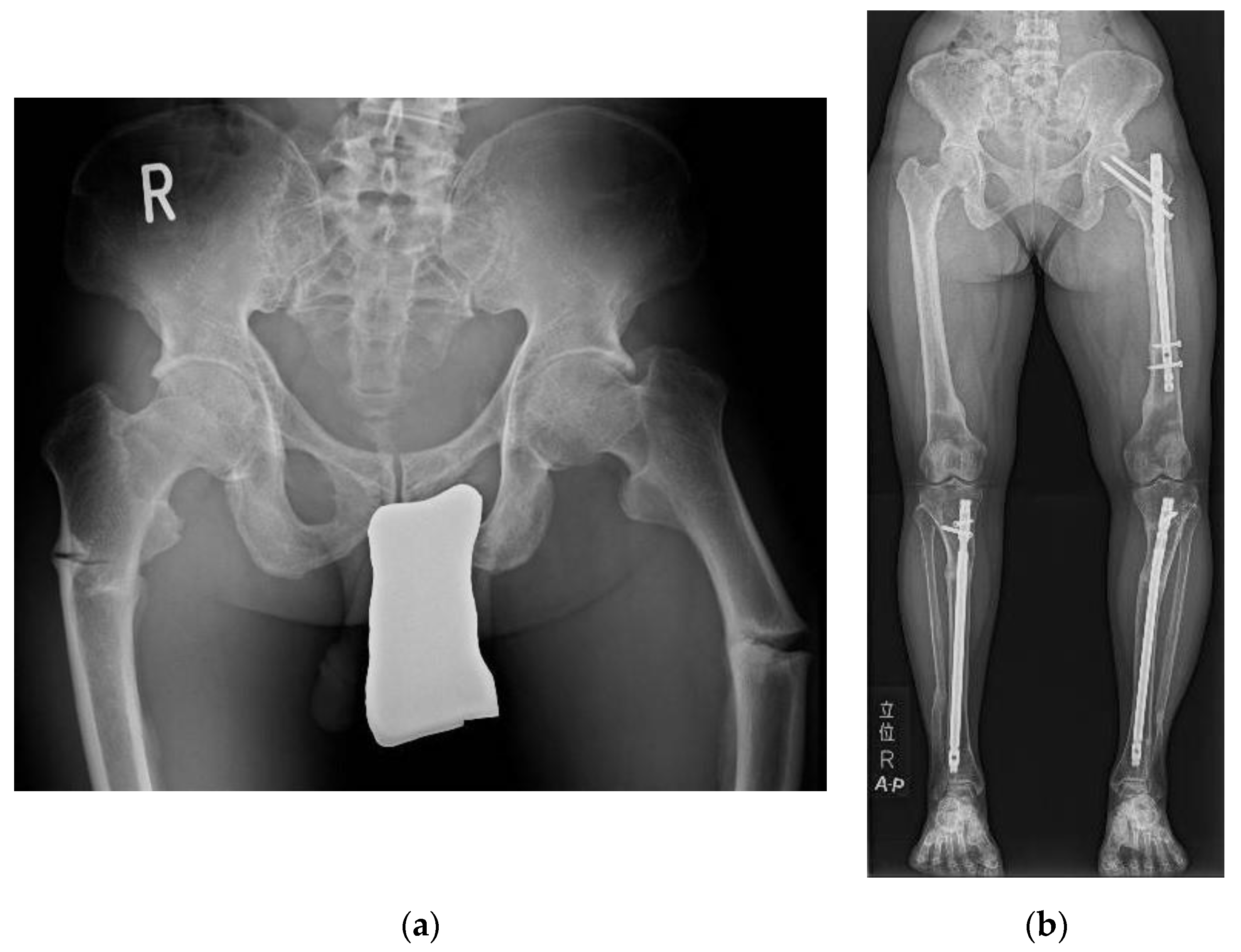
Figure 1. Pseudofracture and fracture in the femurs and tibiae in adult XLH patients. (a) Pseudofractures developed spontaneously in both femurs in an adult XLH patient after years of conventional therapy cessation. (b) Fractures developed spontaneously in the left femur and both tibiae in an adult XLH patient with conventional therapy.
Endocrines 03 00030 g001
Figure 2. Enthesopathies in adult XLH patients. (a) Severe ossification of the posterior longitudinal ligament and ossification of the anterior longitudinal ligament presented in an adult XLH patient. (b) A large osteophyte developed around the left hip joint in an adult XLH patient, causing severe impingement syndrome. (c) Enthesopathy developed in the right Achilles tendon in an adult XLH patient.
Endocrines 03 00030 g002
Figure 3. Early osteoarthritis in adult XLH patients. (a) Osteoarthritis with a Kellgren–Lawrence grade of 3 in the left hip joint of a 29-year-old male XLH patient. (b) Osteoarthritis with a Kellgren–Lawrence grade of 3 in the right knee joint of a 30-year-old female XLH patient.
Endocrines 03 00030 g003
Table 1. Suggested indication and selection of therapy for adult XLH patients.
ObservationConventional TherapyBurosumab
BAP (≤upper limit of the reference range)BAP (>upper limit of reference range)
with eGFR ≥ 45 mL/min/1.73 m2
BAP (>upper limit of the reference range) with conventional treatment
Stature (≥−1.0 SD)Short stature (−2.0 ≤ −1.0 SD) with
eGFR ≥ 45 mL/min/1.73 m2
Short stature (<−2.0 SD)
Mechanical axis of the leg within zone 1 [43]Deviation of the mechanical axis of the leg into zone 2 [43] with eGFR ≥ 45 L/min/1.73 m2Deviation of the mechanical axis of the leg into zone 3 or greater [43] or history of
corrective surgery for leg deformity
No history of pseudofracture/bone pain/fracture in weight-bearing bone or odontitis/tooth abscessesPseudofracture/bone pain in weight-bearing bone or odontitis/tooth abscess once
with eGFR ≥ 45 mL/min/1.73 m2
Pseudofracture/bone pain in weight-bearing bone or odontitis/tooth abscesses more than two times or more than once with conventional treatment or once requiring impending surgery
No response or trivial response to burosumabFracture in weight-bearing bone
Severe adverse event with burosumabUncontrolled severe SHPT with conventional treatment
(e.g., peak intact PTH ≥ twice the upper limit of the reference range);
THPT;
Patients with symptoms described in the “conventional therapy”
and eGFR < 45 mL/min/1.73 m2;
Severe adverse event with conventional therapy
Table 2. Suggested guidance of conventional therapy for adult XLH patients.
PriorityEventAction
Initiation of treatmentStart with active vitamin D
(0.50 μg b.i.d. for calcitriol or 1.0 μg s.i.d. for alfacalcidol)
After 1 to 2 weeks, start phosphate supplementation (800 mg q.i.d.)
Range of dosageCalcitriol: 0.50 to 0.75 μg b.i.d., alfacalcidol: 0.75 to 1.5 μg s.i.d.
Phosphate supplementation: 750 to 1600 mg q.i.d.
Initial phaseAdjust phosphate supplementation for a period of time by 100 mg
Goal: peak phosphate 1 within lower 50% of the reference range; laboratory test: every one to four weeks
Maintenance phase
(after 12 to 18 months)
Adjust phosphate supplementation for a period of time by 100 mg
Goal: BAP within the reference range
Laboratory test: every 3 months
Uncontrolled BAP: change the treatment to burosumab
HighSevere SHPT
(peak intact PTH ≥ 2 ×
upper limit of the reference range)
Immediately decrease phosphate supplementation by 100 mg or more
Increase active vitamin D (0.25 μg daily for calcitriol and 0.25 to 0.5 μg daily for alfacalcidol)
After 1 to 2 weeks, increase phosphate supplementation
Uncontrolled severe SHPT: change the treatment to burosumab
HighDevelopment of THPT
(hyperparathyroidism with the value of calcium ≥ the middle of the reference range)
Immediately quit conventional therapy
Initiate allosteric modulation of calcium sensing receptor
Try to prevent dehydration
Conduct parathyroidectomy for all recognizable glands,
otherwise continue allosteric modulation of the calcium sensing receptor in patients with contraindication for surgery
Change the treatment to burosumab afterward
HighOveradjustment of peak phosphate within upper 25% of the reference range or overImmediately decrease phosphate supplementation by 100 mg or more
HighProgression of CKD
to eGFR < 45 mL/min/1.73 m2
Decrease conventional therapy
Change the treatment to burosumab in patients who still need phosphate supplementation (uncontrolled BAP with active vitamin D).
1 Peak phosphate: phosphate level 1 to 2 h after phosphate supplementation.
Table 3. Suggested guidance for the use of burosumab for adult XLH patients.
PriorityEventAction
Initiation of treatment1.0 mg/kg body weight (up to 90 mg) subcutaneous injection every four weeks
Response to burosumab should be confirmed by peak phosphate 1;
Concomitant use of active vitamin D and phosphate supplementation is not recommended or is prohibited unless another medical condition requires it
Trough phosphate 2 within the higher half of the
reference range
Decrease burosumab dose by 0.2 to 0.3 mg/kg
Subsequently, fine tune burosumab dose by 0.1 mg/kg to target trough phosphate within lower half of the reference range
HighTrough phosphate over the reference rangeSuspend burosumab until phosphate values fall below the normal range
Restarted burosumab at approximately half the initial starting dose
No or trivial response at peak phosphateRule out vitamin D deficiency by measuring 25OHD
Severe SHPT: continue burosumab 3 to 6 times and confirm improvement in peak phosphate
Continuing no response or trivial response after problems above are ruled out or
addressed; change treatment to conventional therapy
HighTHPTInitiate allosteric modulation of the calcium sensing receptor
Try to prevent dehydration
Conduct parathyroidectomy for all recognizable glands, otherwise continue allosteric
modulation of the calcium sensing receptor in patients with contraindication for surgery
1 Peak phosphate: phosphate level 1 to 2 weeks after the last injection of burosumab. 2 Trough phosphate: phosphate level 4 weeks after the last injection of burosumab.
Publisher’s Note: MDPI stays neutral with regard to jurisdictional claims in published maps and institutional affiliations.