Article

Differences in Exercise-Linked Biomarkers between Premenopausal and Postmenopausal Middle-Aged Females

by
Anthony J. Giannopoulos
1,
Ahmad Mohammad
2,
Maria I. Retsidou
1,
Jessica A. L. Tucker
3,
Derek P. D. Bornath
3,
Seth F. McCarthy
3,
Rebecca E. K. MacPherson
2,
Tom J. Hazell
3 and
Panagiota Klentrou
1,*
1
Department of Kinesiology, Faculty of Applied Health Sciences, Brock University, St. Catharines, ON L2S 3A1, Canada
2
Department of Health Sciences, Faculty of Applied Health Sciences, Brock University, St. Catharines, ON L2S 3A1, Canada
3
Department of Kinesiology and Physical Education, Wilfrid Laurier University, Waterloo, ON N2L 3C5, Canada
*
Author to whom correspondence should be addressed.
Endocrines 2024, 5(3), 290-303; https://doi.org/10.3390/endocrines5030021
Submission received: 25 April 2024 / Revised: 8 July 2024 / Accepted: 18 July 2024 / Published: 23 July 2024
(This article belongs to the Special Issue Feature Papers in Endocrines: 2024)

Abstract

:
While the exercise-induced responses of circulated biomarkers related to inflammation and brain health are well documented in humans, little is known about the effect of menopausal status on these responses. This study compared the responses of inflammatory cytokines and brain-derived neurotrophic factor (BDNF) to high-intensity exercise between pre- and postmenopausal middle-aged females. Eight premenopausal (44 ± 3 years) and seven postmenopausal (57 ± 2 years) females performed a high-intensity interval training (HIIT) session consisting of 10 × 1 min running intervals (90% maximum heart rate) separated by 1 min passive recovery intervals. Blood samples were collected at baseline (fasted), pre-exercise (postprandial), and at 0, 30, and 90 min post-HIIT and analyzed for interleukin (IL-6) and 10 (IL-10), tumour necrosis factor-alpha (TNF-α), and BDNF. IL-6 significantly increased from pre-exercise to 0 min post-HIIT in postmenopausal (+40%, p = 0.01) and to 30 min post-HIIT in premenopausal females (+60%, p = 0.02). IL-6 remained elevated at 90 min post-HIIT in premenopausal (+104%, p = 0.05) and to a higher degree in postmenopausal females (+385%, p < 0.001). IL-10 showed no response. TNF-α increased from pre- to 0 min post-HIIT (+10%, p = 0.05), then decreased to below pre-exercise at 30 min (−10%, p = 0.02) and 90 min (−5%, p = 0.04) in both groups. BDNF increased immediately post-HIIT in premenopausal (+60%, p < 0.001) but not postmenopausal females. The differences in IL-6 and BDNF responses to HIIT between pre- and postmenopausal females provide evidence of the role of female reproductive hormones in the regulation of these exercise-induced responses.

1. Introduction

Menopause is a pivotal time in female physiology characterized by the cessation of the menstrual cycle and the concomitant reductions in the female hormone concentrations, i.e., estrogen and progesterone [1]. One of the multiple physiological changes that females experience following menopause is the increase in circulating inflammatory cytokines, which has typically been attributed to the decrease in estrogen (or estradiol in its most common form in humans) concentrations [2]. Indeed, estrogen deficiency has been associated with increased expression and secretion of pro-inflammatory cytokines [3] such as tumour necrosis factor-alpha (TNF-α), as well as interleukin 6 (IL-6), which is considered both pro- and anti-inflammatory [4], suggesting an interaction between estrogen and these cytokines [3]. This chronic inflammation, commonly shown by elevated resting concentrations of pro-inflammatory cytokines, is also associated with increased visceral fat postmenopause [5] and can lead to other pathologies including metabolic syndrome, hypertension, atherosclerosis, ischemic stroke and heart disease [5,6,7,8]. In contrast, chronic exercise is associated with overall anti-inflammatory effects, which can mitigate chronic inflammatory conditions [9]; this has been attributed as the cause of the transient spikes in cytokine concentrations following acute exercise bouts [10,11]. Specifically, acute exercise triggers changes in the circulating concentrations of IL-6, IL-1β, and TNF-α and anti-inflammatory cytokines such as interleukin 10 (IL-10), all of which typically peak immediately after exercise and return to baseline within 5 to 24 h [12,13]. However, the extent and duration of these exercise-induced changes depend on various factors such as the exercise intensity, duration, and frequency [12,14], as well as on the individual’s training status [15], biological sex [16,17], and age [18,19]. Thus, it is also possible that the reductions in estrogen concentrations postmenopause, which have previously been shown to be associated with an increase in pro-inflammatory cytokines [4], could alter the magnitude and timing of these exercise-induced factors. However, studies in postmenopausal females examining the acute responses of these cytokines to exercise are lacking.
Moreover, menopause has been found to play a role in cognitive decline along with the progression of neurodegenerative diseases associated with low concentrations of brain-derived neurotrophic factor (BDNF) which is a crucial molecule involved in brain plasticity and cognition [20]. BDNF concentrations also increase post-exercise in an intensity-dependent manner [21,22,23,24] but most of these exercise studies are in male populations. According to a systematic review in older rodents, there are potential sex-based differences in the BDNF response to exercise, with healthy older or ovariectomized females exhibiting overall greater increases and cognitive improvements compared to healthy older males [25], suggesting female reproductive hormones may influence exercise-induced changes in BDNF. Indeed, BDNF expression is regulated by estrogen as there is an estrogen response element on the gene [26,27]. In humans, menopausal females have also been found with lower circulating BDNF [28], although human studies examining the relationship between BDNF and endogenous ovarian hormones have produced conflicting results due to large participant heterogeneity and a lack of cycle phase standardization across studies [29,30]. Additionally, these studies only apply to resting conditions as there are no exercise studies in females involving BDNF, especially in postmenopausal and advanced age groups. Since postmenopausal females have lower resting BDNF concentrations when compared with premenopausal groups [28], the lack of information on how BDNF responds to acute exercise in these individuals is concerning. Examining the effects of various exercise modalities on BDNF in postmenopausal females is critical for prescribing exercise programs to manage menopause-induced chronic inflammation and cognitive decline.
The purpose of this secondary analysis was to compare the potential responses of IL-6, IL-10, TNF-α, and BDNF to a single high-intensity interval training (HIIT) exercise trial between middle-aged females differing in menopausal status, postmenopausal females with low concentrations of reproductive hormones and premenopausal females during the luteal phase of their menstrual cycle when estrogen and progesterone are highest. Examining how exercise influences inflammatory cytokines and BDNF in postmenopausal females compared to premenopausal counterparts could provide valuable insights into the role of hormones in modulating exercise-induced responses and inform strategies for promoting health and wellness in women across the lifespan.

2. Materials and Methods

2.1. Participants

This secondary analysis involves blood samples collected from a study examining the role of ovarian hormones in appetite regulation in pre- versus postmenopausal middle-aged (i.e., 40–60 years) females. Specifically, samples from 8 premenopausal (44 ± 3 years) and 7 postmenopausal females (57 ± 2 years) recruited to participate in the original study were included in the present analysis.
To be included in this study, all participants were assessed as being healthy by the Get Active Questionnaire [30], had no metabolic diseases such as diabetes, were considered recreationally active (i.e., exercising for approximately 150 min of moderate to vigorous physical activity per week), were not taking any medications, and were free of injuries and other contraindications to exercise. Additional inclusion/exclusion criteria applied to each group. Specifically, postmenopausal females had an absence of menses for >12 months, no experience with hormonal replacement therapy (including bilateral oophorectomy in which both ovaries have been removed), and an estradiol concentration of less than 108 pmol/L and a progesterone concentration of less than 4 nmol/L [31,32]. Premenopausal participants were included if they were eumenorrheic, non-hormonal contraceptive users, and they were excluded if they were pregnant, had been pregnant (for >3 months) within the past 3 years, or had plans to become pregnant during the time of study participation. These participants had to have tracked their cycle for at least two months prior to participation. To do so, they were provided with a thermometer (HealthSmart® International, Waukegan, IL, USA), instructed to monitor body temperature orally upon awakening each day following the onset of menstruation, and reported daily to the researcher via text/email.
This study was conducted per the Declaration of Helsinki and received ethics approval from the Wilfrid Laurier University Research Ethics Board (REB #6900). Ethics approval for the secondary analysis was also received from the Brock University Research Ethics Board (REB #23-260). All participants agreed to participate in this study by signing informed consent. Experimental procedures took place in the Energy Metabolism Research Laboratory of Wilfrid Laurier University.

2.2. Familiarization

All participants completed a familiarization session during which they provided written informed consent and became acclimated with the laboratory as well as how to use the motorized treadmill (4Front, Woodway, Waukesha, WI, USA) to be used in this study. In the case that participants were unfamiliar with using a treadmill, participants were offered the opportunity to address any concerns as well as practice by walking on the treadmill ahead of time in the presence of at least two researchers. Body mass and height were measured using standard procedures (Health-o-meter Professional, Sunbeam Products Inc., Chicago, IL, USA). Participants then completed a graded exercise test to exhaustion on the treadmill to determine VO2max where oxygen consumption (VO2) and carbon dioxide production (VCO2) were measured continuously with an online gas collection system (MAX-II, AEI Technologies, Pittsburgh, PA, USA) and a silicon facemask (7400 series Vmask, Hans Rudolph Inc., Shawnee, KS, USA), as previously described [33]. Participants began the test with a 5 min warmup walking at 4.8–8 km/h followed by fast walking or running at a self-selected pace between 6.4 and 11.2 km/h with incremental increases in grade (2%) applied every 2 min until volitional fatigue. Heart rate (HR) was continuously recorded using an integrated HR monitor (FT1, Polar Electro; Cambridge Bay, NU, Canada). Following completion of the test and a 5 min walking cooldown period (4.8–8 km/h), a verification phase was completed to volitional fatigue where participants fast walked or ran to exhaustion at 105% of the speed and grade used to confirm VO2max [33]. Following the VO2max test and verification phase, the target intensity for the HIIT session (90% HRmax) was calculated using the ACSM running equation [34], using the speed and grade from the VO2max test and VO2 data. Participants completed two practice 1 min intervals (treadmill speed eliciting 90% HRmax) interspersed with 1 min of passive rest, where participants straddled the sides of the treadmill during each rest period, to confirm the calculated exercise intensity was correct. Finally, at the end of the familiarization session, participants were asked if they had any food allergies that may prevent them from consuming the planned standardized breakfast during the HIIT session.
Before leaving the familiarization, premenopausal participants were provided with the ovulation kits (Easy@Home, Easy Healthcare Corporation, Burr Ridge, IL, USA) and asked to report the result via text/email to the researchers, to confirm surges in luteinizing hormone and determine the appropriate session timing. In addition, these participants were asked to continually monitor and report their body temperature upon awakening, as increases of ~0.3 °C indicated ovulation [33]. The premenopausal HIIT session was scheduled for ~7 d following a positive LH surge on the ovulation strips. The menstrual phase was confirmed to be within the luteal phase through a fasted blood sample of estradiol (E2) between 294 and 918 pmol/L and progesterone (P4) concentration > 16 nmol/L [31].

2.3. Experimental Session

Participants arrived at the laboratory at 0800 h after a 12 h overnight fast (i.e., only water after 2000 h). Moderate to vigorous activity and alcohol consumption were restricted for 24 h while caffeine consumption was restricted for 12 h before the session. A fasted blood draw (baseline sample) was taken at ~0815 h followed by a standardized breakfast previously used [35], with a macronutrient profile similar to that of a typical balanced breakfast (smoothie; 7 kcal/kg; 56% carbohydrate, 23% fat, and 21% protein; Gruppo, ON, Canada) at 0815–0830 h. During breakfast, participants were asked to fill out a short questionnaire about their typical sleep patterns, including how much sleep they received the night before coming into the lab. Following breakfast, participants were allowed 30 min for digestion to ensure no gastric discomfort during exercise [35]. A subsequent postprandial blood sample was taken at ~0900 h (pre-HIIT sample). From 0900 to 0930 h, participants performed a HIIT exercise protocol, which began with a 5 min walking or jogging warmup at a self-selected pace between 4.8 and 8.0 km/h on the motorized treadmill (see above). Participants then started the HIIT protocol, which consisted of 10 × 1 min bouts of running at a speed and grade that would elicit 90% of their maximum heart rate followed by 60 s of passive recovery, as described above. Heart rate was monitored throughout the session. Following the completion of the HIIT protocol, participants completed a 5 min walking cooldown at a self-selected pace. Upon completion of the cooldown, the first post-HIIT blood sample was taken at ~0930 h (0 min post-HIIT sample), and subsequent post-HIIT blood sampling occurred at ~1000 h (30 min post-HIIT sample) and 1100 h (90 min post-HIIT sample).

2.4. Blood Collection and Analysis

A total of ~3 mL of whole blood was originally collected into pre-chilled Vacutainer SST tubes (gold top) from each participant’s median cubital vein in the antecubital fossa while participants were lying in a supine position using a standard venipuncture technique during each session at each time point (baseline, pre-exercise, and 0 min, 30 min, and 90 min post-HIIT). Blood samples were inverted 8–10 times before sitting for 30 min at room temperature. The samples were then centrifuged at 3000× g for 10 min at 4° before the serum was aliquoted into microcentrifuge tubes and stored at −80 °C until analysis. In the present study, serum was used to measure circulating concentrations of IL-6, IL-10, TNF-α, and BDNF.
Although this was a secondary analysis of samples from another study, the cytokines reported herein were analyzed for the first time for the present study using new commercially available immunoassay assay kits. Specifically, IL-6, IL-10, and TNF-α were measured with ELLA single-assay kits (cat. # SPCKBPS-003028, ST01B-PS-000276, and ST01B-PS-002803, respectively; ProteinSimple, San Jose, CA). The inter-assay coefficients of variation were 9.9% for IL-6, 6.6% for IL-10, and 8.7% for TNF-α. The intra-assay coefficients of variation for IL-6, IL-10, and TNF-α were 4.6%, 6.69%, and 2.54%, respectively. BDNF was analyzed using an ELISA kit (cat. #CYT306; EMD Millipore) with an inter-assay coefficient of variation of +8.5% and an intra-assay coefficient of variation of +3.7%.

2.5. Statistical Analysis

The data were assessed for normality using the Shapiro–Wilk test, and z-scores for skewness and kurtosis. There were a few missing values for each analyte due to low serum availability or non-detectable values, so the number of participants is different for each marker. A series of two-way repeated-measures analyses of variance (RM-ANOVAs) was used to examine the main effects of group (premenopausal versus postmenopausal) and time (5 points), as well as the time-by-trial interactions, for IL-6, IL-10, TNF-α, and BDNF. Statistical significance was set at p 0.05. Effect sizes, including partial eta squared (η2) for ANOVA and Cohen’s d for post hoc comparisons, were calculated and interpreted using the Cohen criteria: 0.01 = small, 0.06 = moderate, 0.14 = large effect for partial η2, and 0.2 = small, 0.5 = medium, 0.8 = large effect for Cohen’s d [36,37]. The statistical analysis was performed using IBM SPSS Statistics 28 (SPSS Inc., Chicago, IL, USA).

3. Results

3.1. Group Characteristics
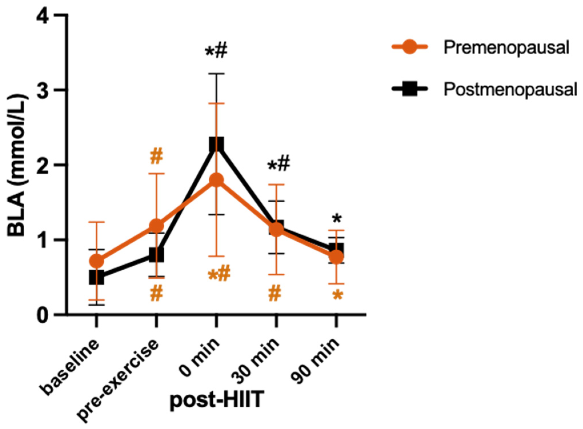
Premenopausal and postmenopausal groups had similar BMIs and VO2max (Table 1). As expected, the premenopausal females had significantly higher concentrations than the postmenopausal females of both estradiol and progesterone (Table 1).

3.2. HIIT Session

During HIIT, there were no significant differences between the premenopausal and postmenopausal females in the average speeds (7.6 ± 1.6 km/h and 6.9 ± 0.5 km/h, respectively). Blood lactate concentrations were also not different (p > 0.05) between groups (Table 1). However, there was a group-by-time interaction (F = 7.51, p = 0.059, η2 = 0.16) as the increase in blood lactate from pre-exercise to 0 min post-HIIT was of a greater magnitude in postmenopausal compared to premenopausal females (+51% vs. +185%, respectively). Additionally, although in the premenopausal group, lactate concentrations returned to pre-exercise levels by 30 min post-HIIT and to the fasted levels by 90 min following HIIT, in the postmenopausal group, lactate concentrations remained higher than pre-exercise at 30 min (+46%), returning to pre-exercise 90 min later (Figure 1).

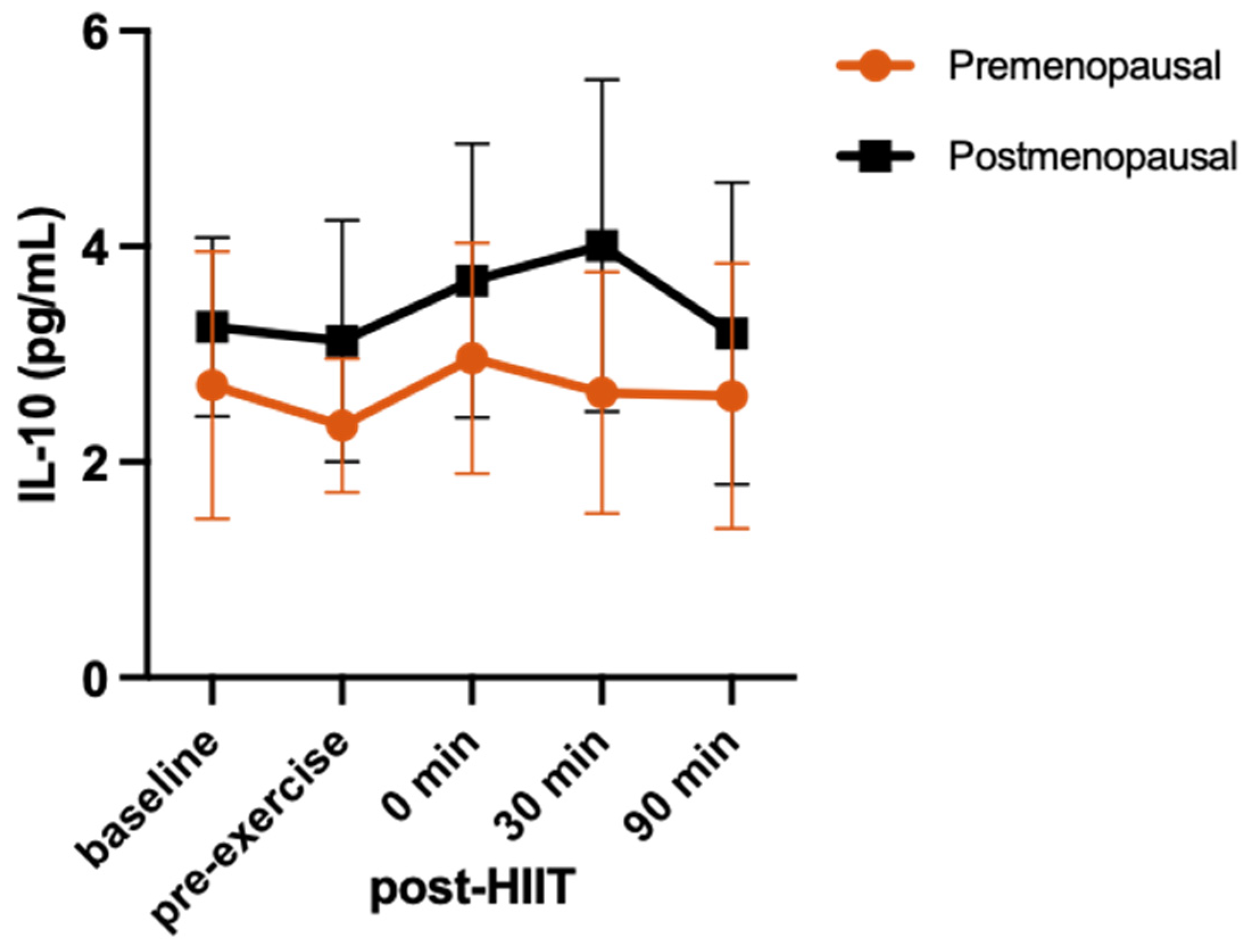
3.3. Cytokine Response
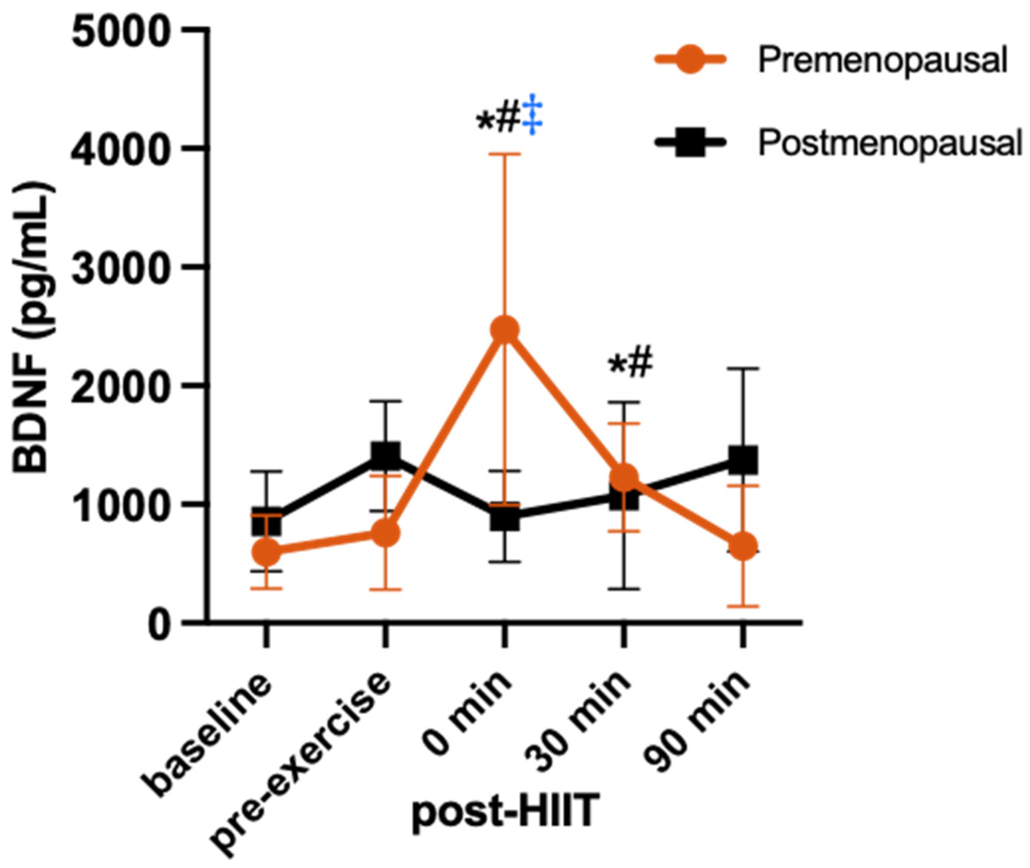
IL-6 showed a significant group-by-time interaction (F = 10.9; p = 0.003; η2 = 0.52). Compared to pre-exercise, the IL-6 increase at 0 min post-HIIT was not significant (+18%, p = 0.189, d = 0.62), but it was significant at 30 min (+60%, p = 0.02, d = 1.02) and 90 min (+104%, p = 0.05, d = 2.19) post-HIIT in the premenopausal group. In contrast, IL-6 increased significantly from pre-exercise to 0 min post-HIIT (+40%, p = 0.01, d = 0.55) in the postmenopausal group, and its increase from pre-exercise to 30 min (+80%, p < 0.001, d = 0.48) and 90 min post-HIIT (+385%, p < 0.001, d = 3.18) was of greater magnitude (2 to 3-fold) than in the premenopausal group, resulting in a significant difference between groups at 90 min following HIIT (Figure 2).
For IL-10 (Figure 3), there was no group-by-time interaction (F = 0.81; p = 0.49; η2 = 0.08) and no main effect for time (F = 2.02; p = 0.11; η2 = 0.18) or group (F = 1.65; p = 0.23; η2 = 0.15).
There was no group-by-time interaction for TNF-α (F = 0.37; p = 0.73; η2 = 0.39). There was a main effect for time (F = 4.51; p = 0.018; η2 = 0.33), reflecting a 10% (p = 0.05, d = 0.38) increase in TNF-α from pre-exercise to 0 min post-exercise, which was followed by a decrease at 30 min (10%, p = 0.02, d = 0.26) and 90 min (5%, p = 0.04, d = 0.14) post-exercise to concentrations below pre-exercise, in both groups (Figure 4).

3.4. BDNF Response

For BDNF, there was a significant group-by-time interaction (F = 7.998; p < 0.001; η2 = 0.25), reflecting an increase in BDNF at 0 min following HIIT in the premenopausal group (+69%, p < 0.001, d = 1.56) but not in the postmenopausal group (−56%, p > 0.99, d = 1.2 Figure 5). This was followed by +38% at 30 min (p = 0.94; d = 1.0) and −17% at 90 min (p > 0.99; d = 0.23) compared to pre-exercise in the premenopausal group and −31% at 30 min (p > 0.99; d = 0.52) and −3% at 90 min (p > 0.99; d = 0.05) in the postmenopausal group.

4. Discussion

This study provides valuable insights into the distinct acute response of exercise-induced factors following a single bout of HIIT in postmenopausal compared to premenopausal middle-aged females. Specifically, postmenopausal participants exhibited an immediate increase in IL-6 concentrations following HIIT, whereas premenopausal females experienced a delayed spike in IL-6 30 min following HIIT. These effects were of a large effect size and suggest that the time course of the increase in IL-6 following HIIT was different between groups. In contrast, no significant exercise-induced changes were observed in IL-10 in either group, while TNF-α concentrations slightly increased immediately after HIIT and then decreased to below pre-exercise concentrations 90 min post-HIIT in both groups. This suggests that the exercise-induced IL-10 and TNF-α response in females may be less influenced by ovarian hormonal status compared to IL-6. Moreover, BDNF concentrations increased after HIIT only in the premenopausal group, with no apparent response observed in the postmenopausal group. This difference in the BDNF response to HIIT, which was of a large effect size, further underscores the potential influence of female reproductive hormones on BDNF regulation in response to exercise.
Greater concentrations of circulating inflammatory cytokines, leading to a higher risk of metabolic syndrome, are often seen in postmenopausal women due to the increase in visceral fat [5]. Considering the pre- and postmenopausal females in this study had similar BMIs, the similarity in their fasted and pre-exercise concentrations of circulating IL-6, IL-10, and TNFα was not surprising. However, the time course of the IL-6 response to HIIT was different between groups, with concentrations being elevated immediately post-HIIT in the postmenopausal group, while the elevation was at 30 min post-HIIT in the premenopausal group. IL-6 remained elevated up to 90 min following HIIT in both groups, but this increase was of almost 3-fold greater magnitude in the postmenopausal group. We previously found IL-6 to be elevated compared to its pre-exercise concentrations 1 h after high-intensity running and cycling (39%, p<0.05) in young adult females [38], which supports the current finding of the IL-6 elevation 30 and 90 min following HIIT in both groups. However, we cannot explain mechanistically why the postmenopausal group had an earlier significant increase in IL-6 (i.e., immediately) following the HIIT protocol, or why 90 min later they had a much larger IL-6 increase compared to their premenopausal counterparts. The main suggestion is that the difference in the timing and magnitude of the IL-6 response is related to the low concentrations of female reproductive hormones, especially estrogen deficiency, which has previously been associated with elevated concentrations of circulating inflammatory cytokines [2,39]. This speculation is based on the fact that the immediate exercise-induced response seen in postmenopausal females aligns with previous research in young adult males who also demonstrated exercise-induced increases in the circulating IL-6 immediately following different modes of a single bout of high-intensity interval exercise, although, in those studies, IL-6 returned to pre-exercise levels after 60 min [40,41]. In another study, however, both IL-6 and TNF-α increased immediately following an acute bout of high-intensity intermittent walking or continuous moderate-intensity walking in young adult males and remained elevated until at least 4 h post-exercise [42]. Interestingly, in the present study, neither at the baseline nor pre-exercise did we find any differences between groups, which seems contradictory to the suggestion that the low-grade chronic inflammation previously reported in postmenopausal females is directly related to their low concentrations of reproductive hormones [2,39]. However, in these previous studies, the difference in resting cytokine concentration could reflect an aging effect because of the larger age differences between the pre- and postmenopausal groups. The novelty of our study was that, although the average age was statistically different between groups, both pre- and postmenopausal participants were middle-aged. In addition, these previous studies only measured resting cytokine concentrations. Thus, the different time course and magnitude of the IL-6 response to the current running protocol between pre- and postmenopausal women support the hypothesis of a direct relationship between female reproductive hormones and the IL-6 response to exercise because it happened irrespective of similar baseline and pre-exercise concentrations between groups. This hypothesis is further supported by the finding of a previous study showing that the changes in female reproductive hormones due to the menstrual cycle phase affected the IL-6 response during an extended recovery period from intensive exercise in eumenorrheic females [43].
Furthermore, the exercise-induced increase in IL-6 is typically directly related to exercise intensity, duration, muscle mass recruited, and muscle glycogen used [44]. While there were no differences between the pre- and postmenopausal females in the HIIT speeds, the exercise-induced relative increase in blood lactate concentration at 0 min post-HIIT was of a greater magnitude in the postmenopausal females compared to the premenopausal females and remained higher than pre-exercise until 30 min post-HIIT and higher than fasted levels 90 min following HIIT. These data suggest a higher lactate production and slower lactate clearance in the postmenopausal group. This is consistent with the aging-related decrease in oxidative muscle fibres, and increased dependency on carbohydrate energy production, which alters lactate production and kinetics during exercise and subsequently IL-6 secretion from muscle [44]. This lactate effect on the IL-6 release from skeletal muscle was shown in young adult males during high-intensity interval cycling and was confirmed using electrically stimulated muscle cell cultures [45]. Thus, further research is required to understand why the IL-6 increase in the postmenopausal group may appear immediately post-exercise (similar to in men) and continue to increase for 90 min post-exercise at a higher magnitude than that observed in the premenopausal group.
The increase in TNF-α immediately following the HIIT session is in line with our previous work, where TNF-α increased immediately following a high-intensity interval running session (10%, p < 0.05) in young adult females and males, but in that case, its concentrations at 1 h and 24 h post-exercise were no different than pre-exercise [38]. The present study also provides a novel finding that after this initial exercise-induced increase, TNF-α was significantly decreased at 30 and 90 min post-HIIT to concentrations slightly below pre-exercise. According to previous studies, exercise-induced increases in TNF-α may take as long as 24 to 48 h to return to pre-exercise levels following intense resistance training [46] and after a marathon race [47] in adult males 18–55 years. Other studies examining the response of TNF-α following exercise have also observed no change, which has been attributed to the suppression of TNF-α by the muscle-derived, anti-inflammatory IL-6 [48], which is consistent with the elevated IL-6 for up to 90 min after our HIIT protocol. Thus, the later decrease in TNF-α found herein is important as it could indicate a healthy balance of pro- versus anti-inflammatory status post-exercise. Indeed, post-exercise inflammation is a natural response of the immune system to tissue damage caused by physical exertion, but excess or prolonged inflammation can be harmful and slow the recovery process. Therefore, it is important to balance the increase in pro-inflammatory cytokines with an increase in anti-inflammatory cytokines. IL-10 is such a cytokine that plays a critical role in the anti-inflammatory properties, is mediated by IL-6, and is produced during exercise [49]. Indeed, in our previous study [38], IL-10 was elevated 5 min after running (20%, p < 0.05) and 1 h after both running and cycling (41% and 64%, respectively, p < 0.05) in both young adult females and males. In the present study, IL-10 did not change in response to HIIT, which somewhat contradicts the suggestion that IL-6 mediates the IL-10 response [49] since IL-6 did increase in both our groups. Because the previous study was performed on younger participants (18–25 years), it is possible that in the present study, the IL-10 response happened after the 90 min study period since IL-6 continued to increase further at 30 and 90 min following HIIT. Later blood draws, at 120 or 150 min post-exercise, could potentially show whether IL-10 would increase in response to the substantial increase in IL-6 and would also help us understand the kinetics of the IL-6 response, i.e., how much longer it would take to return to baseline. In any case, corroborating previous studies [38,46,49,50,51], both inflammatory cytokines (IL-6 and TNF-α) acutely increased following HIIT in our pre- and postmenopausal females.
BDNF was originally characterized for its role in the brain for enhancing neuronal plasticity, which is essential for learning and memory [20,52,53]. Additionally, BDNF is thought to play a role in central and peripheral molecular processes of energy metabolism and homeostasis [54,55,56]. Specifically, BDNF has been linked not only to brain health but also to whole-body glucose homeostasis and insulin sensitivity, as well as muscle plasticity and regeneration [54,57]. Given this, the reduction in BDNF with menopause likely contributes to the increased risk of several age-related metabolic diseases—thus, it is important to find interventions that can maintain or increase BDNF levels throughout menopause [58]. Engaging in physical exercise has consistently been demonstrated to produce an elevation in the expression of BDNF in brain regions such as the hypothalamus, striatum, and various cortical areas, as well as in muscle [21,28,57,58]. In this study, we found BDNF to increase by 69% from pre-exercise to 0 min post-HIIT, and, although non-significantly, to remain 38% higher than pre-exercise at 30 min post-HIIT in premenopausal females but not in the premenopausal group. An increase in circulating BDNF has also been reported after high-intensity exercise in groups of females and males aged 20–50 years [21,22,23,24,59,60]. However, exercise studies in postmenopausal females are lacking. Only one previous study examined the BDNF response to a single bout of 30 min aerobic exercise on the treadmill between pre- and postmenopausal women and found that exercise did not lead to a significant change in BDNF at 0 and 30 h post-exercise in either group [61]. In their study, there was a large difference in average ages between the premenopausal (~28 years) and postmenopausal (~57 years) participants, making it difficult to conclude whether the difference in the BDNF response to HIIT was due to female reproductive hormones or to age. On the other hand, resting BDNF concentrations have been reported to be lower in postmenopausal females compared to eumenorrheic premenopausal females and this difference has been attributed to their lower estradiol and progesterone, as it was restored by a 6-month hormonal replacement treatment [62]. Herein, we found no differences in resting BDNF concentrations, probably because the ages of our groups were not as far apart as in the study by Begliuomini et al. (2007) [28], whose premenopausal participants were in their early 20s. Indeed, in their study, BDNF was positively correlated with estradiol and progesterone and negatively correlated with menopausal age. Our study strengthens this claim on the role of female reproductive hormones in regulating BDNF postmenopause by closing the age gap between the premenopausal and postmenopausal groups (44 ± 3 vs. 57 ± 2 years, respectively). However, other factors related to aging and neurodegenerative processes may still contribute to the observed differences. For example, in our previous study of young adult males (23 ± 3 years), who have very low estrogen and progesterone, we also found an acute exercise-induced increase following bouts of moderate- and vigorous-intensity, and high-intensity interval training [24]. Thus, further research is needed to elucidate the mechanisms underlying the differential BDNF response to exercise in postmenopausal females and better understand the role of reproductive hormones. Understanding these mechanisms could have implications for developing targeted interventions to promote brain health and cognitive function across the lifespan.
Acknowledging limitations is crucial for interpreting the findings of any study. This was a secondary analysis of available samples from another study, and the minimum sample size of 12 participants (6 per group) was calculated to detect differences in appetite hormones using G*Power-2 with a large effect size of 0.467 [35,63]. We were able to reach significance in IL-6, TNFα, and BDNF with this sample, which was comparable to that of a previous study examining post-exercise changes in BDNF [24]. However, we did have fewer participants than our previous study detecting changes in IL-10 [38]. Not accounting for changes in plasma volume during the exercise period is another limitation, as it could influence the observed changes in circulating markers. However, the substantial changes in IL-6 (ranging from 40% to 385%) and BDNF (60%) observed herein indicate responses beyond the effects of exercise-induced hemoconcentration, as evidenced by the modest changes in plasma volume we previously calculated (−3.8%) during a similar HIIT protocol on the treadmill in premenopausal females [38]. Therefore, this study provides valuable insights despite these limitations. Finally, given the limited information available on the clinical relevance of BDNF (especially during exercise) in relatively healthy and younger (vs. elderly) individuals, another limitation is the lack of a secondary marker of cognitive decline.
In conclusion, the comparison of pre- and postmenopausal females’ responses to a session of high-intensity interval exercise provides evidence of potential differences in inflammatory and neurotrophic responses based on hormonal status, emphasizing the importance of considering hormonal status in future exercise investigations and its impact on health outcomes in women across different stages of life. Moving forward, future studies with larger sample sizes and more comprehensive methods for measuring changes in plasma volume could further elucidate the observed differences and contribute to a better understanding of the relationship between age, hormonal status, exercise, and circulating markers related to inflammation and brain health.

Author Contributions

Conceptualization, P.K., R.E.K.M. and T.J.H.; methodology, T.J.H., P.K., R.E.K.M., J.A.L.T. and S.F.M.; formal analysis, A.J.G., A.M., P.K. and R.E.K.M.; investigation, A.J.G., A.M., M.I.R., J.A.L.T., D.P.D.B., S.F.M., P.K., R.E.K.M. and T.J.H.; resources, P.K., R.E.K.M. and T.J.H.; writing—original draft preparation, A.J.G., M.I.R. and P.K.; writing—review and editing, T.J.H., R.E.K.M., J.A.L.T. and S.F.M.; supervision, P.K., T.J.H. and R.E.K.M.; project administration, P.K. and T.J.H.; funding acquisition, P.K., R.E.K.M. and T.J.H. All authors have read and agreed to the published version of the manuscript.

Funding

This research was funded by the Natural Sciences and Engineering Research Council of Canada (NSERC), grant numbers RGPIN-2023-03572 (P.K.), RGPIN-2016-06118 (T.J.H.), and RGPIN-2017-03904 (R.E.K.M).

Institutional Review Board Statement

This study was conducted per the Declaration of Helsinki and received ethics approval from the Wilfrid Laurier University Research Ethics Board (REB #6900). Ethics approval for the present secondary analysis was also received from the Brock University Research Ethics Board (REB #23-260). All participants agreed to participate in this study by signing informed consent. All experimental procedures took place in the Energy Metabolism Research Laboratory of Wilfrid Laurier University.

Informed Consent Statement

Informed consent was obtained from all participants involved in this study.

Data Availability Statement

The raw data supporting the findings of this study are available from the corresponding author P.K. upon reasonable request.

Conflicts of Interest

The authors declare no conflicts of interest.

References

  1. Nelson, H.D. Menopause. Lancet 2008, 371, 760–770. [Google Scholar] [CrossRef] [PubMed]
  2. Malutan, A.M.; Dan, M.; Nicolae, C.; Carmen, M. Proinflammatory and anti-inflammatory cytokine changes related to menopause. Menopause Rev. 2014, 13, 162–168. [Google Scholar] [CrossRef] [PubMed]
  3. Pfeilschifter, J.; Köditz, R.; Pfohl, M.; Schatz, H. Changes in Proinflammatory Cytokine Activity after Menopause. Endocr. Rev. 2002, 23, 90–119. [Google Scholar] [CrossRef] [PubMed]
  4. Pacifici, R.; Brown, C.; Puscheck, E.; Friedrich, E.; Slatopolsky, E.; Maggio, D.; McCracken, R.; Avioli, L.V. Effect of surgical menopause and estrogen replacement on cytokine release from human blood mononuclear cells. Proc. Natl. Acad. Sci. USA 1991, 88, 5134. [Google Scholar] [CrossRef] [PubMed]
  5. Sinatora, R.; Chagas, E.; Mattera, F.; Mellem, L.; de Oliveira dos Santos, A.; Pereira, L.; Aranão, A.; Landgraf Guiguer, E.; Cressoni Araujo, A.; Haber, J.; et al. Relationship of Inflammatory Markers and Metabolic Syndrome in Postmenopausal Women. Metabolites 2022, 12, 73. [Google Scholar] [CrossRef] [PubMed]
  6. Caiazzo, E.; Sharma, M.; Rezig, A.O.; Morsy, M.; Ialenti, A.; Pellicori, P.; Bruzzese, D.; Maffia, P.; Guzik, T.J. Inflammatory Cytokines and Risk of Developing Hypertension: A Systematic Review and Meta-Analysis. In Proceedings of the 19th World Congress of Basic & Clinical Pharmacology (WCP2023), Glasgow, Scotland, 2–7 July 2023. [Google Scholar] [CrossRef]
  7. Su, J.-H.; Luo, M.-Y.; Liang, N.; Gong, S.-X.; Chen, W.; Huang, W.-Q.; Tian, Y.; Wang, A.-P. Interleukin-6: A Novel Target for Cardio-Cerebrovascular Diseases. Front. Pharmacol. 2021, 12, 745061. [Google Scholar] [CrossRef] [PubMed]
  8. Feghali, C.A.; Wright, T.M. Cytokines in acute and chronic inflammation. Front. Biosci. 1997, 2, d12–d26. [Google Scholar] [CrossRef] [PubMed]
  9. Docherty, S.; Harley, R.; McAuley, J.J.; Crowe, L.A.N.; Pedret, C.; Kirwan, P.D.; Siebert, S.; Millar, N.L. The effect of exercise on cytokines: Implications for musculoskeletal health: A narrative review. BMC Sports Sci. Med. Rehabil. 2022, 14, 5. [Google Scholar] [CrossRef] [PubMed]
  10. Allen, J.; Sun, Y.; Woods, J. Exercise and the Regulation of Inflammatory Responses. Prog. Mol. Biol. Transl. Sci. 2015, 135, 337–354. [Google Scholar] [CrossRef] [PubMed]
  11. Gleeson, M.; Bishop, N.C.; Stensel, D.J.; Lindley, M.R.; Mastana, S.S.; Nimmo, M.A. The anti-inflammatory effects of exercise: Mechanisms and implications for the prevention and treatment of disease. Nat. Rev. Immunol. 2011, 11, 607–615. [Google Scholar] [CrossRef] [PubMed]
  12. Azizbeigi, K.; Azarbayjani, M.A.; Atashak, S.; Stannard, S. Effect of Moderate and High Resistance Training Intensity on Indices of Inflammatory and Oxidative Stress. Res. Sports Med. 2015, 23, 73–87. [Google Scholar] [CrossRef] [PubMed]
  13. Cerqueira, É.; Marinho, D.A.; Neiva, H.P.; Lourenço, O. Inflammatory Effects of High and Moderate Intensity Exercise—A Systematic Review. Front. Physiol. 2020, 10, 1550. [Google Scholar] [CrossRef] [PubMed]
  14. Zwetsloot, K.A.; John, C.S.; Lawrence, M.M.; Battista, R.A.; Shanely, R.A. High-intensity interval training induces a modest systemic inflammatory response in active, young men. J. Inflamm. Res. 2014, 7, 9–17. [Google Scholar] [CrossRef] [PubMed]
  15. Kapilevich, L.V.; Zakharova, A.N.; Kabachkova, A.V.; Kironenko, T.A.; Orlov, S.N. Dynamic and Static Exercises Differentially Affect Plasma Cytokine Content in Elite Endurance- and Strength-Trained Athletes and Untrained Volunteers. Front. Physiol. 2017, 8, 35. [Google Scholar] [CrossRef] [PubMed]
  16. Kraemer, W.J.; Ratamess, N.A. Hormonal Responses and Adaptations to Resistance Exercise and Training. Sports Med. 2005, 35, 339–361. [Google Scholar] [CrossRef] [PubMed]
  17. Fragala, M.S.; Kraemer, W.J.; Denegar, C.R.; Maresh, C.M.; Mastro, A.M.; Volek, J.S. Neuroendocrine-Immune Interactions and Responses to Exercise. Sports Med. 2011, 41, 621–639. [Google Scholar] [CrossRef] [PubMed]
  18. Bogdanis, G.C. Effects of Physical Activity and Inactivity on Muscle Fatigue. Front. Physiol. 2012, 3, 142. [Google Scholar] [CrossRef] [PubMed]
  19. Kany, S.; Vollrath, J.T.; Relja, B. Cytokines in Inflammatory Disease. Int. J. Mol. Sci. 2019, 20, 6008. [Google Scholar] [CrossRef] [PubMed]
  20. Pluchino, N.; Russo, M.; Santoro, A.N.; Litta, P.; Cela, V.; Genazzani, A.R. Steroid hormones and BDNF. Neuroscience 2013, 239, 271–279. [Google Scholar] [CrossRef] [PubMed]
  21. Gyorkos, A.; Baker, M.H.; Miutz, L.N.; Lown, D.A.; Jones, M.A.; Houghton-Rahrig, L.D. Carbohydrate-restricted Diet and Exercise Increase Brain-derived Neurotrophic Factor and Cognitive Function: A Randomized Crossover Trial. Cureus 2019, 11, e5604. [Google Scholar] [CrossRef] [PubMed]
  22. Marston, K.J.; Newton, M.J.; Brown, B.M.; Rainey-Smith, S.R.; Bird, S.; Martins, R.N.; Peiffer, J.J. Intense resistance exercise increases peripheral brain-derived neurotrophic factor. J. Sci. Med. Sport 2017, 20, 899–903. [Google Scholar] [CrossRef] [PubMed]
  23. Rasmussen, P.; Brassard, P.; Adser, H.; Pedersen, M.V.; Leick, L.; Hart, E.; Secher, N.H.; Pedersen, B.K.; Pilegaard, H. Evidence for a release of brain-derived neurotrophic factor from the brain during exercise. Exp. Physiol. 2009, 94, 1062–1069. [Google Scholar] [CrossRef] [PubMed]
  24. Reycraft, J.T.; Islam, H.; Townsend, L.K.; Hayward, G.C.; Hazell, T.J.; Macpherson, R.E.K. Exercise Intensity and Recovery on Circulating Brain-derived Neurotrophic Factor. Med. Sci. Sports Exerc. 2020, 52, 1210–1217. [Google Scholar] [CrossRef] [PubMed]
  25. Barha, C.K.; Falck, R.S.; Davis, J.C.; Nagamatsu, L.S.; Liu-Ambrose, T. Sex differences in aerobic exercise efficacy to improve cognition: A systematic review and meta-analysis of studies in older rodents. Front. Neuroendocrinol. 2017, 46, 86–105. [Google Scholar] [CrossRef] [PubMed]
  26. Sohrabji, F.; Miranda, R.C.; Toran-Allerand, C.D. Identification of a putative estrogen response element in the gene encoding brain-derived neurotrophic factor. Proc. Natl. Acad. Sci. USA 1995, 92, 11110–11114. [Google Scholar] [CrossRef] [PubMed]
  27. Solum, D.T.; Handa, R.J. Estrogen Regulates the Development of Brain-Derived Neurotrophic Factor mRNA and Protein in the Rat Hippocampus. J. Neurosci. 2002, 22, 2650–2659. [Google Scholar] [CrossRef]
  28. Begliuomini, S.; Casarosa, E.; Pluchino, N.; Lenzi, E.; Centofanti, M.; Freschi, L.; Pieri, M.; Genazzani, A.D.; Luisi, S.; Genazzani, A.R. Influence of endogenous and exogenous sex hormones on plasma brain-derived neurotrophic factor. Hum. Reprod. 2007, 22, 995–1002. [Google Scholar] [CrossRef] [PubMed]
  29. El-Sayes, J.; Turco, C.V.; Skelly, L.E.; Nicolini, C.; Fahnestock, M.; Gibala, M.J.; Nelson, A.J. The Effects of Biological Sex and Ovarian Hormones on Exercise-Induced Neuroplasticity. Neuroscience 2019, 410, 29–40. [Google Scholar] [CrossRef] [PubMed]
  30. Fowles, J.R.; O’Brien, M.W.; Wojcik, W.R.; d’Entremont, L.; Shields, C.A. A pilot study: Validity and reliability of the CSEP-PATH PASB-Q and a new leisure time physical activity questionnaire to assess physical activity and sedentary behaviours. Appl. Physiol. Nutr. Metab. 2017, 42, 677–680. [Google Scholar] [CrossRef] [PubMed]
  31. Hagobian, T.A.; Braun, B. Physical activity and hormonal regulation of appetite: Sex differences and weight control. Exerc. Sport Sci. Rev. 2010, 38, 25–30. [Google Scholar] [CrossRef] [PubMed]
  32. Elliott-Sale, K.J.; Minahan, C.L.; de Jonge, X.A.K.J.; Ackerman, K.E.; Sipilä, S.; Constantini, N.W.; Lebrun, C.M.; Hackney, A.C. Methodological Considerations for Studies in Sport and Exercise Science with Women as Participants: A Working Guide for Standards of Practice for Research on Women. Sports Med. 2021, 51, 843–861. [Google Scholar] [CrossRef] [PubMed]
  33. McCarthy, S.F.; Leung, J.M.P.; Hazell, T.J. Is a verification phase needed to determine VO2max across fitness levels? Eur. J. Appl. Physiol. 2021, 121, 861–870. [Google Scholar] [CrossRef] [PubMed]
  34. American College of Sports Medicine. ACSM’s Guidelines for Exercise TESTING and Prescription, 9th ed.; Lippincott Williams & Wilkins: Philadelphia, PA, USA, 2013. [Google Scholar]
  35. Islam, H.; Townsend, L.K.; Hazell, T.J. Modified sprint interval training protocols. Part I. Physiological responses. Appl. Physiol. Nutr. Metab. 2017, 42, 339–346. [Google Scholar] [CrossRef] [PubMed]
  36. Cohen, J. Statistical Power Analysis for the Behavioral Sciences, 2nd ed.; Routledge: London, UK, 1988. [Google Scholar] [CrossRef]
  37. Cumming, G. Cohen’s d needs to be readily interpretable: Comment on Shieh (2013). Behav. Res. Methods 2013, 45, 968–971. [Google Scholar] [CrossRef] [PubMed]
  38. Kouvelioti, R.; Kurgan, N.; Falk, B.; Ward, W.E.; Josse, A.R.; Klentrou, P. Cytokine and Sclerostin Response to High-Intensity Interval Running versus Cycling. Med. Sci. Sports Exerc. 2019, 51, 2458–2464. [Google Scholar] [CrossRef]
  39. Karim, R.; Stanczyk, F.Z.; Hodis, H.N.; Cushman, M.; Lobo, R.A.; Hwang, J.; Mack, W.J. Associations between markers of inflammation and physiological and pharmacological levels of circulating sex hormones in postmenopausal women. Menopause 2010, 17, 785–790. [Google Scholar] [CrossRef] [PubMed]
  40. Arent, S.M.; Senso, M.; Golem, D.L.; McKeever, K.H. The effects of theaflavin-enriched black tea extract on muscle soreness, oxidative stress, inflammation, and endocrine responses to acute anaerobic interval training: A randomized, double-blind, crossover study. J. Int. Soc. Sports Nutr. 2010, 7, 11. [Google Scholar] [CrossRef] [PubMed]
  41. Mezil, Y.A.; Allison, D.; Kish, K.; Ditor, D.; Ward, W.E.; Tsiani, E.; Klentrou, P. Response of Bone Turnover Markers and Cytokines to High-Intensity Low-Impact Exercise. Med. Sci. Sports Exerc. 2015, 47, 1495–1502. [Google Scholar] [CrossRef]
  42. Febbraio, M.A.; Pedersen, B.K. Muscle-derived interleukin-6: Mechanisms for activation and possible biological roles. FASEB J. Off. Publ. Fed. Am. Soc. Exp. Biol. 2002, 16, 1335–1347. [Google Scholar] [CrossRef] [PubMed]
  43. Hackney, A.C.; Kallman, A.L.; Ağgön, E. Female sex hormones and the recovery from exercise: Menstrual cycle phase affects responses. Biomed. Hum. Kinet. 2019, 11, 87–89. [Google Scholar] [CrossRef] [PubMed]
  44. Hojman, P.; Brolin, C.; Nørgaard-Christensen, N.; Dethlefsen, C.; Lauenborg, B.; Olsen, C.K.; Åbom, M.M.; Krag, T.; Gehl, J.; Pedersen, B.K. IL-6 release from muscles during exercise is stimulated by lactate-dependent protease activity. American Journal of Physiology. Endocrinol. Metab. 2019, 316, E940–E947. [Google Scholar] [CrossRef]
  45. Zaldivar, F.; Wang-Rodriguez, J.; Nemet, D.; Schwindt, C.; Galassetti, P.; Mills, P.J.; Wilson, L.D.; Cooper, D.M. Constitutive pro- and anti-inflammatory cytokine and growth factor response to exercise in leukocytes. J. Appl. Physiol. 2006, 100, 1124–1133. [Google Scholar] [CrossRef] [PubMed]
  46. de Sousa, C.A.Z.; Sierra, A.P.R.; Martínez Galán, B.S.; Maciel, J.F.d.S.; Manoel, R.; Barbeiro, H.V.; de Souza, H.P.; Cury-Boaventura, M.F. Time Course and Role of Exercise-Induced Cytokines in Muscle Damage and Repair After a Marathon Race. Front. Physiol. 2021, 12, 752144. [Google Scholar] [CrossRef] [PubMed]
  47. Petersen, A.M.W.; Pedersen, B.K. The anti-inflammatory effect of exercise. J. Appl. Physiol. 2005, 98, 1154–1162. [Google Scholar] [CrossRef] [PubMed]
  48. Islam, H.; Neudorf, H.; Mui, A.L.; Little, J.P. Interpreting “anti-inflammatory” cytokine responses to exercise: Focus on interleukin-10. J. Physiol. 2021, 599, 5163–5177. [Google Scholar] [CrossRef] [PubMed]
  49. Kasapis, C.; Thompson, P.D. The effects of physical activity on serum C-reactive protein and inflammatory markers: A systematic review. J. Am. Coll. Cardiol. 2005, 45, 1563–1569. [Google Scholar] [CrossRef] [PubMed]
  50. Townsend, J.R.; Hoffman, J.R.; Fragala, M.S.; Jajtner, A.R.; Gonzalez, A.M.; Wells, A.J.; Mangine, G.T.; Fukuda, D.H.; Stout, J.R. TNF-α and TNFR1 responses to recovery therapies following acute resistance exercise. Front. Physiol. 2015, 6, 48. [Google Scholar] [CrossRef] [PubMed]
  51. Bathina, S.; Das, U.N. Brain-derived neurotrophic factor and its clinical implications. Arch. Med. Sci. 2015, 11, 1164–1178. [Google Scholar] [CrossRef] [PubMed]
  52. Knaepen, K.; Goekint, M.; Heyman, E.M.; Meeusen, R. Neuroplasticity—Exercise-induced response of peripheral brain-derived neurotrophic factor: A systematic review of experimental studies in human subjects. Sports Med. 2010, 40, 765–801. [Google Scholar] [CrossRef] [PubMed]
  53. Sleiman, S.F.; Henry, J.; Al-Haddad, R.; El Hayek, L.; Abou Haidar, E.; Stringer, T.; Ulja, D.; Karuppagounder, S.S.; Holson, E.B.; Ratan, R.R.; et al. Exercise promotes the expression of brain derived neurotrophic factor (BDNF) through the action of the ketone body β-hydroxybutyrate. eLife 2016, 5, e15092. [Google Scholar] [CrossRef] [PubMed]
  54. Fulgenzi, G.; Hong, Z.; Tomassoni-Ardori, F.; Barella, L.F.; Becker, J.; Barrick, C.; Swing, D.; Yanpallewar, S.; Croix, B.S.; Wess, J.; et al. Novel metabolic role for BDNF in pancreatic β-cell insulin secretion. Nat. Commun. 2020, 11, 1950. [Google Scholar] [CrossRef] [PubMed]
  55. Jo, Y.-H.; Chua, S.C., Jr. The Brain–Liver Connection Between BDNF and Glucose Control. Diabetes 2013, 62, 1367–1368. [Google Scholar] [CrossRef] [PubMed]
  56. Podyma, B.; Parekh, K.; Güler, A.D.; Deppmann, C.D. Metabolic Homeostasis via BDNF and its Receptors. Trends Endocrinol. Metab. 2021, 32, 488–499. [Google Scholar] [CrossRef]
  57. Rentería, I.; García-Suárez, P.C.; Fry, A.C.; Moncada-Jiménez, J.; Machado-Parra, J.P.; Antunes, B.M.; Jiménez-Maldonado, A. The Molecular Effects of BDNF Synthesis on Skeletal Muscle: A Mini-Review. Front. Physiol. 2022, 13, 934714. [Google Scholar] [CrossRef]
  58. Matthews, V.B.; Aström, M.-B.; Chan, M.H.S.; Bruce, C.R.; Krabbe, K.S.; Prelovsek, O.; Akerström, T.; Yfanti, C.; Broholm, C.; Mortensen, O.H.; et al. Brain-derived neurotrophic factor is produced by skeletal muscle cells in response to contraction and enhances fat oxidation via activation of AMP-activated protein kinase. Diabetologia 2009, 52, 1409–1418. [Google Scholar] [CrossRef]
  59. Vaynman, S.; Ying, Z.; Gomez-Pinilla, F. Hippocampal BDNF mediates the efficacy of exercise on synaptic plasticity and cognition. Eur. J. Neurosci. 2004, 20, 2580–2590. [Google Scholar] [CrossRef]
  60. Mazo, C.E.; Miranda, E.R.; Shadiow, J.; Vesia, M.; Haus, J.M. High Intensity Acute Aerobic Exercise Elicits Alterations in Circulating and Skeletal Muscle Tissue Expression of Neuroprotective Exerkines. Brain Plast. 2022, 8, 5–18. [Google Scholar] [CrossRef] [PubMed]
  61. Wiet, R. The Effects of Acute Aerobic Exercise on BDNF Levels and Cognition in Postmenopausal Women; Cleveland State University: Cleveland, OH, USA, 2018; Available online: https://etd.ohiolink.edu/acprod/odb_etd/etd/r/1501/10?clear=10&p10_accession_num=csu1527873335485353 (accessed on 17 June 2024).
  62. Berchtold, N.C.; Kesslak, J.P.; Pike, C.J.; Adlard, P.A.; Cotman, C.W. Estrogen and exercise interact to regulate brain-derived neurotrophic factor mRNA and protein expression in the hippocampus. Eur. J. Neurosci. 2001, 14, 1992–2002. [Google Scholar] [CrossRef] [PubMed]
  63. Vanderheyden, L.W.; McKie, G.L.; Howe, G.J.; Hazell, T.J. Greater lactate accumulation following an acute bout of high-intensity exercise in males suppresses acylated ghrelin and appetite postexercise. J. Appl. Physiol. 2020, 128, 1321–1328. [Google Scholar] [CrossRef] [PubMed]
Figure 1. Blood lactate concentrations (mean ± SD) over time during the high-intensity interval training (HIIT) session in premenopausal (n = 8) and postmenopausal (n = 7) females. Significant differences across time are denoted as # p 0.05 from baseline and * p 0.05 from pre-exercise (red = premenopausal; black = postmenopausal) in the post hoc pairwise comparisons.
Endocrines 05 00021 g001
Figure 2. Serum concentrations (mean ± SD) of interleukin 6 (IL-6) before and after high-intensity interval training exercise in premenopausal (n = 8) and postmenopausal (n = 7) females. Significant differences over time are denoted as # p 0.05 from baseline and * p 0.05 from pre-exercise (red = premenopausal; black = postmenopausal) in the post hoc pairwise comparisons. A significant difference between groups is denoted as p     0.05 (blue).
Endocrines 05 00021 g002
Figure 3. Serum concentrations (mean ± SD) of interleukin 10 (IL-10) before and after high-intensity interval training exercise in premenopausal (n = 8) and postmenopausal (n = 7) females.
Endocrines 05 00021 g003
Figure 4. Serum concentrations (mean ± SD) of tumour necrosis factor-alpha (TNF-α) before and after high-intensity interval exercise in premenopausal (n = 8) and postmenopausal (n = 7) females. Significant differences over time are denoted as # p 0.05 from baseline and * p 0.05 from pre-exercise (blue = groups combined, i.e., no interaction) in the post hoc pairwise comparisons.
Endocrines 05 00021 g004
Figure 5. Serum concentrations (mean ± SD) of brain-derived neurotrophic factor (BDNF) before and after high-intensity interval exercise in premenopausal (n = 8) and postmenopausal (n = 7) females. Significant differences over time are denoted as # p 0.05 from baseline and * p 0.05 from pre-exercise (red = premenopausal; black = postmenopausal) in the post hoc pairwise comparisons. A significant difference between groups is denoted as p 0.05 (blue).
Endocrines 05 00021 g005
Table 1. Physical and hormonal characteristics (mean ± standard deviation) of the pre- and postmenopausal females.
Premenopausal Females (n = 8)Postmenopausal Females (n = 7)
Age (years)44 ± 3 57 ± 2
BMI (kg/m2)28 ± 528 ± 2
VO2max (mL/kg/min)29.2 ± 7.430.2 ± 6.5
Estradiol (pmol/L)137.3 ± 31.9 56.7 ± 19.2
Progesterone (nmol/L)19.7 ± 4.8 0.60 ± 0.27
 p  0.05 denotes significant differences between groups.
Disclaimer/Publisher’s Note: The statements, opinions and data contained in all publications are solely those of the individual author(s) and contributor(s) and not of MDPI and/or the editor(s). MDPI and/or the editor(s) disclaim responsibility for any injury to people or property resulting from any ideas, methods, instructions or products referred to in the content.

