Review

3D Printing for Medical Applications: Current State of the Art and Perspectives during the COVID-19 Crisis

1
Department of Electrical and Computer Engineering, University of Victoria, 3800 Finnerty Road, Victoria, BC V8W 2Y2, Canada
2
Centre for Biomedical Research, University of Victoria, 3800 Finnerty Road, Victoria, BC V8W 2Y2, Canada
3
Biomedical Engineering Program, University of Victoria, 3800 Finnerty Road, Victoria, BC V8W 2Y2, Canada
4
Department of Mechanical Engineering, University of Victoria, 3800 Finnerty Road, Victoria, BC V8W 2Y2, Canada
5
Division of Medical Sciences, University of Victoria, 3800 Finnerty Road, Victoria, BC V8W 2Y2, Canada
*
Author to whom correspondence should be addressed.
Surgeries 2021, 2(3), 244-259; https://doi.org/10.3390/surgeries2030025
Submission received: 18 June 2021 / Revised: 14 July 2021 / Accepted: 21 July 2021 / Published: 23 July 2021
(This article belongs to the Special Issue 3D Printing in Surgical Strategies)

Abstract

:
The coronavirus SARS-CoV-2 pandemic has affected over one hundred million people worldwide and has resulted in over two million deaths. In addition to the toll that coronavirus takes on the health of humans infected with the virus and the potential long term effects of infection, the repercussions of the pandemic on the economy as well as on the healthcare system have been enormous. The global supply of equipment necessary for dealing with the pandemic experienced extreme stress as healthcare systems around the world attempted to acquire personal protective equipment for their workers and medical devices for treating COVID-19. This review describes how 3D printing is currently being used in life saving surgeries such as heart and lung surgery and how 3D printing can address some of the worldwide shortage of personal protective equipment, by examining recent trends of the use of 3D printing and how these technologies can be applied during and after the pandemic. We review the use of 3D printed models for treating the long term effects of COVID-19. We then focus on methods for generating face shields and different types of respirators. We conclude with areas for future investigation and application of 3D printing technology.

1. Introduction

SARS-CoV-2 has affected over a hundred million people globally since it began to spread in 2019 [1]. The virus was first detected when multiple citizens in Wuhan, China developed a pneumonia of unknown origins in the fall of 2019 [2]. Then it quickly spread throughout China and to other countries, including Thailand, Japan, and South Korea with Asia serving as the epicenter of the outbreak [2,3]. Next, it was transmitted throughout South-East Asia and the Western pacific, eventually making its way to Europe and North America [4]. The first European countries affected by the virus were France and Germany in late 2019 [5]. The World Health Organization (WHO) Director General Tedros Adhanom Ghebreyesus declared SARS-CoV-2 a public health emergency worldwide on 30 January 2020 [6]. At this time SARS-CoV-2 quickly began spreading throughout the other European countries [5]. On 11 March 2020, COVID-19 was to be termed a pandemic—the first pandemic WHO declared since 2009’s H1N1 outbreak [6]. Only days after this there were 156,622 confirmed cases of COVID-19 throughout 154 countries [3]. To date there have been over 120 million confirmed cases and 2.7 million deaths due to SARS-CoV-2 [1]. The most common symptoms observed in individuals infected with COVID-19 include a fever, dry cough, and tiredness. Other symptoms recorded that are less common and affect only some patients include loss of taste or smell, aches and pains, headaches, sore throat, nasal congestion, red eyes, diarrhea, or a skin rash. The WHO recommends individuals that have difficulty breathing, loss of speech or mobility or chest pain to seek immediate medical care as they will possibly require hospitalization [7].
The effects of COVID-19 go far beyond the numbers of those infected, with substantial global consequences in healthcare and the economy [8]. As hospitals became overwhelmed with COVID-19 patients, wearing proper personal protective equipment (PPE) has become essential for the safety of healthcare professionals, without whom the entire healthcare system would collapse [9,10]. Unfortunately, uncoordinated responses to COVID-19 and lockdowns have precipitated a disruption in global supply chains, leading to issues in goods importation [8]. Due to these disruptions, there has been a lack of PPE and medical equipment available to healthcare professionals and other essential frontline workers [8,9]. The disrupted supply chain has also led to a shortage of ventilators which are essential for treating patients with severe acute respiratory failure [8,11]. These shortages often led to PPE and other devices being used under emergency authorization from health authorities. One solution that has been put into place to solve this lack of equipment is the use of 3D printing to quickly and efficiently produce more PPE and other supplies desperately needed such has ventilator valves [12,13]. Further, 3D printing can produce specialized equipment locally and on demand, reducing the need for international supply and overcome the shortcomings of available PPE. In addition to addressing this need, 3D printing can also produce the supplies and tools needed to treat the long-lasting health conditions related to COVID-19 in a post-pandemic world. This paper reviews recent trends and challenges when using 3D printing for medical applications. We first detail the process for 3D printing and commonly used materials for these applications. We then discuss the use of 3D printing for generating PPE and then for other medical applications. We discuss potential long-term medical effects of COVID-19 and how 3D printing and 3D printed medical supplies could assist in the lasting effects of the pandemic. Finally, we give areas for future work to use 3D printing for medical applications.

2. The 3D Printing Process and Medical Applications

The recent mainstream emergence of 3D printing has taken many industries and hobbyists by storm, but this technology has been around since the 1980s with Charles W. Hall inventing stereolithography (SLA) printing in 1984 which uses ultraviolet (UV) light to cure photopolymers into a designed 3D shape [14,15]. In 1980s and 1990s, the technology was reserved for specific industries and uses. In the early 2000s, initiatives began to increase accessibility of printers through low-cost and non-proprietary printers [16]. Since then, 3D printers have advanced to the point where it is common to see small 3D printers used in both the workplace and in an at-home settings. Not only can 3D printers be used for rapid prototyping and part design in industry, but they can also be enjoyed by the casual hobbyist [14].
3D modeling creates a three-dimensional virtual model of a physical object using software. The virtual model can then be 3D printed into a physical representation of the original object. The process of 3D printing can be broken down into a series of sequential steps. The first step is to create a 3D computer aided design (CAD) model, which should be converted and saved as a specific filetype that the slicing program can understand [14], typically an STL file. The second step is to open the previously generated file into a program referred to as a slicer [14]. The slicer takes the 3D model and “slices” it into layers based on a given set of parameters, and subsequently generates a text code that the 3D printer understands—hence, the name [14]. This file can be given to the 3D printer, either through an external storage device or wirelessly. From there, the printer typically extrudes the material of choice layer by layer, until the model is complete. This review will focus on extrusion-based methods. An example of how different types of medical equipment are developed in CAD, printed into parts, and then placed together can be seen in Figure 1. Accordingly, 3D printing falls under the category of “additive manufacturing” as material is added to create each additional layer. The way in which the material is added is dependent on the type of 3D printer being used, as printers that use filament, resin, or powder function in different ways [17]. This type of printing allows for the tangible creation of models that could not be produced in other ways, such as models with complex geometries or internal pieces [17].

2.1. 3D Printing in Medicine

3D printing has become a more mainstream technology and, as a result, is being utilized in many different industries for diverse applications. Specifically, 3D printing has become an important tool for medical applications, with two key areas being preoperative planning using virtual and patient specific 3D printed implants. Further, 3D modelling can be described as the process of acquiring data points of an object in a physical environment and converting it into a 3D virtual model that can be visually interacted with on a computer or printed into a physical model [19]. In addition, 3D modeling has many applications in medicine such as patient interaction, education, and research, as well as surgical preparation. Virtual and physical patient specific 3D models of anatomy or equipment used in surgery provide a considerably better understanding of a medical condition or procedure [20,21,22,23]. Such models can allow a surgeon to preoperatively plan beforehand a specific operation with a patient to plan around and resolve any issues before the surgery begins. Virtual 3D models can allow for direct visualization of the entire object of interest and surrounding environment, which is advantageous over simple 2D representation such as scans or images. Doctors commonly use 3D printed physical models in surgical preparation. The use of 3D models allows for doctors to practice on physical representations of the specific anatomy of individual patients prior to surgery itself. This can lead to reduced time in the operating room (OR), complications and recovery time for patients [21,24]. Preoperative planning using 3D printed anatomical models is used in a wide variety of surgical specialties, including, but not limited to: neurosurgery [20,23], craniofacial and maxillofacial surgery [22,23], cardiovascular surgery [20,22,25], and orthopedic surgery [22,25].
The use of 3D printed medical implants has allowed for specialized treatments that could otherwise not be done. For example, it was previously impossible to construct implants with an accurate representation of the anatomy of bone given its complex geometries [26]. Now, however, it is possible using the layer-by-layer material deposition technique of 3D printing. Not only does 3D printing allow for the creation of complex implants, but also allows those implants to be made patient-specific [27]. Prototypes can be 3D printed to assess structural integrity and ensure the prosthetic will work in the desired context of the device. Dimensional accuracy is critical during this process to ensure no areas of the model would lead to mechanical failure. The model can then be printed in a biocompatible material once the prototype is deemed successful [26]. This personalized approach to implants increases the success-rates of said treatments and is more time efficient than traditional forms of manufacturing [28,29,30]. On-going work includes attempts to better mimic the properties of the different tissues found in the human body [31]. Further, 3D printing could be advantageous in treating some of the long-term effects of COVID-19 infection that require extensive planning or surgical intervention. In total, 80% of people with COVID-19 have at least one effect that lasts beyond two weeks [32]. Common long term effects found in COVID-19 patients include neurological disorders, fatigue, lung dysfunction, and abnormal chest X-rays [32]. In the following subsections the applications of 3D printing for heart and lung complications—two areas commonly effected by COVID-19—are discussed.

2.1.1. 3D Printing for Heart Surgery

Cardiovascular damage due to infection with SARS-CoV-2 has been documented in some patients, resulting in acute cardiac injury, myocarditis, acute coronary syndrome, arrhythmias, heart failure, cerebrovascular disease, cardiovascular stress, or acute myocardial injury [33,34]. Furthermore, patients with pre-existing cardiovascular diseases may have both an increased risk of SARS-CoV-2 infection, and worse outcomes as a result [33]. In a study done by Zhou et al., 48% of hospitalized SARS-CoV-2 patients had an initial comorbidity, including hypertension in 30% of patients, and coronary heart disease in 8% of patients [35]. They also found incidence of heart failure and acute cardiac injury in 23% and 17% of patients, respectively [35].
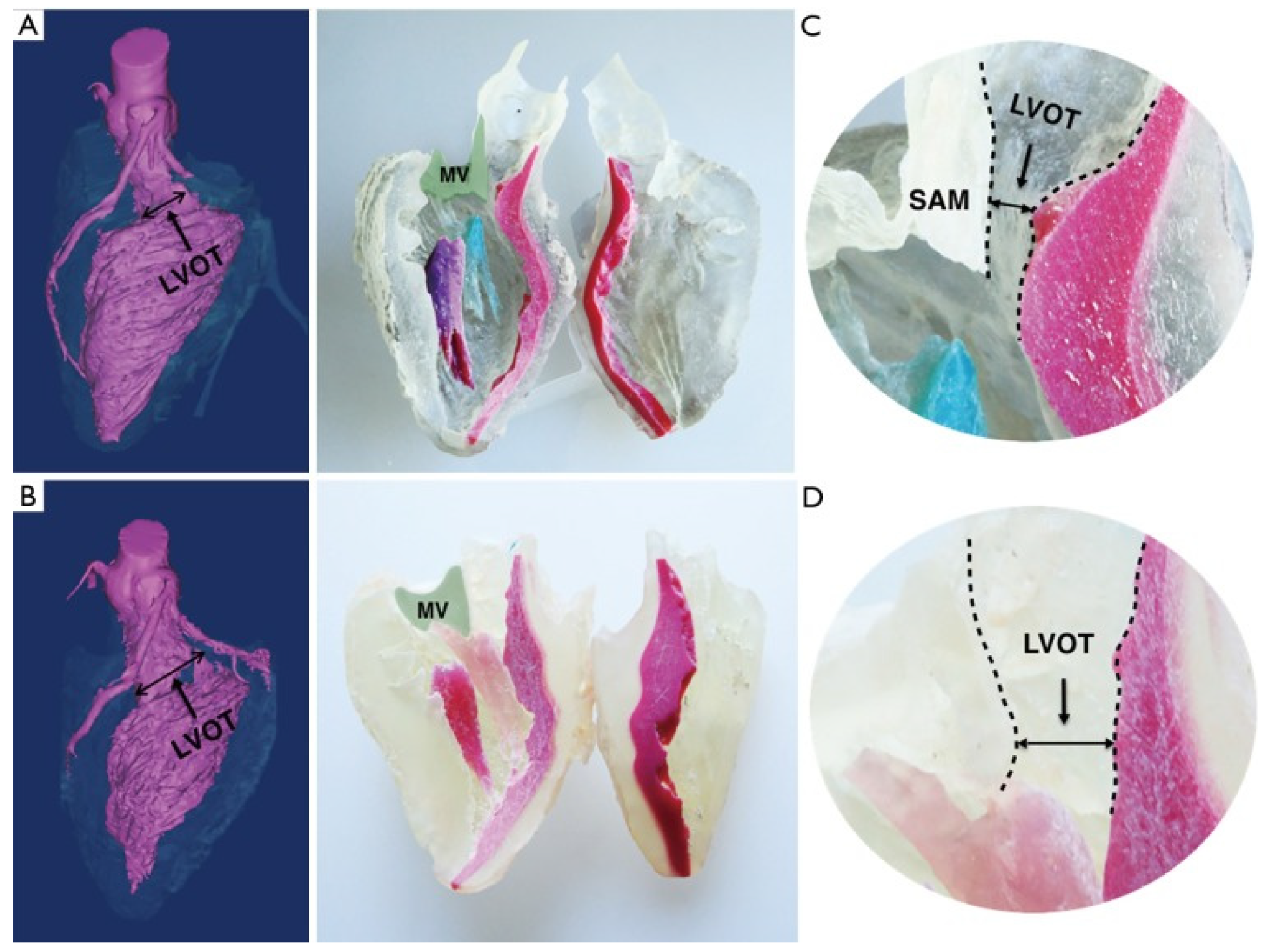
The number of those inflicted with cardiovascular diseases will increase proportionately with the COVID-19 infection rates. This relationship will subsequently increase the need for cardiac treatments. Considering this need, 3D printing could be a beneficial platform for treating cardiovascular effects of COVID-19 through planning cardiac surgeries and evaluating patients. Further, 3D cardiovascular models are typically used to treat structural heart disease, where they are used for modelling heart abnormalities, evaluating patients, and post-surgical evaluations [36,37,38,39]. Figure 2 shows 3D models of a hypertrophic obstructive cardiomyopathy that were used to plan the operation and evaluate the surgical outcome [37]. The 3D models can be used to attempt various approaches in a specific surgery, invent new procedures and practice high-risk operations [40,41]. While at this point, 3D printed cardiovascular implants cannot be used in a clinical setting, advancing research proves this could be a possibility. Zhang et al., printed a 3D model of endothelialized myocardium capable of rhythmic beating using a syringe-based extrusion bioprinter [42]. Their work shows a possibility for the development of more accurate organ modelling and 3D printed myocardial surrogates [42]. Furthermore, tissue engineering can be used to generate cardiac valves, muscles, ventricles, blood vessels, and entire hearts from bioinks (mixtures of cells and biomaterials) that in the future could be used to replace cardiovascular components or in certain cases the entire heart [43,44,45]. The process of printing a human heart starts from a MRI-generated map of the patient’s heart, followed by a patient skin biopsy that is used to generate induced pluripotent stem cells that can form contracting cardiomyocytes along with endothelial cells, cardiac fibroblasts, and smooth muscle cells [43]. These are combined with necessary biomaterials and printed as a bioink, are undergo bioreactor conditioning before being implanted into patients [43]. Much work in 3D printing needs to be done to get to this stage, as there are still challenges in replicating complex heart components, specifically valves and tension apparatuses, and improvement of printing materials to better match that of cardiac muscle needs to be developed.

2.1.2. 3D Printing for Lung Surgery

SARS-CoV-2 can cause lung complications such as pneumonia and, in severe cases, acute respiratory distress syndrome (ARDS) and sepsis, which can cause lasting harm to the lungs and other organs [46]. Pneumonia causes the lungs to fill with fluid and become inflamed. The presence of fluid in the lungs can lead to breathing complications that may cause patients, in severe cases, to require hospital treatment and the use of ventilators for assisted breathing [46]. COVID-19 induced pneumonia could result in breathing difficulties that can take months to improve—even after the virus has passed [46]. In severe cases of pneumonia, ARDS (a form of lung failure) can occur. This pathology happens when fluid in the lungs leaks into the pulmonary blood vessels, causing them to be unable to perform gaseous exchange.
Patients with ARDS are unable to breath independently and require a ventilator to circulate oxygen throughout their body. ARDS can be fatal and people who survive may have lasting pulmonary scarring. Similarly, patients can develop sepsis in severe cases when an infection spreads through the bloodstream, causing tissue damage throughout the body. If patients survive sepsis, they will have lasting effects such as damage to the lungs and other organs and in addition, being more vulnerable to infections and in cases could be deadly [46]. Hasan et al. found that approximately 20% of SARS-CoV-2 patients require hospitalization primarily due to severe respiratory complications such as ARDS [47]. According to their study, patients with ARDS had a mortality rate of 28% [47]. In another study, Tzotzos et al. found approximately 33% COVID-19 patients developed ARDS, 25% required transfer to an intensive care unit (ICU), 16% required a ventilator and 16% died [48]. Of the patients that were transferred to an ICU, 66% required to be put on a ventilator, 75% developed ARDS and 40% died. Of the 40% of patients who passed in this study, 59% were put on a ventilator [48]. In total, 90% of patients who did not survive their COVID-19 infection had ARDS [48].
A similar trend to those long term effects observed in cardiovascular diseases as a result of COVID-19 infections will also extend to pulmonary issues. This increase in lung disorders will generate a great need for treatments to overcome the increased rates of illness. Further, 3D printing can be a resource to help treat lung complications caused by COVID-19 through 3D printed parts for ventilators and 3D bioprinted scaffolds to research the disease. The first 3D printed device used to help with lessening lung complications due to SARS-CoV-2 was a 3D printed ventilator splitter, seen in Figure 3 [49]. Researchers at Johns Hopkins University developed a 3D-splitter that allowed a single ventilator to treat multiple patients. Additionally, they were also able to diminish any concerns of cross contamination between patients [49]. Finding a way to help multiple patients with a single machine safely was vital as ventilators are in great requirement across all hospitals and are limited in resources due to the shortage of machines. The 3D printed splitter allows for an air-flow controller, meaning patients can receive a specific amount of air flow. Each 3D printed splitter was designed to accept a filter to prevent cross-contamination between patients [49]. Earlier devices that did not use 3D printing which had great cross-contamination between patients, making the infected patients complications worsen and did not provide necessary levels of oxygen which resulted in higher mortality rates [49].
Other 3D printed devices to help reduce lung complications due to SARS-CoV-2 include 3D bioprinted scaffolds for lung tissue. A study from Rezaei et al. use 3D bioprinted chitosan/polycaprolactone (CS/PCL) scaffolds to study how COVID-19 infects the lungs by engineering new lung tissue which can be used to test drugs and vaccines to combat the virus [51]. Unlike 2D cell cultures, the 3D bioprinted CS/PCL scaffolds inherit enough porosity to simulate a lung structure effectively allowing for fluids and oxygen to penetrate the scaffold for cell attachment, growth, and proliferation [51]. Infecting the scaffold with COVID-19 to test drugs and vaccines can lead to accurate results for studying pneumonia, ARDS and sepsis, and provide information on how to treat these diseases in the future [51]. With limited donor lung availability, graft rejections and post-operation problems, successful lung transplantation is a modern issue [52]. Further, 3D bioprinting is an option to develop fully functioning engineered lung tissue that can be used to treat lung complications caused by COVID-19 [51]. Human lung tissues can be developed using biological or synthetically developed scaffolds, or with decellularized scaffolds from an animal or human donor, and incorporating various cell types native to lung tissues [53]. Thus, 3D printing/bioprinting could be used to treat many different health complications in medicine where standard resources are limited.

2.1.3. 3D Bioprinting in Vaccine Testing and Drug Delivery

Vaccines and therapeutic treatments are currently in development, with several COVID-19 vaccines being widely distributed and administered with haste. This global endeavor is limited by several factors, including supply shortages, critical requirement of cold storage, distribution challenges, and some population aversion to needles or vaccines [54,55,56]. Microneedles are one area of study in addressing these concerns that has been in development since the early 2000s [57]. Microneedles are small patches with a collection of micron-scale hollow or dissolvable needles for transdermal drug or vaccine administration [57]. These patches are stable at room temperature, reducing the need for cold storage, and are painless, which may help to reduce vaccine hesitancy caused by needle phobias. They can also be self-administered, reducing the need for vaccine clinics in remote areas [58].
The use of 3D printing to fabricate microneedles is quickly gaining popularity [59]. Advances in 3D bioprinting for medical applications allow for standard SLA, DLP, and even FDM printers to reliably produce microneedle patches for a variety of applications, including drug or vaccine delivery [60,61,62]. Various organizations of researchers and medical professionals are conducting studies on the application and relative efficacy of COVID-19 vaccines administered through microneedles, with some initial findings showing stronger antibody responses from microneedle delivery than traditional injection [63].
Furthermore, 3D printing to make drug loaded particles is another emerging strategy for drug delivery. Nishiguchi et al., used a multiphoton lithography 3D printing method to manufacture antigen nanoparticles [64]. Their 3D printing method allowed for the production of particles with various controlled shapes and correspondent immune responses [64]. Moreover, Acevedo et al. used direct laser writing 3D printing to make microparticles containing liquid-cores and shells with controlled characteristics [65]. The controlled features of these 3D printed particles allows for a promising future for 3D printed drug delivery vessels.

3. 3D Printing PPE in Response to COVID-19

Currently, both medical staff and other front-line workers are required to wear more PPE than typically required due to the severity of the COVID-19 pandemic. Additionally, members of the general public have begun voluntarily wearing face protecting PPE as well as non-medical masks to reduce the spread of the virus. As the demand for PPE products has seen a sudden increase, supply lines are scrambling to keep medical staff stocked with important equipment. The recommended protective equipment worn by hospital staff for reducing the spread of COVID-19 include face shields, powered air purifying respirator (PAPR) ventilators, and respirators. In an attempt to fill the need for these vital protective equipment, the Food and Drug Administration (FDA) has relaxed their guidelines to allow companies to design and manufacture PPE using 3D printing technology [66]. While the FDA recommends 3D printed PPE be used as a last resort, the pandemic has illustrated the growing potential for 3D printing technology to be used in place of outsourcing and importing materials, as it allows companies to produce components locally and on demand. These practices benefit scalability of protective equipment and help decrease supply chain interruptions.
There are a variety of international organizations providing consolidated repositories of well-developed open-source models for easy, reliable, high quality 3D printing by local communities, subject to federal guidelines [67]. These are most commonly models for high-output items such as face shields that community members can produce to support their medics and other professionals, as well as other finer devices and components [68,69]. Open-source models allow for personalization of medical devices, although this creates a host of new issues. These issues largely revolve around meeting and maintaining regulatory standards, limiting the usage of this tool to low-risk applications, and managing liability [70].

3.1. Face Shields

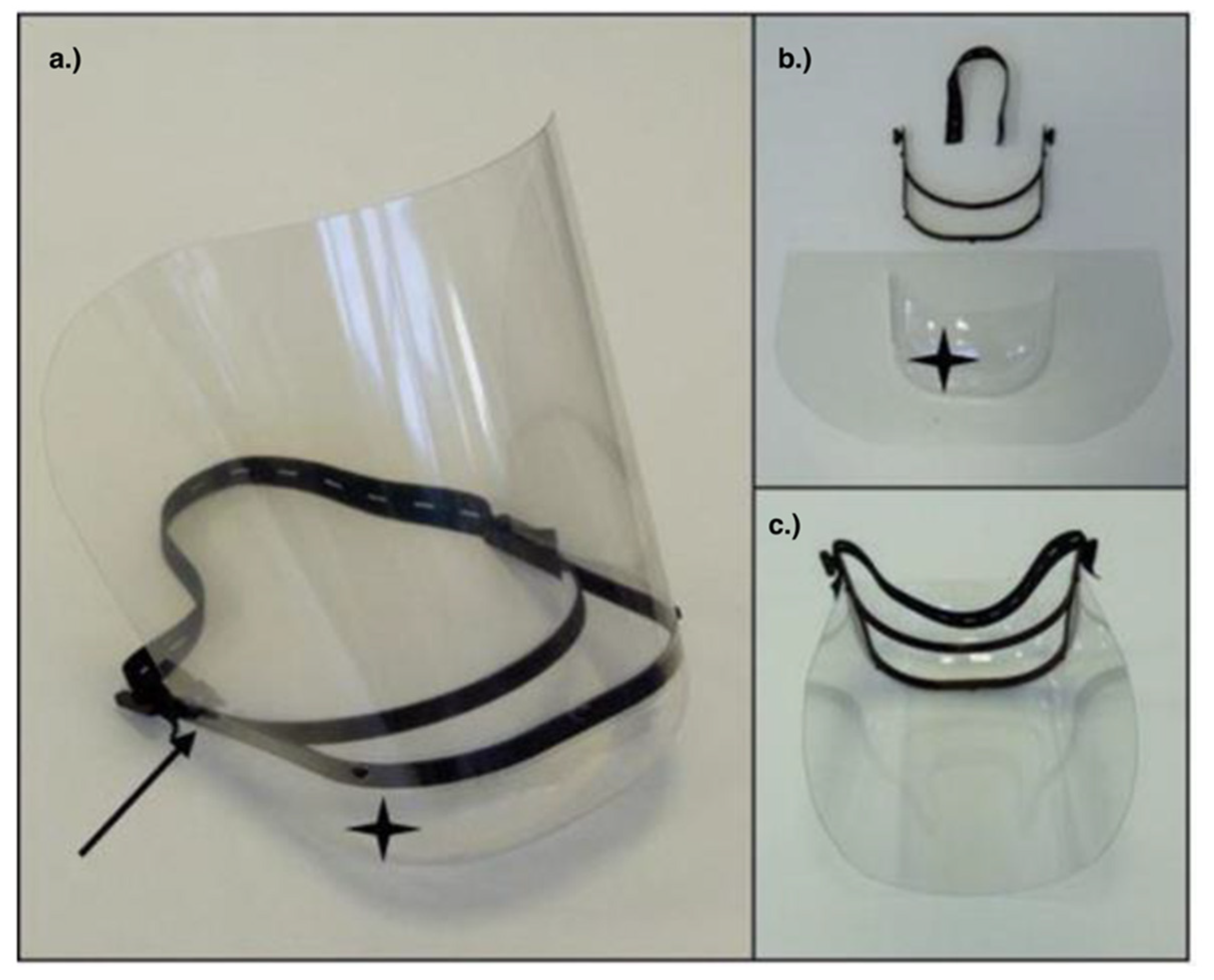
A face shield creates a physical barrier of transparent plastic between the user’s face and their environment and are an easy-to-make 3D printable PPE used by medical staff. In response to the COVID-19 pandemic, face shields have the largest number of 3D printable designs out of any type of protective equipment [71,72]. The printable aspect of each design typically involves the visor that holds the plastic shield which can be seen in Figure 4. Currently, there are many design variations of the visor, each having their own strengths between being quick to print, reliable, and comfortable to wear.
One study compared the differences of protection, fit, printing time, and comfort of the PRUSA RC1 and RC2, Budmen V3, and Easy 3D face shield designs [71]. The Easy 3D face shield design held a slight advantage over the other designs in terms of comfort and protection, but the RC2’s scalability (in the form of the RC3, a design that stacks 4 RC2 models on top of each other to save print time) make the RC2 preferable for large scale printing operations. It is possible for these ratings to be affected by the material and printer settings for the face shield being used as the study only accounted for one material, a lignin-based material called GreenTEC Pro by Extrudr, and printer setting for all the designs. GreenTec Pro was chosen as it has a similar heat resistance and low failure rate to polylactic acid (PLA) while maintaining a high fracture strength. It is worth mentioning that the sample size of the fit testing involved only 10 clinicians, which may not reflect the opinion of the entire medical populace [73].
The materials used to create these face shields can vary. One of the most obvious, medically safe material to use for their creation is PLA [74]. While using PLA is feasible, the flexible nature of the face shield requires a material that is less prone to fracturing. Comparing acrylonitrile butadiene styrene (ABS), polyethylene terephthalate (PETG), and PLA, it was found that PETG is the preferred material to create face shields. This is because PETG is less brittle than PLA, and less prone to shrinkage than ABS. The downsides to using PETG are that the material is not biodegradable, and could be considered difficult to work with as it is less flexible [73].
Re-sterilization of the face shields should be considered to preserve supplies and reduce waste. The materials used for the shield will have to be considered for this task as some plastics like PLA can breakdown quicker than others. One study has shown that 3D printed PETG will degrade less and more predictably when cleaned with hydrogen peroxide than its PLA counterparts. These results were obtained by comparing the deformities developed by a 3D printed part by studying the CT-scans of before and after hydrogen peroxide sterilization using a program called CloudCompare. The largest average difference found between the sterilized parts and their original scans between any two points of data were 0.1887 mm for PLA and 0.0976 mm for PETG [75]. It should be noted that this study only tested 20 products of each material, and the study itself has some inconsistencies with another paper which details the effectiveness of autoclaving 3D printed materials [76]. This study evaluated the effectiveness of different common sterilization methods that would be used in a hospital setting. It was observed that 3D printed parts deformed under steam heat due to the high temperature and pressure of the process. However, the sterilization for this study used a temperature of 134 °C and a pressure of 2 bar which higher than conventional sterilization techniques [76]. Overall, the 3D printing of various types of face shields was a common practice during the early days of the pandemic.

3.2. PAPR

Powered air purifying respirator (PAPR) masks are an important part of hospital protective equipment consisting of a hermetically sealed helmet, which pumps air in and out through a filtration unit mounted on the user’s hip. These masks offer the protection of the high-efficiency particulate air (HEPA) filter respirators but offer easier breathing, as well as ear and eye protection which result in a higher assigned protection factor (APF) rating than the N95 respirator [77]. Manufacturing PAPR masks traditionally is a strenuous task and completely 3D printing them is extremely difficult. However, retrofitting other masks or helmets with 3D printed components to be able to facilitate powered air filter units and improve safety has been proven to be a possibility [78].
One study used a less robust medical ventilated helmet system to generate the mask and improved its safety parameters to meet a modified set of HEPA standards when field tested [78]. This design modified the Stryker Flyte helmet system, which is certified to Association for the Advancement of Medical Instrumentation (AAMI) Level 4 standards but is not cleared as a respiratory protective product [79]. The helmet was improved by replacing the fan cover with a 3D printed manifold that attaches to two breathing circuit filters which the helmet can be seen in Figure 5 below. These filters are attached to anesthesia tubing which pipes the filtered air back out into the user’s environment. By using Tegaderm tape to affix an AAMI Level 3 hood to the mask portion of the helmet, a seal can be made to protect the user. This system was able to pass the modified HEPA standards testing protocol developed previously by Duke University Medical Center [79].
Another study went one step further, taking into account the creation of the air purifying unit and then retrofitting this system onto a full-face snorkel system using a 3D printed adapter [81]. These units are much cheaper to create than sourcing actual PAPR units, however, require a lot of components and assembly. The unit itself costed approximately $185 to make and conventional PAPR units cost from $1000 to over $3400 [81]. The study also mentions that the airflow of the masks need improvement, which is most likely a result of the fan in their design only being able to achieve a post-filter airflow of around 30 L per minute. For reference, the modified National Institute for Occupational Safety (NIOSH) standards that the mask was tested against requires a continuous airflow rate of 85 L per minute [82]. Still, this model could currently be a last resort in terms of use and may be an acceptable low-cost option if PAPR units are unavailable. This work illustrates how 3D printing can lower the cost of PPE production especially during a pandemic.

3.3. Respirators

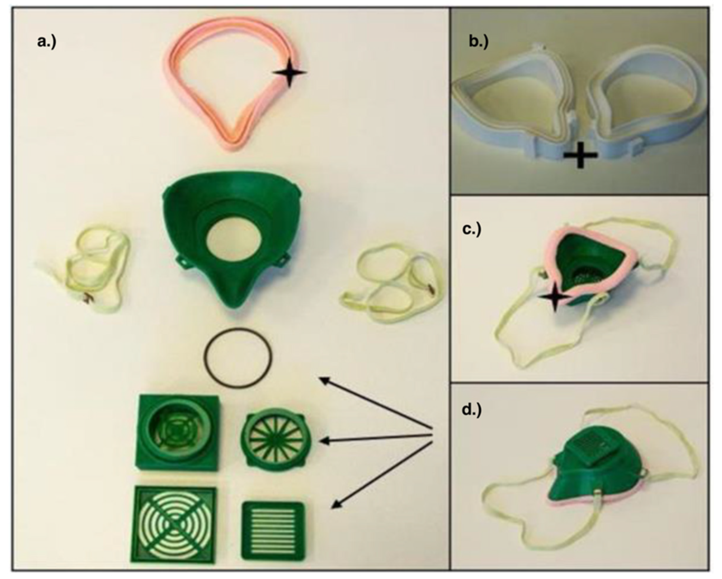
Respirators include a tighter fitting facemask that uses a more sophisticated filter to protect the user from airborne particulates. The need for a better fit is primarily due to the complex nature of the filtration material, which are the main component of these respirators [13]. An example 3D printed respirator mask can be seen below in Figure 6. Multiple designs have been made available to the public in the event that PPE supplies continue to run out. Further, 3D printed respirators have a small advantage as these masks can be fitted on an individual basis with a notable improvement over the mass produced N95. In terms of mass production, it has been proposed that Shellac or other polyurethane finishes be used to correct for manufactured inconsistencies and to create a better seal on the 3D printed parts [83]. Testing the validity of the Shellac approach would be the next step in verifying the mass production process and creating a system to eliminate user error when applying the finish would be of the utmost importance to ensure quality control.
A study conducted by Washington University compares four respirator designs: the Nanohack by Copper 3D, the COVID-19 Mask by Lafactoria3d, the Nanohack 2 (an edit on the Nanohack by multiple creators), and the Single Filter Face Mask (with no credited creator), while using different printers and filaments to find the best fit outcome [83]. Ultimately, these prints were made of PLA as the authors of the study found the most consistent fit results between prints using this material when compared to making TPU or ABS. The fit tests involved an initial basic negative pressure seal test, requiring the user to place a hand over the filter opening and inhale to test for leaks around edge seals. If this test was passed, the user would then wear the mask inside a fume hood with a Bitrex aerosol present to see if they could taste the chemical through the respirator. The study found that the Lafactoria 3D design as the best of these models as it was the most likely to pass a suction test according to the nine participants who initially tested them [83]. The filtration component on these respirators needs to be considered as well. One study suggests using MERV 16 filter material, which has a filtration efficiency of roughly 95% on particles that are 0.3–1.0 micrometers [84]. In order to prevent the user from inhaling fiberglass particles from the MERV 16 filter, it is recommended that it is surrounded by an additional MERV 13 filter. This system has passed at least one hospital test, showing that its filtration capabilities are reported to be as good as the N95 masks that it is trying to replicate [83].
Another study suggests the use of premade electrostatic or mechanical filters that can be retrofitted on the 3M 6603 and 7501 half face reusable respirators using 3D parts [85]. As these respirators typically allow for unfiltered air to be exhaled from the mask, the addition of 3D printed parts can allow for the second filter to be involved in the exhalation process. Depending on whether or not hospital staff currently have fitted respirators, this method allows for a very effective (>99.999% of bacterial and viral filter efficiency for all the filters tested this study) reusable respirator. If supplies and sterilization for the N95 masks prove inadequate, it is possible that the FDA will allow the use of these alternative filters in 3D printed masks.

4. Future Directions and Conclusions

Given the SARS-CoV-2 virus is affecting the majority of the world and it remains unknown how long the virus will last, new innovations to create personal protective equipment to protect frontline healthcare workers were needed to overcome the shortages of supplies that use traditional forms of manufacturing. Further, 3D printing has proven to be one solution currently used in the medical field to overcome challenges in surgery and prosthetics due to their ease of manufacturing and preciseness. Long-term effects of COVID-19, including cardiovascular and lung complications, are becoming more prominent in research, showing a need for additional long-term care of those inflicted with the virus. Further, 3D modelling can be used in heart and lung surgeries as models for planning and practicing difficult surgeries, 3D bioprinting replacement heart and lung tissues and post-surgical evaluations. The 3D printed devices are also being used to study the direct effects SARS-CoV-2, vaccines and drugs have on these organs. Further, 3D printing has been used to create face shields, PAPR masks and respirators, which all have the possibility to be used in the future for healthcare with success and decrease the shortcomings of supply of PPE. These methods have shown that such solutions can be printed on demand to meet such issues with the supply chain.
3D printing can become more of a mainstream technology for production instead of being mainly used for prototyping and small productions. Companies could turn to using 3D printing in their large production lines as it is fast, cheap, and adaptable. To replace standard manufacturing techniques, 3D printers should be able to print a greater volume of product in a shorter timeframe than traditional techniques. Further, 3D printed parts can be interchangeable from one product to another with ease, making it advantageous for large manufactures to use 3D printing when manufacturing more versatile products. To ensure the diverse and versatile applications of 3D printing, these printers should be able to print efficiently and with a wide range of materials. This is important for their use in the production of PPE and medical devices as these must be safe and non-toxic for extended use. In addition, 3D printers can print multiple materials from the same machine, even simultaneously, and as the list of materials capable of seamless printing grows so does the potential for the 3D printing community. Regarding 3D bioprinting, there are many challenges related to printing with biomaterials. Many aspects of the chosen biomaterial must be taken into account for proper printing, including their printability (ability to be printed in a controlled and accurate fashion), biocompatibility, biodegradability, and mechanical properties [86]. The properties of the biomaterial needed will differ based on the type of 3D printer being used, what is being printed, including various cell types and the use of scaffolds. The COVID-19 pandemic also illustrated the importance of being able to source materials for production for both 3D printing and other applications, which is another important consideration for high-throughput manufacturing of any product.
With the success of the current technologies illustrated in this paper, the rapid adoption of these devices could potentially allow for more 3D printed products to be used consistently and phase out traditional forms of manufacturing in the future. If future crises occur, instead of relying on standard manufactures for important PPE, regulatory and governments could turn to the 3D printing community instead. The government could orchestrate supply lines from their own country using the 3D printing community which could supply the country quickly and inexpensively with vital PPE. While the materials used in 3D printing may not be entirely spared from future supply shortages, the ability of 3D printing to use a variety of materials would be beneficial to maintain production. For the smooth production of 3D printed supplies, the maintenance and regulation of materials may be needed at times to ensure proper supplies are ready in times of mass distress. Reasonable material substitutions should be researched and recognized for important PPE and medical equipment before times of crises occur, with a reasonable supply of both the common and substitute material available. Such options can be developed in collaboration with the health regulatory bodies to ensure compliance with the standards for medical devices.
Within the medical field, future directions of 3D printing could for see more medical devices and prosthetics being made from 3D printing more and more due to the flexibility 3D printing allows designers to better their models for specific uses or match a patient’s needs [87]. In addition, instead of only using 3D printing for PPE, 3D printing could be used to make surgical instruments as an accurate and cost effective way to quickly manufacture required medical supplies and alleviate the same bottlenecks which was seen for PPE [87]. In addition to 3D printing medical supplies in the future, 3D printed medical devices can be improved using bioelectronics and robotics. Improving how 3D printing can be done and incorporated into new treatments can open the applications of 3D printing greatly. Combining biological materials with already active devices and standard 3D printing practices can lead to new creations of devices with unique functionalities and properties [88]. This advancement could redefine treatments in the field of smart prosthetics and biomedical devices [88]. The use of robotics incorporated with 3D printing could also benefit the medical field immensely. Using robots that are precise but also fast could speed up the manufacturing of PPE and other medical devices. Overall, the use of 3D will only become more widely adopted as this technology progresses for medical applications.

Author Contributions

A.H., M.C., K.H., T.K., L.A., I.F., S.B., R.K. and J.M. contributed to the writing of the initial draft. I.F., L.A., J.M., J.W.G., R.K. and S.M.W. edited and formatted the draft for submission. All authors have read and agreed to the published version of the manuscript.

Funding

This work was funded through the support of TECHNATION and Innovate BC student funding programs. Willerth receives support from the Canada Research Chairs program and the NSERC Discovery Grant Program.

Institutional Review Board Statement

Not applicable.

Informed Consent Statement

Not applicable.

Data Availability Statement

Not applicable.

Conflicts of Interest

There are no known competing financial interests or personal relationships that could have appeared to influence the work reported in this paper.

References

  1. WHO. Coronavirus Disease (COVID-19) Situation Report 141; WHO: Geneva, Switzerland, 2020. [Google Scholar]
  2. WHO. Novel Coronavirus (2019-nCoV) Situtation Report 1; WHO: Geneva, Switzerland, 2020. [Google Scholar]
  3. Khachfe, H.H.; Chahrour, M.; Sammouri, J.; Salhab, H.A.; Makki, B.E.; Fares, M.Y. An Epidemiological Study on COVID-19: A Rapidly Spreading Disease. Cureus 2020, 12, e7313. [Google Scholar] [CrossRef] [Green Version]
  4. WHO. Novel Coronavirus (2019-nCoV) Situation Report—7; WHO: Geneva, Switzerland, 2020. [Google Scholar]
  5. WHO. Novel Coronavirus (2019-nCoV) Situation Report—9; WHO: Geneva, Switzerland, 2020. [Google Scholar]
  6. Rafiq, D.; Batool, A.; Bazaz, M.A. Three months of COVID-19: A systematic review and meta-analysis. Rev. Med. Virol. 2020, 30, e2113. [Google Scholar] [CrossRef] [PubMed]
  7. CDC Infographic on COVID-19 Symptoms. Available online: covid19-infographic-symptoms-final.pdf (accessed on 20 March 2021).
  8. Nicola, M.; Alsafi, Z.; Sohrabi, C.; Kerwan, A.; Al-Jabir, A.; Iosifidis, C.; Agha, M.; Agha, R. The socio-economic implications of the coronavirus pandemic (COVID-19): A review. Int. J. Surg. 2020, 78, 185–193. [Google Scholar] [CrossRef] [PubMed]
  9. World Health Organization (WHO). Rational Use of Personal Protective Equipment (PPE) for Coronavirus Disease 2019 (COVID-19); WHO: Geneva, Switzerland, 2020. [Google Scholar]
  10. Ranney, M.L.; Griffeth, V.; Jha, A.K. Critical Supply Shortages—The Need for Ventilators and Personal Protective Equipment during the Covid-19 Pandemic. N. Engl. J. Med. 2020, 382, e41. [Google Scholar] [CrossRef] [PubMed]
  11. Kobokovich, A. Ventilator Stockpiling and Availability in the US; Center of Health Security: Baltimore, MD, USA, 2020; Available online: https://www.centerforhealthsecurity.org/resources/COVID-19/COVID-19-fact-sheets/200214-VentilatorAvailability-factsheet.pdf (accessed on 22 July 2021).
  12. Flanagan, S.T.; Ballard, D.H. 3D Printed Face Shields: A Community Response to the COVID-19 Global Pandemic. Acad. Radiol. 2020, 27, 905–906. [Google Scholar] [CrossRef] [PubMed]
  13. Ishack, S.; Lipner, S.R. Applications of 3D Printing Technology to Address COVID-19−Related Supply Shortages. Am. J. Med. 2020, 133, 771–773. [Google Scholar] [CrossRef] [PubMed]
  14. Noorani, R. 3D Printing Technology, Applications, and Selection, 1st ed.; CRC Press: Boca Raton, FL, USA, 2018. [Google Scholar]
  15. Milewski, J.O. Origins of 3D Metal Printing. Superconductivity 2017, 258, 119–129. [Google Scholar] [CrossRef]
  16. Savini, A.; Savini, G.G. A short history of 3D printing, a technological revolution just started. In Proceedings of the 2015 ICOHTEC/IEEE International History of High-Technologies and their Socio-Cultural Contexts Conference (HISTELCON), Tel-Aviv, Israel, 18–19 August 2015. [Google Scholar] [CrossRef]
  17. Horvath, J.; Cameron, R. Mastering 3D Printing, 2nd ed.; Technology in Action; Apress: New York, NY, USA, 2020. [Google Scholar]
  18. Rendeki, S.; Nagy, B.; Bene, M.; Pentek, A.; Toth, L.; Szanto, Z.; Told, R.; Maroti, P. An overview on personal protective equipment (PPE) fabricated with additive manufacturing technologies in the era of COVID-19 pandemic. Polymers 2020, 12, 2703. [Google Scholar] [CrossRef]
  19. Remondino, F.; El-hakim, S. Image-based 3D Modelling: A Review. Photogramm. Rec. 2006, 21, 269–291. [Google Scholar] [CrossRef]
  20. Lee, V. Medical applications for 3D printing: Current and projected uses. Pharm. Ther. 2014, 39, 704–711. [Google Scholar]
  21. Aimar, A.; Palermo, A.; Innocenti, B. The Role of 3D Printing in Medical Applications: A State of the Art. J. Health Eng. 2019, 2019, 1–10. [Google Scholar] [CrossRef] [PubMed] [Green Version]
  22. Matsumoto, J.S.; Morris, J.M.; Foley, T.A.; Williamson, E.E.; Leng, S.; McGee, K.P.; Kuhlmann, J.L.; Nesberg, L.E.; Vrtiska, T.J. Three-dimensional physical modeling: Applications and experience at mayo clinic. Radiographics 2015, 35, 1989–2006. [Google Scholar] [CrossRef]
  23. Mitsouras, D.; Liacouras, P.; Imanzadeh, A.; Giannopoulos, A.A.; Cai, T.; Kumamaru, K.K.; George, E.; Wake, N.; Caterson, E.J.; Pomahac, B.; et al. Medical 3D printing for the radiologist. Radiographics 2015, 35, 1965–1988. [Google Scholar] [CrossRef] [PubMed]
  24. Witowski, J.S.; Pędziwiatr, M.; Major, P.; Budzyński, A. Cost-effective, personalized, 3D-printed liver model for preoperative planning before laparoscopic liver hemihepatectomy for colorectal cancer metastases. Int. J. Comput. Assist. Radiol. Surg. 2017, 12, 2047–2054. [Google Scholar] [CrossRef] [Green Version]
  25. Paul, G.M.; Rezaienia, A.; Wen, P.; Condoor, S.; Parkar, N.; King, W.; Korakianitis, T. Medical Applications for 3D Printing: Recent Developments. Mo. Med. 2018, 115, 75–81. [Google Scholar]
  26. Mohammed, M.I.; Fitzpatrick, A.P.; Gibson, I. Customised design of a patient specific 3D printed whole mandible implant. KnE Eng. 2017, 2, 104–111. [Google Scholar] [CrossRef]
  27. Capelli, C.; Schievano, S. Computational analyses and 3D printed models: A combined approach for patient-specific studies. 3D Print. Med. 2017, 73–90. [Google Scholar] [CrossRef]
  28. Mommaerts, M.Y.; Büttner, M.; Vercruysse, H.; Wauters, L.; Beerens, M. Orbital Wall Reconstruction with Two-Piece Puzzle 3D Printed Implants: Technical Note. Craniomaxillofac. Trauma Reconstr. 2016, 9, 55–61. [Google Scholar] [CrossRef] [Green Version]
  29. Dawood, A.; Marti, B.M.; Sauret-Jackson, V.; Darwood, A. 3D printing in dentistry. Br. Dent. J. 2015, 219, 521–529. [Google Scholar] [CrossRef] [PubMed]
  30. Dodziuk, H. Applications of 3D printing in healthcare. Pol. J. Cardio Thoracic Surg. 2016, 3, 283–293. [Google Scholar] [CrossRef] [PubMed]
  31. Rastogi, P.; Kandasubramanian, B. Review of alginate-based hydrogel bioprinting for application in tissue engineering. Biofabrication 2019, 11, 042001. [Google Scholar] [CrossRef] [PubMed]
  32. López-León, S.; Wegman-Ostrosky, T.; Perelman, C.; Sepulveda, R.; Rebolledo, P.A.; Cuapio, A.; Villapol, S. More than 50 Long-Term Effects of COVID-19: A Systematic Review and Meta-Analysis. SSRN Electron. J. 2021. [Google Scholar] [CrossRef]
  33. Goha, A.; Mezue, K.; Edwards, P.; Nunura, F.; Baugh, D.; Madu, E. COVID-19 and the heart: An update for clinicians. Clin. Cardiol. 2020, 43, 1216–1222. [Google Scholar] [CrossRef] [PubMed]
  34. Ackermann, M.; Werlein, C.; Länger, F.; Kühnel, M.P.; Jonigk, D.D. COVID-19: Auswirkungen auf Lunge und Herz. Pathologe 2021, 42, 164–171. [Google Scholar] [CrossRef]
  35. Zhou, F.; Yu, T.; Du, R.; Fan, G.; Liu, Y.; Liu, Z.; Xiang, J.; Wang, Y.; Song, B.; Gu, X.; et al. Clinical course and risk factors for mortality of adult inpatients with COVID-19 in Wuhan, China: A retrospective cohort study. Lancet 2020, 395, 1054–1062. [Google Scholar] [CrossRef]
  36. Sun, X.; Zhang, H.; Zhu, K.; Wang, C. Patient-specific three-dimensional printing for Kommerell’s diverticulum. Int. J. Cardiol. 2018, 255, 184–187. [Google Scholar] [CrossRef]
  37. Guo, H.C.; Wang, Y.; Dai, J.; Ren, C.W.; Li, J.H.; Lai, Y.Q. Application of 3D printing in the surgical planning of hypertrophic obstructive cardiomyopathy and physician-patient communication: A preliminary study. J. Thorac. Dis. 2018, 10, 867–873. [Google Scholar] [CrossRef] [Green Version]
  38. Wang, Z.; Luo, H.; Gao, C.; Xu, Y. Three-dimensional printing model for the postoperative follow-up of atrial septal defect. Int. J. Cardiol. 2016, 222, 891–892. [Google Scholar] [CrossRef]
  39. Wang, H.; Song, H.; Yang, Y.; Cao, Q.; Hu, Y.; Chen, J.; Guo, J.; Wang, Y.; Jia, D.; Cao, S.; et al. Three-dimensional printing for cardiovascular diseases: From anatomical modeling to dynamic functionality. Biomed. Eng. Online 2020, 19, 1–16. [Google Scholar] [CrossRef]
  40. Yoo, S.J.; Spray, T.; Austin, E.H.; Yun, T.J.; van Arsdell, G.S. Hands-on surgical training of congenital heart surgery using 3-dimensional print models. J. Thorac. Cardiovasc. Surg. 2017, 153, 1530–1540. [Google Scholar] [CrossRef] [Green Version]
  41. Hermsen, J.L.; Burke, T.M.; Seslar, S.P.; Owens, D.S.; Ripley, B.; Mokadam, N.A.; Verrier, E.D. Scan, plan, print, practice, perform: Development and use of a patient-specific 3-dimensional printed model in adult cardiac surgery. J. Thorac. Cardiovasc. Surg. 2017, 153, 132–140. [Google Scholar] [CrossRef] [Green Version]
  42. Zhang, Y.S.; Arneri, A.; Bersini, S.; Shin, S.-R.; Zhu, K.; Goli-Malekabadi, Z.; Aleman, J.; Colosi, C.; Busignani, F.; Dell’Erba, V.; et al. Bioprinting 3D microfibrous scaffolds for engineering endothelialized myocardium and heart-on-a-chip. Biomaterials 2016, 110, 45–59. [Google Scholar] [CrossRef] [Green Version]
  43. Birla, R.K.; Williams, S.K. 3D bioprinting and its potential impact on cardiac failure treatment: An industry perspective. APL Bioeng. 2020, 4, 010903. [Google Scholar] [CrossRef] [Green Version]
  44. Liu, N.; Ye, X.; Yao, B.; Zhao, M.; Wu, P.; Liu, G.; Zhuang, D.; Jiang, H.; Chen, X.; He, Y.; et al. Advances in 3D bioprinting technology for cardiac tissue engineering and regeneration. Bioact. Mater. 2021, 6, 1388–1401. [Google Scholar] [CrossRef]
  45. Birla, R.K. Current State of the Art in Ventricle Tissue Engineering. Front. Cardiovasc. Med. 2020, 7, 591581. [Google Scholar] [CrossRef] [PubMed]
  46. COVID-19 Lung Damage|Johns Hopkins Medicine. Available online: https://www.hopkinsmedicine.org/health/conditions-and-diseases/coronavirus/what-coronavirus-does-to-the-lungs (accessed on 16 June 2021).
  47. Hasan, S.S.; Capstick, T.; Ahmed, R.; Kow, C.S.; Mazhar, F.; a Merchant, H.; Zaidi, S.T.R. Mortality in COVID-19 patients with acute respiratory distress syndrome and corticosteroids use: A systematic review and meta-analysis. Expert Rev. Respir. Med. 2020, 14, 1149–1163. [Google Scholar] [CrossRef] [PubMed]
  48. Tzotzos, S.J.; Fischer, B.; Fischer, H.; Zeitlinger, M. Incidence of ARDS and outcomes in hospitalized patients with COVID-19: A global literature survey. Crit. Care 2020, 24, 1–4. [Google Scholar] [CrossRef] [PubMed]
  49. Johns Hopkins Engineers Develop 3D-Printed Ventilator Splitters|Hub. Available online: https://hub.jhu.edu/2020/04/02/3d-printed-ventilator-splitters-for-covid-19/ (accessed on 16 June 2021).
  50. Vaughan, T.J.; Kirrane, F.; Moerman, K.M.; Cahill, T.; O’Regan, A.; O’Keeffe, D.T. A Novel Dual Non-Invasive Ventilator Continuous Positive Airway Pressure Non-Aerosolization Circuit for Emergency Use in the COVID-19 Pandemic. J. Open Hardw. 2020, 4. [Google Scholar] [CrossRef]
  51. Rezaei, F.S.; Khorshidian, A.; Beram, F.M.; Derakhshani, A.; Esmaeili, J.; Barati, A. 3D printed chitosan/polycaprolactone scaffold for lung tissue engineering: Hope to be useful for COVID-19 studies. RSC Adv. 2021, 11, 19508–19520. [Google Scholar] [CrossRef]
  52. Prakash, Y.S.; Tschumperlin, D.J.; Stenmark, K.R. Coming to terms with tissue engineering and regenerative medicine in the lung. Am. J. Physiol. Lung Cell. Mol. Physiol. 2015, 309, L625–L638. [Google Scholar] [CrossRef] [Green Version]
  53. Tebyanian, H.; Karami, A.; Nourani, M.R.; Motavallian, E.; Barkhordari, A.; Yazdanian, M.; Seifalian, A. Lung tissue engineering: An update. J. Cell. Physiol. 2019, 234, 19256–19270. [Google Scholar] [CrossRef] [PubMed]
  54. Menon, I.; Bagwe, P.; Gomes, K.; Bajaj, L.; Gala, R.; Uddin, M.; D’Souza, M.; Zughaier, S. Microneedles: A New Generation Vaccine Delivery System. Micromachines 2021, 12, 435. [Google Scholar] [CrossRef] [PubMed]
  55. Taddio, A.; Ipp, M.; Thivakaran, S.; Jamal, A.; Parikh, C.; Smart, S.; Sovran, J.; Stephens, D.; Katz, J. Survey of the prevalence of immunization non-compliance due to needle fears in children and adults. Vaccine 2012, 30, 4807–4812. [Google Scholar] [CrossRef] [PubMed] [Green Version]
  56. Understanding the Unique Distribution Challenges of COVID-19 Vaccines. Available online: https://www.cnacanada.ca/web/guest/cnacanada/about/listofauthors/authorbio/blogdetails/SA-Ruth-Stewart/understanding-the-unique-distribution-challenges-of-covid-19-vaccines (accessed on 13 July 2021).
  57. Bariya, S.H.; Gohel, M.C.; Mehta, T.A.; Sharma, O.P. Microneedles: An emerging transdermal drug delivery system. J. Pharm. Pharmacol. 2011, 64, 11–29. [Google Scholar] [CrossRef] [PubMed]
  58. O’Shea, J.; Prausnitz, M.R.; Rouphael, N. Dissolvable Microneedle Patches to Enable Increased Access to Vaccines against SARS-CoV-2 and Future Pandemic Outbreaks. Vaccines 2021, 9, 320. [Google Scholar] [CrossRef]
  59. Krieger, K.J.; Bertollo, N.; Dangol, M.; Sheridan, J.T.; Lowery, M.M.; O’Cearbhaill, E.D. Simple and customizable method for fabrication of high-aspect ratio microneedle molds using low-cost 3D printing. Microsyst. Nanoeng. 2019, 5, 1–14. [Google Scholar] [CrossRef] [Green Version]
  60. Economidou, S.N.; Pere, C.P.P.; Reid, A.; Uddin, J.; Windmill, J.; Lamprou, D.A.; Douroumis, D. 3D printed microneedle patches using stereolithography (SLA) for intradermal insulin delivery. Mater. Sci. Eng. C 2019, 102, 743–755. [Google Scholar] [CrossRef]
  61. Barret, A. COVID-19 Vaccine ‘Smart Patch’ Uses Microneedles to Deliver Immunity. BBC Science Focus Magazine. 6 January 2021. Available online: https://www.sciencefocus.com/news/covid-19-vaccine-smart-patch-uk/ (accessed on 14 July 2021).
  62. A Vaccine Patch Could Someday Be an Ouchless Option. Available online: https://www.nibib.nih.gov/news-events/newsroom/vaccine-patch-could-someday-be-ouchless-option (accessed on 14 July 2021).
  63. Microneedle Coronavirus Vaccine Triggers Immune Response in Mice|National Institutes of Health (NIH). Available online: https://www.nih.gov/news-events/nih-research-matters/microneedle-coronavirus-vaccine-triggers-immune-response-mice (accessed on 13 July 2021).
  64. Nishiguchi, A.; Shima, F.; Singh, S.; Akashi, M.; Moeller, M. 3D-Printing of Structure-Controlled Antigen Nanoparticles for Vaccine Delivery. Biomacromolecules 2020, 21, 2043–2048. [Google Scholar] [CrossRef]
  65. Acevedo, R.; Restaino, M.A.; Yu, D.; Hoag, S.W.; Flank, S.; Sochol, R.D. 3D Nanoprinted Liquid-Core-Shell Microparticles. J. Microelectromech. Syst. 2020, 29, 924–929. [Google Scholar] [CrossRef]
  66. U.S. FDA. FAQs on 3D Printing of Medical Devices, Accessories, Components, and Parts during the COVID-19 Pandemic. Available online: https://www.fda.gov/medical-devices/3d-printing-medical-devices/faqs-3dprinting-%0Amedical-devices-accessories-components-and-parts-during-covid-19-pandemic (accessed on 28 May 2020).
  67. 3D Printing and Other Manufacturing of Personal Protective Equipment in Response to COVID-19. Available online: https://www.canada.ca/en/health-canada/services/drugs-health-products/medical-devices/covid-19-unconventional-manufacturing-personal-protective-equipment.html (accessed on 14 July 2021).
  68. 3D Printed Face Shields for Medics and Professionals—Prusa3D—3D Printers from Josef Průša. Available online: https://www.prusa3d.com/covid19/ (accessed on 14 July 2021).
  69. COVID-19 Response|NIH 3D Print Exchange. Available online: https://3dprint.nih.gov/collections/covid-19-response (accessed on 14 July 2021).
  70. Wang, D.; Zhang, J.; Liu, Q.; Chen, B.; Liang, Y.; Hu, L.; Jiang, G. 3D Printing Challenges in Enabling Rapid Response to Public Health Emergencies. Innovation 2020, 1, 100056. [Google Scholar] [CrossRef]
  71. Vordos, N.; Gkika, D.A.; Maliaris, G.; Tilkeridis, K.E.; Antoniou, A.; Bandekas, D.V.; Mitropoulos, A.C. How 3D printing and social media tackles the PPE shortage during Covid-19 pandemic. Saf. Sci. 2020, 130, 104870. [Google Scholar] [CrossRef]
  72. Throckmorton, A.L.; Bass, E.J.; Ferrick, B.; Ramakrishnan, A.; Eichmann, S.; Catucci, N.; Eshelman, B.; McNamara, J.; Sundquist, E.; Beatson, N.; et al. A Cross University-Led COVID-19 Rapid-Response Effort: Design, Build, and Distribute Drexel AJFlex Face Shields. Ann. Biomed. Eng. 2021, 49, 950–958. [Google Scholar] [CrossRef] [PubMed]
  73. Wesemann, C.; Pieralli, S.; Fretwurst, T.; Nold, J.; Nelson, K.; Schmelzeisen, R.; Hellwig, E.; Spies, B.C. 3-D Printed Protective Equipment during COVID-19 Pandemic. Materials 2020, 13, 1997. [Google Scholar] [CrossRef] [PubMed]
  74. Konta, A.A.; García-Piña, M.; Serrano, D.R. Personalised 3D printed medicines: Which techniques and polymers are more successful? Bioengineering 2017, 4, 79. [Google Scholar] [CrossRef] [PubMed] [Green Version]
  75. Oth, O.; Dauchot, C.; Orellana, M.; Glineur, R. How to Sterilize 3D Printed Objects for Surgical Use? An Evaluation of the Volumetric Deformation of 3D-Printed Genioplasty Guide in PLA and PETG after Sterilization by Low-Temperature Hydrogen Peroxide Gas Plasma. Open Dent. J. 2019, 13, 410–417. [Google Scholar] [CrossRef] [Green Version]
  76. Aguado-Maestro, I.; de Frutos-Serna, M.; González-Nava, A.; de Santos, A.B.M.; García-Alonso, M. Are the common sterilization methods completely effective for our in-house 3D printed biomodels and surgical guides? Injury 2021, 52, 1341–1345. [Google Scholar] [CrossRef] [PubMed]
  77. Wong, W.-Y.; Kong, Y.-C.; See, J.-J.; Kan, R.K.; Lim, M.P.; Chen, Q.; Lim, B.; Ong, S. Anaesthetic management of patients with COVID-19: Infection prevention and control measures in the operating theatre. Br. J. Anaesth. 2020, 125, e239–e241. [Google Scholar] [CrossRef] [PubMed]
  78. Erickson, M.M.; Richardson, E.S.; Hernandez, N.M.; Bobbert, D.W.; Gall, K.; Fearis, P. Helmet Modification to PPE With 3D Printing During the COVID-19 Pandemic at Duke University Medical Center: A Novel Technique. J. Arthroplast. 2020, 35, S23–S27. [Google Scholar] [CrossRef]
  79. Stryker. Flyte Personal Protection System Flyte and T4/T5 Togas and Hoods Are Components of Stryker’ s Personal Protection System and Are Intended to Protect the Patient, Healthcare Personnel and Operating Room Personnel against Contamination, Exposure of Inf; Stryker: Kalamazoo, MI, USA, 2020. [Google Scholar]
  80. Temmesfeld, M.J.; Jakobsen, R.B.; Grant, P. Does a surgical helmet provide protection against aerosol transmitted disease? Acta Orthop. 2020, 91, 538–542. [Google Scholar] [CrossRef]
  81. Khoo, D.; Yen, C.C.; Chow, W.T.; Jain, P.; Loh, N.-H.W.; Teo, W.W.; Koh, C. Ultra-portable low-cost improvised powered air-purifying respirator: Feasibility study. Br. J. Anaesth. 2020, 125, e264–e266. [Google Scholar] [CrossRef]
  82. National Institute for Occupational Safety and Health. Determination of Air Flow for Powered Air-Purifying Respirators Standard Testing Procedure (STP); National Institute for Occupational Safety and Health: Washington, DC, USA, 2005. [Google Scholar]
  83. Provenzano, D.; Rao, Y.J.; Mitic, K.; Obaid, S.N.; Pierce, D.; Huckenpahler, J.; Berger, J.; Goyal, S.; Loew, M.H. Rapid Prototyping of Reusable 3D-Printed N95 Equivalent Respirators at the George Washington University. Preprints 2020. [Google Scholar] [CrossRef] [Green Version]
  84. National Air Filtration Association. Understanding MERV|NAFA User’s Guide to ANSI/ASHRAE 52.2. 2018. Available online: https://www.nafahq.org/understanding-merv-nafa-users-guide-to-ansi-ashrae-52-2/ (accessed on 28 May 2020).
  85. Liu, D.C.Y.; Koo, T.H.; Wong, J.K.K.; Wong, Y.H.; Fung, K.S.C.; Chan, Y.; Lim, H.S. Modifying reusable elastomeric respirators to utilise breathing system filters with 3D printed adapters, a safe alternative to N95 during COVID-19. medRxiv 2020. [Google Scholar] [CrossRef] [Green Version]
  86. Mao, H.; Yang, L.; Zhu, H.; Wu, L.; Ji, P.; Yang, J.; Gu, Z. Recent advances and challenges in materials for 3D bioprinting. Prog. Nat. Sci. 2020, 30, 618–634. [Google Scholar] [CrossRef]
  87. How 3D Printing has Changed the Game in the Medical Field. 2019. Available online: https://www.docwirenews.com/future-of-medicine/infographics/how-3d-printing-has-changed-the-game-in-the-medical-field/ (accessed on 20 March 2021).
  88. Using 3D Printing to Blend Biology and Electronics. Available online: https://www.designnews.com/materials-assembly/using-3d-printing-blend-biology-and-electronics (accessed on 14 July 2021).
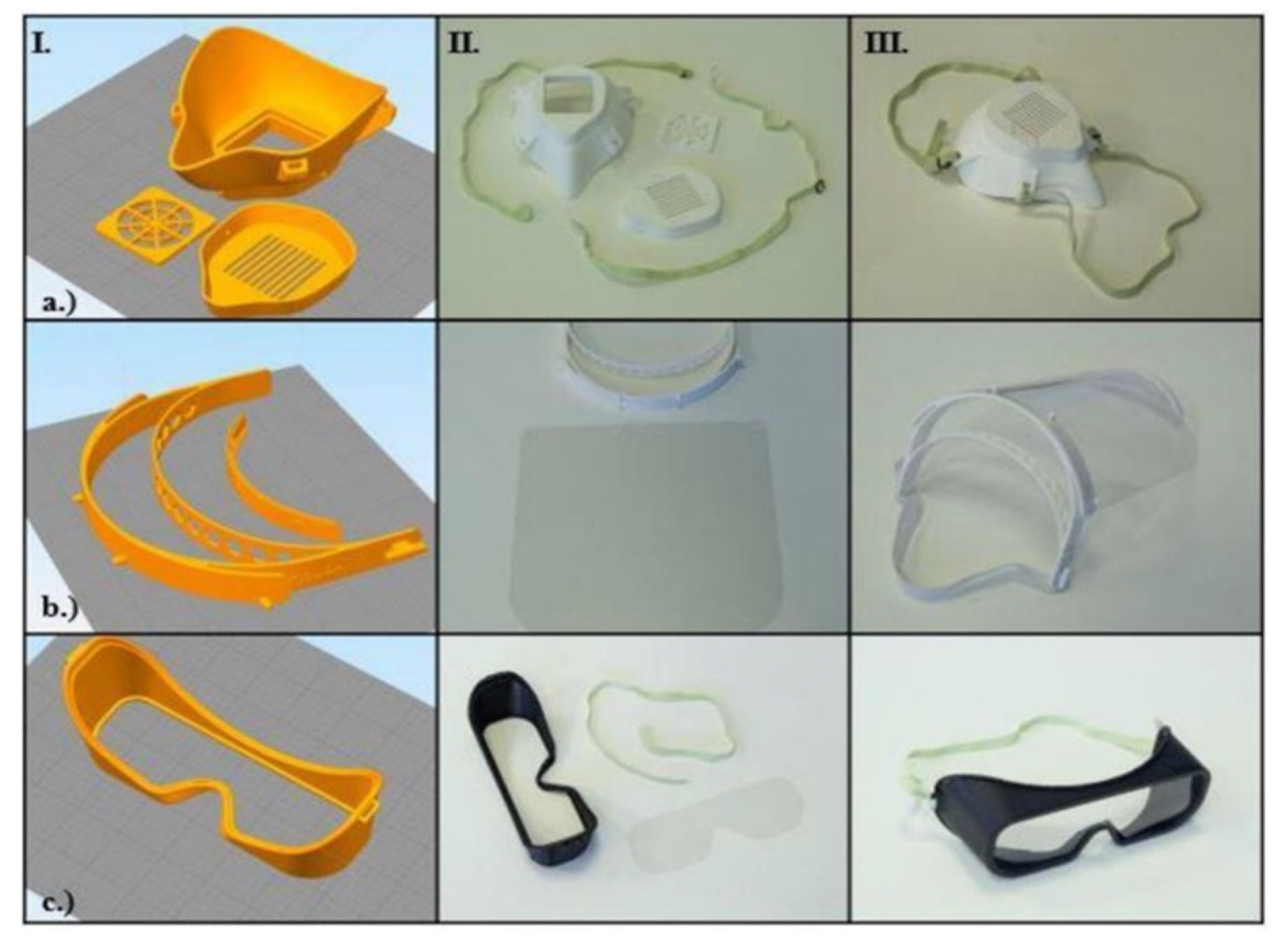
Figure 1. Overview of personal protective equipment (PPE) manufacturing process using 3D printing. With Column I—demonstrating CAD assembly, II—3D printed parts, III—the assembled models. The rows show (a) a respirator face mask; (b) a face shield; (c) a pair of safety googles. This image is reprinted under a Creative Commons Attribution 4.0 International License and it is taken from [18].
Surgeries 02 00025 g001
Figure 2. The 3D models of a hypertrophic obstructive cardiomyopathy pre and post-surgical intervention: (A,B) show the pre-surgical model; (C,D) show the post-surgical model, significantly the enlarged left ventricle outflow tract post-surgery. This image is reprinted under a Creative Commons Attribution 4.0 International License and it is taken from [37].
Surgeries 02 00025 g002
Figure 3. A 3D printed T-Piece Ventilator Splitter connected to a ventilator. This image is reprinted under a Creative Commons Attribution 4.0 International License and it is from [50].
Surgeries 02 00025 g003
Figure 4. The 3D printed face shield. (a) Assembled face shield; (b) disassembled face shield with 3D printed face shield holder, plastic face shield and strap; (c) reverse look of assembled face shield. This image is reprinted under a Creative Commons Attribution 4.0 International License and it is from [18].
Surgeries 02 00025 g004
Figure 5. Stryker Flyte modified ventilated helmet using 3D printing to facilitate powered air filter units and improve safety. This image is reprinted under a Creative Commons Attribution 4.0 International License and it is taken from [80].
Surgeries 02 00025 g005
Figure 6. The 3D printed face mask respirator. (a) Disassembled face mask respirator. (b) Silicone ring layer to promote better face-fitting which had its mold 3D printed. (c) Reverse angle of respirator with silicon ring visible. (d) Assembled face mask respirator. This image is reprinted under a Creative Commons Attribution 4.0 International License and it is taken from [18].
Surgeries 02 00025 g006
Publisher’s Note: MDPI stays neutral with regard to jurisdictional claims in published maps and institutional affiliations.