Review

Advances in Instrumentation and Implant Technology for Spine Oncology: A Focus on Carbon Fiber Technologies

by
Iheanyi Amadi
1,
Jean-Luc K. Kabangu
2,*,
Adip G. Bhargav
2,* and
Ifije E. Ohiorhenuan
2
1
School of Medicine, University of Kansas, Kansas City, KS 66160, USA
2
Department of Neurological Surgery, University of Kansas Medical Center, Kansas City, KS 66160, USA
*
Authors to whom correspondence should be addressed.
Surgeries 2024, 5(3), 499-516; https://doi.org/10.3390/surgeries5030041
Submission received: 13 February 2024 / Revised: 15 June 2024 / Accepted: 26 June 2024 / Published: 30 June 2024

Abstract

:
The challenges inherent in spinal oncology are multi-dimensional, stemming from the complex anatomy of the spine, the high risk of neurological complications, and the indispensability of personalized treatment plans. These challenges are further compounded by the variability in tumor types and locations, which complicates the achievement of optimal treatment outcomes. To address these complexities, the manuscript highlights the pivotal role of technological advancements in surgical practices. The review focuses on the evolution of spinal oncology instrumentation, with a special emphasis on the adoption of carbon fiber implants in the management of spinal tumors. The advancements in instrumentation and implant technology are underscored as vital contributors to the improvement in patient outcomes in spine surgery. Carbon fiber implants are lauded for their reduced imaging artifacts, biocompatibility, and favorable mechanical properties. When combined with other technological innovations, these implants have substantially elevated the efficacy of surgical interventions. The review articulates how these advancements emphasize precision, customization, and the integration of innovative materials, significantly enhancing the effectiveness of surgical procedures. This collective progress marks a considerable advancement in the treatment of spinal tumors, highlighting a shift towards more effective, patient-focused outcomes in spinal oncology.

1. Introduction

The progress registered in the early diagnosis and management of tumors has led to prolonged survival of cancer patients. Consequently, there is a significant rise in the number of patients with metastatic spine tumors which are noted to be more prevalent than primary spine tumors [1,2,3]. The past few decades have witnessed major advances in spinal oncology as it continues to face the challenges posed by the complex nature of spinal tumors. These challenges include the intricate anatomy of the spine, the potential for neurological complications, and the need for comprehensive, patient-specific treatment plans. Additionally, the high variability in tumor types and locations poses difficulties in achieving optimal treatment outcomes.
The management of spinal tumors requires a delicate balance between achieving oncological control and preserving spinal function and stability. Surgical intervention for spinal tumors often necessitated extensive tissue disruption and destabilization, leading to significant morbidity and compromised patient outcomes [3,4]. However, with the advent of advanced instrumentation and technologies, surgeons can now approach these cases with greater precision, efficacy, and safety [4,5]. This has arguably revolutionized the field of spine oncology, offering new hope and improved outcomes for patients facing spinal tumors.
Carbon fiber has garnered considerable attention in spine surgery due to its unique mechanical properties and biocompatibility [6]. One of the key applications of carbon fiber technology in spine oncology is in the development of implants and instrumentation that help optimize postoperative management. By leveraging advanced imaging modalities such as Computed Tomography (CT) and Magnetic Resonance Imaging (MRI), carbon fiber implants can enhance the effectiveness of post-resection radiotherapy [7]. Furthermore, surgeons can monitor for recurrence or progression of tumor [7].
In this narrative review, we explore the latest developments in instrumentation and implants specifically tailored for spine oncology, with a special emphasis on the applications and benefits of carbon fiber materials. We delve into the evolving landscape of carbon fiber technologies in spine oncology, examining their clinical applications, outcomes, and future directions. We aim to provide insights into the transformative potential of carbon fiber instrumentation and implants in optimizing patient care and advancing the field of spine oncology. As the demand for minimally invasive yet effective solutions for spinal tumors continues to grow, carbon fiber technologies stand poised to play a pivotal role in shaping the future of surgical management. By harnessing the inherent properties of this innovative material, surgeons can navigate the complexities of spine oncology with precision and confidence, ultimately improving patient outcomes.

2. Historical Overview of Instrumentation in Spine Surgery

The history of instrumentation in spine surgery is marked by innovative milestones. Accounts of surgical intervention for spinal deformity in the modern era date back to the 19th century when Berthold Hadra, Jules Gerin, and W. T. Wilkins treated patients with spinal conditions including spinal bifida, scoliosis, and Potts disease [8,9]. In the early 20th century, efforts focused on treating spinal deformities associated with diseases like tuberculous spondylitis and poliomyelitis. The Hibbs method in 1911 introduced spinal arthrodesis, using autologous bone grafts via transposition of spinous processes [10,11]. Plaster casts were used to attempt a correction of the loss of curvature and stabilize the spine.
The introduction of the stainless-steel Harrington Rod in the mid-20th century addressed critical gaps in the treatment of neuromuscular scoliosis, thereby revolutionizing spinal surgery [12]. The invention of segmental pedicle screws, pioneered by Roy-Camille [13] paved the way for systems like the Cotrel–Dubousset technique in the 1980s. This approach utilized segmental spinal instrumentation, allowing selective distraction and compression for tailored corrections at individual spinal levels. The Cotrel–Dubousset system, with vertebral screws and hooks made of steel, showed enhanced alignment in sagittal, coronal, and axial planes [14].
A simple representation of the major evolution points of spinal instrumentation over the past century is highlighted in Figure 1. Contemporary spine surgery has seen significant progress with materials evolving beyond stainless steel to advanced options like titanium and cobalt chrome. Pedicle screws remain crucial, adapting to robotics and minimally invasive techniques. Fusion techniques, incorporating bone graft substitutes and biologics, have reduced pseudoarthrodesis rates. The historical evolution reflects a journey from early attempts to address spinal deformities to the introduction of corrective instrumentation, setting the stage for sophisticated and patient-tailored approaches in modern spine oncology.

3. Recent Advances in Instrumentation for Spine Oncology

The quest to achieve comprehensive tumor control has driven the adoption of en bloc resections. These procedures involve extensive excision of the tumor and surrounding tissues to minimize the risk of local recurrence. En bloc resections have proven to be a useful technique in effectively managing primary and metastatic spine tumors. However, accurately delineating tumor margins can be challenging due to anatomical constraints [15]. This often leads to sacrificing important structures like nerves, blood vessels, and the dura [7]. Additionally, some studies have shown that en bloc resections are associated with substantial blood loss, infection risks, and the need for extensive reconstructive surgery to stabilize the spine [15].
To address these challenges, various surgical techniques have been developed. Decompression surgeries, coupled with implant-based stabilization, have gained prominence since the late twentieth century [16]. The widespread adoption of pedicle screw rod systems in the 1980s facilitated the increased utilization of spinal implants [16]. Posterior spinal fixation and decompression have become popular techniques with the advent of implants, while anterior decompression, with or without posterior fixation, was introduced subsequently [16].
These advances in techniques and approaches of spine oncology instrumentation are further amplified by the constant evolution of and advances in computer systems and information technology. Computer-aided surgical systems represent advanced technologies designed to support surgeons in both the planning and execution of surgical procedures. These systems harness the power of computer algorithms, imaging, and real-time feedback to improve surgical techniques by enhancing precision and decision-making throughout surgery [17]. This proves particularly valuable in delicate surgeries or when dealing with tumors invading intricate anatomical structures [18,19]. Surgeons leverage these systems for preoperative planning, utilizing patient-specific data to craft a personalized surgical plan. The integration of computer-aided surgical systems with robotic platforms significantly enhances the capabilities of robot-assisted surgery [17,20,21]. This fusion of technologies not only amplifies the precision and efficiency of surgical interventions but also expands the scope of what can be achieved through robotic assistance in the operating room.
Furthermore, advances in imaging technologies have markedly enhanced intraoperative navigation for spine surgery, empowering surgeons with superior visualization and precision [22]. The integration of cutting-edge imaging modalities, including 3D imaging, CT scans, and MRI data, furnishes surgeons with detailed and real-time visualization of the surgical site. They provide crucial insights into instrument positioning, alignment, and proximity to critical structures. This real-time information provides timely adjustments, improving overall surgical control [8].
In procedures that leverage imaging guidance, computer-aided systems play a pivotal role in minimizing the reliance on extensive intraoperative fluoroscopy [19,23]. This reduction not only reduces radiation exposure for both the patient and the surgical team but also underscores the commitment to safety. Collectively, these innovations contribute to the enhancement of safety and precision in spine surgeries. Surgeons benefit from improved capabilities to navigate intricate anatomy, visualize critical structures, and ultimately achieve optimal outcomes for their patients.

4. Introduction to Implant Technologies

Traditionally, a variety of metals and alloys have been adopted as implant materials in spine oncology. Common options for spine oncology implants include metals like titanium, cobalt, chromium, zirconium, steel, and tantalum [24,25,26,27]. These materials offer crucial qualities such as strength and stability vital in spine oncology instrumentation [28]. Non-metallic materials like the traditional polyetheretherketone (PEEK) have also gained prominence in spinal instrumentation. PEEK, a polymer, is sometimes favored in spine surgery due to its radiolucent properties which improve imaging visibility. In addition, its modulus of elasticity closely aligns with that of bone, distinguishing it from metal implants [29].
While these implant materials have become widely utilized in spine surgery, they present a myriad of challenges. The relatively high density and weight of metals, particularly in the context of spinal fusion constructs, raise concerns about physiological compatibility and functional outcomes. This weight can significantly increase the overall spinal load, especially in patients with compromised bone density [29]. The rigidity of metal implants may also hinder natural spine movement, potentially resulting in a reduced range of motion and stress on adjacent segments. Moreover, the radiopacity of traditional metals often interferes with imaging techniques like X-rays, making it challenging to assess bone healing and fusion.
Non-metal implants such as PEEK are noted to be hydrophobic and biologically inert. This makes it challenging to achieve solid fusion and robust osseointegration due to their inability to properly fuse with bone [7,8]. This characteristic poses a significant challenge as it may hinder the seamless integration of the implant with surrounding bone tissue, potentially compromising its stability and longevity within the body.
Furthermore, the absence of inherent antimicrobial properties in PEEK further exacerbates the risk of implant failure [30]. Without the ability to resist microbial colonization and infection, implants made solely of PEEK are susceptible to microbial contamination, which can trigger inflammatory responses, impair healing processes, and ultimately jeopardize the success of the implantation procedure [30]. These issues often result in the need for revision surgery and the consequent complications including difficulties in removing implants due to possible bone ingrowth, tissue adhesion, and potential damage to surrounding structures.

5. Carbon Fiber in Spine Oncology

In response to these challenges, ongoing research and technological advancements in materials science are ushering in alternative materials. The past few decades have witnessed increasing adoption of carbon fiber-based implants. Carbon fiber is a polymer that possesses several unique properties that make it well-suited for spinal oncology implants. To begin with, it is generally biocompatible and bioinert [31,32]. Thus, it is well tolerated by the human body and does not elicit a significant immune response [33,34]. This property is crucial for spinal implants to minimize the risk of adverse reactions, inflammation, or rejection while promoting a stable environment around the implant when integrated into the spinal column.
Carbon fiber-reinforced (CFR) materials, functioning as electrically conductive microcircuits within a polymer matrix composite, have demonstrated promising experimental reliability in stimulating tissue growth [6]. This effect is achieved by their capacity to mitigate the detrimental effects of excess electrons generated during respiratory stress. Oxygen serves as the ultimate electron acceptor during efficient energy synthesis. However, in its absence or insufficiency, cells may produce free radicals and acidic byproducts, which can be damaging to cellular structures. Here, carbon fiber’s conductivity plays a pivotal role. By acting as a semi-antioxidant and establishing electrochemical gradients, carbon fiber facilitates the removal of excess damaging electrons, directing them towards areas of lower negative charges and lower concentrations [6]. This mechanism helps in maintaining cellular homeostasis and mitigating oxidative stress.
In addition, carbon fiber is comparable to metals such as titanium in terms of strength and stability despite being lightweight [35]. Table 1 shows a comparison of carbon fiber with traditional implant metals in terms of biomechanical properties and imaging compatibility. In spinal oncology, implants often need to provide stability and support to the spine. The strength of carbon fiber allows for the creation of implants that can withstand spinal loads while maintaining structural integrity [36]. Its excellent fatigue resistance is beneficial in spinal implants subjected to repetitive stress or load-bearing conditions [29]. This contributes to the long-term durability of the implant.
Carbon fiber offers a notable advantage in its radiolucency, making it highly beneficial in scenarios where minimizing electromagnetic interference is critical, such as during CT or MRI scans [31,35]. The radiolucent nature of carbon fiber allows for comprehensive visualization of anatomical structures during imaging studies. This enhanced clarity enables surgeons to accurately pinpoint the location, extent, and characteristics of spinal tumors, facilitating informed treatment decisions.
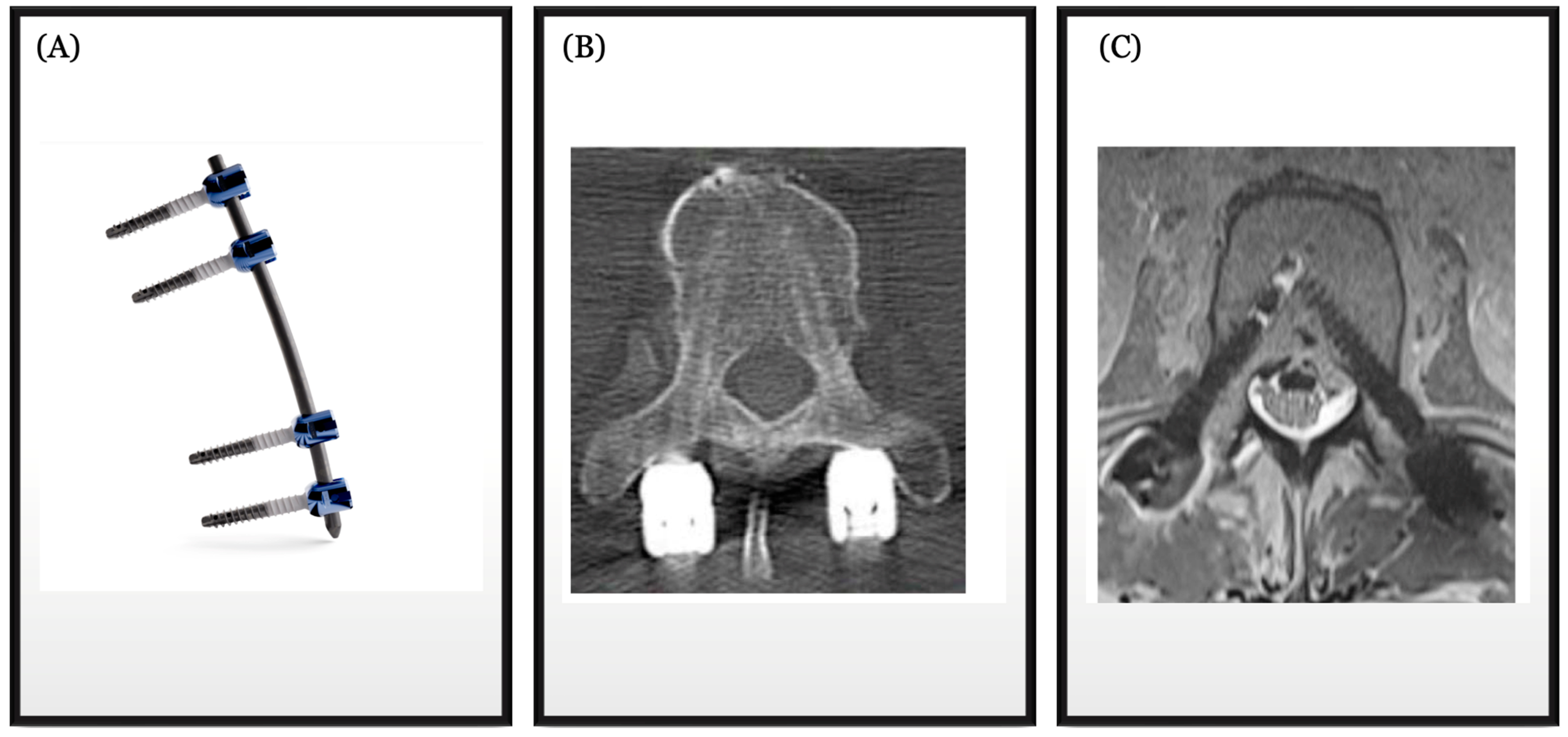
Unlike traditional metal implants, carbon fiber is nearly invisible on imaging and does not distort surrounding structures [35]. The improved imaging compatibility of carbon fiber-based implants in spinal instrumentation is demonstrated in Figure 2. The absence of interference from carbon fiber implants ensures that imaging artifacts are minimized, resulting in clearer and more accurate images. The enhanced visibility of tissues and bones on imaging, facilitated by carbon fiber’s radiolucency, enhances the accuracy of diagnosis and postoperative assessment of spinal tumors. Consequently, this enhances the overall quality of patient care, allowing for prompt intervention and optimized patient management, ensuring that treatment plans are tailored to individual needs and that any issues are promptly addressed [20].
Furthermore, the use of carbon fiber-reinforced instrumentation has been shown to improve postoperative radiation therapy planning [1,37,38]. The radiolucency of carbon fiber implants allows for precise delineation of tumor margins and surrounding anatomical structures, enabling oncologists to target radiation therapy more effectively while minimizing damage to healthy tissues. The value of this quality can be seen in proton beam radiation planning. Proton beams are sensitive to material changes as they pass through structures during targeted therapy [39]. This often results in range uncertainties and setup errors, thereby negatively affecting the quality of the therapy. Metallic implants, like titanium, can cause streak artifacts in 3D CT scans, leading to dose distribution uncertainties in treatment planning. CT scan artifacts can lead to errors in dose calculation [39,40]. As such, carbon fiber implants might be more suited for adequate planning and delivery of proton beam therapy.
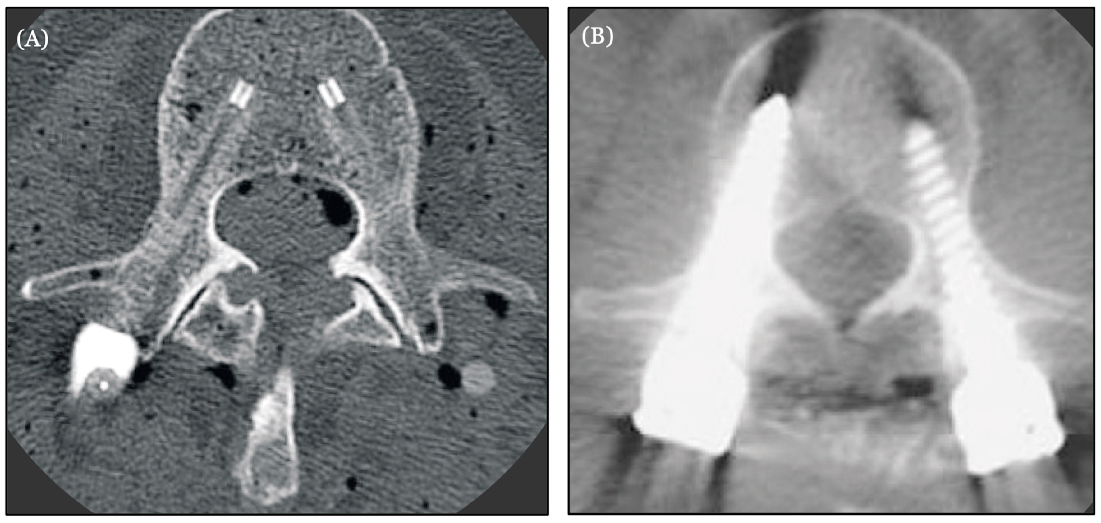
Additionally, carbon fiber implants contribute to improved postoperative monitoring for tumor recurrence. As can be seen in Figure 3, imaging studies free from artifacts provide clearer visualization and interpretation of postoperative changes, aiding clinicians in early detection and management of recurrent disease [37,38,41].
In the last few years, carbon fiber-reinforced polyetheretherketone (CFR-PEEK) has now been adopted in spine surgery. CFR-PEEK composite implants harness the properties of carbon fiber and PEEK to develop a system with biomechanical properties comparable to cortical bone [42,43]. CFR-PEEK represents a viable option for semirigid stabilization in spinal surgery, offering a range of advantages over traditional titanium systems [42]. Studies have demonstrated that CFR-PEEK implants possess bending stiffness, yield strength, and ultimate loads that are on par with or even exceed those of rigid titanium constructs [29]. While CFR-PEEK may exhibit slightly lower torsional stiffness and yield torque compared to titanium, it remains robust enough to effectively withstand the physiological loads encountered in the spine [42].
Moreover, the utilization of CFR-PEEK rods in spinal stabilization procedures brings about notable enhancements in primary stability, kinematics, and load-sharing dynamics when compared to conventional titanium implants [42,44]. This improvement is particularly significant in scenarios where maintaining spinal motion and flexibility is desired while ensuring adequate support and alignment.
In terms of pedicle screw fixation, CFR-PEEK systems have shown remarkable pullout strength, surpassing that of traditional implants [42]. Furthermore, they demonstrate equivalent or superior resistance to screw loosening when compared to titanium alternatives [42,45,46]. This superiority is largely attributed to the absence of a mismatch between the mechanical properties of CFR-PEEK and the surrounding bone tissue, resulting in reduced stress concentrations at the bone–implant interface and thus mitigating the risk of implant failure.
Beyond mechanical performance, CFR-PEEK possesses favorable biocompatibility, enhanced osteogenic properties, and bioactivity [47,48]. Modifying CFR-PEEK implants with amino groups can result in significantly improved osseointegration and bone healing [47,49]. These qualities contribute to ensuring a seamless and stable interface between the implant and the surrounding bone [6] which often results in long-term stability and functionality following spinal surgery. With its combination of mechanical robustness, enhanced stability, and biological compatibility, CFR-PEEK stands as a promising material for advancing spinal instrumentation technology and improving patient outcomes in spinal surgery. Table 2 highlights some of the studies that evaluated specific outcomes of carbon fiber and CFR-PEEK in the management of spinal conditions. Overall, CFR-PEEK has been shown to possess a relatively higher Young’s modulus than PEEK alone [7] and better strength and fatigue resistance, in addition to radiolucency and artifact-free imaging properties [7,29,50,51].

6. New Techniques and Potential Horizons in Spine Oncology

Advances in spine oncology have led to the development of new techniques aimed at improving the treatment of spinal tumors, preserving spinal function, and enhancing patient outcomes. Minimally invasive surgery (MIS) techniques in spine surgery utilize smaller incisions, minimize muscle dissection, and employ specialized instruments for treating various spinal conditions. Robot-assisted minimally invasive spine surgery techniques have also been implemented in percutaneous pedicle screw fixation, kyphoplasty, and vertebroplasty [17,19,58]. They offer several advantages, including expedited recovery, diminished postoperative pain, reduced muscle trauma, lower infection risk, and shorter hospital stays.
In contrast to traditional open surgery, the primary goal of these techniques is to limit tissue damage, decrease blood loss, and facilitate a speedier recovery process [20,59]. Spinal decompression procedures are often combined with radiotherapy in a holistic approach to the treatment of spinal tumors [23]. These procedures entail removing a section of vertebrae elements or tumors, particularly in cases of radioresistant tumors and spinal cord or nerve root impingement. This is then followed by radiotherapy [23,35].
Significant advances have been made in spinal stabilization after resection of both primary and metastatic tumors [37]. There seems to be an increasing emphasis on dynamic stabilization as an important element in the recovery of patients after spinal surgery [60]. Dynamic stabilization systems allow controlled motion at the stabilized segment of the spine. These systems aim to preserve some nonpathological spinal mobility while providing stability, reducing the risk of adjacent segment degeneration and promoting better long-term outcomes [60]. However, it remains to be seen if there could be a role for dynamic stabilization techniques in spinal tumor reconstructive surgery.
Preserving spinal alignment and function is often an important consideration in spine surgery. There are questions of whether spinal fusion is necessary in the management of spinal tumors, especially metastatic tumors [61]. This issue becomes more pressing when considering the survival time of patients after fusion surgery and how long it takes to achieve fusion. That said, advancements in the development of materials for spinal fusion could lead to faster fusion times. Consequently, patients with expected improved survival time might benefit from fusion surgery.
For patients who might be candidates for spinal fusion, balanced fusion techniques involve meticulous consideration of the number of fused segments and their alignment [62,63]. The use of biological agents, including bone morphogenetic proteins (BMPs) enhances spinal fusion outcomes by promoting bone formation, restoring vertebral height, reducing deformity, and stabilizing the spine [64]. In the context of spinal tumor treatment, where maintaining spinal stability and repairing bone defects is crucial, there might be an expectation for bone morphogenetic proteins (BMPs) to play a beneficial role in improving surgical outcomes. However, their potential to cause cancer has been under investigation for some time, and therefore, they are not currently used in spine oncology [65,66,67,68]. Using traditional autologous bone grafts might be considered another option for spinal reconstructive surgery after tumor resection, but concerns about tumor cell seeding make it less favorable [69]. Structural grafts, such as vascularized strut grafts from neighboring ribs or non-vascularized grafts with donor bone fragments placed in synthetic cages, have been suggested as potential solutions to some of these challenges. Nonetheless, further research is necessary to assess their effectiveness fully [70].
Stem cell therapy represents a promising frontier in the management of primary spinal tumors, offering potential advantages in tissue regeneration, immunomodulation, and targeted drug delivery [71]. While its application in spine oncology remains limited and necessitates further investigation, there are documented instances of its use in immunotherapy for spinal conditions [72]. Additionally, progress has been made in identifying the stem cell lineage implicated in spinal metastasis [73,74]. As research continues to advance and clinical experience grows, leveraging stem cell-based techniques alongside bone grafting holds promise for substantially enhancing outcomes in patients with spinal tumors. These reflect the evolving landscape of spine surgery, where the emphasis is adequate tumor control and achieving optimal functional outcomes.

7. Integration of Carbon Fiber Technology with Advanced Techniques

As highlighted so far, carbon fiber and carbon fiber composites, thanks to their remarkable strength-to-weight ratio, enable the creation of robust and enduring spinal implants capable of withstanding the biomechanical demands on the spine [29]. Spinal cages and stabilization systems have been used in various spinal procedures, including fusion surgeries and motion-preserving interventions [50]. Indeed, the role of carbon fiber-reinforced PEEK implants in achieving fusion in managing degenerative spinal diseases has been demonstrated in biomedical and clinical studies [42,75]. Similarly, they have shown promise in biomechanical studies for interbody fusion and vertebral reconstruction in cancer patients, especially compared to bone allografts and titanium devices [76,77,78]. The distinctive properties of carbon fiber contribute to an environment conducive to bone fusion, minimize micromotions, and enhance load-sharing, supporting the long-term stability of the spinal construct without significant subsidence [42,79,80,81]. These qualities position carbon fiber-based spinal cages and stabilization systems as a promising and innovative option in contemporary spine surgery [50,82].
Minimally invasive surgery (MIS) approaches increasingly employ carbon fiber instrumentation. By integrating with computer-assisted MIS systems, carbon fiber instruments facilitate preoperative planning and intraoperative navigation using various imaging modalities [38]. Surgeons can leverage this integration for precise tumor interventions, resulting in reduced invasiveness and improved patient outcomes. This often translates to smaller incisions, minimizing disruption to surrounding tissues and reducing blood loss compared to traditional open surgeries, thereby enhancing the accuracy and efficacy of spinal oncology procedures [37].
Moreover, the integration of carbon fiber technology with customizable and modular systems in spine oncology represents another significant advancement in surgical instrumentation [21,83]. Achieving the goal of preserving spinal alignment and function often requires a customized approach, considering the specific pathology, patient characteristics, and treatment goals. Modular spinal stabilization systems provide flexibility and versatility in addressing complex cases. The adaptability of carbon fiber systems to the specific characteristics of tumors ensures a personalized solution aligned with individual patient pathology [21]. Modular spinal stabilization systems offer flexibility and adaptability in addressing complex cases. The adjustability of carbon fiber systems allows for precise modifications, ensuring an optimal anatomical fit that promotes stability while minimizing disturbances to surrounding tissues.
Furthermore, carbon fiber-based modular polyaxial pedicle screws offer options for adaptive connection and configuration and provide great intraoperative visualization [8,84]. Surgeons can customize the implant constructs based on the specific needs of the patient, allowing for better adaptation to the individual’s anatomy and pathology [85]. These ongoing advancements in technology, surgical techniques, and an enhanced understanding of spinal biomechanics drive continuous innovations aimed at optimizing patient outcomes and sustaining spinal health.

8. Patient-Centered Outcomes and Carbon Fiber Implants

Carbon fiber implants contribute to several factors that positively influence the overall quality of life for individuals undergoing spine surgery with these advanced implants [42]. As previously emphasized, carbon fiber implants, especially in minimally invasive procedures, lead to smaller incisions. They have also led to improved postoperative radiation planning and enhanced the ability to detect tumor recurrence due to their radiolucency. The adaptability and modular design of carbon fiber systems allow for a customized fit, minimizing disruption to surrounding tissues during surgery. This often results in decreased postoperative pain and discomfort, thereby enhancing the immediate post-surgery experience for patients and lowering the risk of complications [51,52,86].
It is essential to acknowledge that individual experiences may vary, and the overall quality of life post-operation is influenced by various factors, including the patient’s overall health, as well as the specific pathology being addressed. Nevertheless, the unique properties of carbon fiber implants offer promising advantages that contribute to positive postoperative functional outcomes for many individuals [38]. The positive impact of improved spinal function can significantly affect the psychological well-being of patients [87,88]. Reduced pain, enhanced mobility, and a quicker return to normal activities contribute to an overall sense of well-being and satisfaction.
While carbon fiber implants in spine surgery offer numerous advantages, potential complications and challenges accompany their use. While carbon fiber itself is resistant to corrosion, the potential for infection exists, especially during the surgical placement of implants. Indeed, it has been reported that CFR-PEEK screws are likely to loosen in infectious cases, probably due to a stronger adhesion to bacteria than traditional metal implants [53,56]. As such, strict adherence to aseptic techniques is essential to minimize the risk of infections.
Beyond postoperative complications, technical challenges arise with carbon fiber in spine oncology. Carbon fiber’s radiolucency can make visualizing the implant on X-rays, CT scans, and MRI challenging, affecting the ability to monitor its position or fusion progress or detect potential issues [7,55]. Thus, postoperative monitoring with imaging, such as CT scans or MRIs, may be challenging in some patients with carbon fiber implants. Cost considerations may also impact the widespread adoption of carbon fiber technology, as it may be more expensive than traditional materials, potentially limiting accessibility for some patients [36,89].
While carbon fiber implants show promise in providing positive functional outcomes and durability, the long-term durability of carbon fiber implants remains a subject of ongoing research. This necessitates continuous monitoring and studies to assess their performance over extended periods. Furthermore, there is the question of the application of carbon fiber for longer fusion. The rod contour in carbon fiber composite implants can prove challenging to shape to match the screws on the operative field [7,89]. As such, longer follow-up studies are needed [36], as well as large prospective cohort studies to further evaluate oncological outcomes in patients treated with carbon fiber-reinforced implants [57,90].

9. The Future of Spine Oncology Instrumentation and Implants

Spine oncology has come a long way, with significant progress recorded over the past decades. That said, there are still unexplored frontiers and unexploited potentials that could further lead to improved diagnosis, treatment, and overall patient outcomes. This is particularly evident with the recent advancements in information technology and artificial intelligence (AI). The application of data analytics and AI in spine oncology is still in its early stages. AI will be an invaluable tool in the development of better data-driven predictive algorithms and clinical decision-making tools in spine oncology [91]. AI tools, particularly deep neural networks, offer the potential to predict surgical outcomes and postoperative complications [15]. These sophisticated algorithms can analyze vast amounts of data, including patient demographics, medical history, imaging results, laboratory values, and surgical details, to generate predictive models with high accuracy and precision. Recent research has focused on developing models capable of accurately predicting spine infections by analyzing various factors [15,92,93]. By harnessing the power of machine learning algorithms, these predictive models can identify subtle patterns affording clinicians valuable insights into individual patient risk profiles, allowing for personalized risk stratification and tailored interventions [15,92].
Furthermore, the utilization of customizable and modular systems enables the customization of surgical procedures based on specific patient characteristics. There is a growing interest in expanding this concept by integrating the genetic profiles of individual patients into the treatment of spinal tumors. Consequently, a genomics-driven approach to spine oncology shows great potential for the future [94].
Another largely uncharted horizon is in the implementation of Enhanced Recovery After Surgery (ERAS). ERAS protocols aim to expedite recovery and reduce surgical stress through the implementation of optimized multimodal pre- and postoperative care methods and pathways to reduce surgical stress [95,96,97]. Although not yet relatively well-defined in neurosurgical settings [97,98,99], ERAS has been widely recognized and implemented globally in many surgical fields [100,101,102]. Carbon fiber instrumentation can play an invaluable role in further developing and implementing Enhanced Recovery After Surgery (ERAS) protocols in spine oncology patients after surgery. As earlier discussed, its lightweight, strength, and radiolucent properties often result in significantly reduced surgical trauma, enhanced stability, and compatibility with advanced imaging techniques. These characteristics align with ERAS goals. As such, carbon fiber-based implants could be incorporated in the ERAS framework and recommendations for spine oncology and spine surgery in general, thereby expediting postoperative mobilization and adjuvant treatment, facilitating quicker recovery, shorter hospital stays, and improved patient outcomes.
All these point to the importance of a multidisciplinary approach in advancing the field of spine oncology [103,104]. The complexity of spinal tumors calls for the collaboration of diverse professionals, including healthcare providers, scientists, engineers, and industry leaders to drive advancements in research and technology. This will most likely foster innovation in treatment modalities, surgical techniques, and diagnostic tools to deliver comprehensive, specialized care and improve overall outcomes in spinal oncology.

10. Conclusions

Significant progress has been recorded in spine oncology, particularly in instrumentation and materials. Computer-aided surgical systems, integrated with robotic platforms, enhance precision and decision-making in surgeries. Imaging technologies, including 3D imaging, CT scans, and MRI, improve intraoperative navigation, reducing reliance on fluoroscopy and minimizing radiation exposure. Carbon fiber and carbon fiber composite instrumentation have emerged as a tool that further advances the areas of progress outlined above, offering strength, stability, and biocompatibility. Carbon fiber’s radiolucency aids in imaging, facilitating postoperative monitoring and radiation therapy planning.
This has led to improvements in patient-centered outcomes including the use of smaller incisions, decreased postoperative pain, and enhanced mobility, contributing to an improved quality of life. However, there is still need for ongoing research in spine oncology, with a focus on information technology, artificial intelligence (AI), and genomics. A multidisciplinary approach involving healthcare providers, scientists, engineers, and industry leaders is crucial for advancing research, technology, and treatment modalities in spinal oncology.

Author Contributions

Conceptualization, A.G.B., J.-L.K.K. and I.A.; methodology, I.A., A.G.B. and J.-L.K.K.; Software, I.A.; validation, I.A., A.G.B., J.-L.K.K. and I.E.O.; formal analysis, I.A.; investigation, I.A.; resources, I.A., A.G.B., J.-L.K.K. and I.E.O.; data curation, I.A.; writing original draft preparation, I.A. and J.-L.K.K., writing-review and editing, I.A., A.G.B., J.-L.K.K. and I.E.O.; visualization, I.A.; supervision, I.E.O.; project administration, J.-L.K.K. and I.E.O. All authors have read and agreed to the published version of the manuscript.

Funding

This research received no external funding.

Conflicts of Interest

The authors declare no conflict of interest.

References

  1. Neal, M.T.; Richards, A.E.; Curley, K.L.; Patel, N.P.; Ashman, J.B.; Vora, S.A.; Kalani, M.A. Carbon fiber-reinforced PEEK instrumentation in the spinal oncology population: A retrospective series demonstrating technique, feasibility, and clinical outcomes. Neurosurg. Focus 2021, 50, E13. [Google Scholar] [CrossRef] [PubMed]
  2. Charest-Morin, R.; Fisher, C.G.; Sahgal, A.; Boriani, S.; Gokaslan, Z.L.; Lazary, A.; Reynolds, J.; Bettegowda, C.; Rhines, L.D.; Dea, N. Primary Bone Tumor of the Spine-An Evolving Field: What a General Spine Surgeon Should Know. Glob. Spine J. 2019, 9 (Suppl. S1), 108S–116S. [Google Scholar] [CrossRef] [PubMed] [PubMed Central]
  3. Missenard, G.; Bouthors, C.; Fadel, E.; Court, C. Surgical strategies for primary malignant tumors of the thoracic and lumbar spine. Orthop. Traumatol. Surg. Res. 2020, 106, S53–S62. [Google Scholar] [CrossRef] [PubMed]
  4. Harel, R.; Doron, O.; Knoller, N. Minimally Invasive Spine Metastatic Tumor Resection and Stabilization: New Technology Yield Improved Outcome. Biomed. Res. Int. 2015, 2015, 948373. [Google Scholar] [CrossRef] [PubMed] [PubMed Central]
  5. Shiber, M.; Kimchi, G.; Knoller, N.; Harel, R. The Evolution of Minimally Invasive Spine Tumor Resection and Stabilization: From K-Wires to Navigated One-Step Screws. J. Clin. Med. 2023, 12, 536. [Google Scholar] [CrossRef] [PubMed] [PubMed Central]
  6. Petersen, R. Carbon Fiber Biocompatibility for Implants. Fibers 2016, 4, 1. [Google Scholar] [CrossRef] [PubMed] [PubMed Central]
  7. Tedesco, G.; Gasbarrini, A.; Bandiera, S.; Ghermandi, R.; Boriani, S. Composite PEEK/Carbon fiber implants can increase the effectiveness of radiotherapy in the management of spine tumors. J. Spine Surg. 2017, 3, 323–329, Erratum in J. Spine Surg. 2018, 4, 167. [Google Scholar] [CrossRef] [PubMed] [PubMed Central]
  8. Warburton, A.; Girdler, S.J.; Mikhail, C.M.; Ahn, A.; Cho, S.K. Biomaterials in Spinal Implants: A Review. Neurospine 2020, 17, 101–110. [Google Scholar] [CrossRef] [PubMed] [PubMed Central]
  9. Virk, S.; Qureshi, S.; Sandhu, H. History of Spinal Fusion: Where We Came from and Where We Are Going. HSS J. 2020, 16, 137–142. [Google Scholar] [CrossRef] [PubMed] [PubMed Central]
  10. Cho, S.K.; Kim, Y.J. History of spinal deformity surgery part I: The Pre-modern Era. Korean J. Spine 2011, 8, 1–8. [Google Scholar] [CrossRef]
  11. Miller, D.J.; Vitale, M.G. Dr. Russell A. Hibbs: Pioneer of Spinal Fusion. Spine 2015, 40, 1311–1313. [Google Scholar] [CrossRef] [PubMed]
  12. Desai, S.K.; Brayton, A.; Chua, V.B.; Luerssen, T.G.; Jea, A. The lasting legacy of Paul Randall Harrington to pediatric spine surgery: Historical vignette. J. Neurosurg. Spine 2013, 18, 170–177. [Google Scholar] [CrossRef] [PubMed]
  13. Roy-Camille, R.; Roy-Camille, M.; Demeulenaere, C. Ostéosynthèse du rachis dorsal, lombaire et lombo-sacré par plaques métalliques vissées dans les pédicules vertébraux et les apophyses articulaires [Osteosynthesis of dorsal, lumbar, and lumbosacral spine with metallic plates screwed into vertebral pedicles and articular apophyses]. Presse Med. 1970, 78, 1447–1448. (In French) [Google Scholar] [PubMed]
  14. Shufflebarger, H.L.; Clark, C.E. Cotrel-Dubousset instrumentation. Orthopedics 1988, 11, 1435–1440. [Google Scholar] [CrossRef] [PubMed]
  15. Costăchescu, B.; Niculescu, A.G.; Iliescu, B.F.; Dabija, M.G.; Grumezescu, A.M.; Rotariu, D. Current and Emerging Approaches for Spine Tumor Treatment. Int. J. Mol. Sci. 2022, 23, 15680. [Google Scholar] [CrossRef] [PubMed] [PubMed Central]
  16. Kumar, N.; Patel, R.; Wadhwa, A.C.; Kumar, A.; Milavec, H.M.; Sonawane, D.; Singh, G.; Benneker, L.M. Basic concepts in metal work failure after metastatic spine tumour surgery. Eur. Spine J. 2018, 27, 806–814. [Google Scholar] [CrossRef] [PubMed]
  17. Pérez de la Torre, R.A.; Ramanathan, S.; Williams, A.L.; Perez-Cruet, M.J. Minimally-Invasive Assisted Robotic Spine Surgery (MARSS). Front. Surg. 2022, 9, 884247. [Google Scholar] [CrossRef] [PubMed] [PubMed Central]
  18. Móga, K.; Ferencz, A.; Haidegger, T. What Is Next in Computer-Assisted Spine Surgery? Advances in Image-Guided Robotics and Extended Reality. Robotics 2022, 12, 1. [Google Scholar] [CrossRef]
  19. Sayari, A.J.; Pardo, C.; Basques, B.A.; Colman, M.W. Review of robotic-assisted surgery: What the future looks like through a spine oncology lens. Ann. Transl. Med. 2019, 7, 224. [Google Scholar] [CrossRef] [PubMed] [PubMed Central]
  20. Porras, J.L.; Pennington, Z.; Hung, B.; Hersh, A.; Schilling, A.; Goodwin, C.R.; Sciubba, D.M. Radiotherapy and Surgical Advances in the Treatment of Metastatic Spine Tumors: A Narrative Review. World Neurosurg. 2021, 151, 147–154. [Google Scholar] [CrossRef] [PubMed]
  21. Soriano-Baron, H.; Martinez-del-Campo, E.; Crawford, N.R.; Theodore, N. Robotics in spinal surgery: The future is here. Concern 2016, 13, 49. [Google Scholar]
  22. Massaad, E.; Shankar, G.M.; Shin, J.H. Novel Applications of Spinal Navigation in Deformity and Oncology Surgery-Beyond Screw Placement. Oper. Neurosurg. 2021, 21 (Suppl. 1), S23–S38. [Google Scholar] [CrossRef] [PubMed]
  23. Barzilai, O.; Bilsky, M.H.; Laufer, I. The Role of Minimal Access Surgery in the Treatment of Spinal Metastatic Tumors. Glob. Spine J. 2020, 10 (Suppl. S2), 79S–87S. [Google Scholar] [CrossRef] [PubMed] [PubMed Central]
  24. Dick, J.C.; Bourgeault, C.A. Notch sensitivity of titanium alloy, commercially pure titanium, and stainless steel spinal implants. Spine 2001, 26, 1668–1672. [Google Scholar] [CrossRef] [PubMed]
  25. Litak, J.; Szymoniuk, M.; Czyżewski, W.; Hoffman, Z.; Litak, J.; Sakwa, L.; Kamieniak, P. Metallic Implants Used in Lumbar Interbody Fusion. Materials 2022, 15, 3650. [Google Scholar] [CrossRef] [PubMed] [PubMed Central]
  26. Mao, J.Z.; Fritz, A.G.; Lucas, J.P.; Khan, A.; Popoola, D.O.; Becker, A.B.; Adetunji, A.; Levy, B.R.; Agyei, J.O.; O’Connor, T.E.; et al. Assessment of Rod Material Types in Spine Surgery Outcomes: A Systematic Review. World Neurosurg. 2021, 146, e6–e13. [Google Scholar] [CrossRef] [PubMed]
  27. Raso, J.; Chi, J.; Labaran, L.A.; Frank, C.; Shen, F.H. Carbon fiber lumbopelvic reconstruction following sacral giant cell tumor resection: Illustrative case. J. Neurosurg. Case Lessons 2023, 5, CASE22555. [Google Scholar] [CrossRef] [PubMed] [PubMed Central]
  28. Han, S.; Hyun, S.J.; Kim, K.J.; Jahng, T.A.; Kim, H.J. Comparative Study Between Cobalt Chrome and Titanium Alloy Rods for Multilevel Spinal Fusion: Proximal Junctional Kyphosis More Frequently Occurred in Patients Having Cobalt Chrome Rods. World Neurosurg. 2017, 103, 404–409. [Google Scholar] [CrossRef] [PubMed]
  29. Uri, O.; Folman, Y.; Laufer, G.; Behrbalk, E. A Novel Spine Fixation System Made Entirely of Carbon-Fiber-Reinforced PEEK Composite: An In Vitro Mechanical Evaluation. Adv. Orthop. 2020, 2020, 4796136. [Google Scholar] [CrossRef] [PubMed] [PubMed Central]
  30. Zheng, Z.; Liu, P.; Zhang, X.; Xin, J.; Wang, Y.; Zou, X.; Mei, X.; Zhang, S.; Zhang, S. Strategies to improve bioactive and antibacterial properties of polyetheretherketone (PEEK) for use as orthopedic implants. Mater. Today Bio 2022, 16, 100402. [Google Scholar] [CrossRef] [PubMed] [PubMed Central]
  31. Yeung, C.M.; Bhashyam, A.R.; Patel, S.S.; Ortiz-Cruz, E.; Lozano-Calderón, S.A. Carbon Fiber Implants in Orthopaedic Oncology. J. Clin. Med. 2022, 11, 4959. [Google Scholar] [CrossRef] [PubMed] [PubMed Central]
  32. Ha, S.W.; Kirch, M.; Birchler, F.; Eckert, K.L.; Mayer, J.; Wintermantel, E.; Sittig, C.; Pfund-Klingenfuss, I.; Textor, M.; Spencer, N.D.; et al. Surface activation of polyetheretherketone (PEEK) and formation of calcium phosphate coatings by precipitation. J. Mater. Sci. Mater. Med. 1997, 8, 683–690. [Google Scholar] [CrossRef] [PubMed]
  33. Katzer, A.; Marquardt, H.; Westendorf, J.; Wening, J.V.; von Foerster, G. Polyetheretherketone--cytotoxicity and mutagenicity in vitro. Biomaterials 2002, 23, 1749–1759. [Google Scholar] [CrossRef] [PubMed]
  34. Jockisch, K.A.; Brown, S.A.; Bauer, T.W.; Merritt, K. Biological response to chopped-carbon-fiber-reinforced peek. J. Biomed. Mater. Res. 1992, 26, 133–146. [Google Scholar] [CrossRef] [PubMed]
  35. Krätzig, T.; Mende, K.C.; Mohme, M.; Kniep, H.; Dreimann, M.; Stangenberg, M.; Westphal, M.; Gauer, T.; Eicker, S.O. Carbon fiber-reinforced PEEK versus titanium implants: An in vitro comparison of susceptibility artifacts in CT and MR imaging. Neurosurg. Rev. 2021, 44, 2163–2170. [Google Scholar] [CrossRef] [PubMed] [PubMed Central]
  36. Murthy, N.K.; Wolinsky, J.P. Utility of carbon fiber instrumentation in spinal oncology. Heliyon 2021, 7, e07766. [Google Scholar] [CrossRef] [PubMed] [PubMed Central]
  37. Schwendner, M.; Ille, S.; Kirschke, J.S.; Bernhardt, D.; Combs, S.E.; Meyer, B.; Krieg, S.M. Clinical evaluation of vertebral body replacement of carbon fiber-reinforced polyetheretherketone in patients with tumor manifestation of the thoracic and lumbar spine. Acta Neurochir. 2023, 165, 897–904. [Google Scholar] [CrossRef] [PubMed] [PubMed Central]
  38. Müther, M.; Lüthge, S.; Gerwing, M.; Stummer, W.; Schwake, M. Management of Spinal Dumbbell Tumors via a Minimally Invasive Posterolateral Approach and Carbon Fiber-Reinforced Polyether Ether Ketone Instrumentation: Technical Note and Surgical Case Series. World Neurosurg. 2021, 151, 277–283.e1. [Google Scholar] [CrossRef] [PubMed]
  39. Lin, H.; Ding, X.; Yin, L.; Zhai, H.; Liu, H.; Kassaee, A.; Hill-Kayser, C.; Lustig, R.A.; McDonough, J.; Both, S. The effects of titanium mesh on passive-scattering proton dose. Phys. Med. Biol. 2014, 59, N81–N89. [Google Scholar] [CrossRef]
  40. Verburg, J.M.; Seco, J. Dosimetric accuracy of proton therapy for chordoma patients with titanium implants. Med. Phys. 2013, 40, 071727. [Google Scholar] [CrossRef] [PubMed] [PubMed Central]
  41. Galloway, R.; Gikas, N.; Golomohammad, R.; Sherriff, J.; Czyz, M.S.r. Separation Surgery, Fixation With Carbon-Fiber Implants, and Stereotactic Body Radiotherapy for Oligometastatic Spinal Disease. Cureus 2022, 14, e31370. [Google Scholar] [CrossRef] [PubMed] [PubMed Central]
  42. Ghermandi, R.; Tosini, G.; Lorenzi, A.; Griffoni, C.; La Barbera, L.; Girolami, M.; Pipola, V.; Barbanti Brodano, G.; Bandiera, S.; Terzi, S.; et al. Carbon Fiber-Reinforced PolyEtherEtherKetone (CFR-PEEK) Instrumentation in Degenerative Disease of Lumbar Spine: A Pilot Study. Bioengineering 2023, 10, 872. [Google Scholar] [CrossRef] [PubMed] [PubMed Central]
  43. Avanzini, A.; Battini, D.; Petrogalli, C.; Pandini, S.; Donzella, G. Anisotropic behaviour of extruded short carbon fibre reinforced peek under static and fatigue loading. Appl. Compos. Mater. 2022, 29, 1041–1060. [Google Scholar] [CrossRef]
  44. Bruner, H.J.; Guan, Y.; Yoganandan, N.; Pintar, F.A.; Maiman, D.J.; Slivka, M.A. Biomechanics of polyaryletherketone rod composites and titanium rods for posterior lumbosacral instrumentation. Presented at the 2010 Joint Spine Section Meeting. Laboratory investigation. J. Neurosurg. Spine 2010, 13, 766–772. [Google Scholar] [CrossRef] [PubMed]
  45. Oikonomidis, S.; Greven, J.; Bredow, J.; Eh, M.; Prescher, A.; Fischer, H.; Thüring, J.; Eysel, P.; Hildebrand, F.; Kobbe, P.; et al. Biomechanical effects of posterior pedicle screw-based instrumentation using titanium versus carbon fiber reinforced PEEK in an osteoporotic spine human cadaver model. Clin. Biomech. 2020, 80, 105153. [Google Scholar] [CrossRef] [PubMed]
  46. Lindtner, R.A.; Schmid, R.; Nydegger, T.; Konschake, M.; Schmoelz, W. Pedicle screw anchorage of carbon fiber-reinforced PEEK screws under cyclic loading. Eur. Spine J. 2018, 27, 1775–1784. [Google Scholar] [CrossRef] [PubMed]
  47. Yu, W.; Zhang, H.A.L.; Yang, S.; Zhang, J.; Wang, H.; Zhou, Z.; Zhou, Y.; Zhao, J.; Jiang, Z. Enhanced bioactivity and osteogenic property of carbon fiber reinforced polyetheretherketone composites modified with amino groups. Colloids Surf. B Biointerfaces 2020, 193, 111098. [Google Scholar] [CrossRef] [PubMed]
  48. Han, X.; Yang, D.; Yang, C.; Spintzyk, S.; Scheideler, L.; Li, P.; Li, D.; Geis-Gerstorfer, J.; Rupp, F. Carbon Fiber Reinforced PEEK Composites Based on 3D-Printing Technology for Orthopedic and Dental Applications. J. Clin. Med. 2019, 8, 240. [Google Scholar] [CrossRef] [PubMed] [PubMed Central]
  49. Wang, J.; Yu, W.; Shi, R.; Yang, S.; Zhang, J.; Han, X.; Zhou, Z.; Gao, W.; Li, Y.; Zhao, J. Osseointegration behavior of carbon fiber reinforced polyetheretherketone composites modified with amino groups: An in vivo study. J. Biomed. Mater. Res. B Appl. Biomater. 2023, 111, 505–512. [Google Scholar] [CrossRef] [PubMed]
  50. Li, C.S.; Vannabouathong, C.; Sprague, S.; Bhandari, M. The Use of Carbon-Fiber-Reinforced (CFR) PEEK Material in Orthopedic Implants: A Systematic Review. Clin. Med. Insights Arthritis Musculoskelet. Disord. 2015, 8, 33–45. [Google Scholar] [CrossRef] [PubMed] [PubMed Central]
  51. Boriani, S.; Tedesco, G.; Ming, L.; Ghermandi, R.; Amichetti, M.; Fossati, P.; Krengli, M.; Mavilla, L.; Gasbarrini, A. Carbon-fiber-reinforced PEEK fixation system in the treatment of spine tumors: A preliminary report. Eur. Spine J. 2018, 27, 874–881. [Google Scholar] [CrossRef] [PubMed]
  52. Alvarez-Breckenridge, C.; de Almeida, R.; Haider, A.; Muir, M.; Bird, J.; North, R.; Rhines, L.; Tatsui, C. Carbon Fiber-Reinforced Polyetheretherketone Spinal Implants for Treatment of Spinal Tumors: Perceived Advantages and Limitations. Neurospine 2023, 20, 317–326. [Google Scholar] [CrossRef] [PubMed] [PubMed Central]
  53. Carbon-Fiber International Collaboration Initiative Research Group. Complications of patients with bone tumors treated with carbon-fiber plates: An international multicenter study. Sci. Rep. 2022, 12, 18969. [Google Scholar] [CrossRef] [PubMed] [PubMed Central]
  54. Henzen, D.; Schmidhalter, D.; Guyerm, G.; Stenger-Weisser, A.; Ermiş, E.; Poel, R.; Deml, M.C.; Fix, M.K.; Manser, P.; Aebersold, D.M.; et al. Feasibility of postoperative spine stereotactic body radiation therapy in proximity of carbon and titanium hybrid implants using a robotic radiotherapy device. Radiat. Oncol. 2022, 17, 94. [Google Scholar] [CrossRef] [PubMed] [PubMed Central]
  55. Hubertus, V.; Wessels, L.; Früh, A.; Tkatschenko, D.; Nulis, I.; Bohner, G.; Prinz, V.; Onken, J.; Czabanka, M.; Vajkoczy, P.; et al. Navigation accuracy and assessability of carbon fiber-reinforced PEEK instrumentation with multimodal intraoperative imaging in spinal oncology. Sci. Rep. 2022, 12, 15816. [Google Scholar] [CrossRef] [PubMed] [PubMed Central]
  56. Joerger, A.K.; Shiban, E.; Krieg, S.M.; Meyer, B. Carbon-fiber reinforced PEEK instrumentation for spondylodiscitis: A single center experience on safety and efficacy. Sci. Rep. 2021, 11, 2414. [Google Scholar] [CrossRef] [PubMed] [PubMed Central]
  57. Oh, J.; Visco, Z.R.; Ojukwu, D.I.; Galgano, M.A. Applications of Carbon Fiber Instrumentation in Spinal Oncology: Recent Innovations in Spinal Instrumentation and 2-Dimensional Illustrative Operative Video. Oper. Neurosurg. 2023, 24, 182–193. [Google Scholar] [CrossRef] [PubMed]
  58. Hu, X.; Scharschmidt, T.J.; Ohnmeiss, D.D.; Lieberman, I.H. Robotic assisted surgeries for the treatment of spine tumors. Int. J. Spine Surg. 2015, 9, 1. [Google Scholar] [CrossRef] [PubMed] [PubMed Central]
  59. Raysi Dehcordi, S.; Marzi, S.; Ricci, A.; Di Cola, F.; Galzio, R.J. Less invasive approaches for the treatment of cervical schwannomas: Our experience. Eur. Spine J. 2012, 21, 887–896. [Google Scholar] [CrossRef] [PubMed] [PubMed Central]
  60. Meyer, B.; Thomé, C.; Vajkoczy, P.; Kehl, V.; Dodel, R.; Ringel, F.; DYNORFUSE Study Group. Lumbar dynamic pedicle-based stabilization versus fusion in degenerative disease: A multicenter, double-blind, prospective, randomized controlled trial. J. Neurosurg. Spine 2022, 37, 515–524. [Google Scholar] [CrossRef] [PubMed]
  61. Park, S.J.; Lee, K.H.; Lee, C.S.; Jung, J.Y.; Park, J.H.; Kim, G.L.; Kim, K.T. Instrumented surgical treatment for metastatic spinal tumors: Is fusion necessary? J. Neurosurg. Spine 2019, 32, 456–464. [Google Scholar] [CrossRef] [PubMed]
  62. Blumenthal, C.; Curran, J.; Benzel, E.C.; Potter, R.; Magge, S.N.; Harrington JFJr Coumans, J.V.; Ghogawala, Z. Radiographic predictors of delayed instability following decompression without fusion for degenerative grade I lumbar spondylolisthesis. J. Neurosurg. Spine 2013, 18, 340–346. [Google Scholar] [CrossRef] [PubMed]
  63. Thomas, K.; Faris, P.; McIntosh, G.; Manners, S.; Abraham, E.; Bailey, C.S.; Paquet, J.; Cadotte, D.; Jacobs, W.B.; Rampersaud, Y.R.; et al. Decompression alone vs. decompression plus fusion for claudication secondary to lumbar spinal stenosis. Spine J. 2019, 19, 1633–1639. [Google Scholar] [CrossRef] [PubMed]
  64. Zhang, Y.; Jiang, Y.; Zou, D.; Yuan, B.; Ke, H.Z.; Li, W. Therapeutics for enhancement of spinal fusion: A mini review. J. Orthop. Translat. 2021, 31, 73–79. [Google Scholar] [CrossRef] [PubMed] [PubMed Central]
  65. Elder, B.D.; Ishida, W.; Goodwin, C.R.; Bydon, A.; Gokaslan, Z.L.; Sciubba, D.M.; Wolinsky, J.P.; Witham, T.F. Bone graft options for spinal fusion following resection of spinal column tumors: Systematic review and meta-analysis. Neurosurg. Focus 2017, 42, E16. [Google Scholar] [CrossRef] [PubMed]
  66. Cooper, G.S.; Kou, T.D. Risk of cancer after lumbar fusion surgery with recombinant human bone morphogenic protein-2 (rh-BMP-2). Spine 2013, 38, 1862–1868. [Google Scholar] [CrossRef] [PubMed] [PubMed Central]
  67. Mazur-Hart, D.J.; Yamamoto, E.A.; Yoo, J.; Orina, J.N. Bone morphogenetic protein and cancer in spinal fusion: A propensity score-matched analysis. J. Neurosurg. Spine 2023, 39, 722–728. [Google Scholar] [CrossRef] [PubMed]
  68. Beschloss, A.M.; DiCindio, C.M.; Lombardi, J.S.; Shillingford, J.N.; Laratta, J.L.; Holderread, B.; Louie, P.; Pugely, A.J.; Sardar, Z.; Khalsa, A.S.; et al. The Rise and Fall of Bone Morphogenetic Protein 2 Throughout the United States. Clin. Spine Surg. 2022, 35, 264–269. [Google Scholar] [CrossRef] [PubMed]
  69. Feler, J.; Sun, F.; Bajaj, A.; Hagan, M.; Kanekar, S.; Sullivan, P.L.Z.; Fridley, J.S.; Gokaslan, Z.L. Complication Avoidance in Surgical Management of Vertebral Column Tumors. Curr. Oncol. 2022, 29, 1442–1454. [Google Scholar] [CrossRef] [PubMed] [PubMed Central]
  70. Glennie, R.A.; Rampersaud, Y.R.; Boriani, S.; Reynolds, J.J.; Williams, R.; Gokaslan, Z.L.; Schmidt, M.H.; Varga, P.P.; Fisher, C.G. A Systematic Review With Consensus Expert Opinion of Best Reconstructive Techniques After Osseous En Bloc Spinal Column Tumor Resection. Spine 2016, 41 (Suppl. 20), S205–S211. [Google Scholar] [CrossRef] [PubMed]
  71. Safari, M.; Khoshnevisan, A. An overview of the role of cancer stem cells in spine tumors with a special focus on chordoma. World J. Stem Cells 2014, 6, 53–64. [Google Scholar] [CrossRef] [PubMed] [PubMed Central]
  72. Pour-Rashidi, A.; Mohammadi, E.; Rezaei, N.; Hanaei, S. Stem Cells and Targeted Gene Therapy in Brain and Spinal Cord Tumors. Adv. Exp. Med. Biol. 2023, 1394, 137–152. [Google Scholar] [CrossRef] [PubMed]
  73. Grady, C.; Melnick, K.; Porche, K.; Dastmalchi, F.; Hoh, D.J.; Rahman, M.; Ghiaseddin, A. Glioma Immunotherapy: Advances and Challenges for Spinal Cord Gliomas. Neurospine 2022, 19, 13–29. [Google Scholar] [CrossRef] [PubMed] [PubMed Central]
  74. Sun, J.; Hu, L.; Bok, S.; Yallowitz, A.R.; Cung, M.; McCormick, J.; Zheng, L.J.; Debnath, S.; Niu, Y.; Tan, A.Y.; et al. A vertebral skeletal stem cell lineage driving metastasis. Nature 2023, 621, 602–609. [Google Scholar] [CrossRef] [PubMed] [PubMed Central]
  75. Kang, K.T.; Koh, Y.G.; Son, J.; Yeom, J.S.; Park, J.H.; Kim, H.J. Biomechanical evaluation of pedicle screw fixation system in spinal adjacent levels using polyetheretherketone, carbon-fiber-reinforced polyetheretherketone, and traditional titanium as rod materials. Compos. Part B Eng. 2017, 130, 248–256. [Google Scholar] [CrossRef]
  76. Brantigan, J.W.; Steffee, A.D.; Geiger, J.M. A carbon fiber implant to aid interbody lumbar fusion: Mechanical testing. Spine 1991, 16 (Suppl. S6), S277–S282. [Google Scholar] [CrossRef] [PubMed]
  77. Disch, A.C.; Luzzati, A.; Melcher, I.; Schaser, K.D.; Feraboli, F.; Schmoelz, W. Three-dimensional stiffness in a thoracolumbar en-bloc spondylectomy model: A biomechanical in vitro study. Clin. Biomech. 2007, 22, 957–964. [Google Scholar] [CrossRef] [PubMed]
  78. Disch, A.C.; Schaser, K.D.; Melcher, I.; Luzzati, A.; Feraboli, F.; Schmoelz, W. En bloc spondylectomy reconstructions in a biomechanical in-vitro study. Eur. Spine J. 2008, 17, 715–725. [Google Scholar] [CrossRef] [PubMed] [PubMed Central]
  79. Cawley, D.T.; Alzakri, A.; Fujishiro, T.; Kieser, D.C.; Tavalaro, C.; Boissiere, L.; Obeid, I.; Pointillart, V.; Vital, J.M.; Gille, O. Carbon-fibre cage reconstruction in anterior cervical corpectomy for multilevel cervical spondylosis: Mid-term outcomes. J. Spine Surg. 2019, 5, 251–258. [Google Scholar] [CrossRef] [PubMed] [PubMed Central]
  80. Chen, Y.; Chen, D.; Guo, Y.; Wang, X.; Lu, X.; He, Z.; Yuan, W. Subsidence of titanium mesh cage: A study based on 300 cases. J. Spinal Disord. Tech. 2008, 21, 489–492. [Google Scholar] [CrossRef] [PubMed]
  81. Zhang, Y.; Quan, Z.; Zhao, Z.; Luo, X.; Tang, K.; Li, J.; Zhou, X.; Jiang, D. Evaluation of anterior cervical reconstruction with titanium mesh cages versus nano-hydroxyapatite/polyamide66 cages after 1- or 2-level corpectomy for multilevel cervical spondylotic myelopathy: A retrospective study of 117 patients. PLoS ONE 2014, 9, e96265. [Google Scholar] [CrossRef] [PubMed] [PubMed Central]
  82. Rousseau, M.A.; Lazennec, J.Y.; Saillant, G. Circumferential arthrodesis using PEEK cages at the lumbar spine. J. Spinal Disord. Tech. 2007, 20, 278–281. [Google Scholar] [CrossRef] [PubMed]
  83. Shen, F.H.; Gasbarrini, A.; Lui, D.F.; Reynolds, J.; Capua, J.; Boriani, S. Integrated Custom Composite Polyetheretherketone/Carbon fiber (PEEK/CF) Vertebral Body Replacement (VBR) in the Treatment of Bone Tumors of the Spine: A Preliminary Report From a Multicenter Study. Spine 2022, 47, 252–260. [Google Scholar] [CrossRef] [PubMed]
  84. Pugely, A.J.; Lindsay, C.P.; Hall, J.; Orness, M.E.; Toossi, N.; Bucklen, B. Are modular pedicle screws associated with a high complication rate following posterior spinal fixation? J. Spine Surg. 2023, 9, 133–138. [Google Scholar] [CrossRef] [PubMed] [PubMed Central]
  85. Hsieh, J.Y.; Chuang, S.M.; Chen, C.S.; Wang, J.H.; Chen, P.Q.; Huang, Y.Y. Novel Modular Spine Blocks Affect the Lumbar Spine on Finite Element Analysis. Spine Surg. Relat. Res. 2022, 6, 533–539. [Google Scholar] [CrossRef] [PubMed] [PubMed Central]
  86. Perez-Roman, R.J.; Boddu, J.V.; Bashti, M.; Bryant, J.P.; Amadasu, E.; Gyedu, J.S.; Wang, M.Y. The Use of Carbon Fiber-Reinforced Instrumentation in Patients with Spinal Oncologic Tumors: A Systematic Review of Literature and Future Directions. World Neurosurg. 2023, 173, 13–22. [Google Scholar] [CrossRef] [PubMed]
  87. Jackson, K.L.; Rumley, J.; Griffith, M.; Agochukwu, U.; DeVine, J. Correlating Psychological Comorbidities and Outcomes After Spine Surgery. Glob. Spine J. 2020, 10, 929–939. [Google Scholar] [CrossRef] [PubMed] [PubMed Central]
  88. de Heer, E.W.; Ten Have, M.; van Marwijk, H.W.J.; Dekker, J.; de Graaf, R.; Beekman, A.T.F.; van der Feltz-Cornelis, C.M. Pain as a risk factor for common mental disorders. Results from the Netherlands Mental Health Survey and Incidence Study-2: A longitudinal, population-based study. Pain 2018, 159, 712–718. [Google Scholar] [CrossRef] [PubMed]
  89. Denton, H.; Dodds, M.; Delaney, F.; Kavanagh, E. Use of Carbon Fibre Implants in Metastatic Spinal Surgery. Ir. Med. J. 2021, 114, P473. [Google Scholar]
  90. Khan, H.A.; Ber, R.; Neifert, S.N.; Kurland, D.B.; Laufer, I.; Kondziolka, D.; Chhabra, A.; Frempong-Boadu, A.K.; Lau, D. Carbon fiber-reinforced PEEK spinal implants for primary and metastatic spine tumors: A systematic review on implant complications and radiotherapy benefits. J. Neurosurg. Spine 2023, 39, 534–547. [Google Scholar] [CrossRef] [PubMed]
  91. Massaad, E.; Fatima, N.; Hadzipasic, M.; Alvarez-Breckenridge, C.; Shankar, G.M.; Shin, J.H. Predictive Analytics in Spine Oncology Research: First Steps, Limitations, and Future Directions. Neurospine 2019, 16, 669–677. [Google Scholar] [CrossRef] [PubMed] [PubMed Central]
  92. Hopkins, B.S.; Mazmudar, A.; Driscoll, C.; Svet, M.; Goergen, J.; Kelsten, M.; Shlobin, N.A.; Kesavabhotla, K.; Smith, Z.A.; Dahdaleh, N.S. Using artificial intelligence (AI) to predict postoperative surgical site infection: A retrospective cohort of 4046 posterior spinal fusions. Clin. Neurol. Neurosurg. 2020, 192, 105718. [Google Scholar] [CrossRef] [PubMed]
  93. Benzakour, A.; Altsitzioglou, P.; Lemée, J.M.; Ahmad, A.; Mavrogenis, A.F.; Benzakour, T. Artificial intelligence in spine surgery. Int. Orthop. 2023, 47, 457–465. [Google Scholar] [CrossRef] [PubMed]
  94. Barzilai, O.; Martin, A.; Reiner, A.S.; Laufer, I.; Schmitt, A.; Bilsky, M.H. Clinical reliability of genomic data obtained from spinal metastatic tumor samples. Neuro Oncol. 2022, 24, 1090–1100. [Google Scholar] [CrossRef] [PubMed] [PubMed Central]
  95. Ljungqvist, O.; Scott, M.; Fearon, K.C. Enhanced Recovery After Surgery: A Review. JAMA Surg. 2017, 152, 292–298. [Google Scholar] [CrossRef]
  96. Altman, A.D.; Helpman, L.; McGee, J.; Samouëlian, V.; Auclair, M.H.; Brar, H.; Nelson, G.S.; Society of Gynecologic Oncology of Canada’s Communities of Practice in ERAS and Venous Thromboembolism. Enhanced recovery after surgery: Implementing a new standard of surgical care. CMAJ 2019, 191, E469–E475. [Google Scholar] [CrossRef] [PubMed] [PubMed Central]
  97. Zaed, I.; Bossi, B.; Ganau, M.; Tinterri, B.; Giordano, M.; Chibbaro, S. Current state of benefits of Enhanced Recovery After Surgery (ERAS) in spinal surgeries: A systematic review of the literature. Neurochirurgie 2022, 68, 61–68. [Google Scholar] [CrossRef] [PubMed]
  98. Debono, B.; Wainwright, T.W.; Wang, M.Y.; Sigmundsson, F.G.; Yang, M.M.H.; Smid-Nanninga, H.; Bonnal, A.; Le Huec, J.C.; Fawcett, W.J.; Ljungqvist, O.; et al. Consensus statement for perioperative care in lumbar spinal fusion: Enhanced Recovery After Surgery (ERAS®) Society recommendations. Spine J. 2021, 21, 729–752. [Google Scholar] [CrossRef] [PubMed]
  99. Naftalovich, R.; Singal, A.; Iskander, A.J. Enhanced Recovery After Surgery (ERAS) protocols for spine surgery—Review of literature. Anaesthesiol. Intensive Ther. 2022, 54, 71–79. [Google Scholar] [CrossRef] [PubMed] [PubMed Central]
  100. Bogani, G.; Sarpietro, G.; Ferrandina, G.; Gallotta, V.; DIDonato, V.; Ditto, A.; Pinelli, C.; Casarin, J.; Ghezzi, F.; Scambia, G.; et al. Enhanced recovery after surgery (ERAS) in gynecology oncology. Eur. J. Surg. Oncol. 2021, 47, 952–959. [Google Scholar] [CrossRef] [PubMed]
  101. Engelman, D.T.; Ben Ali, W.; Williams, J.B.; Perrault, L.P.; Reddy, V.S.; Arora, R.C.; Roselli, E.E.; Khoynezhad, A.; Gerdisch, M.; Levy, J.H.; et al. Guidelines for Perioperative Care in Cardiac Surgery: Enhanced Recovery After Surgery Society Recommendations. JAMA Surg. 2019, 154, 755–766. [Google Scholar] [CrossRef] [PubMed]
  102. Melloul, E.; Lassen, K.; Roulin, D.; Grass, F.; Perinel, J.; Adham, M.; Wellge, E.B.; Kunzler, F.; Besselink, M.G.; Asbun, H.; et al. Guidelines for Perioperative Care for Pancreatoduodenectomy: Enhanced Recovery after Surgery (ERAS) Recommendations 2019. World J. Surg. 2020, 44, 2056–2084. [Google Scholar] [CrossRef] [PubMed]
  103. Dunbar, E.M. Multidisciplinary spine oncology care across the disease continuum. Neurooncol. Pract. 2020, 7 (Suppl. S1), i1–i4. [Google Scholar] [CrossRef] [PubMed] [PubMed Central]
  104. Conti, A.; Acker, G.; Kluge, A.; Loebel, F.; Kreimeier, A.; Budach, V.; Vajkoczy, P.; Ghetti, I.; Germano’, A.F.; Senger, C. Decision Making in Patients With Metastatic Spine. The Role of Minimally Invasive Treatment Modalities. Front. Oncol. 2019, 9, 915. [Google Scholar] [CrossRef] [PubMed] [PubMed Central]
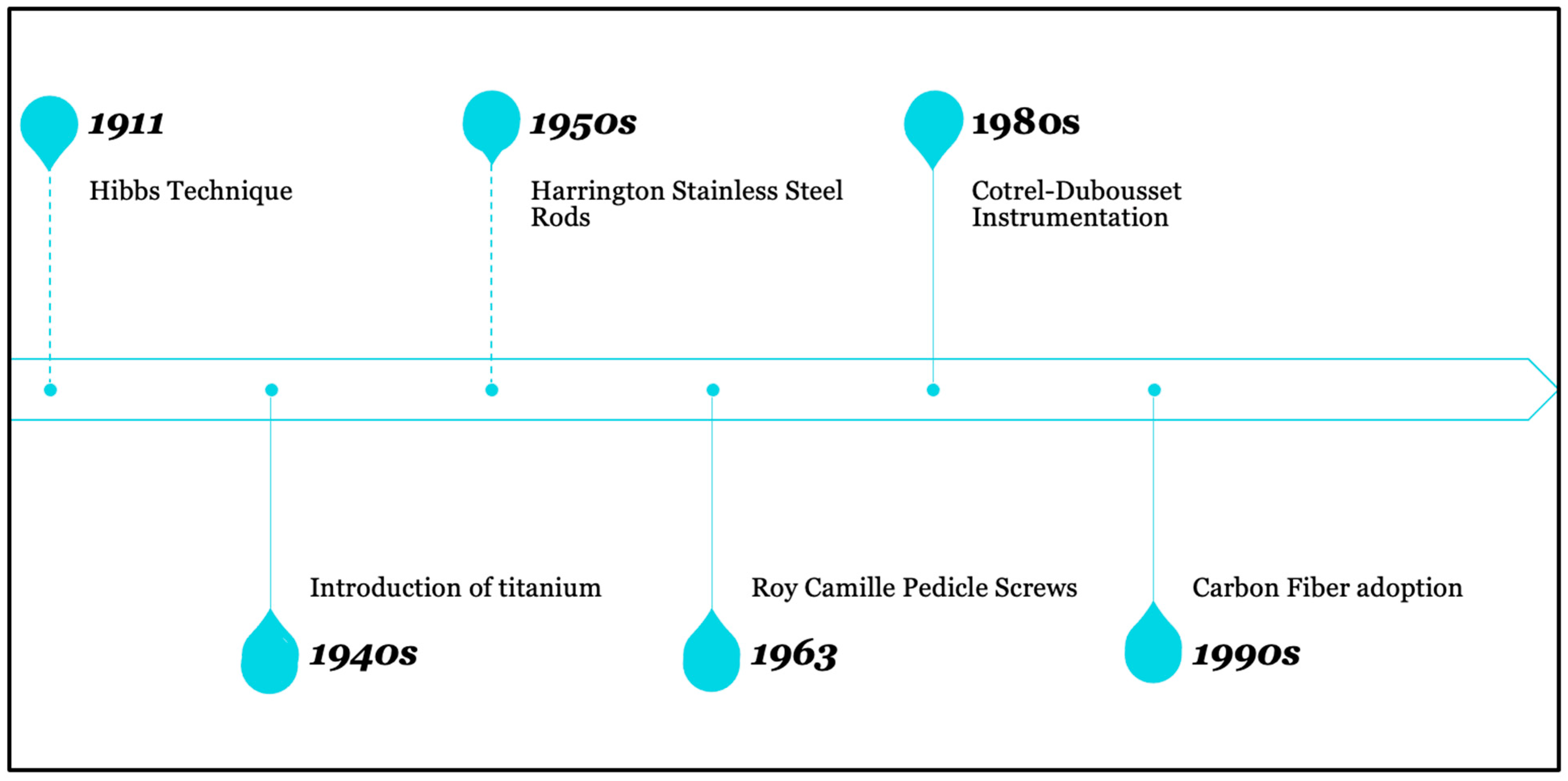
Figure 1. Timeline of the recent evolution of spinal instrumentation.
Surgeries 05 00041 g001
Figure 2. (A) Carbon fiber-reinforced PEEK pedicle screw system; (B) Carbon fiber-reinforced PEEK pedicle screws and titanium rod system; (C) Carbon fiber pedicle screws. Images by Icotec Medical (Alkstätten, Switzerland).
Surgeries 05 00041 g002
Figure 3. Images from Icotec Medical (Alkstätten, Switzerland). (A) MR artifact-free imaging of carbon fiber-reinforced PEEK pedicle screws; (B) Titanium screws creating significant artifacts on imaging.
Surgeries 05 00041 g003
Table 1. Comparison of carbon fiber and some traditional materials used in spinal instrumentation.
Carbon FiberTitaniumSteel
Strength-to weight ratioHigh tensile strength of 3.6 GPa and low density of about 1.8 g/cm3. This gives it exceptional strength-to-weight ratioDensity of about 4.51 g/cm3. Tensile strength of 240 Mpa. Good strength-weight-ratio Tensile strength of ~250 Mpa. Density of about 8 g/cm3. Thus, worst strength-to-weight ratio of the three.
Energy ConductivityLow thermal conductivity and not a conductor of electricityModerate thermal conductivity. Conductor of electricityModerate thermal conductivity. Conductor of electricity
Corrosion and Fatigue ResistanceElastic modulus of 233 GPa. Good fatigue resistance, particularly with repetitive loading. Excellent corrosion resistantExceptional fatigue resistance. Elastic modulus of 120 GPa. Outstanding corrosion resistanceGood fatigue resistance; with elastic modulus of 200 GPa. Not corrosion resistant
Imaging CompatibilitySafe MRI magnetic field. Does not cause artifact on CTSafe on MRI magnetic field. Some artifact on CTIncompatible with MRI imaging. Significant artifact on CT
Table 2. Summary of studies focusing on carbon fiber implants and outcomes.
Title (Year)MaterialsStudy/TestsConclusionOverall Positive Outcome (Y/N)
Alvarez-Breckenridge et al. (2023)Carbon fiber reinforced PEEK (CFR-PEEK)Assessment of the perceived advantages of improved imaging quality, postradiation treatment planning and recurrent tumor detection in spinal tumor patients treated with CFR-PEEKCFR-PEEK is safe and effective for spinal stabilization in both primary and metastatic tumors. Better postoperative radiating planning and local tumor recurrence detection were achieved as a result of improved postoperative imaging quality [52]Y
Carbon-Fiber International Collaboration Initiative Research Group (2022)Carbon Fiber platesPostoperative complications in bone tumor patients treated with Carbon Fiber platesThe use of Carbon Fiber plates in post-tumor resection reconstructions is safe and showed seemingly low complication profile [53]Y
Ghermandi et al. (2023)Carbon fiber reinforced PEEK (CFR-PEEK)Evaluation of Carbon Fiber-Reinforced PEEK in the treatment of degenerative lumbar spine diseaseCFR-PEEK implants is safe and effective in the treatment of spinal degenerative diseases [42]Y
Henzen et al. (2022)Carbon fiber reinforced PEEK and Titanium (CFP-T) hybrid implantFeasibility of postoperative stereotactic body radiation therapy (SBRT)Dosimetry and delivery of postoperative SBRT is feasible in patients with CFP-T implants using a CyberKnife system [54]Y
Hubertus et al. (2022)Carbon fiber reinforced PEEK (CFR-PEEK)Evaluation of the performance and precision of 3D intraoperative imaging and navigation systems in thoraco-lumbar instrumentation with CFR-PEEK pedicle screwsNavigation accuracy was considerably lower for CFR-PEEK pedicle screws than reported for titanium implants. CT may be the best imaging modality for CFR-PEEK instrumentation assessment [55]N
Joerger et al. (2021)Carbon fiber reinforced PEEK (CFR-PEEK)Comparative assessment of pedicle screw loosening and relapse with CFR-PEEK vs titanium in spondylodiscitis patientsMore CFR-PEEK pedicle screw loosening than with titanium likely due to stronger bacterial adhesion in spondylodiscitis patients. No difference in fusion rates [56]N
Neal et al. (2021)Carbon fiber reinforced PEEK (CFR-PEEK)Feasibility and advantages of CFR-PEEK implants in both primary and secondary osseous spinal tumorsCFR-PEEK is comparable to titanium in functionality. Additionally, the imaging characteristics of CFR-PEEK implants result in enhanced safety and efficacy for postoperative radiation planning and surveillance [1]Y
Oh et al. (2023)Carbon fiber reinforced PEEK (CFR-PEEK)Feasibility of CFR-PEEK to provide structural stability while allowing monitoring and treatment of spinal oncologic deformityCFR-PEEK is safe, effective and may provide relatively more benefit than existing instrumentation for treatment of thoracolumbar posterior spinal pathology [57]Y
Schwendner et al. (2023)Carbon fiber reinforced PEEK (CFR-PEEK)Assessment of the clinical and radiological outcomes in patients treated with CFR-PEEK dorsoventral instrumentationThe use of CFR-PEEK for vertebral body replacement in thoracic and lumbar spinal tumor patients offers an improved management option, particularly in terms of postoperative radiotherapy and MRI-based follow-up [37]Y
Uri et al. (2020)Carbon fiber reinforced PEEK (CFR-PEEK)In vitro evaluation of Carbon Fiber-Reinforce PEEKCFR-PEEK composite pedicle screw has superior fatigue properties than titanium-made implants of comparable mechanical properties. The fatigue resistance is similar to bone; it is radiolucent and results in artifact-free images on CT/MRI [29]Y
Disclaimer/Publisher’s Note: The statements, opinions and data contained in all publications are solely those of the individual author(s) and contributor(s) and not of MDPI and/or the editor(s). MDPI and/or the editor(s) disclaim responsibility for any injury to people or property resulting from any ideas, methods, instructions or products referred to in the content.