Case Report

A Case Report of Radiation-Induced Morphea Treated with Completion Mastectomy and Delayed Closure

1
Department of Surgery, Stony Brook University Hospital, Stony Brook, NY 11794, USA
2
Department of Plastic Surgery, Stony Brook University Hospital, Stony Brook, NY 11794, USA
*
Author to whom correspondence should be addressed.
Surgeries 2024, 5(3), 758-763; https://doi.org/10.3390/surgeries5030060
Submission received: 4 July 2024 / Revised: 20 August 2024 / Accepted: 27 August 2024 / Published: 2 September 2024

Abstract

:
Radiation-induced morphea (RIM) is a progressive and irreversible scleroderma encountered after breast radiation therapy. This condition is often underdiagnosed, with confounding differentials including post-radiation fibrosis, infection, inflammatory breast cancer, and cancer recurrence, amongst others. Treatment paradigms differ, and early diagnosis of RIM is essential in preventing further fibrosis and improving quality of life. First-line therapy includes immunosuppressive therapy; surgical management is infrequently described in the literature. Here, we report one of the few reports of completion mastectomy for RIM with postoperative outcomes described. This is a case of a patient who was treated with completion mastectomy with delayed wound closure after developing debilitating RIM status post right breast lumpectomy with radiation for invasive ductal carcinoma.

1. Introduction

Morphea is a rare, idiopathic inflammatory disease of the skin and underlying tissues. Unlike systemic sclerosis, morphea does not involve internal organs, sclerodactyly, nails, or Raynaud’s disease. Morphea is a rare disease with an incidence between 0.34 and 2.7 cases per 100,000 population per year, affecting mostly white women [1]. Peterson et al. classified morphea into plaque morphea, generalized morphea, bullous morphea, linear morphea, and deep morphea [2].
Radiation induced morphea (RIM) is a well-documented phenomenon, with an incidence as high as 1 in every 500 patients [3]. Most frequently, it is described in patients who received radiotherapy for breast cancer, but it has been reported in patients who received radiation therapy for other pathologies [3,4]. RIM was found to be more severe in patients with a history of obesity, active smoking status, coexisting autoimmune disease, and breast augmentation [5]. RIM usually occurs within the first year of exposure, but delayed presentations have been described [5]. The differential diagnosis includes sclerosing carcinoma, cellulitis, radiation dermatitis, and fat necrosis. On histology, RIM has early lymphocytic inflammation, followed by collagen-bundle hypertrophy [3].
The most widely accepted treatment for morphea is methotrexate and systemic glucocorticoids, usually in combination [1]. Another therapy modality includes phototherapy with UV-A1, which induces apoptosis of Langerhans cells and T lymphocytes and reduces collagen synthesis [1]. Mycophenolate has been described as a treatment in children with morphea who did not respond to treatment with glucocorticoids and methotrexate [6]. Based on the available literature, Marsol described a treatment algorithm for generalized morphea including phototherapy followed by immunosuppressant therapy, and mycophenolate if no response was had [1]. Surgical management for RIM is not well described in the literature [7]. One case report by Dancey et al. describes resolution of RIM after modified breast reduction, with no recurrence 6 months post operatively [8].
In this case, the major reason mastectomy with delayed closure was selected for the patient was due to the presence of a necrotic ulcer with episodes of cellulitis. The patient continued to have infections, including osteomyelitis, postoperatively. A retrospective case-control study by Olsen et al. showed that of patients following major breast surgery, patients who undergo mastectomy, have a history of radiation, and are current smokers were significantly more likely to develop surgical site infections [9]. Infection control continues to carry significant import in surgery and implant procedures, with recent developments of nanozymes, biological inhibitors, and biomaterials to mitigate risk of infection [10,11,12].
While surgery, radiation, and chemotherapy remain the mainstays of treatment modalities for breast cancer, photodynamic therapy has emerged as a promising non-invasive oncologic treatment that has been gaining attention due to its ability to target cancer cells with minimal damage to surrounding tissue [13,14]. Photodynamic therapy is being explored in combination with chemotherapy and radiation therapy to enhance its therapeutic effects [13]. While photodynamic therapy is currently being studied in conjunction with radiation, perhaps it can potentially be used in lieu of radiation to decrease radiation associated complications. We present a case report of a patient with T1N0M0 right breast cancer, who underwent lumpectomy and radiation, complicated by pathology-confirmed radiation induced morphea, which was refractory to medical treatment, ultimately requiring a mastectomy.

2. Case Presentation

A 74-year-old female with active tobacco use and a history of chronic lymphocytic leukemia (CLL) treated with bendamustine and rituximab therapy initially presented to the emergency department in April 2019 with a palpable right axillary mass, failure to thrive, and anemia. She was diagnosed with ER positive, HER2 negative invasive ductal carcinoma of the right breast (Figure 1). She underwent a course of neoadjuvant chemotherapy with paclitaxel without resolution, and subsequently fulvestrant, letrozole, and palbociclib. In December 2020, a right lumpectomy with low level 1 axillary lymph node dissection was performed, and a stage pT1a (involving dermis and pectoralis, with clear margins), N0 invasive ductal carcinoma was confirmed on pathology. She then underwent a one-month course of adjuvant radiation therapy (RT) culminating in February 2021, with 4256 cGy in 16 fractions, with a boost to the lumpectomy bed of 1000 cGy using 3D conformal radiation therapy.
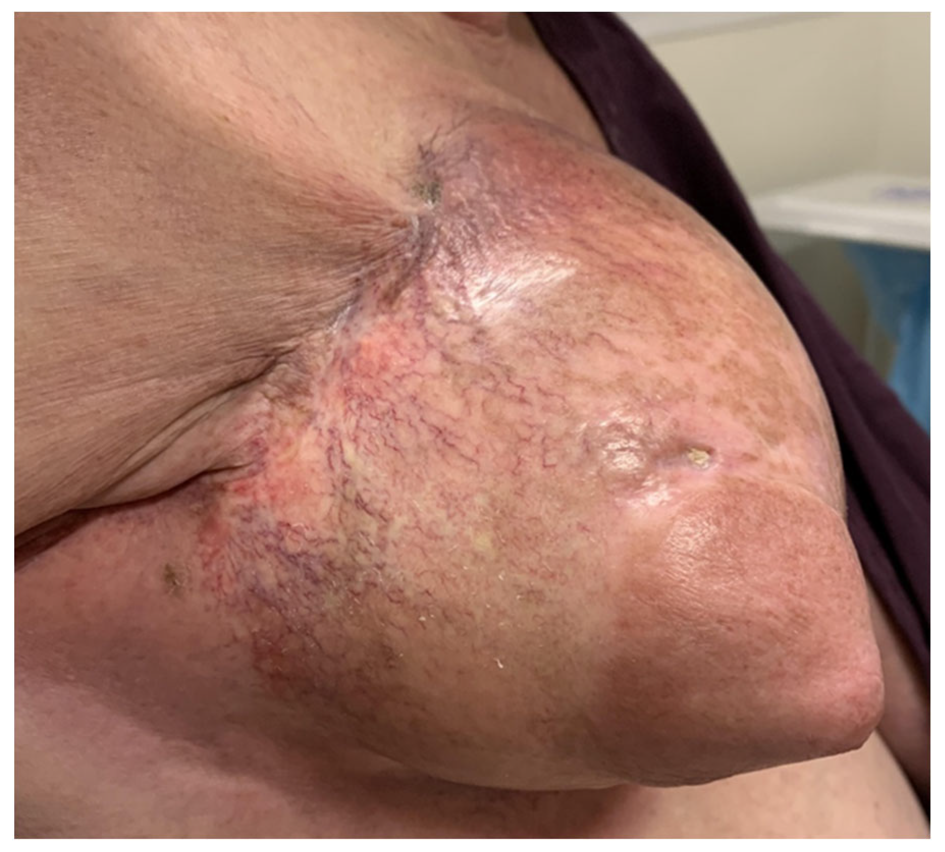
Six months after RT, the patient developed concentric shrinkage with dependent edematous changes of the nipple areolar complex, hyperpigmentation of skin, and some tautness at the axillary tail with overhead extension (Figure 2). In October 2021, punch biopsy showed lymphocytic inflammatory cell infiltrate within the dermis, with thickened collagen bundles, consistent with RIM. She was trialed on Vitamin E and pentoxifylline, and later, systemic prednisone and methotrexate were added without symptom improvement.
The patient also developed a necrotic ulcer on the right lateral breast, likely secondary to the sclerosis of the underlying tissues, with two episodes of superimposed cellulitis requiring antibiotic therapy. At this point, the decision was made to perform a completion mastectomy in December 2022, with wound vac application and planning for delayed wound coverage in the setting of her active tobacco use, chronic ulcerative wound, and fibrotic wound base (Figure 3).
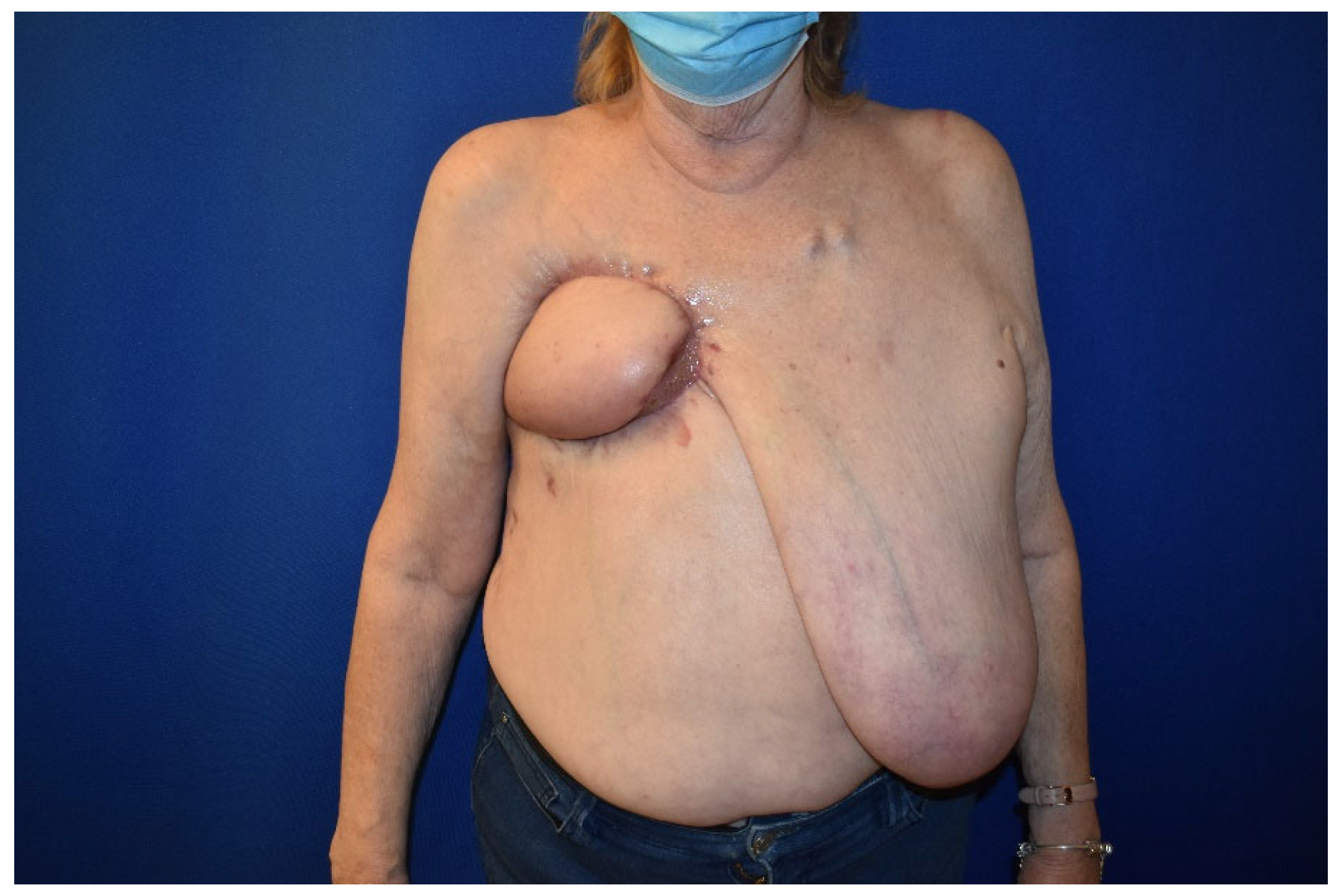
One week postoperatively, the patient returned to the ED for inability to tolerate wound vac change with subsequent multiple readmissions for sixth rib osteomyelitis and wound cultures positive for pseudomonas and corynebacterium. She was treated with a course of vancomycin and meropenem and then underwent two operative debridements (Figure 3). This was followed by reconstruction with a right pedicled latissimus dorsi myocutaneous flap in February 2023 (Figure 4).
She has since healed well from surgery and is considering a left-sided breast reduction for symmetry. She has decreased her smoking to four cigarettes per day. She continues on letrozole.

3. Results and Discussion

In this case report, we describe a 74-year-old female with active tobacco use and a history of CLL with Stage T1N0M0 right breast cancer, who underwent lumpectomy and radiation, complicated by pathology-confirmed radiation induced morphea. The patient developed ulcers and cellulitis of the right chest wall, requiring mastectomy and eventual breast reconstruction.
Of the established risk factors associated with severity of RIM [5], this patient’s only risk factor was active smoking. Partl et al. postulated that neoadjuvant chemotherapy may be associated with RIM, which could be applicable in this case as she received neoadjuvant chemotherapy with paclitaxel, fulvestrant, letrozole, and palbociclib [15]. Her disease was refractory to topical and oral treatment, and the patient eventually underwent mastectomy with delayed wound coverage due to the fibrotic nature of the wound and the active smoking status. There are currently limited reports of the use of surgery in RIM due to the rarity of the disease. Dancey et al. described resolution of RIM after modified breast reduction [8]. Walsh et al. described one case of RIM treated with mastectomy, but the patient did not follow up, so outcomes were unable to be ascertained [16]. To our knowledge, this is the first case report that describes completion mastectomy for RIM with postoperative outcomes. After mastectomy, the patient underwent a latissimus dorsi myocutaneous flap; she has healed well and is considering a contralateral breast reduction for aesthetic purposes.
Radiation-induced morphea (RIM) is a rare inflammatory disease, most frequently reported in patients who have received radiation for breast cancer. RIM is difficult to diagnose due to the rarity of the disease and wide differential diagnoses, including sclerosing carcinoma, cellulitis, radiation dermatitis, and fat necrosis. However, early recognition of RIM is essential in improving quality of life. The treatment for RIM is not well established, but usually involves medical management via immunosuppressants; there are few reports of surgical management of the disease. In this case report, we reviewed a patient with RIM successfully treated with mastectomy and subsequent reconstruction with resolution of symptoms. Further work is necessary to better understand the pathophysiology, risk factors, and potential treatment of RIM. In the future, upcoming oncologic treatments, such as photodynamic therapy [13], can potentially be used in lieu of radiation, thereby decreasing rates of radiation-related complications.

4. Conclusions

Radiation-induced morphea (RIM) is a progressive and irreversible scleroderma encountered after breast radiation therapy, with an incidence as high as 1 in every 500 patients. Risk factors include obesity, smoking, autoimmune disease, and breast augmentation. Treatment most often includes immunosuppression and phototherapy; surgery has not often been described in the management of RIM. In this case report, we analyze a patient with RIM and necrotic ulcer refractory to immunosuppressive therapy, necessitating a completion mastectomy, followed by delayed wound coverage with a right pedicled latissimus dorsi myocutaneous flap.

Author Contributions

Conceptualization, A.B.; investigation, Z.N., N.S., D.B. and A.B.; writing—original draft preparation, N.S. and Z.N.; writing—review and editing, N.S. and D.B. All authors have read and agreed to the published version of the manuscript.

Funding

This research received no external funding.

Institutional Review Board Statement

Case reports submitted for publication do not strictly meet the criteria as research. Although a case report (defined as a retrospective analysis of one (1), two (2), or three (3) clinical cases) may be illustrative, it does not meet the Federal regulation definition of research at 45 CFR 46.102(d). The Human Research Protection Program Standard Operating Procedures at Stony Brook University do not consider a case report/case series of up to three cases as human subject research that needs approval from the Institutional Review Board. Ethical review and approval were waived for this study.

Informed Consent Statement

Written informed consent has been obtained from the patient to publish this paper.

Data Availability Statement

The data presented in this study are available on request from the corresponding author due to privacy reasons.

Conflicts of Interest

The authors declare no conflicts of interest.

References

  1. Bielsa Marsol, I. Update on the classification and treatment of localized scleroderma. Actas Dermosifiliogr. 2013, 104, 654–666. [Google Scholar] [CrossRef] [PubMed]
  2. Peterson, L.S.; Nelson, A.M.; Su, W.P. Classification of morphea (localized scleroderma). Mayo Clin. Proc. 1995, 70, 1068–1076. [Google Scholar] [CrossRef] [PubMed]
  3. Kushi, J.; Csuka, M.E. Generalized morphea after breast cancer radiation therapy. Case Rep. Rheumatol. 2011, 2011, 951948. [Google Scholar] [CrossRef] [PubMed]
  4. Colver, G.B.; Rodger, A.; Mortimer, P.S.; Savin, J.A.; Neill, S.M.; Hunter, J.A.A. Post-irradiation morphoea. Br. J. Dermatol. 1989, 120, 831–835. [Google Scholar] [CrossRef] [PubMed]
  5. Ramseier, J.Y.; Ferreira, M.N.; Leventhal, J.S. Dermatologic toxicities associated with radiation therapy in women with breast cancer. Int. J. Womens Dermatol. 2020, 6, 349–356. [Google Scholar] [CrossRef] [PubMed]
  6. Martini, G.; Ramanan, A.V.; Falcini, F.; Girschick, H.; Goldsmith, D.P.; Zulian, F. Successful treatment of severe or methotrexate-resistant juvenile localized scleroderma with mycophenolate mofetil. Rheumatology 2009, 48, 1410–1413. [Google Scholar] [CrossRef] [PubMed]
  7. Finnegan, P.; Kiely, L.; Gallagher, C.; Mhaolcatha, S.N.; Feeley, L.; Fitzgibbon, J.; White, J.; Bourke, J.; Murphy, L.A. Radiation-induced morphea of the breast-A case series. Skin Health Dis. 2023, 3, e148. [Google Scholar] [CrossRef] [PubMed]
  8. Dancey, A.L.; Waters, R.A. Morphea of the breast. Two case reports and discussion of the literature. J. Plast. Reconstr. Aesthet. Surg. 2006, 59, 1114–1117. [Google Scholar] [CrossRef] [PubMed]
  9. Olsen, M.A.; Lefta, M.; Dietz, J.R.; Brandt, K.E.; Aft, R.; Matthews, R.; Mayfield, J.; Fraser, V.J. Risk factors for surgical site infection after major breast operation. J. Am. Coll. Surg. 2008, 207, 326–335. [Google Scholar] [CrossRef] [PubMed]
  10. Hosseini Hooshiar, M.; Badkoobeh, A.; Kolahdouz, S.; Tadayonfard, A.; Mozaffari, A.; Nasiri, K.; Salari, S.; Safaralizadeh, R.; Yasamineh, S. The potential use of nanozymes as an antibacterial agents in oral infection, periodontitis, and peri-implantitis. J. Nanobiotechnol. 2024, 22, 207. [Google Scholar] [CrossRef] [PubMed]
  11. Muzammil, K.; Hooshiar, M.H.; Varmazyar, S.; Omar, T.M.; Karim, M.M.; Aadi, S.; Kalavi, S.; Yasamineh, S. Potential use of proprotein convertase subtilisin/kexin type 9 (PCSK9) inhibition and prevention method in viral infection. Microb. Cell Factories 2024, 23, 90. [Google Scholar] [CrossRef] [PubMed]
  12. Yari, A.; Fasih, P.; Ghotbi, N.; Badkoobeh, A.; Goodarzi, A.; Hosseini Hooshiar, M. Do Platelet-Rich Concentrates Improve the Adverse Sequelae of Impacted Mandibular Third Molar Removal? J. Oral Maxillofac. Surg. Off. J. Am. Assoc. Oral Maxillofac. Surg. 2024, 82, 671–683. [Google Scholar] [CrossRef] [PubMed]
  13. Jiang, W.; Liang, M.; Lei, Q.; Li, G.; Wu, S. The Current Status of Photodynamic Therapy in Cancer Treatment. Cancers 2023, 15, 585. [Google Scholar] [CrossRef] [PubMed]
  14. Amirjani, A.; Shokrani, P.; Sharif, S.A.; Moheb, H.; Ahmadi, H.; Ahmadiani, Z.S.; Paroushi, M.S. Plasmon-enhanced nano-photosensitizers: Game-changers in photodynamic therapy of cancers. J. Mater. Chem. 2023, 11, 3537–3566. [Google Scholar] [CrossRef] [PubMed]
  15. Partl, R.; Regitnig, P.; Tauber, G.; Pötscher, M.; Bjelic-Radisic, V.; Kapp, K.S. Radiation-induced morphea-a rare but severe late effect of adjuvant breast irradiation: Case report and review of the literature. Strahlenther. Und Onkol. 2018, 194, 1060–1065. [Google Scholar] [CrossRef] [PubMed]
  16. Walsh, N.; Rheaume, D.; Barnes, P.; Tremaine, R.; Reardon, M. Postirradiation morphea: An underrecognized complication of treatment for breast cancer. Hum. Pathol. 2008, 39, 1680–1688. [Google Scholar] [CrossRef] [PubMed]
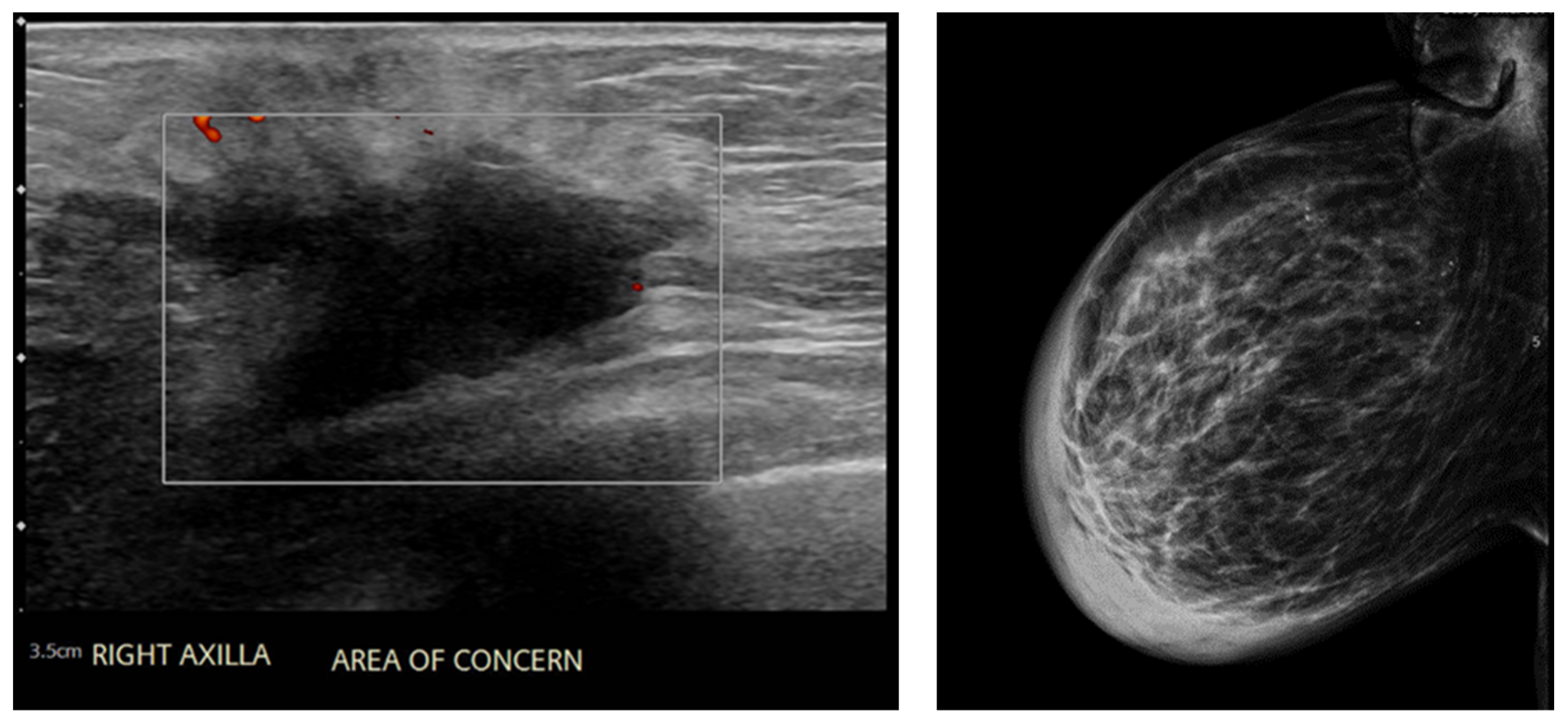
Figure 1. Ultrasound (left) and mammogram (right) showing right axillary mass.
Surgeries 05 00060 g001
Figure 2. Clinical presentation of radiation induced morphea.
Surgeries 05 00060 g002
Figure 3. Patient’s wound after mastectomy (left) and after multiple debridements (right).
Surgeries 05 00060 g003
Figure 4. Patient after right pedicled latissimus dorsi myocutaneous flap.
Surgeries 05 00060 g004
Disclaimer/Publisher’s Note: The statements, opinions and data contained in all publications are solely those of the individual author(s) and contributor(s) and not of MDPI and/or the editor(s). MDPI and/or the editor(s) disclaim responsibility for any injury to people or property resulting from any ideas, methods, instructions or products referred to in the content.