Article

Effect of High-Fat Diet and Lactiplantibacillus plantarum 299v on the Gut Microbiome of Adolescent and Adult Rats

by
Samantha N. Atkinson
1,2,
Caron Dean
3,4,
Victoria L. Woyach
3,4,
Keri R. Hainsworth
3,5 and
Hershel Raff
6,7,8,9,*,†
1
Department of Microbiology and Immunology, Medical College of Wisconsin, Milwaukee, WI 53226, USA
2
Center for Microbiome Research, Medical College of Wisconsin, Milwaukee, WI 53226, USA
3
Department of Anesthesiology, Medical College of Wisconsin, Milwaukee, WI 53226, USA
4
Research Division, Zablocki Veterans Affairs Medical Center, Milwaukee, WI 53295, USA
5
Jane B. Pettit Pain and Headache Center, Children’s Wisconsin, Milwaukee, WI 53226, USA
6
Division of Endocrinology and Molecular Medicine, Department of Medicine, Medical College of Wisconsin, Milwaukee, WI 53226, USA
7
Department of Surgery, Medical College of Wisconsin, Milwaukee, WI 53226, USA
8
Department of Physiology, Medical College of Wisconsin, Milwaukee, WI 53226, USA
9
Cardiovascular Center, Medical College of Wisconsin, Milwaukee, WI 53226, USA
*
Author to whom correspondence should be addressed.
Current address: Endocrinology Research M4150, Medical College of Wisconsin, 8701 Watertown Plank Road, Milwaukee, WI 53226, USA.
Obesities 2025, 5(1), 17; https://doi.org/10.3390/obesities5010017
Submission received: 11 February 2025 / Revised: 25 February 2025 / Accepted: 14 March 2025 / Published: 16 March 2025

Abstract

:
Childhood and adolescent obesity and its associated morbidities are increasing in part due to the ingestion of diets high in fat (HFD). Changes in the gastrointestinal microbiome have been associated with these morbidities, including insulin resistance, cardiovascular disease, and inflammatory states. The use of dietary probiotics may mitigate these microbiome-associated morbidities and improve health during maturation. Using our established model of obesity in rats consuming an HFD from weaning, we examined the gut microbiome with a single-strain probiotic in the drinking water [Lactiplantibacillus plantarum 299v (Lp299v, LP299V®)] of adolescent and adult rats. Our main finding was a differential effect of HFD and probiotic on the gut microbiome that was associated with maturation (adolescence vs. adulthood). Specifically, probiotic treatment of adolescent rats on an HFD led to alterations in the enrichment of the gut microbiome, which were associated with the morbidities of obesity, while adult rats under the same conditions exhibited minimal changes, demonstrating differences in plasticity associated with maturation. Of particular relevance in this regard is the fact that Oscillospiraceae and Lachnospiraceae, associated with beneficial short-chain fatty acid production, were enriched in adolescent rats on an HFD and treated with Lp299v. Our data suggest that the use of probiotics in childhood and adolescence may improve health in adulthood by potentially affecting the developing gastrointestinal microbiome.

1. Introduction

Adolescent obesity is a major worldwide public health problem and is increasing in prevalence [1,2,3,4]. Adolescent obesity results in many clinical syndromes including insulin resistance, cardiovascular disease, a heightened inflammatory state, and liver disease [2,5,6,7,8,9,10,11]. Furthermore, obesity during puberty predisposes the individual to many of these disorders in adulthood [12,13,14,15]. Studying the development of obesity in laboratory animals using a high-fat diet (HFD) is useful in modeling the consequences of accelerated developmental weight gain in humans.
The microbiome consists of a variety of microorganisms, including bacteria, fungi, and viruses and their genetic material in a singular environment. Evidence suggests that the gut microbiome is involved in the control of body weight, energy homeostasis, and inflammation and, thus, plays an important role in the pathophysiology of obesity [16,17,18]. Furthermore, the gut microbiome and diet are major factors contributing to the obesity epidemic [19,20,21,22]. HFD causes dysbiosis of the microbiome, and the bacterial metabolites produced from dietary components have downstream effects [23,24]. Alterations to the equilibrium of the energy balance between energy intake from food and expenditure through resting and thermic metabolism can readily promote obesity [25]. The gut microbiome has been shown to provide an increased capacity for energy harvest that could affect the energy balance, resulting in body weight gain [26,27]. The “microbiota–gut–brain axis” is a multi-organ system dependent upon bidirectional communication to maintain homeostasis [19,22,28]. Specifically, because of this bidirectional relationship, increased gut permeability can stimulate inflammatory and immune responses through lipopolysaccharide leakage, which, in turn, allows proinflammatory adipocytokines to maintain a cycle of systemic inflammation [16,29].
Probiotics are live bacteria that affect the microbiome and have the potential to counteract the effects of HFD, leading to health benefits [7], including anti-obesity outcomes [30,31]. Probiotic use has been associated with attenuated weight gain in animals fed an HFD [16,32], although we have not previously found this with Lactiplantibacillus plantarum 299v (Lp299v) in rats [33] but others have in rabbits [34]. Lp299v is a strain of bacteria that was isolated from healthy human intestinal mucosa in the 1980s [35]. It is the most widely studied strain of L. plantarum in the world and has a wide array of clinical applications. It has been shown to survive the human gastrointestinal tract, and importantly, it has been shown to reduce systemic inflammation [36].
The principal effects of probiotics on the gut microbiome are improved function of the intestinal barrier and improved health of the GI tract and immune system [7], all of which are particularly important to children and adults with obesity [18]. Probiotic supplementation in youth with obesity has been associated with benefits ranging from improvements in anthropometrics and fasting glucose to improvements in the lipid profile (although results on the latter are mixed) [7,18,28]. Furthermore, it appears that in animals, a change in the gut microbiome associated with an HFD provides an important humoral signal to alter insulin sensitivity and glucose homeostasis [37]. This alteration in the microbiome–gut–brain–neuroendocrine axis may be involved in the proinflammatory state of obesity we and others have previously demonstrated [5,6,28].
We have previously reported body composition, serum cytokine, and adipokine data from the current cohort of rats on an HFD with and without a dietary probiotic [33]. While there is evidence that both HFD and probiotic supplementation affect the microbiome [38,39], very little is known about the potentially moderating effects of development on the interaction between these factors [40]. The purpose of the current study was to assess the change in the gut microbiome associated with an HFD, obesity, and probiotic supplementation in adolescent and adult male rats. Several rat models of HFD-induced obesity, probiotic administration, and the microbiome have been developed to model the human condition [41,42,43,44]. In our established rat model of the development of obesity from weaning using an HFD compared to a control diet [33], the gut microbiome was treated with a probiotic (Lp299v) or a placebo vehicle freely provided in drinking water. We hypothesized that the effect of an HFD on the gut microbiome would be altered by ingestion of a single-strain probiotic started at weaning and may be affected by the stage of development.

2. Methods

The protocols for this study were approved by the Animal Care and Use Committee at the Zablocki Department of Veterans Affairs Medical Center. The current microbiome protocol is an extension of our previously published study in this cohort of rats [33]. We opted to assess the microbiome in males because our previous study identified more dramatic effects in male rats on body composition, leptin, and inflammatory cytokines [33], and we anticipate assessing sex differences in the gut microbiome in a future study. Sixty male offspring from fourteen pregnant Long Evans dams obtained from Envigo at 14 days gestation (Indianapolis, IN 46250 USA) were weaned at 3 weeks of age (28–66 g males). Long Evans rats were selected for study as this strain has been shown to develop obesity and its associated phenotypes on an HFD [45,46]. We also chose the Long Evans rat as the basis for future studies with inbred and sequenced Long Evans substrains that are available at our institution (https://rgd.mcw.edu/rgdweb/report/strain/main.html?id=39456107 (accessed on 14 March 2025)).
Rats were maintained and used according to the NIH Guide for the Care and Use of Laboratory Animals and in compliance with federal, state, and local laws and the ARRIVE guidelines [47]. Male animals from a single family were randomly assigned to diet and treatment groups (Table 1) and housed 2–3/cage containing a Beta Chip (Warrensburg NY). The housing room was set on a reverse 12 h light/12 h dark cycle from 6 am/6 pm and maintained at 22 ± 1 °C at 35–65% humidity. Animals had free access to food and water. Rats were weighed weekly from day of weaning (see [33] and Supplemental File).
The standard-fat (control) diet had a 4.5% crude fat content (Purina laboratory rodent diet 5001; Waldschmidt, Milwaukee WI; https://www.labdiet.com/product/detail/5001-laboratory-rodent-diet (accessed on 14 March 2025)). The high-fat diet had 45 kcal% from saturated fats (D12451, Research Diets, Inc., New Brunswick, NJ, USA; https://www.researchdiets.com/formulas/d12451 (accessed on 14 March 2025)). The placebo or single-strain probiotic Lactiplantibacillus plantarum 299v (Lp299v; 10 bn CFU/150 mL), both from Probi USA Inc. (Redmond, WA, USA), were supplied in the drinking water (n = 6–8 rats per group).
We chose to use a single-strain probiotic because it (Lp299v) eliminates difficulties associated with multi-strain probiotics when attempting to determine mechanisms associated with certain associations, such as with obesity and pain [33]. Furthermore, we chose Lp299v specifically because it has been associated with “strong” anti-inflammatory effects in men with stable coronary vascular disease [36]. Administration of a probiotic in drinking water is a validated approach for long-term studies in laboratory animals [48,49]. A fresh solution of Lp299v in the drinking water was made every 3 days. Water bottles were refilled and shaken daily, and intake was qualitatively monitored to ensure adequate and appropriate fluid intake. Based on the average daily water intake and a previous study, each rat consumed ~1 billion CFU of Lp299v per 100 g body weight per day [33,50]. To assess the effect of treatment across puberty in the adult rats, the HFD and probiotic were provided at weaning in all groups rather than equalizing the length of time the adolescent vs. adult rats were treated.
Fecal pellets from individual rats were collected at weaning for baseline data (Pre) prior to group assignment and at 6 weeks (adolescent) or 11 weeks (adult) from start of diet +/− probiotic treatment. As per standard procedure, the pellets were stored in RNAlater, a solution that preserves RNA and DNA, for 2 weeks at 4 °C, before freezing at −20 °C until analysis.

2.1. DNA Extraction and Sequencing

Genomic DNA was extracted from fecal pellets using a Qiagen DNeasy PowerLyzer PowerSoil Kit (Catalog# 12855-100) with a slight modification in the protocol [51]. After addition of solution C1 and heating the samples at 65 °C for 10 min, the sample was subjected to further heating at 95 °C for 10 min followed by vigorous bead beating using PowerLyzer (Qiagen, Germantown, MD 20874, USA). Both incubation steps at 2–8 °C were implemented. DNA was then sequenced for the V3–V4 region of the 16S gene (341F–806R) using the 2 × 300 bp protocol on an Illumina MiSeq device (University of Wisconsin Madison Biotechnology Center, Madison, WI, USA).

2.2. Microbiome Analysis and Statistical Analyses

Statistical analyses were performed on data from fecal pellets of individual rats, but as the rats were housed 2–3 per cage by diet grouping, between-cage analyses were also performed to account for coprophagia.
QIIME2 (v.2023.9) was used to analyze the paired-end 16S rDNA sequencing reads [52]. Sequences were imported and summarized to check quality. Cutadapt was used to trim primers from the reads [53]. Representative sequences were chosen using DADA2, which also removes chimeric sequences [54]. The representative sequences were then aligned [55], masked for hypervariable regions [56], and phylogenetic trees were produced [57]. A classifier was generated to assign taxonomy to the reads using the 99% similarity files of the SILVA v. 138 and the 341–806 region (V3/V4) of the 16S gene [58]. Taxonomy was assigned to the feature table to make taxonomy bar plots and to generate relative abundance tables.
Diversity metrics were run using the core-metrics-phylogenetic command of QIIME2. Alpha and beta diversity were analyzed using their respective commands, alpha-group-significance and adonis [59,60,61]. Alpha diversity metrics used a Kruskal–Wallis test to test for significance, while beta diversity metrics used an ADONIS test; the Kruskal–Wallis test used the Benjamini–Hochberg multiple comparison tests. Principal Coordinate Analysis (PCoA) plots were examined using Emperor [62,63], and finalized figures were made using Python v.3.12.4. LEfSe, linear discriminant analysis (LDA) effect size, was run to determine enriched organisms for each treatment group [64]. Input tables had singletons removed to account for cage bias. Final LEfSe LDA plots were generated using Inkscape v. 1.0.1 (https://inkscape.org/ (accessed on 14 March 2025)). Cladograms were generated using GraPhlAn [65]. Paired samples over time were evaluated by the Wilcoxon rank sum test. In all cases, p < 0.05 was considered significant.

3. Results

The increase in body weight of male rats on an HFD was greater than that of rats on the control diet irrespective of probiotic or placebo consumption (data reported previously [33] and shown in Supplemental Figure S1).
Rat fecal pellets were processed for 16S rDNA sequencing and analyzed using QIIME2. The presence of Lactobacillus genera was identified but not specifically Lactiplantibacillus plantarum (formerly known as Lactobacillus plantarum) (Figure 1A) [66]. Our findings are likely a limitation of 16S rDNA sequencing that often does not have species-level resolution. The only unknown Lactobacillus genus (Lactobacillus;__) showed higher relative abundance in the adolescent samples than in the adult samples (Figure 1B).
Upon initial assessment of beta diversity, the baseline samples taken at weaning (Pre) were clustered separately from the remaining groups for both the control diet and HFD (Supplemental Figure S2). This is not unexpected, as the rats had not been exposed to the solid diet that was started at weaning. Since our direct focus was on diet and the effect of the probiotic on the gut microbiome, we parsed the data into subsets to exclude Pre samples, thereby focusing on each diet separately.
When examining the microbiome of rats fed the control diet, there were no differences between the placebo and probiotic treatment for any alpha or beta diversity metric run on adolescent rat samples when controlling for cage effects (Supplemental Figure S2A, purple dots). Adult rats fed the control diet had beta diversity differences between the placebo- and probiotic-treated groups when controlling for cage effects using the Unweighted UniFrac, Jaccard, and Bray–Curtis metrics (Supplemental Figure S2A, orange dots; Unweighted UniFrac p = 0.008, R2 = 0.108). In adolescent rats fed an HFD, there were no alpha diversity differences between the placebo- and probiotic-treated rats. With beta diversity testing, we found differences between the groups when using the Unweighted UniFrac, Jaccard, and Bray–Curtis metrics when controlling for cage effects. We focused on Unweighted UniFrac as it accounted for the highest variance (Figure 2A; p = 0.001, R2 = 0.200). When comparing the same groups for the adult rats, all beta diversity metrics were statistically significant when controlling for cage effects; we focused on Unweighted UniFrac to have consistency with the adolescent rats (Figure 2B; Unweighted UniFrac p = 0.002, R2 = 0.0825). These data demonstrate that the microbiomes of the adolescent and adult rats were altered by the HFD and probiotic treatment. Since our focus was to determine differences in the microbiomes of adolescent compared to adult rats that responded to probiotic treatment, and since the adolescent rats did not respond to the probiotic when given the control diet, we excluded the control diet samples from the subsequent analyses.
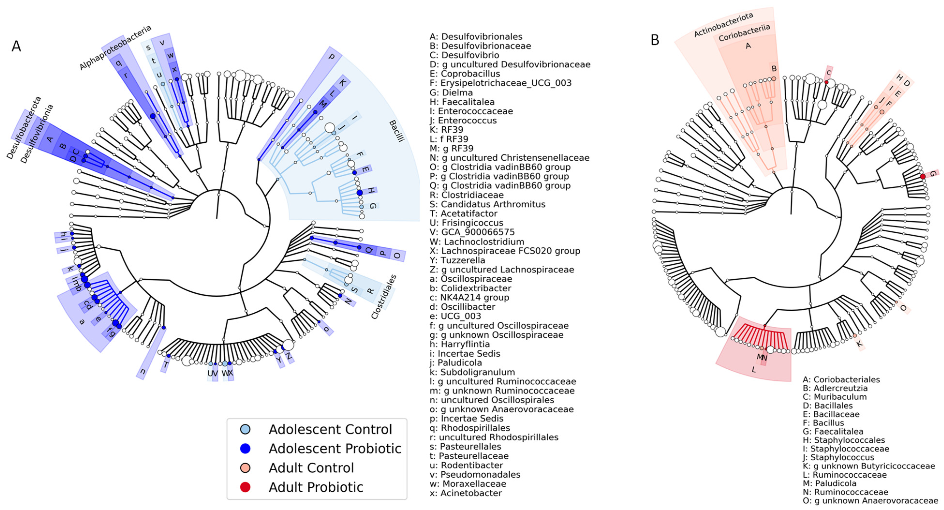
To further assess the microbiome differences between the placebo-treated and probiotic-treated rats on an HFD, we used LEfSe analysis [64]. Adolescent rats fed an HFD and treated with a probiotic showed numerous and diverse changes in gut microbial composition across many clades of bacteria compared to placebo-treated adolescent rats (Figure 3A and Supplemental Figure S3A). The genera of two of the main families of bacteria enriched in the probiotic-treated adolescent rats fed an HFD were identified as Oscillospiraceae and Lachnospiraceae. In contrast to the adolescent diversity, the LEfSe analysis [64] of the data from the adult rats showed an enrichment in only three taxa in the probiotic-treated group (Figure 3B and Supplemental Figure S3B). These data emphasize that the gut microbiome of adolescent rats has an expansive enrichment of taxa when fed an HFD and probiotic compared to minimal enrichment in adults.
There were several significant effects of the probiotic in adolescent rats on an HFD (Figure 4). Specifically, the abundances of members of the Lachnospiraceae and Oscillospiraceae families were enriched in the adolescent rats. Within the Oscillospiraceae family, the specific increase in the abundance of Colidextribacter was of particular interest.
There was only one genus, Faecalitalea, that maintained enrichment from adolescence to adulthood (Figure 5A). The abundance of the Lachnospiraceae and Oscillospiraceae families that were enriched by the probiotic in the adolescent rats on an HFD (see Figure 4) were not maintained into adulthood (Figure 5B–D). These data demonstrate a lack of enriched taxa in the gut microbiome in adults vs. adolescents fed an HFD and single-strain probiotic.

4. Discussion

We used our established rat model of obesity to evaluate the interaction of an HFD and a single-strain probiotic on the gut microbiome in adolescent and adult male rats. To summarize our main findings, the microbiomes of adolescent rats fed a control diet were similar, regardless of the addition of a probiotic or placebo to the water. Adult rats fed a control diet had a shift in the gut microbiome in response to the probiotic treatment. However, differential effects in the gut microbiome of rats on an HFD were associated with maturation (adolescence vs. adulthood) and probiotic treatment. Probiotic treatment of adolescent rats on an HFD led to many changes in the gut microbiome, while adult rats under the same conditions exhibited minimal changes in the gut microbiome. These data support the concept that microbiome plasticity is greater in adolescence than in adulthood, a phenomenon thought to facilitate healthy aging [40].
Two primary taxa, Oscillospiraceae and Lachnospiraceae, enriched in adolescent rats on an HFD and treated with Lp299v, are associated with beneficial short-chain fatty acid (SCFA) production [67,68]. Our findings may have translational applications associated with the downstream effects of this enrichment. Oscillospiraceae are a member of the class Clostridia, obligate anaerobes, and they are an abundant commensal bacterium in the gut involved in gastrointestinal disorders and inflammation [69,70]. Although there have been mixed outcomes of Oscillospira abundance in different obesity models, it has been shown to have a positive association with low fat, leanness, and human health (reviewed in [68]). It can produce many kinds of SCFAs but is dominated by butyrate. SCFA production has been shown to benefit the host through anti-obesity effects such as appetite regulation, energy expenditure, and immunomodulation (reviewed in [71]). SCFAs have also been found to modulate innate and adaptive immune cell homeostasis (reviewed in [72]).
Lachnospiraceae is a large family of anaerobic bacteria that is among the most abundant taxa in the gastrointestinal tract. The health effects associated with this family depend upon the specific taxa, as some studies have identified both increased and decreased associations with inflammatory disorders and human health [69,73], and this is reviewed in [74]. For example, Lachnospiraceae has been associated with type 1 diabetes and different intestinal diseases, but it is also thought to promote health through beneficial metabolite production including SCFAs (reviewed in [67]).
Colidextribacter [75], considered to be beneficial bacteria, also increases SCFA production. This was found to be especially true of the production of butyric acid when fucoidan was added to a dextran sulfate sodium-induced ulcerative colitis model [76]. Relevant to our findings is the fact that Colidextribacter has been associated with successful aging via its effect on lipid metabolism [77]. We are currently performing serum lipidomic analysis of samples from this cohort of rats and adolescent humans to pursue this exciting possibility and to extend our model from previous observational studies in obese adolescent humans [78]. Furthermore, the data are consistent with our previous study showing that the increase in certain serum cytokines (IL-12p70 and MIP1α) due to HFD was attenuated by Lp299v [33].
The only taxon enriched in both the adolescent and adult probiotic-treated groups on an HFD was Faecalitalea, a genus of anaerobic bacteria that has been isolated from human, chicken, and pig guts [79] and has recently been identified as having promising applications to human health [80]. Low levels of this bacterial group have been associated with several diseases and states of ill health. For example, this taxon has been found to have decreased abundance in type 1 diabetes rat models through a decrease in butyric acid production, leading to decreased insulin secretion [81].
With respect to plasticity, the effects we found in our study could be specific to L. Plantarum 299v, as other studies have found limited effectiveness in probiotic treatment of adolescent obesity. One clinical trial using a single strain of Lactobacillus salivarius Ls-33 and two clinical trials using mixed strains, VSL#3 and Visbiome, did not alter gut microbial community abundances or body weight in human adolescent patients with obesity [reviewed in [82]]. Further studies would be needed to conclusively determine if the effect was specifically due to L. plantarum 299v or whether an organism with similar metabolic properties would have the same effect.
Strengths and limitations: This study fills a gap in the literature by identifying specific developmental changes to the microbiome associated with an HFD and a single-strain probiotic. Considering the outcomes of this study, the main limitation is that it was not longitudinal. We plan to extend our analyses to follow individual rats with regular fecal sampling from weaning to older adulthood to determine the timing of the change in microbiome composition. This will allow us to assess the optimal window for probiotic supplementation as a therapeutic intervention to improve healthy development. Of course, variation in the microbiomes between animal models and humans should always be considered in this type of translational research [83]. Finally, it is acknowledged that the duration of dietary treatments was longer for adults than adolescents, as this paradigm fit our research design to assess our primary interest of the full treatment in the pre- to post-pubertal state. We recognize that prolonged probiotic supplementation may affect probiotic efficacy, and future studies will consider delaying the probiotic in the adult groups to equalize probiotic use and distinguish between developmental and adaptation effects.
Perspectives: The use of probiotics to improve human health has been increasing [84,85]. In addition to their effects on gastrointestinal development and function, it appears that probiotics improve immune function, inflammatory states, lipid profiles, and reproductive and psychological health and decrease the risk of cardiovascular disease [85]. The current study showed that single-strain probiotic supplementation in rats on an HFD can be beneficial for adolescents by developing the gastrointestinal microbiome in a way that can potentially improve health in adulthood. This suggests that our current focus on obesity, inflammation, pain, and lipid metabolism in adolescence is warranted considering the theoretical simplicity and cost-effectiveness of giving probiotics to children as they mature to adulthood [6,33,78,86,87,88]. The development of our animal model using an HFD and a probiotic from weaning is an advantage in this regard. While our findings imply that benefits associated with probiotic supplementation in adolescence are not maintained in adulthood, future research is needed to determine whether probiotic supplementation during early stages of the lifespan are associated with developmental and physiological benefits manifested later in the lifespan.

Supplementary Materials

The following supporting information can be downloaded at: https://www.mdpi.com/article/10.3390/obesities5010017/s1. Figure S1: Body weights; Figure S2: Pre-weaning samples cluster separately from control and HFD samples; Figure S3: LDA plots of LEfSe results.

Author Contributions

H.R., K.R.H., V.L.W. and C.D. conceived and designed the research; H.R., V.L.W. and C.D. performed the experiments; H.R., K.R.H., V.L.W., S.N.A. and C.D. analyzed the data; H.R., K.R.H., S.N.A. and C.D. interpreted the results of the experiments; S.N.A. prepared the figures; S.N.A. drafted the manuscript; H.R., K.R.H., V.L.W., S.N.A. and C.D. edited and revised the manuscript; H.R., K.R.H., V.L.W., S.N.A. and C.D. approved the final version of the original and revised manuscripts. All authors have read and agreed to the published version of the manuscript.

Funding

This research received no external funding.

Institutional Review Board Statement

The animal study protocol was approved by the Institutional Animal Care and Use Committee at the Zablocki Department of Veterans Affairs Medical Center (protocol code 1765-14 and 7 July 2022).

Informed Consent Statement

Not applicable.

Data Availability Statement

The raw data supporting the conclusions of this article will be made available by the authors on request.

Acknowledgments

This study is supported in part by an award from the Medical College of Wisconsin Center for Microbiome Research and an award from the Advancing a Healthier Wisconsin Endowment. C.D. is funded by a Merit Review Award RX002747 from the US Department of Veterans Affairs Rehabilitation, Research, and Development Service. Thanks to the MCW Center for Microbiome Research (CMR) personnel, Nita Salzman and Jennifer Ziegelbauer, and the University of Wisconsin-Madison Biotechnology Center. The probiotic strain LP299V® is a proprietary strain developed by Probi® and was donated by Probi USA. In memory of Dorothy Weihrauch.

Disclaimer

The contents do not represent the views of the US Department of Veterans Affairs or the US Government.

Conflicts of Interest

The authors declare no conflicts of interest.

References

  1. Chrissini, M.K.; Panagiotakos, D.B. Public health interventions tackling childhood obesity at European level: A literature review. Prev. Med. Rep. 2022, 30, 102068. [Google Scholar] [CrossRef] [PubMed]
  2. Skinner, A.C.; Ravanbakht, S.N.; Skelton, J.A.; Perrin, E.M.; Armstrong, S.C. Prevalence of Obesity and Severe Obesity in US Children, 1999–2016. Pediatrics 2018, 141, e20173459. [Google Scholar] [CrossRef] [PubMed]
  3. NCD Risk Factor Collaboration (NCD-RisC). Worldwide trends in underweight and obesity from 1990 to 2022: A pooled analysis of 3663 population-representative studies with 222 million children, adolescents, and adults. Lancet 2024, 403, 1027–1050. [Google Scholar] [CrossRef]
  4. Deng, Y.; Yli-Piipari, S.; El-Shahawy, O.; Tamura, K. Trends and key disparities of obesity among US adolescents: The NHANES from 2007 to 2020. PLoS ONE 2024, 19, e0290211. [Google Scholar]
  5. Hainsworth, K.R.; Simpson, P.M.; Raff, H.; Grayson, M.H.; Zhang, L.; Weisman, S.J. Circulating inflammatory biomarkers in adolescents: Evidence of interactions between chronic pain and obesity. Pain Rep. 2021, 6, e916. [Google Scholar] [CrossRef] [PubMed]
  6. Raff, H.; Phillips, J.M.; Simpson, P.M.; Weisman, S.J.; Hainsworth, K.R. Serum soluble urokinase plasminogen activator receptor in adolescents: Interaction of chronic pain and obesity. Pain Rep. 2020, 5, e836. [Google Scholar] [CrossRef]
  7. Luzzi, A.; Briata, I.M.; Di Napoli, I.; Giugliano, S.; Di Sabatino, A.; Rescigno, M.; Cena, H. Prebiotics, probiotics, synbiotics and postbiotics to adolescents in metabolic syndrome. Clin. Nutr. 2024, 43, 1433–1446. [Google Scholar] [CrossRef]
  8. Oliveira, R.G.; Guedes, D.P. Physical Activity, Sedentary Behavior, Cardiorespiratory Fitness and Metabolic Syndrome in Adolescents: Systematic Review and Meta-Analysis of Observational Evidence. PLoS ONE 2016, 11, e0168503. [Google Scholar] [CrossRef]
  9. Daniels, S.R. Complications of obesity in children and adolescents. Int. J. Obes. 2009, 33 (Suppl. 1), S60–S65. [Google Scholar] [CrossRef]
  10. Lawlor, D.A.; Martin, R.M.; Gunnell, D.; Galobardes, B.; Ebrahim, S.; Sandhu, J.; Ben-Shlomo, Y.; McCarron, P.; Davey Smith, G. Association of body mass index measured in childhood, adolescence, and young adulthood with risk of ischemic heart disease and stroke: Findings from 3 historical cohort studies. Am. J. Clin. Nutr. 2006, 83, 767–773. [Google Scholar] [CrossRef]
  11. Bendor, C.D.; Bardugo, A.; Pinhas-Hamiel, O.; Afek, A.; Twig, G. Cardiovascular morbidity, diabetes and cancer risk among children and adolescents with severe obesity. Cardiovasc. Diabetol. 2020, 19, 79. [Google Scholar] [CrossRef] [PubMed]
  12. Simic, B.S. Childhood obesity as a risk factor in adulthood and its prevention. Prev. Med. 1983, 12, 47–51. [Google Scholar] [CrossRef] [PubMed]
  13. Johnston, F.E. Health implications of childhood obesity. Ann. Intern. Med. 1985, 103, 1068–1072. [Google Scholar] [CrossRef]
  14. Jarvis, S.; Giles, H.; Jarvis, P.; New, K. The weight status of children in late childhood within south East Wales and predictions for their future health. J. Public Health 2022, 44, e557–e561. [Google Scholar] [CrossRef]
  15. Llewellyn, A.; Simmonds, M.; Owen, C.G.; Woolacott, N. Childhood obesity as a predictor of morbidity in adulthood: A systematic review and meta-analysis. Obes. Rev. 2016, 17, 56–67. [Google Scholar] [CrossRef]
  16. Kobyliak, N.; Conte, C.; Cammarota, G.; Haley, A.P.; Styriak, I.; Gaspar, L.; Fusek, J.; Rodrigo, L.; Kruzliak, P. Probiotics in prevention and treatment of obesity: A critical view. Nutr. Metab. 2016, 13, 14. [Google Scholar] [CrossRef] [PubMed]
  17. Kobyliak, N.; Falalyeyeva, T.; Boyko, N.; Tsyryuk, O.; Beregova, T.; Ostapchenko, L. Probiotics and nutraceuticals as a new frontier in obesity prevention and management. Diabetes Res. Clin. Pract. 2018, 141, 190–199. [Google Scholar] [CrossRef] [PubMed]
  18. Benitez-Paez, A.; Gomez Del Pugar, E.M.; Lopez-Almela, I.; Moya-Perez, A.; Codoner-Franch, P.; Sanz, Y. Depletion of Blautia Species in the Microbiota of Obese Children Relates to Intestinal Inflammation and Metabolic Phenotype Worsening. mSystems 2020, 5, e00857-19. [Google Scholar] [CrossRef]
  19. Stanislawski, M.A.; Dabelea, D.; Lange, L.A.; Wagner, B.D.; Lozupone, C.A. Gut microbiota phenotypes of obesity. NPJ Biofilms Microbiomes 2019, 5, 18. [Google Scholar] [CrossRef]
  20. Pinart, M.; Dotsch, A.; Schlicht, K.; Laudes, M.; Bouwman, J.; Forslund, S.K.; Pischon, T.; Nimptsch, K. Gut Microbiome Composition in Obese and Non-Obese Persons: A Systematic Review and Meta-Analysis. Nutrients 2021, 14, 12. [Google Scholar] [CrossRef]
  21. Gong, J.; Shen, Y.; Zhang, H.; Cao, M.; Guo, M.; He, J.; Zhang, B.; Xiao, C. Gut Microbiota Characteristics of People with Obesity by Meta-Analysis of Existing Datasets. Nutrients 2022, 14, 2993. [Google Scholar] [CrossRef]
  22. Duan, M.; Wang, Y.; Zhang, Q.; Zou, R.; Guo, M.; Zheng, H. Characteristics of gut microbiota in people with obesity. PLoS ONE 2021, 16, e0255446. [Google Scholar] [CrossRef]
  23. Conlon, M.A.; Bird, A.R. The impact of diet and lifestyle on gut microbiota and human health. Nutrients 2014, 7, 17–44. [Google Scholar] [CrossRef] [PubMed]
  24. Lin, H.; An, Y.; Hao, F.; Wang, Y.; Tang, H. Correlations of Fecal Metabonomic and Microbiomic Changes Induced by High-fat Diet in the Pre-Obesity State. Sci. Rep. 2016, 6, 21618. [Google Scholar] [CrossRef] [PubMed]
  25. Flegal, K.M.; Troiano, R.P. Changes in the distribution of body mass index of adults and children in the US population. Int. J. Obes. Relat. Metab. Disord. 2000, 24, 807–818. [Google Scholar] [CrossRef]
  26. Turnbaugh, P.J.; Ley, R.E.; Mahowald, M.A.; Magrini, V.; Mardis, E.R.; Gordon, J.I. An obesity-associated gut microbiome with increased capacity for energy harvest. Nature 2006, 444, 1027–1031. [Google Scholar] [CrossRef] [PubMed]
  27. Riedl, R.A.; Atkinson, S.N.; Burnett, C.M.L.; Grobe, J.L.; Kirby, J.R. The Gut Microbiome, Energy Homeostasis, and Implications for Hypertension. Curr. Hypertens. Rep. 2017, 19, 27. [Google Scholar] [CrossRef]
  28. Romani-Perez, M.; Liebana-Garcia, R.; Flor-Duro, A.; Bonillo-Jimenez, D.; Bullich-Vilarrubias, C.; Olivares, M.; Sanz, Y. Obesity and the gut microbiota: Implications of neuroendocrine and immune signaling. FEBS J. 2024. [CrossRef]
  29. Savytska, M.; Kyriienko, D.; Zaychenko, G.; Ostapchenko, D.; Falalyeyeva, T.; Kobyliak, N. Probiotic co-supplementation with absorbent smectite for pancreatic beta-cell function in type 2 diabetes: A secondary-data analysis of a randomized double-blind controlled trials. Front. Endocrinol. 2024, 15, 1276642. [Google Scholar] [CrossRef]
  30. Suzumura, E.A.; Bersch-Ferreira, A.C.; Torreglosa, C.R.; da Silva, J.T.; Coqueiro, A.Y.; Kuntz, M.G.F.; Chrispim, P.P.; Weber, B.; Cavalcanti, A.B. Effects of oral supplementation with probiotics or synbiotics in overweight and obese adults: A systematic review and meta-analyses of randomized trials. Nutr. Rev. 2019, 77, 430–450. [Google Scholar] [CrossRef]
  31. Dechelotte, P.; Breton, J.; Trotin-Picolo, C.; Grube, B.; Erlenbeck, C.; Bothe, G.; Fetissov, S.O.; Lambert, G. The Probiotic Strain H. alvei HA4597® Improves Weight Loss in Overweight Subjects under Moderate Hypocaloric Diet: A Proof-of-Concept, Multicenter Randomized, Double-Blind Placebo-Controlled Study. Nutrients 2021, 13, 1902. [Google Scholar] [CrossRef]
  32. Savytska, M.; Kozyk, M.; Strubchevska, K.; Yosypenko, K.; Falalyeyeva, T.; Kobyliak, N.; Boccuto, L.; Pellicano, R.; Fagoonee, S.; Scarpellini, E.; et al. Association between intestinal microflora and obesity. Minerva Gastroenterol. 2024, 70, 342–352. [Google Scholar] [CrossRef] [PubMed]
  33. Raff, H.; Hainsworth, K.R.; Woyach, V.L.; Weihrauch, D.; Wang, X.; Dean, C. Probiotic and high-fat diet: Effects on pain assessment, body composition, and cytokines in male and female adolescent and adult rats. Am. J. Physiol. Regul. Integr. Comp. Physiol. 2024, 327, R123–R132. [Google Scholar] [CrossRef]
  34. Bouaziz, A.; Dib, A.L.; Lakhdara, N.; Kadja, L.; Espigares, E.; Moreno, E.; Bouaziz, O.; Gagaoua, M. Study of Probiotic Effects of Bifidobacterium animalis subsp. lactis BB-12 and Lactobacillus plantarum 299v Strains on Biochemical and Morphometric Parameters of Rabbits after Obesity Induction. Biology 2021, 10, 131. [Google Scholar]
  35. Nordstrom, E.A.; Teixeira, C.; Montelius, C.; Jeppsson, B.; Larsson, N. Lactiplantibacillus plantarum 299v (LP299V®): Three decades of research. Benef. Microbes 2021, 12, 441–465. [Google Scholar] [CrossRef] [PubMed]
  36. Hofeld, B.C.; Puppala, V.K.; Tyagi, S.; Ahn, K.W.; Anger, A.; Jia, S.; Salzman, N.H.; Hessner, M.J.; Widlansky, M.E. Lactobacillus plantarum 299v probiotic supplementation in men with stable coronary artery disease suppresses systemic inflammation. Sci. Rep. 2021, 11, 3972. [Google Scholar] [CrossRef]
  37. Shin, J.H.; Bozadjieva-Kramer, N.; Shao, Y.; Lyons-Abbott, S.; Rupp, A.C.; Sandoval, D.A.; Seeley, R.J. The gut peptide Reg3g links the small intestine microbiome to the regulation of energy balance, glucose levels, and gut function. Cell Metab. 2022, 34, 1765–1778.e1766. [Google Scholar] [CrossRef]
  38. Lefebvre, C.; Tiffay, A.; Breemeersch, C.E.; Dreux, V.; Bole-Feysot, C.; Guerin, C.; Breton, J.; Maximin, E.; Monnoye, M.; Dechelotte, P.; et al. Sex-dependent effects of a high fat diet on metabolic disorders, intestinal barrier function and gut microbiota in mouse. Sci. Rep. 2024, 14, 19835. [Google Scholar] [CrossRef]
  39. Wang, J.; Tang, H.; Zhang, C.; Zhao, Y.; Derrien, M.; Rocher, E.; van-Hylckama Vlieg, J.E.; Strissel, K.; Zhao, L.; Obin, M.; et al. Modulation of gut microbiota during probiotic-mediated attenuation of metabolic syndrome in high fat diet-fed mice. ISME J. 2015, 9, 1–15. [Google Scholar] [CrossRef]
  40. Thriene, K.; Michels, K.B. Human Gut Microbiota Plasticity throughout the Life Course. Int. J. Environ. Res. Public Health 2023, 20, 1463. [Google Scholar] [CrossRef]
  41. Fak, F.; Ahrne, S.; Linderoth, A.; Molin, G.; Jeppsson, B.; Westrom, B. Age-related effects of the probiotic bacterium Lactobacillus plantarum 299v on gastrointestinal function in suckling rats. Dig. Dis. Sci. 2008, 53, 664–671. [Google Scholar] [CrossRef] [PubMed]
  42. Fak, F.; Ahrne, S.; Molin, G.; Jeppsson, B.; Westrom, B. Maternal consumption of Lactobacillus plantarum 299v affects gastrointestinal growth and function in the suckling rat. Br. J. Nutr. 2008, 100, 332–338. [Google Scholar] [CrossRef]
  43. Wang, L.; Jacobs, J.P.; Lagishetty, V.; Yuan, P.Q.; Wu, S.V.; Million, M.; Reeve, J.R., Jr.; Pisegna, J.R.; Tache, Y. High-protein diet improves sensitivity to cholecystokinin and shifts the cecal microbiome without altering brain inflammation in diet-induced obesity in rats. Am. J. Physiol. Regul. Integr. Comp. Physiol. 2017, 313, R473–R486. [Google Scholar] [CrossRef]
  44. Klingbeil, E.; de La Serre, C.B. Microbiota modulation by eating patterns and diet composition: Impact on food intake. Am. J. Physiol. Regul. Integr. Comp. Physiol. 2018, 315, R1254–R1260. [Google Scholar] [CrossRef]
  45. Song, Z.; Xie, W.; Chen, S.; Strong, J.A.; Print, M.S.; Wang, J.I.; Shareef, A.F.; Ulrich-Lai, Y.M.; Zhang, J.M. High-fat diet increases pain behaviors in rats with or without obesity. Sci. Rep. 2017, 7, 10350. [Google Scholar] [CrossRef]
  46. Song, Z.; Xie, W.; Strong, J.A.; Berta, T.; Ulrich-Lai, Y.M.; Guo, Q.; Zhang, J.M. High-fat diet exacerbates postoperative pain and inflammation in a sex-dependent manner. Pain 2018, 159, 1731–1741. [Google Scholar] [CrossRef] [PubMed]
  47. Percie du Sert, N.; Hurst, V.; Ahluwalia, A.; Alam, S.; Avey, M.T.; Baker, M.; Browne, W.J.; Clark, A.; Cuthill, I.C.; Dirnagl, U.; et al. The ARRIVE guidelines 2.0: Updated guidelines for reporting animal research. BMJ Open Sci. 2020, 4, e100115. [Google Scholar]
  48. Sengupta, P. The Laboratory Rat: Relating Its Age With Human’s. Int. J. Prev. Med. 2013, 4, 624–630. [Google Scholar] [PubMed]
  49. Schneider, M. Adolescence as a vulnerable period to alter rodent behavior. Cell Tissue Res. 2013, 354, 99–106. [Google Scholar] [CrossRef]
  50. Green, P.G.; Alvarez, P.; Levine, J.D. A role for gut microbiota in early-life stress-induced widespread muscle pain in the adult rat. Mol. Pain 2021, 17, 17448069211022952. [Google Scholar] [CrossRef]
  51. Kommineni, S.; Bretl, D.J.; Lam, V.; Chakraborty, R.; Hayward, M.; Simpson, P.; Cao, Y.; Bousounis, P.; Kristich, C.J.; Salzman, N.H. Bacteriocin production augments niche competition by enterococci in the mammalian gastrointestinal tract. Nature 2015, 526, 719–722. [Google Scholar] [CrossRef]
  52. Bolyen, E.; Rideout, J.R.; Dillon, M.R.; Bokulich, N.A.; Abnet, C.C.; Al-Ghalith, G.A.; Alexander, H.; Alm, E.J.; Arumugam, M.; Asnicar, F.; et al. Reproducible, interactive, scalable and extensible microbiome data science using QIIME 2. Nat. Biotechnol. 2019, 37, 852–857. [Google Scholar] [CrossRef]
  53. Martin, M. Cutadapt removes adapter sequences from high-throughput sequencing reads. EMBnet J. 2011, 17, 3. [Google Scholar] [CrossRef]
  54. Callahan, B.J.; McMurdie, P.J.; Rosen, M.J.; Han, A.W.; Johnson, A.J.; Holmes, S.P. DADA2: High-resolution sample inference from Illumina amplicon data. Nat. Methods 2016, 13, 581–583. [Google Scholar] [CrossRef] [PubMed]
  55. Katoh, K.; Standley, D.M. MAFFT multiple sequence alignment software version 7: Improvements in performance and usability. Mol. Biol. Evol. 2013, 30, 772–780. [Google Scholar] [CrossRef]
  56. Lane, D. Nucleic Acid Techniques in Bacterial Systematics; John Wiley and Sons: Hoboken, NJ, USA, 1991. [Google Scholar]
  57. Price, M.N.; Dehal, P.S.; Arkin, A.P. FastTree 2—Approximately maximum-likelihood trees for large alignments. PLoS ONE 2010, 5, e9490. [Google Scholar] [CrossRef] [PubMed]
  58. Quast, C.; Pruesse, E.; Yilmaz, P.; Gerken, J.; Schweer, T.; Yarza, P.; Peplies, J.; Glockner, F.O. The SILVA ribosomal RNA gene database project: Improved data processing and web-based tools. Nucleic Acids Res. 2013, 41, D590–D596. [Google Scholar] [CrossRef]
  59. Kruskal, W.H.; Wallis, W.A. Use of ranks in one-criterion variance analysis. J. Am. Stat. Assoc. 1952, 47, 583–621. [Google Scholar] [CrossRef]
  60. Anderson, M.J. A new method for non-parametric multivariate analysis of variance. Austral Ecol. 2001, 26, 32–46. [Google Scholar]
  61. Caporaso, J.G.; Kuczynski, J.; Stombaugh, J.; Bittinger, K.; Bushman, F.D.; Costello, E.K.; Fierer, N.; Pena, A.G.; Goodrich, J.K.; Gordon, J.I.; et al. QIIME allows analysis of high-throughput community sequencing data. Nat. Methods 2010, 7, 335–336. [Google Scholar] [CrossRef]
  62. Vazquez-Baeza, Y.; Gonzalez, A.; Smarr, L.; McDonald, D.; Morton, J.T.; Navas-Molina, J.A.; Knight, R. Bringing the Dynamic Microbiome to Life with Animations. Cell Host Microbe 2017, 21, 7–10. [Google Scholar] [CrossRef] [PubMed]
  63. Vazquez-Baeza, Y.; Pirrung, M.; Gonzalez, A.; Knight, R. EMPeror: A tool for visualizing high-throughput microbial community data. Gigascience 2013, 2, 16. [Google Scholar] [CrossRef] [PubMed]
  64. Segata, N.; Izard, J.; Waldron, L.; Gevers, D.; Miropolsky, L.; Garrett, W.S.; Huttenhower, C. Metagenomic biomarker discovery and explanation. Genome Biol. 2011, 12, R60. [Google Scholar] [CrossRef]
  65. Asnicar, F.; Weingart, G.; Tickle, T.L.; Huttenhower, C.; Segata, N. Compact graphical representation of phylogenetic data and metadata with GraPhlAn. PeerJ 2015, 3, e1029. [Google Scholar] [CrossRef]
  66. Zheng, J.; Wittouck, S.; Salvetti, E.; Franz, C.; Harris, H.M.B.; Mattarelli, P.; O’Toole, P.W.; Pot, B.; Vandamme, P.; Walter, J.; et al. A taxonomic note on the genus Lactobacillus: Description of 23 novel genera, emended description of the genus Lactobacillus Beijerinck 1901, and union of Lactobacillaceae and Leuconostocaceae. Int. J. Syst. Evol. Microbiol. 2020, 70, 2782–2858. [Google Scholar] [CrossRef]
  67. Vacca, M.; Celano, G.; Calabrese, F.M.; Portincasa, P.; Gobbetti, M.; De Angelis, M. The Controversial Role of Human Gut Lachnospiraceae. Microorganisms 2020, 8, 573. [Google Scholar] [CrossRef]
  68. Yang, J.; Li, Y.; Wen, Z.; Liu, W.; Meng, L.; Huang, H. Oscillospira—A candidate for the next-generation probiotics. Gut Microbes 2021, 13, 1987783. [Google Scholar] [CrossRef] [PubMed]
  69. Benard, M.V.; de Goffau, M.C.; Blonk, J.; Hugenholtz, F.; van Buuren, J.; Paramsothy, S.; Kaakoush, N.O.; D’Haens, G.; Borody, T.J.; Kamm, M.A.; et al. Fecal Microbiota Transplantation Outcome and Gut Microbiota Composition in Ulcerative Colitis: A Systematic Review and Meta-analysis. Clin. Gastroenterol. Hepatol. 2024. [Google Scholar] [CrossRef]
  70. Cai, J.; Wang, N.; Chen, J.; Wu, A.; Nepovimova, E.; Valis, M.; Long, M.; Wu, W.; Kuca, K. Bacillus velezensis A2 Inhibited the Cecal Inflammation Induced by Zearalenone by Regulating Intestinal Flora and Short-Chain Fatty Acids. Front. Nutr. 2022, 9, 806115. [Google Scholar] [CrossRef]
  71. Blaak, E.E.; Canfora, E.E.; Theis, S.; Frost, G.; Groen, A.K.; Mithieux, G.; Nauta, A.; Scott, K.; Stahl, B.; van Harsselaar, J.; et al. Short chain fatty acids in human gut and metabolic health. Benef. Microbes 2020, 11, 411–455. [Google Scholar] [CrossRef]
  72. Steinmeyer, S.; Lee, K.; Jayaraman, A.; Alaniz, R.C. Microbiota metabolite regulation of host immune homeostasis: A mechanistic missing link. Curr. Allergy Asthma Rep. 2015, 15, 24. [Google Scholar] [CrossRef] [PubMed]
  73. Dicks, L.M.T. How important are fatty acids in human health and can they be used in treating diseases? Gut Microbes 2024, 16, 2420765. [Google Scholar] [CrossRef] [PubMed]
  74. Lobionda, S.; Sittipo, P.; Kwon, H.Y.; Lee, Y.K. The Role of Gut Microbiota in Intestinal Inflammation with Respect to Diet and Extrinsic Stressors. Microorganisms 2019, 7, 271. [Google Scholar] [CrossRef]
  75. Ricaboni, D.; Mailhe, M.; Cadoret, F.; Vitton, V.; Fournier, P.E.; Raoult, D. ’Colidextribacter massiliensis’ gen. nov., sp. nov., isolated from human right colon. N. Microbes N. Infect. 2017, 17, 27–29. [Google Scholar] [CrossRef]
  76. Liu, X.; Zhang, Y.; Li, W.; Zhang, B.; Yin, J.; Liuqi, S.; Wang, J.; Peng, B.; Wang, S. Fucoidan Ameliorated Dextran Sulfate Sodium-Induced Ulcerative Colitis by Modulating Gut Microbiota and Bile Acid Metabolism. J. Agric. Food Chem. 2022, 70, 14864–14876. [Google Scholar] [CrossRef]
  77. Sardzikova, S.; Gajewska, M.; Galka, N.; Stefanek, M.; Balaz, A.; Garaiova, M.; Holic, R.; Swiderek, W.; Soltys, K. Can longer lifespan be associated with gut microbiota involvement in lipid metabolism? FEMS Microbiol. Ecol. 2024, 100, fiae135. [Google Scholar] [CrossRef] [PubMed]
  78. Gonzalez, P.A.; Simcox, J.; Raff, H.; Wade, G.; Von Bank, H.; Weisman, S.; Hainsworth, K. Lipid signatures of chronic pain in female adolescents with and without obesity. Lipids Health Dis. 2022, 21, 80. [Google Scholar] [CrossRef]
  79. De Maesschalck, C.; Van Immerseel, F.; Eeckhaut, V.; De Baere, S.; Cnockaert, M.; Croubels, S.; Haesebrouck, F.; Ducatelle, R.; Vandamme, P. Faecalicoccus acidiformans gen. nov., sp. nov., isolated from the chicken caecum, and reclassification of Streptococcus pleomorphus (Barnes et al. 1977), Eubacterium biforme (Eggerth 1935) and Eubacterium cylindroides (Cato et al. 1974) as Faecalicoccus pleomorphus comb. nov., Holdemanella biformis gen. nov., comb. nov. and Faecalitalea cylindroides gen. nov., comb. nov., respectively, within the family Erysipelotrichaceae. Int. J. Syst. Evol. Microbiol. 2014, 64, 3877–3884. [Google Scholar]
  80. Martin, R.; Rios-Covian, D.; Huillet, E.; Auger, S.; Khazaal, S.; Bermudez-Humaran, L.G.; Sokol, H.; Chatel, J.M.; Langella, P. Faecalibacterium: A bacterial genus with promising human health applications. FEMS Microbiol. Rev. 2023, 47, fuad039. [Google Scholar] [CrossRef]
  81. Ma, Q.; Li, Y.; Wang, J.; Li, P.; Duan, Y.; Dai, H.; An, Y.; Cheng, L.; Wang, T.; Wang, C.; et al. Investigation of gut microbiome changes in type 1 diabetic mellitus rats based on high-throughput sequencing. Biomed. Pharmacother. 2020, 124, 109873. [Google Scholar] [CrossRef]
  82. Carson, M.D.; Westwater, C.; Novince, C.M. Adolescence and the Microbiome: Implications for Healthy Growth and Maturation. Am. J. Pathol. 2023, 193, 1900–1909. [Google Scholar] [CrossRef] [PubMed]
  83. Nagpal, R.; Wang, S.; Solberg Woods, L.C.; Seshie, O.; Chung, S.T.; Shively, C.A.; Register, T.C.; Craft, S.; McClain, D.A.; Yadav, H. Comparative Microbiome Signatures and Short-Chain Fatty Acids in Mouse, Rat, Non-human Primate, and Human Feces. Front. Microbiol. 2018, 9, 2897. [Google Scholar] [CrossRef] [PubMed]
  84. Suez, J.; Zmora, N.; Segal, E.; Elinav, E. The pros, cons, and many unknowns of probiotics. Nat. Med. 2019, 25, 716–729. [Google Scholar] [CrossRef] [PubMed]
  85. Khalesi, S.; Bellissimo, N.; Vandelanotte, C.; Williams, S.; Stanley, D.; Irwin, C. A review of probiotic supplementation in healthy adults: Helpful or hype? Eur. J. Clin. Nutr. 2019, 73, 24–37. [Google Scholar] [CrossRef]
  86. Hainsworth, K.R.; Davies, W.H.; Khan, K.A.; Weisman, S.J. Co-occurring chronic pain and obesity in children and adolescents: The impact on health-related quality of life. Clin. J. Pain 2009, 25, 715–721. [Google Scholar] [CrossRef]
  87. Hainsworth, K.R.; Miller, L.A.; Stolzman, S.C.; Fidlin, B.M.; Davies, W.H.; Weisman, S.J.; Skelton, J.A. Pain as a Comorbidity of Pediatric Obesity. Infant. Child. Adolesc. Nutr. 2012, 4, 315–320. [Google Scholar] [CrossRef]
  88. Stoner, A.M.; Jastrowski Mano, K.E.; Weisman, S.J.; Hainsworth, K.R. Obesity impedes functional improvement in youth with chronic pain: An initial investigation. Eur. J. Pain 2017, 21, 1495–1504. [Google Scholar] [CrossRef]
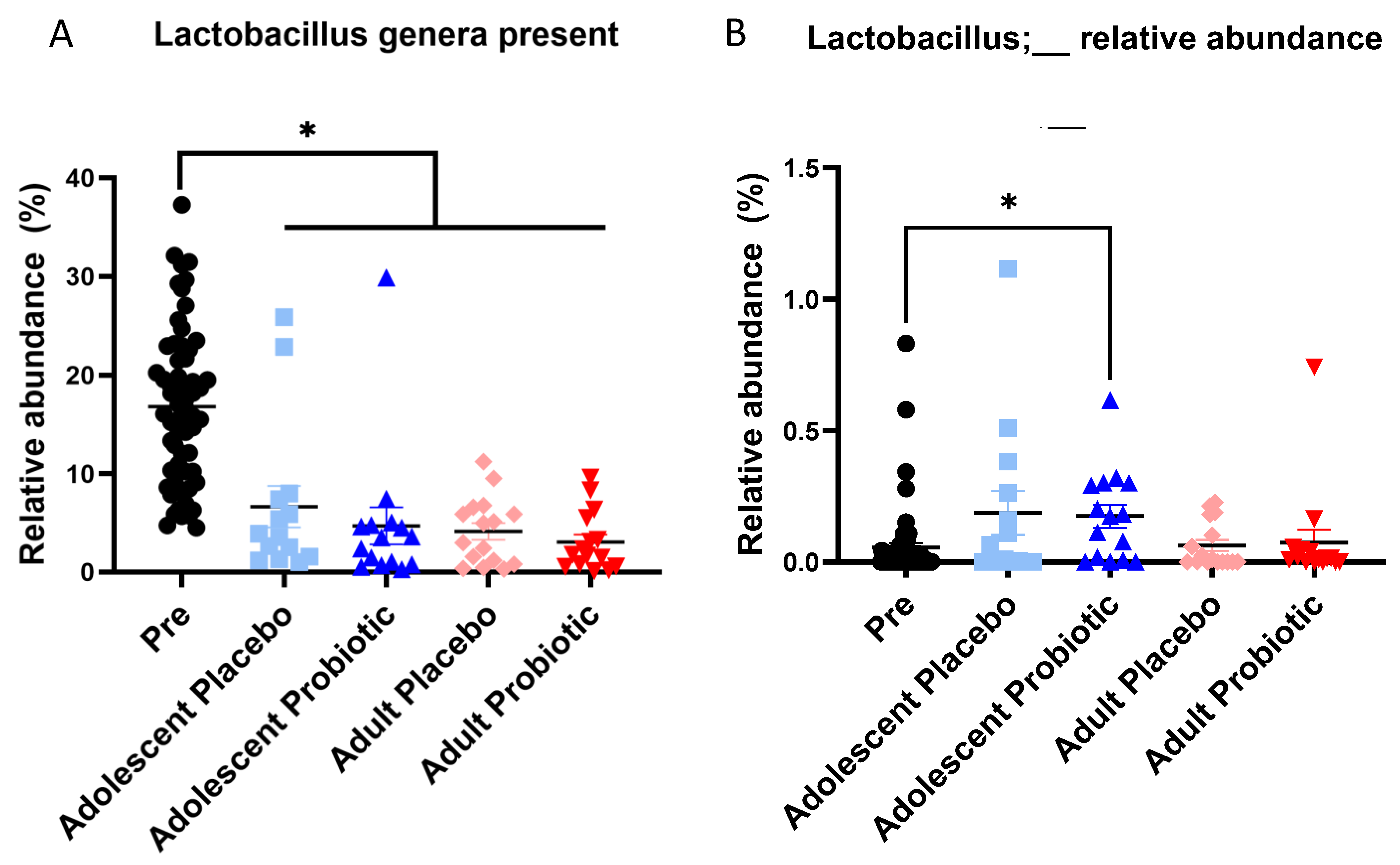
Figure 1. Presence of single-strain probiotic in fecal samples. The left panel (A) shows the relative abundance of all Lactobacillus genera found in the dataset (unknown species, L. intestinalis, L. johnsonii, L. murinus, and L. reuteri). The right panel (B) shows the relative abundance of the unknown Lactobacillus species, which could be L. plantarum. Pre indicates fecal samples at weaning before HFD and/or probiotic was added to the diet. Wilcoxon’s signed rank test: * indicates p < 0.05 between Pre and each treatment indicated with a horizontal line. Pre N = 60, adolescent placebo N = 14, adolescent probiotic N = 15, adult placebo N = 16, adult probiotic N = 15.
Obesities 05 00017 g001
Figure 2. Adolescent and adult rats fed a high-fat diet and treated with probiotic had an altered microbiome compared to rats not given a probiotic. (A). Unweighted UniFrac PCoA of adolescent rats fed a high-fat diet treated with either control (light blue) or probiotic (dark blue); probiotic: p = 0.01, R2 = 0.200; cage: p = 0.041, R2 = 0.390. (B). Unweighted UniFrac PCoA of adult rats fed a high-fat diet treated with either control (light red) or probiotic (dark red); p = 0.002, R2 = 0.0825; cage: p = 0.001, R2 = 0.792. ADONIS tests were run on Unweighted UniFrac distance matrixes with cage as a covariate.
Obesities 05 00017 g002
Figure 3. Adolescent rats fed a high-fat diet and treated with a probiotic had many differentially enriched taxa, while adult rats on the same treatment regimen did not. (A). Cladogram of LEfSe results showing differentially abundant taxa for placebo-treated and probiotic-treated adolescent rats. (B). Cladogram of LEfSe results showing differentially abundant taxa for placebo-treated and probiotic-treated adult rats. LEfSe was run on the relative abundance tables (collapsed to the genus level) of adolescent and adult rats on an HFD, separately, with parameters set to exclude the Wilcoxon subclass test and an LDA cutoff of 2 was used for significance. All taxa shown in the cladograms were significant via LEfSe.
Obesities 05 00017 g003
Figure 4. Lachnospiraceae and Oscillospiracea are two families with many genera that were differentially abundant based on treatment group in adolescent rats on an HFD. Left panel: relative abundances of genera in the Lachnospiraceae family that were significantly enriched in the probiotic-treated adolescent rats on an HFD. Middle panel: relative abundance of the Oscillospiraceae family that was significantly enriched in the probiotic-treated adolescent rats on an HFD. Right panel: relative abundances of genera in the Oscillospiraceae family that were significantly enriched in the probiotic-treated adolescent rats on an HFD. All taxa chosen were deemed significantly enriched in the adolescent probiotic-treated rats on an HFD compared to placebo-treated adolescent rats on an HFD from the LEfSe test described in Figure 3. Control N = 8, probiotic N = 7. Mann–Whitney, * indicates p < 0.05.
Obesities 05 00017 g004
Figure 5. Adult rats only shared one enriched taxon with the probiotic-treated adolescent rats. (A). Faecalitalea relative abundance for adolescent and adults fed an HFD and given a probiotic. This taxon was statistically enriched via LEfSe in both the adolescent and adult rat groups given a probiotic on an HFD compared to their respective placebo control. (BD). Relative abundance of significant features found in adolescent rats in Figure 4 for adult samples (light and dark red). These taxa were not enriched via LEfSe for the adult rats. Adolescent control N = 8, adolescent probiotic N = 7, adult control N = 8, adult probiotic N = 8. Mann–Whitney, * indicates p < 0.05.
Obesities 05 00017 g005
Table 1. Diet and treatment groups.
Standard-Fat DietHigh-Fat Diet (HFD)Probiotic
Lp299v
PlaceboDuration of Diet +/− Probiotic from Weaning
(Weeks)
n
Adolescent++66
++68
++68
++67
Adult++118
++118
++117
++118
Disclaimer/Publisher’s Note: The statements, opinions and data contained in all publications are solely those of the individual author(s) and contributor(s) and not of MDPI and/or the editor(s). MDPI and/or the editor(s) disclaim responsibility for any injury to people or property resulting from any ideas, methods, instructions or products referred to in the content.