Article

Effect of Degree of Substitution and Molecular Weight on Transfection Efficacy of Starch-Based siRNA Delivery System

1
Department of Chemical Engineering, Ben-Gurion University of the Negev, Beer-Sheva 84105, Israel
2
The Shraga Segal Department of Microbiology, Immunology and Genetics, Faculty of Health Sciences, Ben-Gurion University of the Negev, Beer-Sheva 84105, Israel
*
Author to whom correspondence should be addressed.
These authors contributed equally to this work.
Polysaccharides 2024, 5(4), 580-597; https://doi.org/10.3390/polysaccharides5040037
Submission received: 27 June 2024 / Revised: 15 August 2024 / Accepted: 1 October 2024 / Published: 7 October 2024
(This article belongs to the Special Issue Latest Research on Polysaccharides: Structure and Applications)

Abstract

:
RNA interference (RNAi) is a promising approach for gene therapy in cancers, but it requires carriers to protect and deliver therapeutic small interfering RNA (siRNA) molecules to cancerous cells. Starch-based carriers, such as quaternized starch (Q-Starch), have been shown to be biocompatible and are able to form nanocomplexes with siRNA, but significant electrostatic interactions between the carrier and siRNA prevent its release at the target site. In this study, we aim to characterize the effects of the degree of substitution (DS) and molecular weight (Mw) of Q-Starch on the gene silencing capabilities of the Q-Starch/siRNA transfection system. We show that reducing the DS reduces the electrostatic interactions between Q-Starch and siRNA, which now decomplex at more physiologically relevant conditions, but also affects additional parameters such as complex size while mostly maintaining cellular uptake capabilities. Notably, reducing the DS renders Q-Starch more susceptible to enzymatic degradation by α-amylase during the initial Q-Starch pretreatment. Enzymatic cleavage leads to a reduction in the Mw of Q-Starch, resulting in a 25% enhancement in its transfection capabilities. This study provides a better understanding of the effects of the DS and Mw on the polysaccharide-based siRNA delivery system and indicates that the polysaccharide Mw may be the key factor in determining the transfection efficacy of this system.

1. Introduction

RNAi-based therapy is an emerging approach with immense potential in silencing aberrant genes across various cancers and holds promise for treating other genetic disorders [1]. The precision of siRNA gene regulation offers targeted treatments for various genetic conditions, potentially revolutionizing treatment options and offering hope to patients with previously untreatable conditions. Patisiran, the first FDA-approved RNAi therapeutic, exemplifies this potential by targeting and silencing the expression of the mutant transthyretin protein responsible for amyloid formation [2,3,4]. Similarly, the GalNAc-siRNA conjugate Givosiran shows promise in treating acute hepatic porphyria [5,6]. While RNAi holds great potential for increased efficacy and reduced side effects compared to conventional cancer treatments like chemotherapy, the effective delivery of siRNA into cancer cells remains a critical challenge [3]. This is due to the short blood circulation, poor cellular uptake, and low transfection rates of siRNA [7,8]. RNA-based systems exhibit poor stability due to their 2′-hydroxy group, which can be easily cleaved by enzymes, resulting in no therapeutic effect [4,9]. Thus, there is an urgent need to develop a safe and effective delivery method for siRNA molecules.
An effective siRNA carrier should exhibit certain properties, such as the following: (i) a high complexation ability for siRNA, (ii) protection against enzymatic degradation, and (iii) the ability to facilitate cellular uptake followed by the intracellular release of cargo [4]. Encapsulating siRNA within carrier systems such as lipid nanoparticles or polymer-based vehicles safeguards fragile molecules from degradation and bolsters their stability in the bloodstream [10,11]. Biopolymers, especially polysaccharides, have gained new attention in the field of drug delivery systems [12,13,14,15]. Such carriers are easily chemically modified; are known for their non-toxicity, biocompatibility, and biodegradability; and are also an economically viable alternative to other systems [16,17,18,19,20]. Previous research in our lab has been focused on quaternized starch (Q-Starch), which has been shown to have many applications in the delivery of therapeutic compounds [21,22,23]. Amar-Lewis et al. [23], Blitsman et al. [24], Sieradzki et al. [25], and Lifshiz Zimonet et al. [26] have modified starch using a quaternary amine group to form a highly substituted, positively charged non-viral carrier for a wide range of therapeutic molecules, such as siRNA, pDNA, and phosphatidylinositol (3,4,5)-trisphosphate (PIP3). This Q-Starch can self-assemble with negatively charged therapeutic molecules to form a nano-delivery system (complex) that can overcome charge- and size-related cellular barrier systems [27,28], with the intracellular path of Q-Starch/cargo complexes being guided by the cargo itself aligning with its unique biological activity site [24]. In terms of Q-Starch/siRNA complexes, Amar-Lewis et al. [23] have indicated that achieving precise release at the target site remains a significant challenge. While Q-Starch provides protection and guidance, the main obstacle lies in siRNA release at the target site which limits transfection kinetics and therapeutic efficacy.
Here, we hypothesized that siRNA release is impeded because of strong electrostatic interactions between Q-Starch and siRNA, ultimately reducing gene silencing efficacy. In this study, we explored the effect of different parameters, such as the degree of substitution (DS) and molecular weight (Mw) of the starch carrier, on the transfection efficacy of the system. In this work, different Q-Starches will be referred to according to their DS, using the nomenclature of Q-Starch(DS). As Q-Starch(DS)/siRNA complexes self-assemble electrostatically, we infer that they should also likely spontaneously dissociate as a result of local fluctuations in ionic strength, which result in a reduction in siRNA affinity towards positively charged Q-Starch(DS). The total approximate ionic strength in the blood plasma and intercellular space is around 0.15 M [29,30,31], which should serve as an approximate target ionic strength for decomplexation. We assume that lowering the DS of Q-Starch(DS) will weaken its electrostatic affinity for siRNA, thus facilitating decomplexation under more physiologically relevant conditions, but note that this may have an adverse effect on cellular uptake and complex stability [32]. There is no general rule for the effect of molecular weights and degrees of substitutions on transfection efficacy for polymer-based siRNA carriers. For example, in the case of chitosan-based siRNA carriers, it has been reported that a higher DS is correlated with improved transfection [33,34,35], with lower-Mw chitosan also showing improved transfection efficacy relative to higher-Mw chitosan [36]. However, in the case of polyethyleneimine (PEI), a synthetic cationic polymer, a higher Mw has been associated with improved transfection efficacy, albeit with higher cytotoxicity [37,38]. We find starch a more attractive candidate with respect to these polymers, as unlike the case of chitosan, the amount of positive charge can be easily controlled, and unlike PEI, the cytotoxicity is almost non-existent.
Thus, it is of extreme importance to characterize the effect of these two parameters on our starch-based transfection system, as there is no universal rule for the effect of these parameters on the efficacy of different polymer-based siRNA carriers. In this work, we aim to find a sweet spot that balances complex stability with siRNA release. Regarding Mw, we estimate that a higher Mw may result in improved encapsulation efficiency and complex stability but may hinder intracellular siRNA release and adversely affect the efficiency of transfection.

2. Materials and Methods

2.1. Starch Quaternization

Quaternary starch synthesis was carried out as previously described by Amar Lewis et al. [23]. A range of quaternized starches were synthesized, with varying degrees of substitution (DSs). For each sample, 500 mg of soluble potato starch (hydrolyzed potato starch, Mw 105 kDa, Sigma S-2630, St. Louis, MI, USA) was dissolved in 10 mL of sodium hydroxide solution (0.19 g/mL) to obtain 50 mg/mL starch concentration. The solution was then stirred (100 rpm, AGE magnetic stirrer, Velp Scientifica, Usmate Velate, Italy) for 40 min at room temperature. At this point, different amounts of the quaternization reagent, 3-chloro-2-hydroxypropyltrimethylammonium chloride (CHMAC) solution, were used for each synthesis, in order to achieve a different DS for each Q-Starch sample. The highest DS was achieved by adding 9.00 g (0.048 mol, 7.8 mL) of the quaternization reagent slowly to the starch solution. For the remaining five samples, 6.75 g (0.036 mol, 5.9 mL), 4.50 g (0.024 mol, 3.9 mL), 2.25 g (0.012 mol, 1.9 mL), 0.98 g (0.005 mol, 0.85 mL), and 0.60 g (0.003 mol, 0.50 mL) were added, respectively, in descending order of the degree of quaternization.
The product was then precipitated by slowly adding it to a 160 mL solution of an acidified mixture of ethanol and acetone (comprising 40 mL ethanol and 118 mL acetone, acidified with 1.6 mL of HCl 32% wt). The precipitate was then washed 4 times with 25 mL of 80% ethanol (v/v %, 100 mL total washing volume), dissolved in a small volume (5–10 mL) of double-distilled water (DDW, 18.3 MΩ·cm), and poured into a 12–14 kDa Mw cut-off dialysis bag (D9652 Dialysis cellulose membrane, Sigma-Aldrich Inc). The dialysis bag was placed in a vessel containing 5 L of DDW, which was replaced twice with fresh DDW during 48 h of dialysis. The dialyzed product was then freeze-dried and lyophilized for 72 h to obtain the purified quaternized starch (Q-Starch) products.
The different Q-Starches will be referred to according to their DS, using the nomenclature of Q-Starch(DS), as summarized in Table 1 below.

2.2. Quaternized Starch Chemical Analysis

Starch quaternization was confirmed by Fourier transform infrared spectroscopy (FTIR) and elemental analysis (EA). FTIR spectra were obtained in a Thermo-Nicolet FTIR spectrophotometer (Model-Nicolet iS50 FTIR, Waltham, MA, USA), and samples were prepared in the form of potassium bromide (KBr) pellets. Nitrogen content (N% weight) in the Q-Starch(DS) product was measured by the elemental analysis method using a Thermo Scientific FLASH 2000 NC Analyzer, Waltham, MA, USA.
The DS of each Q-Starch was calculated based on the results of the EA measurements using Equation S1 (Supplementary Information) and is summarized in Table 1 (for nomenclature purposes) together with the concentration used for each sample.

2.3. Q-Starch(DS)/siRNA Complex Preparation

Noncoding siRNA NC5 (siNC5) was purchased from Integrated DNA Technologies Inc. (IDT, ref no. 231781443, Coralville, IA, USA). The siNC5 sense sequence was 5′-CUAACGCGACUAUACGCGCAAUAUGGU-3′ (Mw of around 16,500 g/mol). For in vitro cellular uptake experiments, fluorescently labeled siRNA NC5 (siNC5cy5) was purchased from Dharmacon™, Lafayette, CO, USA, with a sense sequence of 5′-cy5-CUAACGCGACUAUACGCGCAAUAUGGU-3′ (identical to siNC5 other than the cy5 label on the 5′ end, Mw of around 17,300 g/mol). For in vitro gene silencing experiments, siRNA targeting the EGFR gene (siRNAEGFR) was purchased from Dharmacon™, with a sense sequence of 5′- GUAACAAGCUCACGCAGUU-3′ (Mw of around 13,400 g/mol).
Q-Starch(DS)/siRNA complexes were prepared at different N/P molar ratios. The N/P ratio refers to the molar ratio between the positively charged amine groups (N) of quaternized starch and the negatively charged nucleic acid phosphate groups (P) of siRNA, with the amount of siRNA remaining fixed in each experiment. Q-Starch(DS) was dissolved in diethyl pyrocarbonate (DEPC)-treated water (Sterile, RNase- and DNase-free water) to a concentration of 0.4 mg/mL for Q-Starch(0.30), Q-Starch(0.44), and Q-Starch(0.59), while a concentration of 1 mg/mL was used for the next Q-Starch(0.08) and Q-Starch(0.12). The least substituted Q-Starch(0.05) was dissolved at a concentration of 2 mg/mL, as a lower concentration would require an excessive amount of Q-Starch(DS) solution. siRNA was dissolved in DEPC-treated water to a concentration of 20 μM. Aliquots of Q-Starch(DS) solution were added to the siRNA solution, which was diluted to the required concentration and N/P ratio, and the solution was gently vortexed (Vortex-Genie 2, Scientific Industries, Inc., Bohemia, NY, USA) and incubated at room temperature for 40 min to allow complexes to electrostatically self-assemble. For different N/P ratios, the volume of starch solution was changed accordingly.

2.4. Q-Starch(DS)/siRNA Complex Characterization

Q-Starch(DS)/siRNA complexes were characterized using the following methods. All characterizations were conducted using siNC5 which was around 27 base pairs long. Other siRNA cargoes may slightly alter the physical properties of the complexes, but this is likely not significant.

2.4.1. Agarose Gel Electrophoresis

Agarose gel electrophoresis was conducted to evaluate the complexes’ formation and the desired N/P molar ratio for full Q-Starch(DS)/siRNA complexation. In some experiments, Q-Starch(DS) was pretreated with the α-amylase enzyme (10069, Merk, Burlington, MA, USA) for 24 h at 37 °C before complex formation, to assess the effect of Q-Starch(DS) cleavage on complexation ability. A total of 4.5 g of agarose powder was dissolved in 150 mL ×1 Tris/acetate/EDTA (TAE) buffer solution (composed of 40 mM Tris/acetate and 1 mM EDTA sodium salt dehydrate, pH 8.0). The solution was heated in a microwave until all of the powder dissolved, then stained with 8 μL ethidium bromide (0.2 μg/mL), and then poured into a horizontal electrophoresis apparatus (Wide Mini-Sub cell GT, BioRad, Hercules, CA, USA) with a 15-well plastic comb to form the loading wells. After letting the gel solidify, samples containing 0.8 μg (3 μL of 20 μM solution) of siNC5, either alone or complexed with Q-Starches at a desired N/P ratio, were mixed with ×6 loading buffer and loaded (24 μL) into the wells in agarose gel (3% w/v). The gel was exposed to an electric field (100 V) for 30 min and then visualized by UV illumination (Visible and ultraviolet transilluminator, DNR Bio-Imaging Systems, Jerusalem, Israel).

2.4.2. Dynamic Light Scattering (DLS)

The hydrodynamic radius of the different Q-Starch(DS)/siRNA complexes was measured by DLS. Samples were prepared at different N/P molar ratios as described above, at a final siRNA concentration of 250 nM in a 1.7 mL Eppendorf tube with a final complex solution volume of 240 μL. The spectra were collected using a CGS-3 (ALV, Langen, Germany) goniometer. The laser was powered with 20 mW at the He-Ne laser line (632.8 nm). Correlograms were calculated by an ALV/LSE 5003 cross-correlator, oriented at 90° for 15 runs at 20 s per run, at 25 °C. Each sample was placed into a thin-walled cylindrical glass cuvette and then placed in a vat filled with toluene as the optical matching fluid. Each complex sample was measured a minimum of three times.

2.4.3. Ionic Strength Agarose Gel Electrophoresis

Agarose gel electrophoresis was conducted to evaluate the complexes’ relative electrostatic strength. Ethidium bromide-stained agarose gels were prepared in a manner identical to the one described in Section 2.4.1 above. A total of 20 μL of Q-Starch(DS)/siRNA complexes was prepared at an N/P ratio of 2, as described above. After waiting approximately 30 min to allow for complex formation, equal parts of NaCl solution were added to the complex solution. Different NaCl solutions were prepared by diluting a master NaCl 5 M solution, which was then mixed with the complex solution, in order to reach a final ionic strength between 0 and 1.5 M. After incubation for another 5 min, the complex/salt solutions were mixed with ×6 loading buffer and loaded in duplicates (24 μL each) into the wells of agarose gel (3% w/v). The gel was exposed to an electric field (100 V) for 30 min and then visualized by UV illumination (Visible and Ultraviolet Transilluminator, DNR Bio-Imaging Systems).

2.4.4. Zeta Potential

The zeta potential of the complexes, indicative of each complex’s effective surface charge, was determined by Zetasizer. The Q-Starch(DS)/siRNA complexes, which were prepared for the DLS measurements, were quickly diluted after measurement to a final volume of 1 mL Samples were transferred to a U-tube cuvette (DTS1070C, Malvern, UK) for the measurement of zeta potential using Zetasizer (ZN-NanoSizer, Malvern, UK). Each sample was measured in automatic mode, at 25 °C, and the Smoluchowski model was used to calculate the zeta potential.

2.4.5. Cryo-Transmission Electron Microscopy (cryoTEM)

The morphology of some of the complexes, as well as the confirmation of their size, was evaluated by cryo-TEM. Complexes were prepared in an identical manner to the preparation of the complexes used for DLS described above. A drop of 2.5 µL of the complex solution was placed on a copper grid coated with a perforated lacey carbon 300 mesh (Ted Pella Inc., Redding, CA, USA) under controlled temperature (cryogenic temperature). The excess liquid was blotted with filter paper to form a thin liquid layer, and the specimen was vitrified via rapid plunging into liquid ethane at its freezing point (−183 °C) using Plunger (Leica EM GP, Wetzlar, Germany). The specimens were transferred into liquid nitrogen for storage. Samples were analyzed using the FEI Tecnai 12 G2 Transmission Electron Microscope at 120 kV with a Gatan cryo-holder maintained at −180 °C. Images were recorded on a slow-scan cooled charge-coupled device camera (Gatan, Pleasanton, CA, USA) at low-dose conditions to minimize electron-beam radiation damage. The recording was carried out using the Digital Micrograph software package version 3.5.

2.5. Cell Culture Handling

HNSCC cell line Cal33 (human, tongue squamous cell carcinoma (SCC)) was selected as an in vitro model in order to study the cellular uptake and transfection efficacy of Q-Starch(DS)/siRNA complexes. Cal33 cells were cultured in Dulbecco’s Modified Eagle Medium (DMEM) growth media containing 10% vol. fetal bovine serum (FBS), 1% vol. L-glutamine (2 mM), and 1% vol. penicillin/streptomycin. Cells were grown in a 75 cm2 flask. The splitting of cells was performed with 2–3 mL of trypsin/EDTA every 3–4 days into 3–4 flasks to prevent the high cell confluence and overpopulation of the culture. Following splitting, trypsin was neutralized with 10 mL of growth medium. Cells were then well-pipetted to ensure a homogeneous suspension and counted by the Countess™ II FL Automated Cell Counter. Trypan blue stain was used for counting viable cells by mixing 20 µL of the cell’s suspension extract with 20 µL of 0.4% trypan blue solution (1:1 v/v ratio). All cell culture procedures were performed inside a laminar-flow hood that was sterilized before each procedure by UV light and by wiping the working area with 70% ethanol.

2.6. In Vitro Cellular Uptake

For in vitro cellular uptake experiments, fluorescently labeled siNC5cy5 (excitation at 640 nm) was complexed with the different Q-Starches, and cellular uptake was quantified using flow cytometry. Cal33 cells were seeded in a 6-well plate 24 h before transfection in DMEM growth media at a density of around 1 × 10⁵ cells/well. On the day of transfection, the culture medium was removed, and 1600 μL of fresh growth media was added. Q-Starch(DS)/siNC5cy5 complexes were prepared at an N/P molar ratio of 2 as described above, and 400 μL of the complex master solution was added to the cells to reach a final siRNA concentration of 50 nM in each well.
Cells were incubated for 24 h with the complexes, after which the cells were washed gently twice with PBS to ensure the removal of free particles that could stay in the media. The adherent cells were then disconnected from the wells by adding 300 μL of trypsin/EDTA and incubating for 10 min in a sterile incubator. Following incubation, 1 mL of growth medium was added to neutralize trypsin activity. The suspended cells were pipetted 3–6 times to assure uniform suspension and then transferred into a sterilized Eppendorf tube (1.5 mL). The cells were centrifuged for 5 min at 260× g at room temperature. After centrifugation, the medium was removed gently, and the cells were resuspended in 500 μL of buffer solution (1% FBS in PBS) and saved on ice in the dark for less than 30 min. The fluorescence unit of the cells was measured by FACSAria III (three repetitions for each experimental group, n = 3). The Relative Fluorescent Unit (RFU) of the treated cells was normalized to the RFU of the untreated cells (i.e., reduced autofluorescence). Untreated cells were taken to have 0% uptake.

2.7. siRNA: EGFR Gene Silencing

EGFR gene silencing experiments were conducted by seeding Cal33 cells in a 6-well plate for 24 h in DMEM growth media at a concentration of 1 × 10⁵ cells/well to reach a confluence of approximately 60–70%. On the day of transfection, Q-Starch(DS)/siRNA complexes, either with non-targeting siRNA (siNC5) or with siRNA targeting the EGFR gene (siRNAEGFR), were prepared at an N/P molar ratio of 2. Some of the Q-Starches were pretreated with the α-amylase enzyme for 24 h at 37 °C to induce cleavage. Before transfection, cell medium was replaced, and complexes were placed on top of the cells at a final siRNA concentration of 50 nM and a final volume of 1 mL/well and incubated at 37 °C and 5% CO2 for 72 h. Following incubation, a series of RNA extraction and purification steps were performed using the PureLinkTM RNA Mini Kit (12183020, Invitrogen, Waltham, MA, USA) to extract the cells’ RNA. Samples were then prepared using the QuibitTM RNA BR Assay Kit (Q10210, Invitrogen), and RNA concentration was measured by the Qubit 3.0 Fluorometer (Q33216, Life Technologies, Carlsbad, CA, USA). In line with the manufacturer’s instructions, a master mix solution was prepared on ice by gently mixing nuclease-free water, buffer, and enzymes from a qScriptTM cDNA Synthesis Kit (95047-100, Quanta-bio, Beverly, MA, USA) in a vol. % ratio of 5:4:1 (i.e., 50 µL of nuclease-free water was mixed with 40 µL buffer and 10 µL enzymes). RNA samples were prepared for polymerase chain reaction (PCR) analysis as follows: 10 µL from the master mix was added to 20 µL of the RNA samples in a PCR tube to reach a concentration of 20 ng/µL RNA; then, it was placed in a PCR (Peltier-based Thermal Cycler MGL96+, MyGeneTM L Series, HY-Labs, Rehovot, Israel) to obtain cDNA. The produced cDNAs were subjected to real-time polymerase chain reaction (RT- PCR) testing (StepOnePlus™ Real-Time PCR System, 4376600, Rhenium, Modi’in-Maccabim-Re’ut, Israel) to determine gene silencing by quantifying EGFR’s mRNA expression. cDNAs were diluted from the produced PCR stock to 10 ng/µL (1:2 with nuclease-free water). A stock solution was prepared on ice by gently mixing nuclease-free water, master mix solution (TaqMan™ Fast Advanced Master Mix, 4444557, Applied Biosystems, Waltham, MA, USA), and TaqMan probe and primers (TaqMan®® Gene Expression Assay, EGFR: Hs1076078, HPRT: Hs99999909, Rhenium) in a ratio of 6:10:1 (i.e., 60 µL of nuclease-free water was mixed with 100 µL from the master mix and 10 µL of probe and primer assay). Then, 3 µL of the cDNA sample was added to a well in a MicroAmp™ Fast Optical 96-Well Reaction Plate with Barcode (0.1 mL, 4346906, Applied Biosystems), followed by the addition of 17 µL of stock solution to reach a total volume of 20 µL in each well. Each group consisted of three different wells, resulting in three repetitions for each group. All gene expression values were normalized to the siRNANC5 treatment, which was considered as 100% gene expression and normalized to an unaffected housekeeping gene HPRT.

2.8. Iodine Starch Test

An iodine starch test was conducted to test the ability of α-amylase to break down Q-Starch(DS) into individual maltose units. Uncleaved starch forms a dark blue-purple complex, attributed to the structural arrangement of amylose chains within starch molecules. A 1 wt.% aqueous solution of Q-Starch(DS) was prepared and was combined with equal parts of an α-amylase solution at concentrations ranging from 0.0625 to 1 wt.% (at a final volume of 200 µL). This mixture was incubated for 10 min at 37 °C after thorough mixing. Subsequently, after incubation, an equal part of iodine solution (prepared by dissolving 4 g of KI and 1.3 g of I2 in 100 mL of DDW) was added, for a final volume of 400 µL. The solution’s color change was observed.

2.9. Viscosity Measurements

The viscosity of Q-Starch(DS) solutions after incubation with α-amylase was measured on an Anton Parr MCR702 dual-motor rheometer using measuring cup CCC27/MD/PTD/TS and cylinder ME27-4.635-40 geometries and an advanced Peltier system for temperature control. The experimental shear rate range was in 1–1000 1/s. The molecular weight of native starch (105 kDa) was assessed by measuring intrinsic viscosity, using Mark Houwink equation parameters of alfa = 0.5 and k = 0.098.

2.10. Statistical Analysis

Statistical analysis was performed using GraphPad Prism 10.2 software, presented as the mean ± SD/SEM. All cellular experiments were repeated at least three times. For experiments involving two groups, a two-tailed Student’s unpaired t test was performed to compare the control versus treatment groups. Values of p ≤ 0.05 were considered significant.

3. Results and Discussion

3.1. Q-Starch Synthesis and Chemical Characterization

To determine the effects of the DS on the decomplexation process, a series of Q-Starch(DS) was synthesized. Q-Starch(DS) synthesis was performed to produce a positively charged carrier, intended to electrostatically interact with negatively charged siRNA, to form a positively charged complex capable of penetrating the cellular membrane. A biodegradable, biocompatible, natural starch was chosen as a non-toxic polysaccharide carrier. The polymer’s backbone was then modified for the creation of the desired positively charged carrier by substituting a quaternary amine group to the polymer’s backbone, as described by Amar et al. [23]. The quaternization reagent used was 2-chloro-2-hydroxypropyltrimethylammonium chloride (CHMAC). As can be seen in Figure 1A, the DS can be highly dependent on steric hinderances and the electrostatic repulsion of the different groups of the quaternized starch product. It can be deduced that the most reactive hydroxyl group is likely the one on the 6′ position in each glucose monomer, since the other hydroxyl groups (on the 3′ and 4′ positions) in each monomer are more sterically hindered. Quaternization synthesis is also influenced by temperature, humidity, the duration of the reaction, and the concentrations of the reagents.
Table 1 presents the elemental analysis (EA) results, which calculates the nitrogen content (wt.%) of the different samples according to the dynamic flash combustion method. The nitrogen content for the most substituted sample was found to be 3.3%, which is consistent with previous work conducted in our lab [23,24,25,26]. The DS and its reciprocal are also presented in Table 1, which serves as an indication of approximately how often a substitution occurs, in the number of glucose units. The results show that even in the case of Q-Starch(0.59), with the highest DS, the most substituted Q-Starch, there is on average less than one substitution per monomer, as indicated by the inverse of the DS. The nitrogen content of the other samples was shown to decrease, as the amount of quaternization reagent was reduced, as shown in Figure 1B. This serves as an indication of the relative simplicity of the control of the DS of our carriers, which can be controlled easily by reducing or increasing the amount of quaternization reagent while keeping the other parameters constant.
Fourier transform infrared spectroscopy (FTIR) was used to identify the presence of C-N bonds in the synthesized Q-Starches. The FTIR spectrum of the Q-Starches reveals an important peak relative to the spectrum of native starch, at around 1479 cm−1 (Figure 1C). A magnification of the relevant peak is shown in Figure 1D. This peak is typical of carbon/nitrogen vibrations, indicating the presence of a C-N bond in the Q-Starches. It can also be seen that the intensity of said peak decreases as the DS of the Q-Starch decreases, which is also consistent with the fact that the nitrogen content decreases accordingly. This peak, together with the EA results, confirms the substitution of starch by a quaternary amine group for all Q-Starches.

3.2. Q-Starch(DS)/siRNA Complex Formation and Characterization

Q-Starch(DS)/siRNA complexes are formed due to electrostatic attraction forces between the positively charged nitrogen groups and the negatively charged phosphate groups of siRNA sequences. We hypothesize that reducing the DS should lead to a reduction in electrostatic affinity towards negatively charged siRNA and thus lead to electrostatically weaker complexes with improved decomplexation. This is shown representatively in Figure 2A, for the two Q-Starches, Q-Starch(0.05) and Q-Starch(0.59), with the lowest and highest DS, respectively.
The desired molar ratio between the nitrogen and phosphate groups (N/P ratio) for the synthesized Q-Starches was determined by agarose gel electrophoresis. A representative image of the results for Q-Starch(0.12) is shown in Figure 2B, while the results for the remaining Q-Starches are shown in Figures S1–S5. It is important to determine at what N/P molar ratio full siRNA complexation is achieved, as free siRNA that is not complexed with Q-Starch(DS) will undergo rapid enzymatic degradation and thus has little therapeutic value as it will likely not contribute to gene silencing. However, one must also consider that the N/P ratio affects the electrostatic strength of the complexes (i.e., the interactions between the drug and the carrier), which in turn determines their condensation, which ultimately might prevent the siRNAs’ decomplexation and participation in gene silencing. Thus, an optimal N/P ratio (in terms of electrostatic considerations) would be the minimal N/P ratio at which full siRNA complexation is observed. However, one must also ensure that at this N/P ratio, the complexes exhibit suitable sizes and effective surface charges for cellular uptake. As can be seen in Figure 2B, a running siRNA band can be observed for free siRNA and for an N/P ratio of 0.5, meaning that at this ratio, there is still free siRNA that has not been complexed with the Q-Starch(DS). There is no noticeable siRNA band at an N/P ratio of 1, meaning that at around this ratio, full complexation is achieved. From this ratio onwards, the bands are the most prominent near the cathode, meaning that siRNA remains in the well as part of the Q-Starch(DS) complex. Similar results were received for the other Q-Starches (Figures S1–S5), indicating that despite the difference in the DS, all Q-Starches exhibit the same behavior in terms of complexation abilities. However, more Q-Starch(DS) is required to complex the same amount of siRNA for less substituted Q-Starches.
Figure 2C presents the zeta potential for the four Q-Starch(DS)/siRNA complexes with the highest DS, which were characterized at N/P ratios of 1, 2, and 3. It can be seen that at an N/P molar ratio of 1, a negative zeta potential is observed for all complexes except for Q-Starch(0.12). This may correspond to free siRNA or siRNA that is more concentrated on the surfaces of the complexes and may indicate that the full complexation which was assumed by the results of gel electrophoresis (Figure 2B) is not entirely correct. As the N/P ratio increases, an increase is noted in the zeta potential as expected, since there are more positive charges. However, the increase in zeta potential when increasing the N/P ratio from 2 to 3 is not statistically significant. It should be noted that for all complexes, the zeta potential is under the stability threshold. This indicates that the complexes may tend to aggregate over time, which on one hand might prevent their penetration through the cell membrane once in an aggregative state but on the other hand may enhance decomplexation if it were to occur after cellular uptake. Because of these zeta potential findings, it was decided to continue characterizing the Q-Starch(DS)/siRNA complexes at an N/P molar ratio of 2 and above to ensure full siRNA complexation. It should be noted that we were unable to successfully measure the zeta potential for the two least substituted Q-Starches, likely because of complex instability.

3.3. Q-Starch(DS)/siRNA Electrostatic Strengths and Size Distributions

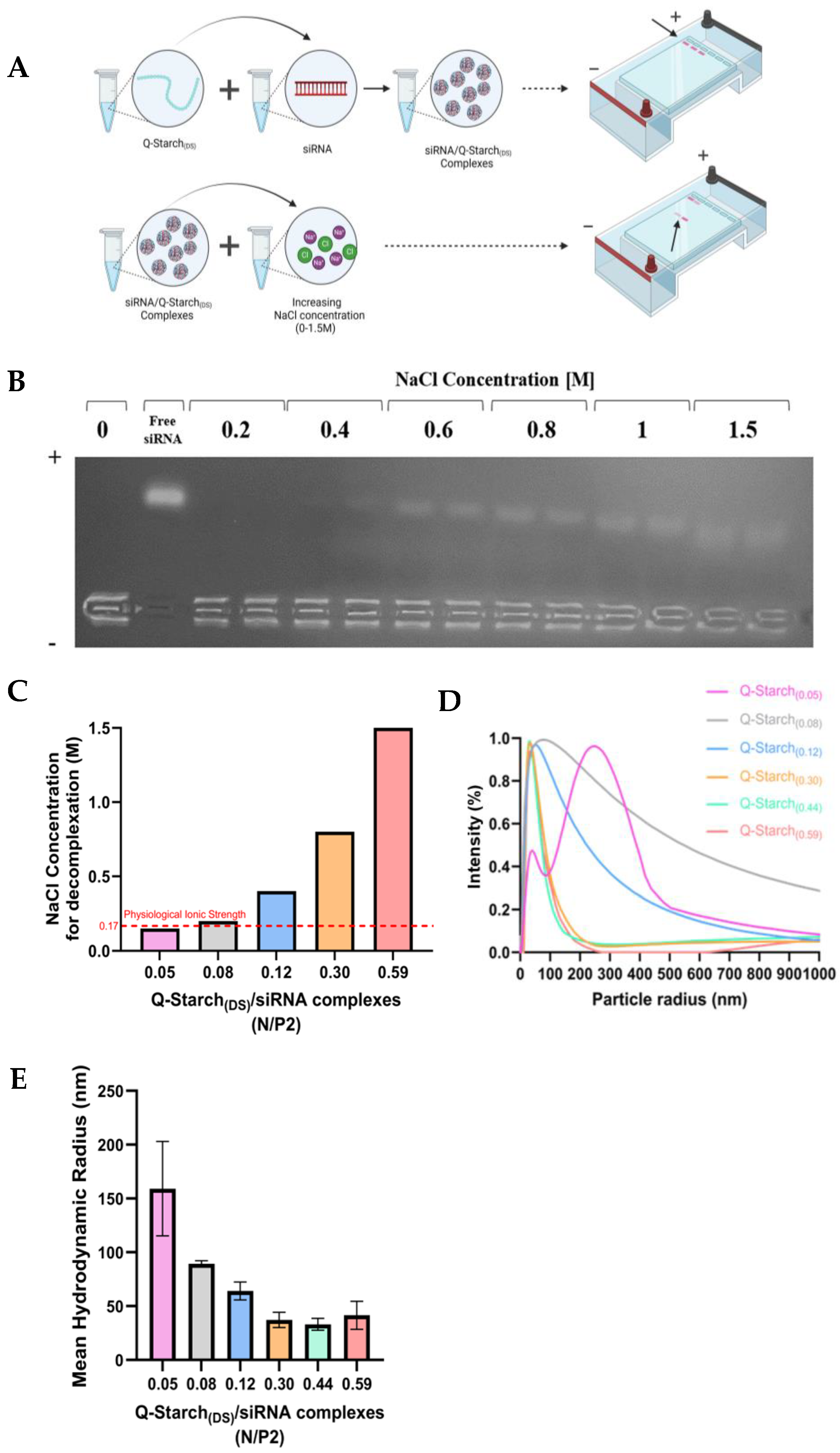
The electrostatic strength of Q-Starch(DS)/siRNA was tested by preparing and incubating the complexes with different NaCl solutions, at varying ionic strengths, to see whether decomplexation is observed under gel electrophoresis. Salt competes with Q-Starch’s positively charged groups for interactions with siRNA, meaning that at higher ionic strengths, more siRNA should be observed free-running in the gel; an illustration depicting the experimental procedure appears in Figure 3A. Examining the ionic strength at which decomplexation is observed is crucial, as this is most likely the main mechanism for decomplexation in our system. As Q-Starch(DS)/siRNA complexes self-assemble electrostatically, they should likely spontaneously dissociate as a result of local fluctuations in ionic strength, which leads to a reduction in siRNA affinity towards positively charged Q-Starch(DS). When looking at biological systems, the total approximate ionic strength in the blood plasma and intercellular space is around 0.15 M [29,30,31], mainly due to sodium ions which are prevalent. The ionic strength inside the cell is slightly higher and stands at around 0.2 M, mainly because of the potassium ion concentration [39]. Thus, the Q-Starch(DS) complexes should be stable in the blood and intercellular space but undergo decomplexation as a result of the increased ionic strength inside the cell.
As can be seen in Figure 3B, free siRNA bands become more distinct as NaCl concentration increases. The same trend persisted for the other Q-Starches (Figures S6–S9), with these experiments showing that a lower salt concentration is required for decomplexation as the DS decreases, indicative of electrostatically weaker complexes. For example, in Figure 3B, the complexes of Q-Starch(0.12) show no visible band at 0.2 M, while the Q-Starch(0.05) complex shows a very distinct band at this concentration (Figure S6). Figure 3C summarizes NaCl concentration in which decomplexation (free siRNA) was first observed in each Q-Starch(DS) complex (at an N/P ratio of 2) and indicates a clear correlation between the DS of Q-Starch(DS) and the electrostatic strength of the Q-Starch(DS)/siRNA complexes, which in turn affects the decomplexation step in siRNA transfection. As stated, a physiologically relevant salt concentration is in the order of 0.15 M, and thus, an effective carrier for siRNA should be stable enough at these conditions. Because of this, both Q-Starch(0.05) and Q-Starch(0.08) are unsuitable candidates for transfection, as they will decomplex before reaching the target site. This is also strengthened by the fact that these complexes also showed decomplexation after incubation in DMEM cell culture media (ionic strength of approximately 0.17 M, Figure S10) [40]. Q-Starch(0.30) and Q-Starch(0.59) will also not likely result in effective gene silencing, as they release siRNA at ionic strengths around 4 and 10 times higher than physiological conditions, and thus, from an electrostatic standpoint, Q-Starch(0.12) is likely the most suitable candidate, exhibiting suitable stability with decomplexation occurring at ionic strengths of around 0.4 M. Additional experiments (Figure S11) also show that increasing the N/P ratio above 2 results in electrostatically stronger complexes, which require higher salt concentrations for decomplexation with the same Q-Starch(DS). Thus, it is likely not beneficial to increase the N/P ratio in terms of transfection efficacy, and an N/P ratio of 2 was chosen for further characterizations and in vitro experiments.
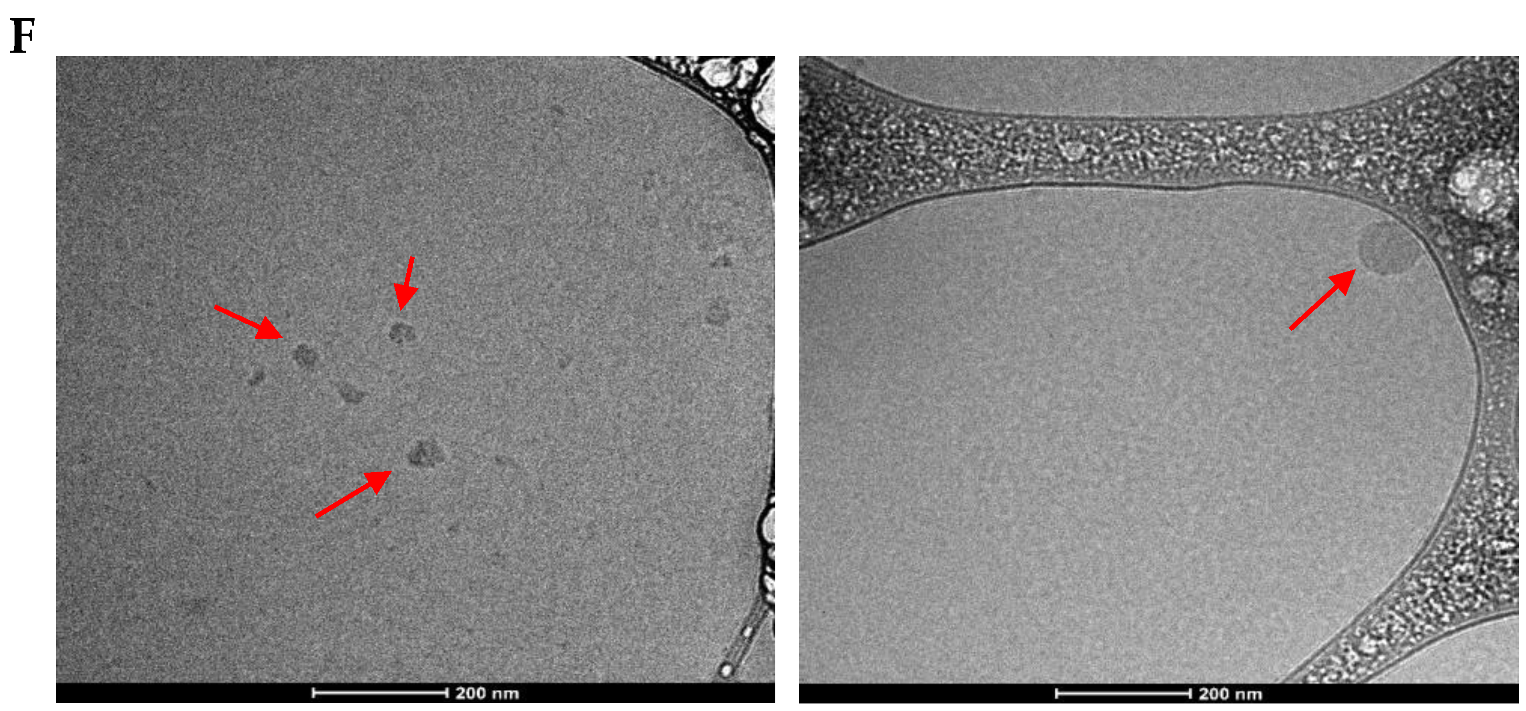
The hydrodynamic radius distribution of our Q-Starch(DS)/siRNA complexes (prepared at an N/P ratio of 2), obtained through dynamic light scattering (DLS), is shown in Figure 3D. The size distributions become progressively wider as the DS decreases, with the mean radius being between 50 and 70 nm for all Q-Starches except Q-Starch(0.08) and Q-Starch(0.05), as shown in Figure 3E. We can attribute the increase in size between the different Q-Starches to the fact that at a certain N/P ratio, more Q-Starch(DS) (N) is required to complex the same amount of siRNA (P) for less substituted Q-Starches, which results in larger complexes. The two least substituted carriers form inconsistently sized complexes, with high variance between measurements. This likely also explains why we were unable to measure the zeta potential of these complexes, which are unstable. Thus, it seems that there is a minimal DS at which Q-Starch(DS) is able to form complexes suitable for cellular uptake, which in our case was found to be 0.12, with resultant complexes having a hydrodynamic radius of around 70 nm. As Q-Starch(0.12) was found to be the most suitable carrier in terms of electrostatic considerations, its complexes were also characterized via cryo-TEM (Figure 3F) to confirm the distribution acquired from DLS measurements. These results demonstrate sphere-like complexes with an approximate diameter in the range of 40–100 nm, which corresponds nicely with the size distribution observed by DLS and establishes these Q-Starch(0.12)/siRNA complexes as nanoparticles capable of penetrating the cellular membrane. After confirming that the four most substituted Q-Starches are all able to form stable nano-sized complexes with siRNA, we proceeded with characterizing their cellular uptake and gene silencing capabilities to test the effect of the DS on these parameters.

3.4. Q-Starch(DS)/siRNA Cellular Uptake and Gene Silencing Capabilities

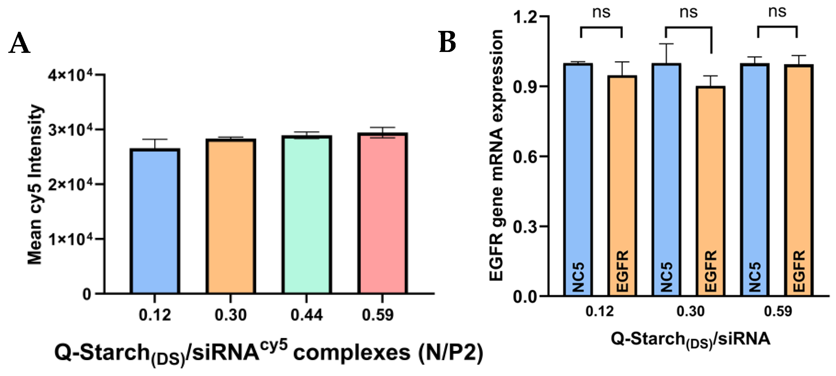
The cellular uptake of Q-Starch(DS)/siRNA complexes was assessed on HNSCC Cal33 cells via flow cytometry, using the four Q-Starches which exhibited stability under biologically relevant ionic strengths, which were complexed with cy5-labeled siRNA. The results are presented in Figure 4A. As shown in Figure 4A, no statistically significant difference was observed in the mean cy5 intensity measured in Cal33 cells following 24 h of incubation with the different Q-Starch(DS) complexes. This indicates that varying the DS does not significantly affect the cellular uptake capabilities of Q-Starch(DS) carriers, i.e., the DS of all carriers likely results in sufficient interaction with the cell membrane. These results aligned with expectations, given the preservation of the particles’ physical characteristics, such as size and charge. Furthermore, from these results, it can be seen that reducing the DS does not hinder the cellular uptake of the complexes, i.e., no new rate-limiting step is introduced.
Three of these Q-Starches were used to conduct in vitro gene silencing experiments on Cal33 cells, assessed via RT-PCR (Figure 4B), in order to test whether reducing the DS indeed leads to improved gene silencing, as a result of the improved decomplexation (Figure 3C). Up to 90% of HNSCC tumors exhibit the overexpression of epidermal growth factor receptor (EGFR), which plays a critical role in HNSCC growth, invasion, metastasis, and angiogenesis [41] .Therefore, we chose to target this receptor as a gene-silencing target. From Figure 4B, it can be seen that no statistically significant reduction in EGFR mRNA expression was observed for any of the Q-Starch(DS) complexes tested. We assess that this is because even in the case of Q-Starch(0.12), decomplexation still occurs at ionic strengths around twice as high as the ionic strengths of biological systems. The remaining Q-Starches likely bind siRNA much too tightly and do not release their cargo at biologically relevant conditions. Despite the fact that some of these Q-Starches displayed siRNA release at ionic strengths between 4 and 10 times higher than physiological ionic strengths, it was still necessary to assess whether an increase in the DS may lead to improved efficacy through other unidentified mechanisms.

3.5. Enzymatic Cleavage of Q-Starch(DS) by α-Amylase

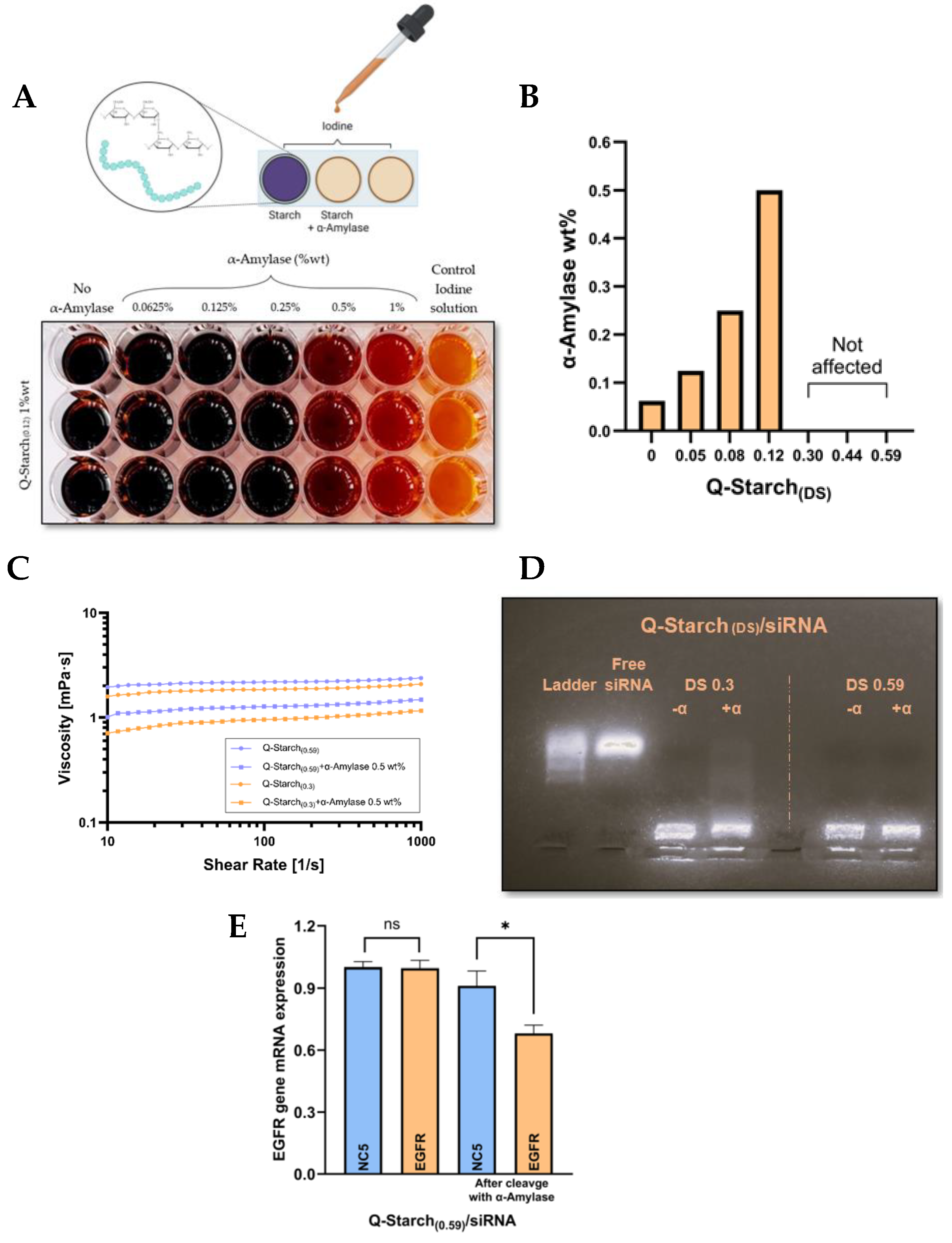
Given that our study indicated that reducing the DS of Q-Starch results in no impact on transfection efficacy, we explored the effects of polymer chain length on the efficacy of the system. As limited data are available regarding the influence of Mw on the efficiency of the starch-based siRNA delivery system, investigating this factor in the context of the Q-Starch(DS)/siRNA transfection system became imperative. We conducted an assessment using enzymatic cleavage by α-amylase (Figure 5A). Since there is an inherent difficulty in precisely controlling the chain lengths of diverse natural polymers, our approach involved regulating starch chain fragmentation by applying the α-amylase enzyme. We first conducted an iodine test to evaluate the capacity of the enzyme to break down Q-Starch(DS) into maltose units. When iodine interacts with starch, it produces a dark blue- or purple-colored complex, attributed to the arrangement of amylose chains within starch molecules. However, this color formation is absent after enzymatic cleavage, establishing a straightforward method to screen the effectiveness of the enzyme across different degrees of Q-Starch(DS) substitution (Figure 5A). The positive groups of Q-Starch(DS) affect its ability to be cleaved by the enzyme, likely through steric interferences. The results of this experiment are summarized in Figure 5B, which demonstrates a clear correlation between the DS and the required enzyme concentration for Q-Starch cleavage. For example, a higher concentration of α-amylase is needed for the cleavage of Q-starch(0.12) in comparison to native starch (Figure 5A,B). As depicted, a decrease in the DS results in more efficient starch degradation in the presence of the enzyme. As can be seen, a DS of 0.30 and above resulted in no observable cleavage at all enzyme concentrations. However, while there is no breakdown into individual maltose units, there is still likely some cleavage to longer starch chains. Viscosity average molecular weight (Mv) determination for Q-Starch (DS) after cleavage with α-amylase is unreliable due to the presence of positive groups on the starch backbone that alters rheological properties. Since the Mark Houwink equation constants, α and k, for modified starch cannot be found in the literature, we base our conclusion on the reduction in Q-Starch (DS) viscosity before and after cleavage, which correlates with chain length [42,43]. A higher molecular weight results in a higher solution viscosity, and a decrease in solution viscosity after enzyme treatment is indicative of successful enzymatic degradation, to some extent. An examination of Q-Starches’ viscosity at varying DSs (0.30 and 0.59) is depicted in Figure 5C.
As anticipated, both solutions exhibit decreased viscosity post-enzyme addition, signifying successful cleavage by the enzyme. There is a larger decrease in the chain length for Q-Starch(0.30), in comparison to Q-Starch(0.59), as expected.
We next opted to generate new complexes from the different Q-Starches post-enzyme cleavage, to test the effect of a reduced Mw. Initially, we cleaved both Q-Starches at DSs of 0.30 and 0.59. Subsequently, we formed complexes and assessed whether the newly formed Q-Starch could bind the same amount of siRNA. As observed in Figure 5D, when cleaving the lesser substituted Q-Starch(0.30), the capacity to form complexes was impaired, and free RNA was observed running towards the anode. However, complete complexation was observed in the highly substituted Q-Starch(0.59), where the cleavage is likely more controlled due to increased steric hindrances. After the successful cleavage of Q-Starch(0.59) into smaller fractions, while still maintaining full siRNA encapsulation capabilities, we proceeded to examine the biological efficacy of the complexes. As observed in Figure 5E, gene suppression was increased by 25% compared to the non-cleaved Q-Starch complexes.
This increase in gene suppression, albeit at an mRNA level, is particularly meaningful, as no silencing was observed without enzyme cleavage, as well as in any other Q-Starches. These experimental findings underscore the critical role of polymer chain length in siRNA release, aligning with the literature suggesting that optimizing chain length is pivotal for enhancing transfection efficiency and therapeutic efficacy. For example, Techaarpornkul et al. show that as the Mw of chitosan decreases, transfection efficiency improves. They argue that the apparent influence of a high Mw of the polymer could lead to a chain entanglement effect, while a short chain may facilitate the release of RNA [36]. Additional research has shown that the efficacy of chitosan is notably suboptimal with a Mw below 10 kDa, as the polymer struggles to form stable complexes and compact siRNA effectively [44], with a higher Mw between 20 and 100 kDa resulting in improved efficacy [36,45]. Another polymer commonly used for siRNA transfection is polyethyleneimine (PEI), a synthetic cationic polymer. High-Mw PEI (HMW-PEI, approximately 25–100 kDa) exhibits higher biological activity (and higher toxicity) relative to low-Mw PEIs (approximately 4–10 kDa) [37,38]. While smaller PEIs show lower cytotoxicity, they also display reduced transfection efficiency due to weaker nucleic acid binding abilities and insufficient protection from nucleases. Branched PEIs are also more effective at delivering siRNA than their linear counterparts [37,38]. This indicates that there is no universal rule guiding the effect of Mw on siRNA transfection efficacy, and an optimal Mw should be found for each system, which should be optimized together with the DS. In our starch-based siRNA transfection system, a reduced Mw, indicated by a significant reduction in the polymer solution viscosity from 2.39 to 1.49 mPas, was associated with improved transfection efficacy, likely as a result of reduced chain entanglement.

4. Conclusions

This research provides a better understanding of the effects that the DS and Mw have on the Q-Starch/siRNA delivery system. From electrostatic considerations alone, one may tend to decrease the DS as much as possible. However, this parameter should be optimized together with Mw to take into account additional parameters, such as complex size and cellular uptake capabilities. Notably, varying the DS had little effect on the physical characteristics of the complexes, as well as their cellular uptake capabilities, but significantly improved the decomplexation step. This, however, was not translated into an improved therapeutic effect, which was only achieved after pre-cleaving Q-Starch with the α-amylase enzyme. This indicates that starch Mw is likely the key factor in determining transfection efficacy in this system.
The future development of the personalized gene delivery system could also integrate backbone degradation, i.e., creating a delivery system which will break down as a result of the reducing environment present in cells. Such a system may be based on Q-Starch, with a reduced DS and Mw, conjugated with disulfide groups that will be reduced inside the cell, thus further enhancing siRNA release.

Supplementary Materials

The following supporting information can be downloaded at https://www.mdpi.com/article/10.3390/polysaccharides5040037/s1, Equation S1: Degree of substitution (DS) calculation of quaternized starch; Figures S1–S5. Q-Starch(DS)/siRNA complex formation evaluated by agarose gel electrophoresis at increasing N/P molar ratios; Figures S6–S9: The agarose gel electrophoresis of Q-Starch(DS)/siRNA complexes formed at an N/P molar ratio of 2 at increasing ionic strengths; Figure S10: Agarose gel electrophoresis with free and full culture media for different Q-Starch(DS)/siRNA complexes formed at an N/P molar ratio of 2; Figure S11: NaCl concentration in which decomplexation was first observed in each Q-Starch.

Author Contributions

Conceptualization, A.R., C.B., R.G., T.T., M.E. and J.K.; methodology, A.R. and C.B.; validation, A.R. and C.B.; investigation, A.R. and C.B.; writing—original draft preparation, A.R. and C.B.; writing—review and editing, R.G., T.T., M.E. and J.K.; supervision, M.E. and J.K. All authors have read and agreed to the published version of the manuscript.

Funding

This research received no external funding.

Institutional Review Board Statement

Not applicable.

Data Availability Statement

The raw data supporting the conclusions of this article will be made available by the authors on request.

Acknowledgments

The authors are grateful to Bar Elisha from the Ilse Katz Institute for Nanoscale Science and Technology for his assistance with elemental analysis measurements. The authors, Chen Benafsha and Amir Regev, would also like to extend their appreciation for the fellowship from the Dubrowski Scholarship for Cancer Research and the Interdisciplinary Research Fund, respectively.

Conflicts of Interest

The authors declare no conflicts of interest.

References

  1. Egorova, A.; Petrosyan, M.; Maretina, M.; Bazian, E.; Krylova, I.; Baranov, V.; Kiselev, A. IRGD-Targeted Peptide Nanoparticles for Anti-Angiogenic RNAi-Based Therapy of Endometriosis. Pharmaceutics 2023, 15, 2108. [Google Scholar] [CrossRef] [PubMed]
  2. Weng, Y.; Xiao, H.; Zhang, J.; Liang, X.J.; Huang, Y. RNAi Therapeutic and Its Innovative Biotechnological Evolution. Biotechnol. Adv. 2019, 37, 801–825. [Google Scholar] [CrossRef]
  3. Xie, X.; Yue, T.; Gu, W.; Cheng, W.; He, L.; Ren, W.; Li, F.; Piao, J.-G. Recent Advances in Mesoporous Silica Nanoparticles Delivering SiRNA for Cancer Treatment. Pharmaceutics 2023, 15, 2483. [Google Scholar] [CrossRef] [PubMed]
  4. Hülsmann, J.; Lindemann, H.; Wegener, J.; Kühne, M.; Godmann, M.; Koschella, A.; Coldewey, S.M.; Heinze, T.; Heinzel, T. Dually Modified Cellulose as a Non-Viral Vector for the Delivery and Uptake of HDAC3 SiRNA. Pharmaceutics 2023, 15, 2659. [Google Scholar] [CrossRef]
  5. Scott, L.J. Givosiran: First Approval. Drugs 2020, 80, 335–339. [Google Scholar] [CrossRef]
  6. Akinc, A.; Maier, M.A.; Manoharan, M.; Fitzgerald, K.; Jayaraman, M.; Barros, S.; Ansell, S.; Du, X.; Hope, M.J.; Madden, T.D.; et al. The Onpattro Story and the Clinical Translation of Nanomedicines Containing Nucleic Acid-Based Drugs. Nat. Nanotechnol. 2019, 14, 1084–1087. [Google Scholar] [CrossRef] [PubMed]
  7. Yonezawa, S.; Koide, H.; Asai, T. Recent Advances in SiRNA Delivery Mediated by Lipid-Based Nanoparticles. Adv. Drug Deliv. Rev. 2020, 154–155, 64–78. [Google Scholar] [CrossRef]
  8. Zhang, M.; Huang, Y. SiRNA Modification and Delivery for Drug Development. Trends Mol. Med. 2022, 28, 892–893. [Google Scholar] [CrossRef]
  9. Layzer, J.M.; Mccaffrey, A.P.; Tanner, A.K.; Huang, Z.; Kay, M.A.; Sullenger, B.A. In Vivo Activity of Nuclease-Resistant SiRNAs. RNA 2004, 10, 766–771. [Google Scholar] [CrossRef]
  10. Arshad, R.; Fatima, I.; Sargazi, S.; Rahdar, A.; Karamzadeh-Jahromi, M.; Pandey, S.; Díez-Pascual, A.M.; Bilal, M. Novel Perspectives towards Rna-Based Nano-Theranostic Approaches for Cancer Management. Nanomaterials 2021, 11, 3330. [Google Scholar] [CrossRef]
  11. Zhao, W.; Hou, X.; Vick, O.G.; Dong, Y. RNA Delivery Biomaterials for the Treatment of Genetic and Rare Diseases. Biomaterials 2019, 217, 119291. [Google Scholar] [CrossRef] [PubMed]
  12. Auriemma, G.; Russo, P.; Del Gaudio, P.; García-González, C.A.; Landín, M.; Aquino, R.P. Technologies and Formulation Design of Polysaccharide-Based Hydrogels for Drug Delivery. Molecules 2020, 25, 3156. [Google Scholar] [CrossRef] [PubMed]
  13. Barclay, T.G.; Day, C.M.; Petrovsky, N.; Garg, S. Review of Polysaccharide Particle-Based Functional Drug Delivery. Carbohydr. Polym. 2019, 221, 94–112. [Google Scholar] [CrossRef]
  14. Zhang, M.; Ma, H.; Wang, X.; Yu, B.; Cong, H.; Shen, Y. Polysaccharide-Based Nanocarriers for Efficient Transvascular Drug Delivery. J. Control. Release 2023, 354, 167–187. [Google Scholar] [CrossRef] [PubMed]
  15. Dattilo, M.; Patitucci, F.; Prete, S.; Parisi, O.I.; Puoci, F. Polysaccharide-Based Hydrogels and Their Application as Drug Delivery Systems in Cancer Treatment: A Review. J. Funct. Biomater. 2023, 14, 55. [Google Scholar] [CrossRef]
  16. Jurak, M.; Wiącek, A.E.; Ładniak, A.; Przykaza, K.; Szafran, K. What Affects the Biocompatibility of Polymers? Adv. Colloid Interface Sci. 2021, 294, 102451. [Google Scholar] [CrossRef]
  17. Naveed, M.; Phil, L.; Sohail, M.; Hasnat, M.; Baig, M.M.F.A.; Ihsan, A.U.; Shumzaid, M.; Kakar, M.U.; Mehmood Khan, T.; Akabar, M.D.; et al. Chitosan Oligosaccharide (COS): An Overview. Int. J. Biol. Macromol. 2019, 129, 827–843. [Google Scholar] [CrossRef]
  18. Li, S.; Xiong, Q.; Lai, X.; Li, X.; Wan, M.; Zhang, J.; Yan, Y.; Cao, M.; Lu, L.; Guan, J.; et al. Molecular Modification of Polysaccharides and Resulting Bioactivities. Compr. Rev. Food Sci. Food Saf. 2016, 15, 237–250. [Google Scholar] [CrossRef]
  19. Wiącek, A.E.; Dul, K. Effect of Surface Modification on Starch/Phospholipid Wettability. Colloids Surf. A Physicochem. Eng. Asp. 2015, 480, 351–359. [Google Scholar] [CrossRef]
  20. Prasher, P.; Sharma, M.; Mehta, M.; Satija, S.; Aljabali, A.A.; Tambuwala, M.M.; Anand, K.; Sharma, N.; Dureja, H.; Jha, N.K.; et al. Current-Status and Applications of Polysaccharides in Drug Delivery Systems. Colloid Interface Sci. Commun. 2021, 42, 100418. [Google Scholar] [CrossRef]
  21. Engelberth, S.A.; Hempel, N.; Bergkvist, M. Chemically Modified Dendritic Starch: A Novel Nanomaterial for SiRNA Delivery. Bioconjug Chem. 2015, 26, 1766–1774. [Google Scholar] [CrossRef] [PubMed]
  22. Engelberth, S.A.; Hempel, N.; Bergkvist, M. Cationic Dendritic Starch as a Vehicle for Photodynamic Therapy and SiRNA Co-Delivery. J. Photochem. Photobiol. B 2017, 168, 185–192. [Google Scholar] [CrossRef] [PubMed]
  23. Amar-Lewis, E.; Azagury, A.; Chintakunta, R.; Goldbart, R.; Traitel, T.; Prestwood, J.; Landesman-Milo, D.; Peer, D.; Kost, J. Quaternized Starch-Based Carrier for SiRNA Delivery: From Cellular Uptake to Gene Silencing. J. Control. Release 2014, 185, 109–120. [Google Scholar] [CrossRef] [PubMed]
  24. Blitsman, Y.; Benafsha, C.; Yarza, N.; Zorea, J.; Goldbart, R.; Traitel, T.; Elkabets, M.; Kost, J. Cargo-Dependent Targeted Cellular Uptake Using Quaternized Starch as a Carrier. Nanomaterials 2023, 13, 1988. [Google Scholar] [CrossRef]
  25. Sieradzki, R.; Traitel, T.; Goldbart, R.; Geresh, S.; Kost, J. Tailoring Quaternized Starch as a Non-viral Carrier for Gene Delivery Applications. Polym. Adv. Technol. 2014, 25, 552–561. [Google Scholar] [CrossRef]
  26. Lifshiz Zimon, R.; Lerman, G.; Elharrar, E.; Meningher, T.; Barzilai, A.; Masalha, M.; Chintakunta, R.; Hollander, E.; Goldbart, R.; Traitel, T.; et al. Ultrasound Targeting of Q-Starch/MiR-197 Complexes for Topical Treatment of Psoriasis. J. Control. Release 2018, 284, 103–111. [Google Scholar] [CrossRef]
  27. Faggio, C.; Morabito, M.; Minicante, S.A.; Lo Piano, G.; Pagano, M.; Genovese, G. Potential Use of Polysaccharides from the Brown Alga Undaria Pinnatifida as Anticoagulants. Braz. Arch. Biol. Technol. 2015, 58, 798–804. [Google Scholar] [CrossRef]
  28. Zheng, Y.; Bai, L.; Zhou, Y.; Tong, R.; Zeng, M.; Li, X.; Shi, J. Polysaccharides from Chinese Herbal Medicine for Anti-Diabetes Recent Advances. Int. J. Biol. Macromol. 2019, 121, 1240–1253. [Google Scholar] [CrossRef]
  29. Milo, R.; Phillips, R. Cell Biology by the Numbers; Garland Science: New York, NY, USA, 2015; ISBN 9781317230694. [Google Scholar]
  30. Melkikh, A.V.; Sutormina, M.I. Model of Active Transport of Ions in Cardiac Cell. J. Theor. Biol. 2008, 252, 247–254. [Google Scholar] [CrossRef]
  31. Terry, J. The Major Electrolytes: Sodium, Potassium, and Chloride. J. Intraven. Nurs. 1994, 17, 240–247. [Google Scholar]
  32. Wei, W.-H.; Dong, X.-M.; Liu, C.-G. In Vitro Investigation of Self-Assembled Nanoparticles Based on Hyaluronic Acid-Deoxycholic Acid Conjugates for Controlled Release Doxorubicin: Effect of Degree of Substitution of Deoxycholic Acid. Int. J. Mol. Sci. 2015, 16, 7195–7209. [Google Scholar] [CrossRef] [PubMed]
  33. Al-Absi, M.Y.; Caprifico, A.E.; Calabrese, G. Chitosan and Its Structural Modifications for SiRNA Delivery. Adv. Pharm. Bull. 2023, 13, 275–282. [Google Scholar] [CrossRef]
  34. Alameh, M.; Lavertu, M.; Tran-Khanh, N.; Chang, C.-Y.; Lesage, F.; Bail, M.; Darras, V.; Chevrier, A.; Buschmann, M.D. SiRNA Delivery with Chitosan: Influence of Chitosan Molecular Weight, Degree of Deacetylation, and Amine to Phosphate Ratio on in Vitro Silencing Efficiency, Hemocompatibility, Biodistribution, and in Vivo Efficacy. Biomacromolecules 2018, 19, 112–131. [Google Scholar] [CrossRef]
  35. Cao, Y.; Tan, Y.F.; Wong, Y.S.; Liew, M.W.J.; Venkatraman, S. Recent Advances in Chitosan-Based Carriers for Gene Delivery. Mar. Drugs 2019, 17, 381. [Google Scholar] [CrossRef]
  36. Techaarpornkul, S.; Wongkupasert, S.; Opanasopit, P.; Apirakaramwong, A.; Nunthanid, J.; Ruktanonchai, U. Chitosan-Mediated SiRNA Delivery In Vitro: Effect of Polymer Molecular Weight, Concentration and Salt Forms. AAPS PharmSciTech 2010, 11, 64–72. [Google Scholar] [CrossRef]
  37. Ewe, A.; Noske, S.; Karimov, M.; Aigner, A. Polymeric Nanoparticles Based on Tyrosine-Modified, Low Molecular Weight Polyethylenimines for SiRNA Delivery. Pharmaceutics 2019, 11, 600. [Google Scholar] [CrossRef]
  38. Jin, Y.; Adams, F.; Möller, J.; Isert, L.; Zimmermann, C.M.; Keul, D.; Merkel, O.M. Synthesis and Application of Low Molecular Weight PEI-Based Copolymers for SiRNA Delivery with Smart Polymer Blends. Macromol. Biosci. 2023, 23, 2200409. [Google Scholar] [CrossRef]
  39. Scopes, R.K. Enzyme Activity and Assays. In Encyclopedia of Life Sciences; Wiley: Hoboken, NJ, USA, 2002. [Google Scholar]
  40. Kwon, D.; Lee, S.H.; Kim, J.; Yoon, T.H. Dispersion, Fractionation and Characterization of Sub-100nm TiO2 Nanoparticles in Aqueous Media. Toxicol. Environ. Health Sci. 2010, 2, 78–85. [Google Scholar] [CrossRef]
  41. Kalyankrishna, S.; Grandis, J.R. Epidermal Growth Factor Receptor Biology in Head and Neck Cancer. J. Clin. Oncol. 2006, 24, 2666–2672. [Google Scholar] [CrossRef]
  42. Fouissac, E.; Milas, M.; Rinaudo, M. Shear-Rate, Concentration, Molecular Weight, and Temperature Viscosity Dependences of Hyaluronate, a Wormlike Polyelectrolyte. Macromolecules 1993, 26, 6945–6951. [Google Scholar] [CrossRef]
  43. Kwaambwa, H.M.; Goodwin, J.W.; Hughes, R.W.; Reynolds, P.A. Viscosity, Molecular Weight and Concentration Relationships at 298K of Low Molecular Weight Cis-Polyisoprene in a Good Solvent. Colloids Surf. A Physicochem. Eng. Asp. 2007, 294, 14–19. [Google Scholar] [CrossRef]
  44. Fernandes, J.C.; Qiu, X.; Winnik, F.M.; Benderdour, M.; Zhang, X.; Dai, K.; Shi, Q. Low Molecular Weight Chitosan Conjugated with Folate for SiRNA Delivery in Vitro: Optimization Studies. Int. J. Nanomed. 2012, 7, 5833–5845. [Google Scholar] [CrossRef]
  45. Liu, X.; Howard, K.A.; Dong, M.; Andersen, M.Ø.; Rahbek, U.L.; Johnsen, M.G.; Hansen, O.C.; Besenbacher, F.; Kjems, J. The Influence of Polymeric Properties on Chitosan/SiRNA Nanoparticle Formulation and Gene Silencing. Biomaterials 2007, 28, 1280–1288. [Google Scholar] [CrossRef] [PubMed]
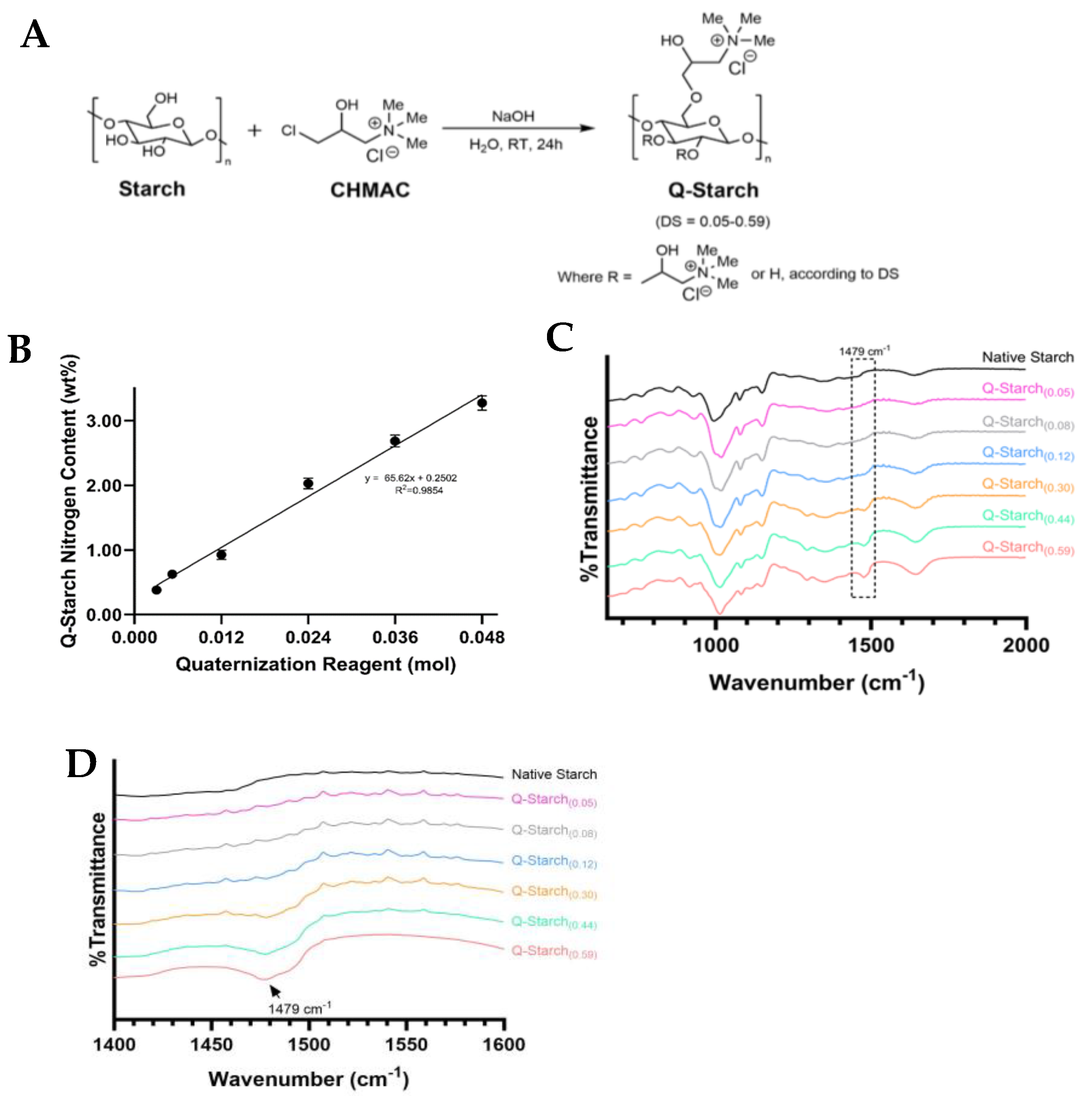
Figure 1. (A) Reaction scheme for starch quaternization by CHMAC. (B) Q-Starch(DS) nitrogen content as function of amount of quaternization reagent. (C) FTIR spectrum of native starch and different Q-Starches. (D) Magnification of relevant peak in FTIR spectrum of (C).
Polysaccharides 05 00037 g001
Figure 2. (A) Representative mechanism for Q-Starch(DS)/siRNA complex formation through self-assembly. (B) Q-Starch(0.12)/siRNA complex formation evaluated by agarose gel electrophoresis at increasing N/P molar ratios. (C) Zeta potential of Q-Starch(DS)/siRNA complexes at increasing DS at N/P molar ratios of 1, 2, and 3.
Polysaccharides 05 00037 g002
Figure 3. (A) An illustration depicting the ionic strength agarose gel electrophoresis experimental procedure; free siRNA is observed after complex exposure to increasing ionic strength. (B) The agarose gel electrophoresis of Q-Starch(0.12)/siRNA complexes formed at an N/P molar ratio of 2 at increasing ionic strengths. (C) NaCl concentration in which decomplexation (free siRNA) was first observed in each Q-Starch(DS) at an N/P ratio of 2. (D) Particle size distribution for Q-Starch(DS)/siRNA complexes at an N/P molar ratio of 2. (E) The mean particle size for the different Q-Starch(DS)/siRNA complexes at an N/P molar ratio of 2. (F) Two representative cryoTEM images of Q-Starch(0.12)/siRNA complexes (marked with a red arrows) at an N/P molar ratio of 2.
Polysaccharides 05 00037 g003aPolysaccharides 05 00037 g003b
Figure 4. Cellular uptake and gene silencing of Q-Starch(DS)/siRNA complexes. (A) Mean cy5 intensity following 24 h of incubation with Q-Starch(DS) complexes as assessed via FACSAria III; (B) EGFR gene mRNA expression following 72 h of incubation with Q-Starch(DS) complexes as assessed via RT-PCR (ns: no statistical significance).
Polysaccharides 05 00037 g004
Figure 5. (A) Illustration depicting expected experimental results for iodine test following Q-Starch(DS) cleavage, together with experimental results for Q-Starch(0.12). (B) Required α-amylase concentration for Q-Starch(DS) cleavage at different DSs via iodine starch test. (n = 3) (C) Shear viscosity as function of applied shear rate for Q-Starch(0.59) and Q-Starch(0.30) before and after cleavage with α-amylase enzyme. (D) Agarose gel electrophoresis results of Q-Starch (0.30)/siRNA and Q-Starch (0.59)/siRNA complexes with or without pre-cleavage of Q-Starch with α-amylase enzyme at 1:1 wt. %. All complexes were formed at N/P molar ratio of 2. (E) RT-PCR results of EGFR gene mRNA expression in Cal33 cells, 72 h post-transfection with Q-Starch(0.59)/siRNA complexes; four examined groups include cells treated with Q-Starch(DS)/siRNANC5 and Q-Starch(DS)/siRNAEGFR complexes at siRNA concentration of 50 nM, N/P molar ratio of 2, with and without pre-cleavage of Q-Starch(0.59) with α-amylase 24 h before transfection. Average + SEM (n = 3). ns: not statistically significant (p > 0.05), *: statistically significant difference (p < 0.05).
Polysaccharides 05 00037 g005
Table 1. Q-Starch(DS) elemental analysis measurement results, calculated degrees of substitution, and respective DS reciprocals.
Q-Starch Average Nitrogen Content (wt. %)DS *(DS)−1
0.38 ± 0.000.0521.71
0.63 ± 0.000.0812.87
0.93 ± 0.070.128.39
2.03 ± 0.080.303.32
2.68 ± 0.090.442.29
3.3 ± 0.10.591.70
* The different Q-Starches will be referred to according to their degree of substitution (DS), using the nomenclature of Q-Starch(DS).
Disclaimer/Publisher’s Note: The statements, opinions and data contained in all publications are solely those of the individual author(s) and contributor(s) and not of MDPI and/or the editor(s). MDPI and/or the editor(s) disclaim responsibility for any injury to people or property resulting from any ideas, methods, instructions or products referred to in the content.