Article

Gallic Acid Functionalization Improves the Pharmacological Profile of Fucoidan B: A Polysaccharide with Antioxidant Properties

by
Joicy Ribeiro dos Santos
1,2,
Diego Araujo Sabry
3,
Guilherme Lanzi Sassaki
4 and
Hugo Alexandre Oliveira Rocha
1,2,*
1
Graduate Program in Biochemistry and Molecular Biology, Center of Biosciences, Federal University of Rio Grande do Norte-UFRN, Natal 59078-970, Brazil
2
Natural Polymer Biotechnology Laboratory (BIOPOL), Department of Biochemistry, Center of Biosciences, Federal University of Rio Grande do Norte-UFRN, Natal 59078-970, Brazil
3
Federal Institute of Piauí-IFPI, São João do Piauí 64.760-000, Brazil
4
Department of Biochemistry and Molecular Biology, Federal University of Paraná-UFPR, Curitiba 81.531-980, Brazil
*
Author to whom correspondence should be addressed.
Polysaccharides 2025, 6(4), 89; https://doi.org/10.3390/polysaccharides6040089
Submission received: 29 June 2025 / Revised: 23 August 2025 / Accepted: 30 September 2025 / Published: 8 October 2025

Abstract

Fucoidan B (FucB) is a sulfated polysaccharide with recognized biological activity. In this study, FucB was chemically modified through redox conjugation with gallic acid (GA) to obtain FucB-GA, aiming to enhance its antioxidant properties. Structural characterization using FTIR, NMR, and electrophoresis confirmed the successful covalent binding of GA to FucB without major structural degradation. The conjugation increased the phenolic content and reduced crystallinity, as shown by XRD and SEM, indicating greater amorphous character, which can favor biological applications. Thermogravimetric analysis demonstrated enhanced thermal stability in FucB-GA. Antioxidant activity was evaluated through various in vitro assays. FucB-GA showed superoxide radical scavenging activity of 91.96%, copper chelating capacity of 43.2%, antioxidant capacity of 37 mg AEE/g, and reducing power of 94.22%, significantly higher results than FucB, while no sample chelated iron. Under the conditions analyzed, gallic acid alone showed minimal or no activity in most assays. These results suggest that conjugation with GA increases the antioxidant potential of FucB, while also improving the activity and bioavailability of GA, likely due to the increase in electron-donating and metal-binding groups. Overall, the study supports the development of FucB-GA as a promising antioxidant compound for pharmaceutical or nutraceutical applications.

Graphical Abstract

1. Introduction

Redox processes, intrinsic to cellular metabolism, are fundamental for sustaining the vital functions of living organisms. However, these processes can result in the generation of reactive oxygen species (ROS), which exhibit a dual role, exerting either beneficial or detrimental effects depending on various cellular conditions [1].
ROS play essential roles in various cellular processes, such as intracellular signaling, regulation of gene expression, apoptosis, biosynthesis of other molecules, and defense against pathogens [2]. However, cellular homeostasis depends on the balance between the generation and elimination of these species. When this balance is disrupted, a condition known as oxidative stress arises, characterized by excessive ROS production that exceeds the antioxidant system’s capacity to neutralize them adequately [3]. This imbalance can result in damage to cellular structures, including DNA, proteins, and lipids, compromising their structural and functional integrity. Such damage is associated with the onset and/or progression of various pathologies [4], including cardiovascular diseases, neurodegenerative disorders, neoplasms, and conditions such as premature aging [5,6].
As a defense mechanism against oxidative stress, the body relies on endogenous and exogenous antioxidant molecules that protect cells by neutralizing ROS. These substances are capable of delaying or preventing oxidative damage to cellular structures, thereby contributing to the prevention of the pathogenesis of various clinical conditions [5]. In this context, with the aim of supporting the maintenance of the balance between ROS generation and elimination, there has been increasing interest in the search for natural or synthetic compounds with antioxidant properties that hold potential as therapeutic agents in antioxidant strategies [7,8]. Among these promising molecules, sulfated polysaccharides (SP) extracted from brown seaweeds and their derivatives stand out [9].
The SP described in brown algae are, for the most part, composed of sulfated α-L-fucose, typically forming a long linear chain, along with smaller amounts of other monosaccharides such as mannose, galactose, glucose, xylose, and uronic acids, which may form branches [10]. These polymers are currently classified as fucans (composed of more than 90% fucose) and fucoidans (heterofucans) [11].
Fucoidans exhibit diverse structures, and each newly identified fucoidan may present a unique structural conformation, potentially resulting in distinct pharmacological activities and offering new prospects for drug development [12]. Accordingly, fucoidans from different species have been reported to display a range of pharmacological activities, including antitumor [13], anticoagulant [14], immunoinflammatory [15], and antioxidant effects [16].
Polysaccharides are widely recognized for their inherent biological properties, including antioxidant activity. However, these activities can vary significantly depending on the polysaccharide’s origin, structure, and molecular weight. To enhance their antioxidant potential, several chemical modification strategies have been explored, such as sulfation, phosphorylation, carboxymethylation, methylation, selenization, acetylation [17] and, more recently, conjugation with phenolic compound, such as flavonoids via grafting methods including chemical coupling [18], enzymatic catalysis [19] and free-radical grafting [20] has become an effective strategy to enhance the antioxidant, antimicrobial, and other bioactivities of polysaccharides. These modifications aim to improve the electron-donating capacity, introduce new functional groups, or increase solubility, ultimately boosting the polysaccharides’ ability to scavenge free radicals, chelate metal ions, and act as reducing agents [17,18,19,20].
Among these strategies, the conjugation of polysaccharides with gallic acid has shown promising results. For instance, the conjugation of animal-derived chitosan with gallic acid, as reported by Curcio et al. [21] yielded a compound with greater water solubility than chitosan and significantly enhanced antioxidant activity in three out of four in vitro assays: total antioxidant capacity (twice as potent), iron chelation (90 times more effective), and reducing power (five times more effective). In the fourth assay, copper chelation, no significant difference was observed between chitosan and the chitosan–gallic acid conjugate. To the best of our knowledge, only one study has reported the conjugation of gallic acid with a sulfated polysaccharide [22]. In that study, the authors obtained a fucoidan–gallic acid conjugate that demonstrated greater antioxidant activity than both gallic acid and unconjugated fucoidan.
Gallic acid (GA) is a naturally occurring triphenolic compound whose bioactivity is well established [23], particularly its ability to protect biological systems against oxidative damage caused by ROS [3]. GA has been associated with antimelanogenic, antioxidant [24], anti-inflammatory, and antiallodynic [25] properties. The molecular structure of GA favors conjugation with polysaccharides, with three hydroxyl groups in the meta and para positions of the aromatic ring contributing to increased antioxidant activity [22]. However, GA presents certain bioavailability limitations when obtained through dietary sources, as it is rapidly metabolized and excreted, thereby reducing its effectiveness in providing sustained antioxidant protection [26]. Therefore, Fernandes-Negreiros et al. [27] proposed that the conjugation of polysaccharides with GA can improve the antioxidant properties of the polysaccharide matrix, while simultaneously enhancing the stability and bioavailability of GA. This dual effect results in increased therapeutic potential and greater protection against oxidative stress in biological systems. Therefore, chemical conjugation emerges as a promising strategy not only to stabilize GA, but also to amplify its biological activity through association with biopolymers.
Thus, recognizing the importance of identifying new molecules with antioxidant potential and enhancing their activity through chemical modifications, this study aimed to obtain a fucoidan, referred to as FucB, from the brown seaweed Spatoglossum schröederi. This polysaccharide exhibited stronger antithrombotic activity than heparin, as it was able to stimulate endothelial cells to synthesize antithrombotic sulfated heparan [28]. However, the other biological activities of this compound have not yet been evaluated. Therefore, in this study, FucB was conjugated with GA, chemically characterized, and its antioxidant activity was compared with that of GA and unmodified FucB. The structural and physicochemical characteristics were assessed using Fourier-transform infrared spectroscopy (FT-IR), proton nuclear magnetic resonance (1H-NMR), scanning electron microscopy (SEM), energy-dispersive X-ray spectroscopy (EDS), X-ray diffraction (XRD), and thermogravimetric analysis (TG and DTG). The in vitro antioxidant activity was evaluated through assays for metal chelation, electron donation, and radical scavenging.

2. Materials and Methods

2.1. Materials

Nitro blue tetrazolium (NBT), toluidine blue, 1,3-diaminopropane, acetone, Folin–Ciocalteau, Coomassie brilliant blue R-250, 2,2′,2″,2′′′-(Ethane-1,2-diyldinitrilo) tetra-acetic acid (EDTA), ascorbic acid, methionine, ammonium molybdate, gallic acid, L-ascorbic acid, and Dulbecco’s Modified Eagle Medium (DMEM) were all purchased from Sigma (St. Louis, MO, USA). Methanol, ethanol, acetone, acetic acid, sulfuric acid, pyridine, and N-cetyl-N,N,N-trimethylammonium bromide (CTV) were purchased from CRQ (São Paulo, SP, Brazil). All other solvents and chemical products used in this study were of analytical-grade purity.
Specimens of Spatoglossum schröederi were collected along the coast of Nísia Floresta, Rio Grande do Norte, Brazil. The seaweed was identified according to its morphology [29]. The material collection occurred under the authorization of the Brazilian National System of Management of Genetic Heritage and Associated Traditional Knowledge SISGEN n° A0D4240.

2.2. Collection and Extraction of FucB from Spatoglossum Schröederi

After collecting, the algae were thoroughly washed, dried at 60 °C, and subsequently ground. Lipids and pigments were removed by treatment with ethyl alcohol. A total of 100 g of the dried and depigmented algal biomass was suspended in 1200 mL of 0.25 M NaCl, and the pH was adjusted to 8.0 using NaOH. Proteolytic digestion was carried out by adding 1.5 g of Prozyme® (Prozyn Biosolutions, São Paulo, SP, Brazil), an enzymatic preparation containing alkaline protease. The suspension was incubated for 18 h at 60 °C in a water bath. After enzymatic hydrolysis, the mixture was filtered, and the resulting filtrate was subjected to further purification steps to isolate FucB, according to the methodology previously described by Rocha et al. [30].

2.3. Conjugation with Gallic Acid

The conjugation of FucB with gallic acid was carried out based on the method described by Curcio et al. [21] with minor modifications [31]. Briefly, 250 mg of FucB was dissolved in 25 mL of distilled water. Subsequently, 500 µL of 1 M hydrogen peroxide and 27 mg of ascorbic acid were added to the solution. After a 30 min incubation at room temperature, 120 mg of gallic acid was introduced. The reaction mixture was maintained in a water bath at 60 °C for 5 min and then allowed to react at room temperature for an additional 24 h. Following the reaction period, the solution was subjected to ultracentrifugation at 5000× g for 50 min at 4 °C. The resulting precipitate was lyophilized, weighed, and designated as FucB-GA.

2.4. Physicochemical Analyses

2.4.1. Quantification of Sulfate, Protein, and Phenolic Compounds

Sulfate content in the samples was quantified using the gelatin-barium method described by Dodgson and Price [32]. Absorbance was measured at 500 nm using a spectrophotometer, with sodium sulfate employed as the standard. Protein content was determined by the Bradford method [33], with absorbance measured at 595 nm and bovine serum albumin used as the standard. The quantification of phenolic compounds was conducted using the colorimetric Folin–Ciocalteu method, as described by Singleton and Rossi [34]. Absorbance was measured at 765 nm, and gallic acid served as the standard.

2.4.2. Molecular Weight Determination

The molecular weight of the FucB-GA and FucB was assessed using high-performance size-exclusion chromatography (HPSEC) with a TSK-Gel® 3000 column (30 cm × 0.75 cm) from Sigma-Aldrich (St. Louis, MO, USA), operated on a system from GE Healthcare Biosciences (Pittsburgh, PA, USA). The mobile phase consisted of 0.2 M sodium chloride in 0.05 M acetate buffer. Chromatographic runs were carried out at 60 °C with a flow rate of 1.0 mL/min. The column was calibrated using dextran standards with molecular weights of 10, 47, 74, and 147 kDa, also obtained from Sigma-Aldrich. A refractive index detector was used to monitor the eluted fractions.

2.4.3. Agarose Gel Electrophoresis Analyses

Electrophoresis was carried out following the protocol described by Dietrich and Dietrich [35] to evaluate the presence or absence of sulfated polysaccharides (SP) in the samples. Agarose gels were prepared at a concentration of 0.6% (w/v) in 0.05 M 1,3-diaminopropane-acetate (PDA) buffer. The gels were cast onto glass slides, and wells were formed for sample application. A total of 1 μg of each sample was loaded into the wells. Electrophoretic separation was performed at 4 °C in a refrigerated electrophoresis chamber under a constant current of 0.3 A and a voltage of 110 V, with migration from the negative to the positive electrode.
Upon completion of the run, the gel was immersed in 0.1% (w/v) cetyltrimethylammonium bromide (CETAVLON) for approximately 2 h to promote SP precipitation. Subsequently, the gel was air-dried and stained with 0.1% (w/v) toluidine blue. Excess dye was removed using a decolorizing solution composed of 1% (v/v) acetic acid, 50% (v/v) ethanol, and 49% (v/v) distilled water. The gel was then dried once more at room temperature for analysis.

2.4.4. Infrared Spectroscopy (FT-IR)

Infrared spectroscopic analyses of the samples were performed following standard protocols at the Institute of Chemistry, Federal University of Rio Grande do Norte (IQ-UFRN). A PerkinElmer Frontier Fourier Transform Infrared (FT-IR) spectrometer (Waltham, MA, USA) equipped with a Quest ATR (Attenuated Total Reflectance) accessory from Specac Ltd. (Orpington, UK) containing a diamond crystal, was used. The system operated with a helium-neon (HeNe) laser and a MIR TGS detector.
Measurements were conducted in the mid-infrared (MIR) range by placing a small quantity of each sample directly onto the diamond crystal at room temperature. Spectra were acquired over a scanning range of 4000–400 cm−1, with a resolution of 4.0 cm−1, and an average of 16 scans per spectrum. A background spectrum was collected prior to each sample measurement to ensure proper cleaning of the crystal and to maintain the integrity and accuracy of the spectral data.

2.4.5. Nuclear Magnetic Resonance (NMR)

For 1H NMR analysis, 10 mg of each FucB and FucB-GA were dissolved in 0.5 mL of deuterium oxide 99% (D2O). Structural characterization was carried out using a Bruker AVANCE III 600 MHz spectrometer (Billerica, MA, USA) operating at 14.1 Tesla, equipped with a N2-cooled 5 mm TCI CryoProbe Prodigy. Analysis was performed using presaturation pulse sequence (zgpr) for water suppression, with 16 number of scans (NS = 16). Spectra were acquired at 343 K, and chemical shifts were expressed in δ (ppm) relative to TMSP-d4 (2,2,3,3-tetradeuterium-3-trimethylsilylpropionate, δ = 0.00 ppm).

2.4.6. Scanning Electron Microscopy (SEM) and Energy-Dispersive X-Ray Spectroscopy (EDS)

Microscopic images were acquired using a Tescan Mira 4 scanning electron microscope (Kohoutovice, Czech Republic), operated with a secondary electron detector at an accelerating voltage of 5 keV. Surface elemental composition was analyzed via energy-dispersive X-ray spectroscopy (EDS) at 20 keV using Aztec software (AZtec v6.2-AZtecLive 6.2, Oxford Instruments NanoAnalysis, Oxford Instruments plc, Abingdon, Oxfordshire, UK) coupled to an Ultim Max EDS detector (Oxford Instruments NanoAnalysis, Oxford Instruments plc, Abingdon, Oxfordshire, UK). Samples were mounted on carbon adhesive tape and sputter-coated with gold using a Denton Vacuum Desk V sputter coater (Denton Vacuum Moorestown, New Jersey, USA) prior to analysis.

2.4.7. X-Ray Diffraction (XRD)

X-ray diffraction (XRD) patterns of the samples were obtained using a Bruker D2 Phaser diffractometer (Bruker AXS GmbH, Karlsruhe, Germany) equipped with a Lynxeye position-sensitive detector (Bruker AXS GmbH, Karlsruhe, Germany) and a copper anode (Cu Kα radiation, λ = 1.54 Å) filtered with a nickel filter. The instrument was operated at 30 kV and 10 mA. Data acquisition was conducted in the 2θ range of 2° to 40°, using a step size of 0.01° and a count time of 0.2 s per step. A 0.1 mm divergent slit and a 1 mm receiving slit were used during the measurements.

2.4.8. Thermogravimetric Analysis (TGA)

Thermogravimetric analysis was conducted using a NETZSCH Tarsus TH 209 F3 analyzer (NETZSCH-Gerätebau GmbH, Selb, Germany). Samples were heated from 26 °C to 900 °C at a constant heating rate of 10 K/min under a continuous nitrogen flow of 20 mL/min.

2.5. Antioxidant Activity

Six assays were performed to analyze the FucB, FucB-GA and GA antioxidant activity as described earlier [36,37]: total antioxidant capacity (TAC), hydrogen peroxide scavenging, superoxide radical scavenging, cupric and ferric chelating, and reducing power. As it was verified that FucB-GA contained 3% gallic acid (Table 1), gallic acid was therefore evaluated at concentrations of 3 and 30 µg/mL, corresponding to FucB-GA concentrations of 100 and 1000 µg/mL, respectively.

2.5.1. Hydrogen Peroxide Scavenging Assay

The hydrogen peroxide scavenging activity was evaluated by incubating the samples with a 40 mM hydrogen peroxide solution prepared in 100 mM sodium phosphate buffer (pH 7.4). Samples were protected from light and maintained at room temperature during the assay. A 100 mM sodium phosphate buffer (pH 7.4) was used as a negative control, while the 40 mM hydrogen peroxide solution served as the positive control. Absorbance was measured at 230 nm using a UV-Vis spectrophotometer. Results were expressed as the percentage of protection relative to the positive control.

2.5.2. Superoxide Anion Scavenging Assay

The assay was based on the ability of the samples to inhibit the photochemical reduction of nitroblue tetrazolium (NBT) in the riboflavin–light–NBT system. Riboflavin, in the presence of light, reduces oxygen, forming oxidized riboflavin and the superoxide anion. Superoxide, in turn, reduces NBT. When the sample has superoxide scavenging capacity, the superoxide is unable to reduce NBT.
To perform the assay, 200 µL of methionine (65 mM), 200 µL of EDTA (0.5 mM), 200 µL of NBT (0.375 mM), and 200 µL of riboflavin (0.5 mM) were added to the samples. The blank consisted only of the buffer solution and was protected from light during the addition of riboflavin. All tubes, except the blank, were incubated in a Styrofoam box lined with aluminum foil, under fluorescent light, for 15 min. Subsequently, the absorbance was measured in a microplate reader at 560 nm. The results were expressed as the percentage of scavenging, calculated using the following equation:
% scavenging = [(AcA)/(AcAb)] × 100
where Ac is the absorbance of the control, Ab is the absorbance of the blank, and A is the absorbance of the sample.

2.5.3. Ferrous Ion (Fe2+) Chelation Assay

The ferrous ion chelation assay was performed using a colorimetric test based on the formation of a colored complex between Fe2+, from ferrous chloride, and ferrozine. The assay was conducted in 96-well microplates, where FeCl2 (2 mM) and ferrozine (5 mM) were added to the samples. The plate was shaken and incubated for 10 min at 37 °C. Absorbance was measured at 562 nm using a spectrophotometer. The results were expressed as a percentage of chelation using the following equation:
% chelation = [(AbAa)/Ab] × 100
where Ab is the absorbance of the blank and Aa is the absorbance of the sample.

2.5.4. Copper (Cu2+) Chelation Assay

The copper chelation assay is based on the ability of pyrocatechol violet to form colored complexes with copper ions. The assay was carried out in 96-well microplates, where a solution of pyrocatechol violet (4 mM) and copper (II) sulfate pentahydrate (50 µg/mL) was added to the samples. The wells were homogenized, and absorbance was measured at 632 nm using a spectrophotometer. The results were expressed as a percentage of chelation using the following equation:
% chelation = [(AbAa)/Ab] × 100
where Ab is the absorbance of the blank and Aa is the absorbance of the sample.

2.5.5. Total Antioxidant Capacity (TAC) Assay

To perform the assay, the samples were incubated with a reagent solution containing 0.6 M sulfuric acid, 28 mM sodium phosphate, and 4 mM ammonium molybdate. The incubation was carried out in an oven at 100 °C for 90 min. After cooling, the absorbance was measured at 695 nm using a spectrophotometer. Ascorbic acid was used as the standard, and the results were expressed as milligrams of ascorbic acid equivalents (mg AAE) per gram of sample.

2.5.6. Reducing Power Assay

For this assay, the samples were incubated with 0.2 M phosphate buffer (pH 6.6) and 1% (w/v) potassium ferricyanide for 20 min at 50 °C in a water bath. Then, 10% (w/v) trichloroacetic acid (TCA) was added to stop the reaction, followed by the addition of 0.1% (w/v) ferric chloride. Ascorbic acid was used as the standard. Absorbance was measured at 700 nm using a spectrophotometer. The result was expressed as a percentage of reducing power relative to the value obtained for ascorbic acid, using the following equation:
% scavenging = [(AcA)/(AcAb)] × 100,
where Ac is the absorbance of the control, Ab is the absorbance of the blank, and A is the absorbance of the sample.

2.6. Language Editing

For the enhancement of the English language in the manuscript, the authors utilized ChatGPT version 4.0, an AI language model developed by OpenAI (San Francisco, CA, USA). This tool was employed to improve clarity and coherence throughout the text.

2.7. Statistical Analysis

Data were expressed as mean ± standard deviation from at least three independent experiments, each performed in triplicate. Statistical analysis was conducted using one-way ANOVA, followed by Bonferroni’s post hoc test, with a significance level set at p < 0.05. All statistical tests were performed using GraphPad Prism 5 software (GraphPad Software, San Diego, CA, USA).

3. Results and Discussion

3.1. Method for Modifying FucB with GA

The process of modifying FucB with GA was carried out using the redox method. This method has been widely employed due to its advantages, such as being more environmentally friendly (producing low-toxicity byproducts), yielding high reaction efficiency [21] and being a simple procedure that can be conducted at room temperature [38,39].
The method utilizes hydrogen peroxide (H2O2) as an oxidizing agent and ascorbic acid (AA) as a reducing agent. The reaction mechanism was proposed by Liu et al. [40], based on data obtained through electron paramagnetic resonance. In the presence of H2O2, ascorbic acid is oxidized to form the ascorbate radical (AA1). This radical is capable of abstracting hydrogen atoms from the polysaccharide, consequently generating macroradicals along the entire polymer chain. Following this process, GA can form covalent bonds with the polysaccharide macroradicals, resulting in the formation of a modified compounding, in this case, FucB-GA. Figure 1 schematically illustrates the main steps in the addition of gallic acid to FucB to form FucB-GA.
At the end of the conjugation process, the yield of the final product was approximately 60%. The losses observed during the procedure can be attributed to several factors, such as the washing step, where some material might have been unintentionally removed during handling. Additionally, during centrifugation, part of the material may not have been fully recovered, which also contributed to the reduced overall yield. Similar observations have been reported in previous studies [17,41].

3.2. Physicochemical Characterization of FucB and FucB-GA

3.2.1. Chemical Composition

According to reference [28], fucoidan B (FucB) has a molecular weight of approximately 21.5 kDa and is composed of galactose, fucose, xylose, and sulfate in a molar ratio of approximately 2.0:1.0:0.5:2.0, respectively, along with minor amounts of uronic acids. The proposed structure consists of a linear backbone of 4-linked, partially 3-sulfated β-galactose units, with side branches of 3-sulfated, 4-linked α-fucose residues attached at the C-2 position of the central chain. Additionally, about half of the β-xylose residues and only minor amounts of fucose are found at the non-reducing ends of the fucose side chains, while glucuronic acid is present in trace amounts. Due to the abundance of hydroxyl groups in its structure, FucB offers multiple reactive sites that are potentially suitable for conjugation with gallic acid.
As described in the Methods section, the physicochemical characterization tests of FucB and FucB-GA were performed, and the results are summarized in Table 1. No protein was detected in the samples under the evaluated conditions, indicating the absence of protein contamination. Therefore, the activities evaluated in this study are not related to the presence of these classes of molecules.
As observed in Table 1, the phenolic compound content in FucB-GA was nearly three times higher than in FucB. This finding provides strong evidence for the presence of GA molecules covalently bound to FucB. As this is the first report of FucB being modified with GA, there are no existing studies available for direct comparison with the results presented in Table 1.
Table 1. Chemical composition of FucB and FucB-GA.
SamplesProtein (%)Phenolic Compounds (%)Sulfate (%)Molecular Mass (kDa)
FucBnd* 0.1 ± 0.1# 23.8 ± 1.621.5
FucB-GAnd* 2.8 ± 0.1# 26.8 ± 1.522.8
FucB: fucan B; FucB-GA: fucan B modified with gallic acid; nd: not detected. # no statistical differences were found between the samples (p < 0.05). * p < 0.001.
According to the data shown in Table 1, the sulfate content of FucB was 23.8% ± 1.6, while that of FucB-GA was 26.8% ± 1.5. However, there was no statistically significant difference in the percentage of sulfate groups between the samples. Only one other study was identified in which GA was conjugated to a sulfated polysaccharide; in that case, the authors likewise did not observe any significant variation in sulfate content following the modification [22].
In the HPLC analysis, a single peak was detected for each sample, which was used to determine the apparent molecular mass. The molecular mass of FucB was 21.5 kDa, a value consistent with that reported by Rocha et al. [30]. For FucB-GA, the molecular mass was determined to be 22.6 kDa, indicating that no depolymerization occurred. Other authors who have conjugated GA to polysaccharides also reported derivatives with lower molecular mass compared to the unmodified forms; for example, both chitosans [42] and dextrans [43] exhibited a 25% reduction in apparent molecular mass following the conjugation process with GA.
There are structural features in FucB that may protect it from the depolymerization that can occur during the conjugation process with GA. FucB is branched, whereas dextrans have few branches and chitosan is linear, which could represent a protective factor for FucB. However, it is important to note that when a linear fungal chitosan was modified with gallic acid (GA), Paiva et al. [31] observed an increase in molecular mass of 0.6 kDa following the conjugation process. Therefore, the presence or absence of branching does not appear to be a decisive factor in depolymerization. Another notable structural difference between dextrans, chitosans, and fucoidans is that fucoidans are sulfated, whereas the other polymers are not. In future work, desulfation of FucB is planned to investigate the potential role of sulfate groups as protective agents against depolymerization.

3.2.2. Agarose Gel Electrophoresis

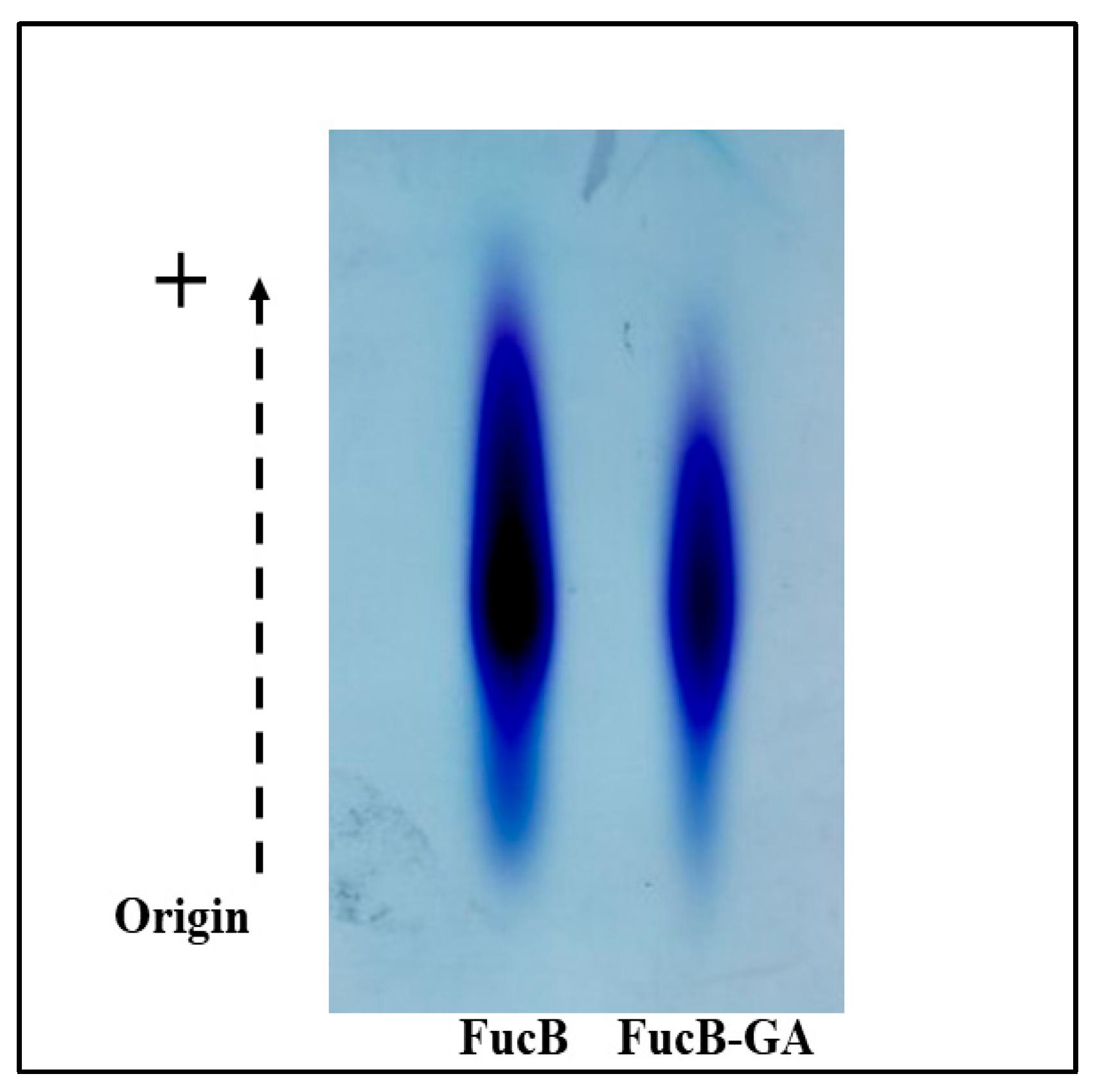
The agarose gel electrophoresis technique in PDA buffer was originally proposed by Dietrich and Dietrich [35]. This technique enables the visualization of bands containing sulfated polysaccharides present in the samples and, thus, confirms the presence of sulfate groups covalently bound to the polysaccharides. The ability of toluidine blue, a cationic dye, to interact with sulfate groups in sulfated polysaccharides allows the development of a blue-violet coloration, known as metachromasia. The PDA buffer promotes migration from the positive to the negative pole, contributing to the separation and visualization of the bands of interest.
The FucB and FucB-GA samples were subjected to electrophoretic separation, and the results are presented in Figure 2. The electrophoresis system using PDA buffer is capable of separating samples based on their interaction with the diamine present in the buffer. This interaction depends, among other factors, on the conformation assumed by the sulfated polysaccharide. Therefore, structurally similar polysaccharides exhibit similar electrophoretic migration, as observed for chondroitin-4-sulfate and chondroitin-6-sulfate [44]. Thus, the fact that FucB and FucB-GA display similar electrophoretic mobilities is a strong indication that conjugation with GA did not induce major structural changes in FucB.
But why did FucB exhibit greater staining intensity than FucB-GA? It has been demonstrated that heparan sulfate and heparin—two structurally similar polysaccharides from the same family—have comparable electrophoretic mobilities, despite the much higher sulfate content in heparin compared to heparan sulfate [45]. Furthermore, heparin does not stain more intensely than heparan sulfate, because many of its sulfate groups are not accessible for interaction with toluidine blue [35]. Therefore, it is believed that the covalent binding of GA to FucB-GA may have hindered the access of toluidine blue to some sulfate groups, resulting in less intense staining of FucB-GA compared to FucB.

3.2.3. Infrared Spectroscopy (FTIR)

The FTIR spectrum enables rapid identification of the functional groups present in molecules. The spectra of FucB and FucB-GA (Figure 3) displayed similar bands at 1617 cm−1, which are characteristic of asymmetric and symmetric carbonyl stretching vibrations [46]. The bands at 846 and 1220 cm−1 indicate the presence of sulfur atoms bound to the molecular backbone. As reported in previous studies by Zheng et al. [46] and He et al. [47], these regions correspond to axially bound groups (C–O–S) and asymmetric stretching vibrations of sulfate groups (S = O), respectively.
Spectral differences between FucB and FucB-GA can be observed in the region around 1726 cm−1, where a low-intensity band, identified as ester carbonyl, appears [48,49]. This band, absent in the FucB spectrum, provides strong evidence of the covalent bond formed during the conjugation of GA to the polysaccharide, as illustrated in the diagram in Figure 1.

3.2.4. Nuclear Magnetic Resonance (NMR)

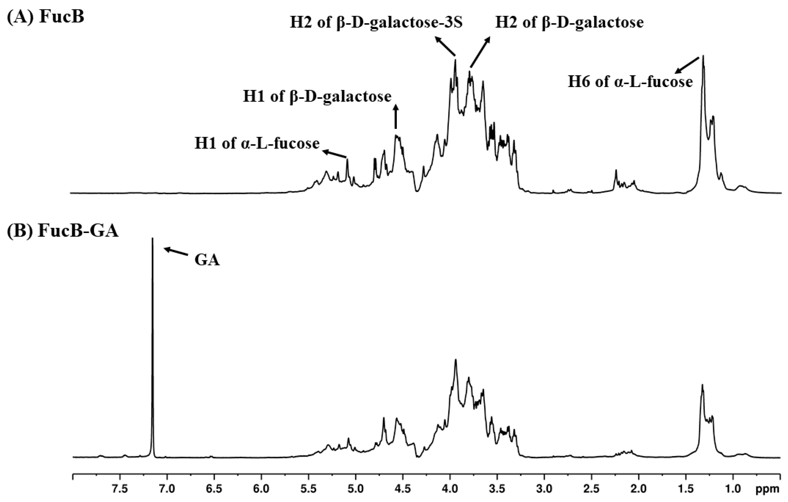
FucB was subjected to nuclear magnetic resonance (NMR) analysis to confirm its identity and to compare it with the spectral data previously reported by Rocha et al. [30], who first purified and characterized this sulfated polysaccharide. The 1H NMR spectrum of FucB, presented in Figure 4, shows significant correspondence with the spectra published by Rocha et al. [30]. The signals for H1 and H6 of α-1-fucose-4 appear at 5.10 and 1.23 ppm, respectively. The H1 signal of β-4-galactose-1 was observed at 4.59 ppm. The signals at 3.95 and 3.81 ppm are related to H2 of β-4-galactose-3S-1 and β-4,2-galactose-1, respectively.
Regarding the spectrum of FucB-GA (Figure 4B), the similarity in peak patterns between the two spectra further supports that the modification method used did not induce significant alterations in the original polysaccharide structure. Additionally, the appearance of a signal at 7.17 ppm, which is absent in the FucB spectrum, is indicative of the protons on the aromatic ring of GA, reinforcing that GA was successfully conjugated to the polysaccharide structure. This signal has been previously reported in spectra of sulfated polysaccharides modified with gallic acid [50,51,52].

3.2.5. X-Ray Diffraction (XRD)

Having confirmed the successful conjugation of GA to FucB, various analyses were conducted to identify possible physical changes in FucB-GA compared to FucB.
X-ray diffraction was the first technique applied. XRD patterns provide information on the physical characteristics of different molecules, including polysaccharides. Sharp and narrow peaks in these patterns are indicative of crystalline reflections, while their absence denotes samples with predominantly amorphous structures [53]. Thus, XRD analysis was performed to compare the microstructures of FucB before and after GA conjugation and to determine whether any microstructural changes had occurred.
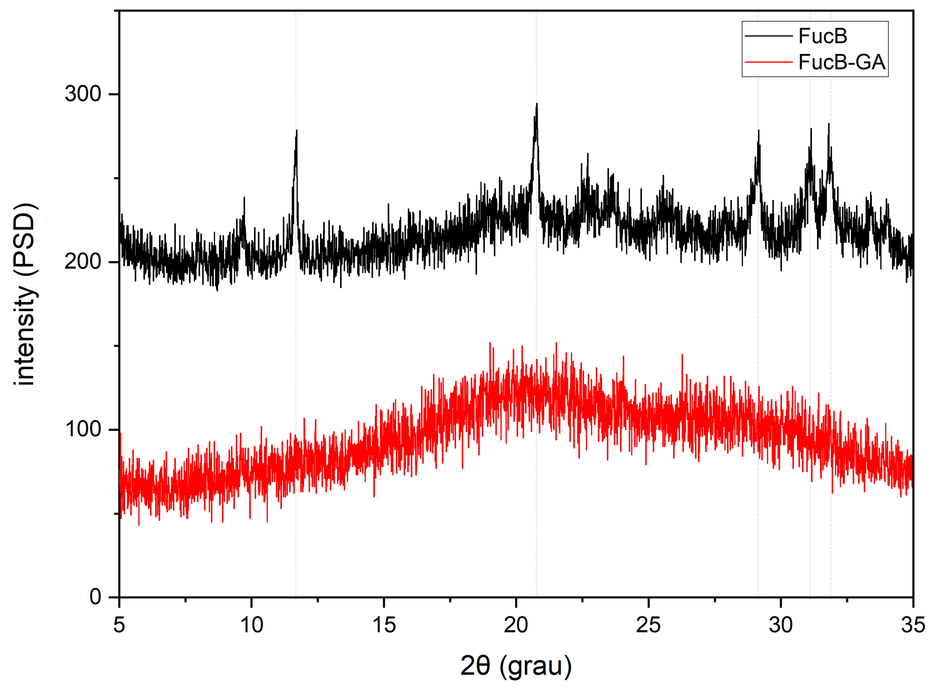
In the XRD pattern of FucB (Figure 5), distinct diffraction peaks were observed at approximately 11°, 20°, and 29–31° 2θ. These peaks suggest the presence of a semi-crystalline structure, characterized by partial molecular ordering within the polysaccharide. Although detailed crystallographic patterns of fucoidans are still limited in the literature, similar XRD features have been reported in fucoidans extracted from Ecklonia maxima [54] and Saccharina japonica [55] which exhibited a prominent crystallinity peak around 23° 2θ, attributed to partial chain alignment and ordered domains. Furthermore, fucoidans from Sargassum wightii, Sargassum swartzii, Sargassum polycystum, and Turbinaria ornata displayed a significant diffraction peak between 32° and 33° 2θ, further reinforcing the semi-crystalline nature of these marine-derived biopolymers [56].
The additional peaks observed near 11° 2θ range in our sample may correspond to heterogeneous crystalline orientations or local stacking variations within the FucB chains. Similar features have also been reported for other semi-crystalline polysaccharides such as chitosan [57] and pectin [58], which often display multiple broad peaks because of short-range molecular organization.
In contrast, the diffractogram of the FucB-GA conjugate (Figure 5) shows disappearance of these peaks and a more amorphous profile. This change supports the successful incorporation of gallic acid into FucB, as phenolic compounds are known to disrupt the molecular organization of polymers, thereby destroying their packing [41,59]. Similar reductions in crystallinity after conjugation with gallic acid have been previously reported for other polymers, such as chitosan [39,60]. Our data suggest that the gallic acid in FucB-GA perturbs intermolecular hydrogen bonding, resulting in a fully amorphous structure without detectable crystalline peaks.
From a pharmacological standpoint, this is a desirable modification, as increased amorphous content enhances water solubility. Amorphous polymers tend to have weaker intermolecular interactions compared to crystalline materials, allowing them to interact more readily with water [42]. Additionally, these characteristic influences key polymer properties such as viscosity, density, and functional behavior, making them more suitable candidates for pharmacological applications [61].

3.2.6. SEM and EDS Analyses

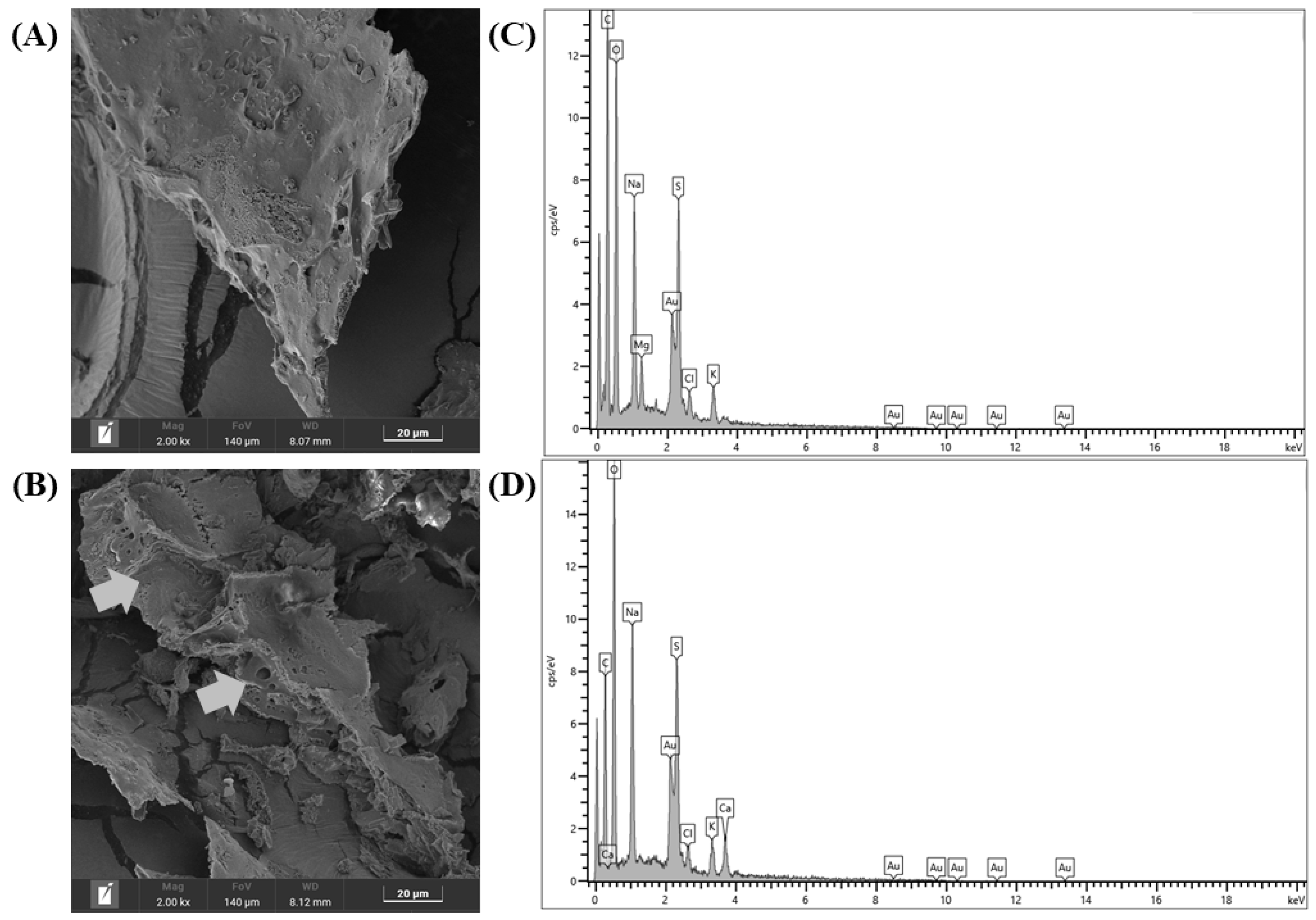
The morphological analysis of the samples was conducted using scanning electron microscopy (SEM). Figure 6A presents a representative image of the results obtained. The surface of FucB exhibited irregular edges, lacking a defined shape, with a rough texture and an amorphous character, consistent with the amorphous profile observed in the diffractograms shown in Figure 5. This amorphous character was retained in FucB-GA (Figure 6B), although a rougher surface was observed in comparison to FucB. Additionally, the presence of pores, absent in the FucB surface, was noted. A similar morphological change was reported by Hu et al. [62] upon conjugating chitosan with GA. According to the authors, this alteration results from the insertion of GA into the polysaccharide, suggesting a reduction in hydrogen bonding and crystallinity, which supports the findings from the XRD analysis.
The chemical composition of the samples was further examined using energy-dispersive spectroscopy (EDS). As shown in Figure 6C,D, the EDS spectra revealed peaks corresponding to carbon, oxygen, sodium, chlorine, and sulfur. These results indicate that the samples are primarily composed of carbon and oxygen, major elements of the polysaccharide backbone. The detection of sulfur confirms the presence of sulfate groups in the polymeric structure, while the presence of sodium and chlorine reflects the presence of NaCl in the samples. Furthermore, the absence of nitrogen peaks suggests that no protein contamination is present, corroborating the data previously reported in Table 1.

3.2.7. Thermogravimetric Analysis (TG/DTG)

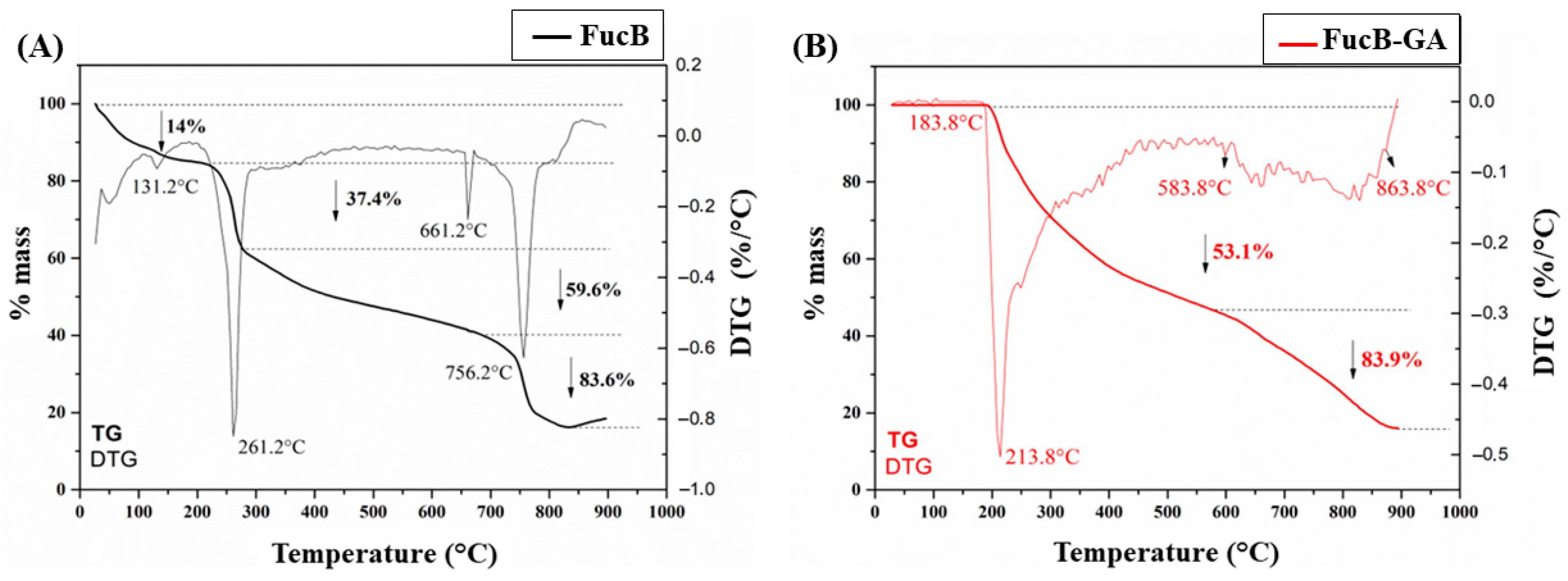
Understanding the thermal stability of biomolecules is crucial, as this property directly influences the applicability of a compound across various fields such as pharmacy, chemistry, and biotechnology [63]. With this objective, FucB and FucB-GA samples were analyzed for thermal stability using thermogravimetric analysis (TG/DTG), and the resulting thermograms are presented in Figure 7.
The first mass loss of FucB (14%) occurred at 131.2 °C and is attributed to the loss of water molecules bound within the structure [64]. The polysaccharide’s mass then decreased through three additional stages: the first stage involved a 37.4% loss of total mass around 261.2 °C, the second stage involved a 59.6% loss at 661.2 °C, and the third stage (83.6% mass loss) occurred at 756.2 °C, leaving 16.4% ash residue at the end. These stages are commonly observed in similar molecules and correspond to depolymerization and decomposition processes [65,66].
FucB-GA maintained mass stability up to approximately 183.8 °C. Subsequently, a progressive mass loss occurred, with the first main decomposition stage (53.1% mass loss) at 213.8 °C, followed by a second decomposition stage (83.9% mass loss) in the range of 583.8 to 863.8 °C. In the end, 16.1% of the total mass of FucB-GA remained as residue. Although the water loss profile was less evident in FucB-GA compared to the native polysaccharide, it is noteworthy that the first decomposition stage of FucB-GA occurred at a lower temperature (213.8 °C) than that of FucB (261.2 °C). Similar behavior was reported for κ-carrageenan derivatives, where functionalization led to a decrease in the degradation temperature, attributed to the disruption of intra- and intermolecular hydrogen bonds [67]. Thus, rather than indicating increased thermal stability, conjugation with gallic acid may induce structural modifications that reduce the decomposition onset temperature. However, intermolecular interactions between the GA hydroxyl groups and the polysaccharide backbone can still influence the overall thermal profile, as observed in other systems, such as poly(vinyl alcohol)/gum tragacanth blend films [68].
Therefore, thermogravimetric analysis demonstrated that the incorporation of GA into the fucoidan structure altered the thermal properties of the resulting compound compared to native fucoidan. These findings highlight that the modification with GA affects the degradation profile and must be carefully considered in potential applications, particularly in contexts involving moderate to high-temperature processes.

3.3. In Vitro Antioxidant Activities

FucB, FucB-GA, and GA samples were subjected to different in vitro assays to evaluate their antioxidant activity. The concentrations selected for FucB and FucB-GA were 100 and 1000 µg/mL. As the proportion of gallic acid in FucB-GA was determined to be approximately 3% (Table 1), the corresponding concentrations of free GA used were 3 and 30 µg/mL, respectively, to match the amounts present in 100 and 1000 µg/mL of FucB-GA.

3.3.1. Hydrogen Peroxide Scavenging

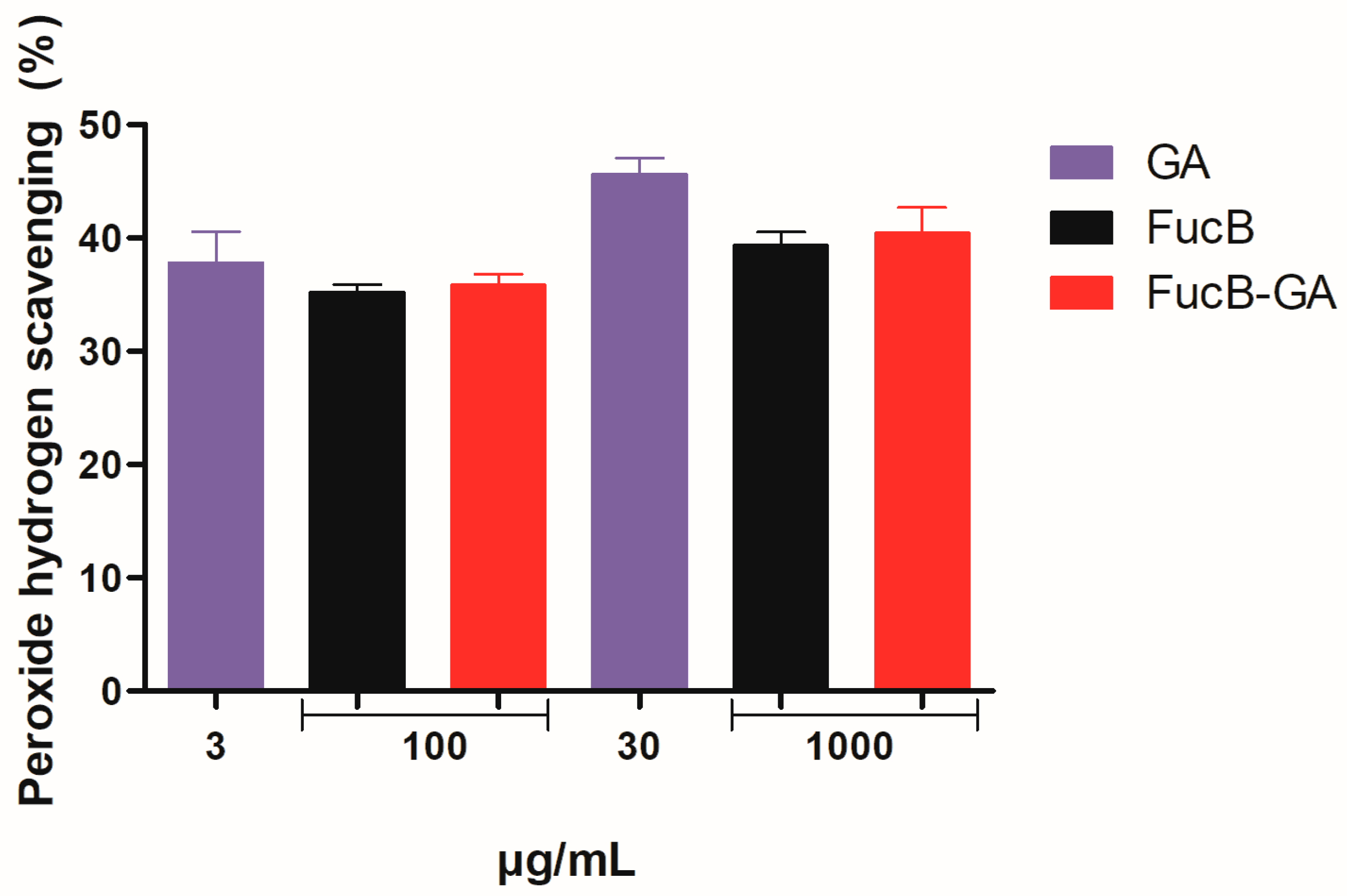
Although hydrogen peroxide is not classified as a free radical, it is considered a reactive oxygen species (ROS) due to its high reactivity. It can diffuse across cell membranes and react with metal ions such as copper and iron, actively participating in Fenton and Haber-Weiss reactions to generate hydroxyl radicals, which are highly toxic and can induce cellular damage [69]. Therefore, the capacity of FucB, FucB-GA, and GA to scavenge hydrogen peroxide was assessed, and the results are presented in Figure 8.
As shown, there were no statistically significant differences among FucB, FucB-GA, and gallic acid at either concentration tested. These results suggest that the chemical modification of FucB with GA did not alter its ability to scavenge hydrogen peroxide.
The hydrogen peroxide scavenging ability of sulfated polysaccharides has been previously reported, as seen with fucoidan-rich extracts from Sargassum vachellianum [70] and with commercial fucoidans [71]. However, to date, no studies have reported the activity of sulfated polysaccharides modified with gallic acid.
It is believed that the absence of statistical differences among the samples may be attributed to the sulfate groups present in both FucB and FucB-GA, which are likely responsible for the observed scavenging activity. This hypothesis suggests that chemical modification with gallic acid did not alter the exposure or reactivity of the sulfate groups, thereby resulting in similar antioxidant activity in both samples.

3.3.2. Superoxide Radical Scavenging

Superoxide anion is a key reactive oxygen species (ROS) in biological systems, playing a role in oxygen consumption for ATP synthesis and storage within mitochondria [72]. However, excessive levels of superoxide anion can contribute to oxidative stress and, consequently, to the development of various diseases [73].
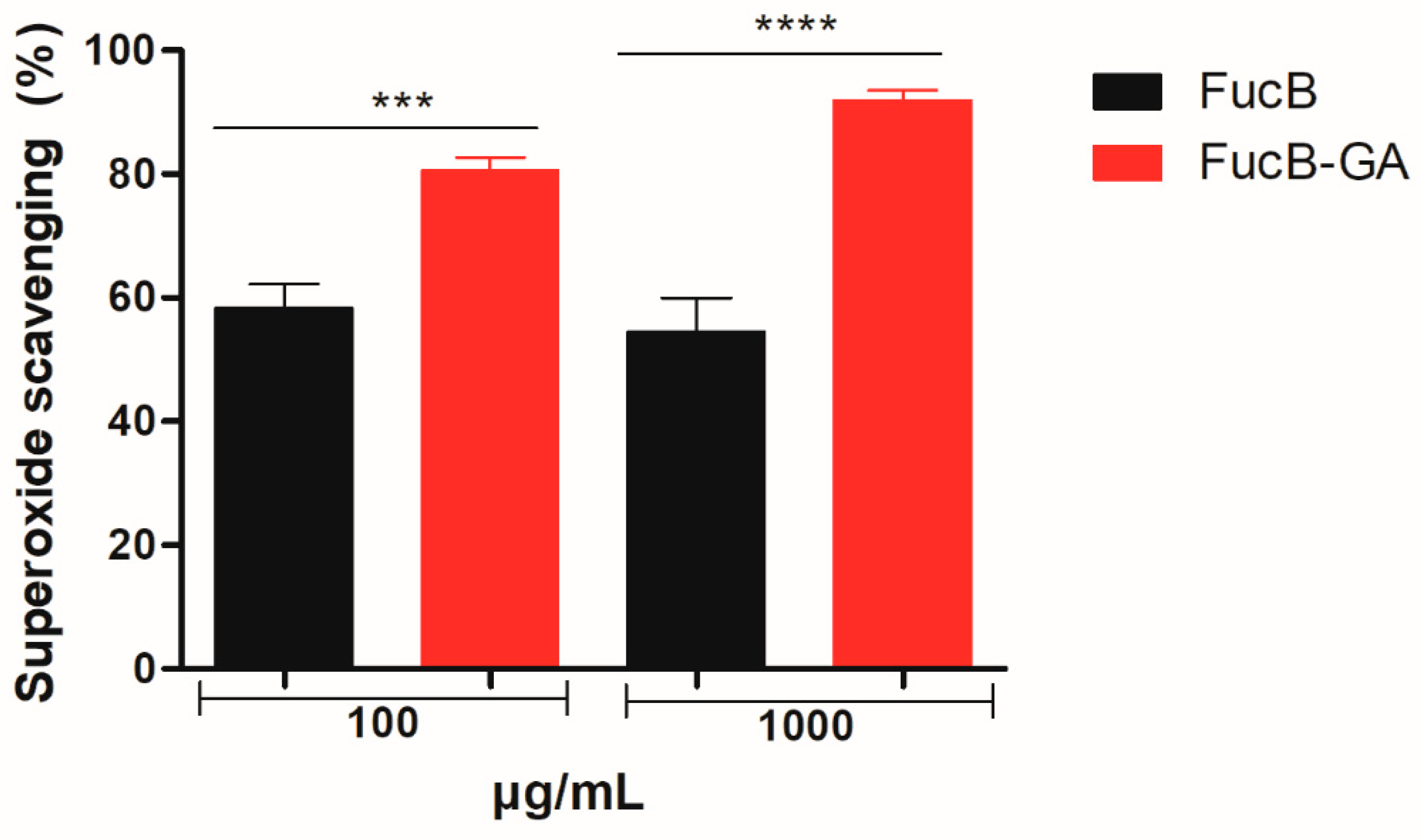
Therefore, FucB, FucB-GA, and GA samples were evaluated for their ability to scavenge superoxide anions. As shown in Figure 9, FucB and FucB-GA at a concentration of 100 µg/mL exhibited scavenging capacities of 58.59% and 80.51%, respectively. At 1000 µg/mL, the scavenging capacities were 51.53% for FucB and 91.96% for FucB-GA. Gallic acid alone did not exhibit any measurable scavenging activity in this assay.
The activity of FucB-GA was superior to that reported for other polysaccharides in the literature. For example, dextran modified with gallic acid (0.5 mg/mL) showed approximately 60% superoxide scavenging activity [43], while fucoidan (0.25 mg/mL) from Dictyopteris justii exhibited only 29.4% activity [36]. Some authors suggest that the neutralization of superoxide anions may occur through mechanisms involving electrostatic interactions or electron donation [74,75]. Further studies are planned to better characterize the mechanism of action of FucB-GA and to determine whether it follows these proposed pathways or involves a novel mechanism.

3.3.3. Chelation of Ferric and Cupric Ions

Metal chelation assays are commonly employed to evaluate the in vitro antioxidant activity of various substances. Accordingly, GA, FucB, and FucB-GA samples were tested for their capacity to chelate iron and copper ions.
Iron chelators play a protective role against oxidative stress by removing Fe2+ ions, which are known to participate in hydroxyl radical generation via Fenton reactions. However, under the conditions tested, none of the samples exhibited measurable iron-chelating activity. This lack of activity has also been reported for other sulfated polysaccharides, including gallic acid–modified fucan A from S. schröederi [22] and commercial fucoidans derived from U. pinnatifida, M. pyrifera, and F. vesiculosus [71].
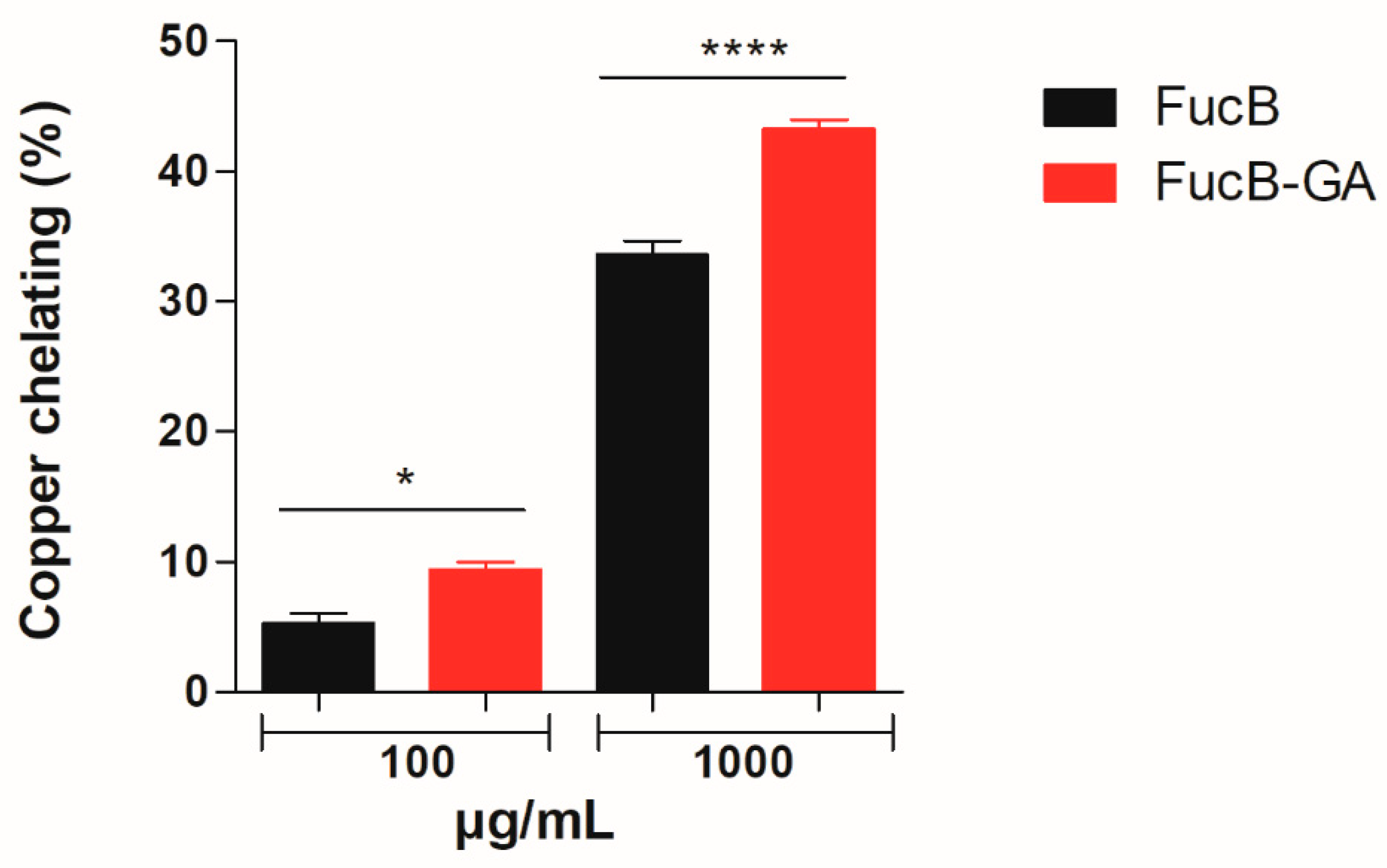
In contrast, the samples demonstrated the ability to chelate copper ions, as shown in Figure 10. At a concentration of 100 µg/mL, FucB exhibited a copper chelating capacity of 5.3%, while FucB-GA showed 9.4%. At 1000 µg/mL, the chelating capacities increased to 33.6% for FucB and 43.2% for FucB-GA.
The data demonstrated that conjugation of FucB with gallic acid resulted in the formation of a compound significantly more effective in chelating copper ions than either of its precursors, FucB or gallic acid alone. Similar outcomes have been reported in other studies where the modification of biomolecules led to enhanced metal-chelating activities compared to the unmodified substances [76].
Excess copper accumulation in the body, due to overconsumption, exposure to toxic levels, or genetic disorders such as Wilson’s disease and Menkes syndrome, can lead to oxidative stress and the induction of apoptosis in several organs, including the brain, kidneys, and corneas [77,78,79]. Current treatments focus on reducing copper intake and administering chelating agents such as D-penicillamine or trientine, which enhance copper excretion via the urine [80]. Nevertheless, there remains a continuous need for alternative chelators with improved properties.
The results obtained in this study suggest that FucB-GA is a promising candidate for future in vivo studies as a copper-chelating compound. Furthermore, due to its metal-chelating ability, FucB-GA can be classified as a preventive antioxidant.

3.3.4. Total Antioxidant Capacity (TAC)

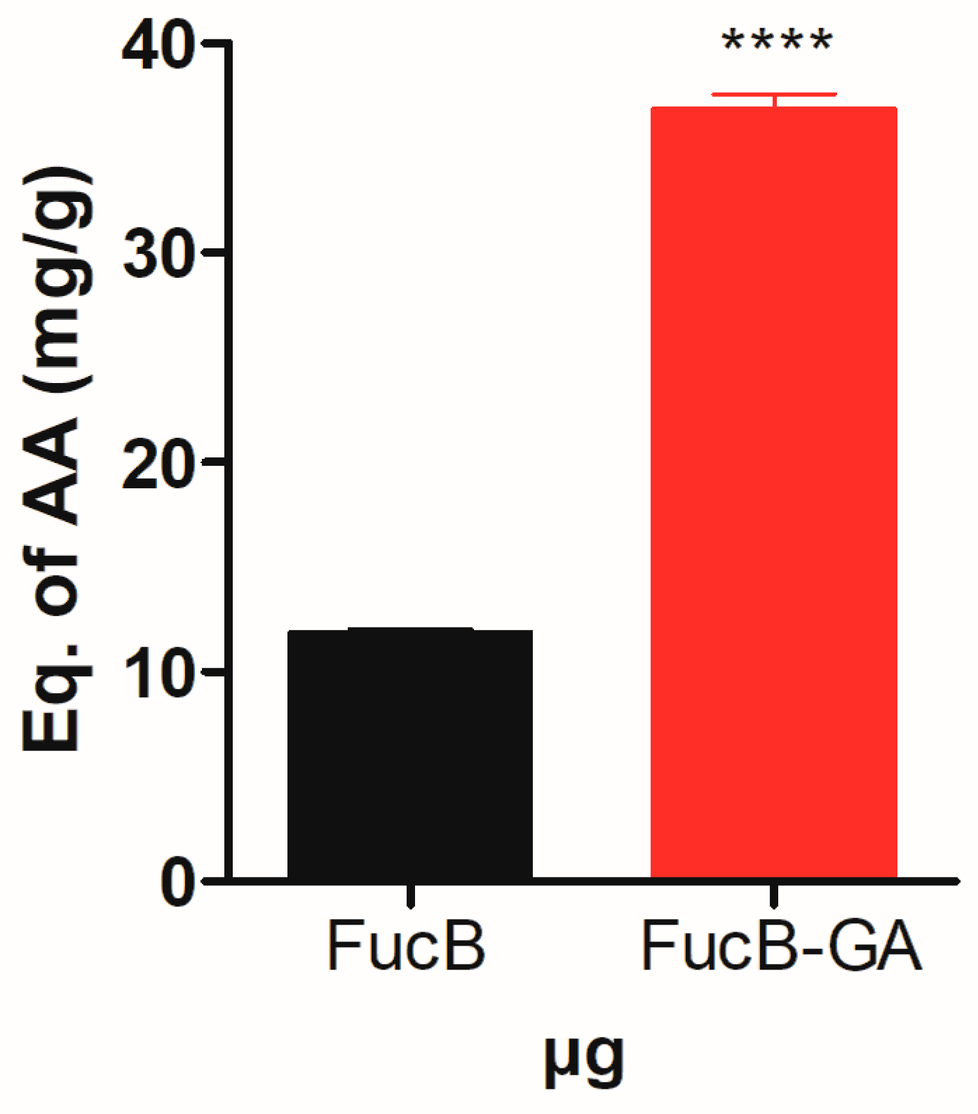
The samples FucB, FucB-GA, and GA were evaluated for their electron-donating capacity under acidic conditions using the total antioxidant capacity (TAC) assay. As shown in Figure 11, FucB-GA exhibited approximately three times greater activity than FucB, with a value of approximately 37 mg ascorbic acid equivalents (AAE) per gram of sample. This result indicates that the modification was effective in enhancing the electron-donating capacity of the polysaccharide. Gallic acid alone did not demonstrate any measurable activity in this assay.
The antioxidant activity observed for FucB-GA was higher than that reported for other polysaccharides modified with gallic acid, such as chitosan, which exhibited TAC values of just over 600 µg AAE/g of sample [81]. In contrast, the gallic acid–laminarin conjugate showed a TAC value of 89 mg AAE/g [27]. However, the literature suggests that sulfated polysaccharides with TAC values exceeding 9.65 mg/g are already considered to have high total antioxidant capacity [82,83]. Therefore, the FucB-GA conjugate demonstrated a notably high antioxidant capacity, supporting the hypothesis that the presence of gallic acid within the structure enhances the compound’s overall antioxidant potential.

3.3.5. Reducing Power

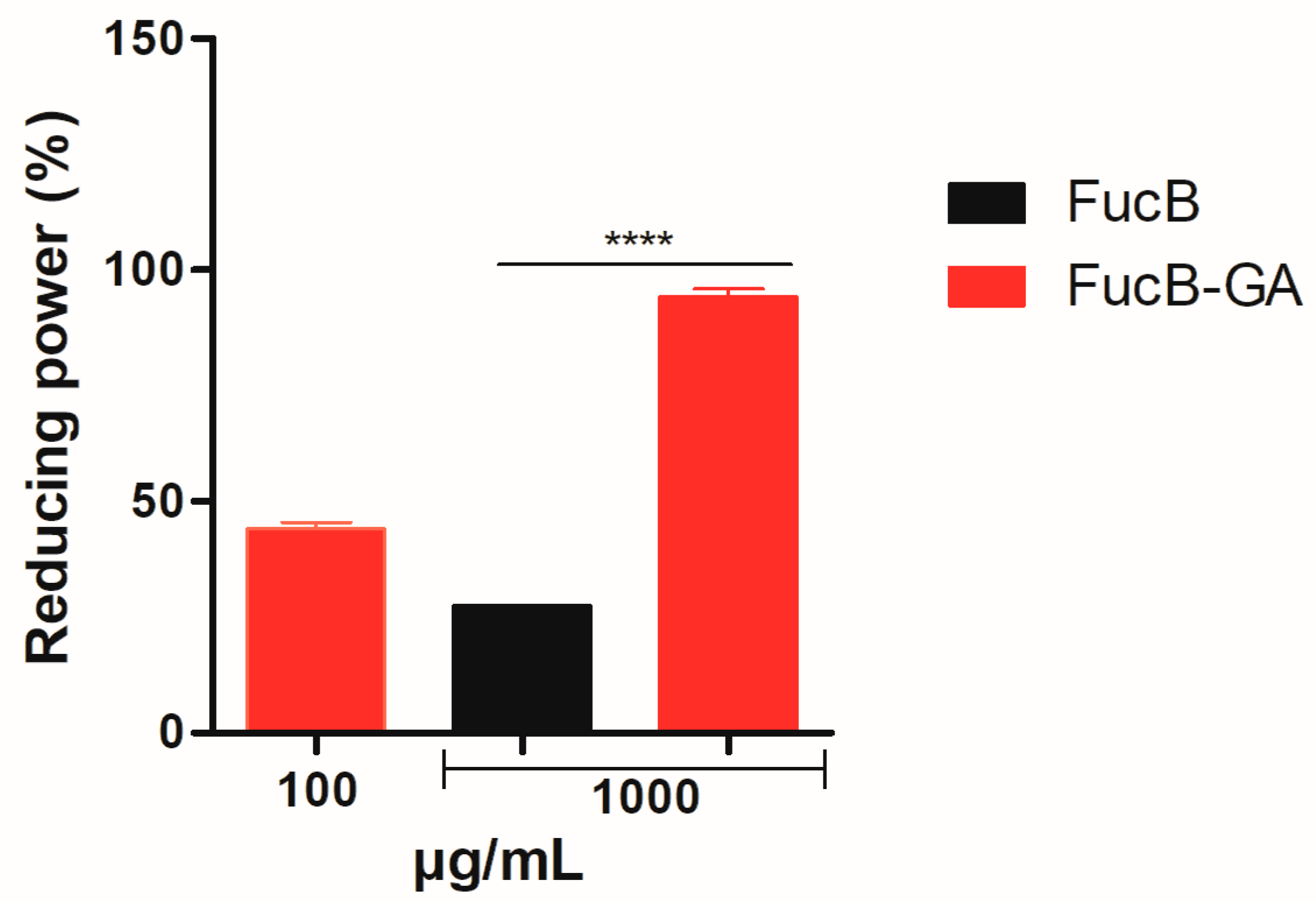
The reducing power assay evaluates the electron-donating capacity of compounds in a neutral medium. According to the results presented in Figure 12, at the lowest concentration tested, FucB showed no detectable activity, whereas FucB-GA exhibited 44.08% activity. At the highest concentration, FucB and FucB-GA demonstrated 27.46% and 94.22% activity, respectively. These findings are consistent with previous studies reporting similar outcomes for other gallic acid-conjugated molecules, such as chitosan [81] and fucan A [22]. Gallic acid alone did not exhibit electron-donating capacity under the conditions of this assay.
The data clearly indicate that the conjugation process significantly enhanced the reducing power of FucB, resulting in over 90% activity for FucB-GA. These findings support our initial hypothesis that conjugation of gallic acid to FucB would substantially improve its in vitro antioxidant activity compared to the individual components.
Gallic acid (GA) has strong antioxidant potential due to its ability to donate electrons, but its effectiveness can be influenced by structural factors such as stability and spatial freedom. When GA was conjugated to FucB, a branched and sulfated polysaccharide, these conditions may have improved. Unlike linear polysaccharides, like as chitosan [21] and dextrans [84], FucB may offer a more flexible and favorable microenvironment, allowing GA to maintain its reactivity and act efficiently as a reducing agent. This could explain the strong performance of FucB-GA in reducing power assays. Although these structural effects were not directly investigated in this study; they provide a plausible explanation for the observed antioxidant activity.

4. Conclusions

In this study, FucB was successfully chemically modified through covalent conjugation with gallic acid (GA), resulting in the FucB-GA derivative. Structural characterization confirmed the presence of GA moieties covalently linked to the polysaccharide without inducing significant depolymerization or structural disruption. Physicochemical analyzes demonstrated changes in thermal stability and greater amorphous character in FucB-GA.
Most notably, the modified compound exhibited significantly enhanced antioxidant properties, including superior total antioxidant capacity, reducing power, and superoxide radical and copper scavenging activities, when compared to native FucB and GA alone. These findings suggest that the incorporation of GA effectively improved the electron-donating and metal-chelating capabilities of FucB, likely due to increased availability of functional groups such as hydroxyls, carboxylate groups and sulfates.
However, the study also presents limitations. The lack of activity against hydrogen peroxide and the inability to chelate ferric ions highlight the specificity of FucB-GA’s antioxidant mechanisms and point to the need for deeper mechanistic understanding. Furthermore, all experiments were conducted in vitro, limiting the predictive value regarding biological efficacy in vivo.
Future studies should focus on elucidating the molecular mechanisms underlying the antioxidant actions of FucB-GA, as well as evaluating its bioavailability, biocompatibility, and therapeutic potential in cellular and animal models. Additionally, investigation into the compound’s anti-inflammatory or cytoprotective properties may broaden its applicability in pharmaceutical and nutraceutical formulations.

Author Contributions

Conceptualization, J.R.d.S. and H.A.O.R.; Formal analysis, J.R.d.S., D.A.S. and G.L.S.; Investigation, J.R.d.S., D.A.S. and H.A.O.R.; Methodology, J.R.d.S., D.A.S. and G.L.S.; Resources, G.L.S. and H.A.O.R.; Writing—original draft, J.R.d.S.; Writing—review and editing, H.A.O.R. All authors have read and agreed to the published version of the manuscript.

Funding

Research was supported by Ministério de Ciência, Tecnologia, Informação e Comércio (MCTIC—Brazil), Coordenação de Aperfeiçoamento de Pessoal de Nível Superior (CAPES—Brazil) (Finance Code 001). H. A. O. Rocha and G. L. Sassaki are CNPq fellowship honored researcher. J.R.d.S. had a Ph.D. scholarship from CAPES.

Institutional Review Board Statement

Not applicable.

Informed Consent Statement

Not applicable.

Data Availability Statement

Dataset generated during the current study are available from the corresponding author upon reasonable request.

Acknowledgments

The authors would like to express their gratitude to the Conselho Nacional de Desenvolvimento Científico e Tecnológico—CNPq and Coordenação de Aperfeiçoamento de Pessoal de Nível Superior—CAPES for their financial support. We would like to thank the Institute of Chemistry at UFRN for the FTIR analysis, and the Laboratory of Molecular Sieves (LABPEMOL) at the Institute of Chemistry, UFRN, for the XRD, TG/DTG, SEM, and EDS analyses. This research was submitted to the Graduate Program in Biochemistry and Molecular Biology at the Federal University of Rio Grande do Norte as part of the Ph.D. thesis of J.R.S. The ChatGTP version 4.0 was used to improve the English.

Conflicts of Interest

The authors declare no conflicts of interest.

Abbreviations

The following abbreviations are used in this manuscript:
XRDX-ray diffraction
FTIRFourier-transform infrared spectroscopy
TG/DTGThermogravimetric analysis
EDSEnergy-dispersive X-ray spectroscopy
FucBFucoidan purified from brown seaweed Spatoglossum schröederi
FucB-GAFucoidan–gallic acid conjugate
NMRNuclear magnetic resonance
ROSReactive oxygen species
SPSulfated polysaccharide
GAGallic acid
SEMScanning electron microscopy
NBTNitro blue tetrazolium
EDTA2,2′,2″,2′′′-(Ethane-1,2-diyldinitrilo) tetra-acetic acid
PDA1,3-diaminopropane-acetate buffer
UFRNFederal University of Rio Grande do Norte
TACTotal antioxidant capacity
ANOVAAnalysis of variance

References

  1. Zhang, L.; Wang, X.; Cueto, R.; Effi, C.; Zhang, Y.; Tan, H.; Qin, X.; Yang, X.; Wang, H. Biochemical basis and metabolic interplay of redox regulation. Redox Biol. 2019, 26, 101284. [Google Scholar] [CrossRef] [PubMed]
  2. Mei, S.; Song, X.; Wang, Y.; Wang, J.; Su, S.; Zhu, J.; Geng, Y. Studies on protection of astaxanthin from oxidative damage induced by H2O2 in RAW 264.7 cells based on 1H NMR metabolomics. J. Agric. Food Chem. 2019, 67, 13568–13576. [Google Scholar] [CrossRef] [PubMed]
  3. Badhani, B.; Sharma, N.; Kakkar, R. Gallic acid: A versatile antioxidant with promising therapeutic and industrial applications. RSC Adv. 2015, 5, 27540–27557. [Google Scholar] [CrossRef]
  4. Neha, K.; Haider, M.R.; Pathak, A.; Yar, M.S. Medicinal prospects of antioxidants: A review. Eur. J. Med. Chem. 2019, 178, 687–704. [Google Scholar] [CrossRef]
  5. Bedlovičová, Z.; Strapáč, I.; Baláž, M.; Salayová, A. A brief overview on antioxidant activity determination of silver nanoparticles. Molecules 2020, 25, 3191. [Google Scholar] [CrossRef]
  6. Pisoschi, A.M.; Pop, A. The role of antioxidants in the chemistry of oxidative stress: A review. Eur. J. Med. Chem. 2015, 97, 55–74. [Google Scholar] [CrossRef]
  7. Forman, H.J.; Zhang, H. Targeting oxidative stress in disease: Promise and limitations of antioxidant therapy. Nat. Rev. Drug Discov. 2021, 20, 689–709. [Google Scholar] [CrossRef]
  8. Liguori, I.; Russo, G.; Curcio, F.; Bulli, G.; Aran, L.; Della-Morte, D.; Gargiulo, G.; Testa, G.; Cacciatore, F.; Bonaduce, D.; et al. Oxidative stress, aging, and diseases. Clin. Interv. Aging 2018, 13, 757–772. [Google Scholar] [CrossRef]
  9. Begum, R.; Howlader, S.; Mamun-Or-Rashid, A.N.M.; Rafiquzzaman, S.M.; Ashraf, G.M.; Albadrani, G.M.; Sayed, A.A.; Peluso, I.; Abdel-Daim, M.M.; Uddin, S. Antioxidant and signal-modulating effects of brown seaweed-derived compounds against oxidative stress-associated pathology. Oxid. Med. Cell. Longev. 2021, 2021, 9974890. [Google Scholar] [CrossRef]
  10. Luthuli, S.; Wu, S.; Cheng, Y.; Zheng, X.; Wu, M.; Tong, H. Therapeutic effects of fucoidan: A review on recent studies. Mar. Drugs 2019, 17, 487. [Google Scholar] [CrossRef] [PubMed]
  11. Deniaud-Bouët, E.; Hardouin, K.; Potin, P.; Kloareg, B.; Hervé, C. A review about brown algal cell walls and fucose-containing sulfated polysaccharides: Cell wall context, biomedical properties and key research challenges. Carb. Polym. 2017, 175, 395–408. [Google Scholar] [CrossRef]
  12. Rocha, H.A.O.; Franco, C.R.; Trindade, E.S.; Carvalho, L.C.; Veiga, S.S.; Leite, E.L.; Dietrich, C.P.; Nader, H.B. A fucan from the brown seaweed Spatoglossum schröederi inhibits chinese hamster ovary cell adhesion to several extracellular matrix proteins. Braz. J. Med. Biol. Res. 2001, 34, 621–626. [Google Scholar] [CrossRef] [PubMed]
  13. Gamal-Eldeen, A.M.; Ahmed, E.F.; Abo-Zeid, M.A. In vitro cancer chemopreventive properties of polysaccha-ride extract from the brown alga, Sargassum latifolium. Food Chem. Toxicol. 2009, 47, 1378–1384. [Google Scholar] [CrossRef]
  14. Silva, T.M.A.; Alves, L.G.; Queiroz, K.C.S.; Santos, M.G.L.; Marques, C.T.; Chavante, S.F.; Rocha, H.A.O.; Leite, E.L. Partial characterization and anticoagulant activity of a heterofucan from the brown seaweed Padina gymnospora. Braz. J. Med. Biol. Res. 2005, 38, 523–533. [Google Scholar] [CrossRef]
  15. Cumashi, A.; Ushakova, N.A.; Preobrazhenskaya, M.E.; D’Incecco, A.; Piccoli, A.; Totani, L.; Tinari, N.; Morozevich, G.E.; Berman Al, E.; Bilian, M.I.; et al. A comparative study of the anti-inflammatory, anticoagulant, antiangiogenic, and antiadhesive activities of nine different fucoidans from brown seaweeds. Glycobiology 2007, 17, 541–552. [Google Scholar] [CrossRef]
  16. Sun, Y.; Hou, S.; Song, S.; Zhang, B.; Ai, C.; Chen, X.; Liu, N. Impact of acidic, water and alkaline extraction on structural features, antioxidant activities of Laminaria japonica polysaccharides. Int. J. Biol. Macromol. 2018, 112, 985–995. [Google Scholar] [CrossRef]
  17. Zhao, T.; Yang, M.; Ma, L.; Liu, X.; Ding, Q.; Chai, G.; Lu, Y.; Wei, H.; Zhang, S.; Ding, C. Structural modifi-cation and biological activity of polysaccharides. Molecules 2023, 28, 5416. [Google Scholar] [CrossRef]
  18. Liu, J.; Wang, X.; Yong, H.; Kan, J.; Jin, C. Recent advances in flavonoid-grafted polysaccharides: Synthesis, struc-tural characterization, bioactivities and potential applications. Int. J. Biol. Macromol. 2018, 116, 1011–1025. [Google Scholar] [CrossRef] [PubMed]
  19. Boundaoui, K.; Le Cerf, D.; Dulong, V. Functionalisation and behaviours of polysaccharides conjugated with phenolic compounds by oxidoreductase catalysis: A review. Int. J. Biol. Macromol. 2024, 283, 137660. [Google Scholar] [CrossRef]
  20. Zhang, W.; Sun, J.; Li, Q.; Liu, C.; Niu, F.; Yue, R.; Zhang, Y.; Zhu, H.; Ma, C.; Deng, S. Free radical-mediated grafting of natural polysaccharides such as chitosan, starch, inulin, and pectin with some polyphenols: Synthesis, structural characterization, bioactivities, and applications–A review. Foods 2023, 12, 3688. [Google Scholar] [CrossRef]
  21. Curcio, M.; Puoci, F.; Iemma, F.; Parisi, O.I.; Cirillo, G.; Spizzirri, U.G.; Picci, N. Covalent insertion of antioxidant molecules on chitosan by a free radical grafting procedure. J. Agric. Food Chem. 2009, 57, 5933–5938. [Google Scholar] [CrossRef] [PubMed]
  22. Melo, K.D.C.M.; Lisboa, L.S.; Queiroz, M.F.; Paiva, W.S.; Luchiari, A.C.; Camara, R.B.G.; Costa, L.S.; Rocha, H.A.O. Antioxidant activity of fucoidan modified with gallic acid using the redox method. Mar. Drugs 2022, 20, 490. [Google Scholar] [CrossRef] [PubMed]
  23. Ji, H.F.; Zhang, H.Y.; Shen, L. Proton dissociation is important to understanding structure–activity relationships of gallic acid antioxidants. Bioorg. Med. Chem. Lett. 2006, 16, 4095–4098. [Google Scholar] [CrossRef]
  24. Kim, Y.J. Antimelanogenic and antioxidant properties of gallic acid. Biol. Pharm. Bull. 2007, 30, 1052–1055. [Google Scholar] [CrossRef]
  25. Couto, A.; Kassuya, C.; Calixto, J.B.; Petrovick, P. R Anti-inflammatory, antiallodynic effects and quantitative analysis of gallic acid in spray dried powders from Phyllanthus niruri leaves, stems, roots and whole plant. Rev. Bras. Farmacogn. 2013, 23, 124–131. [Google Scholar] [CrossRef]
  26. Xiang, Z.; Guan, H.; Zhao, X.; Xie, Q.; Xie, Z.; Cai, F.; Dang, R.; Li, M.; Wang, C. Dietary gallic acid as an antioxidant: A review of its food industry applications, health benefits, bioavailability, nano-delivery systems, and drug interactions. Food Res. Int. 2021, 180, 114068. [Google Scholar] [CrossRef] [PubMed]
  27. Fernandes-Negreiros, M.M.; Batista, L.A.N.C.; Viana, R.L.S.; Sabry, D.A.; Paiva, A.A.O.; Paiva, W.S.; Machado, R.I.A.; Sousa Junior, F.L.; Pontes, D.L.; Vitoriano, J.O.; et al. Gallic acid-laminarin conjugate is a better antioxidant than sulfated or carboxylated laminarin. Antioxidants 2020, 9, 1192. [Google Scholar] [CrossRef]
  28. Rocha, H.A.O.; Bezerra, L.C.L.M.; Albuquerque, I.R.L.; Costa, L.S.; Guerra, C.M.P.; Abreu, L.D.; Nader, H.B.; Leite, E.L. A xylogalactofucan from the brown seaweed Spatoglossum schröederi stimulates the synthesis of an an-tithrombotic heparan sulfate from endothelial cells. Planta Med. 2005, 71, 379–381. [Google Scholar] [CrossRef] [PubMed]
  29. Wynne, M.J. A checklist of benthic marine algae of the tropical and subtropical western Atlantic. Can. J. Bot. 1986, 64, 2239–2281. [Google Scholar] [CrossRef]
  30. Rocha, H.A.O.; Morass, F.A.; Trindade, E.S.; Franco, C.R.C.; Torquato, R.J.S.; Veiga, S.S.; Valente, A.P.; Mourão, P.A.S.; Leite, E.L.; Nader, H.B.; et al. Structural and hemostatic activities of a sulfated galactofucan from the brown alga Spatoglossum schroederi. J. Biol. Chem. 2005, 280, 41278–41288. [Google Scholar] [CrossRef]
  31. Paiva, W.S.; Queiroz, M.F.; Araujo Sabry, D.; Santiago, A.L.C.M.A.; Sassaki, G.L.; Batista, A.C.L.; Rocha, H.A.O. Preparation, structural characterization, and property investigation of gallic acid-grafted fungal chitosan conjugate. J. Fungi 2021, 7, 812. [Google Scholar] [CrossRef]
  32. Dodgson, K.S.; Price, R.G. A note on the determination of the ester sulphate content of sulphated polysaccharides. Biochem. J. 1962, 84, 106–110. [Google Scholar] [CrossRef]
  33. Bradford, M.M. A rapid and sensitive method for the quantitation of microgram quantities of protein utilizing the principle of protein-dye binding. Anal. Biochem. 1976, 72, 248–254. [Google Scholar] [CrossRef] [PubMed]
  34. Singleton, V.L.; Rossi, J.A. Colorimetry of Total Phenolics with Phosphomolybdic-Phosphotungstic Acid Reagents. Am. J. Enol. Vitiv. 1965, 16, 144–158. [Google Scholar] [CrossRef]
  35. Dietrich, C.P.; Dietrich, S.M.C. Electrophoretic behaviour of acidic mucopolysaccharides in diamine buffers. Anal. Biochem. 1976, 70, 645–647. [Google Scholar] [CrossRef] [PubMed]
  36. Melo, K.R.T.; Camara, R.B.G.; Queiroz, M.F.; Vidal, A.A.J.; Lima, C.R.M.; Melo-Silveira, R.F.; Almeida-Lima, L.; Rocha, H.A.O. Evaluation of sulfated polysaccharides from the brown seaweed Dictyopteris justii as antioxidant agents and as inhibitors of the formation of calcium oxalate crystals. Molecules 2013, 18, 14543–14563. [Google Scholar] [CrossRef]
  37. Soeiro, V.C.; Melo, K.R.T.; Alves, M.G.C.F.; Medeiros, M.J.C.; Grilo, M.L.P.M.; Almeida-Lima, J.; Pontes, D.L.; Costa, L.S.; Rocha, H.A.O. Dextran: Influence of molecular weight in antioxidant properties and immunomodulatory potential. Int. J. Mol. Sci. 2016, 17, 1340. [Google Scholar] [CrossRef] [PubMed]
  38. Arizmendi-Cotero, D.; Gómez-Espinosa, R.M.; García, O.D.; Gómez-Vidales, V.; Dominguez-Lopez, A. Electron paramagnetic resonance study of hydrogen peroxide/ascorbic acid ratio as initiator redox pair in the inulin-gallic acid molecular grafting reaction. Carbohydr. Polym. 2016, 136, 350–357. [Google Scholar] [CrossRef]
  39. Liu, J.; Lu, J.; Kan, J.; Jin, C. Synthesis of chitosan-gallic acid conjugate: Structure characterization and in vitro anti-diabetic potential. Int. J. Biol. Macromol. 2013, 62, 321–329. [Google Scholar] [CrossRef]
  40. Liu, J.; Pu, H.; Chen, C.; Liu, Y.; Bai, R.; Kan, J.; Jin, C. Reaction mechanisms and structural and physicochemical properties of caffeic acid grafted chitosan synthesized in ascorbic acid and hydroxyl peroxide redox system. J. Agric. Food Chem. 2018, 66, 279–289. [Google Scholar] [CrossRef]
  41. Liu, T.; Ren, Q.; Wang, S.; Gao, J.; Shen, C.; Zhang, S.; Wang, Y.; Guan, F. Chemical modification of polysaccharides: A review of synthetic approaches, biological activity and the structure–activity relationship. Molecules 2023, 28, 6073. [Google Scholar] [CrossRef] [PubMed]
  42. Wu, C.; Wang, L.; Fang, Z.; Hu, Y.; Chen, S.; Sugawara, T.; Ye, X. The effect of the molecular architecture on the antioxidant properties of chitosan gallate. Mar. Drugs 2016, 14, 95. [Google Scholar] [CrossRef]
  43. Queiroz, M.F.; Sabry, D.A.; Sassaki, G.L.; Rocha, H.A.O.; Costa, L.S. Gallic acid-dextran conjugate: Green syn-thesis of a novel antioxidant molecule. Antioxidants 2019, 8, 478. [Google Scholar] [CrossRef] [PubMed]
  44. Presa, F.B.; Marques, M.L.M.; Viana, R.L.S.; Nobre, L.T.D.B.; Costa, L.S.; Rocha, H.A.O. The protective role of sulfated polysaccharides from green seaweed Udotea flabellum in cells exposed to oxidative damage. Mar. Drugs 2018, 16, 135. [Google Scholar] [CrossRef]
  45. Rabenstein, D.L. Heparin and heparan sulfate: Structure and function. Nat. Prod. Rep. 2002, 19, 312–331. [Google Scholar] [CrossRef]
  46. Zheng, J.; Li, B.; Ji, Y.; Chen, Y.; Lv, X.; Zhang, X.; Linhardt, R.J. Prolonged release and shelf-life of anticoagulant sulfated polysaccharides encapsulated with ZIF-8. Int. J. Biol. Macromol. 2021, 183, 1174–1183. [Google Scholar] [CrossRef]
  47. He, W.; Sun, H.; Su, L.; Zhou, D.; Zhang, X.; Shanggui, D.; Chen, Y. Structure and anticoagulant activity of a sulfated fucan from the sea cucumber Acaudina leucoprocta. Int. J. Biol. Macromol. 2020, 164, 87–94. [Google Scholar] [CrossRef]
  48. Hou, L.; Wu, P. Two-dimensional correlation infrared spectroscopy of heat-induced esterification of cellulose with 1,2,3,4-butanetetracarboxylic acid in the presence of sodium hypophosphite. Cellulose 2019, 26, 2759–2769. [Google Scholar] [CrossRef]
  49. Yang, T.; Liu, T.; Lin, I. Functionalities of chitosan conjugated with stearic acid and gallic acid and application of the modified chitosan in stabilizing labile aroma compounds in an oil-in-water emulsion. Food Chem. 2017, 228, 541–549. [Google Scholar] [CrossRef]
  50. Bai, R.; Yong, H.; Zhang, X.; Liu, J.; Liu, J. Structural characterization and protective effect of gallic acid grafted O-carboxymethyl chitosan against hydrogen peroxide-induced oxidative damage. Int. J. Biol. Macromol. 2020, 143, 49–59. [Google Scholar] [CrossRef] [PubMed]
  51. Cho, Y.; Kim, S.; Ahn, C.; Je, J. Preparation, characterization, and antioxidant properties of gallic acid-grafted-chitosans. Carbohydr. Polym. 2011, 83, 1617–1622. [Google Scholar] [CrossRef]
  52. Pasanphan, W.; Chirachanchai, S. Conjugation of gallic acid onto chitosan: An approach for green and water-based antioxidante. Carbohydr. Polym. 2008, 72, 169–177. [Google Scholar] [CrossRef]
  53. Ali, A.; Chiang, Y.W.; Santos, R.M. X-ray diffraction techniques for mineral characterization: A review for engineers of the fundamentals, applications, and research directions. Minerals 2022, 12, 205. [Google Scholar] [CrossRef]
  54. Daub, C.D.; Mabate, B.; Malgas, S.; Pletschke, B.I. Fucoidan from Ecklonia maxima is a powerful inhibitor of the diabetes-related enzyme, α-glucosidase. Int. J. Biol. Macromol. 2020, 151, 412–420. [Google Scholar] [CrossRef]
  55. Saravana, P.S.; Cho, Y.-J.; Park, Y.-B.; Woo, H.-C.; Chun, B.-S. Structural, antioxidant, and emulsifying activities of fucoidan from Saccharina japonica using pressurized liquid extraction. Carbohydr. Polym. 2016, 153, 518–525. [Google Scholar] [CrossRef]
  56. Puhari, S.S.M.; Yuvaraj, S.; Vasudevan, V.; Ramprasath, T.; Rajkumar, P.; Arunkumar, K.; Amutha, C.; Selvam, G.S. Isolation and characterization of fucoidan from four brown algae and study of the cardioprotective effect of fucoidan from Sargassum wightii against high glucose-induced oxidative stress in H9c2 cardiomyoblast cells. J. Food Biochem. 2022, 46, 14412. [Google Scholar] [CrossRef]
  57. Govindan, S.; Nivethaa, E.A.K.; Saravanan, R.; Narayanan, V.; Stephen, A. Synthesis and characterization of chi-tosan–silver nanocomposite. Appl. Nanosci. 2012, 2, 299–303. [Google Scholar] [CrossRef]
  58. Mishra, R.K.; Majeed, A.B.A.; Banthia, A.K. Development and characterization of pectin/gelatin hydrogel mem-branes for wound dressing. Int. J. Plast. Technol. 2011, 15, 82–95. [Google Scholar] [CrossRef]
  59. Liu, J.; Lu, J.; Kan, J.; Tang, Y.; Jin, C. Preparation, characterization and antioxidant activity of phenolic acids grafted carboxymethyl chitosan. Preparation, characterization and antioxidant activity of phenolic acids grafted carboxymethyl chitosan. Int. J. Biol. Macromol. 2013, 62, 85–93. [Google Scholar] [CrossRef]
  60. Zhang, X.; Wu, H.; Zhang, L.; Sun, Q. Horseradish peroxidase-mediated synthesis of an antioxidant gallic acid-g-chitosan derivative and its preservation application in cherry tomatoes. RSC Adv. 2018, 8, 20363–20371. [Google Scholar] [CrossRef] [PubMed]
  61. Arab, K.; Ghanbarzadeh, B.; Ayaseh, A.; Jahanbin, K. Extraction, purification, physicochemical properties and antioxidant activity of a new polysaccharide from Ocimum album L. seed. Int. J. Biol. Macromol. 2021, 180, 643–653. [Google Scholar] [CrossRef] [PubMed]
  62. Hu, Q.; Wang, T.; Zhou, M.; Xue, J.; Lu, Y. In vitro antioxidant-activity evaluation of gallic-acid-grafted chitosan conjugate synthesized by free-radical-induced grafting method. J. Agric. Food Chem. 2016, 64, 5893–5900. [Google Scholar] [CrossRef]
  63. Hadidi, M.; Amoli, P.I.; Jelyani, A.Z.; Hasiri, Z.; Rouhafza, A.; Ibars, A.; Khaksar, F.B.; Tabrizi, S.T. Polysaccha-rides from pineapple core as a canning by-product: Extraction optimization, chemical structure, antioxidant and functional properties. Int. J. Biol. Macromol. 2020, 163, 2357–2364. [Google Scholar] [CrossRef]
  64. Castaño, J.; Rodríguez-Llamazares, S.; Carrasco, C.; Bouza, R. Physical, chemical and mechanical properties of pehuen cellulosic husk and its pehuenstarch based composites. Carbohydr. Polym. 2012, 90, 1550–1556. [Google Scholar] [CrossRef]
  65. Hajji, M.; Hamdi, M.; Sellimi, S.; Ksouda, G.; Laouer, H.; Li, S.; Nasri, M. Structural characterization, antioxidant and antibacterial activities of a novel polysaccharide from Periploca laevigata root barks. Carbohydr. Polym. 2019, 206, 380–388. [Google Scholar] [CrossRef]
  66. Liu, H.; Tang, W.; Lei, S.; Zhang, Y.; Cheng, M.; Liu, Q.; Wang, W. Extraction optimization, characterization and biological activities of polysaccharide extracts from nymphaea hybrid. Int. J. Mol. Sci. 2023, 24, 8974. [Google Scholar] [CrossRef]
  67. Pongali, P.; Kasim, N.; Hanibah, H.; Rosli, N.H.A. Glutaryl kappa-carrageenan: Synthesis, characterization and investigation of potential as gel-based biopolymer electrolyte. Polym. Bull. 2025, 82, 5693–5715. [Google Scholar] [CrossRef]
  68. Goudar, N.; Vanjeri, V.N.; Dixit, S.; Hiremani, V.; Sataraddi, S.; Gasti, T.; Vootla, S.K.; Masti, S.P.; Chougale, R.B. Evaluation of multifunctional properties of gallic acid crosslinked poly (vinyl alcohol)/tragacanth gum blend films for food packaging applications. Int. J. Biol. Macromol. 2020, 158, 139–149. [Google Scholar] [CrossRef] [PubMed]
  69. Checa, J.; Aran, J.M. Reactive oxygen species: Drivers of physiological and pathological processes. J. Inflamm. Res. 2020, 13, 1057–1073. [Google Scholar] [CrossRef] [PubMed]
  70. Jesumani, V.; Du, H.; Pei, P.; Aslam, M.; Huang, N. Comparative study on skin protection activity of polyphenol-rich extract and polysaccharide-rich extract from Sargassum vachellianum. PLoS ONE 2020, 15, e0227308. [Google Scholar] [CrossRef]
  71. Silva, M.M.C.L.; Lisboa, L.S.; Paiva, W.S.; Batista, L.A.N.C.; Luchiari, A.C.; Rocha, H.A.O.; Camara, R.B.G. Comparison of in vitro and in vivo antioxidant activities of commercial fucoidans from Macrocystis pyrifera, Undaria pinnatifida, and Fucus vesiculosus. Int. J. Biol. Macromol. 2022, 216, 757–767. [Google Scholar] [CrossRef]
  72. Caruso, F.; Incerpi, S.; Pedersen, J.; Belli, S.; Kaur, S.; Rossi, M. Aromatic polyphenol π-π interactions with super-oxide radicals contribute to radical scavenging and can make polyphenols mimic superoxide dismutase activity. Curr. Issues Mol. Biol. 2022, 44, 5209–5220. [Google Scholar] [CrossRef]
  73. Wu, J. Tackle the free radicals damage in COVID-19. Nitric Oxide 2020, 102, 39–41. [Google Scholar] [CrossRef] [PubMed]
  74. Liu, T.; Hong, K.; Yang, T. Functionalities of tremella fuciformis polysaccharides modified with gallic acid. Molecules 2024, 29, 5890. [Google Scholar] [CrossRef]
  75. Niki, E. Assessment of antioxidant capacity in vitro and in vivo. Free Radic. Biol. Med. 2010, 49, 503–515. [Google Scholar] [CrossRef]
  76. Xie, M.; Hu, B.; Wang, Y.; Zeng, X. Grafting of gallic acid onto chitosan enhances antioxidant activities and alters rheological properties of the copolymer. J. Agric. Food Chem. 2014, 62, 9128–9136. [Google Scholar] [CrossRef] [PubMed]
  77. Gromadzka, G.; Tarnacka, B.; Flaga, A.; Adamczyk, A. copper dyshomeostasis in neurodegenerative diseases—Therapeutic implications. Int. J. Mol. Sci. 2020, 21, 9259. [Google Scholar] [CrossRef]
  78. Møller, L.B.; Lenartowicz, M.; Zabot, M.T.; Josiane, A.; Burglen, L.; Bennett, C.; Riconda, D.; Fisher, R.; Janssens, S.; Mohammed, S.; et al. Clinical expression of Menkes disease in females with normal karyotype. Orphanet J. Rare Dis. 2012, 7, 6. [Google Scholar] [CrossRef] [PubMed]
  79. Shribman, S.; Marjot, T.; Sharif, A.; Vimalesvaran, S.; Ala, A.; Alexander, G.; Dhawan, A.; Dooley, J.; Gillett, G.T.; Kelly, D.; et al. Investigation and management of Wilson’s disease: A practical guide from the British Association for the study of the liver. Lancet Gastroenterol. Hepatol. 2022, 7, 560–575. [Google Scholar] [CrossRef]
  80. Brewer, G.J. Novel therapeutic approaches to the treatment of Wilson’s disease. Expert Opin. Pharmacother. 2006, 7, 317–324. [Google Scholar] [CrossRef]
  81. Queiroz, M.F.; Melo, K.R.T.; Sabry, D.A.; Sassaki, G.L.; Rocha, H.A.O.; Costa, L.S. Gallic acid-chitosan conjugate inhibits the formation of calcium oxalate crystals. Molecules 2019, 24, 2074. [Google Scholar] [CrossRef] [PubMed]
  82. Camara, R.B.G.; Costa, L.S.; Fidelis, G.P.; Nobre, L.T.D.B.; Dantas-Santos, N.; Cordeiro, S.L.; Costa, M.S.S.P.; Alves, L.G.; Rocha, H.A.O. Heterofucans from the brown seaweed Canistrocarpus cervicornis with anticoagulant and antioxidant activities. Mar. Drugs 2011, 9, 124–138. [Google Scholar] [CrossRef]
  83. Rodrigues-Souza, I.; Pessatti, J.B.K.; da Silva, L.R.; Bellan, D.d.L.; de Souza, I.R.; Cestari, M.M.; de Assis, H.C.S.; Rocha, H.A.O.; Simas, F.F.; Trindade, E.d.S.; et al. Protective potential of sulfated polysaccharides from tropical seaweeds against alkylating- and oxidizing-induced genotoxicity. Int. J. Biol. Macromol. 2022, 211, 524–534. [Google Scholar] [CrossRef] [PubMed]
  84. Apostolova, E.; Lukova, P.; Baldzhieva, A.; Katsarov, P.; Nikolova, M.; Iliev, I.; Petchev, L.; Trica, B.; Oancea, F.; Delattre, C.; et al. Immunomodulatory and anti-inflammatory effects of fucoidan: A review. Polymers 2020, 12, 2338. [Google Scholar] [CrossRef] [PubMed]
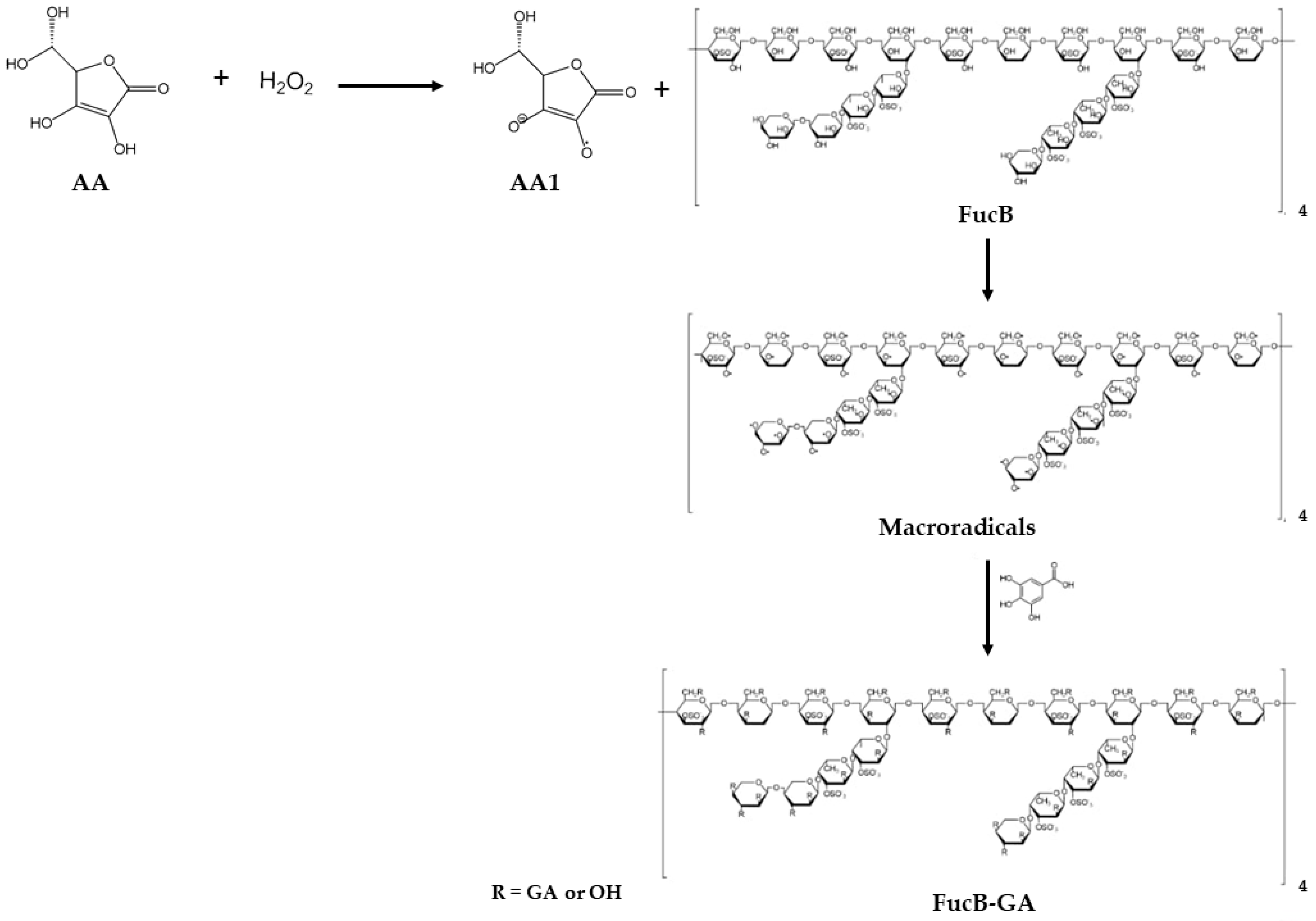
Figure 1. Proposed mechanism of the redox reaction between ascorbic acid (AA) and hydrogen peroxide (H2O2), and the binding of gallic acid (GA) into fucan B (FucB). The upper part illustrates the formation of the ascorbate radical (AA1) from the AA/H2O2 redox process. Subsequently, the formation of FucB macroradicals and the incorporation of GA result in the formation of FucB modified with gallic acid (FucB-GA). R: gallic acid or hydroxyl group.
Polysaccharides 06 00089 g001
Figure 2. Agarose gel electrophoresis of FucB and FucB-GA stained with toluidine blue. The migration profile of the samples is shown from the origin, indicating the migration of the samples along the gel.
Polysaccharides 06 00089 g002
Figure 3. FTIR spectra of FucB (black line) and FucB-GA (red line). The structural difference is highlighted by an arrow in the FucB-GA spectrum, indicating the ester carbonyl stretching band that originated from the covalent binding of GA to FucB.
Polysaccharides 06 00089 g003
Figure 4. 1H-NMR spectra (range: 0–8.23 ppm). Panel (A) 1H-NMR spectrum of FucB: H1 and H6 of α-L-fucose linked-(1→4) at 5.10 and 1.23 ppm, respectively; H1 of β-D-galactose linked-(1→4) at 4.59 ppm; and H2 of β-D-galactose-3S linked-(1→4) and β-D-galactose linked-(1→2,4) at 3.95 and 3.81 ppm, respectively. Panel (B) 1H-NMR spectrum of FucB-GA. An evident peak at 7.17 ppm indicates the presence of aromatic hydrogens from GA, confirming its insertion at the polysaccharide structure.
Polysaccharides 06 00089 g004
Figure 5. X-ray diffraction patterns of FucB and FucB-GA. Both samples exhibit predominantly amorphous profiles; however, the disappearance of these peaks following conjugation suggests a further reduction in crystallinity, likely resulting from the incorporation of gallic acid.
Polysaccharides 06 00089 g005
Figure 6. Morphological characterization by SEM analysis. (A) FucB (scale = 20 µm), exhibiting amorphous morphology; (B) FucB-GA (scale = 20 µm) under the same conditions, with arrows highlighting subtle structural differences following modification. EDS spectra of (C) FucB and (D) FucB-GA. Both spectra show similar elemental peaks, with the absence of nitrogen indicating no protein contamination in the samples.
Polysaccharides 06 00089 g006
Figure 7. TG and DTG curves of (A) FucB (black) and (B) FucB-GA (red). The horizontal regions in the thermograms indicate temperature intervals in which the compounds exhibit thermal stability.
Polysaccharides 06 00089 g007
Figure 8. Hydrogen peroxide scavenging activity of GA, FucB, and FucB-GA samples (p < 0.01). No statistically significant differences were observed among the samples or between the concentrations tested.
Polysaccharides 06 00089 g008
Figure 9. Superoxide radical scavenging activity of FucB and FucB-GA. Asterisks indicate statistically significant differences between sample treatments at the same concentration (**** p < 0.0002; *** p < 0.002).
Polysaccharides 06 00089 g009
Figure 10. Copper chelation activity of FucB and FucB-GA at concentrations of 0.1 and 1.0 mg/mL. Asterisks indicate statistically significant differences between sample treatments at the same concentration (**** p < 0.0001; * p < 0.05).
Polysaccharides 06 00089 g010
Figure 11. Total antioxidant capacity of FucB and FucB-GA samples. Different symbols indicate statistically significant differences between treatments (**** p < 0.0001).
Polysaccharides 06 00089 g011
Figure 12. Comparison of reducing power between FucB and FucB-GA. Asterisks denote statistically significant differences between treatments (**** p < 0.0001).
Polysaccharides 06 00089 g012
Disclaimer/Publisher’s Note: The statements, opinions and data contained in all publications are solely those of the individual author(s) and contributor(s) and not of MDPI and/or the editor(s). MDPI and/or the editor(s) disclaim responsibility for any injury to people or property resulting from any ideas, methods, instructions or products referred to in the content.