Proceeding Paper

A Cell-Based Bioelectric Biosensor for Salmonella spp. Detection in Food †

by
Lazaros Konstantinou
1,2,
Eleni Varda
1,
Stella Pempetsiou
1,
Theofylaktos Apostolou
1,
Konstantinos Loizou
1,
Lazaros Dougiakis
1,
Antonios Inglezakis
1 and
Agni Hadjilouka
1,2,*
1
EMBIO Diagnostics Ltd., Athalassas Avenue 8b, Nicosia 2018, Cyprus
2
Department of Life Sciences, School of Sciences, European University of Cyprus, 6 Diogenous Str., Nicosia 2404, Cyprus
*
Author to whom correspondence should be addressed.
Presented at the 3rd International Electronic Conference on Biosensors, 8–21 May 2023; Available online: https://iecb2023.sciforum.net/.
Eng. Proc. 2023, 35(1), 4; https://doi.org/10.3390/IECB2023-14564
Published: 8 May 2023
(This article belongs to the Proceedings of The 3rd International Electronic Conference on Biosensors)

Abstract

:
The prevalence of foodborne diseases is continuously increasing, causing numerous hospitalizations and deaths, as well as money loss in the agri-food sector and food supply chain worldwide. The standard analyses currently used for bacteria detection have significant limitations with the most important being their long procedural time that can be crucial for foodborne outbreaks. In this study, we developed a biosensor system able to perform robust and accurate detection of Salmonella spp. in meat products after a 3-min analysis. To achieve this, we used a portable device developed by EMBIO Diagnostics called B.EL.D (Bio Electric Diagnostics) and a cell-based biosensor technology (BERA). Results indicated that the new method could detect the pathogen within 24 h after a 3-min analysis and discriminate samples with and without Salmonella with high accuracy (86.1%). The method’s sensitivity, specificity, and positive and negative predictive values ranged from 80 to 90.5%, while the limit of detection was determined to be as low as 10 CFU g−1 in all food substrates.

1. Introduction

Salmonellosis, caused by Salmonella bacteria, is a major global public health issue with around 93.8 million cases of gastroenteritis and approximately 155,000 deaths annually [1]. There are currently about 2500 distinct serotypes or serovars among Salmonella bongori and Salmonella enterica, the two species of Salmonella. Salmonella enterica ser. Enteritidis and Salmonella enterica ser. Typhimurium are two of the most significant Salmonella serotypes that are transmitted from animals to humans, with Enteritidis being the most frequently reported serovar in human salmonellosis cases in the EU and the United States, and Typhimurium being the most prevalent and disseminated serovar globally [2,3].
Salmonella can survive in various environments and adapt to different conditions, making it a common contaminant in food. It can cause illness when people consume contaminated raw or undercooked food or when food handlers have inadequate cleanliness during preparation [4]. Salmonella can also be transmitted to humans through contact with contaminated pets. Foods that commonly cause salmonellosis include raw or undercooked eggs, unpasteurized dairy products, fruits and vegetables, and red meat, poultry, and shellfish [5]. Although measures have been developed to control its incidence, salmonellosis infections still occur and can lead to hospitalizations and deaths [6].
The infectious dose of Salmonella can vary based on various factors, and it is usually low, less than 1000 cells [7]. Therefore, there is a need for pathogen detection and proper control, and new methodologies, such as biosensing techniques, are being developed for this purpose. However, current detection techniques have several drawbacks. Biosensors, such as piezoelectric, optical, and electrochemical biosensors, have been developed to detect Salmonella in food, but very few studies have been done utilizing cell-based biosensors for this purpose [8,9,10].
The study aimed to develop and validate a biosensor system for the detection of Salmonella spp. in food using a cell-based biosensor technology and a portable device called B.EL.D. The system was tested on cured meat samples and frozen ready-to-eat meat and meat products. The study also evaluated seven protocols with different broths and incubation times to detect and discriminate the pathogen among other bacteria, using the ISO 6579-7: 2017 standard as the gold standard method to validate the results [11].

2. Materials and Methods

2.1. Collection of Samples and Experimental Design

A total of hundred samples (n = 100) of cured meat and frozen ready-to-eat meat and meat preparations were collected from a local meat processing company. The study was conducted in two phases (Phase 1 and Phase 2). To assess the biosensor’s ability to detect the pathogen in these food substrates, fifty samples (n = 50) were evaluated in Phase 1 using three alternative protocols (Protocols 1–3) developed based on the ISO 6579-7:2017 methodology [11]. Phase 2 of the project then involved the evaluation of four distinct methods (Protocols 4–7). The ISO 6579-7:2017 methodology served as the foundation for these protocols as well, although the incubation times were greatly shortened to test the biosensor’s ability to detect the pathogen within 30 h or less (Table 1).
From each sample (n = 100), four different samples were prepared for testing. Three were inoculated with Salmonella spp. at 0.6, 1, and 2 log CFU g−1 (see Section 2.2) and were utilized as positive samples, and one was left uninoculated to be used as the negative sample [control]. Pathogen’s absence in the control samples was confirmed through analysis according to the ISO 6579-7:2017 method (Microbiology of the food chain—Horizontal method for the detection, enumeration and serotyping of Salmonella). In the case of presumptive Salmonella spp. colonies in control samples, pathogen’s presence was further investigated through biochemical tests using API20E.

2.2. Bacteria Culturing and Sample Inoculation

Salmonella enterica subsp. enterica serovar Enteritidis WDCM 00030 and Salmonella enterica subsp. enterica serovar Typhimurium WDCM 00031 were used throughout this study. Ahead of any experimental analysis, Salmonella spp. cultures were grown overnight at 37 °C and serially diluted. The samples were then inoculated by placing 25 g of each food substrate in sterile stomacher bags and spraying the bags with the correct pathogen dilution to reach the desired inoculation level (0.6, 1, and 2 log CFU g−1). The following sample treatment was carried out accordingly to the respective protocols (Table 1).

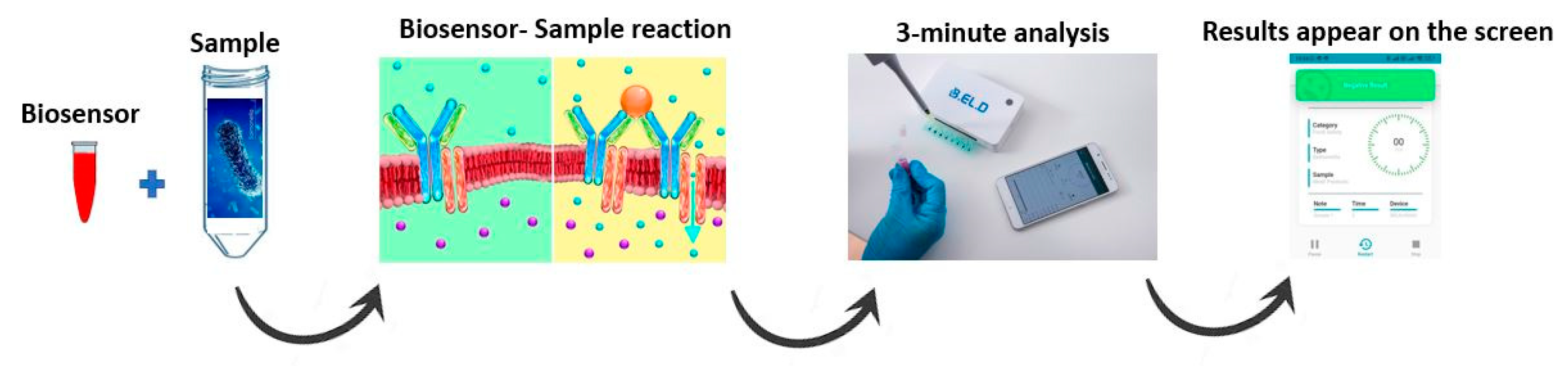
2.3. Bioensor Development and Analysis of the Samples

Cell culture was conducted according to Apostolou et al. [9] utilizing African green monkey kidney epithelial cells (Vero cells). Briefly, cells were cultured with a medium comprising Dulbecco’s medium with 10% fetal bovine serum (FBS), 10% streptomycin/penicillin, and 10% L-glutamine and l-alanine and incubated at 5% CO2 and 37 °C. Vero cells were detached from the culture dishes using Trypsin/EDTA (10 min at 37 °C) and collected by centrifugation (6 min/1000 rpm/25 °C), to a final density of 2.5 × 10⁶ mL−1. The cell pellet was resuspended in 400 μL PBS containing 5 μg mL−1 antibody (monoclonal mouse anti-Salmonella spp. (A, B, C, D & E Groups) from antibodies-online) and incubated on ice for 20 min. Then, the cell-antibody mixture was transferred to electroporation cuvettes (Eppendorf Eporator, Hamburg, Germany) (4 mm) and electroporation was performed by applying two square electric field pulses at 1800 V/cm. The mixture was then transferred to a Petri dish (60 × 15 mm) containing nutrient medium and incubated overnight at 37 °C and 5% CO2. The next day, the medium was discarded from the Petri dish and the membrane-engineered cells were mechanically detached and collected with a fresh medium in Eppendorf tubes [12].
EMBIO DIAGNOSTICS develops and produces the B.EL.D device utilized for the method’s development and the novel biosensor system’s validation (EMBIO DIAGNOSTICS Ltd., Nicosia, Cyprus). The tool monitors electric signals from various biorecognition modules and is a portable multichannel potentiometer with a replaceable connector consisting of eight screen-printed electrodes. The Bioelectric Recognition Assay (BERA), a powerful cell-based biosensor technology, acts as the device’s foundation and high-accuracy A/D converters are employed for measurements, providing high throughput and speedy analysis. Finally, the device connects with an Android or iOS device through Bluetooth 4.0, allowing the end-user to get prompt notification of the test findings. The samples were analysed as previously described by Hadjilouka et al. [10]. In a nutshell, 20 μL of the membrane-engineered cells (~5 × 10⁴ cells) were added on each of the eight screen-printed electrodes and after 120 s, 20 μL of each sample were added on top of the membrane-engineered cells. Every measurement lasted three minutes, and data were sent to a cloud server, where they were used to instantaneously calculate results using a newly created algorithm and display them on the Android/iOS screen. A total of two thousand (n = 2000) tests were conducted for all hundred samples (5 tests per sample) and during each test, the samples underwent eight separate analyses due to the utilization of the set of eight different sensors (Figure 1).

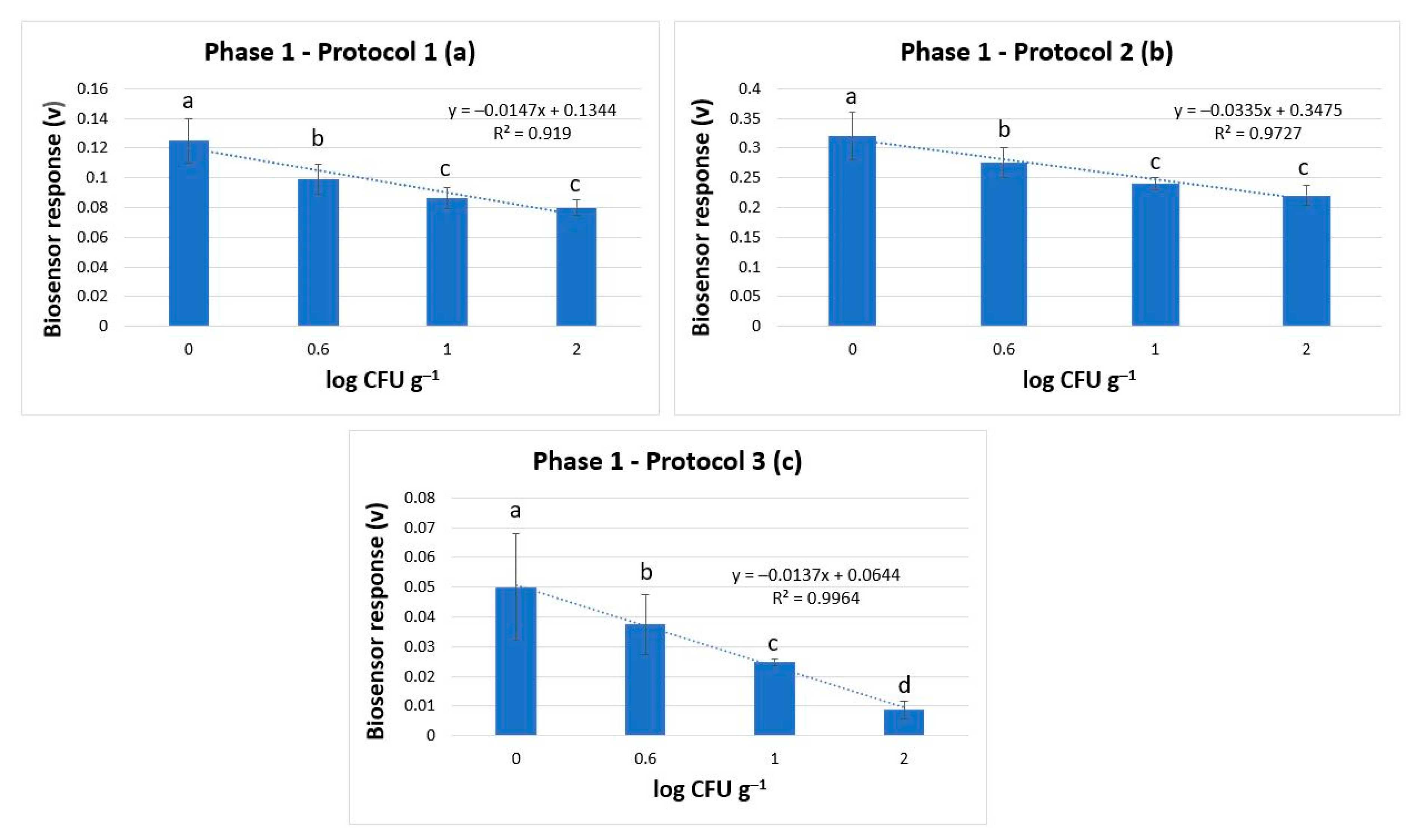
2.4. Algorithm for Response Processing and Statistical Analysis

For each sample, each test produced a time series with 720 voltage detection data points. Data analysis was carried out according to Hadjilouka et al. [12] using libraries in the python programming language. Following an analysis of the data from the positive and negative samples, a one-way analysis of variances (ANOVA) was performed to identify statistical differences. The thresholds that separate positive from negative samples were then defined, along with the limit of detection (LOD). After producing data-stored result arrays for positive and negative samples the system was eventually able to instantly classify the samples as being “above” or “below” the LOD, after each test.
After comparing the results obtained by the biosensor with the conventional techniques, performance indicators for the novel approach were calculated. These indicators were: Accuracy (Acc), Sensitivity (Se), Specificity (Sp), Positive Predictive Value (PPV), and Negative Predictive Value (NPV).

3. Results and Discussion

Table 2 summarizes the findings of the outcomes from all seven protocols. The investigation was conducted in two phases (Phase 1 and Phase 2), and the samples that were analyzed included cured meat samples, frozen ready-to-eat meat, and meat preparations. In Phase 1, the biosensor’s capacity to identify Salmonella spp. was investigated following three different protocols and in Phase 2, four alternative procedures were examined to determine whether the biosensor could identify the pathogen within 30 h or less.
Phase 1:
Results indicated that the biosensor could accurately discriminate between samples with and without Salmonella spp., with a limit of detection (LOD) of 0.6 log CFU g−1 and high accuracies ranging from 83.8% to 97.7%. Incubating samples with different enrichment broths increased the population of Salmonella to high levels (≥5 log CFU g−1). This increase in population enhanced the biosensor’s ability to detect positive samples even when the initial inoculation level was very low (0.6 log CFU g−1) (Figure 2). This ability was observed in all three protocols and showed a statistically significant discrimination power. Protocol 1, however, was found to have the best discrimination ability with the highest accuracy of 97.7%. These results suggested that the newly developed biosensor-based method could detect Salmonella spp. in meat and meat products.
Phase 2
Tests conducted using all four protocols showed that the biosensor could discriminate between samples with and without Salmonella spp. in less than 30 h, but with lower accuracies compared to Phase 1, ranging between 73.3% and 86.1%. Reducing the incubation time resulted in lower levels of Salmonella’s final population (3.5–4 log CFU g−1 increase), leading to reduced accuracy in the biosensor’s ability to discriminate positive from negative samples compared to the first phase of experiments, especially when the initial inoculation level was very low (0.6 log CFU g−1) (Figure 3). Consequently, the limit of detection (LOD) of the method increased to 1 log CFU g−1. Protocol 4 was unable to produce reliable results regarding the pathogen’s absence/presence. Protocols 5 and 7 were found to have the best discrimination power among the four protocols, with an accuracy of 88.8 and 86.1%, respectively. However, since Protocol 7 has the shortest incubation period (24 h), it was selected as the most appropriate for the detection of Salmonella spp. in meat and meat samples using the newly biosensor system.
Four different enrichment and pre-enrichment broths (RVS, MKTTn, M broth, and BPW) were employed for the seven procedures, with various combinations and incubation times for each stage of the process (Table 1). The use of BPW is appropriate for the initial, non-selective enrichment of bacteria, particularly pathogenic Enterobacteria, and for this reason it was utilized in all protocols [13]. Both RVS and MKTTn are selective enrichment broths used for the isolation of Salmonella spp. and they are both suggested by the ISO 6579-7: 2017 method for the detection of the pathogen [14,15]. M broth is an enrichment broth that can be used to grow a range of microorganisms, including Salmonella spp., however it is less sensitive than RVS or MKTTn [16].
Results obtained from all seven protocols indicated that the reduction of incubation time resulted in lower accuracy and performance characteristics of the method. More specifically, when the total incubation time was shortened from 48 h to 30 h and 24 h, respectively, the method’s accuracy fell from 97.7% to 88.6% and 86.1% in the tests where RVS was the primary enrichment broth (Protocols 1, 5, and 7). Similarly, the accuracy decreased from 83.8% to 78% when the total incubation period was shortened from 48 h to 24 h in the assays where MKTTn was the primary enrichment broth (Protocols 2 and 6). This was attributed to the fact that the augmented incubation periods resulted in higher final population levels of the pathogen, thus enhancing the response of the biosensor after the reaction between antibody and antigen. Overall, the RVS broth-based protocols had higher discrimination power than the MKTTn broth-based protocols.
Regarding Protocols 3 and 4, with the longest and shortest incubation periods, respectively, they were considered inappropriate for the newly developed method, for different reasons. Protocol 3 indicated high accuracy (90%) and performance characteristics. Nevertheless, due to its lengthy procedure time it was appropriate for a rapid method. Contrary to this, Protocol 4 was found unsuitable since it was unable to successfully distinguish between positive and negative samples, even when the bacterium was initially present at high population levels (2 log CFU g−1). The protocol’s lack of ability to properly discriminate Salmonella was due to the use of BPW as the only employed broth, since it is a non-selective enrichment broth that encourages the growth of various microorganisms. As a result, the growth of other bacteria in the sample, likely in significantly larger numbers, was not inhibited. Additionally, the results showed that there was no significant difference in the potential dynamic measured in samples tested with and without the pathogen (at initial inoculation levels of 0.6 and 1 log CFU g−1) in this protocol. This similarity was observed because the potential dynamic measured in the samples was not only influenced by the reaction between the antibody and antigen but also by the non-homeostatic membrane potential of microorganisms, which is essential for their behavior and survival. Although a statistically significant decrease in potential dynamic was observed when the initial pathogen inoculation level was at 2 log CFU g−1, the method was still not entirely reliable in distinguishing between samples. Finally, it should be mentioned that the new method was unable to differentiate negative from positive results without the addition of the biosensor, indicating the importance of the reaction between the antibody and the antigen in the newly developed system.

4. Conclusions

The study demonstrated the effectiveness of a portable cell-based biosensor system in detecting Salmonella enterica ser. Enteritidis and Salmonella enterica ser. Typhimurium in meat and meat products. The technique was designed based on the ISO 6579-7:2017 standard and provides results within 3 min after a 24-h enrichment step. The biosensor system is combined with user-friendly software that interfaces with an Android/iOS device through Bluetooth 4.0, allowing for immediate access to test results. The biosensor technology showed an accuracy of 86.1% and a limit of detection of 1 log CFU g−1, making it a useful screening tool for the rapid detection of Salmonella spp. in meat and meat products.

Author Contributions

Conceptualization, A.H. and K.L.; methodology, A.H.; software, L.D.; hardware: A.I.; data curation, A.H., L.K. and L.D.; investigation: L.K., E.V. and S.P.; validation, A.H.; writing—original draft preparation, L.K.; writing—review and editing, A.H. and T.A.; supervision, A.H. All authors have read and agreed to the published version of the manuscript.

Funding

This research received no external funding.

Institutional Review Board Statement

Not applicable.

Informed Consent Statement

Not applicable.

Data Availability Statement

Unavailable due to privacy.

Conflicts of Interest

The authors declare no conflict of interest.

References

  1. Griffith, R.W.; Carlson, S.A.; Krull, A.C. Salmonellosis. In Diseases of Swine; Wiley-Blackwell: Hoboken, NJ, USA, 2019; pp. 912–925. [Google Scholar] [CrossRef]
  2. Sun, H.; Wan, Y.; Du, P.; Bai, L. The Epidemiology of Monophasic Salmonella Typhimurium. Foodborne Pathog. Dis. 2020, 17, 87–97. Available online: https://home.liebertpub.com/fpd (accessed on 31 January 2023). [CrossRef] [PubMed]
  3. Okoro, C.K.; A Kingsley, R.; Connor, T.R.; Harris, S.R.; Parry, C.M.; Al-Mashhadani, M.N.; Kariuki, S.; Msefula, C.L.; A Gordon, M.; de Pinna, E.; et al. Intracontinental spread of human invasive Salmonella Typhimurium pathovariants in sub-Saharan Africa. Nat. Genet. 2012, 44, 1215–1221. [Google Scholar] [CrossRef] [PubMed]
  4. Lemunier, M.; Francou, C.; Rousseaux, S.; Houot, S.; Dantigny, P.; Piveteau, P.; Guzzo, J. Long-Term Survival of Pathogenic and Sanitation Indicator Bacteria in Experimental Biowaste Composts. Appl. Environ. Microbiol. 2005, 71, 5779. [Google Scholar] [CrossRef] [PubMed]
  5. Lynch, M.F.; Tauxe, R.V. Salmonellosis: Nontyphoidal. In Bacterial Infections of Humans: Epidemiology and Control; Springer: Boston, MA, USA, 2009; pp. 677–698. [Google Scholar] [CrossRef]
  6. Ferrari, R.G.; Rosario, D.K.A.; Cunha-Neto, A.; Mano, S.B.; Figueiredo, E.E.S.; Conte-Juniora, C.A. Worldwide epidemiology of Salmonella serovars in animal-based foods: A meta-analysis. Appl. Environ. Microbiol. 2019, 85, e00591-19. [Google Scholar] [CrossRef] [PubMed]
  7. Gharpure, R.; Healy, J.M.; Lauer, A.C.; Tauxe, R.V. Salmonella infections. In Foodborne Infections and Intoxications, 5th ed.; Academic Press: Cambridge, MA, USA, 2021; pp. 65–88. [Google Scholar] [CrossRef]
  8. Silva, N.F.D.; Magalhães, J.M.C.S.; Freire, C.; Delerue-Matos, C. Electrochemical biosensors for Salmonella: State of the art and challenges in food safety assessment. Biosens Bioelectron 2018, 99, 667–682. [Google Scholar] [CrossRef] [PubMed]
  9. Apostolou, T.; Loizou, K.; Hadjilouka, A.; Inglezakis, A.; Kintzios, S. Newly Developed System for Acetamiprid Residue Screening in the Lettuce Samples Based on a Bioelectric Cell Biosensor. Biosensors 2020, 10, 8. [Google Scholar] [CrossRef] [PubMed]
  10. Hadjilouka, A.; Loizou, K.; Apostolou, T.; Dougiakis, L.; Inglezakis, A.; Tsaltas, D. Newly Developed System for the Robust Detection of Listeria monocytogenes Based on a Bioelectric Cell Biosensor. Biosensors 2020, 10, 178. [Google Scholar] [CrossRef] [PubMed]
  11. ISO 6579-1:2017; Microbiology of the Food Chain—Horizontal Method for the Detection, Enumeration and Serotyping of Salmonella—Part 1: Detection of Salmonella spp. ISO: Geneva, Switzerland, 2017. Available online: https://www.iso.org/standard/56712.html (accessed on 28 March 2023).
  12. Hadjilouka, A.; Loizou, K.; Apostolou, T.; Dougiakis, L.; Inglezakis, A.; Tsaltas, D. A Cell-Based Biosensor System for Listeria monocytogenes Detection in Food. Proceedings 2020, 60, 49. [Google Scholar] [CrossRef]
  13. Margot, H.; Zwietering, M.H.; Joosten, H.; O’Mahony, E.; Stephan, R. Evaluation of different buffered peptone water (BPW) based enrichment broths for detection of Gram-negative foodborne pathogens from various food matrices. Int. J. Food Microbiol. 2015, 214, 109–115. [Google Scholar] [CrossRef] [PubMed]
  14. Hyeon, J.Y.; Park, J.-H.; Chon, J.-W.; Wee, S.-H.; Moon, J.-S.; Kim, Y.-J.; Seo, K.-H. Evaluation of selective enrichment broths and chromogenic media for Salmonella detection in highly contaminated chicken carcasses. Poult. Sci. 2012, 91, 1222–1226. [Google Scholar] [CrossRef] [PubMed]
  15. Schönenbrücher, V.; Mallinson, E.T.; Bülte, M. A comparison of standard cultural methods for the detection of foodborne Salmonella species including three new chromogenic plating media. Int. J. Food Microbiol. 2008, 123, 61–66. [Google Scholar] [CrossRef] [PubMed]
  16. Glaize, A.; Young, M.; Harden, L.; Gutierrez-Rodriguez, E.; Thakur, S. The effect of vegetation barriers at reducing the transmission of Salmonella and Escherichia coli from animal operations to fresh produce. Int. J. Food Microbiol. 2021, 347, 109196. [Google Scholar] [CrossRef] [PubMed]
Figure 1. Development of Salmonella spp. biosensor system.
Engproc 35 00004 g001
Figure 2. Phase 1: Biosensor response in protocol 1(a), protocol 2 (b), and protocol 3 (c) in samples without Salmonella spp. (0 log CFU g−1) and with Salmonella spp. at 0.6, 1, and 2 log CFU g−1 (initial inoculation level). The error bars represent the standard errors of the mean value of all replications. The columns marked with different letters indicate that the response was significantly (p < 0.05) different from the respective one obtained from control samples.
Engproc 35 00004 g002
Figure 3. Phase 2: Biosensor response in protocol 4 (a), protocol 5 (b), and protocol 6 (c), and protocol 7 (d) in samples without Salmonella spp. (0 log CFU g−1) and with Salmonella spp. at 0.6, 1, and 2 log CFU g−1 (initial inoculation level). The error bars represent the standard errors of the mean value of all replications. The columns marked with different letters indicate that the response was significantly (p < 0.05) different from the respective one obtained from control samples.
Engproc 35 00004 g003
Table 1. Protocols studied during Phase 1 and 2 for the development and evaluation of the newly developed biosensor system.
PhaseProtocolProcedureTotal
Incubation
Broths/Incubation Time
Phase 11BPW24 h→ RVS24 h 48 h
2BPW24 h→ MKTTn24 h 48 h
3BPW24 h→ RVS6 h→ M broth24 h54 h
Phase 24BPW24 h 24 h
5BPW6 h→ RVS24 h 30 h
6BPW6 h→ RVS18 h 24 h
7BPW6 h→ MKTTn18 h 24 h
Table 2. Performance indices of the seven studied protocols.
Performance Indices Phase 1Phase 2
Protocol 1Protocol 2Protocol 3Protocol 4Protocol 5Protocol 6Protocol 7
RVS 48 hMKTTn 48 hM broth 54 hBPW 24 hRVS 30 hMKTTn 24 hRVS 24 h
Accuracy 97.7%83.8%90%78.5%88.8%78%86.1%
Se.100%66.6%100%50%89.6%84.7%85.7%
Sp.97%88%87.5%87.5%87.5%60.7%86.3%
PPV90.9%57.1%66.6%55.5%92.8%84.7%80%
NPV100%91.6%100%34.8%82.3%60.7%90.5%
Disclaimer/Publisher’s Note: The statements, opinions and data contained in all publications are solely those of the individual author(s) and contributor(s) and not of MDPI and/or the editor(s). MDPI and/or the editor(s) disclaim responsibility for any injury to people or property resulting from any ideas, methods, instructions or products referred to in the content.

