Proceeding Paper

The Synthesis, Characterization, and Antimicrobial Activity of Magnetite (Fe3O4) Nanoparticles by the Sol–Gel Method †

Natubhai V. Patel College of Pure & Applied Sciences, The C.V.M. University, Anand 388 120, Gujarat, India
*
Authors to whom correspondence should be addressed.
Presented at the 4th International Electronic Conference on Applied Sciences, 27 October–10 November 2023; Available online: https://asec2023.sciforum.net/.
Eng. Proc. 2023, 56(1), 293; https://doi.org/10.3390/ASEC2023-15950
Published: 9 November 2023
(This article belongs to the Proceedings of The 4th International Electronic Conference on Applied Sciences)

Abstract

:
Transition metal oxide (TMO) nanoparticles have emerged as promising materials for various applications including color imaging, magnetic recording media, soft magnetic materials, heterogeneous catalysis, and different fields of biomedical science. Apart from the TMO, Fe3O4 nanoparticles hold great promise in a variety of biomedical uses such as drug delivery, cell separation, and MRI imaging. Magnetite (Fe3O4) nanoparticles exhibit their potential as antimicrobial agents due to their unique properties and interactions with microorganisms. This study focuses on the synthesis, characterization, and evaluation of the antimicrobial activity of magnetite (Fe3O4) nanoparticles that are prepared using the sol–gel method. The Fe3O4 nanoparticles were synthesized through a facile and cost-effective sol–gel route, involving the ferric nitrate and ethanol as precursors. Different characterization techniques, including Energy-Dispersive X-ray Spectroscopy (EDAX), X-ray diffraction (XRD), and UV-VIS NIR spectroscopy were employed to analyze the compositional analysis, crystalline structure, and optical properties of the nanoparticles. The EDAX and XRD analysis confirmed that the synthesized nanoparticles are near to stoichiometry and formation of single-phase magnetite nanoparticles. The obtained bandgap of synthesized nanoparticles is 5.03 eV. Additionally, the antibacterial activity of synthesized Fe3O4 nanoparticles was evaluated against a panel of bacteria, which included both Gram-positive (i.e., Staphylococcus aureus) and Gram-negative (i.e., Enterobacter aerogenes) bacteria. Investigations into the nanoparticles’ biocompatibility and long-term effects would be crucial for their safe and effective utilization in real-world applications.

1. Introduction

Transition metal oxide nanoparticles have turned out to be widely explored for many applications such as color imaging, magnetic recording, soft magnetic materials, sensors, supercapacitors, heterogeneous catalysis, and different fields of biomedical applications. Magnetite (Fe3O4) is a natural mineral of iron oxide. The multiple phases of iron oxides are important in academic and industrial research areas. In recent years, there has been a growing interest in the synthesis and characterization of magnetite nanoparticles due to their unique properties and wide range of applications [1]. Magnetite nanoparticles are composed of iron oxide and exhibit magnetic behavior, making them attractive for various fields such as drug delivery, cell separation, imaging (MRI), and in vivo therapy technology [2,3]. Magnetite possesses an FCC structure, where Fe has mixed valency of Fe2+ and Fe3+. The chemical formula of magnetite can be written as [Fe3+]tetra [Fe2+Fe3+]octaO4, which falls into the Inverse spinel group [4]. Magnetite can be synthesized using several methods including coprecipitation [5], microemulsion [6], thermal decomposition [7], hydrothermal [8], ultrasonic [9], and sol–gel [10] methods. The sol–gel method is a chemical method for synthesizing various nanostructures, especially metal oxide nanoparticles. The sol–gel method is cost-effective and allows for good control over the chemical composition as well as the surface area of the nanoparticles. The purpose of this work is the preparation of magnetite nanoparticles via the sol–gel method. The elemental composition, structural analysis, optical properties, and antimicrobial activity of the synthesized magnetite nanoparticles were investigated.

2. Materials and Methods

2.1. Materials

From LOBA CHEMIE PVT.LTD., Mumbai, India, the ferric nitrate (Fe (NO3)3·9H2O and ethanol (C2H5OH) of analytical grade were obtained. The materials were used without any further purification.

2.2. Synthesis of Magnetite Nanoparticles

In this process, first, 1.0 M ferric nitrate was dissolved in 20 mL ethanol and was vigorously stirred for 2 h at 50 °C. Then, the prepared sol was heated to 70 °C to obtain brown gel. The gel was aged at room temperature for about 1 h, and then the xerogel was annealed at 200 °C for 3 h in furnace. The calcined sample was crushed as fine powder by using a mortar and pestle. Finally, brown color magnetite nanoparticles were successfully synthesized.

2.3. Characterization

The EDAX characterization of synthesized magnetite nanoparticles was carried out for determination of elemental composition. The X-ray diffraction spectroscopy (XRD) (Bruker, Ettlingen, Germany, D8 Advance) was used for determination of the structural analysis of magnetite nanoparticles. The absorption spectra of the magnetite nanoparticles were determined by UV-VIS spectroscopy (Perkin Elmer, Waltham, MA, USA, LAMBDA 1050+). Antimicrobial activity of synthesized magnetite nanoparticles was evaluated by standardized test, aiming to establish the minimum inhibitory concentration.

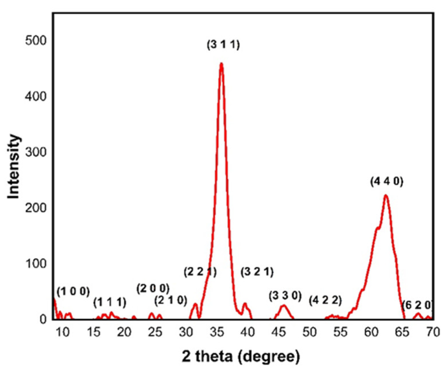
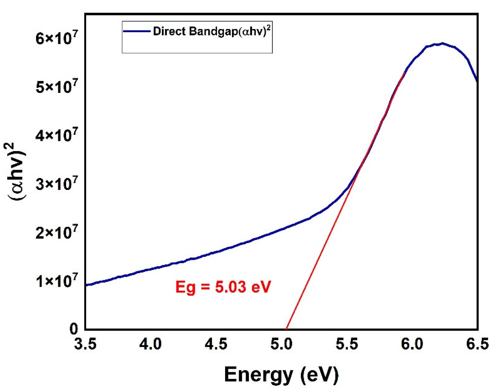
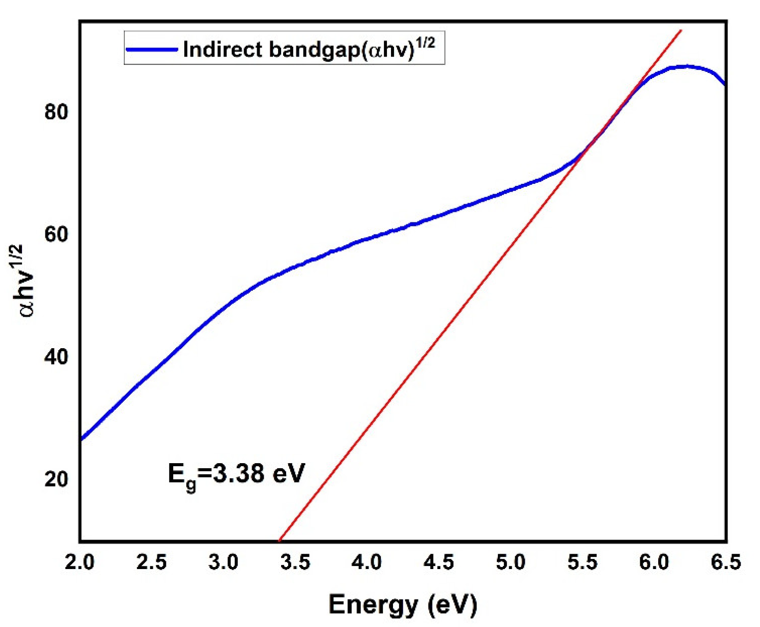
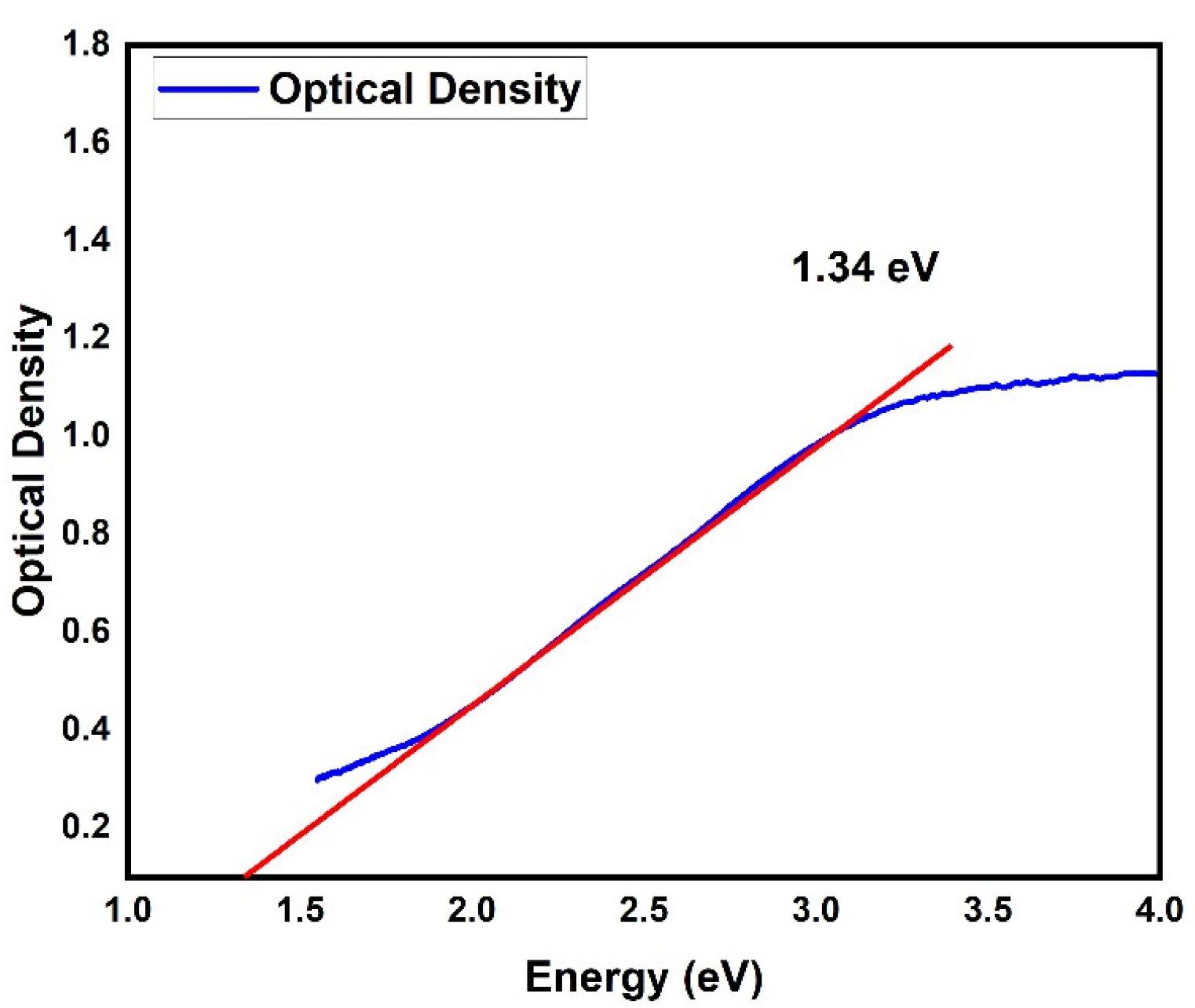
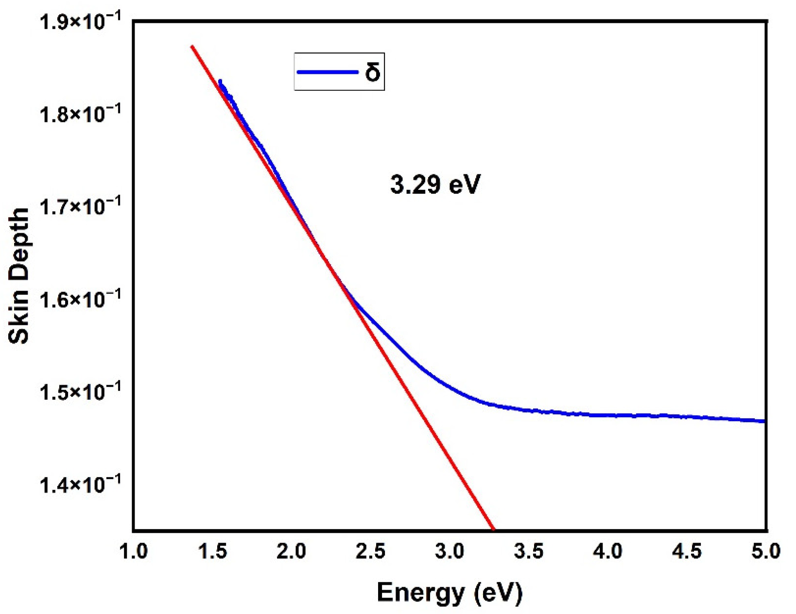
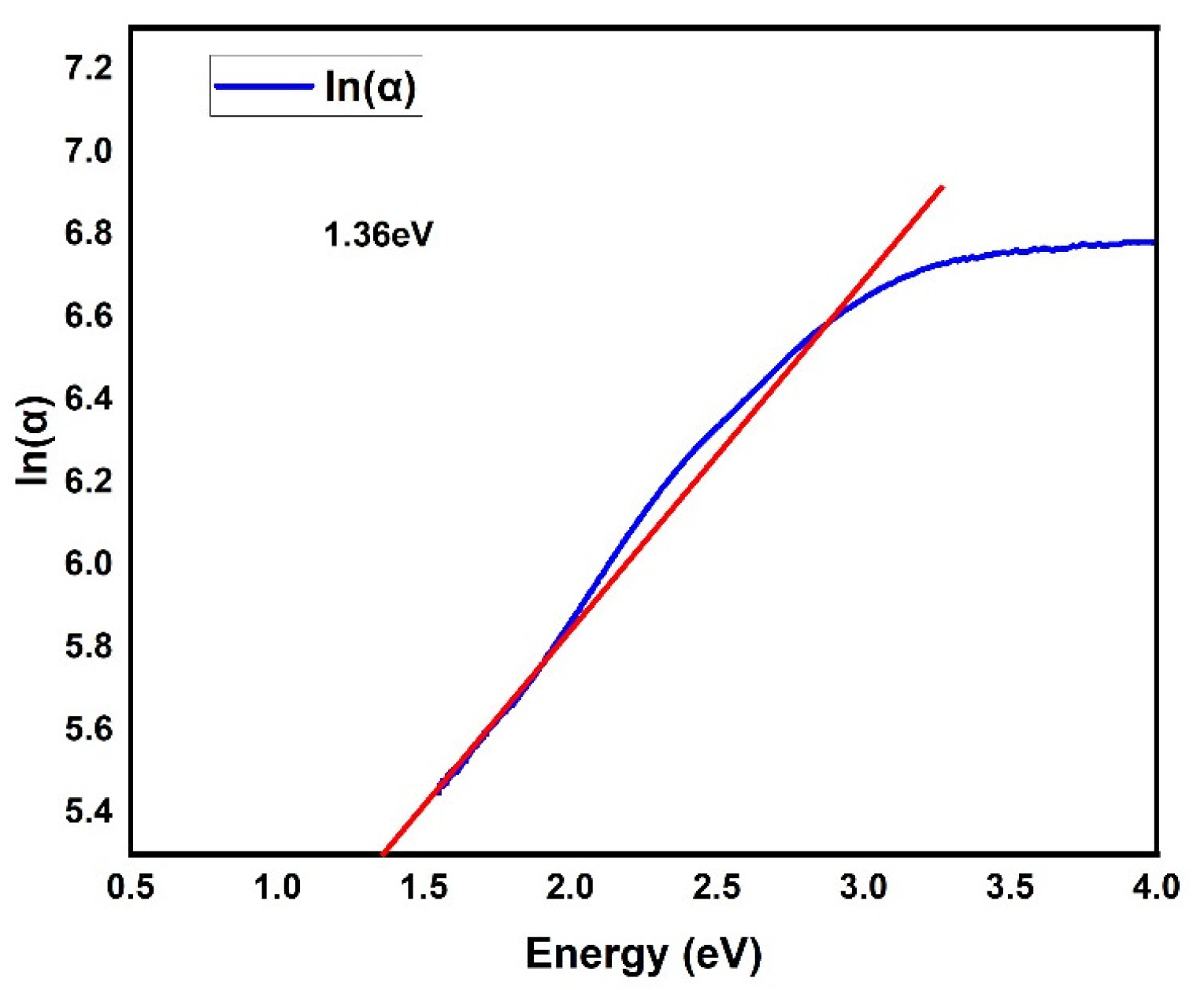
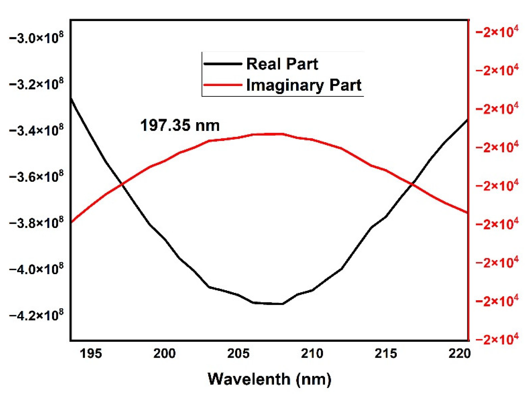
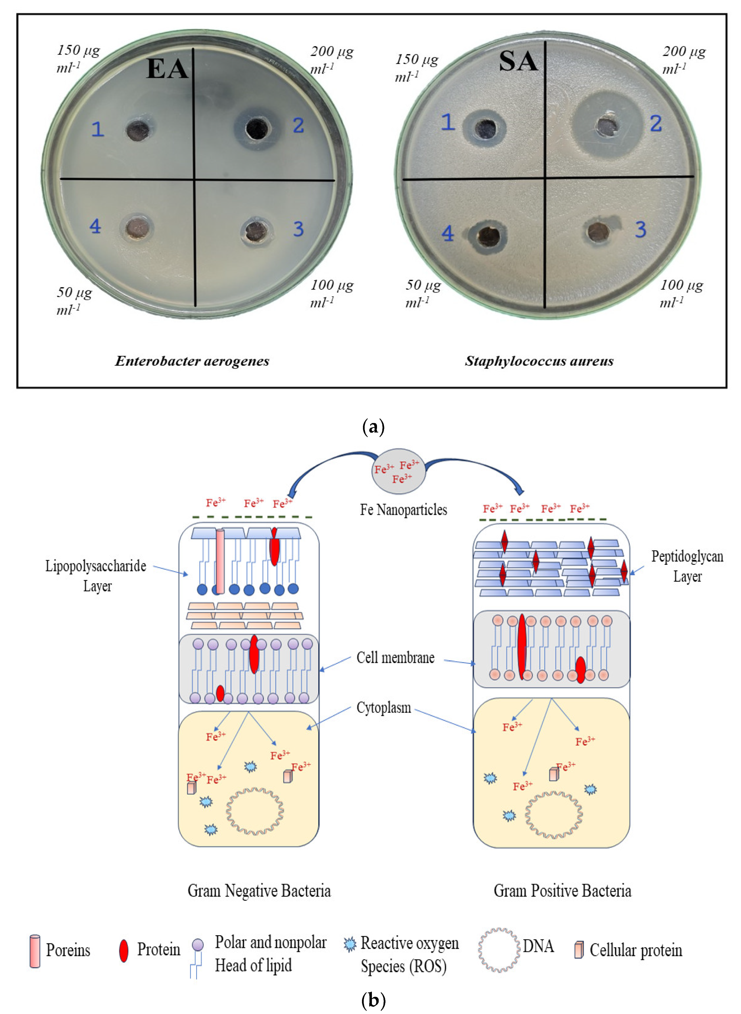
3. Results and Discussion

In order to describe the elemental composition of magnetite (Fe3O4) nanoparticles, EDAX characterization was employed. Table 1 shows the EDAX analysis of magnetite nanoparticles, which shows that the atomic weight % of Fe is 53.38 and the atomic weight % of O is 46.62 in the Fe3O4. Atomic weight % of Fe and O is close to the Stoichiometric composition of Fe3O4. An EDAX image of magnetite nanoparticles is shown in Figure 1.
In order to describe the structural property of magnetite (Fe3O4) nanoparticles, X-ray diffraction (XRD) was employed. The X-ray diffraction (XRD) pattern of magnetite (Fe3O4) nanoparticles is shown in Figure 2. The diffraction peaks at 2θ = 9.77°, 17.99°, 21.64°, 24.36°, 31.40°, 35.75°, 40.20°, 45.93°, 52.77°, 62.47°, and 70.82° can be assigned to (1 0 0), (1 1 1), (2 0 0), (2 1 0), (2 2 1), (3 1 1), (3 2 1), (3 3 0), (4 2 2), (4 4 0), and (6 2 0), respectively. The crystal structure of the obtained magnetite (Fe3O4) nanoparticles is cubic, and the lattice parameter obtained is a = b = c = 8.409 A0 and the obtained data are well matched with JCPDS No: 019-0629.
In order to describe the optical property of magnetite (Fe3O4) nanoparticles, UV-visible spectroscopy was employed. The absorbance spectra, absorption coefficient, refractive index, extinction coefficient optical band gap, Urbach energy, and skin depth were among optical parameters described. The produced magnetite (Fe3O4) nanoparticles were found to have absorption peaks within the typical UV-vis absorption range, with a lower absorption wavelength of 203 nm being observed.
The relation between the absorption coefficient (α) and the incident photon energy (hν) can be determined by using Tauc’s relationship as follows [11]:
α h ν = α 0 ( h ν E g ) n
where α0 is a constant and known as the band tailing parameter, Eg is the optical energy gap, and n is also a constant, which is known as the power factor of the transition mode. The direct optical bandgap is shown in Figure 3, which was found to be 5.03 eV. The indirect optical bandgap is shown in Figure 4 and was found to be 3.38 eV.
The optical density of the absorbance is proportional to both the thickness of the samples and the concentration of the absorbing material. The optical density of the magnetite nanoparticles can be estimated by using this simple equation [11]:
D o p t = α t
where t is the thickness of the sample. The plotting of the optical density (Dopt) against the incident photon energy (hν), which is found to be 1.34 eV, as shown in Figure 5. The skin depth (δ) is related to the absorption coefficient (α) by the following simple relation [11]:
δ = 1 α
The plot of the skin depth against the energy(hν), which is found to be 3.29 eV, as shown in Figure 6. The Urbach energy is calculated by ln α against the energy (hν) plot, which is shown in Figure 7. The Urbach energy of magnetite nanoparticles is given by the following relation [11]:
α = α 0 e x p ( E E U )
where EU is the Urbach energy; E is photon energy, and α0 is constant. The Urbach energy of magnetite (Fe3O4) nanoparticles is 1.36 eV. The plot of complex dielectric against wavelength clearly illustrates the variation of the real and imaginary dielectric constants with wavelength as shown in Figure 8.The corresponding wavelength to intersection of real and imaginary dielectric function is 197.35 nm.
  • Antimicrobial activity
The antimicrobial activity of the magnetite (Fe3O4) nanoparticles was assessed by the agar well diffusion method. The bacterial cultures Staphylococcus aureus and Enterobacter aerogenes were poured over N-agar plates with 1% (W/V) top agar. The plates were allowed to be solidified at room temperature, and four wells were bored by a sterile cup borer. For the antimicrobial activity demonstration, the following concentrations of Fe3O4 nanoparticles were selected—200 ug mL−1, 150 ug mL−1, 100 ug mL−1, and 50 ug mL−1. The agar wells were filled up with magnetite (Fe3O4) nanoparticles solution. The antimicrobial activity was evaluated based on the zone of inhibition that appeared around the agar well, and the diameter was measured in mm. As shown in Figure 9a, the magnetite (Fe3O4) nanoparticles effectively inhibited the growth of Enterobacter aerogenes and Staphylococcus aureus at higher concentrations. The inhibitory effect is concentration-dependent, and the minimum threshold for the growth inhibition was found to be between 100 and 150 ug mL−1. The result indicates that magnetite (Fe3O4) nanoparticles that are synthesized in-house have the potential to be used as a bacteriostatic as well as a bactericidal agent.

4. Conclusions

Magnetite (Fe3O4) nanoparticles were prepared by the sol–gel method at 200 °C. The sol–gel method offers several advantages for preparation of magnetite (Fe3O4) nanoparticles. The EDAX analysis of the magnetite nanoparticles showed that the atomic weight % of Fe is 53.38 and the atomic weight % of O is 46.62 in the Fe3O4. It is close to the Stoichiometric composition of Fe3O4. XRD shows that the crystal structure of the obtained Fe3O4 nanoparticles is cubic, the lattice parameter obtained is a = b = c = 8.409 A0, and the obtained data are well matched with JCPDS No: 019-0629. From UV-Visible analysis, the obtained direct optical bandgap is found to be 5.03 eV, and the indirect optical bandgap is found to be 3.38 eV. The Urbach energy of magnetite (Fe3O4) nanoparticles is 1.36 eV. The result indicates that magnetite (Fe3O4) nanoparticles that are synthesized in-house have the potential to be used as a bacteriostatic as well as a bactericidal agent.

Author Contributions

Conceptualization, P.N.B. and S.R.B.; methodology, P.N.B. and A.B.; formal analysis, P.N.B., S.R.B. and A.B.; investigation, P.N.B., S.R.B. and A.B.; resources, P.N.B., A.B. and M.S.D.; data curation, P.N.B.; writing—original draft preparation, P.N.B. and A.B.; writing—review and editing, S.R.B. and M.S.D. All authors have read and agreed to the published version of the manuscript.

Funding

This research received no external funding.

Institutional Review Board Statement

Not applicable.

Informed Consent Statement

Not applicable.

Data Availability Statement

The data presented in this study are available on request from the corresponding author. The data are not publicly available due to they form part of an ongoing study.

Conflicts of Interest

The authors declare no conflicts of interest.

References

  1. Shah, S.T.; Chowdhury, Z.Z.; Johan, M.R.B.; Badruddin, I.A.; Khaleed, H.M.T.; Kamangar, S.; Alrobei, H. Surface Functionalization of Magnetite Nanoparticles with Multipotent Antioxidant as Potential Magnetic Nanoantioxidants and Antimicrobial Agents. Molecules 2022, 27, 789. [Google Scholar] [CrossRef] [PubMed]
  2. Lemine, O.; Omri, K.; Zhang, B.; El Mir, L.; Sajieddine, M.; Alyamani, A.; Bououdina, M. Sol–gel synthesis of 8nm magnetite (Fe3O4) nanoparticles and their magnetic properties. Superlattices Microstruct. 2012, 52, 793–799. [Google Scholar] [CrossRef]
  3. Cui, H.; Liu, Y.; Ren, W. Structure switch between α-Fe2O3, γ-Fe2O3 and Fe3O4 during the large scale and low temperature sol–gel synthesis of nearly monodispersed iron oxide nanoparticles. Adv. Powder Technol. 2013, 24, 93–97. [Google Scholar] [CrossRef]
  4. Hu, P.; Chang, T.; Chen, W.-J.; Deng, J.; Li, S.-L.; Zuo, Y.-G.; Kang, L.; Yang, F.; Hostetter, M.; Volinsky, A.A. Temperature effects on magnetic properties of Fe3O4 nanoparticles synthesized by the sol-gel explosion-assisted method. J. Alloy. Compd. 2018, 773, 605–611. [Google Scholar] [CrossRef]
  5. Wang, X.L.; Wei, L.; Tao, G.H.; Huang, M.Q. Synthesis and characterization of magnetic and luminescent Fe3O4/CdTe nanocomposites using aspartic acid as linker. Chin. Chem. Lett. 2011, 22, 233–236. [Google Scholar] [CrossRef]
  6. Li, Y.; Jiang, R.; Liu, T.; Lv, H.; Zhou, L.; Zhang, X. One-pot synthesis of grass-like Fe3O4 nanostructures by a novel microemulsion-assisted solvothermal method. Ceram. Int. 2014, 40, 1059–1063. [Google Scholar] [CrossRef]
  7. Eom, Y.; Abbas, M.; Noh, H.; Kim, C. Morphology-controlled synthesis of highly crystalline Fe3O4 and CoFe2O4 nanoparticles using a facile thermal decomposition method. RSC Adv. 2016, 6, 15861–15867. [Google Scholar] [CrossRef]
  8. Zhang, H.; Zhu, G. One-step hydrothermal synthesis of magnetic Fe3O4 nanoparticles immobilized on polyamide fabric. Appl. Surf. Sci. 2012, 258, 4952–4959. [Google Scholar] [CrossRef]
  9. Bhaumik, M.; Maity, A.; Gupta, V.K. Synthesis and characterization of Fe0/TiO2 nano-composites for ultrasound assisted enhanced catalytic degradation of reactive black 5 in aqueous solutions. J. Colloid Interface Sci. 2017, 506, 403–414. [Google Scholar] [CrossRef] [PubMed]
  10. Shaker, S.; Zafarian, S.; Chakra, C.S.; Rao, K.V. Preparation and characterization of magnetite nanoparticles by Sol-Gel method for water treatment. Int. J. Innov. Res. Sci. Eng. Technol. 2013, 7, 2969–2973. [Google Scholar]
  11. Hassanien, A.; Akl, A.A. Effect of Se addition on optical and electrical properties of chalcogenide CdSSe thin films. Superlattices Microstruct. 2016, 89, 153–169. [Google Scholar] [CrossRef]
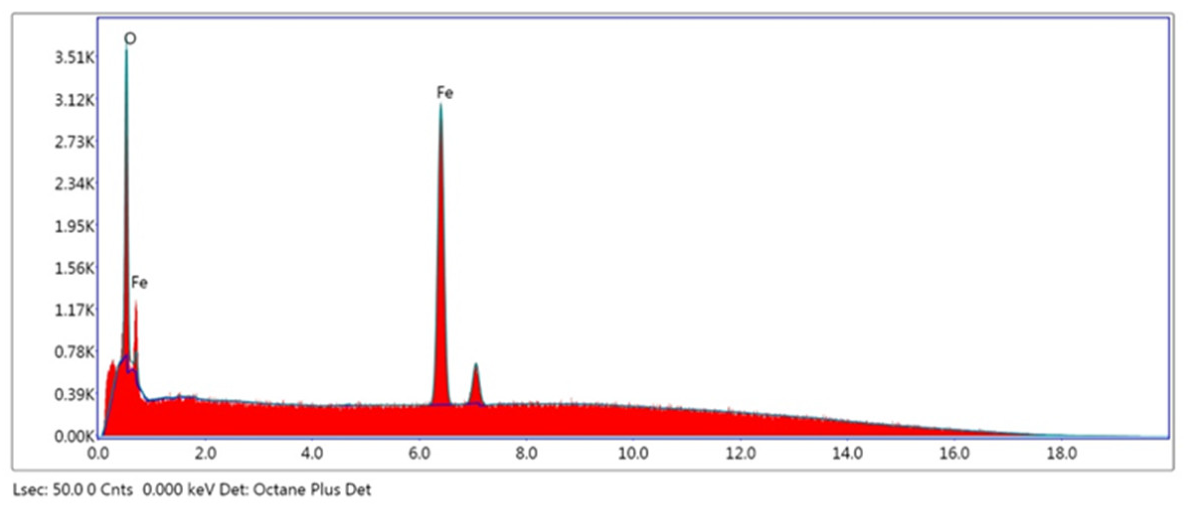
Figure 1. EDAX image of magnetite (Fe3O4) nanoparticles.
Engproc 56 00293 g001
Figure 2. XRD image of magnetite (Fe3O4) nanoparticles.
Engproc 56 00293 g002
Figure 3. Direct optical bandgap of magnetite (Fe3O4) nanoparticles.
Engproc 56 00293 g003
Figure 4. Indirect optical bandgap of magnetite (Fe3O4) nanoparticles.
Engproc 56 00293 g004
Figure 5. Plot of the optical density (Dopt) against energy (hν) of magnetite (Fe3O4) nanoparticles.
Engproc 56 00293 g005
Figure 6. Plot of skin depth against energy (hν) of magnetite (Fe3O4) nanoparticles.
Engproc 56 00293 g006
Figure 7. Plot of ln α against energy (hν) of magnetite (Fe3O4) nanoparticles.
Engproc 56 00293 g007
Figure 8. Plot of complex dielectric constant against wavelength of magnetite (Fe3O4) nanoparticles.
Engproc 56 00293 g008
Figure 9. (a) Antimicrobial activity of magnetite (Fe3O4) nanoparticles against Enterobacter aerogenes and Staphylococcus aureus. (b) Proposed mechanism of antimicrobial action of magnetite (Fe3O4) nanoparticles. Fe3+ ions are attracted to negatively charged lipopolysaccharide layer in Gram-negative bacteria and peptidoglycan layer of Gram-positive bacteria. After entering the cell, metal nanoparticles can disrupt cell membrane, block cellular proteins, disrupt cellular DNA, and generate ROS species, which can lead to death of the microorganism.
Engproc 56 00293 g009
Table 1. Percentage of elements in magnetite (Fe3O4) nanoparticles from EDAX.
ElementWeight%
O K24.7
Fe K75.3
Disclaimer/Publisher’s Note: The statements, opinions and data contained in all publications are solely those of the individual author(s) and contributor(s) and not of MDPI and/or the editor(s). MDPI and/or the editor(s) disclaim responsibility for any injury to people or property resulting from any ideas, methods, instructions or products referred to in the content.

