Proceeding Paper

Bimodal Nanoprobes Containing AgInSe2 Hydrophilic Quantum Dots and Paramagnetic Chelates for Diagnostic Magnetic Resonance Imaging †

by
Rebeca Muniz de Melo
1,*,
Gabriela Marques de Albuquerque
1,
Goreti Pereira
1,2 and
Giovannia Araujo de Lima Pereira
1
1
Department of Fundamental Chemistry, Federal University of Pernambuco, Recife 52171-900, Brazil
2
Department of Chemistry & CESAM, University of Aveiro, 3810-193 Aveiro, Portugal
*
Author to whom correspondence should be addressed.
Presented at the 4th International Electronic Conference on Applied Sciences, 27 October–10 November 2023; Available online: https://asec2023.sciforum.net/.
Eng. Proc. 2023, 56(1), 6; https://doi.org/10.3390/ASEC2023-15272
Published: 26 October 2023
(This article belongs to the Proceedings of The 4th International Electronic Conference on Applied Sciences)

Abstract

:
The development of bimodal systems with signals for two diagnostic techniques has been increasing. Magnetic resonance imaging (MRI) is a non-invasive technique that distinguishes pathological tissues from healthy ones. To improve the images’ contrast, nanoparticulate contrast agents (CAs) have been developed, allowing for the attachment of several CA molecules in one nanoparticle. In this work, we associated AgInSe2 quantum dots (QDs) with gadolinium complexes, obtaining nanoprobes for MRI and optical imaging. The nanosystems showed good optical properties and values of relaxivity superior to the CAs used clinically. Thus, these nanoprobes have the potential to be used as CAs for MRI and optical imaging.

1. Introduction

Most diseases that affect humanity are multifactorial, so diagnostic techniques must detect these factors and distinguish the various mechanisms and/or phases of the investigation. For this reason, there is a growing interest in the development of multimodal probes, seeking complementary forms of diagnosis [1]. Magnetic resonance imaging (MRI) is one of the most used diagnostic techniques, being a non-invasive technique that produces images with anatomical detail capable of drawing inferences about the diagnosis of many pathologies [2]. To improve the resolution of the generated image, contrast agents (CAs) are widely used in clinical procedures. The best-known CAs are those based on Gd, which have a limited contrast-enhancing capacity, making them effective only at concentrations above 0.1 mM. Furthermore, nanoparticulate CAs are capable of concentrating several CA molecules in a single nanoparticle, increasing the local concentration of paramagnetic ions without increasing the dose of administered CAs. These systems have been the subject of various studies in order to overcome this limitation of CA dosage [1].
Paramagnetic CAs can be associated with nanoparticles that have optical properties, obtaining bimodal nanoprobes, such as quantum dots (QDs). QDs are semiconductor nanocrystals that have excellent optical properties. In addition, QDs containing Gd3+ ions associated with their structure have been reported in the literature [1,3], and it has been observed that the main area of biological application of these paramagnetic QDs is in the diagnosis of cancer [3,4]. Thus, the binding of Gd3+ complexes to QDs will generate more efficient contrast agents, with high relaxivities, increasing the sensitivity of the technique. The covalent conjugation of Gd3+ chelates to hydrophilic QDs has been carried out by the occurrence of the carboxylic acid groups of the stabilizer, requiring the use of coupling additives or a ligand (diamine) that allows the association of the QD carboxyl with the chelate carboxyl, adding a step in the experimental procedure [1]. Thus, this work aimed to prepare cadmium-free QDs (AgInSe2) and conjugated them with Gd3+ complexes for the preparation of bimodal nanoprobes that combine paramagnetic and optical properties for diagnostic imaging.

2. Materials and Methods

2.1. Preparation of AgInSe2 QDs

Initially, we studied which were the most suitable conditions for the preparation of AgInSe2 QDs (AISe) in water, varying some parameters such as temperature and the reagents’ molar ratio. In a flask, AgNO3 (0.0054 M), In(NO3)3 (0.0026 M), and MSA were dissolved in 100 mL of ultra-pure water, and the pH was adjusted to 5 with NaOH (2 M). The mixture was heated and left under stirring. Subsequently, Na2SeO3 (0.25 M) and NaBH4 (dissolved in 3 mL of water) were injected, and the system was stirred for 1–2 h under heating. The Ag:In molar radio was fixed at 2:1, and the Se:NaBH4 proportion was 1:2.5; the other parameters are described in Table 1. The QDs were characterized using UV-Vis absorption (Evolution 600 spectrophotometer, Thermo Scientific, Waltham, MA, USA) and emission spectroscopy (LS 55 spectrometer, PerkinElmer, Waltham, MA, USA).

2.2. Preparation of Gd3+ Complexes

The DOTA molecule was dissolved in water, adjusting the pH to 5.5–5.8 with NaOH, and the coupling agents (EDC and NHS) were added; the solution was left for 1 h at 60 °C. The pH was adjusted to 7, a GdCl3 solution was added, and the mixture remained under stirring for 1 h at 60 °C. Cysteamine (CYA) was added, leaving the system under agitation for 4 h. The complex was characterized by relaxometry (Minispec Bruker 20 MHz, at 25 and 37 °C) and the xylenol orange test using a UV-Vis absorption spectrophotometer.

2.3. Preparation and Characterization of Bimodal Systems

The paramagnetic complex was added to the QD suspension, stirring for 48 h. Two different volumes (50 and 100 μL) of the complex solution (0.01 M) were tested. After purification in ultracentrifugation tubes with a 10 kDa MWCO membrane (Vivaspin®, GE Healthecare, Chicago, IL, USA), the bimodal systems were characterized using UV-Vis absorption and emission spectroscopy and relaxometry (20 MHz, at 25 and 37 °C).

3. Results and Discussion

3.1. Preparation of AgInSe2 QDs

The absorption spectra profiles are similar for all the syntheses, with a first absorption band between 500 and 650 nm (Figure 1A). The emission spectra (Figure 1B) presented a maximum of emission around 800 nm for all the samples, with variations in the intensity and full-width at half maximum (FWHM). The synthesis that presented the best result of intensity and FWHM was the synthesis of AISe 2 after 1 h of reaction, so this was the synthesis chosen to proceed with in the experiments. A point worth highlighting when choosing the synthesis is to consider the intensity value and the bandwidth, since a higher intensity and lower FHWM mean a nanocrystal with fewer surface defects.

3.2. Preparation of Gd3+ Complexes

The modified DOTA-Gd complexes showed a complexation yield above 95% by the xylenol test and longitudinal relaxivity similar to commercial DOTA-Gd (DOTA-CYA-Gd r1 = 3.75 Mm−1s−1 per Gd3+ at 20 MHz and 37 °C, in water) [1]. In view of this, the results obtained suggest that the addition of the linker to the activated complex did not cause a significant variation in the values of r1 compared to the clinically used DOTA, showing that the prepared complex was stable and had good relaxometric properties.

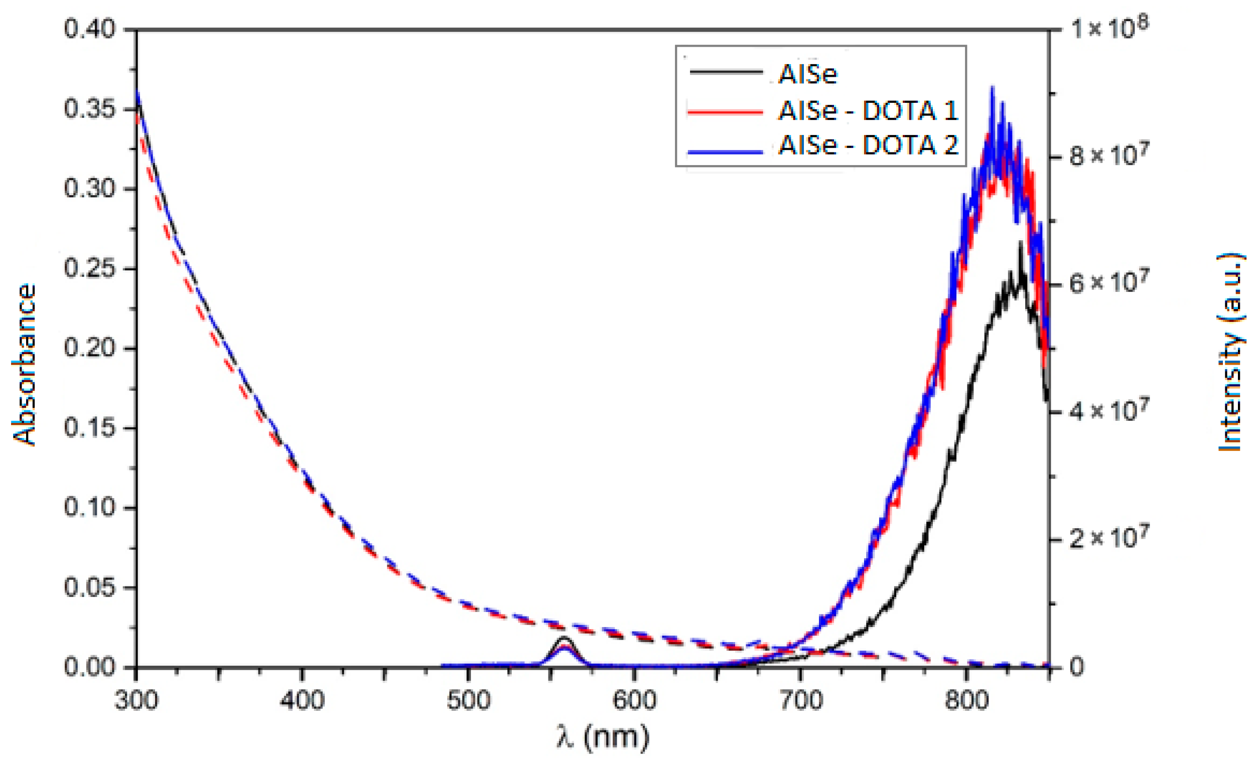
3.3. Preparation and Characterization of Bimodal Systems (AISe-DOTA)

The optical characterization of the prepared systems (Figure 2) showed that there was no change in the maximum absorption wavelength when compared with the QDs, meaning that there was no change in the QD’s core. Furthermore, it can be seen that the system demonstrated an increase in the emission intensity and no significant shift in the emission maximum, indicating that the conjugation was effective and that the interaction between the complexes and the QDs is favorable, since the complexes can passivate the surface of the QD, minimizing its defects.
The estimated r1 values for the bimodal nanoprobes prepared were higher for the system where more complexes were added, indicating that a higher concentration of DOTA-Gd chelates was attached to the surface of these systems. Nevertheless, for all the bimodal probes, the longitudinal relaxivity values were enhanced when compared with the clinically used DOTA-Gd contrast agent (Table 2), thus proving the achievement of a promising bimodal contrast agent for use in optical and magnetic resonance imaging.

4. Conclusions

In this work, we prepared AISe QDs in aqueous medium with good optical properties. The bimodal systems containing AISe QDs were prepared using a thiolated Gd3+ complex by dative bond between the chelate and the QD’s surface. The results showed that the methodology used proved to be effective, with a promising and easy-to-execute conjugation route. Furthermore, the prepared bimodal nanoprobes showed improved optical and relaxometric properties compared to the bare QDs or DOTA-Gd complexes. Thus, the nanoprobes prepared have the potential to be used as contrast agents for optical and magnetic resonance imaging.

Author Contributions

Conceptualization, G.P. and G.A.d.L.P.; methodology, R.M.d.M., G.M.d.A., G.P. and G.A.d.L.P.; formal analysis, R.M.d.M. and G.M.d.A.; investigation, R.M.d.M. and G.M.d.A.; resources, G.P. and G.A.d.L.P.; writing—original draft preparation, R.M.d.M., G.M.d.A., G.P. and G.A.d.L.P.; writing—review and editing, R.M.d.M., G.P. and G.A.d.L.P.; supervision, G.P. and G.A.d.L.P.; project administration, G.P. and G.A.d.L.P.; funding acquisition, G.P. and G.A.d.L.P. All authors have read and agreed to the published version of the manuscript.

Funding

This research was funded by FACEPE-Brazil (APQ-1351-1.06/22) and CNPq (Universal 409319/2021-0).

Institutional Review Board Statement

Not applicable.

Informed Consent Statement

Not applicable.

Data Availability Statement

All data are within this article.

Acknowledgments

Thank you to UFPE, CNPq, and FACEPE for financial support. G.P. is grateful to CESAM/FCT/MCTES (UIDP/50017/2020+UIDB/50017/2020+LA/P/0094/2020) for financial support. R.M.d.M. (BIC-1261-1.06/23) and G.M.d.A. (IBPG-0985-3.03/20) are grateful to FACEPE for their scholarships.

Conflicts of Interest

The authors declare no conflict of interest. The funders had no role in the design of the study; in the collection, analyses, or interpretation of the data; in the writing of the manuscript; or in the decision to publish the results.

References

  1. Albuquerque, G.M.; Souza-Sobrinha, I.; Coiado, S.D.; Santos, B.S.; Fontes, A.; Pereira, G.A.; Pereira, G. Quantum dots and Gd3+ chelates: Advances and challenges towards bimodal nanoprobes for magnetic resonance and optical imaging. Top. Curr. Chem. 2021, 379, 12. [Google Scholar] [CrossRef] [PubMed]
  2. Landini, L.; Positano, V.; Santarelli, M. (Eds.) Advanced Image Processing in Magnetic Resonance Imaging; Taylor & Francis Grouped: Boca Raton, FL, USA, 2005. [Google Scholar]
  3. Liu, Y.; Ai, K.; Yuan, Q.; Lu, L. Fluorescence-enhanced gadolinium-doped zinc oxide quantum dots for magnetic resonance and fluorescence imaging. Biomaterials 2011, 32, 1185–1192. [Google Scholar] [CrossRef] [PubMed]
  4. Mulder, W.J.; Castermans, K.; van Beijnum, J.R.; Oude Egbrink, M.G.; Chin, P.T.; Fayad, Z.A.; Löwik, C.W.; Kaijzel, E.L.; Que, I.; Storm, G.; et al. Molecular imaging of tumor angiogenesis using αvβ3-integrin targeted multimodal quantum dots. Angiogenesis 2009, 12, 17–24. [Google Scholar] [CrossRef] [PubMed]
Figure 1. (A) Absorption spectra of AISe QDs syntheses; (B) emission spectra of AISe QDs syntheses.
Engproc 56 00006 g001
Figure 2. Absorption and emission spectra of AISe-DOTA.
Engproc 56 00006 g002
Table 1. Synthetic parameters for preparation of hydrophilic AgInSe2 QDs.
SampleMSA:(Ag:In) Molar Ratio(Ag:In):Se
Molar Ratio
T (°C)pH
AISe 14:16:1505
AISe 28:110:1505
AISe 38:110:1905
Table 2. Estimated values of the longitudinal relaxivity r1 of the bimodal systems (20 MHz at 25 and 37 °C).
SampleV (µL) of Complex Addedr1 (mM−1s−1 per Gd3+)
25 °C37 °C
AISe-DOTA 1505.205.23
AISe-DOTA 21006.786.30
Disclaimer/Publisher’s Note: The statements, opinions and data contained in all publications are solely those of the individual author(s) and contributor(s) and not of MDPI and/or the editor(s). MDPI and/or the editor(s) disclaim responsibility for any injury to people or property resulting from any ideas, methods, instructions or products referred to in the content.

