Proceeding Paper

A Study on the Suitability of Constant Boundary Elements for the Simulation of Biological Organs †

Department of Mechanical and Industrial Engineering, Manipal Institute of Technology, Manipal Academy of Higher Education, Manipal 576104, Karnataka, India
Presented at the International Conference on Recent Advances in Science and Engineering, Dubai, United Arab Emirates, 4–5 October 2023.
Eng. Proc. 2023, 59(1), 169; https://doi.org/10.3390/engproc2023059169
Published: 17 January 2024
(This article belongs to the Proceedings of Eng. Proc., 2023, RAiSE-2023)

Abstract

:
In the process of designing of surgical simulators, it may be a requirement to simulate biological organs in real-time. About thirty computations per second are required for achieving real-time graphics. Hence, computational techniques employed to simulate biological organs in real-time should be able to perform about thirty computations per second. The computational techniques employed should be fast enough, but at the same time should be accurate enough to realistically simulate the biological organs which are inherently nonlinear. A numerical technique called the Boundary Element Method (BEM) is generally thought of as being faster when one compares the technique with some well-established numerical techniques like the Finite Element Method (FEM). This technique (BEM) is even faster if constant boundary elements are employed. However, the BEM is mostly used to simulate linear behavior, whereas the FEM is more established for simulating nonlinear behavior. The present work investigates whether biological organs may be simulated by using the linear BEM. The reason nonlinear BEM has not been used is that the nonlinear BEM is quite slow and difficult to implement. A human kidney is the biological organ considered in this work. A nonlinear analysis and a linear analysis are carried out on the kidney. A nonlinear analysis is carried out by using the FEM, whereas the linear analysis is carried out by using the BEM. Results from the nonlinear analysis are compared with the results from the linear analysis. The results indicate that there is good agreement between the results from the linear BEM and the nonlinear FEM many times, but there is considerable difference between the results other times. Although the results reinforce the idea that the BEM could be a useful tool while simulating biological organs, further research is needed to definitively say whether the results given by the linear BEM, which uses constant boundary elements, are always good enough for simulating biological organs.

1. Introduction

Surgeons are required to acquire certain surgical skills (e.g., eye–hand coordination) to be able to perform certain surgical procedures (e.g., laparoscopic surgery) [1,2]. Surgical simulators are increasingly becoming popular for preparing surgeons for certain surgical procedures. A few surgical simulators that may be used to train the trainee-surgeons are explained in the references [3,4,5,6,7,8,9].
A surgical simulator capable of simulating a biological organ should be capable of simulating the underlying physics. The degree of realism achieved while simulating the underlying physics is a deciding factor when it comes to the fidelity of a simulator. Realism achieved in turn depends on the accuracy as well as the speed offered by the numerical technique employed for simulating the underlying physics.
There is a vast body of literature related to the simulation of biological organs by realistically simulating the underlying physics through the use of different computational techniques. A study of the representative literature, e.g., refs. [10,11,12,13,14,15,16,17,18,19,20], indicates that continuum mechanics based numerical methods similar to the Finite Element Method (FEM) are good in simulating biological organs accurately. However, biological organs behave nonlinearly, and hence simulations require the nonlinear FEM, which is slower. One should note that the realism offered by a simulator is decided not just by the accuracy of simulations but by the speed of simulations also. Hence, there is a need to find an alternative to the nonlinear FEM that is quite fast but capable of providing reasonably accurate solutions.
It is generally thought that the Boundary Element Method (BEM) is faster than the FEM when solving linear elastostatic problems, because the BEM requires only the boundary of the solution region to be meshed, whereas the FEM requires the interior of the solution region to be meshed also. However, as soon as one tries to implement the BEM to solve a nonlinear problem, one would also need some kind of meshing of the interior of the solution region. This could cause the BEM to also not be fast enough, just like the nonlinear FEM, when solving a nonlinear problem.
Hence, this work tries to employ the linear BEM for simulating biological organs. Employing the linear BEM would ensure that the simulations are faster than the FEM. Now, the question is whether the solutions given by the linear BEM are accurate enough, in the context of simulating biological organs. The purpose of this study is to try to find an answer this very question.
In this study, a biological organ is simulated by using the linear BEM and also by using the nonlinear FEM. The results obtained by these two methods are compared for the purpose of deciding whether it is good to use the linear BEM for simulating biological organs.
The methodology followed in this work is similar to that followed in this author’s work [21]. The main difference between [21] and the present work is that [21] compares the results from the linear FEM with the results from the nonlinear FEM, whereas the present work compares the results from the linear BEM with the results from the nonlinear FEM. Some of the contents of this paper have been borrowed from this author’s PhD thesis [22].

2. Methodology

The constant boundary element is the type of boundary element used for this work. This type of element is chosen because it offers faster performance when compared to other types of elements. Boundary elements of triangular shape are utilized in this work. Elements of triangular shapes help to approximate geometry that is complex.
Constant boundary elements of triangular shapes have only one node per boundary element. The node is located within the triangular element. Using constant boundary elements—in contrast to linear or quadratic boundary elements—helps to perform the simulations faster. This is because it is a well-known fact that lower order elements like constant elements are faster when compared to higher order elements like linear or quadratic elements. Of course, lower order elements provide less accurate solutions when compared to higher order elements for the same total number of elements, but the purpose of this study is to check whether constant boundary elements can provide enough accuracy. One may refer to reference books on BEM (that deal with the fundamentals of BEM) to know how constant boundary elements of triangular shapes are formulated.
For carrying out nonlinear FEM simulations, the commercial software package ANSYS (Version: 2023 R1) is used, and geometric nonlinearity is considered. Codes developed by this author [23] are used to perform linear BEM simulations.
The biological organ considered is the human left kidney. This kidney is the kidney of the Visible Human male [24]. Geometry of the left kidney was obtained from the images that are a part of the Visible Human Dataset [24]. The procedure illustrated in [25] was followed to obtain the geometry. The procedure involved the use of ImageJ [26,27], ITK-SNAP [28], and MeshLab [29]. These are open-source and free software packages. Upon utilizing these three software packages in sequence, a surface model of the kidney made up of surface triangles was obtained. The same surface triangles are used as boundary elements while carrying out the linear BEM analysis. For carrying out nonlinear FEM analysis, a solid model is required, and, hence, the commercial software Rhinoceros (Version: Rhino 8) [30] was used to convert the surface model of the kidney to a solid model, as illustrated in [25].
The material properties used are the same as those used in [31]. These were the material properties used: Young’s modulus = 150 N/mm2; Poisson’s ratio = 0.4. The reference [31] indicates that these values are realistic for a human kidney.
Three problems are solved in this work, with each problem solved using both nonlinear FEM and linear BEM. The three problems are given the names Problem 1, Problem 2, and Problem 3. The geometry as well as the material properties used are the same for each of these three problems. The only difference between the three problems is that they employ different boundary conditions.
The kidney is surrounded by other organs, and it is not easy to simulate the actual boundary conditions that may keep changing during surgical manipulations. Hence, arbitrary boundary conditions are used for the present work. For all of the problems, a small portion of the surface is completely fixed, and a known displacement is enforced at some other point located on the surface. The point on the surface of the kidney where a known displacement is enforced, and the portion of the surface of the kidney that is completely fixed, and these are chosen to be the same location and region for all of the problems considered. With regard to the magnitude of the known enforced displacement, the magnitude is kept the same for all of the three problems. Hence, the only difference between the three problems is that the directions are different for the known enforced displacements. For problem 1, the known displacement is applied in the x direction, whereas the known displacement is enforced in the y and z directions, respectively, for Problem 2 and Problem 3. For each of the three problems, the known applied displacement is enforced to be equal to 5 mm. The reason for applying the 5 mm displacements is to apply physically meaningful displacements. The reason for applying known displacements instead of applying known forces is that surgical simulators focus more on simulating realistic deformation of biological organs, and simulating haptic feedback is not easy and not often a priority.
Figure 1 shows the portion of the surface of the kidney that is completely fixed, and the location where a known displacement is enforced.
The next section presents the results for each of the problems considered.

3. Results

When comparing the results obtained by using the BEM with those obtained by using the FEM, care has to be taken so that the results are obtained for the same locations on the biological organ. The FEM discretizes the volume of the kidney and it uses 3D elements, whereas the BEM discretizes the kidney by using surface triangles. Hence, it is difficult to ensure the location of nodes in the FEM discretization are exactly the same as the location of the nodes as per the discretization that is employed while solving the same problem by using the BEM. Although this may result in some error, this error is ignored in this work. The nodes that form the completely fixed portion of the surface of the kidney are carefully identified in both the BEM and the FEM discretization, carefully. Similarly, corresponding nodes closest to the point where a known displacement is enforced on the surface of the kidney are carefully identified manually.
The results from the linear BEM analysis were compared with the results from the nonlinear FEM by tabulating the displacements. For this purpose, the displacement vector sum is noted at a few points randomly chosen all over the surface of the kidney. Eleven randomly chosen points located on the surface of the kidney were used. This was done to capture the deformation of the kidney by observing the displacement vector sum at eleven points, that are located over the surface of the kidney randomly. Care was taken to ensure that the eleven points were from different locations of the kidney. Table 1, Table 2 and Table 3 list the displacement vector sum at those eleven points for all the problems considered, obtained using the nonlinear FEM analysis and the linear BEM analysis. The last column shows the difference between the results obtained by employing the FEM and the BEM.

4. Discussion

From Table 1, Table 2 and Table 3 in the last section, the difference in the results that are obtained by using the FEM versus the BEM is less than 1 mm in most of the cases. Although the difference is much less than 1 mm for many cases, the difference is quite high for a few cases. Since the biological organ considered here is of a realistic size (not scaled up or scaled down), a difference of about 1 mm appears fine. So, it appears that the linear BEM could be a better alternative to the nonlinear FEM at least for some applications, since the linear BEM is much faster. Since the relevant literature does not clearly say what the allowed error is, further research is needed to determine the allowable errors and to definitively say whether or not the linear BEM is always a better candidate when compared to nonlinear FEM.
This work tries to investigate whether the linear BEM could be a better alternative to the nonlinear FEM for simulating biological organs. The biological organ considered in this work is a kidney. This paper could assume significance when one tries to build a simulator for simulating how a kidney deforms during palpation.
Future work could be to carry out the simulations on a greater number of biological organs. Also, different types of elements may be employed. Future research could also focus on quantifying allowable errors for different applications. The heterogeneous nature of biological organs could also be considered in the future.

5. Conclusions

From the results presented in this work, it appears that the linear BEM could be a better alternative to the nonlinear FEM, at least for some applications. However, further research is needed to definitively say whether the linear BEM is always a better candidate when compared to nonlinear FEM when it comes to simulating biological organs.

Funding

This research received no external funding.

Institutional Review Board Statement

Not applicable.

Informed Consent Statement

Not applicable.

Data Availability Statement

Apart from the results presented in the paper, no new data were created.

Acknowledgments

The author wishes to acknowledge his institution for providing the financial support for presenting this paper in the conference.

Conflicts of Interest

The author declares no conflicts of interest.

References

  1. Wilson, M.; Coleman, M.; McGrath, J. Developing basic hand-eye coordination skills for laparoscopic surgery using gaze training. BJU Int. 2010, 105, 1356–1358. [Google Scholar] [CrossRef] [PubMed]
  2. Topalli, D.; Cagiltay, N.E. Eye-Hand Coordination Patterns of Intermediate and Novice Surgeons in a Simulation-Based Endoscopic Surgery Training Environment. J. Eye Mov. Res. 2018, 11. [Google Scholar] [CrossRef] [PubMed]
  3. TraumaMan Surgical Simulator. Available online: https://simulab.com/products/traumaman-surgical-simulator (accessed on 28 July 2023).
  4. Badash, I.; Burtt, K.; Solorzano, C.A.; Carey, J.N. Innovations in surgery simulation: A review of past, current and future techniques. Ann. Transl. Med. 2016, 4, 453. [Google Scholar] [CrossRef] [PubMed]
  5. Surgical Simulation. Available online: https://www.caehealthcare.com/surgical-simulation/ (accessed on 28 July 2023).
  6. Surgical Simulation. Available online: https://www.healthysimulation.com/surgical-simulation/ (accessed on 28 July 2023).
  7. Simulation Software for Surgical Training and Education. Available online: https://www.insimo.com/ (accessed on 28 July 2023).
  8. Simbionix Simulators. Available online: https://simbionix.com/simulators/ (accessed on 28 July 2023).
  9. Surgery Simulators. Available online: https://www.medicalexpo.com/medical-manufacturer/surgery-simulator-44251.html (accessed on 28 July 2023).
  10. Chen, X.; Hu, J. A review of haptic simulator for oral and maxillofacial surgery based on virtual reality. Expert Rev. Med. Devices 2018, 15, 435–444. [Google Scholar] [CrossRef] [PubMed]
  11. Coveney, P.V.; Hoekstra, A.; Rodriguez, B.; Viceconti, M. Computational biomedicine. Part II: Organs and systems. Interface Focus 2021, 11, 20200082. [Google Scholar] [CrossRef]
  12. Sui, Y.; Pan, J.J.; Qin, H.; Liu, H.; Lu, Y. Real-time simulation of soft tissue deformation and electrocautery procedures in laparoscopic rectal cancer radical surgery. Int. J. Med. Robot. 2017, 13, e1827. [Google Scholar] [CrossRef] [PubMed]
  13. Sacks, M.S.; Motiwale, S.; Goodbrake, C.; Zhang, W. Neural Network Approaches for Soft Biological Tissue and Organ Simulations. J. Biomech. Eng. 2022, 144, 121010. [Google Scholar] [CrossRef] [PubMed]
  14. Villard, P.i.; Koenig, N.; Perrenot, C.; Perez, M.; Boshier, P. Toward a realistic simulation of organ dissection. Stud. Health Technol. Inf. 2014, 196, 452–456. [Google Scholar]
  15. Mendizabal, A.; Tagliabue, E.; Hoellinger, T.; Brunet, J.N.; Nikolaev, S.; Cotin, S. Data-driven simulation for augmented surgery. Dev. Nov. Approaches Biomech. Metamater. 2020, 132, 71–96. [Google Scholar]
  16. Delingette, H. Toward realistic soft-tissue modeling in medical simulation. Proc. IEEE 1998, 86, 512–523. [Google Scholar] [CrossRef]
  17. Moreno-Guerra, M.R.; Martínez-Romero, O.; Palacios-Pineda, L.M.; Olvera-Trejo, D.; Diaz-Elizondo, J.A.; Flores-Villalba, E.; da Silva, J.V.L.; Elías-Zúñiga, A.; Rodriguez, C.A. Soft Tissue Hybrid Model for Real-Time Simulations. Polymers 2022, 14, 1407. [Google Scholar] [CrossRef] [PubMed]
  18. Meier, U.; Lopez, O.; Monserrat, C.; Juan, M.C.; Alcaniz, M. Real-time deformable models for surgery simulation: A survey. Comput. Methods Programs Biomed. 2005, 77, 183–197. [Google Scholar] [CrossRef] [PubMed]
  19. Liu, S.; Zhang, Y.; Zheng, W.; Yang, B. Real-time Simulation of Virtual Palpation System. In IOP Conference Series: Earth and Environmental Science; IOP Publishing: Bristol, UK, 2019; Volume 234. [Google Scholar] [CrossRef]
  20. Zhang, J.; Zhong, Y.; Gu, C. Deformable models for surgical simulation: A survey. IEEE Rev. Biomed. Eng. 2018, 11, 143–164. [Google Scholar] [CrossRef] [PubMed]
  21. Kirana Kumara, P. A Comparison between the Results from Linear Analysis and Nonlinear Analysis in the Context of Simulation of Biological Materials. J. Compos. Sci. 2023, 7, 109. [Google Scholar] [CrossRef]
  22. Kirana Kumara, P. Studies on the Viability of the Boundary Element Method for the Real-Time Simulation of Biological Organs. Ph.D. Thesis, Centre for Product Design & Manufacturing, Indian Institute of Science, Bangalore, India, 2016. [Google Scholar]
  23. Kirana Kumara, P. Codes for Solving Three Dimensional Linear Elastostatic Problems Using Constant Boundary Elements While Ignoring Body Forces. Available online: https://eprints.iisc.ac.in/48088/ (accessed on 28 July 2023).
  24. VHP, n.d., Visible Human Project. Available online: http://www.nlm.nih.gov/research/visible/visible_human.html (accessed on 28 July 2023).
  25. Kirana Kumara, P. Extracting Three Dimensional Surface Model of Human Kidney from the Visible Human Data Set using Free Software. Leonardo Electron. J. Pract. Technol. 2012, 11, 115–126. [Google Scholar]
  26. Rasband, W.S. ImageJ. U. S. National Institutes of Health, Bethesda, Maryland, USA. 1997–2018. Available online: https://imagej.nih.gov/ij/ (accessed on 28 July 2023).
  27. Schneider, C.A.; Rasband, W.S.; Eliceiri, K.W. NIH Image to ImageJ: 25 years of image analysis. Nat. Methods 2012, 9, 671–675. [Google Scholar] [CrossRef] [PubMed]
  28. Yushkevich, P.A.; Piven, J.; Hazlett, H.C.; Smith, R.G.; Ho, S.; Gee, J.C.; Gerig, G. User-guided 3D active contour segmentation of anatomical structures: Significantly improved efficiency and reliability. Neuroimage 2006, 31, 1116–1128. [Google Scholar] [CrossRef] [PubMed]
  29. Cignoni, P.; Callieri, M.; Corsini, M. MeshLab: An Open-Source Mesh Processing Tool. In Proceedings of the Sixth Eurographics Italian Chapter Conference, Salerno, Italy, 2–4 July 2008; pp. 129–136. [Google Scholar]
  30. Rhinoceros. Available online: https://www.rhino3d.com/ (accessed on 28 July 2023).
  31. Monserrat, C.; Meier, U.; Alcaniz, M.; Chinesta, F.; Juan, M.C. A new approach for the real-time simulation of tissue deformations in surgery simulation. Comput. Methods Programs Biomed. 2001, 64, 77–85. [Google Scholar] [CrossRef] [PubMed]
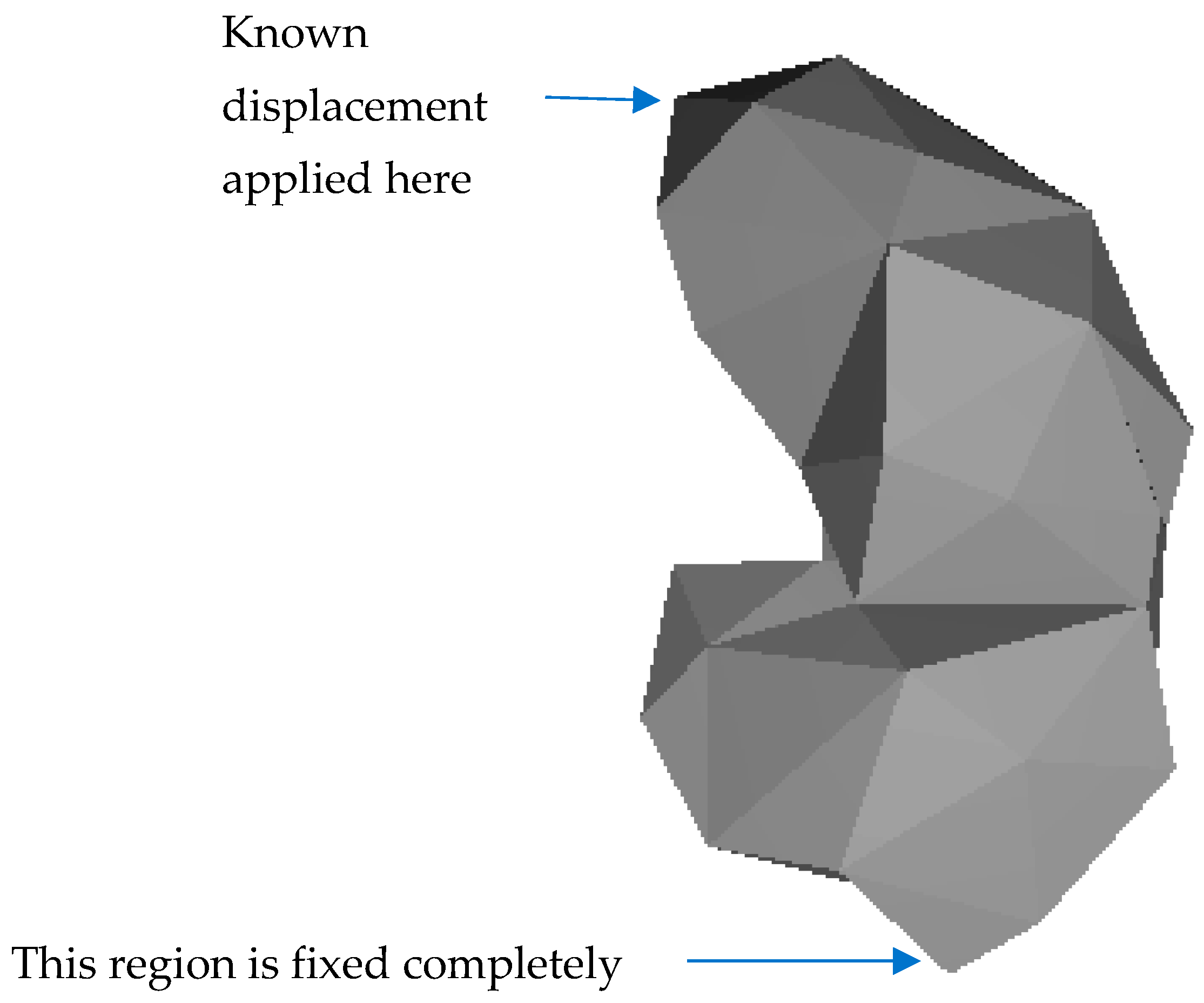
Figure 1. Boundary conditions.
Engproc 59 00169 g001
Table 1. Results for Problem 1.
PointDisplacement Vector Sum from Nonlinear FEM Analysis (mm)Displacement Vector Sum from Linear BEM Analysis (mm)Difference (mm)
14.1763.911−0.265
25.4854.513−0.972
35.1614.932−0.229
45.1774.133−1.044
52.2001.904−0.296
60.0000.0000
70.7280.8520.124
80.7500.9330.183
91.3841.5490.165
101.5511.6250.074
111.8701.723−0.147
Table 2. Results for Problem 2.
PointDisplacement Vector Sum from Nonlinear FEM Analysis (mm)Displacement Vector Sum from Linear BEM Analysis (mm)Difference (mm)
15.0433.025−2.018
26.0483.826−2.222
35.9324.322−1.61
45.2514.115−1.136
52.3232.4040.081
60.0000.0000
70.6610.150−0.511
80.9840.162−0.822
91.3730.389−0.984
101.5840.546−1.038
111.7670.839−0.928
Table 3. Results for Problem 3.
PointDisplacement Vector Sum from Nonlinear FEM Analysis (mm)Displacement Vector Sum from Linear BEM Analysis (mm)Difference (mm)
17.5513.707−3.844
28.9223.775−5.147
39.2313.838−5.393
48.4463.389−5.057
53.1461.717−1.429
60.0000.0000
70.9480.747−0.201
81.4880.792−0.696
91.6100.999−0.611
101.7761.096−0.68
112.0511.183−0.868
Disclaimer/Publisher’s Note: The statements, opinions and data contained in all publications are solely those of the individual author(s) and contributor(s) and not of MDPI and/or the editor(s). MDPI and/or the editor(s) disclaim responsibility for any injury to people or property resulting from any ideas, methods, instructions or products referred to in the content.