Proceeding Paper

Effect of Lanthanum Doping on the Structural, Morphological, and Optical Properties of Spray-Coated ZnO Thin Films †

by
Manu Srivathsa
and
Bharathipura Venkataramana Rajendra
*
Department of Physics, Manipal Institute of Technology, Manipal Academy of Higher Education, Manipal 576104, Karnataka, India
*
Author to whom correspondence should be addressed.
Presented at the International Conference on Recent Advances on Science and Engineering, Dubai, United Arab Emirates, 4–5 October 2023.
Eng. Proc. 2023, 59(1), 32; https://doi.org/10.3390/engproc2023059032
Published: 12 December 2023
(This article belongs to the Proceedings of Eng. Proc., 2023, RAiSE-2023)

Abstract

:
In recent years, transparent conducting oxide semiconductor materials have found applications in both science and technology, especially in the areas of semiconductors, optoelectronics, and a wide range of energy efficiency devices. These TCO materials are the building blocks of various optoelectronic devices, such as transparent thin-film transistors, solar cells, and light-emitting diodes. This work concentrates on the structure, morphology, and optical properties of ZnO and Zn0.95La0.05O thin films at 673 K using a chemical spray technique. The polycrystalline nature and wurtzite structure of ZnO were confirmed by using XRD analysis with preferred growth along the (1 0 1) plane. The Zn0.95La0.05O deposits showed maximum crystallinity of 15.4 nm and a strain value of 2.4 × 10⁻³. The lattice constants increased for lanthanum-doped ZnO thin films due to the ionic radii mismatch of the doping material, which causes lattice expansion. Fibrous morphology was observed for ZnO, and a mixed structure of grains and fibers was observed for Zn0.95La0.05O films, which confirms the insertion of La3+ into the Zn2+ position. The Zn0.95La0.05O deposits showed transmittance above 80% due to the increased crystalline quality and a bandgap of 3.32 eV. The photoluminescence spectra showed peaks corresponding to e-h recombination, zinc defects (Zni and Ozn), and oxygen vacancy (Oi and Vo). The lanthanum-doped ZnO films showed increased band-edge emission and decreased defect-related peaks due to the increased crystalline quality. Hence, the doping of La3+ ions into a ZnO lattice enhances the crystalline quality and increases the transparency of the host ZnO matrix, which is suitable for optoelectric device applications.

1. Introduction

The transparent conducting oxide (TCO) semiconductors are of great interest due to their better optical and electrical properties. Currently, a lot of research works are looking at TCO semiconductor material-based photodetectors [1], UV sensors [2], optical limiters [3], and solar cells [4]. The ZnO, ZnS, CdO, and CdS materials of the II–VI semiconducting groups are good examples of TCO materials. Compared to other materials, ZnO-based materials are of great interest due to their easy availability, bandgap tuning, nontoxic nature, high exciton binding energy (60 MeV), and low cost. ZnO-based semiconducting materials are very sensitive to ultraviolet light. Hence, they can be used in the fabrication of ultraviolet (UV) detectors. These properties of ZnO films can be enhanced by introducing suitable dopants into the host matrix. Lots of work has been carried out on the transition of metal ion-doped ZnO films. But the effect of rare-earth ion doping into the ZnO lattice is an emerging field and needs to be studied in detail. The insertion of rare earth ions into the ZnO lattice is a difficult task because of the difference in the ionic radii of La3+ and Zn2+ ions. Due to this mismatch, the dopant may occupy the interstitial positions and hinder the properties. However, the surface structure, bandgap, transparency, and electrical behavior of ZnO can be modified by the proper insertion of rare-earth ions into the matrix. These rare-earth ion-doped ZnO films can be synthesized using various spin coatings [5] sol–gel methods [6], SILAR [7], and spray techniques [8]. The spray pyrolysis technique is employed in the current study due to its simplicity, low cost, less chemical wastage, large area of deposition, and uniform coating. Here, the properties of the deposits mainly depend on the concentration of the precursor solution, substrate temperature, pressure of the carrier gas, nozzle to substrate distance, spray interval, and duration of the spray. Hence, optimizing all these parameters is very important to obtain better-quality thin films. Lanthanum is a less-studied topic, and due to the inconsistency in the available reports, this work focuses on lanthanum doping on the structure, morphological, and optical properties of spray-coated ZnO films.

2. Materials and Methods

2.1. Film Deposition

The ZnO and La:ZnO films were coated on glass substrates at 673 K using spray pyrolysis method. The chemicals used were zinc acetate dihydrate and lanthanum chloride heptahydrate dissolved in distilled water and constantly stirred for 30 min to obtain a uniform concentration of 0.05 M. The prepared precursor solution was filled in the burette and sprayed on the preheated glass substrates through a spray nozzle in optimized spray intervals. The films were maintained at the same temperature for 30 min to obtain better crystallinity.

2.2. Characterization Techniques

The structural properties of the thin films were determined using X-ray diffraction analysis. A scanning electron microscope was used to study the morphology of the deposited films. The optical properties, such as transmittance and bandgap of the material, were obtained using the UV–vis double-beam spectrometer. The defects in the films were determined using photoluminescence spectra analysis.

3. Result and Discussion

3.1. Structural Analysis

The XRD spectra of the deposited films are shown in Figure 1. Both the films showed a polycrystalline nature, and the noticed peaks matched the wurtzite structure. The spectra showed seven peaks at (1 0 0), (0 0 2), (1 0 1), (1 0 2), (1 1 0), (1 0 3), and (1 1 2). The orientation of the peaks mainly depends on the atom configurations on the subsequent layers and primarily depends on the deposition parameters. The intensity of the (1 0 1) peak increased for Zn0.95La0.05O films. The shift of the diffraction peak toward the lower-angle side after lanthanum doping was observed. The estimated structural parameters are tabulated in Table 1.
The ‘D’ value of the deposited films was determined by using the Scherrer equation given below [9].
D = k λ β   c o s   θ  
The ‘D’ of the Zn0.95La0.05O films was found to be due to the insertion of La3+ ions into the Zn2+ lattice creating lattice expansion and increasing the crystallite size.
The strain ( ε ) values were determined by using the formula given below [10].
ε = β   c o s   θ 4
The strain is due to the incorporation of La3+ ions into Zn2+ positions. The strain values were found to be decreased for Zn0.95La0.05O films.
The ‘a’ and ‘c’ were calculated using the below relation [10].
a = λ 3 s i n   θ 100
c = λ s i n   θ 002
The lattice constant values ‘a’ and ‘c’ were found to be increased for Zn0.95La0.05O films due to the insertion of La3+ ions, which causes the lattice expansion in the doped films.
The dislocation density was calculated using the ‘D’ values by using the below formula [11].
δ = 1 D 2
The dislocation density refers to the number of dislocations per unit area. This is related to the crystallinity and purity of the deposits. The Zn0.95La0.05O films showed lower dislocation density values.

3.2. Scanning Electron Microscopy (SEM)

The morphology of ZnO and Zn0.95La0.05O films is shown in Figure 2. The pure ZnO films showed a fibrous morphology. The change in morphology from fibrous to a mixed granular–fibrous one was observed with the addition of lanthanum into the ZnO lattice. This change is due to the replacement of Zn2+ by La3+ ions. The lanthanum tends to change the morphology from fibrous to granular; however, compared to granular, fibrous was more observed due to the lower doping percentage of lanthanum. Hence, the lanthanum doping into the ZnO lattice changes the morphology of the deposited films.

3.3. Optical Studies

The transmittance and Tauc’s plot of the deposited films are shown in Figure 3a,b. Both ZnO and Zn0.95La0.05O films showed high transparency. Transparency in the range of 70% was noticed for ZnO films. However, the transparency increased after the lanthanum doping due to the increased crystallinity. As the crystallinity increases, the light travels easily through the films without being deflected. However, in the case of ZnO, the XRD results show a smaller crystallite size and higher strain and dislocation density values. Hence, due to the increased defects, the transparency of the film decreased. The shift in the absorption edge of Zn0.95La0.05O films confirms the better optical properties.
The Tauc’s plot relation [12] was used to determine the bandgap.
α h ν = A ( h ν E g ) n
The bandgap of the films showed a strong dependency on lanthanum doping. The ZnO films showed a bandgap of 3.1 eV, whereas the Zn0.95La0.05O films showed an increased bandgap of 3.32 eV. This may be due to the Burstein–Moss shift (B–M shift). Here, the bandgap of the material increases as the absorption edge shifts toward higher energies, and as a result, some states close to the conduction band are populated due to the increased carrier density of the deposited films due to the addition of La3+ ions, which leaves an extra electron in the valance band and helps to increase conductivity. Hence, the lanthanum doping into the ZnO lattice varies the optical properties of the deposited films.

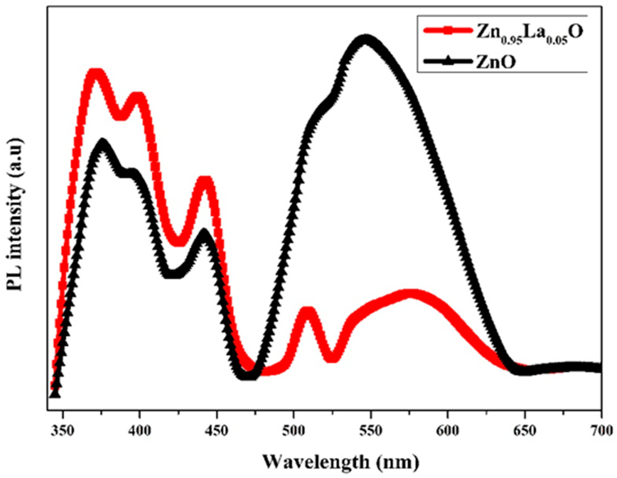
3.4. Photoluminescence Study

The PL spectra of ZnO and Zn0.95La0.05O films are shown in Figure 4. The spectra were fitted using a Gaussian fit, which showed seven peaks corresponding to an e-h recombination at 385 nm, violet emission at 400 nm, blue emission peaks at 425 and 452 nm, green emission at 530 nm, and yellow-orange emissions at 580 and 605 nm. The band-edge peak at 385 nm corresponds to the crystalline quality and homogeneity of the deposited films [13]. As the crystal quality increases, the intensity of the band-edge emission increases. The Zn0.95La0.05O films showed increased band-edge emissions. Hence, the recombination of electron-hole pairs will be more in Zn0.95La0.05O films. The violet emission is due to the interface traps occurring inside the grain boundaries of ZnO [14]. The Zn0.95La0.05O films showed maximum violet emission. The zinc defects cause the blue emission. The intensity of the blue emission peaks increased for Zn0.95La0.05O films. The green emission is due to oxygen-related defects [15]. The intensity of the green emission peak decreased with the addition of La3+ into the ZnO lattice. The presence of singly ionized oxygen vacancies cause yellow-orange emission. The yellow-orange emission was found to be maximum for pure ZnO films due to increased oxygen-related defects.

4. Conclusions

The ZnO and Zn0.95La0.05O films were coated on chemically cleaned glass substrates at 673 K. The spectra confirmed the wurtzite structure of ZnO. The Zn0.95La0.05O films showed a maximum crystallite size of 15.4 nm and lower strain and dislocation density values. SEM images showed the change in morphology from fine fibrous to a mixed granular–fibrous morphology to Zn0.95La0.05O films. An increase in the optical transmittance (80%) and bandgap (3.32 eV) was observed for Zn0.95La0.05O films. The photoluminescence spectra showed peaks corresponding to band-edge emission, zinc-related defects, and oxygen defects. The band-edge emission increased, and defect-related peaks decreased with the inclusion of the lanthanum content in ZnO films. Hence, the lanthanum doping into the ZnO lattice will enhance the properties of ZnO films.

Author Contributions

M.S. is presently working as a research scholar in the Department of Physics, Manipal Institute of Technology, MAHE, Manipal, India. He played a role in Data curation, Formal analysis, Investigation, and Writing—original draft. B.V.R. is a professor in the Department of Physics, Manipal Institute of Technology, MAHE, Manipal, India. He played a role in Funding acquisition, Resources, Software, Supervision, Validation, and Writing—review and editing. All authors have read and agreed to the published version of the manuscript.

Funding

This research work has been funded by UGC-DAE CSR Mumbai Centre (UDCSR/MUM/AO/CRS-M-315/2020/813).

Institutional Review Board Statement

Not applicable.

Informed Consent Statement

Not applicable.

Data Availability Statement

All the data and images are included within the article.

Acknowledgments

The authors would like to thank the Manipal Academy of Higher Education (MAHE) for providing a research facility and Sudha D. Kamath for providing the photoluminescence facility.

Conflicts of Interest

The authors declare no conflict of interest.

References

  1. Gozeh, B.A.; Karabulut, A.; Ameen, M.M.; Yildiz, A.; YakuphanoǦlu, F. Synthesis and characterization of La-doped ZnO (La:ZnO) films for photodetectors. Surf. Rev. Lett. 2020, 27, 1950173. [Google Scholar] [CrossRef]
  2. Srivathsa, M.; Kumar, P.; Rajendra, B.V. Ultraviolet Photoconductivity and Photoluminescence Properties of Spray Pyrolyzed ZnO Nanostructure: Effect of Deposition Temperature. Opt. Mater. 2022, 131, 112726. [Google Scholar] [CrossRef]
  3. Sindhu, H.S.; Maidur, S.R.; Shankaragouda Patil, P.; Rajendra, B.V. Influence of Structure and Surface Morphology on Optical Limiting Property of Spray Pyrolyzed ZCO Thin Films. Chem. Phys. Lett. 2020, 759, 137975. [Google Scholar] [CrossRef]
  4. Wibowo, A.; Marsudi, M.A.; Amal, M.I.; Ananda, M.B.; Stephanie, R.; Ardy, H.; Diguna, L.J. ZnO Nanostructured Materials for Emerging Solar Cell Applications. RSC Adv. 2020, 10, 42838–42859. [Google Scholar] [CrossRef] [PubMed]
  5. Poongodi, G.; Kumar, R.M.; Jayavel, R. Structural, Optical and Visible Light Photocatalytic Properties of Nanocrystalline Nd Doped ZnO Thin Films Prepared by Spin Coating Method. Ceram. Int. 2015, 41, 4169–4175. [Google Scholar] [CrossRef]
  6. Znaidi, L. Sol-Gel-Deposited ZnO Thin Films: A Review. Mater. Sci. Eng. B Solid-State Mater. Adv. Technol. 2010, 174, 18–30. [Google Scholar] [CrossRef]
  7. Raidou, A.; Benmalek, F.; Sall, T.; Aggour, M.; Qachaou, A.; Laanab, L.; Fahoume, M. Characterization of ZnO Thin Films Grown by SILAR Method. OALib 2014, 1, e588. [Google Scholar] [CrossRef]
  8. Srivathsa, M.; Kumar, P.; Goutam, U.K.; Rajendra, B.V. Enhancement in the Transport and Optoelectrical Properties of Spray Coated ZnO Thin Films by Nd Dopant. Electron. Mater. Lett. 2022, 19, 138–160. [Google Scholar] [CrossRef]
  9. Khan, Z.R.; Khan, M.S.; Zulfequar, M.; Shahid Khan, M. Optical and Structural Properties of ZnO Thin Films Fabricated by Sol-Gel Method. Mater. Sci. Appl. 2011, 2, 340–345. [Google Scholar] [CrossRef]
  10. Suryanarayana, C.; Norton, M.G. X-ray Diffraction; Springer: Boston, MA, USA, 1998; ISBN 978-1-4899-0150-7. [Google Scholar]
  11. Mariappan, R.; Ponnuswamy, V.; Suresh, P. Effect of Doping Concentration on the Structural and Optical Properties of Pure and Tin Doped Zinc Oxide Thin Films by Nebulizer Spray Pyrolysis (NSP) Technique. Superlattices Microstruct. 2012, 52, 500–513. [Google Scholar] [CrossRef]
  12. Nassiba, A.; Boubaker, B.H.; Chahnez, S.; Djamel, B.; Leila, S.; Brahim, G.; Rahal, A.; Benhaoua, A.; Hima, A. Effect of La Doping on Zno Thin Films by Spray Pyrolysis. Defect Diffus. Forum 2019, 397, 206–212. [Google Scholar] [CrossRef]
  13. Mousa, A.O.; Habubi, N.F.; Nema, N.A. Substrate Effects on Structural and Optical Properties of ZnO Thin Films Deposited by Chemical Spray Pyrolysis. Int. Lett. Chem. Phys. Astron. 2015, 51, 69–77. [Google Scholar] [CrossRef]
  14. Jin, B.J.; Im, S.; Lee, S.Y. Violet and UV Luminescence Emitted from ZnO Thin Films Grown on Sapphire by Pulsed Laser Deposition. Thin Solid Film. 2000, 366, 107–110. [Google Scholar] [CrossRef]
  15. Lin, B.; Fu, Z.; Jia, Y. Green Luminescent Center in Undoped Zinc Oxide Films Deposited on Silicon Substrates. Appl. Phys. Lett. 2001, 79, 943–945. [Google Scholar] [CrossRef]
Figure 1. X-ray diffraction pattern of ZnO and Zn0.95La0.05O films.
Engproc 59 00032 g001
Figure 2. SEM images of ZnO and Zn0.95La0.05O films.
Engproc 59 00032 g002
Figure 3. (a) Transmittance and (b) Tauc’s plot of ZnO and Zn0.95La0.05O films.
Engproc 59 00032 g003aEngproc 59 00032 g003b
Figure 4. Photoluminescence spectra of ZnO and Zn0.95La0.05O films.
Engproc 59 00032 g004
Table 1. Estimated structural parameters calculated using XRD data.
SampleDeposition TemperatureOrientationCrystallite Size (D) in nmLattice Constants (Å)Microstrain × 10−3Dislocation Density × 1015 (lines/m2)
ac
ZnO673 K(1 0 1)9.83.1685.1352.910.4
Zn0.95La0.05O673 K(1 0 1)15.43.2225.1612.44.2
Disclaimer/Publisher’s Note: The statements, opinions and data contained in all publications are solely those of the individual author(s) and contributor(s) and not of MDPI and/or the editor(s). MDPI and/or the editor(s) disclaim responsibility for any injury to people or property resulting from any ideas, methods, instructions or products referred to in the content.

