Proceeding Paper

Encapsulation of Aqueous Extract of Hancornia speciosa  †

by
Lorrane Soares dos Santos
1,
Jéssica Silva Medeiros
1,
Antonio Matias Navarrete de Toledo
2,
Letícia Fleury Viana
1,
Maria Inês Rodrigues Machado
3 and
Adriana Rodrigues Machado
1,*
1
Department of Food Science, Goiano Federal Institute of Education, Science and Technology, Campus Rio Verde, Rodovia Sul Goiana, Km 01, Rio Verde 75901-970, GO, Brazil
2
Department of Food Engineering and Technology, School of Food Engineering, University of Campinas, 80, Monteiro Lobato St., Campinas 13083-862, SP, Brazil
3
Center for Agricultural Sciences and Biodiversity, Federal University of Cariri—UFCA, Rua Ícaro de Sousa Moreira, s/n, Crato Campus, Crato 63130-025, CE, Brazil
*
Author to whom correspondence should be addressed.
Presented at the 3rd International Electronic Conference on Processes—Green and Sustainable Process Engineering and Process Systems Engineering (ECP 2024), 29–31 May 2024; Available online: https://sciforum.net/event/ECP2024.
Eng. Proc. 2024, 67(1), 22; https://doi.org/10.3390/engproc2024067022
Published: 29 August 2024
(This article belongs to the Proceedings of The 3rd International Electronic Conference on Processes)

Abstract

:
Liposomes are increasingly favored for encapsulation due to their biocompatibility and versatility, making them valuable in drug delivery, cosmetics, and food science. Their ability to encapsulate both hydrophobic and hydrophilic compounds has driven this growing interest. Liposomes composed of soybean lecithins (SLs) were produced by reverse-phase evaporation and used to encapsulate phenolic extracts of mangaba (SL-MAPE). Liposomes were characterized by size distribution, polydispersity index, and ζ-potential. Liposomes, SL-MAPE, exhibited high encapsulation efficiency (SL-MAPE 1.0 mg/mL: 80.14%; SL-MAPE 1.5 mg/mL: 86.18%; and SL-MAPE 2.0 mg/mL: 88.09%, respectively) and sizes ranging between SL-MAPE 1.0 mg/mL: 197.43; SL-MAPE 1.5 mg/mL: 318.2; and SL-MAPE 2.0 mg/mL: 238.33 nm, showing to be good candidates for the encapsulation of phenolic extracts obtained from mangaba (SL-MAPE).

1. Introduction

Mangaba (Hancornia speciosa) is a fruit native to the Cerrado of Goiás, with a composition rich in nutrients such as calcium, zinc, phosphorus, and iron, as well as bioactive compounds such as vitamin C, carotenoids, and considerable antioxidant activity [1]. Thus, due to the instability of phenolic compounds in the face of the external environment, liposomes are an alternative way to preserve these compounds against degradation, improving stability and solubility [2,3,4].
Liposomes are vesicles with a structure composed of a bilayer of lipid molecules that enclose an aqueous volume, and their diameter varies from nanometers to micrometers [5]. Therefore, they are capable of encapsulating hydrophilic and hydrophobic substances, such as carotenoids, phenolics, and vitamins, among others [4,6,7].
In this study, the objective was to perform an assay to evaluate the characteristics of the lecithin used, mainly for the elaboration of liposomes for encapsulation of mangaba phenolic extract, as well as the verification of the average size, polydispersion index, encapsulation efficiency, and stability of the liposome load through the zeta potential.

2. Materials and Methods

2.1. Raw Material

The mangaba fruit (Hancornia speciosa) came from natural unplanted vegetation organized in the region of Montes Claros de Goiás (16°06′20″ S and 51°17′11″ W), harvested manually from November to December 2017 and immediately transported to the Laboratory of Vegetable Culture and Textiles (LCTV) at the Federal Institute of Goiás–Campus Rio Verde, where they were prepared, stored, and evaluated. The fruits were washed, sanitized with 200 ppm sodium hypochlorite for 15 min, then pulped, homogenized, pasteurized at 80 °C for 20 min, packed, and frozen at −26 °C in a freezer until further analysis. Soy lecithin (Attivos Magisttrais, Barueri, Brazil) was purchased in the local market of Rio Verde, Goiás, in powder form.

2.2. Extraction and Determination of Phenolic Compounds

For the preparation of mangaba pulp extracts, 100 mg of the sample was weighed in a screw-capped flask with the subsequent addition of 10 mL of water and left under stirring for one hour on a mechanical stirrer. Afterward, a 0.1 mL aliquot of the obtained filtrate was filtered on filter paper and placed in a threaded test tube, and 7.9 mL of deionized water and 0.5 mL of Folin–Ciocalteu 2 N reagent (diluted 1:10) were added. It was shaken by an agitator. After being kept for 5 min in the dark, 1.5 mL of a 20% sodium carbonate solution was added (Na2CO3 was added to the solution). It was then agitated by an agitator. After incubation at 25 °C for 2 h the absorbance of the solution at 765 nm was measured using a spectrophotometer (BEL/Spectro S-2000, Bel Engineering, Monza, Italy). A standard curve was performed using gallic acid at concentrations from 0 to 500 mg L−1 to construct a calibration curve. All steps of this analysis were performed under light, according to the methodology proposed by Souza [8], adapted by the authors.

2.3. Physical Chemistry Characterization of Soy Lecithin

The following analyses were performed on commercial soy lecithin: acidity index and peroxide index according to A.O.C.S. [9].

2.4. Encapsulation of Phenolic Extracts in Liposomes

Initially, the mangaba pulp was extracted aqueous with subsequent lyophilization, where 0.180 g was yielded as the final sample. The concentrations of 1.0, 1.5, and 2.0 g of lipid matrix (lecithin) were previously determined, and the analyses were always performed in triplicate in each of the respective concentrations. The lipid nanovesicles were acquired through the following process: homogenization of the lyophilized aqueous pulp extract.
The preparation of liposomes was performed through reverse evaporation, with modifications made with the use of sonication, which constitutes a process in stages where the different stages lead to the formation of three vesicular systems of different structural organizations: reverse micelles, organogels, and liposomes [10]. During the process of obtaining lipid nanovesicles, the bioactive compound is added.

2.4.1. Particle Size, Polydispersion Index, and Zeta Potential

All measurements were performed at 25 °C in Malvern Zeta sizer Nano Zs equipment in the department of FEA (Faculty of Food Engineering), Laboratory of Process Engineering, State University of Campinas (UNICAMP). Each particle size and polydispersion measurements were measured with a detection angle of 173° and zeta potential measurements at an angle of 17°, and zeta potential values were calculated by the Smoluchowski model [11].

2.4.2. Encapsulation Efficiency

The encapsulation efficiency was determined indirectly, according to Assis et al. [12] and Machado et al. [4]. First, 0.5 mL of the liposomes were placed in a centrifuge tube with 1 mL of acetone since phosphatidylcholine is insoluble in this solvent. The samples were centrifuged at 5000× g for 30 min at 3 °C, separating into two phases. The supernatant containing the non-encapsulated sample was withdrawn and placed in an oven at 60 °C until complete evaporation of the solvent. The remaining dried stuff was resuspended with 5 mL of distilled water, and the concentration was determined through the phenol method [4]. A 0.5 mL aliquot of the initial sample was withdrawn, and 1 mL of 0.06% Triton X-100 was added to determine the total phenol present in the sample. The equipment was then homogenized in a vortex (Phoenix Luferco AP56, Tecnal, Piracicaba, Brazil) until complete solubilization of phosphatidylcholine. The encapsulation efficiency (EE) was calculated as shown in Equation (1):
E E % = P i n s .   P i n s . + P o u t . × 100
Phenol content inside   ( P i n s . ) . the liposome vesicles quantified compounds after dissolving with Triton; phenols outside (   P o u t . ) the vesicles and quantified compounds that solubilized in acetone were considered.

2.4.3. Optical Microscopy

The formation of microparticles was monitored using an optical microscope model Olympus BX 43 (Olympus, Center Valley, PA, USA) by obtaining the smear of a drop of the sample on a glass slide, followed by observation using magnification objectives corresponding to 40 times.
After obtaining the smear, the diameter and length were expressed in millimeters and the mass presented in grams. The physical–chemical analyses were performed in triplicate, with an arithmetic mean between the values and their respective standard deviations. The mean results were compared using Tukey’s 5% significance test, using the Statisc 7 Software Program.

2.4.4. Scanning Electron Microscopy

For microscopic analysis, the lyophilized liposomes were resuspended in methanol (Synth®), treated in an ultrasonic bath for 20 min, and deposited on a silicon substrate. The substrates were attached to the sample holder of the microscope using a carbon ribbon and then metallized with gold for 3 min and 5 mA of current in a Sanyu Electron Quick Coater Model SC-701 (Sanyu Electron, Tokyo, Japan). The images were obtained on a Shimadzu SSX-550 model microscope (Shimadzu, Co. Ltd., Kyoto, Japan), which has a vacuum coupled with EDS [2,10].

2.5. Statistical Analysis

All determinations were performed in triplicate (expressed as mean ± standard error (SE)), and the results were analyzed with ANOVA and Tukey’s test at a significance level of 5% with the aid of Statistica® software (2004), version 7.0.

3. Results and Discussion

3.1. Quality Analysis of Soy Lecithin

Table 1 demonstrates the quality analysis of soy lecithin.
The moisture content for soy lecithin is by the legislation, according to Table 1, and by the Food Code, which recommends ≤1.5% [13].
Humidity is extremely important to ensure microbiological stability in the product. Its value should be less than 1% (m/m), as it has a great influence on the lecithin viscosity and storage time of the product [14].
Table 1. Quality analysis of soy lecithin.
Quality AnalysisOutcome of the StudyLegislation Standards *
Moisture (%)0.350.40
Acid value (mg/kg)0.0629.00
Peroxide index (meq/kg)0.205.00
Insoluble in acetone (%)96.3862.00
* Source: Zulian [14].
In this study, the acidity index complies with the standards of the legislation, since the acidity index indicates the degree of degradation of lecithin due to the low quality of the soy processed or inadequate process conditions. For acidity to not compromise the quality of the product, the acidity index should not exceed the limit of 29 mg KOH/g.
The peroxide value is a classical method for determining primary oxidation products (hydroperoxides). It indicates the beginning of the deterioration of oil and fat samples [15].
Extrinsic characteristics such as temperature can influence the quality of the product, such as the peroxide indices of lecithin, corroborated by Avila [16], which increased the temperature and increased the formation of peroxides during storage [17]. Mezouari and Eichner [18] explained that the production of peroxides indicates principles of oxidation already occurred at some point. Studies have shown that although lecithins are degraded, they are more stable than other lipids [19].

3.2. Physico-Chemical Characterization of Liposomes

In this study, liposomes containing SL-MAPE (Soybean Liposome with Mangaba Aqueous Phenolic Extract) were developed, varying the bioactive/lipid compound ratio of 1.0, 1.5, and 2.0.
As observed in Table 2, liposomes can be used to encapsulate food bioactive with different structures, but depending on the production method, the characteristics of the liposome, and the material to be encapsulated, several types of structures can be developed with different characteristics, as an example of different encapsulation efficiencies [5,20]. Regarding the classification of liposomes may be large unilamellar or LUVs, comprising the sizes obtained (80 to 1000 nm), the size obtained in this study obtained statistical differences, according to Table 2, for concentrations 1.0,1.5, and 2.0. Machado et al. [4] evaluated the mean size and polydispersion index of liposomes that underwent the homogenization and sonication process and found that the application of a homogenizer or ultrasound did not present differences in obtaining nanosized liposomes; however, they found that liposomes produced through the sonication process have a smaller mean size (279.53 nm) compared to liposomes that underwent homogenization (303.97 nm).
Regarding the polydispersion index higher than 0.2, they indicate that the developed liposomes do not present a good homogeneity. According to Danaei et al. [21], polydispersion provides information on homogeneity of the high (>0.3) size distribution for all samples, indicating the formation of polydisperse systems or a narrow range of sizes [22].
The zeta potential values obtained in this study are from the literature [12]. According to Vogel et al. [23] and Németh et al. [24], the zeta potential must be close to ±30 mV to obtain a good stability of the suspensions. The zeta potential obtained for the different concentrations of liposomes, confirmed in Table 2, presented a negative value, indicating that the vesicles showed an external surface with a predominance of negative charges [24,25].
The results presented for encapsulation efficiency were satisfactory, all concentrations above 80%, according to Table 2. Compared with other studies such as Assis et al. [12] obtained EEs of 87% and 92% for methanolic and ethanolic Spirulina extracts, respectively, and Machado et al. [4] in liposomes subject to homogenization obtained higher encapsulation efficiency (90%) compared to liposomes subject to ultrasound (79%), as well as Machado et al. [4] used liposomes made with rice and soy lecithin, respectively, and obtained 97.35 and 88.28% of encapsulation efficiency for LEB-18 Spirulina phenolic extracts, it can be stated that the method used was very efficient for encapsulation of mangaba phenolic extracts.
Figure 1 presents vesicles in different sizes, being a polydispersed system. With this, the presence of a nanovesicle can be confirmed, but only in more sophisticated microscopes such as scanning electron microscopes for the visualization of spherical liposome formation, as shown in the figure below.
Scanning electron microscopy was employed to characterize the surface of lipid nanostructures and to observe evidence of the dispersion of particles in the lipid matrix, as seen in Figure 1B,C, and confirmed by Table 2.

4. Conclusions

The results of the present study showed that Hancornia speciosa extracts can be protected by lipid systems such as liposomes.
It was also found that liposomes from the aqueous extract were obtained in sizes of lipid nanovesicles with excellent encapsulation efficiency, resulting in great potential for application in food. In this study, it was also observed that, in the lipid concentrations containing mangaba phenolic extracts evaluated, there was an increase in polydispersity, resulting in particle agglomerations, indicating polydisperse systems, as evidenced by the microscopy images.
Overall, the results obtained in this study were satisfactory and provide a foundation for future, more detailed investigations, including the use of lecithins from other sources for encapsulation of various compounds, as well as sophisticated techniques such as gas chromatography for the fatty acid profile of these liposomes, the morphology of these materials, and the in vitro release profile of these extracts for potential applications in different areas.

Author Contributions

Conceptualization, L.S.d.S. and J.S.M.; methodology and A.R.M.; validation, A.R.M.; investigation L.S.d.S. and J.S.M.; data curation, L.S.d.S., J.S.M., A.M.N.d.T. and A.R.M.; writing—original draft preparation, L.S.d.S. and A.R.M.; writing—review and editing, M.I.R.M. and A.R.M.; visualization, L.F.V., A.M.N.d.T., M.I.R.M. and A.R.M.; supervision, A.M.N.d.T., L.F.V. and A.R.M. All authors have read and agreed to the published version of the manuscript.

Funding

This research received no external funding.

Institutional Review Board Statement

Not applicable.

Informed Consent Statement

Not applicable.

Data Availability Statement

Data are contained within the article.

Acknowledgments

The authors would like to thank the FAPEG, CAPES, CNPq, IFGoiano, and UNICAMP.

Conflicts of Interest

The authors declare no conflicts of interest.

References

  1. de Oliveira Filho, J.G.; de Sousa, T.L.; Bertolo, M.R.V.; Bogusz Junior, S.; Mattoso, L.H.C.; Pimentel, T.C.; Egea, M.B. Embalagens Para Alimentos de Última Geração: Filmes Bioativos Comestíveis Com Alginato, Polpa de Mangaba (Hancornia speciosa) e Saccharomyces boulardii. Food Biosci. 2023, 54, 102799. [Google Scholar] [CrossRef]
  2. Machado, A.; Assis, L.M.; Costa, J.A.; Badiale-Furlong, E.; Motta, A.; Micheletto, Y.M.S.; Souza-Soares, L. Application of Sonication and Mixing for Nanoencapsulation of the Cyanobacterium Spirulina Platensis in Liposomes. Int. Food Res. J. 2015, 22, 96–101. [Google Scholar]
  3. Machado, A.R.; Assis, L.M.; Machado, M.R.G.; Souza-Soares, L.A. Biological Evaluation of Casein and Spirulina in Rats and Encapsulation of These Protein Sources in Liposomes. Int. Food Res. J. 2015, 22, 344–350. [Google Scholar]
  4. Machado, A.R.; Pinheiro, A.C.; Vicente, A.A.; Souza-Soares, L.A.; Cerqueira, M.A. Liposomes Loaded with Phenolic Extracts of Spirulina LEB-18: Physicochemical Characterization and Behavior under Simulated Gastrointestinal Conditions. Food Res. Int. 2019, 120, 656–667. [Google Scholar] [CrossRef] [PubMed]
  5. Machado, A.R.; Machado, M.I.R.; de Souza-Soares, L.A. Avaliação do tamanho de particulas lipossomicas em diferentes ensaios. Res. Soc. Dev. 2023, 12, e24512135104. [Google Scholar] [CrossRef]
  6. Santos, L.S.d.; Medeiros, J.S.; Toledo, A.M.N.; Viana, L.F.; Machado, M.I.R.; Machado, A.R. Encapsulation process of lyophilized and non-lyophilized aqueous extract of mangaba in lipid vesicles. Braz. J. Dev. 2020, 6, 2180–2188. [Google Scholar] [CrossRef]
  7. Toniazzo, T.; Berbel, I.F.; Cho, S.; Fávaro-Trindade, C.S.; Moraes, I.C.F.; Pinho, S.C. β-Carotene-Loaded Liposome Dispersions Stabilized with Xanthan and Guar Gums: Physico-Chemical Stability and Feasibility of Application in Yogurt. LWT—Food Sci. Technol. 2014, 59, 1265–1273. [Google Scholar] [CrossRef]
  8. Souza, M. Potencial Antifúngico, Antioxidante e Inibidor Da Produção de Aflatoxina Por Extratos Fenólicos de Chlorella Sp. E Spirulina Platensis. 2012. Available online: https://bdtd.ibict.br/vufind/Record/FURG_19aeb2ea356735fd7c31eda19c011cae (accessed on 13 July 2018).
  9. American Oil Chemists Society. Official Methods and Recommended Practices, 4ed.; American Oil Chemists Society: Champaing, IL, USA, 1993; Volume 3. [Google Scholar]
  10. Mertins, O.; Sebben, M.; Henrique Schneider, P.; Pohlmann, A.R.; Silveira, N.P. da Caracterização da pureza de fosfatidilcolina da soja através de RMN de 1H e de 31P. Quím. Nova 2008, 31, 1856–1859. [Google Scholar] [CrossRef]
  11. Malvern. Dynamic Light Scattering: An Introduction in 30 Minutes. Version 4, Technical Note. 2014. Available online: http://www.malvern.com/br/support/resource-center/technical-notes/tn101104dynamiclightscatteringintroduction.aspx (accessed on 6 November 2018).
  12. de Assis, L.M.; Machado, A.R.; da Motta, A.D.S.; Costa, J.A.V.; de Souza-Soares, L.A. Development and Characterization of Nanovesicles Containing Phenolic Compounds of Microalgae Spirulina Strain LEB-18 and Chlorella Pyrenoidosa. Adv. Mater. Phys. Chem. 2014, 4, 42009. [Google Scholar] [CrossRef]
  13. Castejon, L.V. Parâmetros de Qualidade na Clarificação da Lecitina de Soja; Universidade Federal de Uberlândia: Uberlândia, Brazil, 2015. [Google Scholar]
  14. Zulian, S.L. Adição de Ácido Graxo de Soja Como Agente Estabilizante da Viscosidade de Lecitina de Soja. 2016. Available online: https://repositorio.ufsc.br/xmlui/handle/123456789/176058 (accessed on 12 February 2021).
  15. Sikorski, Z.Z.E.; Kolakowska, A. Chemical and Functional Properties of Food Lipids; CRC Press: Boca Raton, FL, USA, 2010; ISBN 978-1-4200-3199-7. [Google Scholar]
  16. Fernández Avila, C. Stability Assessment of Emulsions Treated by Ultra-High Pressure Homogenization and Their Incorporation in a Uht Milk-Based Product for Delivery of Conjugated Linoleic Acid. 2016. Available online: https://ddd.uab.cat/pub/tesis/2016/hdl_10803_399169/cfa1de1.pdf (accessed on 12 February 2021).
  17. Bordinassi, P.D.; Johann, G.; Coró, F.A.G.; Pedrão, M.R. Adição de Peróxido de Hidrogênio em lecitina de soja e análise de possíveis alterações de seus padrões Físico-Químicos. Res. Soc. Dev. 2022, 11, e50611629316. [Google Scholar] [CrossRef]
  18. Mezouari, S.; Eichner, K. Evaluation of the Stability of Blends of Sunflower and Rice Bran Oil. Eur. J. Lipid Sci. Technol. 2007, 109, 531–535. [Google Scholar] [CrossRef]
  19. Berton-Carabin, C.C.; Ropers, M.-H.; Genot, C. Lipid Oxidation in Oil-in-Water Emulsions: Involvement of the Interfacial Layer. Compr. Rev. Food Sci. Food Saf. 2014, 13, 945–977. [Google Scholar] [CrossRef]
  20. Esposto, B.S.; Jauregi, P.; Tapia-Blácido, D.R.; Martelli-Tosi, M. Liposomes vs. Chitosomes: Encapsulating Food Bioactives. Trends Food Sci. Technol. 2021, 108, 40–48. [Google Scholar] [CrossRef]
  21. Danaei, M.; Dehghankhold, M.; Ataei, S.; Hasanzadeh Davarani, F.; Javanmard, R.; Dokhani, A.; Khorasani, S.; Mozafari, M.R. Impact of Particle Size and Polydispersity Index on the Clinical Applications of Lipidic Nanocarrier Systems. Pharmaceutics 2018, 10, 57. [Google Scholar] [CrossRef] [PubMed]
  22. Hoseini, B.; Jaafari, M.R.; Golabpour, A.; Momtazi-Borojeni, A.A.; Karimi, M.; Eslami, S. Application of Ensemble Machine Learning Approach to Assess the Factors Affecting Size and Polydispersity Index of Liposomal Nanoparticles. Sci. Rep. 2023, 13, 18012. [Google Scholar] [CrossRef] [PubMed]
  23. Vogel, R.; Pal, A.K.; Jambhrunkar, S.; Patel, P.; Thakur, S.S.; Reátegui, E.; Parekh, H.S.; Saá, P.; Stassinopoulos, A.; Broom, M.F. High-Resolution Single Particle Zeta Potential Characterisation of Biological Nanoparticles Using Tunable Resistive Pulse Sensing. Sci. Rep. 2017, 7, 17479. [Google Scholar] [CrossRef] [PubMed]
  24. Németh, Z.; Csóka, I.; Semnani Jazani, R.; Sipos, B.; Haspel, H.; Kozma, G.; Kónya, Z.; Dobó, D.G. Quality by Design-Driven Zeta Potential Optimisation Study of Liposomes with Charge Imparting Membrane Additives. Pharmaceutics 2022, 14, 1798. [Google Scholar] [CrossRef] [PubMed]
  25. Midekessa, G.; Godakumara, K.; Ord, J.; Viil, J.; Lättekivi, F.; Dissanayake, K.; Kopanchuk, S.; Rinken, A.; Andronowska, A.; Bhattacharjee, S.; et al. Zeta Potential of Extracellular Vesicles: Toward Understanding the Attributes That Determine Colloidal Stability. ACS Omega 2020, 5, 16701–16710. [Google Scholar] [CrossRef] [PubMed]
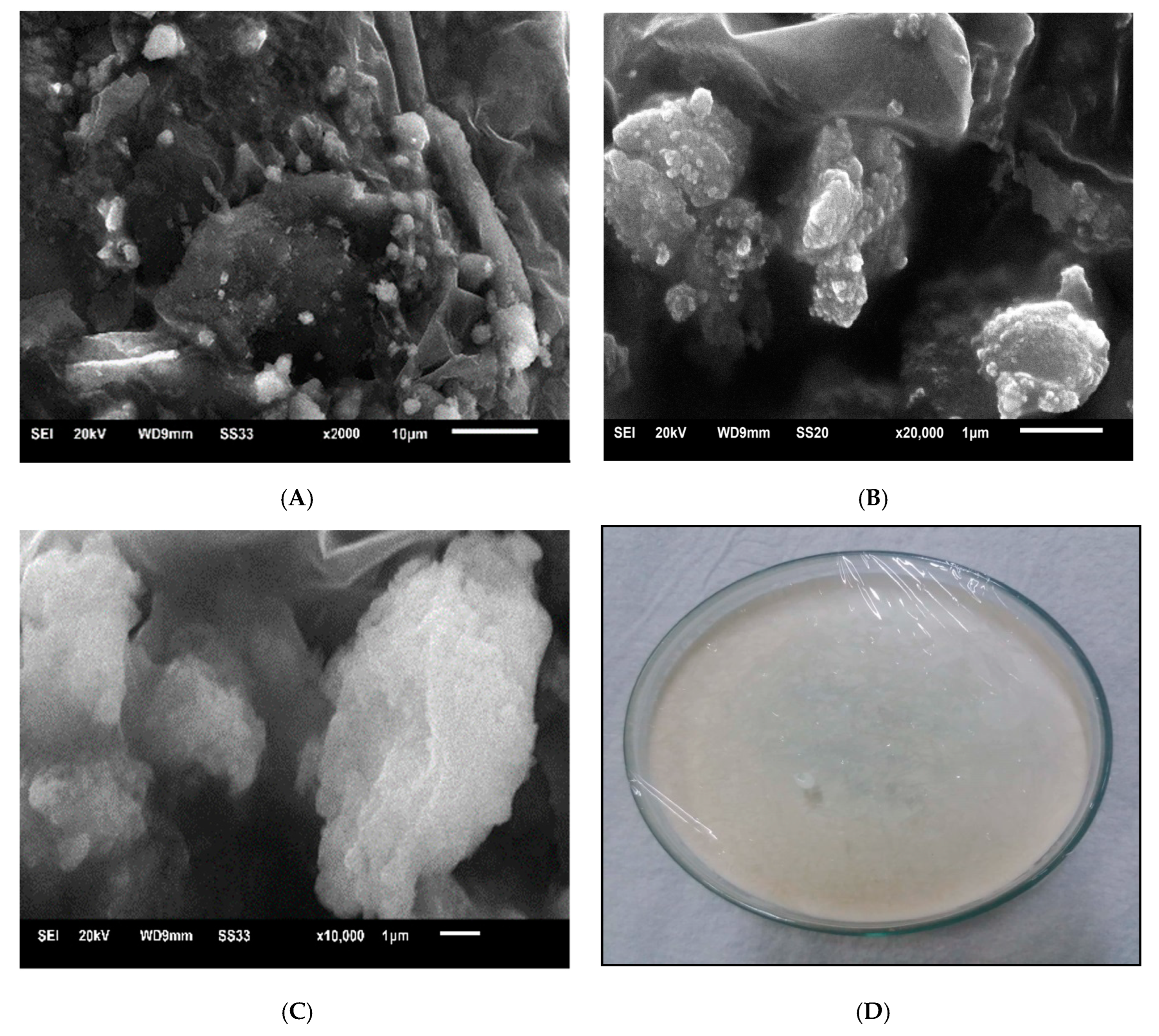
Figure 1. Scanning electron micrograph of mangaba extracts, where: (A): SL-MAPE 1.0 (mg/mL); (B): SL-MAPE 1.5 (mg/mL); (C): SL-MAPE 2.0 (mg/mL). (D): Freeze-dried sample, Liposome-SL-MAPE 2.0 (mg/mL).
Engproc 67 00022 g001
Table 2. Physical characterization of liposomal nanovesicles containing mangaba aqueous extract prepared with soy lecithin (SL-MAPE).
Physical CharacterizationConcentration (mg/mL)
1.01.52.0
Particle diameter (nm)197.43 ± 0.81 c318.2 ± 1.55 a238.33 ± 0.89 b
Polydispersion0.28 ± 0.03 c0.49 ± 0.01 a0.47 ± 0.02 a
Zeta Potential−37.00 ± 0.17 a−33.7 ± 1.48 b−35.7 ± 0.87 b
Encapsulation Efficiency (%)80.14 ± 0.07 c86.18 ± 0.78 b88.09 ± 0.45 a
Note: Values obtained: mean ± standard deviation. For different letters on the same line, there was statistical variation by Tukey’s test a (p < 0.05).
Disclaimer/Publisher’s Note: The statements, opinions and data contained in all publications are solely those of the individual author(s) and contributor(s) and not of MDPI and/or the editor(s). MDPI and/or the editor(s) disclaim responsibility for any injury to people or property resulting from any ideas, methods, instructions or products referred to in the content.