Review

Ferroptosis in Cancer Immunotherapy—Implications for Hepatocellular Carcinoma

Department of Hepatology and Gastroenterology, Charité Universitätsmedizin Berlin, Augustenburger Platz 1, 13353 Berlin, Germany
*
Author to whom correspondence should be addressed.
Immuno 2022, 2(1), 185-217; https://doi.org/10.3390/immuno2010014
Submission received: 30 December 2021 / Revised: 8 February 2022 / Accepted: 11 February 2022 / Published: 15 February 2022
(This article belongs to the Section Cancer Immunology and Immunotherapy)

Abstract

:
Ferroptosis is a recently recognized iron-dependent form of non-apoptotic regulated cell death (RCD) characterized by lipid peroxide accumulation to lethal levels. Cancer cells, which show an increased iron dependency to enable rapid growth, seem vulnerable to ferroptosis. There is also increasing evidence that ferroptosis might be immunogenic and therefore could synergize with immunotherapies. Hepatocellular carcinoma (HCC) is the most common primary liver tumor with a low survival rate due to frequent recurrence and limited efficacy of conventional chemotherapies, illustrating the urgent need for novel drug approaches or combinatorial strategies. Immunotherapy is a new treatment approach for advanced HCC patients. In this setting, ferroptosis inducers may have substantial clinical potential. However, there are still many questions to answer before the mystery of ferroptosis is fully unveiled. This review discusses the existing studies and our current understanding regarding the molecular mechanisms of ferroptosis with the goal of enhancing response to immunotherapy of liver cancer. In addition, challenges and opportunities in clinical applications of potential candidates for ferroptosis-driven therapeutic strategies will be summarized. Unraveling the role of ferroptosis in the immune response could benefit the development of promising anti-cancer therapies that overcome drug resistance and prevent tumor metastasis.

1. Introduction

Cell death is crucial for normal development and homeostasis throughout an organism’s lifetime [1]. Various forms of cell death have been identified, each having its respective modalities and features. Ferroptosis is one of the more recently described forms of non-apoptotic cell death. The term ferroptosis was first mentioned in 2012 by Dixon as a newly identified and unique iron-dependent cell death [2]. Ferroptotic cells display cytological changes that differ from morphological, biochemical, and genetic characteristics of other cell death forms, including decreased mitochondria cristae and ruptured mitochondrial membranes [3,4,5]. Compared to apoptosis, ferroptosis lacks rupture or blebbing of the plasma membrane, chromatin condensation, or rounding of the cell, and the execution of ferroptosis is not known to require specific pro-death protein expression [6]. As implied in its name, ferroptotic cell death is defined by the requirement of iron. Caused by excessive peroxidation of membrane phospholipids and production of reactive oxygen species (ROS), the plasma membrane loses its selective permeability, ultimately resulting in cell death. Regulated by multiple layers of metabolic signaling pathways including iron metabolism, lipid metabolism, and mitochondrial function, ferroptotic cell death is a complex process that will be further discussed in more detail. There is recent evidence suggesting that ferroptotic cell death plays an important role in mediating a wide variety of cellular processes in diseases, including immune activation. Whether ferroptotic cancer cells are immunogenic is currently unclear, but targeting ferroptosis in immunotherapeutic approaches is considered as a promising strategy.
Primary liver cancer is one of the world’s most common causes of cancer-related death and represents a serious health and economic burden [7,8]. Hepatocellular carcinoma (HCC) is the most common primary liver tumor, comprising around 75% of all liver cancer cases and typically developing in the context of liver fibrosis or cirrhosis [9]. In patients with advanced cirrhosis and HCC, liver transplant often remains the only curative treatment option [10]. Classic chemotherapies such as cisplatin are not regularly used in HCC due to the rapid development of chemoresistance [11], toxicity to various organs, and limited efficacy [12]. The first effective systemic therapy in advanced HCC was achieved using multi-kinase inhibitors, such as sorafenib, lenvatinib, carbozantinib, or regorafenib, but all of these substances provide only a rather small improvement of overall patient survival [13,14,15]. Treatment options are usually limited, as surgical resection or organ transplantation is feasible only in a fraction of patients and systemic therapies are not curative [16].
Antitumor immunotherapy has emerged as standard therapy for cancer treatment for many tumors (including HCC), and great progress has been achieved in the past few decades. The most successful immunotherapy to date is the modulation of immune checkpoint pathways, e.g., by targeting the T-cell inhibitory molecules programmed cell death-1 protein (PD-1) and programmed death-ligand 1 (PDL1) or cytotoxic T-lymphocyte-associated protein 4 (CTLA-4) that contribute to the tumor’s escape from cytotoxic T-cell responses [17,18]. Recently, a phase III trial reported that combination of atezolizumab (anti-PD-L1 antibody) and bevacizumab (anti-vascular endothelial growth factor receptor (VEGFR)) resulted in increased overall survival of patients with advanced HCC in comparison to sorafenib, making atezolizumab/bevacizumab the new first-line treatment for unresectable HCC [19]. Two other PD-1 inhibitors (nivolumab and pembrolizumab) have already been approved as a second-line monotherapy in advanced HCC in the USA, but the response rates are still low (15–20%) [20,21]. It has become increasingly clear that the efficacy of cancer immunotherapy depends on several factors including the potential to induce an anti-tumor immune response to overcome resistance. Immunogenic cell death (ICD) is seen as a promising concept in achieving strong and long-lasting anti-cancer immunity by not only killing malignant cells but also activating the immune system [22], which has the potential to synergize with immune checkpoint blockade. Despite ongoing research, less is known about regulators of ferroptosis and its role in immune activation, but there is some evidence that ferroptotic cell death might be immunogenic. These considerations support the concept to develop ferroptosis-based approaches to enhance response to immunotherapy.
Here, we review existing studies and our current understanding regarding the molecular mechanisms of ferroptosis in cancer and immune cells, which may open the door to a new era of cancer immunotherapies targeting non-apoptotic RCD processes in primary liver cancer to improve its poor prognosis.

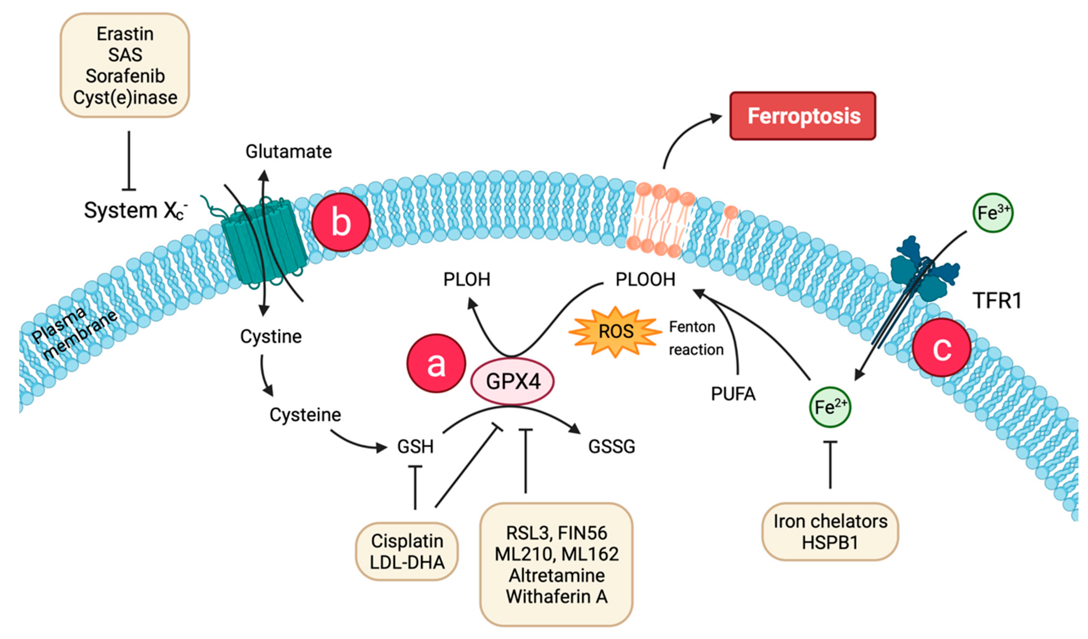
2. Molecular Basis of Ferroptosis

Dixon et al. [2] first proposed ferroptosis in 2012 as a recently recognized non-apoptotic form of cell death. The knowledge about various cell death modalities and especially our understanding of ferroptosis is incomplete. Further research is necessary in order to understand why certain stimuli trigger ferroptosis instead of apoptosis. However, it is clear that ferroptosis describes an oxidative, iron-dependent process characterized by massive lipid peroxidation-mediated membrane damage and accumulation of ROS to toxic levels [1,5]. Its morphology differs from other known cell death types, such as apoptosis, necrosis, autophagy, necroptosis, or pyroptosis, as ferroptotic cells have characteristic mitochondrial atrophy accompanied by a reduction or disappearance of mitochondrial cristae [4,23]. Previous studies concur that ferroptosis mostly resembles necrosis-like morphological changes but depends mainly on iron signaling [24]. The two main biochemical characteristics are lipid peroxidation and iron accumulation, which will be detailed separately in the following sections.

2.1. Lipid Peroxidation and Antioxidant Defense

The peroxidation of polyunsaturated fatty acids (PUFAs) in phospholipids and their incorporation into the cell membrane are the main characteristics of ferroptosis (Figure 1) [25]. Catalyzed by acyl-CoA synthetase long-chain family member 4 (ACSL4), PUFAs are incorporated into phospholipids to form polyunsaturated fatty acid-containing phospholipids (PUFA-PLs). Free PUFAs become a radical target after incorporation into the plasma membrane by ACSL4. Plasma membrane PUFAs in turn are vulnerable to free radical-initiated oxidation and trigger additionally self-amplified Fenton reaction, leading to destruction of the plasma membrane lipid bilayer and affecting membrane function [26]. The exact details remain unclear, but the peroxidation of phospholipid PUFAs is likely catalyzed by lipoxygenase (LOX) family enzymes [27]. There is recent evidence that the amount of PUFAs at the plasma membrane has an influence on the response to ferroptosis: n-3, but also, remarkably, n-6-long chain PUFAs showed cytotoxic effects proportional to the number of double bonds [28]. However, the antioxidant enzyme GPX4 is able to directly reduce phospholipid hydroperoxide to hydroxyphospholipid, which hinders the production of intracellular lipid hydroxyl radicals (Figure 1a). Glutathione (GSH) is considered as an essential cofactor for GPX4, as it can donate reducing equivalents for GPX4 opposing conversion of lipid hydroperoxides (L-OOH) to non-toxic lipid alcohols (L-OH) [29]. GSH synthesis is dependent upon cystine import into the cells by the cysteine/glutamate transporter (also known as system xc, Figure 1b) which consists of two subunits, solute carrier family 7 member 11 (SLC7A11) and solute carrier family 3 member 2 (SLC3A2) [30]. The imported cystine can be oxidized to cysteine, which is then used to synthesize GSH required for GPX4 activity as a reducing cofactor. The GSH-GPX4 antioxidant system has an important role in protecting cells from ferroptosis as its activity determines the extent of ferroptosis by adjusting ROS accumulation and oxidative stress [31]. Pharmacological inhibition of the upstream regulator system xc (see Section 3.1) or the downstream effector GPX4 (see Section 3.2) of GSH induces ferroptosis. The same can be achieved through depletion of system xc (encoded by the SLC7A11 gene) or GPX4 genetic depletion [32].

2.2. Iron Accumulation

As an essential trace element, iron is necessary to maintain cell metabolism [33]. Extracellular iron is internalized into the cell bound to transferrin (TF) as a TF-Fe3+ complex (Figure 1c). Taken up by the transferrin receptor 1 (TFR1) at the plasma membrane through clathrin-mediated endocytosis, it is released into the cytosol as free Fe2+ [34]. During ferroptosis, Fe2+ participates in Fenton chemistry to generate hydroxyl radicals und initiates lipid peroxidation, thus leading to propagation of damage throughout the membrane as already discussed [31].
Therefore, elevated levels of iron and iron derivates (heme or iron-sulfur clusters) can increase the vulnerability to ferroptosis, which plays an important role in tumor cells due to their extraordinary growth. Because of this increased susceptibility of cancer cells, ferroptosis might be a promising tool for anti-cancer treatment. Moreover, several proteins and genes that are involved in iron metabolism have been shown to modulate sensitivity to ferroptosis by changing cellular iron contents [35]. This emphasizes the association of cell death pathways with cell metabolism. Iron dependency also becomes apparent through blocking of TF-mediated iron import or recycling of iron-containing storage proteins, which leads to attenuation of ferroptosis [36]. Previous findings revealed involvement of other cell death types, contributing to ferroptosis and cell death fate: autophagic cell death requires the iron-carrier TF and the amino acid glutamine to trigger death [37]. Gao et al. demonstrated that ferroptosis is a form of autophagic cell death known as ferrotinophagy when autophagy is activated upon ferroptotic cell death induction. Autophagy is activated to degrade cellular ferritin and thereby increases iron levels and sensitizes to ferroptosis. The concentration of free ferrous ions in the cytoplasm determines whether it acts as a beneficial cofactor or as a toxic-free radical catalyst in the cell [38]. Growing evidence suggests that the autophagic machinery, at least under certain conditions and connected to human diseases, contributes to ferroptotic cell death and plays a dual role in tumorigenesis and, with it, cancer therapy [39].
Non-canonical ferroptosis induction by increasing intracellular Fe2+ refers to increasing iron intake through TFR1 expression or decreased expression of iron transporter, or excessive activation of heme oxygenase 1 (HMOX1) [40]. Heme metabolism influences iron contents, as it is a major source of dietary iron in mammals, derived primarily from hemoglobin and myoglobin. It plays an essential role in the liver, which is the principle organ that responds to changes in systemic iron signals in order to maintain body iron homeostasis [41]. As a central metabolic and signaling molecule, it is involved in diverse transcription and cell signaling processes, e.g., heme inhibits the proteasome, and binding to immunoglobulins [42,43]. HMOX1 catalyzes the catabolism of heme, which raises intracellular ferrous iron in the cytosol and is, therefore, crucial for maintaining cellular redox homeostasis. HMOX1 knockout mice showed iron accumulation in liver and kidney, and HMOX1 deficiency in humans is connected to several abnormalities, indicating an important role of HMOX1 in human health [44].

2.3. Regulatory Pathways

As described previously, ferroptosis is a regulated form of cell death driven by accumulation of membrane lipid peroxides from iron overload. Various genetic changes were considered important to contribute to this dynamic type of cell death. The major drivers of ferroptosis “decide” over life or death in response to ferroptotic stimuli. A predictive biomarker for monitoring ferroptosis appears to be the enzyme ACSL4 that is involved in fatty acid metabolism, contributing to accumulation of lipid intermediates (see Section 2.1.) [45]. Upregulation of ACSL4 enhances PUFAs content in phospholipids, which are susceptible to oxidation reactions. Expression levels of ACSL4 correlate with ferroptosis sensitivity—this was demonstrated by suppression of ACSL4 expression by RNA interference (RNAi) in HepG2 and HL60 cells, leading to an increase of ferroptosis resistance, while its overexpression restores ferroptosis sensitization [46]. As with factors involved in fatty acid metabolism, ferroptosis can also be regulated via signal transduction pathways of iron metabolism. Decreasing iron utilization may increase the sensitivity to ferroptosis whereas intracellular iron pools seem to promote ferroptosis [47]. Various genes and proteins related to iron metabolisms are generally upregulated during ferroptosis, including the iron responsive element binding protein 2 (IREB2). IREB2 is considered the core regulator of iron metabolism binding iron-responsive elements in the mRNA of target genes including the TF receptors and knockdown results in resistance to ferroptosis induction [2]. Additionally, overexpression of iron-storage proteins such as ferritin are supposed to play a wide anti-ferroptotic role [48].
GPX4 is the major lipid hydroperoxide (LOOH)-neutralizing enzyme, which in turn represents a target for several regulating proteins. Erastin and Ras-selective lethal small molecule 3 (RSL3) both inactivate GPX4 and are proposed as efficient ferroptosis inducers. Erastin does so indirectly by inhibiting cystine import, which then lacks as an essential cellular building block of GSH. Erastin is also a potential activator of mitochondrial voltage-dependent anion channel 2/3 (VDAC2/3), highlighting the participation of mitochondria dysfunction in erastin-induced ferroptosis [49]. RSL3 directly binds the active site of GPX4 and inhibits the phospholipid peroxidase activity [50].
Research on the mechanism of ferroptosis also required the identification of ferroptosis inhibitors in order to study the role of ferroptosis in vivo and discuss potential pharmacological approaches aiming to subvert lipid peroxidation. Ferrostatin-1 (Fer-1), liproxstatin-1, vitamin E, and zileuton have been identified and suppress ferroptosis as lipoxygenase inhibitors, preventing the propagation of oxidative damage within the membrane [51,52]. Other than that, iron chelators such as deferoxamine inhibit the accumulation of iron [2]. A full understanding of the regulatory network including ferroptosis inhibitors might provide potential health benefits to prevent symptoms of diseases that are related to ferroptosis [53].
In summary, many potent biomarker candidates have already been identified, which could be useful to further study the fundamental basis of lipid metabolism in ferroptosis and to define the interplay of involved regulators. However, further functional studies are required to unravel the critical role of certain biomarkers in order to find applications in ferroptosis-based cancer therapy.

2.4. Immunogenic Features of Ferroptosis (in Cancer Cells)

There is rising evidence supporting the notion that ferroptosis can play a significant role in mediating various functions during immune responses and immunotherapies. For early ferroptotic cancer cells, it has been proven that they release adenosine triphosphate (ATP) and high mobility group box 1 (HMGB1), which can be recognized as damage-associated molecular patterns (DAMPs) by certain immune cells, triggering an inflammatory response [54]. At present, the most effective systemic therapy in advanced HCC is the combinational approach of atezolizumab and bevacizumab [19]. The response rate is largely dependent on the immune status of the tumor: inflamed (“hot”) tumors with high infiltration of tumor-reactive T cells respond significantly better to checkpoint inhibitors than “cold” or “excluded” tumors that are poorly infiltrated by immune cells including antigen-presenting cells (APCs) [55,56]. Immune checkpoint inhibitors (such as PD-1 or CTLA4) mainly act by activating effective anti-cancer immune responses driven by cytotoxic T cells. Ferroptosis in cancer cells can be induced by CD8+ T cells that release INF-γ, which leads to system xc downregulation and consequently lipid peroxidation [57].
Compared to other forms of cell death, ferroptosis is considered an immune-activating process. Although further research is still needed to clarify the interaction between ferroptotic cell death and the immune system, there is some preliminary evidence showing that ferroptosis plays a direct role for the immune system. This is not surprising since iron metabolism and lipid peroxidation were linked to regulation of immune functions, even before ferroptosis was discovered. Further determination of specific immune signals associated with ferroptosis would be useful in order to enhance the immune effect in tumor immunotherapy.
Typically for RCD, DAMPs are released as immune modulators, stimulating immune cells (e.g., macrophages or monocytes) (Figure 2) [58]. These are endogenous molecules that bind to pattern recognition receptors (PRRs), thus activating the production of immune stimulants, including a variety of cytokines and chemokines. A typical DAMP, which is released by ferroptotic cells in an autophagy-dependent manner, is the nuclear transcription factor HMGB1 able to bind chromosomal DNA involved in DNA recombination and repair processes [59]. It was shown to be actively secreted by innate immune cells in response to microbial infections but can also be passively released by somatic cells undergoing cytoplasmic membrane destruction during cell death [60]. HMGB1 acetylation induced by histone deacetylase inhibition is the trigger for HMGB1 release during ferroptosis [61]. Moreover, another study shows that HMGB1 is an essential factor for cancer cell immunity and plays paradoxical roles in tumor development by promoting both cell survival and death [62]. For a more complete investigation of the full immunogenic potential of this cell death pathway, it is important to discover the full repertoire of DAMPs released during ferroptosis. While the role of HMGB1 and ATP release has already been clarified in apoptosis and necroptosis [63], its involvement in ferroptotic cell death processes has remained elusive until now. Recent research highlights the involvement in the regulation of TFR mRNA levels and p38 phosphorylation in the rat sarcoma (RAS)-JNK/p38 pathway [64]. Additionally, Ye et al. found that knockdown of HMGB1 reduces erastin-mediated ferroptosis and the connected ROS generation [64]. Hence, HMGB1 is a critical regulator of ferroptosis.
Furthermore, other immune signals can be intermediate or final products of lipid peroxidation such as 4-hydroxynonenal (4-HNE) or malondialdehyde (MDA), driving inflammation by activating macrophages to produce pro-inflammatory cytokines [65]. They can trigger inflammation partly through activating TLR4 signaling [66]. However, thus far, there is no direct evidence that these toxic aldehydes are alone sufficient to induce ferroptosis. Taken together, despite the mechanisms reviewed here, current evidence is insufficient for a complete understanding of the relation between ferroptosis and the immune system. A more comprehensive understanding of DAMP release mechanisms and their regulation could enable development of potential new drugs for therapeutic interventions in cancer.

2.5. Immune Response to DAMPs Released by Ferroptotic Cells

Until now, two ways for ferroptotic cells to influence inflammatory responses in immune cells have been characterized. On the one hand, the release of DAMPs by non-immune ferroptotic cells attract APCs and other immune cells. Secretion of DAMPs attracts and stimulates dendritic cells (DCs) for tumor-associated antigen (TAA) uptake and processing followed by MHC class I-restricted cross-presentation of these TAAs, which leads to the priming of T cells and clonal expansion of cancer-specific cytotoxic T lymphocytes (Figure 2) [67]. For example, HMGB1 is able to trigger an inflammatory response in peripheral macrophages via activation of the NF-κB pathway [59]. Functioning as an adjuvant, HMGB1 contributes to activation of the innate and adaptive immune systems. Targeting HMGB1 release using neutralizing anti-HMGB1 antibodies limits inflammatory response in macrophages during ferroptosis [59]. On the other hand, ferroptosis in immune cells themselves might compromise immune responses. It has been shown that non-apoptotic cell death, including ferroptosis, is more likely to cause inflammation than apoptosis but little is known about the recognition and clearance mechanisms for cells dying through non-apoptotic cell death pathways. Phagocytic clearance of dead cells plays a vital role in immune homeostasis, especially during cancer therapy, and the exact mechanistic of ferroptotic cell engulfment remains unclear. However, Luo et al. have demonstrated that different types of macrophages readily engulfed ferroptotic cells through a mechanism different from the engulfment of apoptotic cells. Ferroptotic cells can produce oxygenated phosphatidylethanolamines on the outer plasma membrane that can be recognized by TLR2 on macrophages, leading to clearance of ferroptotic cells [68]. Moreover, macrophages can be classified as pro-inflammatory or anti-inflammatory pro-carcinogenic macrophages, both functioning in eliminating pathogens in order to maintain immune homeostasis. Environmental signals decide on pro- and anti-inflammatory polarization, whereas an imbalance contributes to various diseases and tumor development [69], and previous studies indicated a relationship between macrophage polarization and ferroptosis. Dai et al. showed that ferroptotic cancer cells cause autophagy-dependent protein release, triggering pro-carcinogenic macrophage polarization via activation of several signaling pathways, which could restrict anti-tumor immunity [70]. Moreover, macrophage subgroups differ concerning their sensitivities to ferroptosis induction, with pro-inflammatory ones being more susceptible [71]. Regarding the adaptive immune system, T cells can also induce ferroptosis in tumor cells. It has been reported that CD8+ T cells release cytokines (e.g., TNF or IFN-γ), which significantly downregulate expression of system xc components driving tumor cells into ferroptosis due to reduced cysteine uptake [57]. This might provide a direction for further bridging the gap between ferroptosis and immunotherapy in the treatment of malignant cancers.
Despite system xc, the other key regulator of ferroptosis, GPX4, was also reported to play an essential role in the expansion of both antigen-specific CD4+ and CD8+ T cells [72]. A lack of GPX4 leads to ferroptotic cell death induction with an accumulation of lipid peroxides in T cells, thereby preventing their immune response to infection. Moreover, it was recently found that ferroptosis-resistant CD8+ T cells overexpressing GPX4 have normal immune functions, but deletion of ACSL4 impairs their immune response [73]. GPX4 also plays a pivotal role in preventing ferroptosis after T-cell activation by only expanding GPX4-expressing CD4+ and CD8+ T cells [72]. Additionally, the induction of ferroptosis in cancer cells has been shown to be associated with increased release of prostaglandin E2 (PGE2), an immunosuppressive agent directly suppressing cytotoxic T-cell activity (Figure 2 (7)). Therefore, PGE2 promotes the proliferation of tumor cells and plays a central role in impairing anti-tumor immunity, which could be a barrier to the induction of a potent immune response [74]. Thus, understanding the molecular mechanisms underlying the immune response of ferroptotic cell death would help guide the design of new immune-related treatments.
To do so, a more detailed investigation of the TME including activated immune cells that secrete proinflammatory chemokines and cytokines is necessary. Recent studies confirmed the role of ferroptotic cancer cells regulating tumor immunity in different ways. As other cell death types, ferroptotic cells release “find me” signals that attract APCs and other immune cells, leading to the engulfment by macrophages [75]. During maturation and cross-presentation by DCs, ferroptotic cancer cells interact further with the immune system through TLR4 [76].
In a very recent integrative study, a ferroptosis scoring model based on ferroptosis-related genes and their association with immune infiltration in cancer was constructed [77]. For this purpose, 30 genes were classified into ferroptosis driver and suppressor groups, and transcription factors and therapeutic agents targeting ferroptosis regulators in cancer were determined and validated. This score can be a potential biomarker of the immune response in the TME and the therapeutic response to immunotherapy. The described dual effects of ferroptosis on tumor immunity, and with it, patient’s prognosis score, differ from person to person, but the results showed that the ferroptosis score, as an independent prognostic factor, is feasible for predicting the prognosis of different cancers and immunotherapy efficacy. Supporting previously obtained results, high ferroptosis scores had higher immune cell infiltration scores due to CD8+ T cell and T-helper cell activity [57]. This highlights the importance of the TME that should be considered in the design of therapeutic compounds and confirms the powerful combinatorial use of T cell-mediated ferroptosis induction with checkpoint blockade [78]. Despite some limitations of this study (more datasets for verification and combinatorial approaches would be needed), this ferroptosis score might have potential to help improve the survival rate of patients with HCC and predict tumor progression earlier as prognostic markers that currently find application in clinic.

3. Ferroptosis in Cancer Therapy

Ferroptosis has not only been investigated to expand the basic understanding regarding cell death modalities, but its connection to cancer and cancer treatment also exhibits extraordinary potential in tumor treatment [25]. Since iron is important for the regulation of redox homeostasis in normal cells, understanding the role of iron in cancer development is an active area of research. Moreover, there is epidemiological evidence that high intake of dietary iron increases the risk of several cancers including HCC [79]. Higher demand for iron and upregulation of TFR1 make cancer cells more susceptible to iron-dependent cell death modalities such as ferroptosis [71]. By changing cellular iron content, the sensitivity of cells towards ferroptosis can be altered in various processes, e.g., degradation of the iron storage protein ferritin or transferrin regulation [80], resulting in iron overload and inducing ferroptosis in cancer cells. Iron reduction therapy could also be promising; agents (e.g., deferoxamine) that chelate iron by forming a stable complex hindering its further chemical reaction [41] exert antiproliferative effects and antitumor activities in several cancers, including HCC, by depriving cancer cells of essential iron [81]. Prevention of ferroptosis can be, in this case, also applicable for treatment of iron-related disorders [82].
In leukemia cells, ferroptosis induced by iron loading exerts a therapeutic effect [82]. However, excessive iron was identified to be related to poor prognosis of leukemia due to reduced blood cell differentiation, which is why efforts to reduce the amount of iron are still preferable in this context [38]. In addition, systemic administration of iron modulating agents goes along with severe side effects because of their ubiquitous role [25]. A sufficient iron supply is required for the activity of many iron-containing or dependent enzymes involved in cell division, as well as for T-cell growth and expansion [83].
HMOX1 expression correlates with cancer progression, as well as poor prognosis by demonstrating that heme iron dysregulation actively promotes tumor development [84,85]. Li et al. showed in 2017 that excessive heme could directly induce neuronal ferroptosis [86]. However, as for other factors that control iron metabolism, the exact role of HMOX1 in ferroptosis is unclear and has a dual role depending on the context [42]. Several studies showed that heme sequestration could be an effective strategy to manage and treat an array of pathologic conditions, including cancer, by making heme unavailable for tumor cells [87,88]. Heme reduction in the tumor environment by heme-sequestering proteins affects oxygen levels and blood vessel growth limiting tumor hypoxia and normalizing tumor vasculature [89]. Although Sohoni et al. emphasize that it would not be a stand-alone treatment, their findings suggest a potential new path forward in treating cancer cells with heme-sequestering proteins in tandem with chemotherapy or other forms of cancer treatments.
Additionally, INF-γ was found to be involved in regulating tumor cell proliferation, and recently its role in ferroptosis was revealed. It downregulates system xc protein levels via JAK/STAT signaling, which leads to enhanced glutathione depletion and increased lipid peroxidation in HCC cells. This identification provided new insights into the feasibility of using INF-γ in cancer treatment [90].
Due to its multifaceted role, ferroptosis inhibitors should be taken into account such as lipophilic antioxidants that prevent lipid peroxidation [32]. To the best of our knowledge, no clinical trials regarding ferroptosis inhibitors in liver diseases have been performed until now. However, in preclinical studies using mouse models of liver fibrosis or non-alcoholic steatohepatitis (NASH), the use of the lipophilic antioxidants Fer-1 and Lip-1 prevents disease progression [91]. However, unsatisfactory pharmacokinetics preclude their clinical application [92]. Potent Fer-1 analogues were synthesized by introducing structural modifications to the Fer-1 scaffold to improve solubility and stability and showed in vivo efficiency [93].

3.1. System xc Regulators

More suitable for therapeutic induction of ferroptosis in human cancers is targeting the major mediators with the help of inducers that can either act directly on GPX4 or indirectly by inhibiting system xc (Figure 3, Table 1). Erastin and RSL3 were proven to trigger ferroptosis in highly proliferating cancer cell lines including head and neck, gastric, melanoma, and lung cancer [94]. Poor water solubility, renal toxicity, and unstable metabolism limit erastin’s application in vivo [95], but this issue can be overcome by using drug delivery vehicles, such as exosomes, with low immunogenicity, high biocompatibility, and high efficiency [96]. Moreover, scientists have developed erastin derivatives, including piperazine erastin (PE) and imidazole ketone erastin (IKE), showing better water solubility and stability in experimental models of fibrosarcoma and diffuse large B-cell lymphoma [97].
Ferroptosis inducers may also be useful in combination with common therapeutic agents. Sato et al. suggest a synergizing effect of pre-treatment of short exposure to erastin followed by cisplatin, which efficiently induces cell death in a variety of cancer cell lines [150]. Cisplatin was originally considered the most effective and widely used anti-cancer agent for the treatment of solid tumors [151]. However, cisplatin was also proven to be a ferroptosis inducer via GSH and GPX4 inactivation in different cell lines, and its synergistic effect with erastin on their anti-tumor activity opened up a new way for the utility of classic drugs [152]. The combination of erastin and radiation was shown to induce a significant inhibition of tumor growth in a cell model of lung tumor and in mouse model of sarcoma. Other studies confirmed a potentiating effect in models of glioma, melanoma, and breast cancer [153,154]. Another study confirmed a combined effect of erastin with irradiation in non-small cell lung cancer (NSCLC) by inducing ferroptosis [155]. Radiotherapy is still one of the most widely used cancer therapies, but radioresistance remains a major factor leading to the failure of radiotherapy [156]. Several studies revealed that ionizing radiation can enhance the anti-tumor effect by synergizing with ferroptosis triggers and immunotherapy by increasing the expression of ACSL4 and GPX4 [78,157].
An additional potent inhibitor of the xc transporter, sulfasalazine (SAS), may induce eradication of chemotherapy/radiotherapy-resistant cancer cells by potentiating the cytotoxicity of cisplatin in some cancer types [158]. Nevertheless, it was shown that erastin is a substantially more potent inhibitor of system xc function than SAS.
Sorafenib was the first multi-kinase inhibitor approved for HCC treatment, and there is evidence that sorafenib might induce ferroptosis in addition to its kinase inhibitory functions [159]. As for erastin and SAS, there have been approaches combining ferroptosis induction using sorafenib nanoparticles with the common anti-cancer drugs cisplatin or lactoferrin [160]. However, sorafenib’s exact lethal mechanism of action in cells has not been clear until now, and there is disagreement about it. It may not involve a direct kinase inhibition and/or binding to an alternative target such as an unknown kinase whose activity is necessary for system xc [161]. Besides inhibiting tumor proliferation through the RAF/MEK/ERK signaling pathway, sorafenib can block tumor angiogenesis via VEGFR and platelet-derived growth factor receptor (PDGFR) [162]. Sorafenib-induced ferroptotic anticancer activity can be enhanced by blocking another critical regulator named metallothionein (MT)-1G that also plays an antioxidant role [163]. Upregulation of MT-1G was observed in sorafenib-resistant cancer cells and was shown to limit sorafenib action. It could be one regulator contributing to acquired resistance against sorafenib in human HCC cells, which is one reason for poor prognosis [164]. Thus, MT-1G pathway inhibition enhances the ferroptosis-inducing activity of sorafenib. Similarly, expression levels of the retinoblastoma (RB) protein that acts as a negative regulator of cell proliferation impacts the response of HCC cells to sorafenib and the regulation of ferroptosis [165]. Liver cancer cells with reduced RB levels show a 2~3 times higher cell mortality upon exposure to sorafenib than control cells with normal RB levels [166]. Therefore, evaluating the RB status before treatment application could be used to judge the drug resistance to sorafenib in HCC patients.
A combinatorial strategy using sorafenib together with the immune system modulator YIV-906 (PHY906, KD018) is currently under investigation in a phase II clinical study (NCT04000737). Preclinical research suggests that YIV-906 could act as an enhancer of the anti-tumor activity of sorafenib [167]. Combination therapy of sorafenib and the GPX4 inhibitor RSL3 may be a promising strategy in HCC treatment as studies confirmed synergic therapeutic effect on HCC progression in mice [168]. Another recent study aimed to verify the effect of recombinant human adenovirus type 5 (H101), which is an oncolytic adenovirus that can selectively replicate in tumor cells (NCT05113290). H101 showed anti-tumor effects on liver cancer and a synergistic effect with sorafenib on hepatoma cells in vitro [169]. The ongoing clinical study intends to verify this promising evidence to support clinical medication for advanced HCC patients.
An alternative strategy focuses on decreasing intracellular cysteine levels in cancer cells by inhibiting the import of cystine via system xc. Depletion of extracellular cysteine and cystine can be achieved by treatment with cyst(e)inases and was shown to increase ROS and reduce GSH levels [170]. They may represent an effective therapeutic modality as cysteine depletion and the following inactivated antioxidant cellular response successfully suppresses tumor growth [171,172].

3.2. GPX4 Regulators

Independent of system xc, several inhibitors target the other major ferroptosis mediator GPX4 or the metabolic pathways crucial for its action (Figure 3, Table 1). RSL3 ((1S,3R)-methyl 2-(2-chloroacetyl)-2,3,4,9-tetrahydro-1-[4-(methoxycarbonyl)phenyl]-1H
-pyrido [3,4-b]indole-3-carboxylate) was first identified for inducing iron-dependent cell death by directly targeting selenocysteine residue of GPX4, thereby directly inhibiting PL-peroxidase activity [27,50]. As for erastin, the poor water solubility and metabolic instability of RSL3 precludes its systemic in vivo use yet it has to be further optimized for clinical application [144,173]. In contrast, systemic toxicity and pharmacodynamic analysis by Yang at al. [52] observed RSL3 to be well tolerated in animal studies. In mouse models, RSL3 was shown to block the activity of GPX4, leading to ferroptosis induction and thus inhibition of fibrosarcoma growth.
Additional GPX4 inhibitory agents are ML162 (α-[(2-chloroacetyl) (3-chloro-4-methoxyphenyl)amino]-N-(2-phenylethyl)-2-thiophene-acetamide; DPI7: diphenylene iodonium) and ML210 ([4-[bis(4-chlorophenyl)methyl]-1-piperazinyl](5-methyl-4-nitro-3-isoxazolyl)-methanone), both exhibiting similar ferroptosis-inducing activity as RSL3 [25].
A new and specific inducer of ferroptosis, N2,N7-dicyclohexyl-9-(hydroxyimino)-
9H-fluorene-2,7-disulfonamide (FIN56), was discovered by Shimada et al. [125] from a systematic survey. Treatment of cells with FIN56 resulted in degradation of GPX4 protein and lipid ROS generation afterwards [174]. Another GPX4 inhibitor called altretamine (2,4,6-tris (dimethylamino)-1,3,5-triazine) was identified and experimentally confirmed to share the same mechanism of action as SAS as an inhibitor of GPX4 lipid repair activity [149]. However, the precise mechanism remains elusive. Pharmacokinetic and toxicity analysis revealed a great inter- and intrapatient variability regarding bioavailability of oral altretamine [175]. Altretamine has been applied in ovarian cancer, including a phase II study showing ferroptosis induction, and was generally well tolerated and associated with prolonged progression-free and overall survival [176,177].
Several studies that focus on identifying ferroptosis resistance genes revealed additional potent ferroptosis-regulating proteins and agents: the flavoprotein apoptosis-inducing factor mitochondria-associated 2 (AIF2), renamed as ferroptosis suppressor protein 1 (FSP1), is a potent ferroptosis suppressor by preventing lipid peroxidation through an ubiquinone (also known as coenzyme Q10, CoQ10)-mediated mechanism that is distinct from glutathione-dependent protective pathways [178,179].

3.3. Targeting SLC7A11

Attention was shifted to another factor that can influence the effect of ferroptosis: the most frequently mutated gene in human cancer, the TP53 tumor suppressor gene, might also play a role in ferroptosis and especially ferroptosis in tumor suppression [180]. P53 is involved in various cellular processes, including mediating cell cycle arrest, apoptosis, senescence, or differentiation [181]. Recent published studies confirmed the ability of p53 to suppress tumor development by making cells more sensitive to iron by inhibiting SLC7A11 expression, which is a key component of the system xc [182,183,184]. Therefore, novel anticancer drugs that are able to reactivate the mutant p53 should have wide clinical applicability, which is much needed to combat tumors with mutant p53. However, at the same time, other studies showed a negative effect of p53 on ferroptosis in cancer cells different from the previously identified function as a positive regulator of ferroptosis. In this case, p53 seemed to play a role in inducing p21 expression to promote GSH synthesis, thus rendering cancer cells more resistant to ferroptosis [185]. Therefore, a clearer idea of this bidirectional control both as a positive and negative modulator of ferroptosis needs to be attained, and the exact role of p53 is in need of clarification. The pharmacological induction of ferroptosis may be a homeostatic mechanism to prevent tumor formation and an emerging anticancer strategy to combat tumors, but the impact of p53 expression on ferroptosis sensitivity is still poorly understood. Besides p53, there are many more proteins or agents that regulate SLC7A11 and represent an important therapeutic target for HCC patients. The RNA binding protein deleted in azoospermia-associated protein 1 (DAZAP1) was found to be an important oncogene in HCC and plays a significant role in ferroptosis by interacting with SLC7A11 mRNA [116]. Beclin 1 (BECN1) contributes to signaling pathways in ferroptosis by directly blocking system xc activity via SLC7A11 binding [153]. Therefore, an AMP-activated protein kinase (AMPK)-mediated phosphorylation of BECN1 is required for formation of BECN1-SLC7A11 complex and subsequent ferroptosis initiation [186]. Recent results show that the system xc inhibitors erastin and SAS promote BECN1 phosphorylation and AMPK-promoted cancer cell ferroptosis [119]. The aminotransferase branched-chain amino acid aminotransferase 2 (BCAT2) participates as a specific ferroptosis inhibitor being downregulated via the AMPK pathway. With its role in regulation of sulfur amino acid metabolism, it is involved in regulating intracellular glutamate levels and contributes to ferroptotic death in HCC cells [132]. Synergizing with sorafenib, SAS was demonstrated to downregulate BCAT2 levels to induce ferroptosis in vitro.

3.4. Targeting NRF

Interestingly, SLC7A11 is also a transcriptional target of the nuclear factor erythroid 2-related factor 2 (NRF2), which has been shown to play a central role in protecting HCC cells against ferroptosis through the p62-Kelch-like ECH-associated protein 1 (KEAP1)-mediated pathway [122]. NRF2 is involved in iron and ROS metabolism, where it protects HCC cells against ferroptosis through upregulation of multiple genes such as HMOX1 [122]. The common leucine zipper transcription factor NRF2 is essential for redox homeostasis and regulates the metabolism of multiple cancer cells, which makes it a promising target for tumor therapy [162]. Several studies have assessed the role of NRF2 in ferroptosis, and upregulation of iron and ROS by genetic or pharmacological inhibition of NRF2 expression leads to an enhanced anti-tumor effect of erastin and sorafenib [153]. NRF2 is supposed to be the master transcriptional regulator of the expression of antioxidant molecules. It is typically located in the cytoplasm bound by KEAP1 and under stress activates antioxidant response elements (ARE). The sigma 1 receptor (σ1R) acts as mediator of oxidative stress by regulating the key antioxidant pathway NRF2-KEAP1 [187]. σ1R has emerged as a promising treatment for neurodegenerative diseases but was also shown to play a role in other diseases in which oxidative stress is implicated [188]. Bai et al. demonstrated in 2019 a regulatory role for σ1R in protecting HCC cells against ferroptosis by preventing ROS accumulation [139]. Another NRF2-inhibiting protein is the quiescin sulfhydryl oxidase 1 (QSOX1), which was identified as a tumor suppressor in HCC [127]. It was shown to have a cooperative effect in vitro and in vivo with sorafenib, resulting in increased ferroptosis in HCC cells, which makes it a novel candidate for sorafenib-based combination therapeutic strategies [126]. Withaferin A (WA) was identified as a natural ferroptosis-inducing agent by activating NRF2 pathway characterized by an increase in intracellular Fe2+ through HMOX1-mediated heme degradation [189]. In neuroblastoma cells, upregulated HMOX1 expression induces ferroptosis by iron ion enrichment [190].
The alkaloid fenugreek (trigonelline) has been reported to act against NRF2, which improved the pro-ferroptotic effect of sorafenib or erastin in HCC cells [191]. Overall, NRF2 plays a central role in defense against oxidative stress and for protection of HCC cells from ferroptotic cell death inhibition, which makes it a promising therapeutic target for HCC [122]. While inhibition of NRF2 enhances ferroptosis susceptibility, its activation leads to cellular resistance to ferroptosis.

3.5. Nanoparticles

Nanotechnology aimed at engineering small molecules inducing ferroptosis has also attracted substantial research interest. Specifically designed nanoparticles as carriers for iron ions have been reported to disrupt iron metabolism and induce ferroptosis in cancer cells. They are capable of precisely tuning physiochemical properties, for example, by rapidly and massively increasing cellular iron levels, and can be used to overcome tumor resistance in cancer therapy, having a relatively low risk of side effects compared with locally injected agents [192]. Nanoparticles exploit the enhanced permeability retention (EPR) effect to selectively target the tumor. The groundwork for further exploitation of ferroptosis-inducing nanoparticles is laid, but more efforts and in vivo studies are necessary to elucidate the real potential of nanoparticles for induction of ferroptosis [193]. Combinatorial nanodrugs can be constructed like a nanoparticle, consisting of iron-abundant protein ferritin, erastin, and rapamycin that is also an FDA-approved immunosuppressive drug [194]. Polyethylene glycol-coated ultra-small nanoparticles Coined Cornel dots (C’dots) were recently approved by the FDA, causing iron overload and thereby inducing ferroptotic cell death [195]. Nanoparticles containing low-density lipoprotein-docosahexaenoic acid (LDL-DHA) have been demonstrated to trigger ferroptosis in HCC cells by lethal lipid ROS accumulation and the depletion of GSH [196]. Studies confirmed the preventative role of fatty acids in hepatocarcinogenesis, and LDL-DHA nanoparticles could be included in the growing list of ferroptosis-inducing agents with anticancer effects [197,198]. However, there are still potential risks of ferroptosis-driving nanotherapeutics due to cytotoxic side-effects that should be taken into consideration for an efficient and safe nanotherapeutic design. Clinical investigations of nanoparticles in cancer treatment are needed, but clinical investigations are required to establish this [199].

3.6. Other Classes

Determining ferroptosis sensitivity by enriching PUFAs in cellular membranes that can be oxidized, ACSL4 could also be a potential target. One regulator is the ADP ribosylation factor 6 (ARF6), which was proven to sensitize cancer cells to RSL3-induced lipid peroxidation and ferroptosis [200]. Traditional medical plants as new therapeutic biochemical agents can target cancer cells as well [201]. Solasonine is a naturally occurring glycoalkaloid and was shown to participate in ferroptosis by promotion of ferroptotic cell death of HCC cells via GPX4-induced destruction of the GSH redox system [202]. Overloading cells with iron can also induce ferroptosis, which was shown when treating cells with ammonium sulfate and iron chloride [25]. An iron-reducing combinatorial approach using the iron chelator and classic ferroptosis inhibitor deferoxamine, together with conventional transarterial chemoembolization (TACE), is currently under investigation in patients with unresectable HCC (NCT03652467). However, the short half-life of deferoxamine limits its clinical application [203]. Another critical regulator of ferroptotic cancer cell death was confirmed by Sun et al. [164] in 2015, who proved that the heat shock protein beta-1 (HSPB1) is involved in iron uptake and lipid oxidation but also enhances erastin-induced ferroptosis. However, there is also a higher risk of severe side effects because iron metabolism is essential in the regulation of redox hemostasis in normal cells. Artemisinins and its derivates, including the anti-malaria drug artesunate, have been repurposed as anticancer drugs by ROS production and following cell death induction [204]. It accumulates in lysosomes, activates lysosome function, and promotes ferritin degradation and cellular iron accumulation [205]. Eling at al. [167] proved artesunate to be an effective and specific ferroptosis activator as well as a novel pathway for killing pancreatic cancer cells. In HCC cells, another derivate of artemisinim, dihydroartemisinin (DHA), was shown to induce ROS accumulation and thus ferroptosis, leading to reduced HCC development in a dose-dependent manner both in vitro and in vivo [206]. Recently, a synergistic mode of action was also proven for the combined treatment of sorafenib and artesunate in HCC cell lines in vitro and in vivo [207]. The well-tolerated artesunate enhanced the anticancer effects by inducing oxidative stress of low dose sorafenib, which makes it a preferred candidate to synergize with sorafenib to inhibit HCC [63].

3.7. Ferroptosis in Cancer Immunotherapy

Immunotherapy is currently one of the most promising anti-cancer treatment methods enhancing response rates through activation of the immune system, whereby tumor cells may be recognized and eliminated by the immune system with a high degree of specificity [208]. However, immunotherapy is effective in only about 30 percent of cancer patients because of (acquired) resistance as well as inadequate biomarkers for patient stratification [209]. Therefore, new insights from ferroptosis research may provide new opportunities to increase the response rates to cancer immunotherapy and explore the ways in which to make the immune system work for more patients. This could open up a promising direction in the future using combinatorial approaches of immunotherapy, e.g., anti-PD-1/PD-L1 immune checkpoint inhibitor with ferroptosis-promoting modalities.
T cells are critical mediators of antitumor immunity, and ferroptosis has recently been shown to contribute to cancer immunotherapy driven by cytotoxic T cell response [40,210]. INF-γ that is secreted from cytotoxic CD8+ T cells sensitizes tumors to ferroptosis through downregulation of system xc subunits SLC3A2 and SLC7A11 expression. This impairs cystine uptake by tumor cells, followed by enhanced lipid peroxidation and ferroptosis [57]. It was confirmed by correlation of reduced SLC3A2 expression with strong CD8+ T cell signature and IFN-γ expression [211]. The immune checkpoint inhibitor anti-PD-L1 was proven to potentiate erastin- and RSL3-induced tumor growth inhibition in vitro and in vivo, stimulating CD8+ T cells to release INF-γ, ultimately increasing the sensitivity of cancer cells to ferroptosis [57].
A recent study from Xu et al. [174] reported a novel role of GPX4 in regulating immune homeostasis by preventing regulatory T cells (Tregs) from lipid peroxidation and further ferroptosis. This protects and sustains activated Treg function and could be a potential target for new therapeutic strategies to improve cancer treatment as Tregs were shown beforehand to polarize tumor-associated macrophages (TAMs) to maintain a strong suppression of effector T cells within the TME [212].
Besides tumor growth inhibition, ferroptosis could also switch from antitumor to immunosuppressive activity through PGE2 release from ferroptotic cancer cells, which is a key immunosuppressive factor that suppresses the antitumor functions of NK cells, DCs, and cytotoxic T cells [213]. There is no doubt that cancer cells greatly impact the microenvironment by releasing cell signaling molecules influencing the development and progression of cancer. The TME comprises not only cancer cells but also immune cells, stromal cells, extracellular matrix, and various secreted signaling molecules, which also shape the TME [214]. In recent years, with the deepening research in TME features, the role of immune cells in tumor development and progression has been emphasized [214,215,216]. It must be kept in mind that tumor and immune cells share similar growth signals and metabolic properties; therefore, cancer therapy often goes along with an impaired antitumor immunity. Appropriate T-cell functions may not be properly induced or maintained in tumors due to resource competition, emphasizing that balancing T-cell metabolic activity is a key factor when modulating anti-tumor immunity [217]. With better understanding of metabolic features of the TME and tumor-infiltrating lymphocytes (TILs), new opportunities to improve and promote appropriate immune cell function arise [215]. The importance of the TME as a limiting barrier to antitumor immunity and achieving greater treatment efficacy became increasingly clear through expanded research in recent years. One contributor of a protumor TME and promotor of immunotherapy resistance was recently identified by Jiang et al. [209]. The tyrosine-protein kinase receptor TYRO3 suppresses tumor cell ferroptosis by upregulation of key anti-ferroptosis genes such as SLC3A2 and facilitates the development of a pro-tumorigenic microenvironment by decreased levels of pro-inflammatory macrophages and enhanced macrophage polarization towards a pro-carcinogenic phenotype. TYRO3 inhibition promoted tumor ferroptosis and sensitized the tumors to anti-PD-1 therapy, which makes it promising as a therapeutic target to overcome resistance to immunotherapy [210]. However, the influence of TYRO3 on other immune cells remains unclear, and further research of the influence of ferroptosis to the overall TME is required to get a more complete overview about what effect tumor cell ferroptosis has on immune cells.
Two recent studies confirmed a crucial role of the fatty acid transporter CD36 in promoting T-cell ferroptosis: increased CD36 expression in CD8+ TILs promoted lipid peroxidation followed by ferroptosis, thereby resulting in CD8+ T-cell dysfunction by lower INF-γ production in the TME [218,219]. Therefore, blocking of CD36 could be useful to boost anti-tumor immunity.
Ferroptotic cancer cells can also enhance repolarization of macrophages to a pro-carcinogenic phenotype, which restricts anti-tumor immunity and induces higher resistance to ferroptosis induced by deletion of GPX4 [220]. Targeting these cells with ferroptosis inducers could be a promising therapeutic strategy to reverse immunosuppression in the TME [221].
Another potential target to enhance the anti-cancer activity of existing immunotherapies is iron modulation [222]. As previously mentioned, iron metabolism is fundamental for vital biological processes and has been identified as one of the pivotal regulators of ferroptosis by accumulating ROS through the Fenton reaction [41]. Cancer cells exhibit distinct iron metabolism characterized by iron addiction to enhance tumor growth and metastasis [223]. Cancer cells ensure their iron supply through multiple mechanisms, mainly through activation of iron uptake and parallel downregulation of export pathways [224]. In addition, dysregulated iron metabolism can alter the polarization of TAMs to an anti-inflammatory phenotype associated with enhanced release of iron, which helps to meet the metabolic demands of cancer cells [225]. Therefore, within the TME, tumor cells reconfigure immune cells such as TAMs to satisfy their enhanced demand of iron by altered polarization and resulting in enhanced tumorigenesis, as well as immunosuppression [226].
The glycoprotein ceruloplasmin (CP) has been discovered to play a suppressing role in HCC cells by regulating iron homeostasis [227]. Depletion of CP significantly increased the accumulation of intracellular ferrous iron (Fe2+) and lipid ROS, promoting erastin- and RSL3-induced ferroptosis [165].
Ruiz-de-Angulo et al. developed iron oxide-loaded nanovaccines (IONVs) that can enhance immunotherapy-promoted tumor ferroptosis [228]. They were able to demonstrate enhanced immunotherapeutic efficacy with the help of engineered nanocarriers that enable effective vaccine delivery to the tumor and downregulate tumor immune escape mechanisms [229]. This is a good example establishing the combination of ferroptosis and immunotherapy as a promising cancer treatment strategy.
As mentioned previously, scores derived from algorithms evaluating ferroptosis modification patterns and TME immune infiltration characterization could be applied in clinical practice to guide therapeutic regimens [77,230,231]. Ferroptosis-related gene signatures can be employed as prognostic or diagnostic tools and guide novel directions in cancer treatment. Very recently, Liu et al. developed a scoring system based on a defined ferroptosis related gene signature in HCC that considers individual heterogeneity of ferroptosis modification [230].
Collectively, it will be important to weigh the pros and cons of when to apply ferroptosis inducers or inhibitors and how this affects the efficacy of immune checkpoint inhibitors and other immunotherapies. Distinct tumors may require distinct therapeutic approaches, but overall ferroptosis-based immunotherapy may potentially reverse the immunosuppressive TME and therefore resistance. On the one hand, ferroptotic cancer cells secrete DAMPs that foster anti-tumor immunity through APC activation and lymphocyte recruitment. On the other hand, tumor-infiltrating immune cells, which could eventually undergo ferroptosis, release immunosuppressive molecules such as PGE2, disrupting the anti-tumor immunity.
Further research on this complicated crosstalk between tumor and immune cells and the dual role of ferroptosis in tumor immunity is necessary in order to widen the perspective on targeting ferroptosis in cancer immunotherapy. Ferroptosis might be promoted by delivering iron into the TME as an adjuvant; however, hyperinflamed tumors already show an iron overdose, which improves cancer progression and immune evasion. Therefore, iron chelators could be promising, but iron toxicity should be taken into account [222].

3.8. Clinical Trials in HCC Treatment

A variety of clinical trials are ongoing worldwide concerning HCC treatment. Regarding ferroptosis-inducing agents, clinical trials are still limited to the use of sorafenib and mainly study combination therapy, with targeted therapy or immunotherapy. Although the relationship between ferroptosis and immunotherapy is not well understood, a synergistic mode of action of PD-L1 antibodies with ferroptosis inducers in tumor growth inhibition in vitro and in vivo was proven [57].
Preclinically demonstrated antiproliferative effects of sorafenib in liver cancer cell lines and mouse xenograft HCC models were confirmed in clinical trials with advanced HCC patients who showed a nearly 3 month median survival benefit, as compared with those who received placebo [13]. Sorafenib’s mode of action targets two key pathways by inhibition of the Raf-MEK-ERK signaling pathways abrogating tumor growth, as well as VEGFR and PDGFR inhibiting neoangiogenesis [232]. However, further pharmacogenomic studies are required since several studies found that continuous sorafenib treatment was not beneficial and reduces patient’s quality of life, leading to drug interruption or discontinuation [233,234]. This limitation of sorafenib’s effectiveness might be overcome by optimizing the dosing schedule of sorafenib, e.g., by the “ramp-up” strategy starting with a reduced dose to improve patients’ compliance and avoid treatment interruption [235].
Table 2 summarizes currently ongoing trials of sorafenib combination treatments in HCC, mainly focusing on combination strategies with immunotherapies, such as immune checkpoint inhibitors. Even though sorafenib was shown to induce ferroptosis in vitro as well as in mouse models, a recent study suggests that sorafenib does not seem to be a bona fide ferroptosis inducer, and erastin only triggers ferroptosis in certain cancer cells [99]. Therefore, there is significant controversy as to whether ferroptosis is really necessary for anti-tumor effect of sorafenib in vivo and to which extent ferroptosis is considered in clinical trials investigating sorafenib-immunotherapy combinations. The ferroptosis-independent role in other types of cell death (apoptosis, pyroptosis, necroptosis) is still not well understood, and further studies are essential to unveil the underlying mechanisms [236]. Therefore, deeper understanding of sorafenib’s mode of action in HCC is required to uncouple the kinase inhibitory function from the ferroptosis-inducing function to further improve efficacy in HCC systemic therapy.
Ongoing phase III trials targeting the anti-cancer immune response through ICI in first- and second-line treatment in combination with sorafenib and other tumor-targeting drugs will hopefully significantly improve the effectiveness of established cancer treatment. Nevertheless, the revolution of immune therapies that has changed the paradigm in HCC treatment also brings difficulties in developing effective drugs. With increasing numbers of effective agents, complexity of clinical decision making and design of clinical trials will also increase [237]. Moreover, significant drug toxicity is still relevant, especially in cirrhotic patients, since liver dysfunction decreases the threshold for severe side effects [238].
Traditionally, new therapies were compared with standard of care or placebo to demonstrate greater efficacy of the new drug. To date, most systemic therapies tested in phase III trials for advanced HCC have failed to improve on or parallel the efficacy of sorafenib [239]. The way in which to sensitize therapy-resistant cancer cells to ferroptosis remains one of the most important questions that needs to be addressed. Very recently, Yao et al. showed that the loss of the tumor suppressor gene encoding leukemia inhibitory factor receptor (LIFR) confers resistance to ferroptosis-inducing drugs through upregulation of iron-sequestering cytokine lipocalin-2 (LCN2) [240]. Therefore, anti-LCN2 therapy using LCN2-neutralizing antibody could be used in combination with sorafenib to improve liver cancer treatment by targeting ferroptosis. Additionally, there are hints that this may have the potential to sensitize tumors to radiation, which requires further investigation [240].
The growing knowledge of ferroptosis regulators has not been translated into clinical benefits as most of them have been tested only on cell-line models, which are limited in translating therapy response to in vivo settings of patients [240]. Although ferroptosis research is mainly still ongoing in the preclinical phase, the future options are endless, and clinical trials may pave the way to become a prominent therapeutic utility in liver cancer treatment. One key development is targeted ferroptosis induction, e.g., by nanoparticles to increase specificity and reduce side effects [241].
Even though advanced liver cancer remains a significant unmet medical need, in particular for those patients who are resistant to front line systematic therapy, a number of effective drugs are already available to clinicians for the management of advanced HCC, and further investigation including future clinical trials are expected to be conducted in the future.

4. Conclusions and Perspectives

Since its discovery in 2012, extensive research on ferroptosis has helped us understand iron-dependent cell death mechanisms at molecular levels. With the proof of the anti-cancer potential of ferroptosis induction, it can be a powerful weapon to improve cancer prevention and treatment.
Liver cancer remains as one of the major challenges for basic and clinical oncology because of late diagnosis, limited treatment options, and high risk for recurrence. This is likely why the progress of innovative treatment options has lagged behind that of other tumors, and new treatments for advanced liver cancers are needed. A number of ferroptosis-inducing small molecules have been developed, which hold much potential, e.g., by lowering the threshold for activation of ferroptotic cell death [242]. Meanwhile, considering that iron deficiency and iron overload may have an impact on anti-tumor activity, studies are needed to uncover the suitable iron concentration and optimal dose of ferroptosis-related drugs to minimize tumor progression. However, some challenges remain to be overcome as knowledge about the exact role and the mechanism of ferroptosis in physiological and pathological processes, as well as its treatment application, is still limited. A common issue is that translating anti-cancer strategies to the clinical setting may be limited due to the heterogeneity and improved complexity of human cancers in comparison to animal models. In addition, conditions in culture are often non-physiologic, as the behavior and properties of cancer cells in tumors, as well as ferroptosis susceptibility, are influenced by multiple factors, including their in vivo microenvironment [243].
Overcoming the issue of therapy-resistant cancers, ferroptosis inducers create high expectations for the therapeutic potential of ferroptosis induction [244]. Chemotherapy alone or in combination with radiotherapy or immunotherapy showed limited clinical benefits in primary liver cancer patients because of the emergence of drug resistance [245]. To overcome resistance to apoptosis in particular, inducing non-apoptotic cell death such as ferroptosis may provide an alternative strategy. Several approaches have been tested either by direct or indirect inhibition of system xc (e.g., sorafenib, erastin, SAS) or GPX4 (e.g., RSL3, ML210, WA). The specificity and optimal dose of ferroptosis inducers require further clarification, and there are still many questions to answer before the potential of ferroptosis is fully unveiled. The usage of ferroptosis-inducing small molecules may enable a precise and targeted drug delivery because of enhanced permeability and retention, but their poor bioavailability and residual cytotoxicity limits the success of nanomaterials in clinical settings [246].
The growing body of recent evidence supports the notion that ferroptosis can trigger immune responses, which could be leveraged to increase immunotherapy efficacy by increasing the immunogenicity of tumor cells or killing immunosuppressive cells. Ferroptosis has recently been suspected to be involved in T cell-mediated anti-tumor immunity and affects the efficacy of caner immunotherapy [210]. Interactions with factors of the TME shape the vulnerability to ferroptosis but require further research to explore more factors in the TME and a better understanding of the complicated ferroptosis-based crosstalk between tumor cells and immune cells. This is important in order to combine ferroptosis-inducing regiments with immune checkpoint inhibitors, which have become the basis of treatment in many solid cancers.
With this, not only new and effective immunotherapeutic options but also combinatorial therapies with synergistic modes of action might be developed. The efficacy of ferroptosis-mediated cancer therapies could also be boosted with interdisciplinary cooperation of different approaches (e.g., local-ablative radiation) regulating multiple cell death pathways simultaneously.
Moreover, ferroptosis appears to play a dual role such as a “double-edged sword” in promoting and suppressing tumorigenesis. On the one hand, ferroptotic cells can suppress tumor growth by activation of anticancer immunity via stimulation of the immune system. On the other hand, ferroptotic cells can also initiate cancers and other diseases by suppression of an antitumor immune response, which hinders effectiveness of anticancer therapy [247]. This has to be kept in mind and considered in terms of dosage, timing, and type of treatment as the dual role of ferroptosis emphasizes the importance of compartmentalizing the induction of ferroptosis to specific cell types. Considering the full spectrum of the TME landscape will assist in determining conditions under which ferroptosis promotes versus inhibits tumor growth in vivo to evaluate the impact on stroma cells such as T-lymphocytes or tumor-associated macrophages.
Despite the ongoing clinical studies about ferroptosis in immunotherapy for cancer treatment, many details regarding the immunogenic potential of this non-apoptotic cell death modality remain elusive. The release mechanisms of DAMPs and their regulation have been a recent research focus, but an interesting question is whether these DAMPs are unique for ferroptosis or common across several cell death types [248]. Different cell death modalities lead to different immunogenicity outcomes, which highlights the importance of studying the hallmarks of immunogenic cell death such as kinetics and patterns of DAMP exposure for every cell death type individually [63,249]. Future studies should provide the necessary insights for a more complete understanding and validation of ferroptosis inducers in real clinic settings. Expectations are high, but many questions are waiting to be answered. Although several biomarkers have been proposed, it remains a major challenge to specifically and accurately quantify ferroptotic response, especially in vivo [250]. Notwithstanding these limitations, tremendous progress in gaining insights into ferroptosis and its role in cancer has been made during the last years, which opened the door for great strides toward an even better understanding. We can be optimistic about the future, which holds great promise for creating new translational anticancer strategies and the prospects of novel combination therapies based on ferroptosis induction, which will hopefully result in prolonged life expectancy and improved patient-reported outcomes in the forthcoming management of this devastating disease.

Author Contributions

Conceptualization, J.K. and L.H.; investigation, J.K.; writing—original draft preparation, J.K.; writing—review and editing, A.B., F.T. and L.H.; visualization, J.K. All authors have read and agreed to the published version of the manuscript.

Funding

This research was funded by the Deutsche Forschungsgemeinschaft: 461704718 (SPP 2306).

Acknowledgments

The authors thank the members of the Hammerich and Tacke groups for helpful discussions.

Conflicts of Interest

The authors declare no conflict of interest.

References

  1. Yan, G.; Elbadawi, M.; Efferth, T. Multiple cell death modalities and their key features (Review). World Acad. Sci. J. 2020, 2, 39–48. [Google Scholar] [CrossRef] [Green Version]
  2. Dixon, S.J.; Lemberg, K.M.; Lamprecht, M.R.; Skouta, R.; Zaitsev, E.M.; Gleason, C.E.; Patel, D.N.; Bauer, A.J.; Cantley, A.M.; Yang, W.S.; et al. Ferroptosis: An iron-dependent form of nonapoptotic cell death. Cell 2012, 149, 1060–1072. [Google Scholar] [CrossRef] [PubMed] [Green Version]
  3. Cao, J.Y.; Dixon, S.J. Mechanisms of ferroptosis. Cell Mol. Life Sci. 2016, 73, 2195–2209. [Google Scholar] [CrossRef] [PubMed] [Green Version]
  4. Latunde-Dada, G.O. Ferroptosis: Role of lipid peroxidation, iron and ferritinophagy. Biochim. Biophys. Acta Gen. Subj. 2017, 1861, 1893–1900. [Google Scholar] [CrossRef] [Green Version]
  5. Xie, Y.; Hou, W.; Song, X.; Yu, Y.; Huang, J.; Sun, X.; Kang, R.; Tang, D. Ferroptosis: Process and function. Cell Death Differ. 2016, 23, 369–379. [Google Scholar] [CrossRef] [Green Version]
  6. Dixon, S.J. Ferroptosis: Bug or feature? Immunol. Rev. 2017, 277, 150–157. [Google Scholar] [CrossRef]
  7. Siegel, R.L.; Miller, K.D.; Jemal, A. Cancer statistics, 2019. CA Cancer J. Clin. 2019, 69, 7–34. [Google Scholar] [CrossRef] [Green Version]
  8. McGlynn, K.A.; Petrick, J.L.; El-Serag, H.B. Epidemiology of Hepatocellular Carcinoma. Hepatology 2021, 73 (Suppl. 1), 4–13. [Google Scholar] [CrossRef]
  9. Dasgupta, P.; Henshaw, C.; Youlden, D.R.; Clark, P.J.; Aitken, J.F.; Baade, P.D. Global Trends in Incidence Rates of Primary Adult Liver Cancers: A Systematic Review and Meta-Analysis. Front. Oncol. 2020, 10, 171. [Google Scholar] [CrossRef] [Green Version]
  10. Lurje, I.; Czigany, Z.; Bednarsch, J.; Roderburg, C.; Isfort, P.; Neumann, U.P.; Lurje, G. Treatment Strategies for Hepatocellular Carcinoma (-) a Multidisciplinary Approach. Int. J. Mol. Sci. 2019, 20, 1465. [Google Scholar] [CrossRef] [Green Version]
  11. Galluzzi, L.; Senovilla, L.; Vitale, I.; Michels, J.; Martins, I.; Kepp, O.; Castedo, M.; Kroemer, G. Molecular mechanisms of cisplatin resistance. Oncogene 2012, 31, 1869–1883. [Google Scholar] [CrossRef] [PubMed] [Green Version]
  12. Kumari, R.; Sahu, M.K.; Tripathy, A.; Uthansingh, K.; Behera, M. Hepatocellular carcinoma treatment: Hurdles, advances and prospects. Hepat. Oncol. 2018, 5, HEP08. [Google Scholar] [CrossRef] [PubMed]
  13. Llovet, J.M.; Ricci, S.; Mazzaferro, V.; Hilgard, P.; Gane, E.; Blanc, J.F.; de Oliveira, A.C.; Santoro, A.; Raoul, J.L.; Forner, A.; et al. Sorafenib in advanced hepatocellular carcinoma. N. Engl. J. Med. 2008, 359, 378–390. [Google Scholar] [CrossRef] [PubMed] [Green Version]
  14. Ikeda, M.; Okusaka, T.; Mitsunaga, S.; Ueno, H.; Tamai, T.; Suzuki, T.; Hayato, S.; Kadowaki, T.; Okita, K.; Kumada, H. Safety and Pharmacokinetics of Lenvatinib in Patients with Advanced Hepatocellular Carcinoma. Clin. Cancer Res. 2016, 22, 1385–1394. [Google Scholar] [CrossRef] [PubMed] [Green Version]
  15. Abou-Alfa, G.K.; Schwartz, L.; Ricci, S.; Amadori, D.; Santoro, A.; Figer, A.; De Greve, J.; Douillard, J.Y.; Lathia, C.; Schwartz, B.; et al. Phase II study of sorafenib in patients with advanced hepatocellular carcinoma. J. Clin. Oncol. 2006, 24, 4293–4300. [Google Scholar] [CrossRef] [Green Version]
  16. Razumilava, N.; Gores, G.J. Cholangiocarcinoma. Lancet 2014, 383, 2168–2179. [Google Scholar] [CrossRef] [Green Version]
  17. Makarova-Rusher, O.V.; Medina-Echeverz, J.; Duffy, A.G.; Greten, T.F. The yin and yang of evasion and immune activation in HCC. J. Hepatol. 2015, 62, 1420–1429. [Google Scholar] [CrossRef] [Green Version]
  18. Schmidt, N.; Flecken, T.; Thimme, R. Tumor-associated antigen specific CD8(+) T cells in hepatocellular carcinoma—a promising target for immunotherapy. Oncoimmunology 2014, 3, e954919. [Google Scholar] [CrossRef] [Green Version]
  19. Finn, R.S.; Qin, S.; Ikeda, M.; Galle, P.R.; Ducreux, M.; Kim, T.Y.; Kudo, M.; Breder, V.; Merle, P.; Kaseb, A.O.; et al. Atezolizumab plus Bevacizumab in Unresectable Hepatocellular Carcinoma. N. Engl. J. Med. 2020, 382, 1894–1905. [Google Scholar] [CrossRef]
  20. Yau, T.; Park, J.W.; Finn, R.S.; Cheng, A.L.; Mathurin, P.; Edeline, J.; Kudo, M.; Harding, J.J.; Merle, P.; Rosmorduc, O.; et al. Nivolumab versus sorafenib in advanced hepatocellular carcinoma (CheckMate 459): A randomised, multicentre, open-label, phase 3 trial. Lancet Oncol. 2021, 23, 77–90. [Google Scholar] [CrossRef]
  21. Finn, R.S.; Ryoo, B.Y.; Merle, P.; Kudo, M.; Bouattour, M.; Lim, H.Y.; Breder, V.; Edeline, J.; Chao, Y.; Ogasawara, S.; et al. Pembrolizumab As Second-Line Therapy in Patients With Advanced Hepatocellular Carcinoma in KEYNOTE-240: A Randomized, Double-Blind, Phase III Trial. J. Clin. Oncol. 2020, 38, 193–202. [Google Scholar] [CrossRef] [PubMed]
  22. Galluzzi, L.; Vitale, I.; Warren, S.; Adjemian, S.; Agostinis, P.; Martinez, A.B.; Chan, T.A.; Coukos, G.; Demaria, S.; Deutsch, E.; et al. Consensus guidelines for the definition, detection and interpretation of immunogenic cell death. J. Immunother. Cancer 2020, 8, e000337. [Google Scholar] [CrossRef] [PubMed] [Green Version]
  23. Lewerenz, J.; Ates, G.; Methner, A.; Conrad, M.; Maher, P. Oxytosis/Ferroptosis-(Re-) Emerging Roles for Oxidative Stress-Dependent Non-apoptotic Cell Death in Diseases of the Central Nervous System. Front. Neurosci. 2018, 12, 214. [Google Scholar] [CrossRef] [PubMed]
  24. Vanden Berghe, T.; Linkermann, A.; Jouan-Lanhouet, S.; Walczak, H.; Vandenabeele, P. Regulated necrosis: The expanding network of non-apoptotic cell death pathways. Nat. Rev. Mol. Cell Biol. 2014, 15, 135–147. [Google Scholar] [CrossRef] [PubMed]
  25. Wang, L.; Chen, X.; Yan, C. Ferroptosis: An emerging therapeutic opportunity for cancer. Genes Dis. 2020, 9, 334–346. [Google Scholar] [CrossRef]
  26. Kagan, V.E.; Mao, G.; Qu, F.; Angeli, J.P.; Doll, S.; Croix, C.S.; Dar, H.H.; Liu, B.; Tyurin, V.A.; Ritov, V.B.; et al. Oxidized arachidonic and adrenic PEs navigate cells to ferroptosis. Nat. Chem. Biol. 2017, 13, 81–90. [Google Scholar] [CrossRef]
  27. Yang, W.S.; Kim, K.J.; Gaschler, M.M.; Patel, M.; Shchepinov, M.S.; Stockwell, B.R. Peroxidation of polyunsaturated fatty acids by lipoxygenases drives ferroptosis. Proc. Natl. Acad. Sci. USA 2016, 113, E4966–E4975. [Google Scholar] [CrossRef] [Green Version]
  28. Dierge, E.; Debock, E.; Guilbaud, C.; Corbet, C.; Mignolet, E.; Mignard, L.; Bastien, E.; Dessy, C.; Larondelle, Y.; Feron, O. Peroxidation of n-3 and n-6 polyunsaturated fatty acids in the acidic tumor environment leads to ferroptosis-mediated anticancer effects. Cell Metab. 2021, 33, 1701–1715. [Google Scholar] [CrossRef]
  29. Seiler, A.; Schneider, M.; Forster, H.; Roth, S.; Wirth, E.K.; Culmsee, C.; Plesnila, N.; Kremmer, E.; Radmark, O.; Wurst, W.; et al. Glutathione peroxidase 4 senses and translates oxidative stress into 12/15-lipoxygenase dependent- and AIF-mediated cell death. Cell Metab. 2008, 8, 237–248. [Google Scholar] [CrossRef] [Green Version]
  30. Ingold, I.; Berndt, C.; Schmitt, S.; Doll, S.; Poschmann, G.; Buday, K.; Roveri, A.; Peng, X.; Porto Freitas, F.; Seibt, T.; et al. Selenium Utilization by GPX4 Is Required to Prevent Hydroperoxide-Induced Ferroptosis. Cell 2018, 172, 409–422. [Google Scholar] [CrossRef] [Green Version]
  31. Xiaoxuan, W.; Zicheng, L.; Lijuan, M.; Haijie, Y. Ferroptosis and its emerging role in tumor. Biophys. Rep. 2021, 7, 280. [Google Scholar] [CrossRef]
  32. Friedmann Angeli, J.P.; Schneider, M.; Proneth, B.; Tyurina, Y.Y.; Tyurin, V.A.; Hammond, V.J.; Herbach, N.; Aichler, M.; Walch, A.; Eggenhofer, E.; et al. Inactivation of the ferroptosis regulator Gpx4 triggers acute renal failure in mice. Nat. Cell Biol. 2014, 16, 1180–1191. [Google Scholar] [CrossRef] [PubMed] [Green Version]
  33. Torti, S.V.; Manz, D.H.; Paul, B.T.; Blanchette-Farra, N.; Torti, F.M. Iron and Cancer. Annu. Rev. Nutr. 2018, 38, 97–125. [Google Scholar] [CrossRef] [PubMed]
  34. Fleming, M.D.; Romano, M.A.; Su, M.A.; Garrick, L.M.; Garrick, M.D.; Andrews, N.C. Nramp2 is mutated in the anemic Belgrade (b) rat: Evidence of a role for Nramp2 in endosomal iron transport. Proc. Natl. Acad. Sci. USA 1998, 95, 1148–1153. [Google Scholar] [CrossRef] [Green Version]
  35. Hassannia, B.; Vandenabeele, P.; Vanden Berghe, T. Targeting Ferroptosis to Iron Out Cancer. Cancer Cell 2019, 35, 830–849. [Google Scholar] [CrossRef]
  36. Gao, M.; Monian, P.; Quadri, N.; Ramasamy, R.; Jiang, X. Glutaminolysis and Transferrin Regulate Ferroptosis. Mol. Cell 2015, 59, 298–308. [Google Scholar] [CrossRef] [Green Version]
  37. Hou, W.; Xie, Y.; Song, X.; Sun, X.; Lotze, M.T.; Zeh, H.J., 3rd; Kang, R.; Tang, D. Autophagy promotes ferroptosis by degradation of ferritin. Autophagy 2016, 12, 1425–1428. [Google Scholar] [CrossRef]
  38. Lan, H.; Gao, Y.; Zhao, Z.; Mei, Z.; Wang, F. Ferroptosis: Redox Imbalance and Hematological Tumorigenesis. Front. Oncol. 2022, 12. [Google Scholar] [CrossRef]
  39. Liu, J.; Kuang, F.; Kroemer, G.; Klionsky, D.J.; Kang, R.; Tang, D. Autophagy-Dependent Ferroptosis: Machinery and Regulation. Cell Chem. Biol. 2020, 27, 420–435. [Google Scholar] [CrossRef]
  40. Nie, Q.; Hu, Y.; Yu, X.; Li, X.; Fang, X. Induction and application of ferroptosis in cancer therapy. Cancer Cell Int. 2022, 22, 12. [Google Scholar] [CrossRef]
  41. Chen, X.; Yu, C.; Kang, R.; Tang, D. Iron Metabolism in Ferroptosis. Front. Cell Dev. Biol. 2020, 8, 590226. [Google Scholar] [CrossRef]
  42. Tang, D.; Chen, X.; Kang, R.; Kroemer, G. Ferroptosis: Molecular mechanisms and health implications. Cell Res. 2021, 31, 107–125. [Google Scholar] [CrossRef] [PubMed]
  43. Roumenina, L.T.; Rayes, J.; Lacroix-Desmazes, S.; Dimitrov, J.D. Heme: Modulator of Plasma Systems in Hemolytic Diseases. Trends Mol. Med. 2016, 22, 200–213. [Google Scholar] [CrossRef] [PubMed]
  44. NaveenKumar, S.K.; SharathBabu, B.N.; Hemshekhar, M.; Kemparaju, K.; Girish, K.S.; Mugesh, G. The Role of Reactive Oxygen Species and Ferroptosis in Heme-Mediated Activation of Human Platelets. ACS Chem. Biol. 2018, 13, 1996–2002. [Google Scholar] [CrossRef] [PubMed]
  45. Doll, S.; Proneth, B.; Tyurina, Y.Y.; Panzilius, E.; Kobayashi, S.; Ingold, I.; Irmler, M.; Beckers, J.; Aichler, M.; Walch, A.; et al. ACSL4 dictates ferroptosis sensitivity by shaping cellular lipid composition. Nat. Chem. Biol. 2017, 13, 91–98. [Google Scholar] [CrossRef] [PubMed]
  46. Yuan, H.; Li, X.; Zhang, X.; Kang, R.; Tang, D. Identification of ACSL4 as a biomarker and contributor of ferroptosis. Biochem. Biophys. Res. Commun. 2016, 478, 1338–1343. [Google Scholar] [CrossRef] [PubMed]
  47. Alvarez, S.W.; Sviderskiy, V.O.; Terzi, E.M.; Papagiannakopoulos, T.; Moreira, A.L.; Adams, S.; Sabatini, D.M.; Birsoy, K.; Possemato, R. NFS1 undergoes positive selection in lung tumours and protects cells from ferroptosis. Nature 2017, 551, 639–643. [Google Scholar] [CrossRef]
  48. Wang, Y.Q.; Chang, S.Y.; Wu, Q.; Gou, Y.J.; Jia, L.; Cui, Y.M.; Yu, P.; Shi, Z.H.; Wu, W.S.; Gao, G.; et al. The Protective Role of Mitochondrial Ferritin on Erastin-Induced Ferroptosis. Front. Aging Neurosci. 2016, 8, 308. [Google Scholar] [CrossRef] [Green Version]
  49. Yagoda, N.; von Rechenberg, M.; Zaganjor, E.; Bauer, A.J.; Yang, W.S.; Fridman, D.J.; Wolpaw, A.J.; Smukste, I.; Peltier, J.M.; Boniface, J.J.; et al. RAS-RAF-MEK-dependent oxidative cell death involving voltage-dependent anion channels. Nature 2007, 447, 864–868. [Google Scholar] [CrossRef] [Green Version]
  50. Yang, W.S.; SriRamaratnam, R.; Welsch, M.E.; Shimada, K.; Skouta, R.; Viswanathan, V.S.; Cheah, J.H.; Clemons, P.A.; Shamji, A.F.; Clish, C.B.; et al. Regulation of ferroptotic cancer cell death by GPX4. Cell 2014, 156, 317–331. [Google Scholar] [CrossRef] [Green Version]
  51. Yang, W.S.; Stockwell, B.R. Ferroptosis: Death by Lipid Peroxidation. Trends Cell Biol. 2016, 26, 165–176. [Google Scholar] [CrossRef] [PubMed] [Green Version]
  52. Shah, R.; Shchepinov, M.S.; Pratt, D.A. Resolving the Role of Lipoxygenases in the Initiation and Execution of Ferroptosis. ACS Cent. Sci 2018, 4, 387–396. [Google Scholar] [CrossRef] [PubMed]
  53. Sharma, A.; Flora, S.J.S. Positive and Negative Regulation of Ferroptosis and Its Role in Maintaining Metabolic and Redox Homeostasis. Oxid. Med. Cell Longev. 2021, 2021, 9074206. [Google Scholar] [CrossRef]
  54. Efimova, I.; Catanzaro, E.; Van der Meeren, L.; Turubanova, V.D.; Hammad, H.; Mishchenko, T.A.; Vedunova, M.V.; Fimognari, C.; Bachert, C.; Coppieters, F.; et al. Vaccination with early ferroptotic cancer cells induces efficient antitumor immunity. J. Immunother. Cancer 2020, 8. [Google Scholar] [CrossRef] [PubMed]
  55. Bonaventura, P.; Shekarian, T.; Alcazer, V.; Valladeau-Guilemond, J.; Valsesia-Wittmann, S.; Amigorena, S.; Caux, C.; Depil, S. Cold Tumors: A Therapeutic Challenge for Immunotherapy. Front. Immunol. 2019, 10, 168. [Google Scholar] [CrossRef] [PubMed] [Green Version]
  56. Chen, D.S.; Mellman, I. Elements of cancer immunity and the cancer-immune set point. Nature 2017, 541, 321–330. [Google Scholar] [CrossRef]
  57. Wang, W.; Green, M.; Choi, J.E.; Gijon, M.; Kennedy, P.D.; Johnson, J.K.; Liao, P.; Lang, X.; Kryczek, I.; Sell, A.; et al. CD8(+) T cells regulate tumour ferroptosis during cancer immunotherapy. Nature 2019, 569, 270–274. [Google Scholar] [CrossRef]
  58. Tang, D.; Kang, R.; Coyne, C.B.; Zeh, H.J.; Lotze, M.T. PAMPs and DAMPs: Signal 0s that spur autophagy and immunity. Immunol. Rev. 2012, 249, 158–175. [Google Scholar] [CrossRef]
  59. Wen, Q.; Liu, J.; Kang, R.; Zhou, B.; Tang, D. The release and activity of HMGB1 in ferroptosis. Biochem. Biophys. Res. Commun. 2019, 510, 278–283. [Google Scholar] [CrossRef]
  60. Gardella, S.; Andrei, C.; Ferrera, D.; Lotti, L.V.; Torrisi, M.R.; Bianchi, M.E.; Rubartelli, A. The nuclear protein HMGB1 is secreted by monocytes via a non-classical, vesicle-mediated secretory pathway. EMBO Rep. 2002, 3, 995–1001. [Google Scholar] [CrossRef] [Green Version]
  61. Murao, A.; Aziz, M.; Wang, H.; Brenner, M.; Wang, P. Release mechanisms of major DAMPs. Apoptosis 2021, 26, 152–162. [Google Scholar] [CrossRef] [PubMed]
  62. Tang, D.; Kang, R.; Zeh, H.J., 3rd; Lotze, M.T. High-mobility group box 1 and cancer. Biochim. Biophys Acta 2010, 1799, 131–140. [Google Scholar] [CrossRef] [PubMed] [Green Version]
  63. Demuynck, R.; Efimova, I.; Naessens, F.; Krysko, D.V. Immunogenic ferroptosis and where to find it? J. Immunother. Cancer 2021, 9. [Google Scholar] [CrossRef]
  64. Ye, F.; Chai, W.; Xie, M.; Yang, M.; Yu, Y.; Cao, L.; Yang, L. HMGB1 regulates erastin-induced ferroptosis via RAS-JNK/p38 signaling in HL-60/NRAS(Q61L) cells. Am. J. Cancer Res. 2019, 9, 730–739. [Google Scholar] [PubMed]
  65. Zhong, H.; Yin, H. Role of lipid peroxidation derived 4-hydroxynonenal (4-HNE) in cancer: Focusing on mitochondria. Redox Biol. 2015, 4, 193–199. [Google Scholar] [CrossRef] [Green Version]
  66. Wang, Y.; Wang, W.; Yang, H.; Shao, D.; Zhao, X.; Zhang, G. Intraperitoneal injection of 4-hydroxynonenal (4-HNE), a lipid peroxidation product, exacerbates colonic inflammation through activation of Toll-like receptor 4 signaling. Free Radic. Biol. Med. 2019, 131, 237–242. [Google Scholar] [CrossRef]
  67. Kroemer, G.; Galluzzi, L.; Kepp, O.; Zitvogel, L. Immunogenic cell death in cancer therapy. Annu. Rev. Immunol. 2013, 31, 51–72. [Google Scholar] [CrossRef]
  68. Luo, X.; Gong, H.B.; Gao, H.Y.; Wu, Y.P.; Sun, W.Y.; Li, Z.Q.; Wang, G.; Liu, B.; Liang, L.; Kurihara, H.; et al. Oxygenated phosphatidylethanolamine navigates phagocytosis of ferroptotic cells by interacting with TLR2. Cell Death Differ. 2021, 28, 1971–1989. [Google Scholar] [CrossRef]
  69. Murray, P.J. Macrophage Polarization. Annu. Rev. Physiol. 2017, 79, 541–566. [Google Scholar] [CrossRef]
  70. Dai, E.; Han, L.; Liu, J.; Xie, Y.; Kroemer, G.; Klionsky, D.J.; Zeh, H.J.; Kang, R.; Wang, J.; Tang, D. Autophagy-dependent ferroptosis drives tumor-associated macrophage polarization via release and uptake of oncogenic KRAS protein. Autophagy 2020, 16, 2069–2083. [Google Scholar] [CrossRef]
  71. Luo, L.; Wang, H.; Tian, W.; Zeng, J.; Huang, Y.; Luo, H. Targeting ferroptosis for cancer therapy: Iron metabolism and anticancer immunity. Am. J. Cancer Res. 2021, 11, 5508–5525. [Google Scholar] [PubMed]
  72. Matsushita, M.; Freigang, S.; Schneider, C.; Conrad, M.; Bornkamm, G.W.; Kopf, M. T cell lipid peroxidation induces ferroptosis and prevents immunity to infection. J. Exp. Med. 2015, 212, 555–568. [Google Scholar] [CrossRef] [PubMed] [Green Version]
  73. Drijvers, J.M.; Gillis, J.E.; Muijlwijk, T.; Nguyen, T.H.; Gaudiano, E.F.; Harris, I.S.; LaFleur, M.W.; Ringel, A.E.; Yao, C.H.; Kurmi, K.; et al. Pharmacologic Screening Identifies Metabolic Vulnerabilities of CD8(+) T Cells. Cancer Immunol. Res. 2021, 9, 184–199. [Google Scholar] [CrossRef]
  74. Kalinski, P. Regulation of immune responses by prostaglandin E2. J. Immunol. 2012, 188, 21–28. [Google Scholar] [CrossRef] [PubMed] [Green Version]
  75. Kloditz, K.; Fadeel, B. Three cell deaths and a funeral: Macrophage clearance of cells undergoing distinct modes of cell death. Cell Death Discov. 2019, 5, 65. [Google Scholar] [CrossRef] [PubMed]
  76. Apetoh, L.; Ghiringhelli, F.; Tesniere, A.; Obeid, M.; Ortiz, C.; Criollo, A.; Mignot, G.; Maiuri, M.C.; Ullrich, E.; Saulnier, P.; et al. Toll-like receptor 4-dependent contribution of the immune system to anticancer chemotherapy and radiotherapy. Nat. Med. 2007, 13, 1050–1059. [Google Scholar] [CrossRef] [PubMed]
  77. Tang, B.; Yan, R.; Zhu, J.; Cheng, S.; Kong, C.; Chen, W.; Fang, S.; Wang, Y.; Yang, Y.; Qiu, R.; et al. Integrative analysis of the molecular mechanisms, immunological features and immunotherapy response of ferroptosis regulators across 33 cancer types. Int. J. Biol. Sci. 2022, 18, 180–198. [Google Scholar] [CrossRef]
  78. Lang, X.; Green, M.D.; Wang, W.; Yu, J.; Choi, J.E.; Jiang, L.; Liao, P.; Zhou, J.; Zhang, Q.; Dow, A.; et al. Radiotherapy and Immunotherapy Promote Tumoral Lipid Oxidation and Ferroptosis via Synergistic Repression of SLC7A11. Cancer Discov. 2019, 9, 1673–1685. [Google Scholar] [CrossRef] [Green Version]
  79. Fonseca-Nunes, A.; Jakszyn, P.; Agudo, A. Iron and cancer risk--a systematic review and meta-analysis of the epidemiological evidence. Cancer Epidemiol. Biomarkers Prev. 2014, 23, 12–31. [Google Scholar] [CrossRef] [Green Version]
  80. Gao, M.; Monian, P.; Pan, Q.; Zhang, W.; Xiang, J.; Jiang, X. Ferroptosis is an autophagic cell death process. Cell Res. 2016, 26, 1021–1032. [Google Scholar] [CrossRef] [Green Version]
  81. Hatcher, H.C.; Singh, R.N.; Torti, F.M.; Torti, S.V. Synthetic and natural iron chelators: Therapeutic potential and clinical use. Future Med. Chem. 2009, 1, 1643–1670. [Google Scholar] [CrossRef] [PubMed] [Green Version]
  82. Grignano, E.; Birsen, R.; Chapuis, N.; Bouscary, D. From Iron Chelation to Overload as a Therapeutic Strategy to Induce Ferroptosis in Leukemic Cells. Front. Oncol. 2020, 10, 586530. [Google Scholar] [CrossRef] [PubMed]
  83. Capece, T.; Walling, B.L.; Lim, K.; Kim, K.D.; Bae, S.; Chung, H.L.; Topham, D.J.; Kim, M. A novel intracellular pool of LFA-1 is critical for asymmetric CD8(+) T cell activation and differentiation. J. Cell Biol. 2017, 216, 3817–3829. [Google Scholar] [CrossRef] [PubMed]
  84. Nitti, M.; Piras, S.; Marinari, U.M.; Moretta, L.; Pronzato, M.A.; Furfaro, A.L. HO-1 Induction in Cancer Progression: A Matter of Cell Adaptation. Antioxidants 2017, 6, 29. [Google Scholar] [CrossRef] [PubMed]
  85. Morales, M.; Xue, X. Targeting iron metabolism in cancer therapy. Theranostics 2021, 11, 8412–8429. [Google Scholar] [CrossRef] [PubMed]
  86. Li, Q.; Han, X.; Lan, X.; Gao, Y.; Wan, J.; Durham, F.; Cheng, T.; Yang, J.; Wang, Z.; Jiang, C.; et al. Inhibition of neuronal ferroptosis protects hemorrhagic brain. JCI Insight 2017, 2, e90777. [Google Scholar] [CrossRef] [Green Version]
  87. Dey, S.; Ashrafi, A.; Vidal, C.; Jain, N.; Kalainayakan, S.P.; Ghosh, P.; Alemi, P.S.; Salamat, N.; Konduri, P.C.; Kim, J.W.; et al. Heme Sequestration Effectively Suppresses the Development and Progression of Both Lung Adenocarcinoma and Squamous Cell Carcinoma. Mol. Cancer Res. 2022, 20, 139–149. [Google Scholar] [CrossRef]
  88. Ghosh, P.; Guo, Y.; Ashrafi, A.; Chen, J.; Dey, S.; Zhong, S.; Liu, J.; Campbell, J.; Konduri, P.C.; Gerberich, J.; et al. Oxygen-Enhanced Optoacoustic Tomography Reveals the Effectiveness of Targeting Heme and Oxidative Phosphorylation at Normalizing Tumor Vascular Oxygenation. Cancer Res. 2020, 80, 3542–3555. [Google Scholar] [CrossRef]
  89. Wang, T.; Ashrafi, A.; Konduri, P.C.; Ghosh, P.; Dey, S.; Modareszadeh, P.; Salamat, N.; Alemi, P.S.; Berisha, E.; Zhang, L. Heme Sequestration as an Effective Strategy for the Suppression of Tumor Growth and Progression. Mol. Cancer Ther. 2021, 20, 2506–2518. [Google Scholar] [CrossRef]
  90. Kong, R.; Wang, N.; Han, W.; Bao, W.; Lu, J. IFNgamma-mediated repression of system xc(-) drives vulnerability to induced ferroptosis in hepatocellular carcinoma cells. J. Leukoc. Biol. 2021, 110, 301–314. [Google Scholar] [CrossRef]
  91. Li, X.; Wang, T.X.; Huang, X.; Li, Y.; Sun, T.; Zang, S.; Guan, K.L.; Xiong, Y.; Liu, J.; Yuan, H.X. Targeting ferroptosis alleviates methionine-choline deficient (MCD)-diet induced NASH by suppressing liver lipotoxicity. Liver Int. 2020, 40, 1378–1394. [Google Scholar] [CrossRef] [PubMed]
  92. Chen, J.; Li, X.; Ge, C.; Min, J.; Wang, F. The multifaceted role of ferroptosis in liver disease. Cell Death Differ. 2022. [Google Scholar] [CrossRef] [PubMed]
  93. Devisscher, L.; Van Coillie, S.; Hofmans, S.; Van Rompaey, D.; Goossens, K.; Meul, E.; Maes, L.; De Winter, H.; Van Der Veken, P.; Vandenabeele, P.; et al. Discovery of Novel, Drug-Like Ferroptosis Inhibitors with in Vivo Efficacy. J. Med. Chem. 2018, 61, 10126–10140. [Google Scholar] [CrossRef] [PubMed]
  94. Codenotti, S.; Poli, M.; Asperti, M.; Zizioli, D.; Marampon, F.; Fanzani, A. Cell growth potential drives ferroptosis susceptibility in rhabdomyosarcoma and myoblast cell lines. J. Cancer Res. Clin. Oncol. 2018, 144, 1717–1730. [Google Scholar] [CrossRef] [PubMed]
  95. Yu, Y.; Xie, Y.; Cao, L.; Yang, L.; Yang, M.; Lotze, M.T.; Zeh, H.J.; Kang, R.; Tang, D. The ferroptosis inducer erastin enhances sensitivity of acute myeloid leukemia cells to chemotherapeutic agents. Mol. Cell Oncol. 2015, 2, e1054549. [Google Scholar] [CrossRef] [Green Version]
  96. Yu, M.; Gai, C.; Li, Z.; Ding, D.; Zheng, J.; Zhang, W.; Lv, S.; Li, W. Targeted exosome-encapsulated erastin induced ferroptosis in triple negative breast cancer cells. Cancer Sci. 2019, 110, 3173–3182. [Google Scholar] [CrossRef] [Green Version]
  97. Zhang, Y.; Tan, H.; Daniels, J.D.; Zandkarimi, F.; Liu, H.; Brown, L.M.; Uchida, K.; O’Connor, O.A.; Stockwell, B.R. Imidazole Ketone Erastin Induces Ferroptosis and Slows Tumor Growth in a Mouse Lymphoma Model. Cell Chem. Biol. 2019, 26, 623–633. [Google Scholar] [CrossRef]
  98. Zhao, Y.; Li, Y.; Zhang, R.; Wang, F.; Wang, T.; Jiao, Y. The Role of Erastin in Ferroptosis and Its Prospects in Cancer Therapy. OncoTargets Ther. 2020, 13, 5429–5441. [Google Scholar] [CrossRef]
  99. Zheng, J.; Sato, M.; Mishima, E.; Sato, H.; Proneth, B.; Conrad, M. Sorafenib fails to trigger ferroptosis across a wide range of cancer cell lines. Cell Death Dis. 2021, 12, 698. [Google Scholar] [CrossRef]
  100. Sun, Y.; Deng, R.; Zhang, C. Erastin induces apoptotic and ferroptotic cell death by inducing ROS accumulation by causing mitochondrial dysfunction in gastric cancer cell HGC27. Mol. Med. Rep. 2020, 22, 2826–2832. [Google Scholar] [CrossRef]
  101. Basuli, D.; Tesfay, L.; Deng, Z.; Paul, B.; Yamamoto, Y.; Ning, G.; Xian, W.; McKeon, F.; Lynch, M.; Crum, C.P.; et al. Iron addiction: A novel therapeutic target in ovarian cancer. Oncogene 2017, 36, 4089–4099. [Google Scholar] [CrossRef] [PubMed] [Green Version]
  102. Geng, N.; Shi, B.J.; Li, S.L.; Zhong, Z.Y.; Li, Y.C.; Xua, W.L.; Zhou, H.; Cai, J.H. Knockdown of ferroportin accelerates erastin-induced ferroptosis in neuroblastoma cells. Eur. Rev. Med. Pharmacol. Sci. 2018, 22, 3826–3836. [Google Scholar] [CrossRef] [PubMed]
  103. Li, Y.; Yan, H.; Xu, X.; Liu, H.; Wu, C.; Zhao, L. Erastin/sorafenib induces cisplatin-resistant non-small cell lung cancer cell ferroptosis through inhibition of the Nrf2/xCT pathway. Oncol. Lett. 2020, 19, 323–333. [Google Scholar] [CrossRef] [PubMed] [Green Version]
  104. Dixon, S.J.; Patel, D.N.; Welsch, M.; Skouta, R.; Lee, E.D.; Hayano, M.; Thomas, A.G.; Gleason, C.E.; Tatonetti, N.P.; Slusher, B.S.; et al. Pharmacological inhibition of cystine-glutamate exchange induces endoplasmic reticulum stress and ferroptosis. Elife 2014, 3, e02523. [Google Scholar] [CrossRef] [PubMed]
  105. Guo, W.; Zhao, Y.; Zhang, Z.; Tan, N.; Zhao, F.; Ge, C.; Liang, L.; Jia, D.; Chen, T.; Yao, M.; et al. Disruption of xCT inhibits cell growth via the ROS/autophagy pathway in hepatocellular carcinoma. Cancer Lett. 2011, 312, 55–61. [Google Scholar] [CrossRef] [PubMed]
  106. Ye, P.; Mimura, J.; Okada, T.; Sato, H.; Liu, T.; Maruyama, A.; Ohyama, C.; Itoh, K. Nrf2- and ATF4-dependent upregulation of xCT modulates the sensitivity of T24 bladder carcinoma cells to proteasome inhibition. Mol. Cell Biol. 2014, 34, 3421–3434. [Google Scholar] [CrossRef] [PubMed] [Green Version]
  107. Ma, M.Z.; Chen, G.; Wang, P.; Lu, W.H.; Zhu, C.F.; Song, M.; Yang, J.; Wen, S.; Xu, R.H.; Hu, Y.; et al. Xc- inhibitor sulfasalazine sensitizes colorectal cancer to cisplatin by a GSH-dependent mechanism. Cancer Lett. 2015, 368, 88–96. [Google Scholar] [CrossRef]
  108. Chung, W.J.; Lyons, S.A.; Nelson, G.M.; Hamza, H.; Gladson, C.L.; Gillespie, G.Y.; Sontheimer, H. Inhibition of cystine uptake disrupts the growth of primary brain tumors. J. Neurosci. 2005, 25, 7101–7110. [Google Scholar] [CrossRef]
  109. Yu, H.; Yang, C.; Jian, L.; Guo, S.; Chen, R.; Li, K.; Qu, F.; Tao, K.; Fu, Y.; Luo, F.; et al. Sulfasalazineinduced ferroptosis in breast cancer cells is reduced by the inhibitory effect of estrogen receptor on the transferrin receptor. Oncol. Rep. 2019, 42, 826–838. [Google Scholar] [CrossRef]
  110. Louandre, C.; Ezzoukhry, Z.; Godin, C.; Barbare, J.C.; Maziere, J.C.; Chauffert, B.; Galmiche, A. Iron-dependent cell death of hepatocellular carcinoma cells exposed to sorafenib. Int. J. Cancer 2013, 133, 1732–1742. [Google Scholar] [CrossRef]
  111. Lachaier, E.; Louandre, C.; Godin, C.; Saidak, Z.; Baert, M.; Diouf, M.; Chauffert, B.; Galmiche, A. Sorafenib induces ferroptosis in human cancer cell lines originating from different solid tumors. Anticancer Res. 2014, 34, 6417–6422. [Google Scholar] [PubMed]
  112. Chang, Y.S.; Adnane, J.; Trail, P.A.; Levy, J.; Henderson, A.; Xue, D.; Bortolon, E.; Ichetovkin, M.; Chen, C.; McNabola, A.; et al. Sorafenib (BAY 43-9006) inhibits tumor growth and vascularization and induces tumor apoptosis and hypoxia in RCC xenograft models. Cancer Chemother. Pharmacol. 2007, 59, 561–574. [Google Scholar] [CrossRef] [PubMed]
  113. Krainz, T.; Gaschler, M.M.; Lim, C.; Sacher, J.R.; Stockwell, B.R.; Wipf, P. A Mitochondrial-Targeted Nitroxide Is a Potent Inhibitor of Ferroptosis. ACS Cent. Sci. 2016, 2, 653–659. [Google Scholar] [CrossRef] [PubMed]
  114. Kissel, M.; Berndt, S.; Fiebig, L.; Kling, S.; Ji, Q.; Gu, Q.; Lang, T.; Hafner, F.T.; Teufel, M.; Zopf, D. Antitumor effects of regorafenib and sorafenib in preclinical models of hepatocellular carcinoma. Oncotarget 2017, 8, 107096–107108. [Google Scholar] [CrossRef]
  115. Zhang, W.; Konopleva, M.; Shi, Y.X.; McQueen, T.; Harris, D.; Ling, X.; Estrov, Z.; Quintas-Cardama, A.; Small, D.; Cortes, J.; et al. Mutant FLT3: A direct target of sorafenib in acute myelogenous leukemia. J. Natl. Cancer Inst. 2008, 100, 184–198. [Google Scholar] [CrossRef]
  116. Wang, Q.; Guo, Y.; Wang, W.; Liu, B.; Yang, G.; Xu, Z.; Li, J.; Liu, Z. RNA binding protein DAZAP1 promotes HCC progression and regulates ferroptosis by interacting with SLC7A11 mRNA. Exp. Cell Res. 2021, 399, 112453. [Google Scholar] [CrossRef]
  117. Chen, X.; Yu, C.; Kang, R.; Kroemer, G.; Tang, D. Cellular degradation systems in ferroptosis. Cell Death Differ. 2021, 28, 1135–1148. [Google Scholar] [CrossRef]
  118. Hu, F.; Li, G.; Huang, C.; Hou, Z.; Yang, X.; Luo, X.; Feng, Y.; Wang, G.; Hu, J.; Cao, Z. The autophagy-independent role of BECN1 in colorectal cancer metastasis through regulating STAT3 signaling pathway activation. Cell Death Dis. 2020, 11, 304. [Google Scholar] [CrossRef]
  119. Song, X.; Zhu, S.; Chen, P.; Hou, W.; Wen, Q.; Liu, J.; Xie, Y.; Liu, J.; Klionsky, D.J.; Kroemer, G.; et al. AMPK-Mediated BECN1 Phosphorylation Promotes Ferroptosis by Directly Blocking System Xc(−) Activity. Curr. Biol. 2018, 28, 2388–2399. [Google Scholar] [CrossRef] [Green Version]
  120. Zhu, J.; Cai, Y.; Xu, K.; Ren, X.; Sun, J.; Lu, S.; Chen, J.; Xu, P. Beclin1 overexpression suppresses tumor cell proliferation and survival via an autophagydependent pathway in human synovial sarcoma cells. Oncol. Rep. 2018, 40, 1927–1936. [Google Scholar] [CrossRef]
  121. Chen, Z.; Li, Y.; Zhang, C.; Yi, H.; Wu, C.; Wang, J.; Liu, Y.; Tan, J.; Wen, J. Downregulation of Beclin 1 and impairment of autophagy in a small population of colorectal cancer. Dig. Dis. Sci. 2013, 58, 2887–2894. [Google Scholar] [CrossRef] [PubMed] [Green Version]
  122. Sun, X.; Ou, Z.; Chen, R.; Niu, X.; Chen, D.; Kang, R.; Tang, D. Activation of the p62-Keap1-NRF2 pathway protects against ferroptosis in hepatocellular carcinoma cells. Hepatology 2016, 63, 173–184. [Google Scholar] [CrossRef] [PubMed]
  123. Wang, J.; Liu, Z.; Hu, T.; Han, L.; Yu, S.; Yao, Y.; Ruan, Z.; Tian, T.; Huang, T.; Wang, M.; et al. Nrf2 promotes progression of non-small cell lung cancer through activating autophagy. Cell Cycle 2017, 16, 1053–1062. [Google Scholar] [CrossRef]
  124. Zhang, M.; Zhang, C.; Zhang, L.; Yang, Q.; Zhou, S.; Wen, Q.; Wang, J. Nrf2 is a potential prognostic marker and promotes proliferation and invasion in human hepatocellular carcinoma. BMC Cancer 2015, 15, 531. [Google Scholar] [CrossRef] [PubMed] [Green Version]
  125. Wang, X.J.; Sun, Z.; Villeneuve, N.F.; Zhang, S.; Zhao, F.; Li, Y.; Chen, W.; Yi, X.; Zheng, W.; Wondrak, G.T.; et al. Nrf2 enhances resistance of cancer cells to chemotherapeutic drugs, the dark side of Nrf2. Carcinogenesis 2008, 29, 1235–1243. [Google Scholar] [CrossRef] [PubMed] [Green Version]
  126. Sun, J.; Zhou, C.; Zhao, Y.; Zhang, X.; Chen, W.; Zhou, Q.; Hu, B.; Gao, D.; Raatz, L.; Wang, Z.; et al. Quiescin sulfhydryl oxidase 1 promotes sorafenib-induced ferroptosis in hepatocellular carcinoma by driving EGFR endosomal trafficking and inhibiting NRF2 activation. Redox Biol. 2021, 41, 101942. [Google Scholar] [CrossRef]
  127. Zhang, X.F.; Wang, J.; Jia, H.L.; Zhu, W.W.; Lu, L.; Ye, Q.H.; Nelson, P.J.; Qin, Y.; Gao, D.M.; Zhou, H.J.; et al. Core fucosylated glycan-dependent inhibitory effect of QSOX1-S on invasion and metastasis of hepatocellular carcinoma. Cell Death Discov. 2019, 5, 84. [Google Scholar] [CrossRef] [Green Version]
  128. de Andrade, C.R.; Stolf, B.S.; Debbas, V.; Rosa, D.S.; Kalil, J.; Coelho, V.; Laurindo, F.R. Quiescin sulfhydryl oxidase (QSOX) is expressed in the human atheroma core: Possible role in apoptosis. In Vitro Cell Dev. Biol. Anim. 2011, 47, 716–727. [Google Scholar] [CrossRef]
  129. Katchman, B.A.; Ocal, I.T.; Cunliffe, H.E.; Chang, Y.H.; Hostetter, G.; Watanabe, A.; LoBello, J.; Lake, D.F. Expression of quiescin sulfhydryl oxidase 1 is associated with a highly invasive phenotype and correlates with a poor prognosis in Luminal B breast cancer. Breast Cancer Res. 2013, 15, R28. [Google Scholar] [CrossRef] [Green Version]
  130. Fifield, A.L.; Hanavan, P.D.; Faigel, D.O.; Sergienko, E.; Bobkov, A.; Meurice, N.; Petit, J.L.; Polito, A.; Caulfield, T.R.; Castle, E.P.; et al. Molecular Inhibitor of QSOX1 Suppresses Tumor Growth In Vivo. Mol. Cancer Ther. 2020, 19, 112–122. [Google Scholar] [CrossRef] [Green Version]
  131. Pernodet, N.; Hermetet, F.; Adami, P.; Vejux, A.; Descotes, F.; Borg, C.; Adams, M.; Pallandre, J.R.; Viennet, G.; Esnard, F.; et al. High expression of QSOX1 reduces tumorogenesis, and is associated with a better outcome for breast cancer patients. Breast Cancer Res. 2012, 14, R136. [Google Scholar] [CrossRef] [PubMed] [Green Version]
  132. Wang, K.; Zhang, Z.; Tsai, H.I.; Liu, Y.; Gao, J.; Wang, M.; Song, L.; Cao, X.; Xu, Z.; Chen, H.; et al. Branched-chain amino acid aminotransferase 2 regulates ferroptotic cell death in cancer cells. Cell Death Differ. 2021, 28, 1222–1236. [Google Scholar] [CrossRef] [PubMed]
  133. Antanaviciute, I.; Mikalayeva, V.; Cesleviciene, I.; Milasiute, G.; Skeberdis, V.A.; Bordel, S. Transcriptional hallmarks of cancer cell lines reveal an emerging role of branched chain amino acid catabolism. Sci. Rep. 2017, 7, 7820. [Google Scholar] [CrossRef] [Green Version]
  134. Pankevičiūtė, M.; Mikalayeva, V.; Raškevičius, V.; Sarapinienė, I. A Novel chemical compound inhibits BCAT2 in cancer cells. In Proceedings of the FEBS Open Bio: Supplement: 45th FEBS Congress, Molecules of Life: Towards New Horizons, Ljubljana, Slovenia, 3–8 July 2021; Federation of European Biochemical Societies (FEBS). John Wiley & Sons Ltd.: West Sussex, UK, 2021; Volume 11. [Google Scholar]
  135. Lei, M.Z.; Li, X.X.; Zhang, Y.; Li, J.T.; Zhang, F.; Wang, Y.P.; Yin, M.; Qu, J.; Lei, Q.Y. Acetylation promotes BCAT2 degradation to suppress BCAA catabolism and pancreatic cancer growth. Signal. Transduct. Target. Ther. 2020, 5, 70. [Google Scholar] [CrossRef] [PubMed]
  136. Lee, J.H.; Cho, Y.R.; Kim, J.H.; Kim, J.; Nam, H.Y.; Kim, S.W.; Son, J. Branched-chain amino acids sustain pancreatic cancer growth by regulating lipid metabolism. Exp. Mol. Med. 2019, 51, 1–11. [Google Scholar] [CrossRef]
  137. Khalil, M.I.; Ibrahim, M.M.; El-Gaaly, G.A.; Sultan, A.S. Trigonella foenum (Fenugreek) Induced Apoptosis in Hepatocellular Carcinoma Cell Line, HepG2, Mediated by Upregulation of p53 and Proliferating Cell Nuclear Antigen. Biomed. Res. Int. 2015, 2015, 914645. [Google Scholar] [CrossRef] [Green Version]
  138. Al-Oqail, M.M.; Farshori, N.N.; Al-Sheddi, E.S.; Musarrat, J.; Al-Khedhairy, A.A.; Siddiqui, M.A. In Vitro Cytotoxic Activity of Seed Oil of Fenugreek AgainstVarious Cancer Cell Lines. Asian Pac. J. Cancer Prev. 2013, 14, 1829–1832. [Google Scholar] [CrossRef] [Green Version]
  139. Bai, T.; Lei, P.; Zhou, H.; Liang, R.; Zhu, R.; Wang, W.; Zhou, L.; Sun, Y. Sigma-1 receptor protects against ferroptosis in hepatocellular carcinoma cells. J. Cell Mol. Med. 2019, 23, 7349–7359. [Google Scholar] [CrossRef] [Green Version]
  140. Wu, Y.; Bai, X.; Li, X.; Zhu, C.; Wu, Z.P. Overexpression of sigma—1 receptor in MCF—7 cells enhances proliferation via the classic protein kinase C subtype signaling pathway. Oncol Lett. 2018, 16, 6763–6769. [Google Scholar] [CrossRef] [Green Version]
  141. Shin, D.; Kim, E.H.; Lee, J.; Roh, J.L. Nrf2 inhibition reverses resistance to GPX4 inhibitor-induced ferroptosis in head and neck cancer. Free Radic. Biol. Med. 2018, 129, 454–462. [Google Scholar] [CrossRef]
  142. Yang, H.; Zhao, L.; Gao, Y.; Yao, F.; Marti, T.M.; Schmid, R.A.; Peng, R.W. Pharmacotranscriptomic Analysis Reveals Novel Drugs and Gene Networks Regulating Ferroptosis in Cancer. Cancers 2020, 12, 3273. [Google Scholar] [CrossRef] [PubMed]
  143. Ghoochani, A.; Hsu, E.C.; Aslan, M.; Rice, M.A.; Nguyen, H.M.; Brooks, J.D.; Corey, E.; Paulmurugan, R.; Stoyanova, T. Ferroptosis Inducers Are a Novel Therapeutic Approach for Advanced Prostate Cancer. Cancer Res. 2021, 81, 1583–1594. [Google Scholar] [CrossRef]
  144. Sui, X.; Zhang, R.; Liu, S.; Duan, T.; Zhai, L.; Zhang, M.; Han, X.; Xiang, Y.; Huang, X.; Lin, H.; et al. RSL3 Drives Ferroptosis Through GPX4 Inactivation and ROS Production in Colorectal Cancer. Front. Pharmacol. 2018, 9, 1371. [Google Scholar] [CrossRef] [PubMed] [Green Version]
  145. Probst, L.; Dachert, J.; Schenk, B.; Fulda, S. Lipoxygenase inhibitors protect acute lymphoblastic leukemia cells from ferroptotic cell death. Biochem. Pharmacol. 2017, 140, 41–52. [Google Scholar] [CrossRef] [PubMed]
  146. Wang, X.; Lu, S.; He, C.; Wang, C.; Wang, L.; Piao, M.; Chi, G.; Luo, Y.; Ge, P. RSL3 induced autophagic death in glioma cells via causing glycolysis dysfunction. Biochem. Biophys. Res. Commun. 2019, 518, 590–597. [Google Scholar] [CrossRef] [PubMed]
  147. Shimada, K.; Skouta, R.; Kaplan, A.; Yang, W.S.; Hayano, M.; Dixon, S.J.; Brown, L.M.; Valenzuela, C.A.; Wolpaw, A.J.; Stockwell, B.R. Global survey of cell death mechanisms reveals metabolic regulation of ferroptosis. Nat. Chem. Biol. 2016, 12, 497–503. [Google Scholar] [CrossRef] [Green Version]
  148. Gaschler, M.M.; Andia, A.A.; Liu, H.; Csuka, J.M.; Hurlocker, B.; Vaiana, C.A.; Heindel, D.W.; Zuckerman, D.S.; Bos, P.H.; Reznik, E.; et al. FINO2 initiates ferroptosis through GPX4 inactivation and iron oxidation. Nat. Chem. Biol. 2018, 14, 507–515. [Google Scholar] [CrossRef]
  149. Woo, J.H.; Shimoni, Y.; Yang, W.S.; Subramaniam, P.; Iyer, A.; Nicoletti, P.; Rodriguez Martinez, M.; Lopez, G.; Mattioli, M.; Realubit, R.; et al. Elucidating Compound Mechanism of Action by Network Perturbation Analysis. Cell 2015, 162, 441–451. [Google Scholar] [CrossRef] [Green Version]
  150. Sato, M.; Kusumi, R.; Hamashima, S.; Kobayashi, S.; Sasaki, S.; Komiyama, Y.; Izumikawa, T.; Conrad, M.; Bannai, S.; Sato, H. The ferroptosis inducer erastin irreversibly inhibits system xc- and synergizes with cisplatin to increase cisplatin’s cytotoxicity in cancer cells. Sci. Rep. 2018, 8, 968. [Google Scholar] [CrossRef] [Green Version]
  151. Wang, D.; Lippard, S.J. Cellular processing of platinum anticancer drugs. Nat. Rev. Drug Discov. 2005, 4, 307–320. [Google Scholar] [CrossRef]
  152. Guo, J.; Xu, B.; Han, Q.; Zhou, H.; Xia, Y.; Gong, C.; Dai, X.; Li, Z.; Wu, G. Ferroptosis: A Novel Anti-tumor Action for Cisplatin. Cancer Res. Treat. 2018, 50, 445–460. [Google Scholar] [CrossRef] [PubMed] [Green Version]
  153. Wang, H.; Lin, D.; Yu, Q.; Li, Z.; Lenahan, C.; Dong, Y.; Wei, Q.; Shao, A. A Promising Future of Ferroptosis in Tumor Therapy. Front. Cell Dev. Biol. 2021, 9, 629150. [Google Scholar] [CrossRef] [PubMed]
  154. Cobler, L.; Zhang, H.; Suri, P.; Park, C.; Timmerman, L.A. xCT inhibition sensitizes tumors to gamma-radiation via glutathione reduction. Oncotarget 2018, 9, 32280–32297. [Google Scholar] [CrossRef] [PubMed]
  155. Pan, X.; Lin, Z.; Jiang, D.; Yu, Y.; Yang, D.; Zhou, H.; Zhan, D.; Liu, S.; Peng, G.; Chen, Z.; et al. Erastin decreases radioresistance of NSCLC cells partially by inducing GPX4-mediated ferroptosis. Oncol Lett. 2019, 17, 3001–3008. [Google Scholar] [CrossRef] [PubMed] [Green Version]
  156. Delaney, G.; Jacob, S.; Featherstone, C.; Barton, M. The role of radiotherapy in cancer treatment: Estimating optimal utilization from a review of evidence-based clinical guidelines. Cancer 2005, 104, 1129–1137. [Google Scholar] [CrossRef]
  157. Lei, G.; Zhang, Y.; Koppula, P.; Liu, X.; Zhang, J.; Lin, S.H.; Ajani, J.A.; Xiao, Q.; Liao, Z.; Wang, H.; et al. The role of ferroptosis in ionizing radiation-induced cell death and tumor suppression. Cell Res. 2020, 30, 146–162. [Google Scholar] [CrossRef]
  158. Gout, P.W.; Buckley, A.R.; Simms, C.R.; Bruchovsky, N. Sulfasalazine, a potent suppressor of lymphoma growth by inhibition of the x(c)- cystine transporter: A new action for an old drug. Leukemia 2001, 15, 1633–1640. [Google Scholar] [CrossRef] [Green Version]
  159. Wu, J.; Wang, Y.; Jiang, R.; Xue, R.; Yin, X.; Wu, M.; Meng, Q. Ferroptosis in liver disease: New insights into disease mechanisms. Cell Death Discov. 2021, 7, 276. [Google Scholar] [CrossRef]
  160. Shen, Z.; Liu, T.; Li, Y.; Lau, J.; Yang, Z.; Fan, W.; Zhou, Z.; Shi, C.; Ke, C.; Bregadze, V.I.; et al. Fenton-Reaction-Acceleratable Magnetic Nanoparticles for Ferroptosis Therapy of Orthotopic Brain Tumors. ACS Nano 2018, 12, 11355–11365. [Google Scholar] [CrossRef]
  161. Wilhelm, S.M.; Adnane, L.; Newell, P.; Villanueva, A.; Llovet, J.M.; Lynch, M. Preclinical overview of sorafenib, a multikinase inhibitor that targets both Raf and VEGF and PDGF receptor tyrosine kinase signaling. Mol. Cancer Ther. 2008, 7, 3129–3140. [Google Scholar] [CrossRef] [Green Version]
  162. Liao, H.; Shi, J.; Wen, K.; Lin, J.; Liu, Q.; Shi, B.; Yan, Y.; Xiao, Z. Molecular Targets of Ferroptosis in Hepatocellular Carcinoma. J. Hepatocell. Carcinoma 2021, 8, 985–996. [Google Scholar] [CrossRef]
  163. Sun, X.; Niu, X.; Chen, R.; He, W.; Chen, D.; Kang, R.; Tang, D. Metallothionein-1G facilitates sorafenib resistance through inhibition of ferroptosis. Hepatology 2016, 64, 488–500. [Google Scholar] [CrossRef] [PubMed] [Green Version]
  164. Bahnson, R.R.; Basu, A.; Lazo, J.S. The role of metallothioneins in anticancer drug resistance. Cancer Treat. Res. 1991, 57, 251–260. [Google Scholar] [CrossRef] [PubMed]
  165. Chen, S.; Zhu, J.Y.; Zang, X.; Zhai, Y.Z. The Emerging Role of Ferroptosis in Liver Diseases. Front. Cell Dev. Biol. 2021, 9, 801365. [Google Scholar] [CrossRef] [PubMed]
  166. Louandre, C.; Marcq, I.; Bouhlal, H.; Lachaier, E.; Godin, C.; Saidak, Z.; Francois, C.; Chatelain, D.; Debuysscher, V.; Barbare, J.C.; et al. The retinoblastoma (Rb) protein regulates ferroptosis induced by sorafenib in human hepatocellular carcinoma cells. Cancer Lett. 2015, 356, 971–977. [Google Scholar] [CrossRef]
  167. Harding, J.J.; Abou-Alfa, G.K.; Shi, Y.; Whang-Peng, J.; Yuen, M.F.; Saif, W.M.; Tian, A.; Gu, S.; Lam, W.; Liu, S.-H.; et al. A phase II randomized placebo-controlled study investigating the combination of yiv-906 and sorafenib (SORA) in HBV (+) patients (Pts) with advanced hepatocellular carcinoma (HCC). J. Clin. Oncol. 2020, 38, TPS601. [Google Scholar] [CrossRef]
  168. Wang, Q.; Bin, C.; Xue, Q.; Gao, Q.; Huang, A.; Wang, K.; Tang, N. GSTZ1 sensitizes hepatocellular carcinoma cells to sorafenib-induced ferroptosis via inhibition of NRF2/GPX4 axis. Cell Death Dis. 2021, 12, 426. [Google Scholar] [CrossRef]
  169. He, Q.; Liu, Y.; Zou, Q.; Guan, Y.S. Transarterial injection of H101 in combination with chemoembolization overcomes recurrent hepatocellular carcinoma. World J. Gastroenterol. 2011, 17, 2353–2355. [Google Scholar] [CrossRef]
  170. Cramer, S.L.; Saha, A.; Liu, J.; Tadi, S.; Tiziani, S.; Yan, W.; Triplett, K.; Lamb, C.; Alters, S.E.; Rowlinson, S.; et al. Systemic depletion of L-cyst(e)ine with cyst(e)inase increases reactive oxygen species and suppresses tumor growth. Nat. Med. 2017, 23, 120–127. [Google Scholar] [CrossRef]
  171. Chaiswing, L.; Zhong, W.; Liang, Y.; Jones, D.P.; Oberley, T.D. Regulation of prostate cancer cell invasion by modulation of extra- and intracellular redox balance. Free Radic. Biol. Med. 2012, 52, 452–461. [Google Scholar] [CrossRef] [Green Version]
  172. Badgley, M.A.; Kremer, D.M.; Maurer, H.C.; DelGiorno, K.E.; Lee, H.J.; Purohit, V.; Sagalovskiy, I.R.; Ma, A.; Kapilian, J.; Firl, C.E.M.; et al. Cysteine depletion induces pancreatic tumor ferroptosis in mice. Science 2020, 368, 85–89. [Google Scholar] [CrossRef] [PubMed]
  173. Bebber, C.M.; Muller, F.; Prieto Clemente, L.; Weber, J.; von Karstedt, S. Ferroptosis in Cancer Cell Biology. Cancers 2020, 12, 164. [Google Scholar] [CrossRef] [PubMed] [Green Version]
  174. Dixon, S.J.; Winter, G.E.; Musavi, L.S.; Lee, E.D.; Snijder, B.; Rebsamen, M.; Superti-Furga, G.; Stockwell, B.R. Human Haploid Cell Genetics Reveals Roles for Lipid Metabolism Genes in Nonapoptotic Cell Death. ACS Chem Biol 2015, 10, 1604–1609. [Google Scholar] [CrossRef] [PubMed]
  175. Damia, G.; D’Incalci, M. Clinical pharmacokinetics of altretamine. Clin. Pharmacokinet. 1995, 28, 439–448. [Google Scholar] [CrossRef]
  176. Alberts, D.S.; Jiang, C.; Liu, P.Y.; Wilczynski, S.; Markman, M.; Rothenberg, M.L. Long-term follow-up of a phase II trial of oral altretamine for consolidation of clinical complete remission in women with stage III epithelial ovarian cancer in the Southwest Oncology Group. Int J. Gynecol. Cancer 2004, 14, 224–228. [Google Scholar] [CrossRef]
  177. Mendiola, C.; Valdiviezo, N.; Vega, E.; Munoz, A.S.; Ciruelos, E.M.; Manso, L.; Ghanem, I.; Dorta, M.; Manneh, R.; Flores, C.J.; et al. Altretamine for recurrent ovarian cancer or as maintenance after response to second-line therapy. J. Clin. Oncol. 2011, 29, e15589. [Google Scholar] [CrossRef]
  178. Bersuker, K.; Hendricks, J.M.; Li, Z.; Magtanong, L.; Ford, B.; Tang, P.H.; Roberts, M.A.; Tong, B.; Maimone, T.J.; Zoncu, R.; et al. The CoQ oxidoreductase FSP1 acts parallel to GPX4 to inhibit ferroptosis. Nature 2019, 575, 688–692. [Google Scholar] [CrossRef]
  179. Doll, S.; Freitas, F.P.; Shah, R.; Aldrovandi, M.; da Silva, M.C.; Ingold, I.; Goya Grocin, A.; Xavier da Silva, T.N.; Panzilius, E.; Scheel, C.H.; et al. FSP1 is a glutathione-independent ferroptosis suppressor. Nature 2019, 575, 693–698. [Google Scholar] [CrossRef]
  180. Bykov, V.J.N.; Eriksson, S.E.; Bianchi, J.; Wiman, K.G. Targeting mutant p53 for efficient cancer therapy. Nat. Rev. Cancer 2018, 18, 89–102. [Google Scholar] [CrossRef]
  181. Mello, S.S.; Attardi, L.D. Deciphering p53 signaling in tumor suppression. Curr. Opin. Cell Biol. 2018, 51, 65–72. [Google Scholar] [CrossRef]
  182. Li, T.; Kon, N.; Jiang, L.; Tan, M.; Ludwig, T.; Zhao, Y.; Baer, R.; Gu, W. Tumor suppression in the absence of p53-mediated cell-cycle arrest, apoptosis, and senescence. Cell 2012, 149, 1269–1283. [Google Scholar] [CrossRef] [PubMed] [Green Version]
  183. Chu, B.; Kon, N.; Chen, D.; Li, T.; Liu, T.; Jiang, L.; Song, S.; Tavana, O.; Gu, W. ALOX12 is required for p53-mediated tumour suppression through a distinct ferroptosis pathway. Nat. Cell Biol. 2019, 21, 579–591. [Google Scholar] [CrossRef] [PubMed]
  184. Wang, S.J.; Li, D.; Ou, Y.; Jiang, L.; Chen, Y.; Zhao, Y.; Gu, W. Acetylation Is Crucial for p53-Mediated Ferroptosis and Tumor Suppression. Cell Rep. 2016, 17, 366–373. [Google Scholar] [CrossRef] [PubMed] [Green Version]
  185. Tarangelo, A.; Magtanong, L.; Bieging-Rolett, K.T.; Li, Y.; Ye, J.; Attardi, L.D.; Dixon, S.J. p53 Suppresses Metabolic Stress-Induced Ferroptosis in Cancer Cells. Cell Rep. 2018, 22, 569–575. [Google Scholar] [CrossRef] [Green Version]
  186. Liang, C.; Feng, P.; Ku, B.; Dotan, I.; Canaani, D.; Oh, B.H.; Jung, J.U. Autophagic and tumour suppressor activity of a novel Beclin1-binding protein UVRAG. Nat. Cell Biol. 2006, 8, 688–699. [Google Scholar] [CrossRef]
  187. Wang, J.; Shanmugam, A.; Markand, S.; Zorrilla, E.; Ganapathy, V.; Smith, S.B. Sigma 1 receptor regulates the oxidative stress response in primary retinal Muller glial cells via NRF2 signaling and system xc(-), the Na(+)-independent glutamate-cystine exchanger. Free Radic. Biol. Med. 2015, 86, 25–36. [Google Scholar] [CrossRef] [Green Version]
  188. Gupta, R.K.; Patel, A.K.; Shah, N.; Chaudhary, A.K.; Jha, U.K.; Yadav, U.C.; Gupta, P.K.; Pakuwal, U. Oxidative stress and antioxidants in disease and cancer: A review. Asian Pac. J. Cancer Prev. 2014, 15, 4405–4409. [Google Scholar] [CrossRef] [Green Version]
  189. Hassannia, B.; Wiernicki, B.; Ingold, I.; Qu, F.; Van Herck, S.; Tyurina, Y.Y.; Bayir, H.; Abhari, B.A.; Angeli, J.P.F.; Choi, S.M.; et al. Nano-targeted induction of dual ferroptotic mechanisms eradicates high-risk neuroblastoma. J. Clin. Investig. 2018, 128, 3341–3355. [Google Scholar] [CrossRef]
  190. Chang, L.C.; Chiang, S.K.; Chen, S.E.; Yu, Y.L.; Chou, R.H.; Chang, W.C. Heme oxygenase-1 mediates BAY 11-7085 induced ferroptosis. Cancer Lett. 2018, 416, 124–137. [Google Scholar] [CrossRef]
  191. Yu, J.; Wang, J.Q. Research mechanisms of and pharmaceutical treatments for ferroptosis in liver diseases. Biochimie 2021, 180, 149–157. [Google Scholar] [CrossRef]
  192. Cabral, H.; Makino, J.; Matsumoto, Y.; Mi, P.; Wu, H.; Nomoto, T.; Toh, K.; Yamada, N.; Higuchi, Y.; Konishi, S.; et al. Systemic Targeting of Lymph Node Metastasis through the Blood Vascular System by Using Size-Controlled Nanocarriers. ACS Nano 2015, 9, 4957–4967. [Google Scholar] [CrossRef] [PubMed] [Green Version]
  193. Zheng, H.; Jiang, J.; Xu, S.; Liu, W.; Xie, Q.; Cai, X.; Zhang, J.; Liu, S.; Li, R. Nanoparticle-induced ferroptosis: Detection methods, mechanisms and applications. Nanoscale 2021, 13, 2266–2285. [Google Scholar] [CrossRef] [PubMed]
  194. Li, Y.; Wang, X.; Yan, J.; Liu, Y.; Yang, R.; Pan, D.; Wang, L.; Xu, Y.; Li, X.; Yang, M. Nanoparticle ferritin-bound erastin and rapamycin: A nanodrug combining autophagy and ferroptosis for anticancer therapy. Biomater. Sci. 2019, 7, 3779–3787. [Google Scholar] [CrossRef] [PubMed]
  195. Kim, S.E.; Zhang, L.; Ma, K.; Riegman, M.; Chen, F.; Ingold, I.; Conrad, M.; Turker, M.Z.; Gao, M.; Jiang, X.; et al. Ultrasmall nanoparticles induce ferroptosis in nutrient-deprived cancer cells and suppress tumour growth. Nat. Nanotechnol. 2016, 11, 977–985. [Google Scholar] [CrossRef] [PubMed] [Green Version]
  196. Ou, W.; Mulik, R.S.; Anwar, A.; McDonald, J.G.; He, X.; Corbin, I.R. Low-density lipoprotein docosahexaenoic acid nanoparticles induce ferroptotic cell death in hepatocellular carcinoma. Free Radic. Biol. Med. 2017, 112, 597–607. [Google Scholar] [CrossRef]
  197. Lim, K.; Han, C.; Dai, Y.; Shen, M.; Wu, T. Omega-3 polyunsaturated fatty acids inhibit hepatocellular carcinoma cell growth through blocking beta-catenin and cyclooxygenase-2. Mol. Cancer Ther. 2009, 8, 3046–3055. [Google Scholar] [CrossRef] [Green Version]
  198. Weylandt, K.H.; Krause, L.F.; Gomolka, B.; Chiu, C.Y.; Bilal, S.; Nadolny, A.; Waechter, S.F.; Fischer, A.; Rothe, M.; Kang, J.X. Suppressed liver tumorigenesis in fat-1 mice with elevated omega-3 fatty acids is associated with increased omega-3 derived lipid mediators and reduced TNF-alpha. Carcinogenesis 2011, 32, 897–903. [Google Scholar] [CrossRef] [Green Version]
  199. Zaffaroni, N.; Beretta, G. Nanoparticles for Ferroptosis Therapy in Cancer. Pharmaceutics 2021, 13, 1785. [Google Scholar] [CrossRef]
  200. Ye, Z.; Hu, Q.; Zhuo, Q.; Zhu, Y.; Fan, G.; Liu, M.; Sun, Q.; Zhang, Z.; Liu, W.; Xu, W.; et al. Abrogation of ARF6 promotes RSL3-induced ferroptosis and mitigates gemcitabine resistance in pancreatic cancer cells. Am. J. Cancer Res. 2020, 10, 1182–1193. [Google Scholar]
  201. Scheck, A.C.; Perry, K.; Hank, N.C.; Clark, W.D. Anticancer activity of extracts derived from the mature roots of Scutellaria baicalensis on human malignant brain tumor cells. BMC Complement. Altern. Med. 2006, 6, 27. [Google Scholar] [CrossRef] [Green Version]
  202. Jin, M.; Shi, C.; Li, T.; Wu, Y.; Hu, C.; Huang, G. Solasonine promotes ferroptosis of hepatoma carcinoma cells via glutathione peroxidase 4-induced destruction of the glutathione redox system. Biomed. Pharmacother. 2020, 129, 110282. [Google Scholar] [CrossRef] [PubMed]
  203. Xu, J.; Sun, T.; Zhong, R.; You, C.; Tian, M. PEGylation of Deferoxamine for Improving the Stability, Cytotoxicity, and Iron-Overload in an Experimental Stroke Model in Rats. Front. Bioeng. Biotechnol. 2020, 8, 592294. [Google Scholar] [CrossRef] [PubMed]
  204. Wang, Z.; Li, M.; Liu, Y.; Qiao, Z.; Bai, T.; Yang, L.; Liu, B. Dihydroartemisinin triggers ferroptosis in primary liver cancer cells by promoting and unfolded protein responseinduced upregulation of CHAC1 expression. Oncol. Rep. 2021, 46. [Google Scholar] [CrossRef] [PubMed]
  205. Yang, N.D.; Tan, S.H.; Ng, S.; Shi, Y.; Zhou, J.; Tan, K.S.; Wong, W.S.; Shen, H.M. Artesunate induces cell death in human cancer cells via enhancing lysosomal function and lysosomal degradation of ferritin. J. Biol. Chem. 2014, 289, 33425–33441. [Google Scholar] [CrossRef] [Green Version]
  206. Su, Y.; Zhao, D.; Jin, C.; Li, Z.; Sun, S.; Xia, S.; Zhang, Y.; Zhang, Z.; Zhang, F.; Xu, X.; et al. Dihydroartemisinin Induces Ferroptosis in HCC by Promoting the Formation of PEBP1/15-LO. Oxidative Med. Cell. Longev. 2021, 2021, 3456725. [Google Scholar] [CrossRef]
  207. Li, Z.J.; Dai, H.Q.; Huang, X.W.; Feng, J.; Deng, J.H.; Wang, Z.X.; Yang, X.M.; Liu, Y.J.; Wu, Y.; Chen, P.H.; et al. Artesunate synergizes with sorafenib to induce ferroptosis in hepatocellular carcinoma. Acta Pharmacol. Sin. 2021, 42, 301–310. [Google Scholar] [CrossRef]
  208. Lim, W.A.; June, C.H. The Principles of Engineering Immune Cells to Treat Cancer. Cell 2017, 168, 724–740. [Google Scholar] [CrossRef] [Green Version]
  209. Jiang, Z.; Lim, S.O.; Yan, M.; Hsu, J.L.; Yao, J.; Wei, Y.; Chang, S.S.; Yamaguchi, H.; Lee, H.H.; Ke, B.; et al. TYRO3 induces anti-PD-1/PD-L1 therapy resistance by limiting innate immunity and tumoral ferroptosis. J. Clin. Investig. 2021, 131, e139434. [Google Scholar] [CrossRef]
  210. Sun, L.L.; Linghu, D.L.; Hung, M.C. Ferroptosis: A promising target for cancer immunotherapy. Am. J. Cancer Res. 2021, 11, 5856–5863. [Google Scholar]
  211. Talty, R.; Bosenberg, M. The role of ferroptosis in melanoma. Pigment Cell Melanoma Res. 2022, 35, 18–25. [Google Scholar] [CrossRef]
  212. Watson, M.J.; Vignali, P.D.A.; Mullett, S.J.; Overacre-Delgoffe, A.E.; Peralta, R.M.; Grebinoski, S.; Menk, A.V.; Rittenhouse, N.L.; DePeaux, K.; Whetstone, R.D.; et al. Metabolic support of tumour-infiltrating regulatory T cells by lactic acid. Nature 2021, 591, 645–651. [Google Scholar] [CrossRef] [PubMed]
  213. Wang, D.; DuBois, R.N. The Role of Prostaglandin E(2) in Tumor-Associated Immunosuppression. Trends Mol. Med. 2016, 22, 1257–1270. [Google Scholar] [CrossRef] [PubMed] [Green Version]
  214. Binnewies, M.; Roberts, E.W.; Kersten, K.; Chan, V.; Fearon, D.F.; Merad, M.; Coussens, L.M.; Gabrilovich, D.I.; Ostrand-Rosenberg, S.; Hedrick, C.C.; et al. Understanding the tumor immune microenvironment (TIME) for effective therapy. Nat. Med. 2018, 24, 541–550. [Google Scholar] [CrossRef] [PubMed]
  215. Sugiura, A.; Rathmell, J.C. Metabolic Barriers to T Cell Function in Tumors. J. Immunol. 2018, 200, 400–407. [Google Scholar] [CrossRef] [PubMed]
  216. Muri, J.; Kopf, M. Redox regulation of immunometabolism. Nat. Rev. Immunol. 2021, 21, 363–381. [Google Scholar] [CrossRef] [PubMed]
  217. Kishton, R.J.; Sukumar, M.; Restifo, N.P. Metabolic Regulation of T Cell Longevity and Function in Tumor Immunotherapy. Cell Metab. 2017, 26, 94–109. [Google Scholar] [CrossRef] [Green Version]
  218. Ma, X.; Xiao, L.; Liu, L.; Ye, L.; Su, P.; Bi, E.; Wang, Q.; Yang, M.; Qian, J.; Yi, Q. CD36-mediated ferroptosis dampens intratumoral CD8(+) T cell effector function and impairs their antitumor ability. Cell Metab. 2021, 33, 1001–1012. [Google Scholar] [CrossRef]
  219. Xu, S.; Chaudhary, O.; Rodriguez-Morales, P.; Sun, X.; Chen, D.; Zappasodi, R.; Xu, Z.; Pinto, A.F.M.; Williams, A.; Schulze, I.; et al. Uptake of oxidized lipids by the scavenger receptor CD36 promotes lipid peroxidation and dysfunction in CD8(+) T cells in tumors. Immunity 2021, 54, 1561–1577. [Google Scholar] [CrossRef]
  220. Kapralov, A.A.; Yang, Q.; Dar, H.H.; Tyurina, Y.Y.; Anthonymuthu, T.S.; Kim, R.; St Croix, C.M.; Mikulska-Ruminska, K.; Liu, B.; Shrivastava, I.H.; et al. Redox lipid reprogramming commands susceptibility of macrophages and microglia to ferroptotic death. Nat. Chem. Biol. 2020, 16, 278–290. [Google Scholar] [CrossRef]
  221. Zhu, S.; Luo, Z.; Li, X.; Han, X.; Shi, S.; Zhang, T. Tumor-associated macrophages: Role in tumorigenesis and immunotherapy implications. J. Cancer 2021, 12, 54–64. [Google Scholar] [CrossRef]
  222. Sacco, A.; Battaglia, A.M.; Botta, C.; Aversa, I.; Mancuso, S.; Costanzo, F.; Biamonte, F. Iron Metabolism in the Tumor Microenvironment-Implications for Anti-Cancer Immune Response. Cells 2021, 10, 303. [Google Scholar] [CrossRef] [PubMed]
  223. Ludwig, H.; Evstatiev, R.; Kornek, G.; Aapro, M.; Bauernhofer, T.; Buxhofer-Ausch, V.; Fridrik, M.; Geissler, D.; Geissler, K.; Gisslinger, H.; et al. Iron metabolism and iron supplementation in cancer patients. Wien. Klin. Wochenschr. 2015, 127, 907–919. [Google Scholar] [CrossRef] [PubMed] [Green Version]
  224. Pfeifhofer-Obermair, C.; Tymoszuk, P.; Petzer, V.; Weiss, G.; Nairz, M. Iron in the Tumor Microenvironment-Connecting the Dots. Front. Oncol. 2018, 8, 549. [Google Scholar] [CrossRef] [PubMed] [Green Version]
  225. Recalcati, S.; Locati, M.; Marini, A.; Santambrogio, P.; Zaninotto, F.; De Pizzol, M.; Zammataro, L.; Girelli, D.; Cairo, G. Differential regulation of iron homeostasis during human macrophage polarized activation. Eur J. Immunol. 2010, 40, 824–835. [Google Scholar] [CrossRef] [PubMed]
  226. Cassetta, L.; Pollard, J.W. Targeting macrophages: Therapeutic approaches in cancer. Nat. Rev. Drug Discov. 2018, 17, 887–904. [Google Scholar] [CrossRef] [PubMed]
  227. Shang, Y.; Luo, M.; Yao, F.; Wang, S.; Yuan, Z.; Yang, Y. Ceruloplasmin suppresses ferroptosis by regulating iron homeostasis in hepatocellular carcinoma cells. Cell Signal. 2020, 72, 109633. [Google Scholar] [CrossRef]
  228. Ruiz-de-Angulo, A.; Bilbao-Asensio, M.; Cronin, J.; Evans, S.J.; Clift, M.J.D.; Llop, J.; Feiner, I.V.J.; Beadman, R.; Bascaran, K.Z.; Mareque-Rivas, J.C. Chemically Programmed Vaccines: Iron Catalysis in Nanoparticles Enhances Combination Immunotherapy and Immunotherapy-Promoted Tumor Ferroptosis. iScience 2020, 23, 101499. [Google Scholar] [CrossRef]
  229. Irvine, D.J.; Swartz, M.A.; Szeto, G.L. Engineering synthetic vaccines using cues from natural immunity. Nat. Mater. 2013, 12, 978–990. [Google Scholar] [CrossRef] [Green Version]
  230. Liu, D.-L.; Wu, M.-Y.; Zhang, T.-N.; Wang, C.-G. Ferroptosis Regulator Modification Patterns and Tumor Microenvironment Immune Infiltration Characterization in Hepatocellular Carcinoma. Front. Mol. Biosci. 2022, 9. [Google Scholar] [CrossRef]
  231. Gao, L.; Xue, J.; Liu, X.; Cao, L.; Wang, R.; Lei, L. A scoring model based on ferroptosis genes for prognosis and immunotherapy response prediction and tumor microenvironment evaluation in liver hepatocellular carcinoma. Aging 2021, 13, 24866–24881. [Google Scholar] [CrossRef]
  232. Wilhelm, S.M.; Carter, C.; Tang, L.; Wilkie, D.; McNabola, A.; Rong, H.; Chen, C.; Zhang, X.; Vincent, P.; McHugh, M.; et al. BAY 43-9006 exhibits broad spectrum oral antitumor activity and targets the RAF/MEK/ERK pathway and receptor tyrosine kinases involved in tumor progression and angiogenesis. Cancer Res. 2004, 64, 7099–7109. [Google Scholar] [CrossRef] [Green Version]
  233. Hsu, C.H.; Shen, Y.C.; Shao, Y.Y.; Hsu, C.; Cheng, A.L. Sorafenib in advanced hepatocellular carcinoma: Current status and future perspectives. J. Hepatocell. Carcinoma 2014, 1, 85–99. [Google Scholar] [CrossRef] [PubMed] [Green Version]
  234. Sacco, R.; Bargellini, I.; Ginanni, B.; Bertini, M.; Faggioni, L.; Federici, G.; Romano, A.; Bertoni, M.; Metrangolo, S.; Altomare, E.; et al. Long-term results of sorafenib in advanced-stage hepatocellular carcinoma: What can we learn from routine clinical practice? Expert. Rev. Anticancer Ther. 2012, 12, 869–875. [Google Scholar] [CrossRef] [PubMed]
  235. Shingina, A.; Hashim, A.M.; Haque, M.; Suen, M.; Yoshida, E.M.; Gill, S.; Donnellan, F.; Weiss, A.A. In a ‘real-world’, clinic-based community setting, sorafenib dose of 400 mg/day is as effective as standard dose of 800 mg/day in patients with advanced hepatocellular carcimona, with better tolerance and similar survival. Can. J. Gastroenterol. 2013, 27, 393–396. [Google Scholar] [CrossRef] [PubMed] [Green Version]
  236. Kang, R.; Zeng, L.; Zhu, S.; Xie, Y.; Liu, J.; Wen, Q.; Cao, L.; Xie, M.; Ran, Q.; Kroemer, G.; et al. Lipid Peroxidation Drives Gasdermin D-Mediated Pyroptosis in Lethal Polymicrobial Sepsis. Cell Host Microbe 2018, 24, 97–108. [Google Scholar] [CrossRef] [PubMed] [Green Version]
  237. Llovet, J.M.; Montal, R.; Villanueva, A. Randomized trials and endpoints in advanced HCC: Role of PFS as a surrogate of survival. J. Hepatol. 2019, 70, 1262–1277. [Google Scholar] [CrossRef] [PubMed] [Green Version]
  238. Cheng, A.L.; Kang, Y.K.; Lin, D.Y.; Park, J.W.; Kudo, M.; Qin, S.; Chung, H.C.; Song, X.; Xu, J.; Poggi, G.; et al. Sunitinib versus sorafenib in advanced hepatocellular cancer: Results of a randomized phase III trial. J. Clin. Oncol. 2013, 31, 4067–4075. [Google Scholar] [CrossRef] [PubMed]
  239. Villanueva, A.; Llovet, J.M. Targeted therapies for hepatocellular carcinoma. Gastroenterology 2011, 140, 1410–1426. [Google Scholar] [CrossRef] [Green Version]
  240. Yao, F.; Deng, Y.; Zhao, Y.; Mei, Y.; Zhang, Y.; Liu, X.; Martinez, C.; Su, X.; Rosato, R.R.; Teng, H.; et al. A targetable LIFR-NF-kappaB-LCN2 axis controls liver tumorigenesis and vulnerability to ferroptosis. Nat. Commun. 2021, 12, 7333. [Google Scholar] [CrossRef]
  241. Walters, R.; Mousa, S.A. Modulations of ferroptosis in lung cancer therapy. Expert. Opin. Ther. Targets 2022. [Google Scholar] [CrossRef]
  242. Zuo, S.; Yu, J.; Pan, H.; Lu, L. Novel insights on targeting ferroptosis in cancer therapy. Biomark. Res. 2020, 8, 50. [Google Scholar] [CrossRef] [PubMed]
  243. Mbah, N.E.; Lyssiotis, C.A. Metabolic regulation of ferroptosis in the tumor microenvironment. J. Biol. Chem. 2022, 101617. [Google Scholar] [CrossRef] [PubMed]
  244. Gao, R.; Kalathur, R.K.R.; Coto-Llerena, M.; Ercan, C.; Buechel, D.; Shuang, S.; Piscuoglio, S.; Dill, M.T.; Camargo, F.D.; Christofori, G.; et al. YAP/TAZ and ATF4 drive resistance to Sorafenib in hepatocellular carcinoma by preventing ferroptosis. EMBO Mol. Med. 2021, 13, e14351. [Google Scholar] [CrossRef] [PubMed]
  245. Richon, V.M.; Schulte, N.; Eastman, A. Multiple mechanisms of resistance to cis-diamminedichloroplatinum(II) in murine leukemia L1210 cells. Cancer Res. 1987, 47, 2056–2061. [Google Scholar] [PubMed]
  246. Shen, Z.; Song, J.; Yung, B.C.; Zhou, Z.; Wu, A.; Chen, X. Emerging Strategies of Cancer Therapy Based on Ferroptosis. Adv. Mater. 2018, 30, e1704007. [Google Scholar] [CrossRef]
  247. Friedmann Angeli, J.P.; Krysko, D.V.; Conrad, M. Ferroptosis at the crossroads of cancer-acquired drug resistance and immune evasion. Nat. Rev. Cancer 2019, 19, 405–414. [Google Scholar] [CrossRef]
  248. Oppenheim, J.J.; Yang, D. Alarmins: Chemotactic activators of immune responses. Curr. Opin. Immunol. 2005, 17, 359–365. [Google Scholar] [CrossRef]
  249. Zhang, Y.; Cheung, Y.K.; Ng, D.K.P.; Fong, W.P. Immunogenic necroptosis in the anti-tumor photodynamic action of BAM-SiPc, a silicon(IV) phthalocyanine-based photosensitizer. Cancer Immunol. Immunother. 2021, 70, 485–495. [Google Scholar] [CrossRef]
  250. Yang, Y.; Lin, J.; Guo, S.; Xue, X.; Wang, Y.; Qiu, S.; Cui, J.; Ma, L.; Zhang, X.; Wang, J. RRM2 protects against ferroptosis and is a tumor biomarker for liver cancer. Cancer Cell Int. 2020, 20, 587. [Google Scholar] [CrossRef]
Figure 1. Molecular basis of ferroptosis and strategies for therapeutic induction of ferroptosis. A key cause of ferroptosis is the lipid radical chain reaction leading to excessive production and failure of elimination of phospholipid hydroperoxides (PLOOH). Two major mechanisms mediated by (a) glutathione peroxidase 4 (GPX4) and (b) system xc serve to prevent PLOOH accumulation. System xc mediates the uptake of cystine for the production of cysteine and glutathione (GSH). GPX4 converts GSH to glutathione disulfide (GSSG), resulting in reduced PLOOH and inhibition of ferroptosis. (c) Extracellular Fe3+ enters the cell through transferrin receptor 1 (TFR1) on the cell membrane, is reduced to Fe2+, and is combined with reactive oxygen species (ROS) to participate in lethal iron-dependent peroxidation of polyunsaturated fatty acids (PUFAs), and finally induces ferroptosis. Multiple ferroptosis-inducing agents (system xc, GPX4 inhibitors, nanoparticles) hold great promise for cancer therapy. Created with BioRender. Available online: https://biorender.com (accessed on 10 February 2022). Abbreviations: SAS: sulfasalazine, LDL-DHA: ow-density lipoprotein docosahexaenoic acid, RSL3: Ras-selective lethal small molecule 3, FIN56: N2,N7-dicyclohexyl-9-(hydroxyimino)-9H-fluorene-2,7-disulfonamide, ML210:α-[(2-chloroacetyl)(3-chloro-4-methoxyphenyl)amino]-N-(2-phenylethyl)-2-thio-pheneacetamide, ML162:α-[(2-chloroacetyl)(3-chloro-4-methoxy-phenyl)amino]-N-(2-phenylethyl)-2-thiophene-acetamide, HSPB1: heat shock protein beta-1.
Immuno 02 00014 g001
Figure 2. The immunogenic features of ferroptotic cancer cells. Ferroptotic cells release DAMPs, including HMGB1, ATP, and CRT (1), that act as immune modulators inducing maturation of antigen-loaded DCs (2). Activated DC release pro-inflammatory cytokines and present TAA to T cells (3). Activated CD8+ T cells release IFN-γ, activating INFR, which, in turn, downregulates expression of glutamate-cystine antiporter system xc (4), and consequently promotes tumor cell lipid peroxidation and ferroptosis (5). Immune-checkpoint blockade of PD-1 enhances antitumor responses against cancers (6). Simultaneously, ferroptotic cancer cells release PGE2 that suppresses cytotoxic T cell activity (7). Created with BioRender. Available online: https://biorender.com (accessed on 10 February 2022). Abbreviations: APC: antigen-presenting cell; ATP: adenosine triphosphate; CD: cluster of differentiation; CRT: calreticulin; DAMPs: damage-associated molecular patterns; DC: dendritic cell; GPX4: glutathione peroxidase 4; GSH: glutathione; HMGB1: high mobility group box 1; 4-HNE: 4-hydroxynonenal; IFN-γ: interferon-γ; IFNR: interferon-receptor; IL: interleukin; MDA: malondialdehyde; MHC: major histocompatibility complex; PD-1: programmed cell death protein 1; PD-L1: programmed death-ligand 1; PGE2: prostaglandin E2; TAA: tumor associated antigen; TCR: T-cell receptor; TLR: toll-like receptor; TNFα: tumor necrosis factor α.
Immuno 02 00014 g002
Figure 3. Ferroptosis regulators with known potential relevance to cancer therapy. The initiation and execution of ferroptosis is a highly regulated process. Ferroptosis is activated by several classes of ferroptosis inducers including system xc inhibitors, GPX4 inhibitors, other inducing components, and combinatorial approaches. Ferroptosis is also inhibited by several pharmacological and genetic agents that inhibit lipid metabolism, lipid peroxidation, and iron metabolism. At the same time, the tumor suppressor protein p53 exhibits a dual regulatory role in the control of ferroptosis depending upon the stress conditions. These ferroptosis regulators could not only by used as potential therapeutic agents for cancer therapy, but also can provide a valuable tool for ferroptosis studies. The detailed mechanisms of action are shown in Table 1. Created with BioRender. Available online: https://biorender.com (accessed on 10 February 2022). Abbreviations: ARF6: ADP ribosylation factor 6; BCAT2: branched-chain amino acid aminotransferase 2; BECN1: Beclin 1; DPI: diphenylene iodonium; FIN56: N2,N7-dicyclohexyl-9-(hydroxyimino)-9H-fluorene-2,7-disulfonamide; FSP1: ferroptosis suppressor protein 1; GPX4: glutathione peroxidase 4; GSH: glutathione; HSPB1: heat shock protein beta-1; LDL-DHA: low-density lipoprotein-docosahexaenoic acid; ML162: α-[(2-chloroacetyl) (3-chloro-4-methoxyphenyl)amino]-N-(2-phenylethyl)-2-thiopheneacetamide; ML210: ([4-[bis(4-chlorophenyl)methyl]-1-piperazinyl](5-methyl-4-nitro-3-isoxazolyl)-methanone; NRF2: nuclear factor erythroid 2-related factor 2; RSL3: Ras-selective lethal small molecule 3; QSOX1: quiescin sulfhydryl oxidase 1; SAS: sulfasalazine; σ1R: sigma 1 receptor.
Immuno 02 00014 g003
Table 1. Selected drugs and compounds associated with targeting the ferroptotic pathway. This table lists compounds currently known to induce or promote ferroptosis, including their classification, mechanism, suitability for in vivo administration, and in which type of cancer they have already been studied.
CategoryDrug/
Compound
MechanismCancer Research
Tumor TypeEffect on
Ferroptosis
System xc inhibitionErastinInhibition of system xc and VDAC2/3 [2]human lung cancer cell line (A549, Calu-1) [98,99]
human liver cancer cell line (HepG2) [100]
human leukemia cell line (HL60) [95]
human B cell lymphoma cell line (SU-DHL-8, WSU-DLCL-2) [50]
human fibrosarcoma cell line (HT1080) [99]
human kidney carcinoma cell line (Caki-1, 786-O) [99]
human ovarian epithelial-endometroid carcinoma cell line (COV362) [101]
human neuroblastoma cell line (SH-SY5Y) [102]
Induction
lung cancer xenograft mice (N5CP) [103]
high grade serous ovarian carcinoma (HGSOC) xenograft mice [101]
SulfasalazineA prodrug of 5-aminosalicylic acid that inhibits cystine uptake [104]human liver cancer cell lines
(Huh-7, SK-Hep-1, HepG2, PLC/ PRF/5, Hep3B) [105]
human bladder cancer cell line (T24) [106]
human cervix carcinoma cell line (HeLa) [98]
human astrocytoma cell line (U373MG) [98]
human glioblastoma cell line (T98G) [98]
human embryonic kidney cell line (293T) [98]
human colorectal cancer cell lines (HCT116, HT29, LOVO, DLD1) [107]
human glioma cell line (D54-MG, STTG-1, U251-MG, U87-MG) [108]
human breast cancer cell lines (MDA-MB-231, T47D, BT549 and MCF7) [109]
Induction
human glioma xenograft mice [108]
SorafenibInhibits system xc-mediated cystine import, leading to GSH depletion and iron-dependent accumulation of lipid-ROS [104,110]human hepatocellular carcinoma cell line
(Huh7, PLC/PRF5, Hep3B, HepG2) [111]
human kidney carcinoma cell line (Caki-1, ACHN, 786-O) [111,112]
human melanoma cell line (SK-MEL-3) [111]
human lung carcinoma cell line (NCI-H460, N5CP, A549) [103]
human pancreatic carcinoma cell lines (PANC-1) [111]
human colon carcinoma cell lines (HCT116, HT-29) [111]
human fibrosarcoma cell line (HT-1080) [113]
Induction
patient-derived human hepatocellular carcinoma xenograft mice
(HCC-PDX) [114]
thyroid cancer xenograft mice
acute myelogenous leukemia xenograft mice [115]
DAZAP1Maintains SLC7A11 mRNA stability [116]human hepatocellular carcinoma cell lines
(HepG2, SMMC-7721, Hep3B, Bel-7402, Huh7, L02) [116]
Inhibition
BECN1Blocks system xc activity via binding of SLC7A11 [117]human colorectal cancer cell lines (HCT116, CX-1, LoVo, SW48) [118]
Human pancreatic cancer cell line (PANC1) [119]
human fibrosarcoma cell line (HT1080) [119]
human non-small-cell lung cancer cell line (Calu-1) [119]
human synovial sarcoma cell line (SW982) [120]
Induction
human colon cancer xenograft mice (HT-29) [121]
human fibrosarcoma xenograft mice (HT1080) [119]
NRF2Key regulator of antioxidant response including system xc-expression [122]
human lung cell lines (H460, H1975, H1299,95D, A549) [123]
human hepatocellular carcinoma cell lines
(Hep3B, Bel-7402, and HepG2) [124]
human breast carcinoma cell line (MDA-MB-231) [125]
human neuroblastoma cell line (SH-SY5Y) [125]
Inhibition
human lung cancer xenograft mice [123]
QSOX1Inhibition of NRF2 activation [126]human hepatocellular carcinoma cell lines (HCC-LM3, MHCC97L, MHCC97H, SMMC7721, SMMC-7402, HuH7, PLC, Hep3B, HepG2) [127]
cervical carcinoma cell line (HeLa) [128]
human breast carcinoma cell line (BT549, BT474) [129]
human kidney carcinoma cell line (786-O) [130]
Induction
human primary breast tumor clinical study [131]
human kidney carcinoma xenograft mice [130]
BCAT2Regulates intracellular glutamate concentration [132]murine hepatocellular carcinoma cell line (H22) [132]
murine pancreatic carcinoma cell line (Panc02) [132]
human pancreas carcinoma cell line (AsPC-1) [132]
human hepatocellular carcinoma cell line (HepG2) [132]
human fibrosarcoma cell line (HT1080) [132]
human colon cancer cell line (SW-480) [132]
patient-derived human breast carcinoma cell line (BCC, MCF-7) [133]
human ovarian cancer cell line (HeLa) [134]
human colon cancer cell line (HCT116) [134]
Inhibition
hepatocellular carcinoma xenograft mice (H22)
pancreatic carcinoma xenograft mice (SW1990) [135]
pancreatic ductal carcinoma xenograft mice [136]
Fenugreek (trigonelline)Blocks NRF2human hepatocellular carcinoma cell line (HepG2) [137]
human epidermoid cancer cell line (HEp2) [138]
human breast carcinoma cell line (MCF-7) [138]
Induction
σ1RRegulates ROS accumulation via NRF2 [139]
human hepatocellular carcinoma cell line
(HepG2, Huh-7, SMMC-7721, PLC/PRF/5)
hepatocellular carcinoma xenograft mice (Huh-7) [139]
human breast cancer cell lines (MCF-7, MCF-41) [140]
Inhibition
GPX4
Inhibition
RSL3Inhibition of GPX4 phospholipid peroxidase activity [50]human head and neck cancer cell line (AMC-HN2-11) [141]
human lung cancer cell lines (H1650, HCC827, PC9, NCI-H1975) [142]
human prostate cancer cell lines
(DU145, PC-3, 22Rv1, LNCaP, NCI-H660) [143]
human colon cancer cell lines (HCT116, LoVo, HT29) [144]
human acute lymphoblastic leukemia cell line [145]
rat glioma cell line (C6) [146]
human glioma cell line (U373, U87, U251 cell lines) [146]
human pancreatic carcinoma cell lines (PANC-1) [147]
human ovarian carcinoma cell line (IGROV-1) [147]
human bone rhabdomyosarcoma cell line (A-673) [147]
human breast carcinoma cell line (MCF7) [147]
human prostate carcinoma cell line (PC-3) [147]
Induction
head and neck cancer xenograft mice [141]
prostate cancer xenograft mice [143]
FIN56Degradation of GPX4 protein [147]human lung carcinoma cell line (HT-1080, BJeLR, Calu-1) [147]
human osteosarcoma cell line (143B) [147]
human kidney carcinoma cell line (CAKI-1, UO-31, 786-O) [148]
human bone rhabdomyosarcoma cell line (A-673) [147]
human breast adenocarcinoma cell line (MCF7) [147]
human prostate carcinoma cell line (PC-3) [147]
human non-small cell lung cancer cell line (NCI-H1975) [147]
human colorectal carcinoma cell line (LS411N) [147]
human pancreatic adenocarcinoma cell lines (PANC-1) [147]
human ovarian carcinoma cell line (IGROV-1) [147]
Induction
lung carcinoma xenograft mice [50]
DPIsInhibition of GPX4 via GSH depletion [50]human lung carcinoma cell line (BJeLR, Calu-1) [2,50]
human renal cell carcinoma cell lines (A498, TK-10, SN12C, CAKI-1, RXF-393, ACHN, 786-0, UO-31) [50]
human fibrosarcoma cell line (HT1080) [50]
Inhibition
lung carcinoma xenograft mice [50]
AltretamineAnticancer agent inhibiting GPX4 without depleting cells of GSH [149]human B-cell lymphoma cell line (OCI-LY3, OCI-LY7, U-2932 DLBCL) [149]
human osteosarcoma cell line U-2-OS [149]
human breast cancer cell lines (MCF-7, MDA-MB-231) [149]
Induction
Table 2. Ongoing clinical trials with ferroptosis-associated agents for HCC treatment. clinical studies are needed to determine the therapeutic value of ferroptosis induction in patients with HCC as preclinical data are not sufficient.
Trial IdentifierPhaseDrugTargetNStudy StartEstimated Study Completion
NCT03518502IVSorafenib monotherapy vs TACE +
sorafenib
VEGFR1301 March 201228 February 2022
NCT01624285IISorafenib tosylateVEGFR35616 July 2012July 2023
NCT01730937IIISBRT + sorafenib tosylateradiation, VEGFR193April 2013June 2025
NCT01840592IISorafenib + doxorubicin (TACE)cytostatica, VEGFR30April 2013April 2022
NCT02733809IVSorafenibVEGFR40January 2014December 2024
NCT02143401INavitoclax + sorafenib tosylateBcl-2, VEGFR447 November 20147 May 2022
NCT02576509IIINivolumab vs. sorafenibPD-1, VEGFR7437 December 201530 June 2022
NCT02716012IMTL-CEBPA + sorafenibVEGFR511 March 20161 January 2022
NCT03037437IISorafenib + HCQautophagy inhibition, VEGFR6816 February 2017April 2023
NCT03298451IIIDurvalumab + tremelimumab vs.
sorafenib or lenvatinib
PD-1 + CTLA-4, VEGFR150411 October 201730 April 2023
NCT03211416IISorafenib tosylate + pembrolizumabPD-1, VEGFR417 December 20177 December 2022
NCT03412773IIITislelizumab vs. sorafenibPD-1, VEGFR67428 December 2017May 2022
NCT03434379IIIAtezolizumab + bevacizumab vs.
sorafenib
PD-L1 + VEGFR55815 March 201830 June 2022
NCT03439891IINivolumab + sorafenibPD-1, VEGFR2416 April 201830 September 2023
NCT03652467IDeferoxamine + conventional TACEFerric ions1001 September 201831 December 2023
NCT03645980IIDKN-01 + sorafenibDKK-1, VEGFR7010 October 201831 August 2022
NCT03755791IIICabozantinib + atezolizumab vs.
sorafenib
VEGFR74010 December 20181 December 2021
NCT03775395IIIHAIC + sorafenibchemotherapy, VEGFR25012 December 20181 December 2021
NCT02436902IIITACE + sorafenib vs. monotherapyVEGFR2401 February 201930 August 2022
NCT04926532IIToripalimab + sorafenibPD-1, VEGFR301 August 201931 December 2021
NCT03606590IINovoTTF-100L(P) + sorafenibelectrical field + VEGFR2515 February 2019September 2021
NCT03965546ISorafenib + ET140202-T cellAFP/MHC complex, VEGFR2730 May 2019June 2022
NCT04926532IIToripalimab + sorafenibPD-1, VEGFR301 August 201931 December 2021
NCT03971201IISurgery + sorafenib vs. sorafenibVEGFR2006 September 201930 June 2023
NCT04143191IIISorafenib + TACEVEGFR15815 September 201915 September 2023
NCT04039607IIINivolumab + ipilimumab vs. sorafenib or lenvatinibPD-1 + CTLA-4, VEGFR63430 September 20194 January 2025
NCT04232722IISorafenib + arsenicalVEGFR + apoptosis431 January 202030 June 2022
NCT04000737IIYIV-906 + sorafenibVEGFR12510 January 202019 May 2023
NCT04387695IIISBRT + TACE + sorafenibradiation, VEGFR5430 April 20201 June 2023
NCT04518852IITACE + sorafenib and PD-1 mAbPD-1, VEGFR6014 September 202031 January 2023
NCT04599777IITACE + sorafenib and tislelizumabPD-1, VEGFR301 October 2020December 2022
NCT04687163IIISorafenib or lenvatinib + HAICchemotherapy, VEGFR40030 December 20201 December 2022
NCT04229355IIIDEB-TACE + sorafenibVEGFR902 February 202130 December 2022
NCT04763408IVSorafenib, lenvatinibVEGFR10009 April 202131 July 2029
NCT04770896IIIAtezolizumab + lenvatinib or sorafenibPD-L1 + VEGFR55426 April 20218 October 2024
NCT04967495not applicableTACE + sorafenib or lenvatinib + iodion-125 seeds brachytherapy (TACE-MKI-I)radiotherapy, VEGFR
1719 July 20218 January 2025
NCT04992143IITACE + tilelizumab and sorafenibPD-1, VEGFR2020 August 202130 June 2023
NCT04465734IIIHLX10 mAb + HLX04 mAb vs. sorafenibPD-1, VEGFR47715 November 202115 March 2024
Abbreviations: AFP: alpha-fetoprotein; Bcl-2: B-cell lymphoma 2; CEBPA: CCAAT enhancer-binding protein alpha; CTLA-4: cytotoxic T-lymphocyte-associated protein 4; DEB-TACE: drug-eluting bead TACE; DKK-1: dickkopf-related protein 1; DKN-01: DKK1-neutralizing monoclonal antibody; H101: recombinant human type-5 adenovirus; HAIC: hepatic arterial infusion chemotherapy; HCQ: hydroxychloroquine; MHC: major histocompatibility complex; MKI: multikinase inhibitor; PD-1: programmed cell death protein 1; SBRT: stereotactic body radiation therapy; TACE: transarterial chemoembolization; TTF: tumor treating fields; VEGFR: vascular endothelial growth factor receptor.
Publisher’s Note: MDPI stays neutral with regard to jurisdictional claims in published maps and institutional affiliations.

