Article

Synergistic Effect of Co-Administered SARS-CoV-2 Vaccines Improves Immune Responses in BALB/c Mice: A Preliminary Study

by
Nshimirimana Jonas
1,*,
Josephine Kimani
2,
James Kimotho
3,
Matthew Mutinda Munyao
4 and
Samson Muuo Nzou
4,5
1
Department of Molecular Biology and Biotechnology, Pan African University Institute for Basic Sciences, Technology and Innovation, Nairobi P.O. Box 62000-00200, Kenya
2
Department of Biochemistry, Jomo Kenyatta University of Agriculture and Technology, Nairobi P.O. Box 62000-00200, Kenya
3
Innovation and Technology Transfer Department, Kenya Medical Research Institute, Nairobi P.O. Box 54840-00200, Kenya
4
Centre for Microbiology Research, Kenya Medical Research Institute, Nairobi P.O. Box 54840-00200, Kenya
5
Nagasaki University Institute of Tropical Medicine, Kenya Medical Research Institute Project, Nairobi P.O. Box 54840-00200, Kenya
*
Author to whom correspondence should be addressed.
Immuno 2024, 4(2), 172-185; https://doi.org/10.3390/immuno4020012
Submission received: 8 March 2024 / Revised: 22 April 2024 / Accepted: 31 May 2024 / Published: 7 June 2024
(This article belongs to the Section Infectious Immunology and Vaccines)

Abstract

:
Various vaccine platforms have been approved for broad use to prevent the transmission of Severe Acute Respiratory Syndrome Coronavirus 2 (SARS-CoV-2) infection. However, these vaccines exhibit distinct differences in immunogenicity and efficacy, which decline after vaccination and are further exacerbated by the emergence of virus variants and mutants. This study reports the immunization outcomes against the SARS-CoV-2 virus by assessing the immune responses and safety of different SARS-CoV-2 vaccines co-administered in BALB/c mice. Vaccine combinations comprising mRNA/adenovirus26-vector, mRNA/inactivated, adenovirus26-vector/inactivated, and mRNA/adenovirus26-vector/inactivated vaccines were prepared in optimized doses, and their activities upon immunization evaluated in comparison with individual mRNA, adenovirus26-vectored, and inactivated vaccines. Fourteen- and 28-days post-immunization, we measured spike-specific IgG response using Enzyme-Linked Immunosorbent Assay (ELISA), cytokine expression profiles through Quantitative real-time polymerase chain reaction (RT-PCR), and evaluated safety through histopathological examination. The mRNA/Vector/Inactivated group exhibited slightly higher anti-spike IgG levels, albeit not statistically significant (p > 0.132). Importantly, this regimen induced elevated IL-6 and IFN-γ mRNA expression levels (p < 0.0001) compared to immunization with individual vaccines. In summary, this study demonstrated that co-administering the mRNA/adenovirus26 vector/inactivated SARS-CoV-2 vaccines improved spike-specific IgG response, triggered significantly enhanced IL-6 and IFN-γ mRNA expression levels, and proved safe in mice.

1. Introduction

The coronavirus disease 2019 (COVID-19), which emerged in late 2019, posed a global health threat [1] and has led to high morbidity and mortality [2], impacting the global macro-economy on a large scale [3]. The World Health Organization (WHO) reported 771,820,937 confirmed cases of COVID-19 as of 4 November 2023, including 6,978,175 deaths globally [4]. The SARS-CoV-2 virus, characterized by its spherical shape and surface spikes, belongs to β-coronaviruses and is an RNA virus with a single-stranded genome of positive sense. The primary focus of SARS-CoV-2 vaccine development is centered around the spike glycoprotein, which serves as the main viral target [5]. In the fight against COVID-19 infection, various strategies such as antiviral treatment and vaccination have been implemented. As a result, antiviral drugs like Nirmatrelvir have shown efficacy in mitigating the risk of SARS-CoV-2 progression. However, recent studies suggest a negative impact of antiviral drugs on the development of long-term immune responses against SARS-CoV-2 infection [6,7]. In addition, a variety of vaccines including mRNA, adenovirus-vector, inactivated, protein subunit, DNA vaccines, and others were developed and approved by the WHO [8].
Despite being designed to target the same virus and confer protection against COVID-19 infection, these vaccine platforms exhibit significant variations in their immunogenicity and efficacy profiles [9]. While adenovirus vector-based SARS-CoV-2 vaccines like Ad26.COV2.S have shown initial antibody responses that are relatively lower with extended sustainability [10], the mRNA-based vaccines like BNT162b2 and mRNA-1273 vaccines are recognized for inducing higher initial antibody levels that decline over time following vaccination [11]. Conversely, inactivated vaccines exhibit weaker, shorter-lasting antibody responses, demonstrating reduced effectiveness in comparison to both adenovirus-vectored and mRNA-based vaccines [12].
The BioNTech-Pfizer BNT162b2 mRNA vaccine was reported to have 65.5% efficacy against the Omicron variant after one-month post-vaccination [13]. In contrast, the AD26.CoV2.S vaccine demonstrated a vaccine efficacy of 52.9% [14] while the inactivated BBIBP-CorV exhibited an efficacy of 78.10% [15]. Moreover, studies demonstrated that the protective level of the BioNTech-Pfizer and Ad26.COV2.S vaccines declined by 21% over six months following full vaccination [16], while the efficacy of the Sinopharm vaccine decreased up to 64% at six months post-vaccination [17]. This reduced vaccine effectiveness, exacerbated by the introduction of new variants of concern (VOCs) and mutations in the spike glycoprotein-encoding viral gene [18], prompted the implementation of frequent booster doses [19]. As a response, researchers have explored alternative strategies, such as the prime-booster approach “mix and match approach”, involving different SARS-CoV-2 vaccines for initial and subsequent doses with acceptable side effects, which have shown longer-lasting effects [18]. Nevertheless, the effective optimization of booster programs remains an ongoing challenge requiring real-time management [9,20] and consequently, there is a continued need for additional vaccination strategies to improve the immunogenicity and efficacy of urgently developed COVID-19 vaccines and enhance their ability to provide protection [21].
Importantly, tapping into the advantages of heterologous vaccination, and considering the challenges associated with frequent booster doses, it is desirable to combine these benefits by co-administering different SARS-CoV-2 vaccines. This approach has not been comprehensively evaluated. It is anticipated that this immunization strategy could provide a holistic effect on both humoral and cellular immune responses, offering long-term immune protection compared with the full-course immunization with individual vaccines alone and, as a result, avert the regular booster programs. More interestingly, the research underscores the growing significance of safely co-administering vaccines to enhance global immunization efforts and actively promote the integration of new vaccines into immunization programs [22]. Recent studies have shown improved immune responses when COVID-19 vaccines were co-administered with seasonal inactivated influenza vaccine (SIIV) [23], as well as when BCG and H107 subunit vaccines were co-administered, providing significant long-term protection against Mycobacterium tuberculosis [24]. In this study, we, therefore, investigated the humoral and cell-mediated immune responses as well as safety profiles of mRNA-based, adenovirus vector-based, and inactivated SARS-CoV-2 vaccines, along with co-administration regimens in a BALB/c mouse model.

2. Materials and Methods

2.1. SARS-CoV-2 Vaccines Used in This Study

The CORMINATY mRNA, Janssen (Ad26.COV2.S), and Sinopharm inactivated vaccines (Table 1) were obtained from Kenya Medical Research Institute (KEMRI) and were exclusively employed for research purposes.

2.2. Animal Model and Immunization Protocol

Thirty-two female BALB/c mice aged 6–8 weeks old were obtained from the Institute of Primate Research (IPR) in Kenya, and acclimatized for 14 days at KEMRI animal facility under standard conditions of temperature (23 ± 2 °C), humidity (40–70%), and a 12 h light/dark cycle. They were clustered into 7 treatment groups and one control group comprising 4 mice each, and every mouse in the group received the vaccine via intramuscular (IM) injection into either the left or right thigh muscle, or both, following the immunization protocol shown in Table 2.

2.3. Sample Collection

Fifty microliters of blood samples were taken via tail vein puncture on day 14 and day 28 post-immunization, mixed with 50 μL of heparin, and then centrifuged at 1000 rpm for 15 minutes. The plasma was kept at −20 °C awaiting the SARS-CoV-2 spike-specific IgG determination by using Enzyme-Linked Immunosorbent Assay (ELISA). On day 28 post-immunization, mice were euthanized by CO2 asphyxiation. The spleen tissues were harvested and stored at −80 °C awaiting total RNA extraction. The heart and liver tissues were harvested and placed in 10% formalin at room temperature for histopathological examination.

2.4. ELISA for SARS-CoV-2 Spike-Specific IgG

Indirect ELISA was carried out to determine the anti-spike IgG antibodies using the Mouse Anti-SARS-CoV-2 Spike Protein Antibody IgG Titer Serologic ELISA Kit (Solarbio Science & Technology Co., Ltd., Beijing, China, catalog number KEPM-2061) according to the manufacturer’s instructions. The plates, pre-coated with SARS-CoV-2 (2019-nCoV) spike glycoprotein protein, were utilized, and plasma samples were diluted at 1:200 using sample dilution buffer. The ELISA plates were then read at 450 nm using the VersaMax™ ELISA Microplate Reader (Molecular Devices LLC, San Jose, CA, USA), and the Optical density (OD450nm) readings were computed. An OD450nm of ≥0.1 indicated the presence of mouse anti-spike(S) IgG antibodies.

2.5. Total RNA Extraction and cDNA Synthesis

Total RNA was isolated from BALB/c mice splenocytes using the Total RNA Extraction Kit (Solarbio Science & Technology Co., Ltd., Beijing, China) according to the manufacturer’s instructions. Spleen tissues were homogenized in 1 mL of lysis buffer per 100 mg of the sample using copper beads and the Fisherbrand™ Bead Mill 24 homogenizer (Thermo Fisher Scientific, Waltham, MA, USA). The concentration and purity of the extracted RNA were assessed using the NanoDrop™ 2000/2000c spectrophotometer (Thermo Fisher Scientific, Waltham, MA, USA) at an absorbance of 260/280 nm. Following the manufacturer’s instructions, the complementary DNA (cDNA) synthesized from extracted total RNA samples was performed using a Universal RT-PCR Kit (Solarbio Science & Technology Co., Ltd., Beijing, China). The samples were then stored at −80 °C awaiting downstream analysis.

2.6. Quantitative Real-Time Polymerase Chain Reaction (RT-PCR)

The mRNA expression profiles of IL-6 and IFN-γ were evaluated using Quantitative Real-Time Polymerase Chain Reaction (RT-PCR). For RT-PCR setup, 5× HOT FIREPol EvaGreen® (Solis-BioDyne, Tartu, Estonia) was used as per the manufacturer’s protocol to make a final volume of 20 µL comprising 4 μL of HOT FIREPol EvaGreen® qPCR Mix Plus (5×), 0.5 µL each of forward and reverse primers (10 μM), 2 µL of cDNA template, and 13 µL of nuclease-free water. The amplification was carried out on the Applied Biosystems from Quant Studio 5 platform (PE Applied Biosystems, Waltham, MA, USA) using the following thermocycling protocol: one cycle of initial activation at 95 °C for 12 min, followed by 40 cycles of denaturation at 95 °C for 15 s, annealing at 62 °C for 25 s, and extension at 72 °C for 25 s. Specific target regions were amplified with specific primers listed in Table 3. The delta-delta threshold (ΔΔCt) formula was then used to determine the relative gene expression: ΔCt = Ct (gene of interest) − Ct (housekeeping gene) with the unvaccinated control group serving as the calibrator.

2.7. Histopathological Analysis

Histopathological examination was performed following the procedure outlined by Kandeil et al. [30]. Heart and liver histopathology was conducted on immunized and non-immunized BALB/c mice 28 days post-immunization. After euthanizing the mice, the tissues were surgically removed, fixed in 10% neutral buffered formalin, and then embedded in paraffin wax. The resulting tissue blocks were then cut into 5 µm sections and stained with Harris’ hematoxylin and eosin (HE) staining method. The histopathological alterations in the heart and liver sections were analyzed under a light microscope using 20× magnification with the help of an experienced Pathologist.

2.8. Statistical Analysis

The ELISA optical densities as well as the Ct values were initially recorded in Microsoft Excel (2016). Statistical analysis was conducted using Graph Pad Prism version 8.0.2 software. One-way ANOVA and Student’s t-tests were used to determine the statistical significance of differences, with a p-value < 0.05 considered as statistically significant for all conducted tests.

3. Results

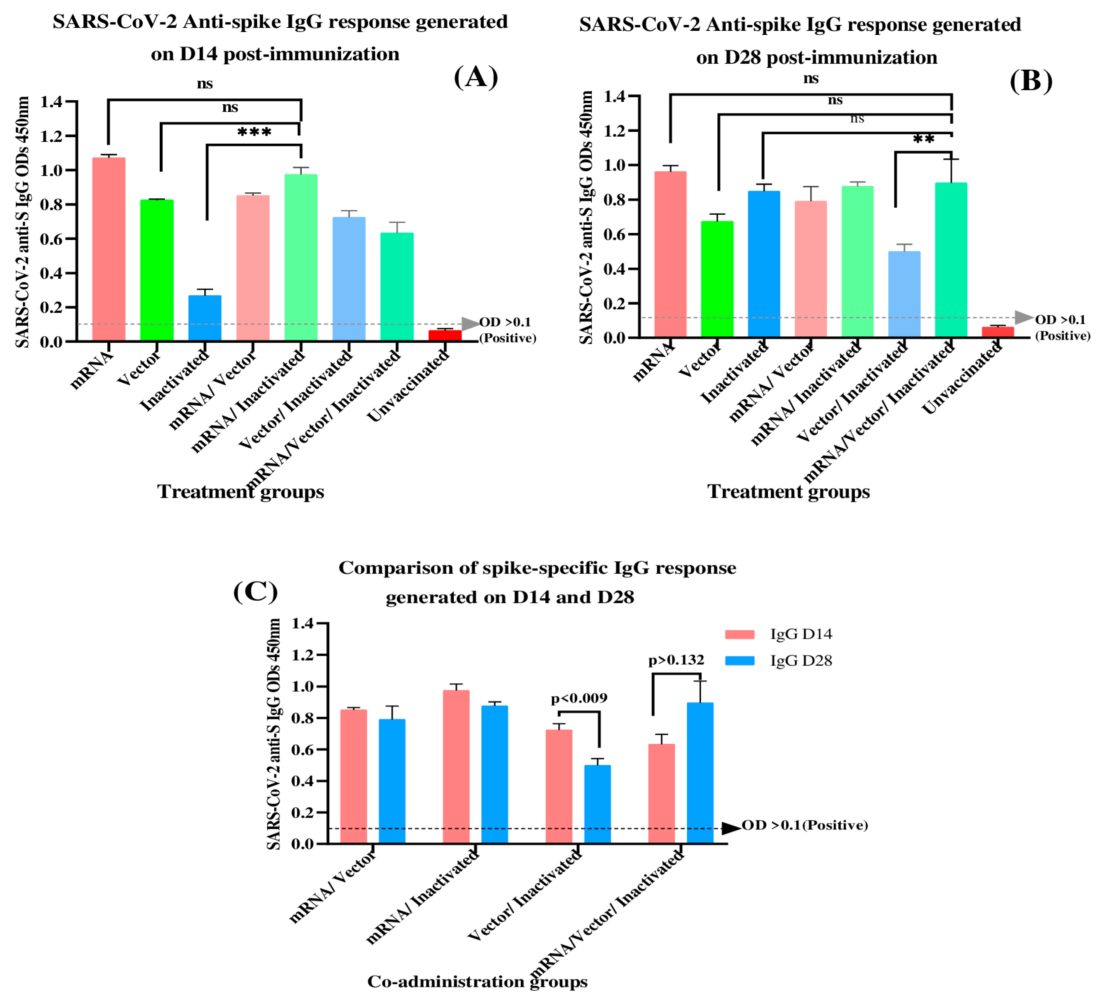
3.1. Determination of SARS-CoV-2 Spike-Specific IgG Antibodies at 14- and 28-Days Post-Immunization

In this study, a qualitative indirect ELISA was carried out on days 14 and 28 after immunization to ascertain whether experimental groups exhibited the production of anti- SARS-CoV-2 spike IgG antibodies after immunization of mice with different SARS-CoV-2 vaccine platforms and co-administration regimens. The anti-spike IgG OD450nm mean values for each treatment group are recorded in Table 4.
The produced anti-spike IgG levels exhibited statistically significant differences between all the treatment groups on both days 14 and 28 post-immunization (p < 0.0001, p < 0.0001, respectively). Additionally, a significant variation in spike-specific IgG antibodies was observed among vaccine co-administration groups (mRNA/Vector, mRNA/Inactivated, Vector/Inactivated, mRNA/Vector/Inactivated) at days 14 and 28 (p < 0.0006, p < 0.0219, respectively). The mRNA/Inactivated group which elicited increased IgG antibodies (OD = 0.975) in comparison to other co-administration regimens (Table 4) exhibited a significant difference (p < 0.0001) compared to the Inactivated group but no statistical significance was observed when compared to the mRNA and Vector groups (p > 0.505, p > 0.076, respectively) as indicated in (Figure 1A).
Whereas a titer decrease was observed for the co-administration groups on day 28 post-immunization, the mRNA/vector/Inactivated group demonstrated an increase in IgG levels (D14 = OD 0.633, D28 = OD 0.897). Nevertheless, the same group exhibited no statistically significant difference in IgG levels (p > 0.05) compared to full-course homologous immunization with individual vaccines (Figure 1B). We further compared the IgG response induced between both time points (day 14 and 28 after immunization) using unpaired t-tests. As a result, the mRNA/Vector and mRNA/Inactivated groups exhibited no statistically significant decline in the IgG levels 28 days after immunization, with 1.077-fold and 1.111-fold decreases, respectively (p > 0.514, p > 0.088, respectively). In contrast, the Vector/Inactivated group demonstrated a significant decline in IgG levels (1.450-fold decrease) with p < 0.009.

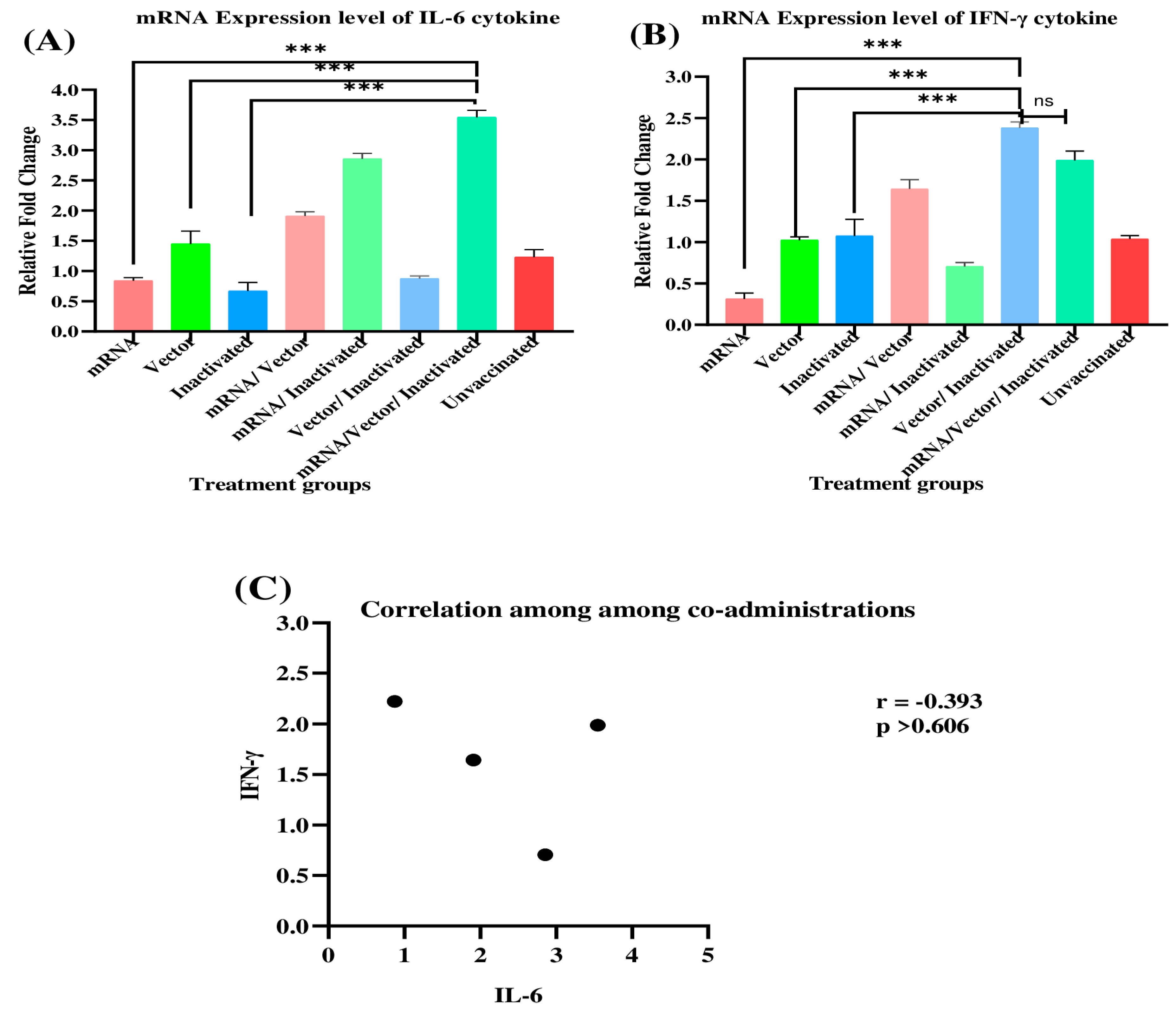
3.2. Evaluation of IL-6 and IFN-γ mRNA Expression Profiles in Immunized BALB/c Mice

Further, we explored the cell-mediated immune response of mice immunized with homologous SARS-CoV-2 vaccines and their combined regimens. Here, the IL-6 and IFN-γ cytokine expression levels from mice splenocytes were quantified using RT-PCR at day 28 post-immunization. A housekeeping gene (HPRT1 gene) was used to normalize the expression profiles of genes of interest. The mRNA/Inactivated and mRNA/Vector/Inactivated groups exhibited elevated IL-6 expression within the co-administration groups. However, as compared to the mRNA/Inactivated group (p < 0.0001), the mRNA/Vector/Inactivated group revealed a 1.240-fold increase. At the same time, BALB/c mice immunized with the combination of mRNA, adenovirus26 vector, and inactivated vaccines expressed significantly elevated levels of IL-6 (p < 0.0001) compared to immunization with individual vaccines, as illustrated in (Figure 2A).
On the contrary, the Vector/Inactivated and mRNA/Vector/Inactivated groups induced an increased expression of IFN-γ in comparison with other co-administration groups. However, the Vector/Inactivated group did not exhibit a significant expression of IFN-γ in comparison to the mRNA/Vector/Inactivated group (p > 0.999). Notably, these combined regimens demonstrated statistically significant IFN-γ expression when compared to a complete course of immunization with the mRNA vaccine, adenovirus 26 vector vaccine, and Inactivated vaccine (p < 0.0001). Taken together, the IL-6 and IFN-γ cytokine expression profiles demonstrated that the IL-6 was significantly downregulated in the Vector/Inactivated group but upregulated in the mRNA/Vector/Inactivated group. Conversely, IFN-γ was significantly downregulated in the mRNA/Inactivated group while being upregulated in the Vector/Inactivated and mRNA/Vector/Inactivated groups. These findings demonstrated the ability of the mRNA/adenovirus26 vector/inactivated SARS-CoV-2 vaccine combined vaccine to generate robust cellular immune responses compared to a complete course of homologous immunization of mRNA vaccine, adenovirus 26 vector vaccine, and Inactivated vaccine.
The correlation between cytokines among the co-administration groups was determined through Pearson correlation analysis (Figure 2C). Notably, a moderate negative correlation was observed between IL-6 and IFN-γ expression levels (r = −0.393).

3.3. Safety Assessment

Histopathological examination in the heart and liver tissues was evaluated in all experimental groups at the end of the experiment (28 days after immunization). Photomicrographs at 20× magnification of the BALB/c mice heart and liver tissue after immunization with different SARS-CoV-2 homologous and combined vaccines are presented in Figure 3. Microscopic examination of the heart and liver in both immunized and non-immunized mice revealed cardiac muscle composed of benign myocytes. Similarly, microscopic analysis of liver photomicrographs from all experimental groups revealed hepatic tissue composed of trabeculae of benign hepatocytes with normal portal tracts and central veins. Notably, the only exception was observed in the mice groups that received the co-administration of 4 × 10⁹ VP adenovirus26 vector /0.8 µg inactivated vaccine (Figure 3L) and the co-administration of 5 µg mRNA/4 × 109 VP adenovirus26 vector/0.8 µg inactivated vaccines (Figure 3N), where the liver exhibited mild necrosis. Importantly, no remarkably significant histopathological alterations in the heart and liver tissues were observed across all mice immunized with SARS-CoV-2 vaccine co-administration regimens.

4. Discussion

SARS-CoV-2 vaccines have been demonstrated to induce diverse immune responses [31]. This study compared immune responses upon immunization with individual vaccines, namely mRNA, adenovirus26 vector, and inactivated SARS-CoV-2 vaccines, and vaccine combinations comprising mRNA/adenovirus26 vector, mRNA/inactivated, adenovirus26 vector/inactivated, and mRNA/adenovirus26 vector vaccines. The findings of this study revealed that the inactivated vaccine induced lower spike-specific IgG levels among the individual immunization groups at day 14. This finding aligns with a study conducted in mice by Zhang et al. [21], who similarly reported at day 14 post-immunization that lower immunogenicity of a single dose of inactivated vaccine was realized compared to one dose of mRNA or one dose of adenovirus vector vaccine. However, their study did not extend to day 28. He et al. [29] demonstrated that the spike-specific IgG levels induced by two doses of inactivated vaccine were higher than a single dose of adenovirus vector vaccine at day 35, which was in contrast to our findings at day 28. The differences in outcomes may be attributed to the delivery system and antigen expression [32]. The viral vector utilized in our study was a recombinant adenovirus26 vectored vaccine that expresses a prefusion-stabilized spike protein, while their study employed a recombinant adenovirus 5 vaccine that expresses a full-length spike protein. Importantly, mRNA and adenovirus vector-based vaccines have proven effective in generating strong humoral responses, as supported by our findings demonstrating higher spike-specific IgG produced on days 14 and 28 post-immunization among individual immunization groups. However, both mRNA and Vector groups exhibited a slower decline in IgG levels by day 28, aligning with findings reported in previous studies [28,33].
The humoral and cell-mediated immune response of vaccine co-administration regimens were assessed. The vaccine co-administration approach has been proven to be effective and has been implemented for use worldwide. Moreover, the application of heterologous vaccines has been effective in previous studies [23]. Considering the distinct immune characteristics associated with various SARS-CoV-2 vaccines, it is logical to investigate innovative approaches for immunization against the virus. Recent studies demonstrated an improvement in immunogenicity when the DNA-based and inactivated vaccines were co-administered in mice [34]. In the present study, we tested four co-administration regimens, namely mRNA/adenovirus26 vector, mRNA/inactivated, adenovirus26 vector/inactivated, and mRNA/adenovirus26 vector/inactivated vaccines. The results demonstrated improved immunogenicity and significant differences in IgG levels among coadministration groups at days 14 or 28 post-immunization (p < 0.0006, p < 0.0219, respectively) despite the decline observed at 28 days post-immunization. Physiological, environmental, and immune system elements have been proven to impact vaccine effectiveness [35]. For deeper comprehension, studies demonstrated that the waning immunity may be linked to the negative immune interference among different vaccine platforms. Notably, interference was more pronounced when inactivated vaccines, such as for influenza, were co-administered with COVID vaccines [36] as well as in heterologous prime-boost approaches involving, for example, viral vector and mRNA vaccines [37]. Additionally, pre-existing anti-vector antibodies [38] and shorter intervals between mRNA doses [39] have been proposed as factors that may contribute to vaccine-induced interference.
Interestingly, our study explored the elevated anti-spike IgG levels at day 28 in the mRNA/Vector/Inactivated group, which was not significant compared to individual immunization groups (p > 0.5). Unlike other co-administration regimens that exhibited a decrease in IgG levels, the reason behind the sustained IgG levels induced by the mRNA/Vector/Inactivated regimen can be attributed to the antigenic mass presented to the immune system. In addition, the respective strengths of each vaccine synergistically complement one another. While mRNA vaccines possess inherent adjuvant properties, activating Toll-like receptor 7 (TLR-7) to swiftly initiate humoral immune responses compared to other alternative vaccine methods [11], the adenovirus vector-based vaccines can lead to long-term immune responses due to prolonged replication [40]. Inactivated vaccines, in contrast, present the whole attenuated virus which mimics a natural infection, prompting the body to elicit a strong, long-lasting immunity [30]. Moreover, adopting heterologous vaccination has been a strategy suggested to alleviate the pre-existing anti-vector immunity and non-spike protein immune responses, hence inducing long-term memory B cells and wider SARS-CoV-2 spike-specific IgG antibodies [40,41]. However, we suggest further investigation to acquire a comprehensive understanding of the mechanisms involved in the stability of antibody generation.
To achieve a more effective vaccination strategy, it is imperative to establish a robust and enduring cellular-mediated immune response. In this effort, the expression profiles of IFN-γ and IL-6 were investigated. Previous studies have indicated that the administration of various SARS-CoV-2 vaccines, including mRNA, adenovirus vector-based, and inactivated vaccines, induces dynamic amounts of cytokines such as IFN-γ and IL-6, in real-time [34,42]. In our experiment, the mRNA/adenovirus26 vector/inactivated vaccine co-administration regimen exhibited higher expression levels of IL-6 compared to other experimental groups. IL-6 has been observed to be an essential soluble regulator for follicular helper T(Tfh) cells differentiation. The latter, identified as distinctive CD4+ T cells, plays a crucial role in the differentiation process of B cells into memory B cells and durable plasma cells, as mechanisms involved in the longevity of antibody response [43]. IFN-γ is predominantly produced by natural killer T cells, CD4 T cells, and CD8 T cells. It is suggested that IFN-γ does not activate B cells; rather, it exerts an inhibitory effect on B cell division, particularly during the later phases of B cell proliferation and maturation [44].
The current study also demonstrated that the co-administration of adenovirus26 vector/inactivated vaccines and mRNA/adenovirus26 vector/inactivated vaccines induced significantly higher levels of IFN-γ expression (p < 0.0001) compared with the other treatment groups. We speculate that each vaccine synergistically complements one another. The inactivated vaccines and nucleic acids-based vaccines like mRNA and adenovirus vector-based vaccines have demonstrated the ability to engage diverse innate cellular sensors, including Toll-like receptors, which further initiate innate immune responses and activate IFN signaling pathways, ultimately resulting in the production of IFN-γ. This, in turn, enhances adaptive immune responses [45,46].
People with genetic variations in the IFN-γ gene that negatively impact the activity of IFN-γ are five times more likely to be susceptible to severe acute respiratory syndrome (SARS) [46]. Given that IFN-γ has antiviral properties, its pivotal role in effective and successful immunization is underscored [47]. Our study suggests that immunization with co-administration regimens could lead to a robust ability to activate macrophages, enhance MHC-1 antigen presentation, and consequently stimulate efficiently Th1 CD4 and CD8 T cell responses [48].
To further assess the practicability of the immunization strategy, the safety of the SARS-CoV-2 vaccine co-administration was assessed in BALB/c mice 28 days post-immunization.
Importantly, no mortality or alarming adverse reactions were observed in any experimental group during this period. Histopathological examination of the heart and liver tissues showed no significant histopathological changes compared to unvaccinated control mice. The literature indicates that IL-6 is a key proinflammatory cytokine in the onset of cytokine storms and acute respiratory distress syndrome (ARDS), leading to tissue damage, and ultimately, death [49]. In our study, we speculate that the observed mild necrosis of hepatocytes in mice immunized with adenovirus vector/inactivated and mRNA/adenovirus vector/inactivated vaccines may be associated with the immune-inflammatory responses provoked by the IL-6 cytokine. To enhance further safety assessment of the SARS-CoV-2 vaccine co-administration, biochemical and hematological analyses are highly recommended.

5. Conclusions

Despite the widespread use of mRNA-based, adenovirus vector-based, and inactivated SARS-CoV-2 vaccines, they exhibit notable differences in their immunogenicity and efficacy, with a decline in effectiveness after vaccination necessitating frequent booster doses. This situation has underscored the necessity for additional novel vaccination strategies such as SARS-CoV-2 vaccine co-administration, an area that requires critical investigation. This study demonstrated that co-administering mRNA, adenovirus-vectored, and inactivated SARS-CoV-2 vaccines in BALB/c mice improved spike-specific IgG response, and promoted IFN-γ and IL-6 mRNA expression compared to the complete immunization with individual vaccines alone. Importantly, co-administration regimens proved to be safe and well tolerated in BALB/c mice. For further comprehensive understanding, studies are recommended, including neutralization assays and cross-protection tests on SARS-CoV-2 variants of concern.

Author Contributions

Conceptualization, S.M.N., J.K. (Josephine Kimani), J.K. (James Kimotho) and N.J.; methodology, S.M.N., J.K. (Josephine Kimani), N.J., M.M.M. and J.K. (James Kimotho); validation, N.J., J.K. (Josephine Kimani) and S.M.N.; formal analysis, N.J., S.M.N. and J.K. (Josephine Kimani); investigation, N.J.; resources, S.M.N., J.K. (James Kimotho) and M.M.M.; data curation, N.J., S.M.N. and J.K. (Josephine Kimani); validation, N.J., S.M.N. and J.K. (Josephine Kimani); writing—original draft preparation, N.J., S.M.N. and J.K. (Josephine Kimani); writing—review and editing; N.J., S.M.N. and J.K. (Josephine Kimani); visualization, N.J., M.M.M., S.M.N., J.K. (Josephine Kimani) and J.K. (James Kimotho); supervision, S.M.N. and J.K. (Josephine Kimani); project administration, J.K. (James Kimotho) and S.M.N.; funding acquisition, S.M.N., J.K. (Josephine Kimani) and N.J. All authors have read and agreed to the published version of the manuscript.

Funding

This work was supported by the African Union Commission through the Pan African University Institute for Basic Sciences, Technology, and Innovation (PAUSTI) and partly by KEMRI internal grant KEMRI/COV/INNOV/003.

Institutional Review Board Statement

All mice studies were carried out in compliance with the Guide for the Care and Use of Laboratory Animals at Kenya Medical Research Institute (KEMRI). The animal study protocols were reviewed and approved by KEMRI Animal Care and Use Committee and Mount Kenya University (MKU) Animal Care and Use Ethics Review Committee with reference numbers KEMRI ACUC/02.06.2023 and REF: MKU/ISERC/2904 (approval number 1948), respectively.

Informed Consent Statement

Not applicable.

Data Availability Statement

All data are provided in this manuscript.

Acknowledgments

The authors extend their gratitude for the support provided by the African Union Commission through the Pan African Union through Pan African University Institute for Basic Sciences, Technology and Innovation (PAUSTI) and partly by KEMRI. Appreciation is also directed to the overall staff and especially Anne Wanjiru Mwangi from the Pan African Hub for Infectious Diseases Research at the Centre for Microbiology Research (CMR) Laboratories in Kenya Medical Research Institute. Additionally, we acknowledge the generous support provided by Vincent Kiplangat Ruttoh, who facilitated our access to qPCR facilities at the Centre for Virus Research, and Shingo Inoue, for providing guidance on animal experiments.

Conflicts of Interest

The authors declare no conflicts of interest.

References

  1. Dhama, K.; Khan, S.; Tiwari, R.; Sircar, S.; Bhat, S.; Malik, Y.S.; Singh, K.P.; Chaicumpa, W.; Bonilla-Aldana, D.K.; Rodriguez-Morales, A.J. Coronavirus Disease 2019–COVID-19. Clin. Microbiol. Rev. 2020, 33, 10–1128. [Google Scholar] [CrossRef] [PubMed]
  2. Chang, D.; Chang, X.; He, Y.; Tan, K.J.K. The Determinants of COVID-19 Morbidity and Mortality across Countries. Sci. Rep. 2022, 12, 5888. [Google Scholar] [CrossRef] [PubMed]
  3. Richards, F.; Kodjamanova, P.; Chen, X.; Li, N.; Atanasov, P.; Bennetts, L.; Patterson, B.J.; Yektashenas, B.; Mesa-Frias, M.; Tronczynski, K.; et al. Economic Burden of COVID-19: A Systematic Review. Clin. Outcomes Res. 2022, 14, 293–307. [Google Scholar] [CrossRef] [PubMed]
  4. WHO. WHO Coronavirus (COVID-19) Dashboard. Geneva: World Health Organization. 2023. Available online: https://covid19.who.int/ (accessed on 11 November 2023).
  5. Chams, N.; Chams, S.; Badran, R.; Shams, A.; Araji, A.; Raad, M.; Mukhopadhyay, S.; Stroberg, E.; Duval, E.J.; Barton, L.M.; et al. COVID-19: A Multidisciplinary Review. Front. Public Health 2020, 8, 383. [Google Scholar] [CrossRef] [PubMed]
  6. Fumagalli, V.; Di Lucia, P.; Ravà, M.; Marotta, D.; Bono, E.; Grassi, S.; Donnici, L.; Cannalire, R.; Stefanelli, I.; Ferraro, A.; et al. Nirmatrelvir Treatment of SARS-CoV-2-infected Mice Blunts Antiviral Adaptive Immune Responses. EMBO Mol. Med. 2023, 15, e17580. [Google Scholar] [CrossRef]
  7. Panza, F.; Fiorino, F.; Pastore, G.; Fiaschi, L.; Tumbarello, M.; Medaglini, D.; Ciabattini, A.; Montagnani, F.; Fabbiani, M. Does Nirmatrelvir/Ritonavir Influence the Immune Response against SARS-CoV-2, Independently from Rebound? Microorganisms 2023, 11, 2607. [Google Scholar] [CrossRef] [PubMed]
  8. Haque, A.; Pant, A.B. Efforts at COVID-19 Vaccine Development: Challenges and Successes. Vaccines 2020, 8, 739. [Google Scholar] [CrossRef] [PubMed]
  9. Barouch, D.H. Covid-19 Vaccines—Immunity, Variants, Boosters. N. Engl. J. Med. 2022, 387, 1011–1020. [Google Scholar] [CrossRef] [PubMed]
  10. Jacob-Dolan, C.; Barouch, D.H. COVID-19 Vaccines: Adenoviral Vectors. Annu. Rev. Med. 2022, 73, 41–54. [Google Scholar] [CrossRef]
  11. Gebre, M.S.; Rauch, S.; Roth, N.; Gergen, J.; Yu, J.; Liu, X.; Cole, A.C.; Mueller, S.O.; Petsch, B.; Barouch, D.H. MRNA Vaccines Induce Rapid Antibody Responses in Mice. NPJ Vaccines 2022, 7, 88. [Google Scholar] [CrossRef]
  12. Kanokudom, S.; Assawakosri, S.; Suntronwong, N.; Auphimai, C.; Nilyanimit, P.; Vichaiwattana, P.; Thongmee, T.; Yorsaeng, R.; Srimuan, D.; Thatsanatorn, T.; et al. Safety and Immunogenicity of the Third Booster Dose with Inactivated, Viral Vector, and MRNA COVID-19 Vaccines in Fully Immunized Healthy Adults with Inactivated Vaccine. Vaccines 2022, 10, 86. [Google Scholar] [CrossRef] [PubMed]
  13. Andrews, N.; Stowe, J.; Kirsebom, F.; Toffa, S.; Rickeard, T.; Gallagher, E.; Gower, C.; Kall, M.; Groves, N.; O’Connell, A.-M.; et al. Covid-19 Vaccine Effectiveness against the Omicron (B.1.1.529) Variant. N. Engl. J. Med. 2022, 386, 1532–1546. [Google Scholar] [CrossRef] [PubMed]
  14. Sadoff, J.; Gray, G.; Vandebosch, A.; Cárdenas, V.; Shukarev, G.; Grinsztejn, B.; Goepfert, P.A.; Truyers, C.; Van Dromme, I.; Spiessens, B.; et al. Final Analysis of Efficacy and Safety of Single-Dose Ad26.COV2.S. N. Engl. J. Med. 2022, 386, 847–860. [Google Scholar] [CrossRef] [PubMed]
  15. Graña, C.; Ghosn, L.; Evrenoglou, T.; Jarde, A.; Minozzi, S.; Bergman, H.; Buckley, B.S.; Probyn, K.; Villanueva, G.; Henschke, N.; et al. Efficacy and Safety of COVID-19 Vaccines. Cochrane Database Syst. Rev. 2022, 2023. [Google Scholar] [CrossRef]
  16. Piechotta, V.; Harder, T. Waning of COVID-19 Vaccine Effectiveness: Individual and Public Health Risk. Lancet 2022, 399, 887–889. [Google Scholar] [CrossRef] [PubMed]
  17. Belayachi, J.; Obtel, M.; Mhayi, A.; Razine, R.; Abouqal, R. Long Term Effectiveness of Inactivated Vaccine BBIBP-CorV (Vero Cells) against COVID-19 Associated Severe and Critical Hospitalization in Morocco. PLoS ONE 2022, 17, e0278546. [Google Scholar] [CrossRef] [PubMed]
  18. Deming, M.E.; Lyke, K.E. A ‘Mix and Match’ Approach to SARS-CoV-2 Vaccination. Nat. Med. 2021, 27, 1510–1511. [Google Scholar] [CrossRef] [PubMed]
  19. Krause, P.R.; Fleming, T.R.; Peto, R.; Longini, I.M.; Figueroa, J.P.; Sterne, J.A.C.; Cravioto, A.; Rees, H.; Higgins, J.P.T.; Boutron, I.; et al. Considerations in Boosting COVID-19 Vaccine Immune Responses. Lancet 2021, 398, 1377–1380. [Google Scholar] [CrossRef] [PubMed]
  20. Rzymski, P.; Camargo, C.A.; Fal, A.; Flisiak, R.; Gwenzi, W.; Kelishadi, R.; Leemans, A.; Nieto, J.J.; Ozen, A.; Perc, M.; et al. COVID-19 Vaccine Boosters: The Good, the Bad, and the Ugly. Vaccines 2021, 9, 1299. [Google Scholar] [CrossRef]
  21. Zhang, J.; He, Q.; An, C.; Mao, Q.; Gao, F.; Bian, L.; Wu, X.; Wang, Q.; Liu, P.; Song, L.; et al. Boosting with Heterologous Vaccines Effectively Improves Protective Immune Responses of the Inactivated SARS-CoV-2 Vaccine. Emerg. Microbes Infect. 2021, 10, 1598–1608. [Google Scholar] [CrossRef]
  22. Bauwens, J.; De Lusignan, S.; Weldesselassie, Y.G.; Sherlock, J.; Künzli, N.; Bonhoeffer, J. Safety of Routine Childhood Vaccine Coadministration versus Separate Vaccination. BMJ Glob. Health 2022, 7, e008215. [Google Scholar] [CrossRef]
  23. Wagenhäuser, I.; Reusch, J.; Gabel, A.; Höhn, A.; Lâm, T.T.; Almanzar, G.; Prelog, M.; Krone, L.B.; Frey, A.; Schubert-Unkmeir, A.; et al. Immunogenicity and Safety of Coadministration of COVID-19 and Influenza Vaccination. Eur. Respir. J. 2023, 61, 2201390. [Google Scholar] [CrossRef] [PubMed]
  24. Woodworth, J.S.; Clemmensen, H.S.; Battey, H.; Dijkman, K.; Lindenstrøm, T.; Laureano, R.S.; Taplitz, R.; Morgan, J.; Aagaard, C.; Rosenkrands, I.; et al. A Mycobacterium Tuberculosis-Specific Subunit Vaccine That Provides Synergistic Immunity upon Co-Administration with Bacillus Calmette-Guérin. Nat. Commun. 2021, 12, 6658. [Google Scholar] [CrossRef]
  25. Li, C.; Chen, Y.; Zhao, Y.; Lung, D.C.; Ye, Z.; Song, W.; Liu, F.-F.; Cai, J.-P.; Wong, W.-M.; Yip, C.C.-Y.; et al. Intravenous Injection of Coronavirus Disease 2019 (COVID-19) MRNA Vaccine Can Induce Acute Myopericarditis in Mouse Model. Clin. Infect. Dis. 2022, 74, 1933–1950. [Google Scholar] [CrossRef] [PubMed]
  26. Vogel, A.B.; Kanevsky, I.; Che, Y.; Swanson, K.A.; Muik, A.; Vormehr, M.; Kranz, L.M.; Walzer, K.C.; Hein, S.; Güler, A.; et al. BNT162b Vaccines Protect Rhesus Macaques from SARS-CoV-2. Nature 2021, 592, 283–289. [Google Scholar] [CrossRef]
  27. Celise, D.A.; Kimotho, J.; Kimani, J.W.; Muriithi, A.K.; Odari, E.O. Increase in the Immune Response in Balb/c Mice after the Co-Administration of a Vector-Based COVID-19 Vaccine with Cytosine Phosphoguanine Oligodeoxynucleotide. Vaccines 2022, 11, 53. [Google Scholar] [CrossRef]
  28. Mudenda, M.; Kimani, J.; Kinyua, J.; Kimotho, J. Preliminary In Vivo Evidence of Oral Selenium Supplementation as a Potentiating Agent on a Vector-Based COVID-19 Vaccine in BALB/c Mice. Vaccines 2022, 11, 57. [Google Scholar] [CrossRef] [PubMed]
  29. He, Q.; Mao, Q.; An, C.; Zhang, J.; Gao, F.; Bian, L.; Li, C.; Liang, Z.; Xu, M.; Wang, J. Heterologous Prime-Boost: Breaking the Protective Immune Response Bottleneck of COVID-19 Vaccine Candidates. Emerg. Microbes Infect. 2021, 10, 629–637. [Google Scholar] [CrossRef] [PubMed]
  30. Kandeil, A.; Mostafa, A.; Hegazy, R.R.; El-Shesheny, R.; El Taweel, A.; Gomaa, M.R.; Shehata, M.; Elbaset, M.A.; Kayed, A.E.; Mahmoud, S.H.; et al. Immunogenicity and Safety of an Inactivated SARS-CoV-2 Vaccine: Preclinical Studies. Vaccines 2021, 9, 214. [Google Scholar] [CrossRef]
  31. Sharif, N.; Alzahrani, K.J.; Ahmed, S.N.; Dey, S.K. Efficacy, Immunogenicity and Safety of COVID-19 Vaccines: A Systematic Review and Meta-Analysis. Front. Immunol. 2021, 12, 714170. [Google Scholar] [CrossRef]
  32. Sakurai, F.; Tachibana, M.; Mizuguchi, H. Adenovirus Vector-Based Vaccine for Infectious Diseases. Drug Metab. Pharmacokinet. 2022, 42, 100432. [Google Scholar] [CrossRef]
  33. Bergami, F.; Arena, F.; Sammartino, J.C.; Ferrari, A.; Zavaglio, F.; Zelini, P.; Paolucci, S.; Comolli, G.; Percivalle, E.; Lilleri, D.; et al. Differential Kinetics of Effector and Memory Responses Induced by Three Doses of SARS-CoV-2 MRNA Vaccine in a Cohort of Healthcare Workers. Vaccines 2022, 10, 1809. [Google Scholar] [CrossRef] [PubMed]
  34. Meng, Z.; Ma, D.; Duan, S.; Zhang, J.; Yue, R.; Li, X.; Gao, Y.; Li, X.; Zeng, F.; Xu, X.; et al. Immunological Study of Combined Administration of SARS-CoV-2 DNA Vaccine and Inactivated Vaccine. Vaccines 2022, 10, 929. [Google Scholar] [CrossRef]
  35. Torresi, J.; Edeling, M.A.; Nolan, T.; Godfrey, D.I. A Complementary Union of SARS-CoV2 Natural and Vaccine Induced Immune Responses. Front. Immunol. 2022, 13, 914167. [Google Scholar] [CrossRef] [PubMed]
  36. Shenyu, W.; Xiaoqian, D.; Bo, C.; Xuan, D.; Zeng, W.; Hangjie, Z.; Qianhui, Z.; Zhenzhen, L.; Chuanfu, Y.; Juan, Y.; et al. Immunogenicity and Safety of a SARS-CoV-2 Inactivated Vaccine (CoronaVac) Co-Administered with an Inactivated Quadrivalent Influenza Vaccine: A Randomized, Open-Label, Controlled Study in Healthy Adults Aged 18 to 59 Years in China. Vaccine 2022, 40, 5356–5365. [Google Scholar] [CrossRef]
  37. Lv, J.; Wu, H.; Xu, J.; Liu, J. Immunogenicity and Safety of Heterologous versus Homologous Prime-Boost Schedules with an Adenoviral Vectored and MRNA COVID-19 Vaccine: A Systematic Review. Infect. Dis. Poverty 2022, 11, 53. [Google Scholar] [CrossRef] [PubMed]
  38. Voysey, M.; Costa Clemens, S.A.; Madhi, S.A.; Weckx, L.Y.; Folegatti, P.M.; Aley, P.K.; Angus, B.; Baillie, V.L.; Barnabas, S.L.; Bhorat, Q.E.; et al. Single-Dose Administration and the Influence of the Timing of the Booster Dose on Immunogenicity and Efficacy of ChAdOx1 NCoV-19 (AZD1222) Vaccine: A Pooled Analysis of Four Randomised Trials. Lancet 2021, 397, 881–891. [Google Scholar] [CrossRef]
  39. Payne, R.P.; Longet, S.; Austin, J.A.; Skelly, D.T.; Dejnirattisai, W.; Adele, S.; Meardon, N.; Faustini, S.; Al-Taei, S.; Moore, S.C.; et al. Immunogenicity of Standard and Extended Dosing Intervals of BNT162b2 MRNA Vaccine. Cell 2021, 184, 5699–5714.e11. [Google Scholar] [CrossRef]
  40. Wang, S.; Liang, B.; Wang, W.; Li, L.; Feng, N.; Zhao, Y.; Wang, T.; Yan, F.; Yang, S.; Xia, X. Viral Vectored Vaccines: Design, Development, Preventive and Therapeutic Applications in Human Diseases. Signal Transduct. Target. Ther. 2023, 8, 149. [Google Scholar] [CrossRef]
  41. Ai, J.; Guo, J.; Zhang, H.; Zhang, Y.; Yang, H.; Lin, K.; Song, J.; Fu, Z.; Fan, M.; Zhang, Q.; et al. Cellular Basis of Enhanced Humoral Immunity to SARS-CoV-2 upon Homologous or Heterologous Booster Vaccination Analyzed by Single-Cell Immune Profiling. Cell Discov. 2022, 8, 114. [Google Scholar] [CrossRef]
  42. Jiang, M.; Väisänen, E.; Kolehmainen, P.; Huttunen, M.; Ylä-Herttuala, S.; Meri, S.; Österlund, P.; Julkunen, I. COVID-19 Adenovirus Vector Vaccine Induces Higher Interferon and pro-Inflammatory Responses than MRNA Vaccines in Human PBMCs, Macrophages and MoDCs. Vaccine 2023, 41, 3813–3823. [Google Scholar] [CrossRef] [PubMed]
  43. Seo, Y.B.; Im, S.J.; Namkoong, H.; Kim, S.W.; Choi, Y.W.; Kang, M.C.; Lim, H.S.; Jin, H.T.; Yang, S.H.; Cho, M.L.; et al. Crucial Roles of Interleukin-7 in the Development of T Follicular Helper Cells and in the Induction of Humoral Immunity. J. Virol. 2014, 88, 8998–9009. [Google Scholar] [CrossRef] [PubMed]
  44. Alspach, E.; Lussier, D.M.; Schreiber, R.D. Interferon γ and Its Important Roles in Promoting and Inhibiting Spontaneous and Therapeutic Cancer Immunity. Cold Spring Harb. Perspect. Biol. 2019, 11, a028480. [Google Scholar] [CrossRef] [PubMed]
  45. Chang, A.; Yu, J. Fighting Fire with Fire: Immunogenicity of Viral Vectored Vaccines against COVID-19. Viruses. 2022, 14, 380. [Google Scholar] [CrossRef] [PubMed]
  46. Sahin, U.; Muik, A.; Derhovanessian, E.; Vogler, I.; Kranz, L.M.; Vormehr, M.; Baum, A.; Pascal, K.; Quandt, J.; Maurus, D.; et al. COVID-19 Vaccine BNT162b1 Elicits Human Antibody and TH1 T Cell Responses. Nature 2020, 586, 594–599. [Google Scholar] [CrossRef] [PubMed]
  47. Primorac, D.; Vrdoljak, K.; Brlek, P.; Pavelić, E.; Molnar, V.; Matišić, V.; Erceg Ivkošić, I.; Parčina, M. Adaptive Immune Responses and Immunity to SARS-CoV-2. Front. Immunol. 2022, 13, 848582. [Google Scholar] [CrossRef]
  48. Costela-Ruiz, V.J.; Illescas-Montes, R.; Puerta-Puerta, J.M.; Ruiz, C.; Melguizo-Rodríguez, L. SARS-CoV-2 Infection: The Role of Cytokines in COVID-19 Disease. Cytokine Growth Fac. Rev. 2020, 54, 62–75. [Google Scholar] [CrossRef]
  49. Qudus, M.S.; Tian, M.; Sirajuddin, S.; Liu, S.; Afaq, U.; Wali, M.; Liu, J.; Pan, P.; Luo, Z.; Zhang, Q.; et al. The Roles of Critical Pro-inflammatory Cytokines in the Drive of Cytokine Storm during SARS-CoV-2 Infection. J. Med. Virol. 2023, 95, e28751. [Google Scholar] [CrossRef]
Figure 1. Humoral immune responses induced by different SARS-CoV-2 vaccine platforms and co-administration regimens in BALB/c mice. (A) 14 days post-immunization using ELISA, (B) 28 days post-immunization. (C) Comparison of anti-spike(S) IgG response generated 14 and 28 days after immunization among co-administration groups. The dashed line indicates the cut-off value for the anti-spike IgG antibodies with positive OD450nm values. One-way ANOVA (A,B) and Unpaired t-test (C) were conducted using GraphPad Prism software 8.0.2 to compare the statistical differences. Error bars represent SEM (n = 4 mice per group)., ** p < 0.001, *** p < 0.0001, ns: p > 0.05.
Immuno 04 00012 g001
Figure 2. Cytokine expression levels induced by different SARS-CoV-2 vaccine platforms and co-administration regimens in BALB/c mice. (A) Relative quantification of IL-6 mRNA expression levels. (B) IFN-γ mRNA expression levels. (C) Correlation analysis between IL-6 and IFN-γ expression levels among co-administered regimens. One-way ANOVA (A,B) and Pearson Correlation analysis (C) were conducted using GraphPad Prism software 8.0.2 to compare the statistical differences. Error bars represent SEM, *** p < 0.0001, ns: p > 0.05.
Immuno 04 00012 g002
Figure 3. Histopathology of heart and liver tissues from the immunized BALB/c mice. Histological findings showing benign heart and liver tissues of mice immunized with 5 µg mRNA vaccine (A,B), 4 × 10⁹ VP adenovirus26 vectored vaccine (C,D), 0.8 µg inactivated vaccine (E,F), co-administration of 5 µg mRNA/4 × 109 VP adenovirus26 vectored vaccines (G,H), co-administration of 5µg mRNA/0.8 µg inactivated vaccines (I,J). The microscopic findings in the mice immunized with the co-administration of 4 × 10⁹ VP adenovirus26 vectored/0.8 µg inactivated vaccines (K,L) and the co-administration of 5 µg mRNA/4 × 109 VP adenovirus26 vectored/0.8 µg inactivated vaccines (M,N) indicate mild necrosis in hepatic tissue and no histopathological changes in heart tissue. No histopathological changes observed in either the heart or liver of unvaccinated control mice (O,P).
Immuno 04 00012 g003
Table 1. Details of SARS-CoV-2 vaccines used in the experiment.
SARS-CoV-2 VaccinesPlatformsDoses Administered in Mice Studies (per Mouse)
COMIRNATY COVID-19 (Pfizer-BioNTech) vaccine. Lot Number: GN6343mRNA (nucleoside modified) vaccine5 µg [25,26]
Janssen (Ad26.COV2.S (recombinant))
vaccine. Lot number: ACB6959
Adenovirus26-vectored vaccine4 × 109 VP [27,28]
SARS-CoV-2 vaccine (Vero Cell), Inactivated
(Sinopharm) Product Code: 2021071947
Inactivated vaccine
0.8 µg [21,29]
VP represents Viral particles.
Table 2. Treatment formulations and animal immunization programs.
Treatment GroupsTreatment and Immunization Protocol
mRNA5 µg (50 μL) of Pfizer-BioNTech vaccine on D0 and D14
Vector4 × 109 VP (40 μL) of Janssen vaccine on D0
Inactivated0.8 µg (100 μL) of Sinopharm vaccine on D0 and D14
mRNA/VectorCoadministration of 5 µg (50 μL) Pfizer-BioNTech and 4 × 10⁹ VP (40 μL) Janssen on D0
mRNA/InactivatedCoadministration of 5 µg (50 μL) Pfizer-BioNTech and 0.8 µg (100 μL) Sinopharm on D0
Vector /InactivatedCoadministration of 4 × 10⁹ VP (40 μL) Janssen and 0.8 µg (100 μL) Sinopharm on D0
mRNA/Vector/InactivatedCoadministration of 5 µg of Pfizer-BioNTech (50 µL), 4 × 10⁹ VP (40 µL) of Janssen and 0.8 µg (100 µL) of Sinopharm on D0
Unvaccinated50 μL of 1× phosphate-buffered saline (PBS) on D0
D0: day 0, D14: day 14.
Table 3. Primer sequences of housekeeping and target genes used in RT-PCR experiment.
Gene of InterestForward Primer
(5′-3′)
Reverse Primer
(5′-3′)
Amplicon Size (bp)Tm (°C)%GCNCBI Accession
IL-6CCCACCAGGA
ACGAAAGTCA
ACTGGCTGGA
AGTCTCTTGC
7059.89
59.96
55.00
55.00
https://www.ncbi.nlm.nih.gov/nucleotide/930945755# (accessed on 5 September 2023)
IFN-γGGATGCATTCA
TGAGTATTGC
CCTTTTCCGC
TTCCTGAGG
12755.42
58.14
42.86
57.89
https://www.ncbi.nlm.nih.gov/nucleotide/926657655# (accessed on 5 September 2023)
HPRT1TGAAGTACTCATTG
ATAGTCAAGGGCA
CTGGTGAAAA
GGACCTCTCG
10961.94
57.91
40.74
55.00
https://www.ncbi.nlm.nih.gov/nucleotide/96975137# (accessed on 5 September 2023)
Abbreviations: IL-6: Interleukin 6, IFN-γ: Interferon gamma, HPRT1: Hypoxanthine Guanine Phosphoribosyl Transferase1, Tm: melting temperature, %GC: proportion of guanine(G) and cytosine(C) nucleotide base pairs.
Table 4. SARS-CoV-2 spike-specific IgG OD450nm readings at 14- and 28-days post-immunization.
Treatment GroupsDay 14Day 28
MeanSDCV (%)MeanSDCV (%)
mRNA1.0700.0393.650.9620.0687.16
Vector0.8240.0131.680.6740.08612.89
Inactivated0.2680.07427.70.8490.0819.57
mRNA/Vector0.8510.0313.660.7900.17121.68
mRNA/Inactivated0.9750.0818.320.8770.0515.84
Vector/Inactivated0.7240.08011.10.4990.08717.61
mRNA/Vector/Inactivated0.6330.12519.80.8970.27630.77
Unvaccinated0.0630.02337.40.0600.02135.49
Abbreviations: SD: Standard deviation, CV (%): Coefficient of variation.
Disclaimer/Publisher’s Note: The statements, opinions and data contained in all publications are solely those of the individual author(s) and contributor(s) and not of MDPI and/or the editor(s). MDPI and/or the editor(s) disclaim responsibility for any injury to people or property resulting from any ideas, methods, instructions or products referred to in the content.

