Article

Eosinophil-Count-Derived Inflammatory Markers and Psoriasis Severity: Exploring the Link

by
Oana Mirela Tiucă
1,2,3,
Silviu Horia Morariu
2,3,*,
Claudia Raluca Mariean
1,4,
Robert Aurelian Tiucă
1,5,6,
Alin Codrut Nicolescu
7 and
Ovidiu Simion Cotoi
4,8
1
Doctoral School of Medicine and Pharmacy, George Emil Palade University of Medicine, Pharmacy, Science, and Technology of Targu Mures, 540142 Targu Mures, Romania
2
Dermatology Department, George Emil Palade University of Medicine, Pharmacy, Science, and Technology of Targu Mures, 540142 Targu Mures, Romania
3
Dermatology Clinic, Mures Clinical County Hospital, 540342 Targu Mures, Romania
4
Pathophysiology Department, George Emil Palade University of Medicine, Pharmacy, Science, and Technology of Targu Mures, 540142 Targu Mures, Romania
5
Endocrinology Department, George Emil Palade University of Medicine, Pharmacy, Science, and Technology of Targu Mures, 540142 Targu Mures, Romania
6
Endocrinology Department, Mures Clinical County Hospital, 540139 Targu Mures, Romania
7
Agrippa Ionescu Emergency Clinical Hospital, 011773 Bucharest, Romania
8
Pathology Department, Mures Clinical County Hospital, 540011 Targu Mures, Romania
*
Author to whom correspondence should be addressed.
Dermato 2024, 4(2), 25-36; https://doi.org/10.3390/dermato4020004
Submission received: 24 December 2023 / Revised: 8 March 2024 / Accepted: 3 April 2024 / Published: 15 April 2024

Abstract

:
Psoriasis is an immune-mediated disease, with various triggering factors, genetic predisposition, and an altered immune response concurring in the development of this disease. The eosinophil is a cell with an important role in various kinds of inflammatory processes. Scarce data are available regarding the role of the eosinophil in psoriasis. This study aims to address the overall relationship between eosinophil-count-derived inflammatory markers and psoriasis severity. There were 366 patients fulfilling the inclusion criteria included in this retrospective study and they were divided based on the body surface area (BSA) scale in mild and moderate-to-severe psoriasis. White blood cell (WBC), neutrophil, lymphocyte, monocyte, and eosinophil count, along with eosinophil-to-monocyte ratio (EMR) and eosinophil-to-neutrophil ratio (ENR) differed significantly between the two study groups. Eosinophil count, EMR, and ENR negatively correlated with disease severity. ENR is the most reliable eosinophil-count-derived marker in assessing psoriasis severity with an AUC of 0.627 and a cut-off value of 0.03. Eosinophil-count-derived inflammatory markers’ usefulness in appreciating disease severity was assessed for the first time in the literature in this study and proved to be reliable for the eosinophil count, EMR, and ENR.

1. Introduction

Psoriasis is an immune-mediated disease, with various triggering factors, genetic predisposition, and an altered immune response concurring in the development of this disease. Characterized by epidermal hyperplasia and keratinocyte hyperactivation, it usually presents with well-defined erythematous lesions covered with thick, silver scales in the classic, chronic plaque form. Nonetheless, multiple subtypes of psoriasis have been described, such as guttate, rupioid, or inverse psoriasis. Moreover, nail and joint involvement complete the clinical picture of this complex disease.
Moderate-to-severe psoriasis is frequently associated with metabolic disorders, such as obesity, dyslipidemia, and metabolic syndrome [1]. The proliferation and activation of Th-1, Th-17, and Th-22 cells lead to increased production of proinflammatory cytokines, such as tumor necrosis factor (TNF)-α, interleukin (IL)-6, IL-17, IL-23, and vascular endothelial growth factor (VEGF) [2,3]. Recently, it was proven that psoriatic inflammation transcends the skin, the proinflammatory mediators’ migration into the joints and bloodstream leading to articular damage, increased oxidative stress, endothelial dysfunction, hypercoagulation, and an increased inflammatory state [4,5].
Recently, various components of complete blood cell count have emerged as reliable indicators of inflammation in various diseases, such as malignant tumors, rheumatic disorders, or heart conditions [6,7,8,9,10]. Moreover, they are accessible, cost-effective, and widely available indices for evaluating systemic inflammation.
The proinflammatory mediators overly produced in psoriasis lead to an increased recruitment, activation, and survival of neutrophils [11,12], and to peripheral lymphopenia due to lymphocyte apoptosis [11]. Platelets, apart from being involved in coagulation and hemostasis, regulate immune cells, thus having additional roles in inflammatory processes [13].
The eosinophil is a cell with an important role in various kinds of inflammatory processes. By accumulating in the peripheral blood and tissues, they release cellular protein and lipid mediators, exerting proinflammatory effects. Scarce data are available regarding the role of the eosinophil in psoriasis. Eosinophil cationic protein (ECP) levels seem to be significantly higher in patients with psoriasis vulgaris while circulating eosinophils seem to be normal [14]. As such, eosinophil-count-derived inflammatory markers may reflect more accurately the involvement of eosinophils in inflammation as compared to eosinophils’ absolute count.
Even though new data are emerging with regard to the inflammatory effects of eosinophils, their role in psoriasis etiopathogenesis is still unclear. However, they seem to interfere with psoriatic inflammation by modulating cytokine production from plasmacytoid dendritic cells [15]. Additionally, they seem to accelerate psoriasis etiopathogenesis by secreting inflammatory cytokines that further induce neutrophil activation upon ligation with a TLR7 agonist and neutrophilic infiltration into lesional psoriatic skin [16]. Eosinophil-derived major basic protein (MBP) leads to superoxide and IL-8 being produced by the neutrophil, therefore promoting an innate immune response.
Eosinophil-to-lymphocyte ratio (ELR), eosinophil-to-monocyte ratio (EMR), and eosinophil-to-neutrophil ratio (ENR) have proven to be reliable indicators for both disease presence and severity in various disorders, such as allergic rhinitis, COVID-19 infections, prostate cancer, and chronic obstructive pulmonary disorder [17,18,19,20,21,22]. In skin disorders, they have been until now evaluated briefly only in relation to atopic dermatitis [23] and urticaria [24]. However, data regarding eosinophil involvement in skin diseases, other than predominantly allergic, are limited.
To the best of our knowledge, this is the first study to address the overall relationship between eosinophil-count-derived inflammatory markers and psoriasis severity.

2. Materials and Methods

2.1. Study Population

We conducted a retrospective observational study that included patients diagnosed with psoriasis vulgaris in the Dermatology Department of Mures Clinical County Hospital, Romania, between January 2017 and December 2022. Patients with psoriasis vulgaris, who were older than 18 years of age and with available data regarding disease severity and laboratory investigations, were included. Patients presenting with other clinical forms of psoriasis, of pediatric age and/or without available laboratory investigations or data referring to disease severity were excluded. Additionally, we excluded patients with a known history of psoriatic arthritis, cardiovascular and liver disease, malignant tumors, active infections, diabetes, or known allergies, and those who had received systemic treatment, phototherapy, or photochemotherapy three months before enrollment.

2.2. Data Collection

The data were collected using the patients’ medical records. Information regarding demographics (age, sex), clinical presentation, and laboratory parameters were reviewed. Psoriasis severity was assessed using the Body Surface Area (BSA) score and defined as follows: mild (BSA < 5%), and moderate-to-severe (BSA > 10%). The complete white blood cell (WBC) and leucocyte subsets (neutrophil, lymphocyte, monocyte, and eosinophil) count were analyzed. For patients presenting multiple times in our department in the aforementioned time interval, data referring to the first presentation were selected.

2.3. Biomarkers

Venous blood samples were collected in the morning and analyzed using the Mindray BC-6200 automatic hematology analyzer (Mindray Medical International Limited, Shenzhen, China). For the included patients, the following eosinophil-count-derived inflammatory markers were calculated: eosinophil-to-lymphocyte ratio (ELR), eosinophil-to-monocyte ratio (EMR), and eosinophil-to-neutrophil ratio (ENR). The formulas for the aforementioned markers are depicted in Table 1. Eosinophilia was defined as an eosinophil absolute count higher than 0.4 × 10³/μL. The absolute eosinophil count presents with diurnal variation, with higher values at night and lower in the morning. To address that, all patients’ samples were collected in the morning.

2.4. Study Outcome

The primary endpoint of our study was to assess whether eosinophil-count-derived inflammatory markers may be predictors of disease severity in patients with psoriasis vulgaris and to determine significant cut-off values. As far as we know, this is the first study focusing on the role of eosinophil-count-derived markers in psoriasis.

2.5. Statistical Analysis

The statistical analysis was performed using the MedCalc Statistic software for Windows, version 22.014. The Shapiro Wilk test was used to assess normality. Continuous variables were expressed as the median or mean and standard deviation, while for categorical variables, the absolute count (n) and proportions were used. The Chi-square test was used to compare categorical variables, while the independent Mann–Whitney test was used for continuous variables. Correlations were evaluated using Spearman’s correlation coefficient. The receiver operating characteristic (ROC) curve analysis and the area under the ROC curves (AUCs) were used to assess various inflammatory scores’ performance in predicting psoriasis severity. The optimal cut-off values for relevant systemic inflammatory markers were determined using the Youden Index from the ROC curve. The DeLong Z test was used to compare the AUCs of the serum models.

3. Results

3.1. Study Population Clinical Profile

Three hundred sixty-six patients were enrolled in this study. The majority of them were males (n = 219) and had a mean age at enrollment of 54.48 years. In terms of disease severity, 180 had mild psoriasis and 186 presented with moderate-to-severe psoriasis. No difference regarding age and gender between the two study groups was noted, as seen in Table 2.
Patients with moderate-to-severe psoriasis had a higher count of white blood cells, neutrophils, and monocytes, but lower values of lymphocytes. One patient from each study group presented with eosinophilia. Regarding the eosinophil-derived markers, all of them had lower values in the moderate-to-severe group compared to patients with milder forms. Statistically significant differences were noted between the two study groups regarding WBC, neutrophil, lymphocyte, and eosinophil count, EMR, and ENR (p < 0.05), as depicted in Table 2. As the disease progressed, a decrease was noted in the lymphocyte and eosinophil count, while neutrophils and monocytes increased.

3.2. Eosinophil-Derived Markers and Disease Severity

Next, the association between markers that were previously proven to be significant and disease severity was assessed. Spearman’s correlation coefficient was performed, identifying a positive significant correlation between disease severity and WBC and neutrophil count, with a rho coefficient of 0.250 and 0.236. On the other hand, between disease severity and EMR, ENR, and lymphocyte and eosinophil count, negative correlations were noted (rho coefficient of −0.198, −0.188, −0.106, and −0.127, respectively) (Table 3).

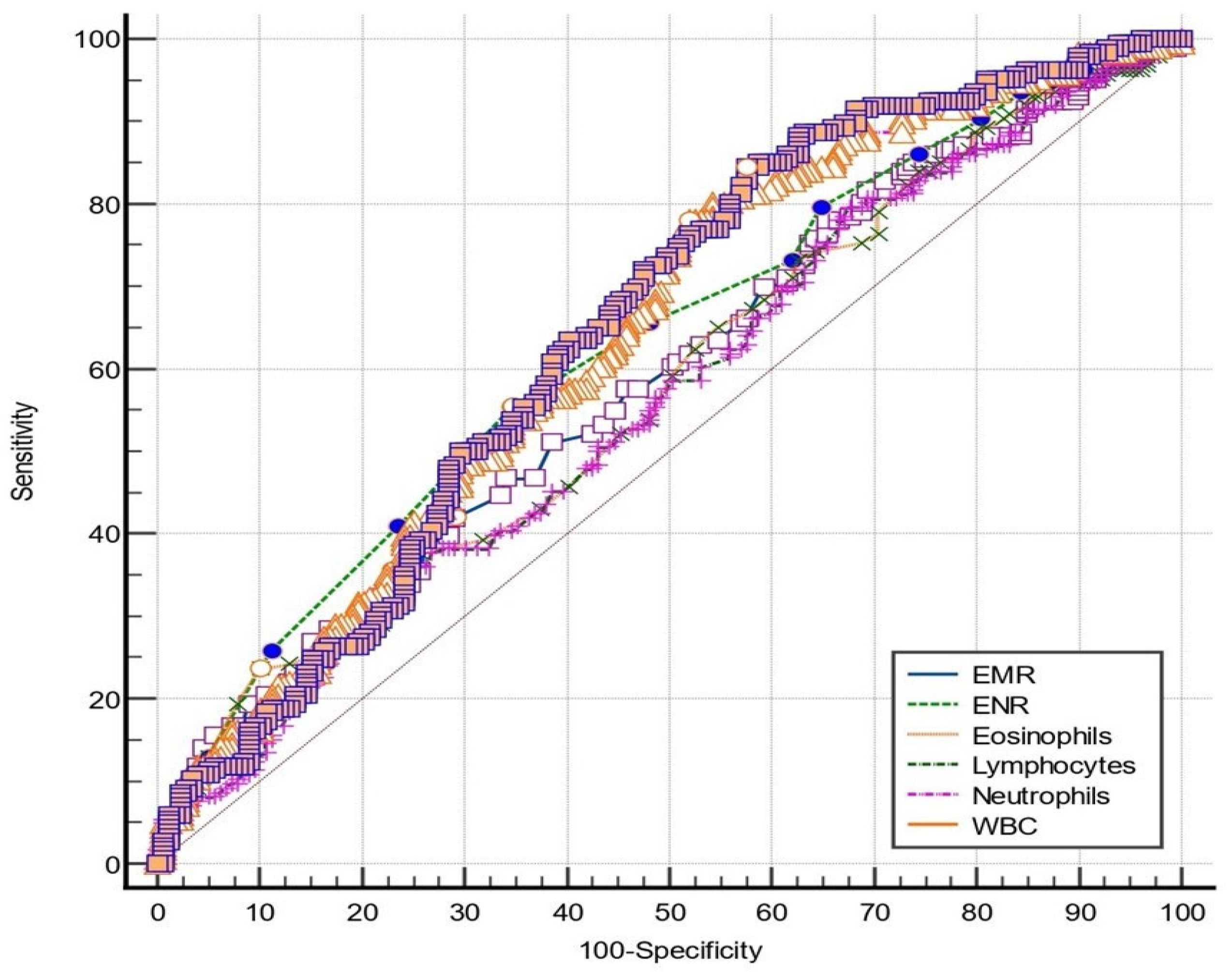
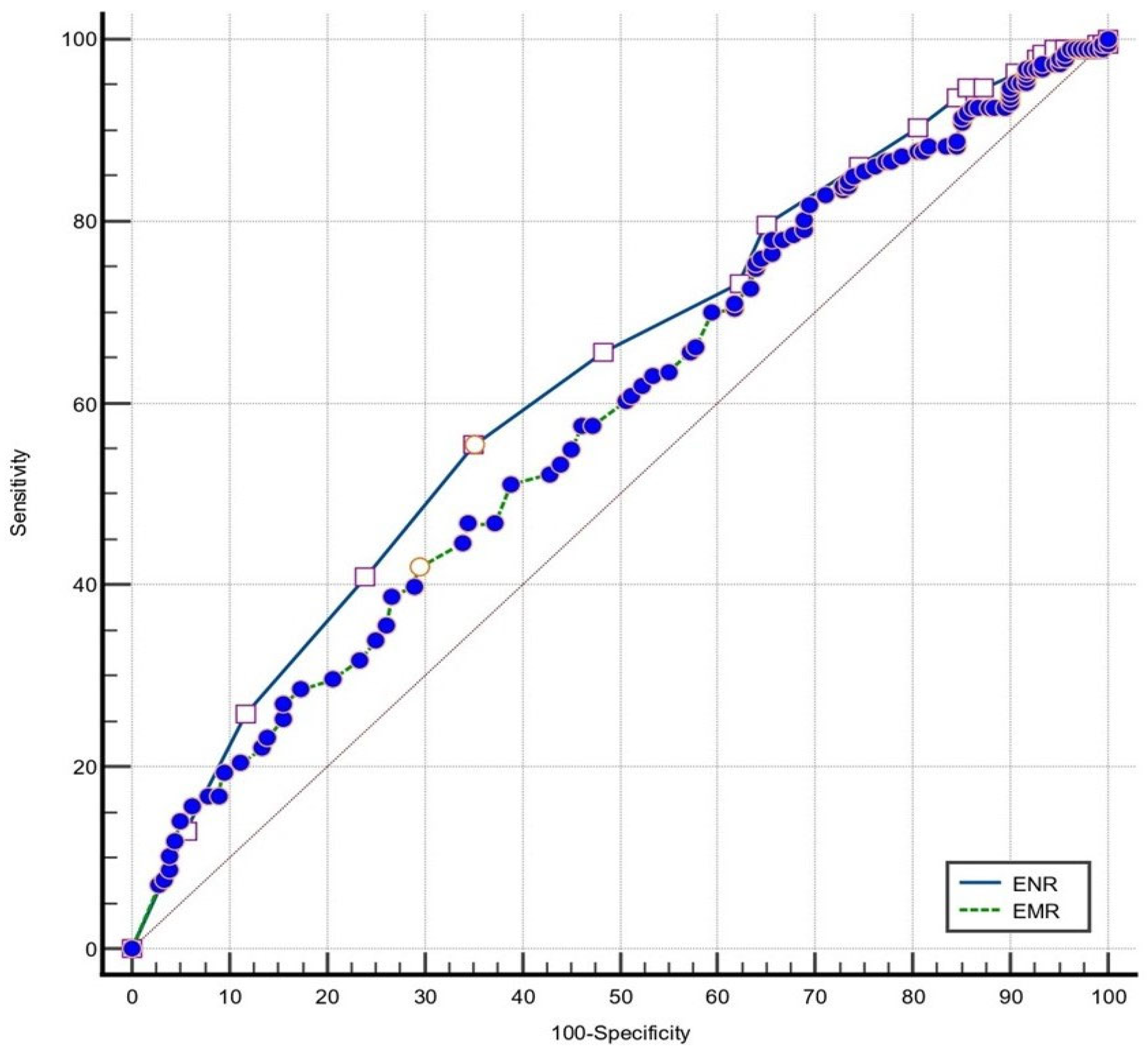
3.3. Eosinophil-Derived Markers Performance for Disease Severity Evaluation

The diagnostic performance of various markers was further analyzed. Table 4 depicts the optimal cut-off values of the analyzed markers and the prediction accuracy of such markers.
When discriminating between mild and moderate-to-severe psoriasis, all markers proved to be statistically significant (p < 0.05). WBC had the highest sensibility, while the eosinophil count had the highest specificity of all tested parameters. Strictly referring to eosinophil-count-derived markers, EMR had higher sensibility and ENR higher specificity. Therefore, values higher than cut-off values of 6.25 for WBC and 3.64 for neutrophil count can confidently predict a moderate-to-severe course of disease. On the other hand, if referring to EMR, ENR, and lymphocyte and eosinophil count, it seems that values below the cut-off values of 0.34, 0.03, 1.7, and 0.05, respectively, adequately predict a milder form of psoriasis.
Next, the ROC curves of all blood-count-derived markers were generated and afterward compared, as seen in Figure 1 and Figure 2. WBC had the highest AUC, but similar to those of neutrophil, lymphocyte, and eosinophil count, EMR, and ENR (p > 0.05). However, when comparing EMR and ENR (Figure 1), it was identified that ENR predicted significantly better psoriasis severity (p = 0.002).

4. Discussion

Previous studies have identified various blood-count-derived inflammatory markers to be associated with psoriasis. The most extensive studies, on neutrophil-to-lymphocyte ratio (NLR) and platelets-to-lymphocyte ratio (PLR), adequately reflect disease severity, treatment response [25], and various comorbidities associated with psoriasis, such as liver fibrosis [26], cardiovascular disease [27,28], or articular damage [29]. Even though several studies focusing on peripheral leucocytes and various subsets are available, as presented earlier, no published reports refer to eosinophil-count-derived inflammatory markers and psoriasis.
In psoriasis, the dermal infiltrate expresses IL-5, IL-8, eotaxin, and monocyte chemoattractant protein 3, molecules that were proven to be able to activate and recruit eosinophils [30]. Moreover, eosinophils were proven to be able to process and stimulate T cells, leading to T cell proliferation and cytokine release [31]. Also, eosinophils release preformed cytokines such as IL-4 and IFN-γ that further promote Th2 or Th1 response [32].
Data referring to the role of the eosinophil are scarce, while information regarding eosinophil-count-derived inflammatory markers in psoriasis was not published until this study. Our analysis identified that patients with moderate-to-severe psoriasis present with higher values of WBC, neutrophils, and monocytes, and lower counts of eosinophils and lymphocytes. These may be due to a continuous activation of neutrophils by proinflammatory cytokines. Decreased lymphocyte levels are directly linked to apoptosis induced by proinflammatory mediators that are produced by the activated keratinocytes and to an increased lymphocytic influx into patients’ skin as severity increases [33]. In this matter, prospective studies focusing on the direct link between the aforementioned proinflammatory mediators and complete blood cell count might give a better understanding of this matter.
Moreover, there is a direct link between eosinophils and lymphocytes, as these two leucocyte subsets were lower in severe forms of psoriasis compared to milder forms. Even though eosinophils cannot be considered classical antigen-presenting cells, they can express on their surface components necessary for antigen presentation and therefore process antigens and stimulate T cells in an antigen-specific manner. Moreover, it was proven that T cells respond to signals provided by the eosinophils [34,35].
Regarding eosinophil-count-derived inflammatory markers ELR, EMR, and ENR, our study identified significantly lower values of EMR and ENR in moderate-to-severe forms compared to milder forms (p values of 0.004 and <0.001, respectively). No significant difference was noted between the two study groups regarding ELR values, suggesting that this marker might not be reliable in assessing the inflammatory status in psoriasis. As the disease progressed, EMR, ENR, and eosinophil count values decreased, as suggested by the significant negative correlation between disease severity and the values of these parameters (rho values of −0.198, −0.188, and −0.127, respectively), probably linked to an increased influx of circulating lymphocytes and eosinophils into the skin.
In discriminating between mild and moderate-to-severe forms of psoriasis, WBC, neutrophil, lymphocyte, and eosinophil count, EMR, and ENR proved to be reliable and significant in predicting disease severity, considering that the AUCs (p < 0.05). EMR (AUC = 0.585), lymphocyte (AUC = 0.561), and eosinophil (AUC = 0.573) count displayed moderate predictive value, while WBC (AUC = 0.644), neutrophil count (AUC = 0.636), and ENR (AUC = 0.627) had good predictive value. Nevertheless, the reliability of these markers should be validated in larger studies.
All eosinophil-count-derived inflammatory markers correlated significantly and negatively with disease severity. As such, moderate-to-severe psoriasis should be defined by ENR values lower than 0.03, EMR values lower than 0.34, and eosinophil counts lower than 0.05. Additionally, WBC values higher than 6.25, neutrophil counts higher than 3.64, and lymphocyte counts lower than 1.7 describe moderate-to-severe psoriasis. However, larger studies should be performed for such values to become widely accepted in predicting disease severity in psoriasis.
The eosinophils have a wide range of roles, interfering in immunomodulation, host response, fibrosis, edema, blister formation, cytotoxic T-cell activation, and B-cell maturation. Moreover, multiple subpopulations of eosinophils have been identified based on cytokine expression [36] and mediator release mechanisms [37]. Different eosinophil subsets exert different functions, and their identification seems like a reliable tool in addressing eosinophil-mediated diseases in the future. Apart from that, they fulfill non-inflammatory roles, such as an appropriate adaptive immune response, tissue integrity, and tissue remodeling [38].
By recognizing receptors through various patterns, eosinophils are involved in host defense. Eosinophil peroxidase, eosinophil cationic protein (ECP), and eosinophil-derived neurotoxin are degranulated due to receptor stimulation. By producing and releasing various cytokines and chemokines, eosinophils interfere in immunomodulation. They are linked to type 2 inflammatory processes and lead to the release of IL-4, IL-9, IL-13, and IL-31, with different cytokine expressions in patients’ skin based on the disease they are presenting. Second, they exert a cytotoxic function by the release of TNF-α. The production and release of IL-31 is a direct link between eosinophils, inflammatory processes, and pruritus [39].
Apart from that, eosinophils have an antimicrobial effect. By forming eosinophil extracellular traps, they have bactericidal properties, and by means of the eosinophil-derived neurotoxin, they seem to exert anti-viral activity [40]. Recently, it was proved that eosinophils also play an important role in the evolution of SARS-CoV-2 infection, with marked eosinopenia being linked to a more severe and prolonged course of disease [41].
Currently, there is a shifting perspective regarding the eosinophil’s role. Apart from their clear involvement in allergic inflammatory diseases, it has become more and more clear that these cells are involved in a much larger variety of disorders, such as post-traumatic tissue repair or granulomatous disorders [42].
Moreover, psoriatic patients present with elevated levels of eosinophil cationic protein (ECP), both in the serum and affected skin [14,43]. Elevated serum ECP is linked to psoriasis, irrespective of disease severity; however, values were observed in patients presenting with moderate and severe psoriasis. Moreover, Michaelsson et al. [43] found that patients with psoriasis presented with low EG2 positive (EG2+) activated eosinophils in the skin and higher levels of EG2+ activated eosinophils in the duodenum compared to those with irritable bowel syndrome, even in association with negative gliadin antibodies. These findings indicate that it is highly unlikely in such cases for elevated serum ECP to be due to eosinophil activation in the skin and thus suggest that elevated EG2+ activated eosinophils in the duodenum may properly reflect the inflammatory activity of psoriatic skin.
In addition, there is some evidence indicating that psoriasis may be associated with peripheric eosinophilia. A linear decrease in high blood eosinophil count and the psoriasis area and severity index (PASI) score was noted after a 54 week course of treatment with vitamin D derivates [44], while additional data describe the positive effect of cyclosporine in reducing peripheral eosinophilia [45,46]. Moreover, it seems that biologics (infliximab, etanercept, adalimumab, and ustekinumab) lead to a sustained increase in peripheral eosinophil count, as early as three months after treatment initiation [47]. Mild peripheral eosinophilia is linked to psoriasis, with conflicting data regarding psoriasis subtype: predominantly chronic plaque, as identified by Zhao et al. [48], or generalized pustular and erythrodermic forms [49].
In our study, the co-occurrence of peripheral eosinophilia and psoriasis was not common; only one patient from each group presented with this modification. When comparing these two patients to the complete study group size, there are not enough data to properly sustain a more than hazardous association between the absolute eosinophil count and psoriasis severity. However, future ideas might include analyzing data from patients presenting with other subtypes of psoriasis and comparing absolute peripheral eosinophil count in psoriatic patients to patients suffering from eosinophil-mediated disorders, such as drug reactions or atopic dermatitis.
Historically, from a histopathological point of view, psoriasis is defined by psoriasiform epidermal hyperplasia, elongated rete ridges, parakeratosis, and intraepidermal neutrophils. Eosinophils are stated to be absent in psoriatic skin; however, studies sustaining this are scarce [50,51]. To this matter, new data related to eosinophil identification on biopsy specimens should be taken into account. On routinely used colorations, i.e., hematoxylin-eosin, psoriasis lesions typically exhibit no or a reduced number of eosinophils. In a study published by Moy et al. [52], perivascular eosinophils were observed in all included cases of erythrodermic psoriasis. Regarding chronic plaque psoriasis, dermal eosinophils were reported in almost half of the reviewed biopsies in studies by Chau et al. [53] and Penn et al. [54]. On the other hand, data analyzed by Rosa et al. [55] identified dermal eosinophils only in 18% of psoriasis cases. An increased immunohistochemical staining for the EG2 monoclonal antibody [56] and ECP [57] compared to normal skin was proved to be linked to psoriasis. The eosinophil count was higher in active lesions and in patients presenting with a severe course of disease. These data are in contradiction with our findings, since peripheral eosinophils were lower in count in advanced forms of the disease, most likely due to these cells being trapped in patients’ skin. It is considered that each additional eosinophil in the peripheral blood leads to a corresponding increase of 100 eosinophils in patients’ skin [49]. Patients with fast-developing psoriasis present with strong ECP signal especially in the upper part of the epidermis, while in slowly progressing lesions the expression of such a marker was reduced [57]. This information suggests once more that eosinophils are part of the inflammatory infiltrate observed in lesional skin.
However, it should be noted that the lack of eosinophils on biopsy specimens can also serve as an important differential diagnosis tool. Absent or minimal eosinophils are an essential clue towards differentiating psoriasis from acute generalized exanthematous pustulosis [58], pityriasis lichenoides [59], and drug-induced psoriasiform eruptions [60,61]. Also, previous treatments may alter the classic histopathological pattern of psoriasis, making the diagnosis more difficult. On the other hand, an appropriate selection of the biopsy place should be made, since superimposed eczemas or sensitivity reactions to various triggers may lead to eosinophil overexpression in that specific biopsy specimen. However, it is worth mentioning that marked tissue eosinophilia is a hallmark of atopic dermatitis, as Th2-type lymphocytes lead to eosinophils recruitment in the affected areas. Compared to atopic dermatitis, chronic plaque psoriasis and pustular psoriasis present with a lower count of epidermal and dermal eosinophils [62]. As such, the presence of eosinophils in skin biopsies should not rule out the diagnosis of psoriasis, especially if other known histologic features are identified.
The main limitation of this study derives from its single-center retrospective character; to address this, future research might benefit from the prospective enrollment of study participants. Disease severity was based on the BSA scale and not on the PASI score, to limit the possible dependence on the examiner’s experience assessment of induration, erythema, and scaling. A comprehensive, multi-score assessment of disease severity ought to be considered in the future. It would be useful to further investigate the association between eosinophil count and derived parameters in larger cohort studies. It is widely accepted that eosinophils present with diurnal variation, with lower values in the morning and higher ones at night. To limit this, all blood samples were obtained in the morning, after an overnight fast.

5. Conclusions

The absolute eosinophil count and eosinophil-count-derived inflammatory markers EMR and ENR are reliable indicators of systemic inflammation and disease severity in psoriasis. Additionally, ENR proves to be the most reliable eosinophil-count-derived inflammatory marker in predicting disease severity across the two study groups.
Reporting for the first time the usefulness of eosinophil-count-derived inflammatory markers, our study highlights the usefulness of these easily obtainable and cost-effective markers in evaluating psoriasis inflammation.

Author Contributions

Conceptualization, O.M.T., S.H.M. and O.S.C.; formal analysis, O.M.T.; methodology, O.M.T., C.R.M. and A.C.N.; resources, C.R.M., R.A.T. and A.C.N.; validation, O.S.C. and S.H.M.; visualization, O.M.T. and R.A.T.; writing–original draft, O.M.T., C.R.M. and R.A.T.; writing—revision and editing, O.M.T. and A.C.N.; supervision, O.S.C. and S.H.M. All authors have read and agreed to the published version of the manuscript.

Funding

This research received no external funding.

Institutional Review Board Statement

The study was conducted in accordance with the Declaration of Helsinki, and approved by the Ethics Committee Mures Clinical County Hospital no. 3770/05.04.2023.

Informed Consent Statement

Informed consent was obtained from all subjects involved in the study.

Data Availability Statement

All data presented can be made available upon request.

Acknowledgments

This article is part of a Ph.D. thesis from the Doctoral School of Medicine and Pharmacy of the University of Medicine, Pharmacy, Science, and Technology George Emil Palade of Targu Mures, titled “The impact of systemic inflammation in modulating disease presentation in psoriasis”, which will be presented by Oana Mirela Tiucă by the fall of 2024.

Conflicts of Interest

The authors declare no conflicts of interest.

References

  1. Singh, S.; Young, P.; Armstrong, A.W. Relationship between psoriasis and metabolic syndrome: A systematic review. G. Ital. Dermatol. Venereol. 2016, 151, 663–677. [Google Scholar] [PubMed]
  2. Luan, L.; Han, S.; Wang, H.; Liu, X. Down-regulation of the Th1, Th17, and Th22 pathways due to anti-TNF-α treatment in psoriasis. Int. Immunopharmacol. 2015, 29, 278–284. [Google Scholar] [CrossRef] [PubMed]
  3. Canavese, M.; Altruda, F.; Ruzicka, T.; Schauber, J. Vascular endothelial growth factor (VEGF) in the pathogenesis of psoriasis—A possible target for novel therapies? J. Dermatol. Sci. 2010, 58, 171–176. [Google Scholar] [CrossRef] [PubMed]
  4. Frieder, J.; Ryan, C. Psoriasis and cardiovascular disorders. G. Ital. Dermatol. Venereol. 2016, 151, 678–693. [Google Scholar] [PubMed]
  5. Grozdev, I.; Korman, N.; Tsankov, N. Psoriasis as a systemic disease. Clin. Dermatol. 2014, 32, 343–350. [Google Scholar] [CrossRef] [PubMed]
  6. Larmann, J.; Handke, J.; Scholz, A.S.; Dehne, S.; Arens, C.; Gillmann, H.J.; Uhle, F.; Motsch, J.; Weigand, M.A.; Janssen, H. Preoperative neutrophil to lymphocyte ratio and platelet to lymphocyte ratio are associated with major adverse cardiovascular and cerebrovascular events in coronary heart disease patients undergoing non-cardiac surgery. BMC Cardiovasc. Disord. 2020, 20, 230. [Google Scholar] [CrossRef] [PubMed]
  7. Modica, R.; Minotta, R.; Liccardi, A.; Cannavale, G.; Benevento, E.; Colao, A. Evaluation of Neutrophil-to-Lymphocyte Ratio (NLR), Platelet-to-Lymphocyte Ratio (PLR) and Systemic Immune–Inflammation Index (SII) as Potential Biomarkers in Patients with Sporadic Medullary Thyroid Cancer (MTC). JPM 2023, 13, 953. [Google Scholar] [CrossRef] [PubMed]
  8. Lin, Z.-Q.; Ma, C.; Cao, W.-Z.; Ning, Z.; Tan, G. Prognostic Significance of NLR, PLR, LMR and Tumor Infiltrating T Lymphocytes in Patients Undergoing Surgical Resection for Hilar Cholangiocarcinoma. Front. Oncol. 2022, 12, 908907. [Google Scholar] [CrossRef]
  9. Gambardella, C.; Mongardini, F.M.; Paolicelli, M.; Bentivoglio, D.; Cozzolino, G.; Ruggiero, R.; Pizza, A.; Tolone, S.; del Genio, G.; Parisi, S.; et al. Role of Inflammatory Biomarkers (NLR, LMR, PLR) in the Prognostication of Malignancy in Indeterminate Thyroid Nodules. Int. J. Mol. Sci. 2023, 24, 6466. [Google Scholar] [CrossRef]
  10. Erre, G.L.; Paliogiannis, P.; Castagna, F.; Mangoni, A.A.; Carru, C.; Passiu, G.; Zinellu, A. Meta-analysis of neutrophil-to-lymphocyte and platelet-to-lymphocyte ratio in rheumatoid arthritis. Eur. J. Clin. Investig. 2019, 49, e13037. [Google Scholar] [CrossRef]
  11. Wu, J.; Yan, L.; Chai, K. Systemic immune-inflammation index is associated with disease activity in patients with ankylosing spondylitis. Clin. Lab. Anal. 2021, 35, e23964. [Google Scholar] [CrossRef] [PubMed]
  12. Kim, Y.; Choi, H.; Jung, S.M.; Song, J.J.; Park, Y.; Lee, S. Systemic immune-inflammation index could estimate the cross-sectional high activity and the poor outcomes in immunosuppressive drug-naïve patients with antineutrophil cytoplasmic antibody-associated vasculitis. Nephrology 2019, 24, 711–717. [Google Scholar] [CrossRef] [PubMed]
  13. Uslu, A.U.; Küçük, A.; Şahin, A.; Ugan, Y.; Yılmaz, R.; Güngör, T.; Bağcacı, S.; Küçükşen, S. Two new inflammatory markers associated with Disease Activity Score-28 in patients with rheumatoid arthritis: Neutrophil-lymphocyte ratio and platelet-lymphocyte ratio. Int. J. Rheum. Dis. 2015, 18, 731–735. [Google Scholar] [CrossRef] [PubMed]
  14. Juhlin, L.; Venge, P. Eosinophilic cationic protein (ECP) in skin disorders. Acta Derm. Venereol. 1991, 71, 495–501. [Google Scholar] [CrossRef] [PubMed]
  15. Skrzeczynska-Moncznik, J.; Wlodarczyk, A.; Zabieglo, K.; Kapinska-Mrowiecka, M.; Marewicz, E.; Dubin, A.; Potempa, J.; Cichy, J. Secretory Leukocyte Proteinase Inhibitor-Competent DNA Deposits Are Potent Stimulators of Plasmacytoid Dendritic Cells: Implication for Psoriasis. J. Immunol. 2012, 189, 1611–1617. [Google Scholar] [CrossRef] [PubMed]
  16. Kim, H.J.; Roh, J.Y.; Jung, Y. Eosinophils Accelerate Pathogenesis of Psoriasis by Supporting an Inflammatory Milieu that Promotes Neutrophil Infiltration. J. Investig. Dermatol. 2018, 138, 2185–2194. [Google Scholar] [CrossRef] [PubMed]
  17. Yenigun, A.; Sezen, S.; Calim, O.F.; Ozturan, O. Evaluation of the Eosinophil-to-lymphocyte Ratio in Pediatric Patients with Allergic Rhinitis. Am. J. Rhinol. Allergy 2016, 30, e21–e25. [Google Scholar] [CrossRef] [PubMed]
  18. Lan, C.; Su, W.; Yang, M.; Chen, S.; Wu, Y. Predictive role of neutrophil-percentage-to-albumin, neutrophil-to-lymphocyte and eosinophil-to-lymphocyte ratios for mortality in patients with COPD: Evidence from NHANES 2011–2018. Respirology 2023, 28, 1136–1146. [Google Scholar] [CrossRef]
  19. Ferro, M.; Musi, G.; Serino, A.; Cozzi, G.; Mistretta, F.A.; Costa, B.; Bianchi, R.; Cordima, G.; Luzzago, S.; Di Trapani, E.; et al. Neutrophil, Platelets, and Eosinophil to Lymphocyte Ratios Predict Gleason Score Upgrading in Low-Risk Prostate Cancer Patients. Urol. Int. 2019, 102, 43–50. [Google Scholar] [CrossRef]
  20. Georgakopoulou, V.E.; Garmpis, N.; Damaskos, C.; Valsami, S.; Dimitroulis, D.; Diamantis, E.; Farmaki, P.; Papageorgiou, C.V.; Makrodimitri, S.; Gravvanis, N.; et al. The Impact of Peripheral Eosinophil Counts and Eosinophil to Lymphocyte Ratio (ELR) in the Clinical Course of COVID-19 Patients: A Retrospective Study. In Vivo 2021, 35, 641–648. [Google Scholar] [CrossRef]
  21. Chen, Y.; Ren, J.; Yang, N.; Huang, H.; Hu, X.; Sun, F.; Zeng, T.; Zhou, X.; Pan, W.; Hu, J.; et al. Eosinophil-to-Monocyte Ratio is a Potential Predictor of Prognosis in Acute Ischemic Stroke Patients After Intravenous Thrombolysis. Clin. Interv. Aging 2021, 16, 853–862. [Google Scholar] [CrossRef] [PubMed]
  22. Deng, X.; Wang, X.; Shen, L.; Yao, K.; Ge, L.; Ma, J.; Zhang, F.; Qian, J.; Ge, J. Association of eosinophil-to-monocyte ratio with 1-month and long-term all-cause mortality in patients with ST-elevation myocardial infarction undergoing primary percutaneous coronary intervention. J. Thorac. Dis. 2018, 10, 5449–5458. [Google Scholar] [CrossRef] [PubMed]
  23. Hagino, T.; Saeki, H.; Fujimoto, E.; Kanda, N. The Eosinophil-to-Lymphocyte Ratio Acts as an Indicator for Improvement of Clinical Signs and Itch by Upadacitinib Treatment in Atopic Dermatitis. JCM 2023, 12, 2201. [Google Scholar] [CrossRef] [PubMed]
  24. Weissmann, S.; Burrack, N.; Babyev, A.S.; Gordon, M.; Golan-Tripto, I.; Horev, A. Eosinophil–Lymphocyte Ratio, Eosinophil–Neutrophil Ratio, and Eosinophil–Monocyte Ratio in Chronic and Severe Urticaria. Am. J. Clin. Dermatol. 2023, 24, 669–671. [Google Scholar] [CrossRef] [PubMed]
  25. Asahina, A.; Kubo, N.; Umezawa, Y.; Honda, H.; Yanaba, K.; Nakagawa, H. Neutrophil-lymphocyte ratio, platelet-lymphocyte ratio and mean platelet volume in Japanese patients with psoriasis and psoriatic arthritis: Response to therapy with biologics. J. Dermatol. 2017, 44, 1112–1121. [Google Scholar] [CrossRef] [PubMed]
  26. Tiucă, O.M.; Morariu, S.H.; Mariean, C.R.; Tiucă, R.A.; Nicolescu, A.C.; Cotoi, O.S. Predictive Performances of Blood-Count-Derived Inflammatory Markers for Liver Fibrosis Severity in Psoriasis Vulgaris. Int. J. Mol. Sci. 2023, 24, 16898. [Google Scholar] [CrossRef] [PubMed]
  27. Amalia, L.; Dalimonthe, N.Z. Clinical significance of Platelet-to-White Blood Cell Ratio (PWR) and National Institute of Health Stroke Scale (NIHSS) in acute ischemic stroke. Heliyon 2020, 6, e05033. [Google Scholar] [CrossRef] [PubMed]
  28. Abdulhadi, B.; Naranjo, M.; Krishnamoorthy, P.; Rangaswami, J. White blood cell count to platelet ratio: A novel biomarker for predicting outcomes in patients on circulatory support devices. J. Am. Coll. Cardiol. 2018, 71, A810. [Google Scholar] [CrossRef]
  29. Yorulmaz, A.; Hayran, Y.; Akpinar, U.; Yalcin, B. Systemic Immune-Inflammation Index (SII) Predicts Increased Severity in Psoriasis and Psoriatic Arthritis. Curr. Health Sci. J. 2020, 46, 352–357. [Google Scholar] [PubMed]
  30. Yawalkar, N.; Shrikhande, M.; Hari, Y.; Nievergelt, H.; Braathen, L.R.; Pichler, W.J. Evidence for a role for IL-5 and eotaxin in activating and recruiting eosinophils in drug-induced cutaneous eruptions. J. Allergy Clin. Immunol. 2000, 106, 1171–1176. [Google Scholar] [CrossRef]
  31. Wang, H.B.; Ghiran, I.; Matthaei, K.; Weller, P.F. Airway Eosinophils: Allergic Inflammation Recruited Professional Antigen-Presenting Cells. J. Immunol. 2007, 179, 7585–7592. [Google Scholar] [CrossRef] [PubMed]
  32. Spencer, L.A.; Szela, C.T.; Perez, S.A.C.; Kirchhoffer, C.L.; Neves, J.S.; Radke, A.L.; Weller, P.F. Human eosinophils constitutively express multiple Th1, Th2, and immunoregulatory cytokines that are secreted rapidly and differentially. J. Leukoc. Biol. 2009, 85, 117–123. [Google Scholar] [CrossRef] [PubMed]
  33. Langewouters, A.M.G.; Van Erp, P.E.J.; De Jong, E.M.G.J.; Van De Kerkhof, P.C.M. Lymphocyte subsets in peripheral blood of patients with moderate-to-severe versus mild plaque psoriasis. Arch. Dermatol. Res. 2008, 300, 107–113. [Google Scholar] [CrossRef] [PubMed]
  34. Mattes, J.; Yang, M.; Mahalingam, S.; Kuehr, J.; Webb, D.C.; Simson, L.; Hogan, S.P.; Koskinen, A.; McKenzie, A.N.; Dent, L.A.; et al. Intrinsic Defect in T Cell Production of Interleukin (IL)-13 in the Absence of Both IL-5 and Eotaxin Precludes the Development of Eosinophilia and Airways Hyperreactivity in Experimental Asthma. J. Exp. Med. 2002, 195, 1433–1444. [Google Scholar] [CrossRef]
  35. MacKenzie, J.R.; Mattes, J.; Dent, L.A.; Foster, P.S. Eosinophils Promote Allergic Disease of the Lung by Regulating CD4+ Th2 Lymphocyte Function. J. Immunol. 2001, 167, 3146–3155. [Google Scholar] [CrossRef] [PubMed]
  36. Roth, N.; Städler, S.; Lemann, M.; Hösli, S.; Simon, H.U.; Simon, D. Distinct eosinophil cytokine expression patterns in skin diseases—The possible existence of functionally different eosinophil subpopulations. Allergy 2011, 66, 1477–1486. [Google Scholar] [CrossRef] [PubMed]
  37. Simon, D.; Hoesli, S.; Roth, N.; Staedler, S.; Yousefi, S.; Simon, H.U. Eosinophil extracellular DNA traps in skin diseases. J. Allergy Clin. Immunol. 2011, 127, 194–199. [Google Scholar] [CrossRef] [PubMed]
  38. Lee, J.J.; Jacobsen, E.A.; McGarry, M.P.; Schleimer, R.P.; Lee, N.A. Eosinophils in health and disease: The LIAR hypothesis. Clin. Exp. Allergy 2010, 40, 563–575. [Google Scholar] [CrossRef] [PubMed]
  39. Kunsleben, N.; Rüdrich, U.; Gehring, M.; Novak, N.; Kapp, A.; Raap, U. IL-31 Induces Chemotaxis, Calcium Mobilization, Release of Reactive Oxygen Species, and CCL26 in Eosinophils, Which Are Capable to Release IL-31. J. Investig. Dermatol. 2015, 135, 1908–1911. [Google Scholar] [CrossRef]
  40. Rugeles, M.T.; Trubey, C.M.; Bedoya, V.I.; Pinto, L.A.; Oppenheim, J.J.; Rybak, S.M.; Shearer, G.M. Ribonuclease is partly responsible for the HIV-1 inhibitory effect activated by HLA alloantigen recognition. AIDS 2003, 17, 481–486. [Google Scholar] [CrossRef]
  41. Roca, E.; Ventura, L.; Zattra, C.M.; Lombardi, C. EOSINOPENIA: An early, effective and relevant COVID-19 biomarker? QJM Int. J. Med. 2021, 114, 68–69. [Google Scholar] [CrossRef] [PubMed]
  42. Gonlugur, U.; Efeoglu Gonlugur, T. Non-allergic Eosinophilic Inflammation. Immunol. Investig. 2006, 35, 29–45. [Google Scholar] [CrossRef] [PubMed]
  43. Michaëlsson, G.; Kraaz, W.; Gerdén, B.; Hagforsen, E.; Lundin, I.P.; Lööf, L.; Sjöberg, O.; Scheynius, A. Patients with psoriasis have elevated levels of serum eosinophil cationic protein and increased numbers of EG2 positive eosinophils in the duodenal stroma. Br. J. Dermatol. 1996, 135, 371–378. [Google Scholar] [CrossRef] [PubMed]
  44. Sueki, H.; Nakada, T.; Iijima, M. A case of psoriasis vulgaris with peripheral blood eosinophilia, parallelling the psoriasis area and severity index (PASI) score. Clin. Exp. Dermatol. 2004, 29, 549–550. [Google Scholar] [CrossRef] [PubMed]
  45. Shupack, J.L.; Kenny, C.; Jondreau, L.; Eckman, I.; Gropper, C.; Stiller, M.J. Decreased Peripheral Blood Eosinophil Counts in Severe Psoriatic Patients Treated with Low-Dose Cyclosporine A. Dermatology 1992, 185, 202–204. [Google Scholar] [CrossRef] [PubMed]
  46. Schopf, R.E.; Hultsch, T.; Lotz, J.; Bräutigam, M. Eosinophils, pruritus and psoriasis: Effects of treatment with etretinate or cyclosporin-A. J. Eur. Acad. Dermatol. Venereol. 1998, 11, 234–239. [Google Scholar]
  47. Akdogan, N.; Dogan, S.; Atakan, N. Long-term effects of biologic therapies on peripheral blood eosinophils in patients with psoriasis: A 3-year single-center study. J. Dermatol. Treat. 2020, 31, 702–706. [Google Scholar] [CrossRef]
  48. Zhao, Y.; Tian, J.; Gao, C.; Liu, L.; Pan, L.; Song, Z. Retrospective Analysis of 397 Dermatoses Inpatients Associated with Blood Eosinophilia. Clin. Cosmet. Investig. Dermatol. 2023, 16, 3455–3463. [Google Scholar] [CrossRef]
  49. Mansur, A.; Göktay, F.; Yaşar, Ş. Peripheral blood eosinophilia in association with generalized pustular and erythrodermic psoriasis. Acad. Dermatol. Venereol. 2008, 22, 451–455. [Google Scholar] [CrossRef]
  50. Patterson, J.W. The psoriasiform reaction pattern. In Weedon’s Skin Pathology, 4th ed.; Elsevier: Philadelphia, PA, USA, 2016; pp. 82–90. [Google Scholar]
  51. Billings, S.D.; Cotton, J. Psoriasiform dermatitis. In Inflammatory Dermatopathology, 2nd ed.; Springer: Cham, Switzerland, 2016; p. 24. [Google Scholar]
  52. Moy, A.P.; Murali, M.; Kroshinsky, D.; Duncan, L.M.; Nazarian, R.M. Immunologic Overlap of Helper T-Cell Subtypes 17 and 22 in Erythrodermic Psoriasis and Atopic Dermatitis. JAMA Dermatol. 2015, 151, 753. [Google Scholar] [CrossRef]
  53. Chau, T.; Parsi, K.K.; Ogawa, T.; Kiuru, M.; Konia, T.; Li, C.; Fung, M.A. Psoriasis or not? Review of 51 clinically confirmed cases reveals an expanded histopathologic spectrum of psoriasis. J. Cutan. Pathol. 2017, 44, 1018–1026. [Google Scholar] [CrossRef] [PubMed]
  54. Penn, L.; Brinster, N.K. Eosinophils Among the Histological Features of Psoriasis. Am. J. Dermatopathol. 2019, 41, 347–349. [Google Scholar] [CrossRef] [PubMed]
  55. Rosa, G.; Fernandez, A.P.; Schneider, S.; Billings, S.D. Eosinophils are rare in biopsy specimens of psoriasis vulgaris. J. Cutan. Pathol. 2017, 44, 1027–1032. [Google Scholar] [CrossRef] [PubMed]
  56. Tai, P.C.; Spry, C.J.F.; Peterson, C.; Venge, P.; Olsson, I. Monoclonal antibodies distinguish between storage and secreted forms of eosinophil cationic protein. Nature 1984, 309, 182–184. [Google Scholar] [CrossRef]
  57. Lundin, A.; Fredens, K.; Michaelsson, G.; Venge, P. The eosinophil granulocyte in psoriasis. Br. J. Dermatol. 1990, 122, 181–193. [Google Scholar] [CrossRef] [PubMed]
  58. Kardaun, S.H.; Kuiper, H.; Fidler, V.; Jonkman, M.F. The histopathological spectrum of acute generalized exanthematous pustulosis (AGEP) and its differentiation from generalized pustular psoriasis. J. Cutan. Pathol. 2010, 37, 1220–1229. [Google Scholar] [CrossRef] [PubMed]
  59. Sharon, V.R.; Konia, T.H.; Barr, K.L.; Fung, M.A. Assessment of the ‘no eosinophils’ rule: Are eosinophils truly absent in pityriasis lichenoides, connective tissue disease, and graft-vs.-host disease? J. Cutan. Pathol. 2012, 39, 413–418. [Google Scholar] [CrossRef] [PubMed]
  60. Laga, A.C.; Vleugels, R.A.; Qureshi, A.A.; Velazquez, E.F. Histopathologic Spectrum of Psoriasiform Skin Reactions Associated With Tumor Necrosis Factor-α Inhibitor Therapy. A Study of 16 Biopsies. Am. J. Dermatopathol. 2010, 32, 568–573. [Google Scholar] [CrossRef] [PubMed]
  61. Basavaraj, K.H.; Ashok, N.M.; Rashmi, R.; Praveen, T.K. The role of drugs in the induction and/or exacerbation of psoriasis. Int. J. Dermatol. 2010, 49, 1351–1361. [Google Scholar] [CrossRef]
  62. Steigleder, G.K.; Inderwisch, R. Eosinophile franulocyten in der efflorescenz bei psoriasis und neurodermitis constitutionalis (atopische dermatitis). Arch. Derm. Res. 1975, 254, 253–255. [Google Scholar] [CrossRef]
Figure 1. ROC comparison of WBC, neutrophils, eosinophils, lymphocytes, ENR, and EMR in predicting psoriasis severity.
Dermato 04 00004 g001
Figure 2. ROC comparison of ENR and EMR in predicting psoriasis severity.
Dermato 04 00004 g002
Table 1. Formulas of analyzed biomarkers.
MarkerFormula
ELREosinophil count/lymphocyte count (×103/μL)
EMREosinophil count/monocyte count (×103/μL)
ENREosinophil count/neutrophil count (×103/μL)
ELR, eosinophil-to-lymphocyte ratio (ELR); EMR, eosinophil-to-monocyte ratio (EMR); and ENR, eosinophil-to-neutrophil ratio (ENR).
Table 2. Clinical and laboratory characteristics of the study population.
VariablesAll PatientsMild Disease (n = 180)Moderate-to-Severe Disease (n = 186)p-Value *
Age54.48 ± 16.4853.86 ± 17.4555.08 ± 15.510.723
Gender
  Male219101 (56%)118 (63.4%)0.074
  Female14779 (44%)68 (36.6%)
WBC (×103/L)7.50 [7.15–7.83]6.75 [6.33–7.24]8.03 [7.62–8.33]<0.001
Neutrophils (×103/L)4.27
[4.09–4.56]
3.78 [3.43–4.24]4.77 [4.30–5.04]<0.001
Lymphocytes (×103/L)2.10 [1.97–2.23]2.22 [1.99–2.30]2.02 [1.94–2.21]0.043
Monocytes (×103/L)0.51 [0.48–0.53]0.49 [0.45–0.52]0.52 [0.48–0.55]0.345
Eosinophils (×103/L)0.15 [0.14–0.17]0.16 [0.14–0.19]0.14 [0.12–0.16]0.012
ELR0.10 [0.09–0.10]0.08 [0.07–0.86]0.07 [0.06–0.08]0.087
EMR0.27 [0.25–0.28]0.33 [0.27–0.39]0.26 [0.23–0.30]0.004
ENR0.04 [0.03–0.04]0.05 [0.04–0.05]0.03 [0.03–0.05]<0.001
WBC, white blood cell count; ELR, eosinophil-to-lymphocyte ratio (ELR); EMR, eosinophil-to-monocyte ratio (EMR); and ENR, eosinophil-to-neutrophil ratio (ENR); data presented as median and confidence interval; * comparison between study groups as previously defined.
Table 3. Correlation between serological markers and disease severity.
Markerrp-Value
WBC0.250<0.001
Neutrophil count0.236<0.001
Lymphocyte count −0.1060.042
Eosinophil count−0.1270.015
EMR−0.198<0.001
ENR−0.188<0.001
WBC, white blood cell count; EMR, eosinophil-to-monocyte ratio (EMR); and ENR, eosinophil-to-neutrophil ratio (ENR).
Table 4. Predictive performance of eosinophil-count-derived inflammatory markers.
ParameterAUC
(95% CI)
p-ValueCut-OffSe (%)Sp (%)Youden Index Jp-Value *
WBC0.644
[0.593–0.693]
<0.0016.2584.4142.220.27-
Neutrophil count0.636
[0.585–0.686]
<0.0013.6477.9647.780.260.59
Lymphocyte count0.561
[0.509–0.613]
0.0411.735.4876.670.120.09
Eosinophil count0.573
[0.521–0.625]
0.0140.0523.2489.940.130.10
EMR0.585
[0.536–0.633]
<0.0010.3478.4948.890.270.14
ENR 0.627
[0.575–0.678]
<0.0010.0355.40650.920.63
WBC, white blood cell count; ELR, eosinophil-to-lymphocyte ratio (ELR); EMR, eosinophil-to-monocyte ratio (EMR); and ENR, eosinophil-to-neutrophil ratio (ENR); * compared to WBC.
Disclaimer/Publisher’s Note: The statements, opinions and data contained in all publications are solely those of the individual author(s) and contributor(s) and not of MDPI and/or the editor(s). MDPI and/or the editor(s) disclaim responsibility for any injury to people or property resulting from any ideas, methods, instructions or products referred to in the content.

