Case Report

Rare Presentation, Critical Diagnosis: Primary Actinomycosis of the Foot

by
Alexandra Maria Dorobanțu
1,
Mihai Lupu
2,
Liliana Gabriela Popa
1,3,*,
Raluca Tatar
3,4,
Calin Giurcaneanu
1,3,
Irina Tudose
5 and
Olguta Anca Orzan
1,3
1
Department of Dermatology, ‘Elias’ Emergency University Hospital, 011461 Bucharest, Romania
2
Panduri Medical Center, 011367 Bucharest, Romania
3
Department of Dermatology, ‘Carol Davila’ University of Medicine and Pharmacy, 020021 Bucharest, Romania
4
Department of Plastic Reconstructive Surgery and Burns, ‘Grigore Alexandrescu’ Clinical Emergency Hospital for Children, 011743 Bucharest, Romania
5
Anatomic Pathology Laboratory, ‘Elias’ University Emergency Hospital, 011461 Bucharest, Romania
*
Author to whom correspondence should be addressed.
Dermato 2024, 4(3), 72-78; https://doi.org/10.3390/dermato4030008
Submission received: 16 February 2024 / Revised: 6 April 2024 / Accepted: 26 June 2024 / Published: 4 July 2024
(This article belongs to the Special Issue What Is Your Diagnosis?—Case Report Collection)

Abstract

:
Actinomycosis, an uncommon granulomatous infection caused by the Actinomyces species, rarely targets as primary involvement the limb and is often linked to traumatic incidents. In this report, we present the case of a 44-year-old female who developed multiple small nodules on her left foot over approximately 12 months. Some nodules exhibited firmness and a violet hue, while others discharged a yellowish fluid. The patient had no significant comorbidities. Despite thorough blood paraclinical assessments, including complete blood count, serological HIV testing, and QuantiFERON-TB Gold testing, no abnormalities were detected. Bacteriological examinations and cultures of the discharge yielded negative results. Dermatoscopic examination revealed ovoid yellowish structures, with confocal microscopy highlighting granulomas. A subsequent skin biopsy confirmed characteristic changes indicative of actinomycosis. Although systemic antibiotic therapy with penicillin derivatives was initially considered, the patient’s documented allergic history to this medication class, verified through allergological testing, prompted the initiation of doxycycline treatment. Notably, significant improvement was observed at the 3-month follow-up. This case underscores the importance of reporting rare instances of actinomycosis due to its diagnostic complexity and management challenges.

1. Introduction

Actinomycosis is a rare and slowly progressing granulomatous infection caused by Actinomyces species, particularly Actinomyces israelii. These Gram-positive bacilli are commensal organisms of the oral cavity, gastrointestinal, and urogenital tract [1]. Actinomycosis can affect the respiratory tract, skin, musculoskeletal system, digestive tract, central nervous system, and other body parts [1]. However, the infection is predominantly localized in the cervicofacial (55%), abdominopelvic (20%), and pulmonothoracic (15%) regions [2]. It is noteworthy that primary limb involvement is exceptionally rare, while extremity involvement is usually secondary to local extension or hematogenous dissemination [3].
The incidence of actinomycosis has been reported to have declined in recent years, especially in developed countries [4]. Although there is little accurate prevalence data in the literature, the overall incidence of actinomycosis appears to have decreased significantly in the last three to four decades [5]. Actinomycosis has been documented in all age groups, with the highest incidence in mid-life and lower frequencies at each end of the age spectrum [6]. Actinomycosis is considered a rare infection, with a reported annual incidence of 1/300,000 persons, although reliable epidemiological data on its prevalence is lacking [7]. Actinomycosis is also known to affect middle-aged individuals and is more prevalent in women [8].
The incidence of actinomycosis in the foot is not extensively documented. However, the rarity of actinomycosis, its tendency to mimic malignancy, and its potential diagnostic overlap with other conditions, such as skeletal tuberculosis (TB), warrant consideration when evaluating the incidence of actinomycosis in the foot, particularly in regions with a high prevalence of TB.
A multitude of risk factors, including demographic, behavioral, and various medical conditions, influence actinomycosis. Immunodeficiency, hypoproteinemia, and diabetes are among the medical conditions that have been linked to an increased susceptibility to actinomycosis [9]. In addition, other known risk factors include poor oral hygiene, sepsis, abdominal surgery, diverticulitis, and the use of intrauterine and intravaginal devices [10]. Moreover, an increased risk of actinomycosis has been associated with smoking, alcohol abuse, and chronic pulmonary diseases [11].

2. Case Report

A previously healthy 44-year-old woman from a rural area presented with multiple small, firm nodules on her left foot. The nodules appeared purplish in hue and were accompanied by multiple discharging sinuses. The patient first observed the lesions a year ago, and they have progressively increased in size since then. She did not recall experiencing any trauma or animal bite but preferred to walk around her house and yard without shoes. The patient had no history of systemic illness, recent weight loss, fever, or significant family history.
Before presenting to our department, the patient had been treated with topical corticosteroids for pustular psoriasis by several physicians over the past six months without any improvement of the lesions.
Upon medical examination, the patient was alert and afebrile with normal vital signs. The cardiovascular, respiratory, abdominal, and central nervous systems showed no abnormalities. However, the local examination revealed multiple small, tender nodules with a purple–red hue and a few dry fistulous tracts on the plantar area of the left foot (Figure 1). No palpable inguinal adenopathy was detected, and peripheral pulses were present.
Laboratory findings included a complete blood count, erythrocyte sedimentation rate (ESR), liver function tests, glucose levels, blood urea, creatinine, and fibrinogen, all within normal limits. The HIV antibody testing has yielded a negative result. Given that Romania is considered an endemic country for tuberculosis, both an acid-fast bacilli smear and QuantiFERON-TB Gold test were carried out, both of which came back negative. Furthermore, the cultures for bacteria and fungus were also negative.
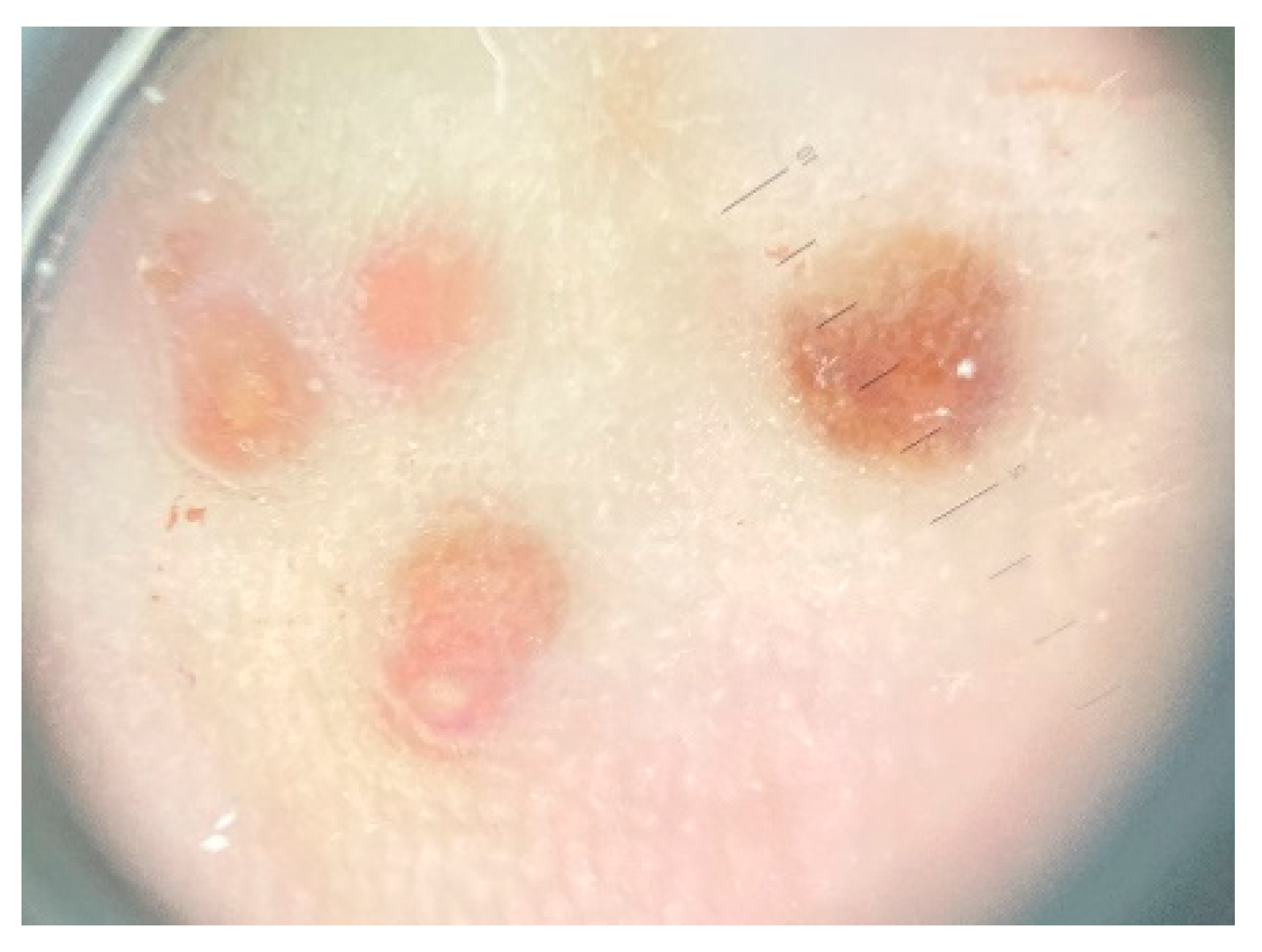
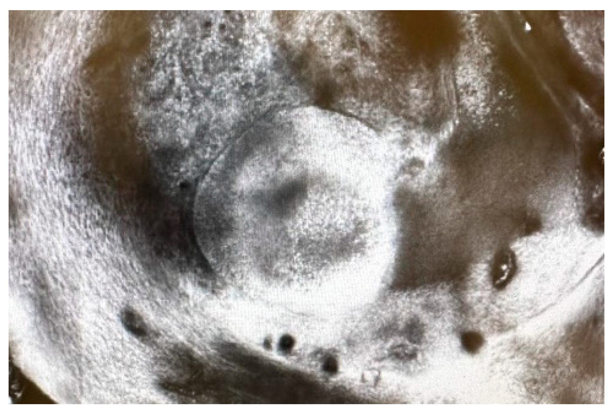
A dermoscopy of the lesions was performed revealing ovoid yellowish structures (Figure 2) that correspond to granulomas identified on confocal microscopy (Figure 3). We did not find any data reported in the literature about dermoscopy or confocal microscopy features seen in actinomycosis.
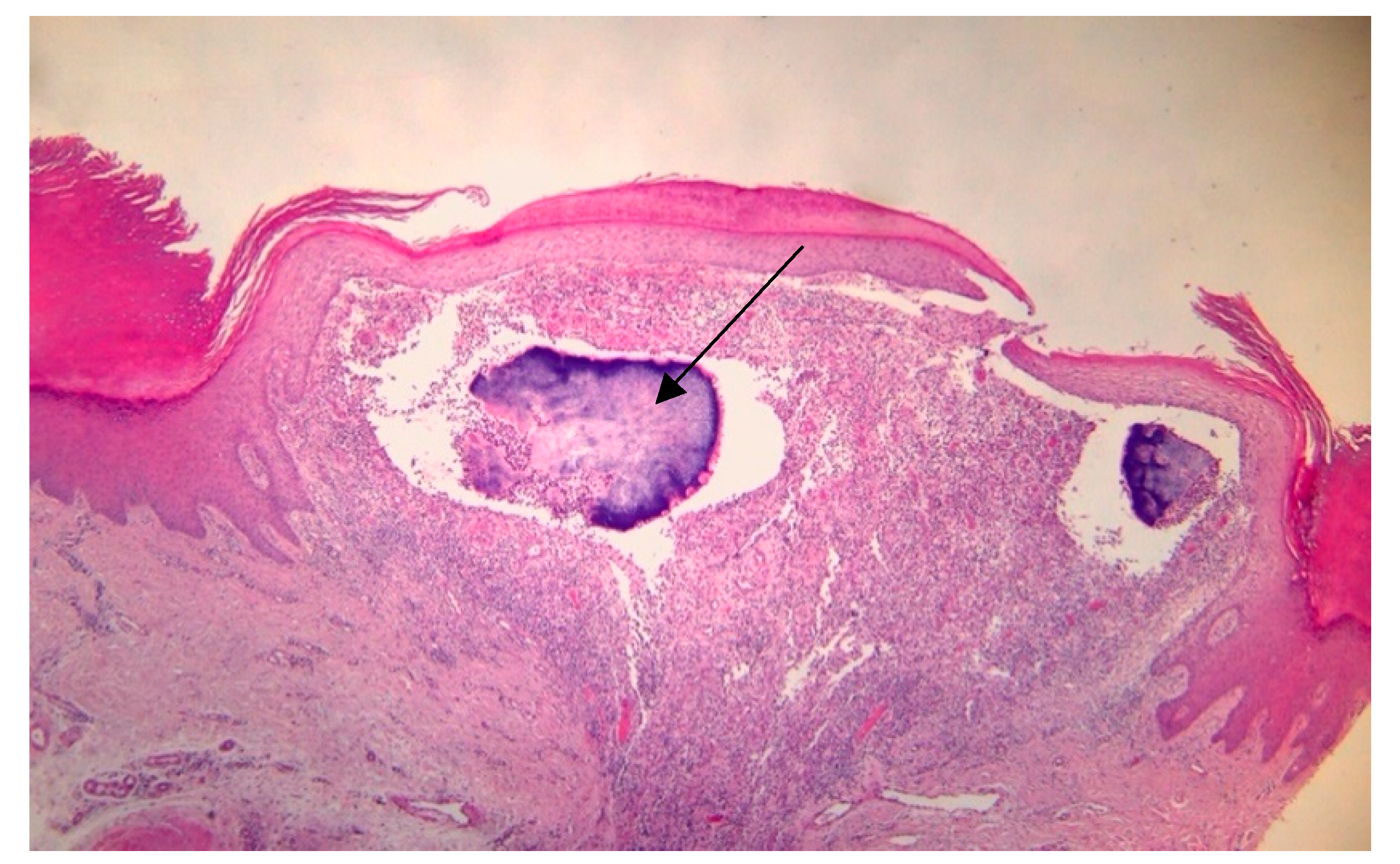
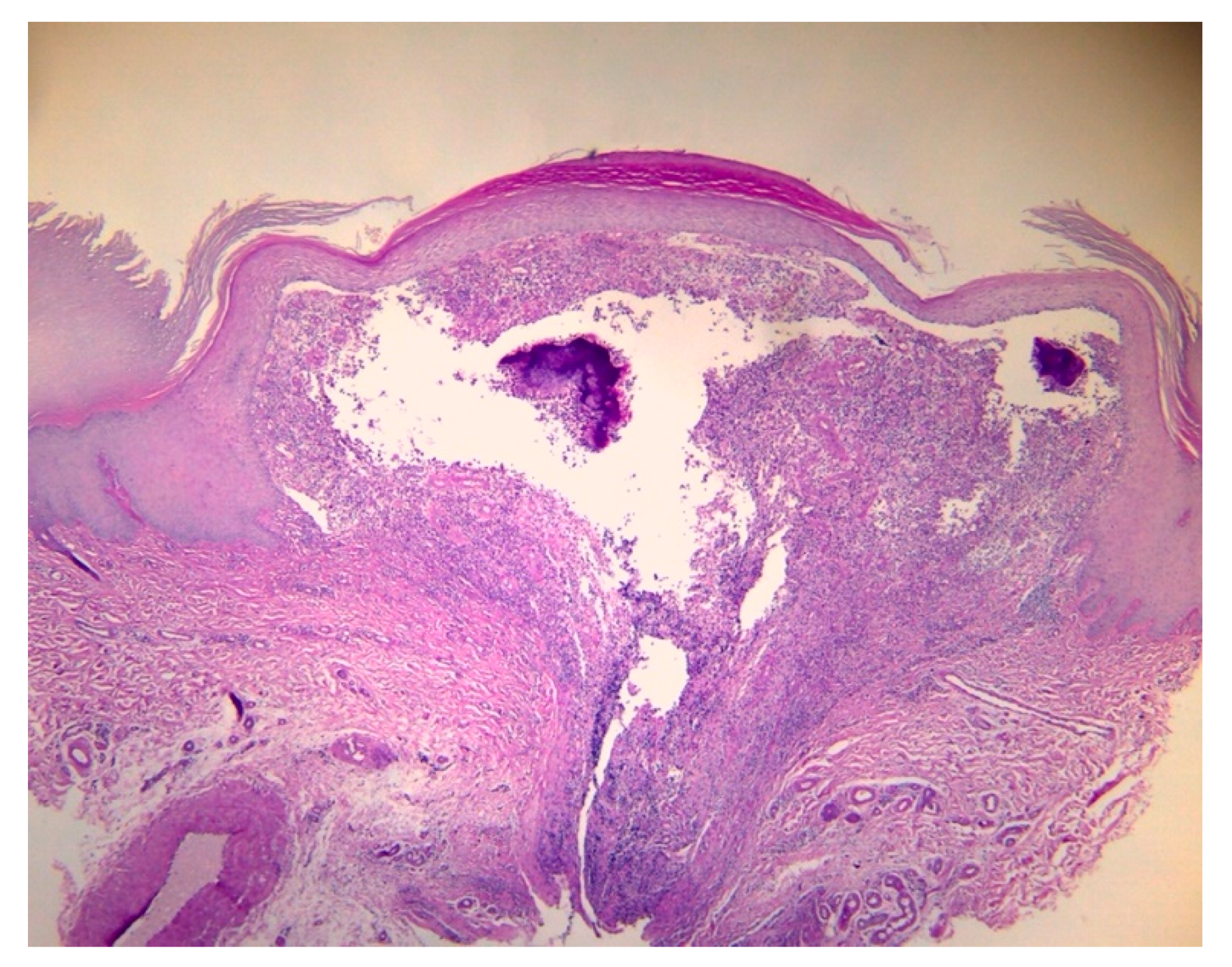
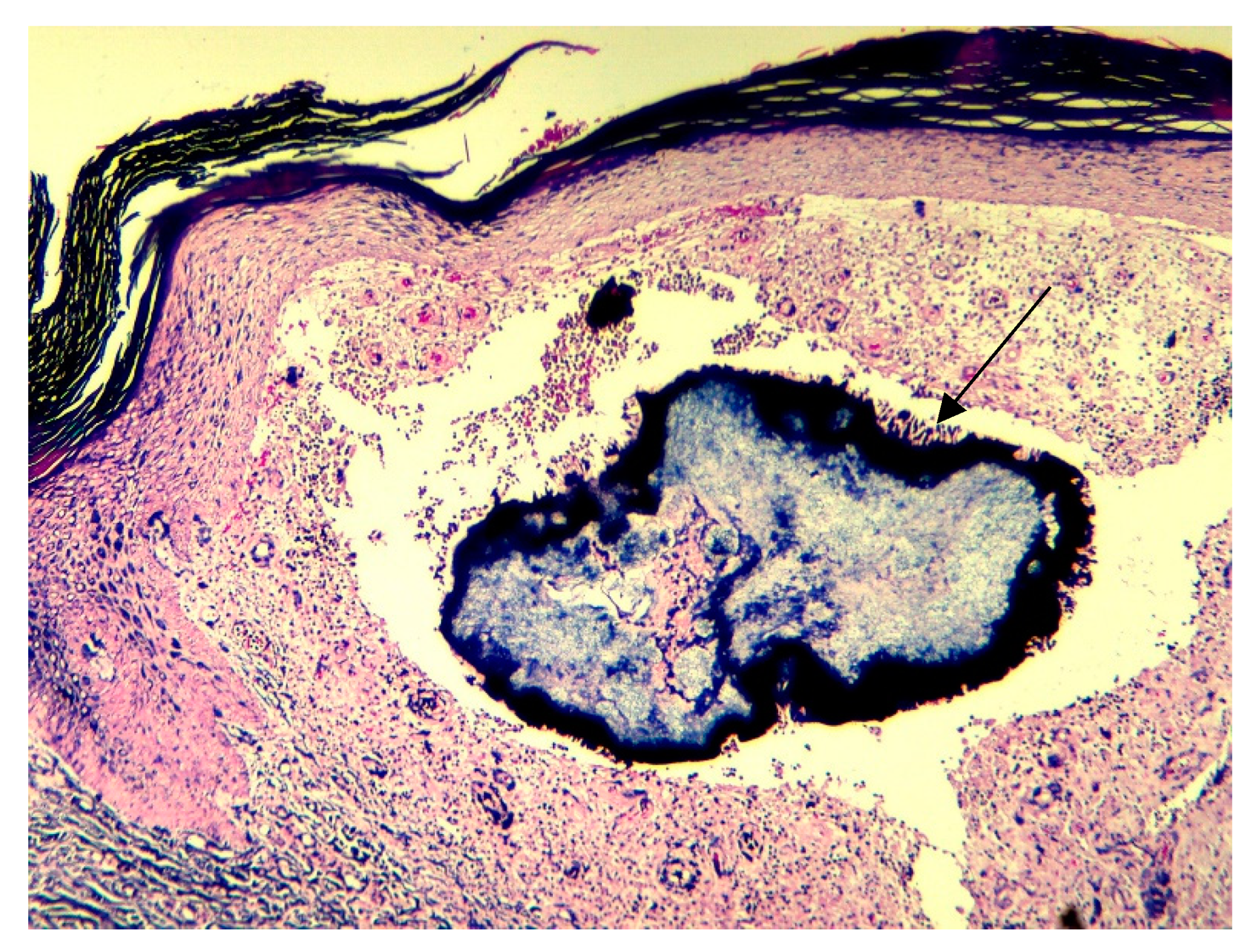
A skin biopsy was performed to exclude the possibility of malignancy, particularly Kaposi sarcoma. The biopsy revealed multiple colonies of filamentous organisms with characteristic neutrophilic outlining (Splendore-Hoeppli phenomenon), tumefied neoformed vessels, and an extensive mixed inflammatory reaction consistent with actinomycosis (Figure 4). The periodic acid-Schiff stain (PAS) was negative for fungal agents (Figure 5). The Gram stain showed conglomeration of filamentous Gram-positive bacilli (Figure 6). The culture of the specimen was negative, which is possible in cases of Actinomyces infection due to previous administration of antibiotics or inadequate culture conditions.
The histopathological report also served to differentiate actinomycosis from cutaneous tuberculosis, pustular psoriasis, Kaposi sarcoma, insect bite reaction, and malignancy. Unfortunately, no pus collection could be obtained from the fistulous tracts due to lack of discharge during the examination, preventing the observation of any discernible sulfur granules.
The initiation of penicillin therapy had been planned but was deferred after the patient revealed a possible allergic reaction to the medication. Therefore, an allergological examination using a drug provocation test was conducted to confirm this claim. The test produced a widespread erythematous and pruritic reaction, necessitating the administration of 4 mg dexamethasone intravenously. Consequently, an alternative medication was deemed necessary, and doxycycline was identified as the drug of choice at a dosage of 200 mg per day. After a follow-up period of three months, a significant improvement in the patient’s lesions was observed.

3. Discussions

Actinomyces species are endogenous microorganisms that are part of the normal microflora. However, they possess parasitic properties and can cause infections when the mucosal barrier is disrupted, allowing the organism to enter soft tissues with impaired blood supply [1]. Cervicofacial, thoracic, and abdominal actinomycosis represent the most prevalent forms of the disease, while systemic illness, regional adenopathy, and extremity involvement are uncommon [12]. Secondary infections may arise when the pathogen spreads from primary sites by hematogenous routes [1,12]. Although primary actinomycosis of the foot is rare, a few cases have been documented in the literature, typically following a traumatic occurrence. In the present case, the patient’s preference to walk around the house and yard barefoot could increase the risk of infection due to potential trauma.
Clinically, the lesions commonly present as one or multiple indurated swellings [12]. As the swellings progress, purulent loculations form, leading to central suppuration and softening [12]. Over time, sinus tracts may arise, which can extend to adjacent tissues such as the skin, bone, or internal organs, depending on the location of the lesion [12]. These sinus tracts may spontaneously open or close in other areas [12]. Most patients experience progressive skin and soft tissue inflammation, which can progress to an abscess or cold mass or nodular lesions with fistulas that must be differentiated from chronic inflammatory skin conditions, such as pustular psoriasis, cutaneous mycobacterial or fungal infections [1,12,13]. In countries where tuberculosis is prevalent, such as Romania, the acid-fast bacilli smear and QuantiFERON-TB Gold test are mandatory, as primary actinomycosis’s clinical manifestations can resemble tuberculosis [13]. In our case, the reports came back negative.
The presence of sulfur granules is a clear indication of actinomycosis infection. These granules can range in size from microscopic to macroscopic and are usually found in loculations containing 1 to 6 sulfur granules [12]. The affected region exhibits a distinctive feature of granulation tissue, which forms multiple loculations, characterized by a purulent center, which contains sulfur granules surrounded by colonies of actinomyces, neutrophils, microorganisms, and cellular debris [12]. Confirming the diagnosis requires isolating the organism from these granules in histopathological samples, but this can be difficult as the samples usually contain only a few granules [12,13]. In such cases, an incisional biopsy showing Actinomyces can raise concern for actinomycosis infection, and appropriate sampling and histopathological examination are necessary to confirm the diagnosis [13].
The successful culture of Actinomyces spp. from clinical specimens poses a significant challenge. Reiner et al. reported that the positive culture rate was less than 50%, primarily due to the organism’s heightened sensitivity to a broad range of antimicrobial agents and the necessity for rigorous anaerobic processing and growth [14]. It is plausible that these factors contributed to the negative results obtained in our case.
The treatment for actinomycosis typically involves a combination of prolonged antibiotic therapy and, in some cases, surgical intervention. The selection of antibiotics is a crucial aspect in managing actinomycosis, with high doses of penicillin or its derivatives being the preferred choice [12]. The duration of antibiotic therapy is typically extended, ranging from six months to a year, to ensure complete resolution of the infection [12]. Initially, parenteral administration of penicillin is recommended, with a dosage of 18 to 24 million units of penicillin G [12]. This is followed by a prolonged course of oral penicillin V or amoxicillin [12]. In instances where there are bone lesions, such as in osteomyelitis associated with actinomycosis, surgical intervention, such as incision and drainage, may be necessary [15,16].
In patients allergic to penicillin, alternative drug options should be considered. Therapeutic options include doxycycline, tetracycline, erythromycin, or clindamycin, as well as third-generation cephalosporins, meropenem, imipenem, and fluoroquinolones [1,12]. In the present case, given the patient’s penicillin allergy, doxycycline was the preferred medication, with partial resolution of the lesions.

4. Conclusions

Primary cutaneous actinomycosis of an extremity is a rare, granulomatous infection that can mimic other diseases, both of infectious etiology (such as cutaneous tuberculosis, deep fungal infections, etc.) and non-infectious causes (such as pustular psoriasis, Kaposi sarcoma, other skin tumors, etc.) making accurate diagnosis challenging. Due to its rarity, medical professionals need to be aware of its symptoms and consider it a possible diagnosis in patients with progressive nodular swelling and fistulous tracts. The accurate diagnosis of actinomycosis requires a careful examination of the clinical presentation and associated symptoms alongside a detailed analysis of the histological features, including special stain management of this condition, is also challenging as it involves a prolonged course of antibiotics and sometimes surgical debridement.

Author Contributions

Conceptualization, A.M.D. and O.A.O.; methodology, A.M.D. and O.A.O.; software, A.M.D., O.A.O., M.L., L.G.P., I.T. and C.G.; validation, A.M.D., O.A.O. and C.G.; formal analysis, A.M.D., O.A.O., M.L., L.G.P., R.T., I.T. and C.G.; investigation, A.M.D., O.A.O., M.L., R.T. and I.T.; resources, A.M.D., O.A.O., M.L. and R.T.; data curation, A.M.D., O.A.O., R.T., C.G. and L.G.P.; writing—original draft preparation, A.M.D., O.A.O., L.G.P. and C.G.; writing—review and editing, A.M.D., O.A.O., C.G. and L.G.P.; visualization, A.M.D. and M.L.; supervision, A.M.D., O.A.O., L.G.P. and C.G.; project administration, A.M.D., O.A.O., R.T. and L.G.P. All authors have read and agreed to the published version of the manuscript.

Funding

This research received no external funding.

Institutional Review Board Statement

Not applicable.

Informed Consent Statement

Informed consent was obtained from the patient.

Data Availability Statement

No new data were created or analyzed in this study. Data sharing is not applicable to this article.

Conflicts of Interest

The authors declare no conflicts of interest.

References

  1. Valour, F.; Sénéchal, A.; Dupieux, C.; Karsenty, J.; Lustig, S.; Breton, P.; Gleizal, A.; Boussel, L.; Laurent, F.; Braun, E.; et al. Actinomycosis: Etiology, clinical features, diagnosis, treatment, and management. Infect. Drug Resist. 2014, 7, 183–197. [Google Scholar] [CrossRef] [PubMed] [PubMed Central]
  2. Najmi, A.H.; Najmi, I.H.; Tawhari, M.M.H.; Sawadi, K.H.; Khbrani, K.A.H.; Tawhari, F.H.; Tawhari, M.A.; Mathkur, M.H.; Al-Attas, K.M. Cutaneous actinomycosis and long-term management through using oral and topical antibiotics: A case report. Clin. Pract. 2018, 8, 1102. [Google Scholar] [CrossRef] [PubMed] [PubMed Central]
  3. Almarzouq, S.F.; Almarghoub, M.A.; Almeshal, O. Primary actinomycosis of the big toe: A case report and literature review. J. Surg. Case Rep. 2019, 2019, rjz292. [Google Scholar] [CrossRef] [PubMed]
  4. Wong, V.; Turmezei, T.; Weston, V. Actinomycosis. BMJ 2011, 343, d6099. [Google Scholar] [CrossRef] [PubMed]
  5. Bonnefond, S.; Catroux, M.; Melenotte, C.; Karkowski, L.; Rolland, L.; Trouillier, S.; Raffray, L. Clinical features of actinomycosis: A retrospective, multicenter study of 28 cases of miscellaneous presentations. Medicine 2016, 95, e3923, Erratum in: Medicine 2016, 95, e5074. [Google Scholar] [CrossRef] [PubMed] [PubMed Central]
  6. Sullivan, D.; Chapman, S. Bacteria that masquerade as fungi: Actinomycosis/nocardia. Proc. Am. Thorac. Soc. 2010, 7, 216–221. [Google Scholar] [CrossRef] [PubMed]
  7. Stájer, A.; Ibrahim, B.; Gajdács, M.; Urbán, E.; Baráth, Z. Diagnosis and management of cervicofacial actinomycosis: Lessons from two distinct clinical cases. Antibiotics 2020, 9, 139. [Google Scholar] [CrossRef] [PubMed]
  8. Çarkman, S.; Ozben, V.; Durak, H.; Karabulut, K.; Ipek, T. Isolated abdominal wall actinomycosis associated with an intrauterine contraceptive device: A case report and review of the relevant literature. Case Rep. Med. 2010, 2010, 340109. [Google Scholar] [CrossRef] [PubMed]
  9. Baierlein, S.A.; Wistop, A.; Looser, C.; Peters, T.; Riehle, H.M.; von Flüe, M.; Peterli, R. Abdominal actinomycosis: A rare complication after laparoscopic gastric bypass. Obes. Surg. 2007, 17, 1123–1126. [Google Scholar] [CrossRef] [PubMed]
  10. Kim, D.; Kim, S. Destruction of the c2 body due to cervical actinomycosis: Connection between spinal epidural abscess and retropharyngeal abscess. Korean J. Spine 2017, 14, 20–22. [Google Scholar] [CrossRef] [PubMed]
  11. Bellazreg, F.; Hachfi, W.; Abdelkader, A.B.; Hattab, Z.; Kaabia, N.; Bahri, F.; Letaief, A. A mass shadow on chest X-ray in a 40-year-old man: What’s your diagnosis? Adv. Infect. Dis. 2012, 2, 148–150. [Google Scholar] [CrossRef]
  12. Bettesworth, J.; Gill, K.; Shah, J. Primary actinomycosis of the foot: A case report and literature review. J. Am. Coll. Certif. Wound Spec. 2009, 1, 95–100. [Google Scholar] [CrossRef] [PubMed] [PubMed Central]
  13. Choi, T.L.; Lui, T.H. Primary Actinomycosis of the Foot in a Patient with Neurofibromatosis. Foot Ankle Spec. 2011, 4, 245–248. [Google Scholar] [CrossRef]
  14. Boushabi, A.; Hicham, A.; Mohamed, S. Actinomycosis of the sole of the foot (Madura foot): A rare case report and literature review. Int. J. Surg. Case Rep. 2023, 113, 109052. [Google Scholar] [CrossRef] [PubMed] [PubMed Central]
  15. Ryu, D.J.; Jeon, Y.S.; Kwon, H.Y.; Choi, S.J.; Roh, T.H.; Kim, M.K. Actinomycotic osteomyelitis of a long bone in an immunocompetent adult: A case report and literature review. BMC Musculoskelet. Disord. 2019, 20, 185. [Google Scholar] [CrossRef] [PubMed] [PubMed Central]
  16. Khan, S.; Khan, B.; Batool, W.; Khan, M.; Khan, A.H. Primary Cutaneous Actinomycosis: A Diagnostic Enigma. Cureus 2023, 15, e37261. [Google Scholar] [CrossRef] [PubMed] [PubMed Central]
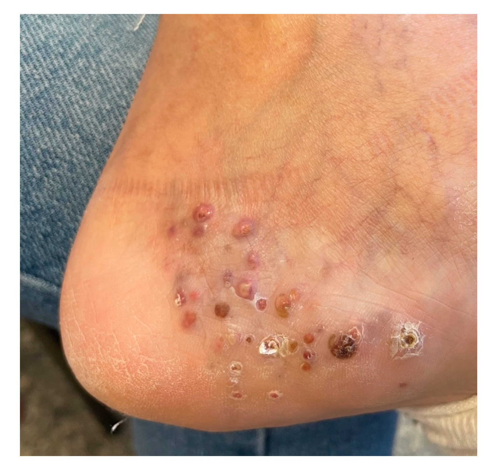
Figure 1. Clinical picture showing small nodules with a purple–red hue and dry fistulous tracts on the plantar area of the left foot.
Dermato 04 00008 g001
Figure 2. Dermoscopy findings consisting of ovoid yellowish structures.
Dermato 04 00008 g002
Figure 3. Confocal microscopy image showing granulomas.
Dermato 04 00008 g003
Figure 4. Histopathology findings suggestive for actinomycosis showing aggregates of bacteria surrounded by intensely eosinophilic material, Spendore–Hoeppli phenomenon reaction (arrow) and a densepolymorphous inflammatory infiltrate (10×, H&E stain).
Dermato 04 00008 g004
Figure 5. Special stain periodic acid-Schiff was performed to exclude a fungal infection (PAS, 4×).
Dermato 04 00008 g005
Figure 6. Gram stain showed granules composed of masses of Gram-positive bacteria (arrow) (Gram stain, 10×).
Dermato 04 00008 g006
Disclaimer/Publisher’s Note: The statements, opinions and data contained in all publications are solely those of the individual author(s) and contributor(s) and not of MDPI and/or the editor(s). MDPI and/or the editor(s) disclaim responsibility for any injury to people or property resulting from any ideas, methods, instructions or products referred to in the content.