Article

Surface Microfabrication of Lactic Acid–Glycolic Acid Copolymers Using a Gas-Permeable Porous Mold

1
Department of Pharmaceutical Engineering, Toyrama Prefectural University, Imizu 939-0398, Toyama, Japan
2
Futuristic Technology Department, Sanko Gosei Ltd., Nanto 939-1852, Toyama, Japan
3
Toyama Industrial Technology Research and Development Center, Takaoka 933-0981, Toyama, Japan
*
Author to whom correspondence should be addressed.
Macromol 2024, 4(3), 544-555; https://doi.org/10.3390/macromol4030032
Submission received: 13 June 2024 / Revised: 29 July 2024 / Accepted: 1 August 2024 / Published: 5 August 2024

Abstract

:
We attempted to perform surface microfabrication of the bioabsorbable material lactic acid–glycolic acid copolymer (LG-80) using a micro-imprint lithography technique with a gas-permeable porous mold at less than 5 °C. As a result, high-resolution surface micromachining with a height of 1.26 μm and a pitch of 2.97 μm was achieved using a convex sapphire mold with a height of 1.3 μm and a pitch of 3 μm. After processing, the LG-80 exhibited high water repellency, and FT-IR analysis of the surface showed no significant change in its chemical structure, confirming that the surface microfabrication was successful, while retaining the properties of the material. This demonstrated new possibilities for surface microfabrication technology for bioabsorbable materials, which are expected to be applied in the medical and life science fields in products such as surgical implants, tissue regeneration materials, and cell culture scaffold materials. In particular, the use of micro-imprint lithography enables low-cost and high-precision processing, which will be a major step toward the practical application of bioabsorbable materials.

Graphical Abstract

1. Introduction

An important consideration for future medical materials is their ability to disappear spontaneously after being implanted or used in the body [1]. These materials gradually degrade while in contact with blood or while present in the body and eventually disappear completely. These materials are important for several reasons. First, some common implantable medical materials are either no longer needed after their intended purpose has been achieved or they may have a negative impact on the organism. Also, many damaged biological tissues have the ability to repair themselves with little help. Thus, a material that provides temporary support to a patient’s body without burdening it is called a bioabsorbable material because it disappears over time and does not remain or accumulate after being implanted in the body [2,3]. Polyglycolic acid, polylactic acid, and polycaprolactone are typical examples of bioabsorbable materials [4,5].
In recent years, bioabsorbable materials have been studied in a wide range of fields [6,7], and it is intended that these materials be degraded, metabolized, and excreted in vivo. Therefore, these materials need to interact with the organism and harmonize with its environment. This means that these materials must not cause harmful reactions on part of the material itself or the living organism, and it is of utmost importance to ensure their safe use. The familiarity of a material with the living body, the safety of the material with respect to the living body, and the property of “not stressing the living body” and “not having a harmful effect on the living tissue” are called biocompatibility [8,9].
Taking advantage of this biocompatibility feature, bioabsorbable materials play various important roles in medical and in vivo applications [10]. For example, they are used in a wide range of applications, including absorbable suture materials [11,12,13], artificial blood vessels [14,15], drug release materials [16,17,18], and tissue engineering and regenerative medicine [19,20,21]. Absorbable suture materials are used for post-surgical suturing and naturally degrade in the body, eliminating the need for re-operation. Artificial blood vessels support the formation of new blood vessels in the body, while drug-releasing materials deliver drugs to specific sites and maintain their effects through gradual release. In the field of tissue engineering and regenerative medicine, they are used as cell scaffolds to promote the regeneration of damaged tissue.
However, it is difficult to apply these bioabsorbable materials for high-resolution surface micromachining [22]. The reasons for this are explained below. For example, polyglycolic acid has a relatively fast decomposition rate, low stability at elevated temperatures, insufficient dissolution stability, a tendency to generate gas during dissolution processing, and significantly poor impact resistance owing to its high crystallinity and hardness [23,24,25]. In addition, polylactic acid has a relatively fast decomposition rate, low stability at elevated temperatures, weak heat resistance with a melting point of approximately 170 °C, and poor flowability and mold release properties [26,27]. Furthermore, polycaprolactone has a low melting point of approximately 60 °C, which restricts processing at high temperatures and results in relatively low crystallinity and reactivity [28,29]. Surface microfabrication methods include beam, laser, and micro-imprint lithography, but these techniques usually involve heat [30,31,32,33]. Using these methods, bioabsorbable materials with low melting points and relatively heat-sensitive properties are at risk of thermal decomposition during processing. Therefore, surface microfabrication of bioabsorbable materials is considered difficult.
Therefore, we developed a gas-permeable porous mold and found a technology to realize microfabrication by micro-imprint lithography by making materials that require high-temperature melting for processing flowability by using a solvent. Conventional non-gas-permeable molds, such as quartz and metal, used in micro-imprint lithography give rise to problems, such as gas entrapped between the mold and the material to be transferred during pressurization and solvent trapped in the mold, which result in molding defects. Gas-permeable porous molds allow gas and solvents to permeate through porous holes in the mold, improving molding defects caused by gas accumulation, and enabling fine patterning on the material to be transferred, including solvents.
In this study, we focused on poly (lactic acid–glycolic acid) (LG-80), a bioabsorbable material currently used mainly in drug delivery systems and medical absorbable sutures, etc. We aimed to add high added value to poly (lactic acid–glycolic acid) by applying surface microfabrication using micro-imprint lithography technology, and to apply it in the medical and life science fields in products such as surgical implants, tissue regeneration materials, and cell culture scaffold materials. An inorganic compound, TiO2-SiO2, was synthesized as a gas-permeable porous mold material to improve the mold-releasing property with an organic-based transfer agent. We attempted to solve the above issues using low-temperature micro-imprint lithography below 5 °C utilizing gas-permeable porous molds, and also attempted surface microfabrication of lactic acid–glycolic acid copolymers, which are bioabsorbable materials.

2. Materials and Methods

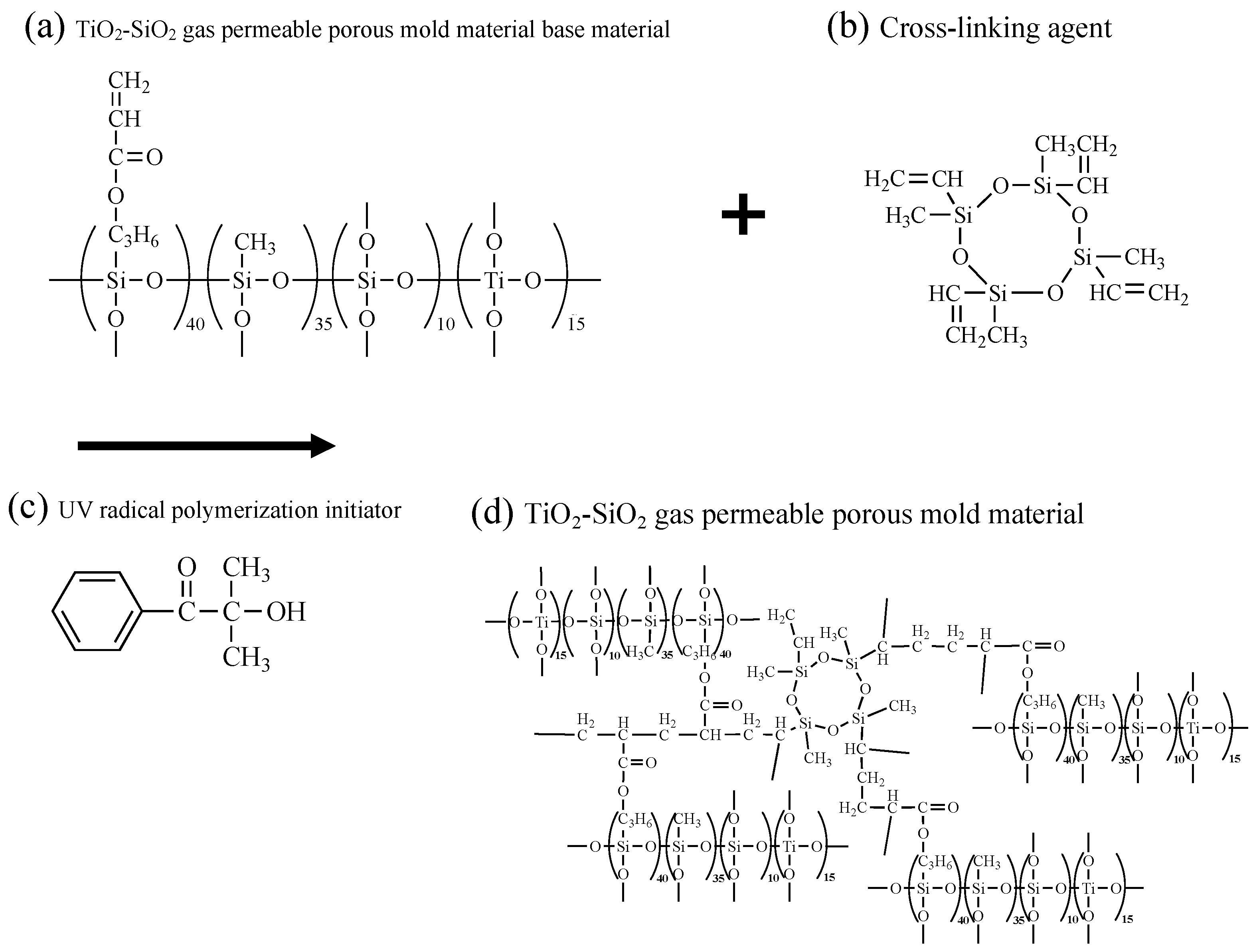
2.1. Synthesis of TiO2-SiO2 Gas-Permeable Porous Mold Material

Figure 1a–d show the chemical structure of the TiO2-SiO2 gas-permeable porous mold material used in this study and the mechanism of the cross-linking reaction.
The TiO2-SiO2 gas-permeable porous mold material (Figure 1a), based on a four-component mixture of 40 wt% 3-(Acryloyloxy) propyltrimethoxysilane, 35 wt% methyltrimethoxysilane, 15 wt% tetraethyl titanate, and 10 wt% tetraethoxysilane, was synthesized by sol-gel polymerization [34,35,36]. The base material was synthesized by mixing 87 wt% TiO2-SiO2 gas-permeable porous mold material (Figure 1a), 10 wt% of 2,4,6,8-tetramethyl-2,4,6,8-tetravinylcyclotetrasiloxane (T2523: Tokyo Chemical Industry) (Figure 1b) as a cross-linking agent, and 3 wt% of 2-hydroxy-2-methylpropiophenone (Omnirad 1173: Toyotsu Chemiplas Co., Ltd., Tokyo, Japan) (Figure 1c) as a UV radical polymerization initiator. The mixture was stirred for 8 h with a roller stirrer (MR-5: AS ONE Co., Ltd., Osaka, Japan) to obtain TiO2-SiO2 gas-permeable porous mold material (Figure 1d).

2.2. Mixing of Lactic Acid–Glycolic Acid Copolymer (LG-80)

The lactic acid–glycolic acid copolymer (LG-80: L-lactic acid content 80 mol%, Taki Chemical Co., Ltd.,Hyogo, Japan) was mixed with 30 wt% and 70 wt% of dichloromethane as a volatile solvent to increase fluidity, and dissolved by sonication at 30 °C for 5 min using an ultrasonic cleaner, the DIGITAL ULTRASONIC CLEANER OZL-2000 (Onezili, Guangzhou, China).

2.3. Mixing of Materials

Non-gas-permeable mold material consisting of 4 components (43 wt% isobornyl acrylate, 33 wt% n-butyl acrylate, 20 wt% triethylene glycol diacrylate, and 4.0 wt% 2-hydroxy-2-methyl propiophenone) was mixed at room temperature (25 °C).

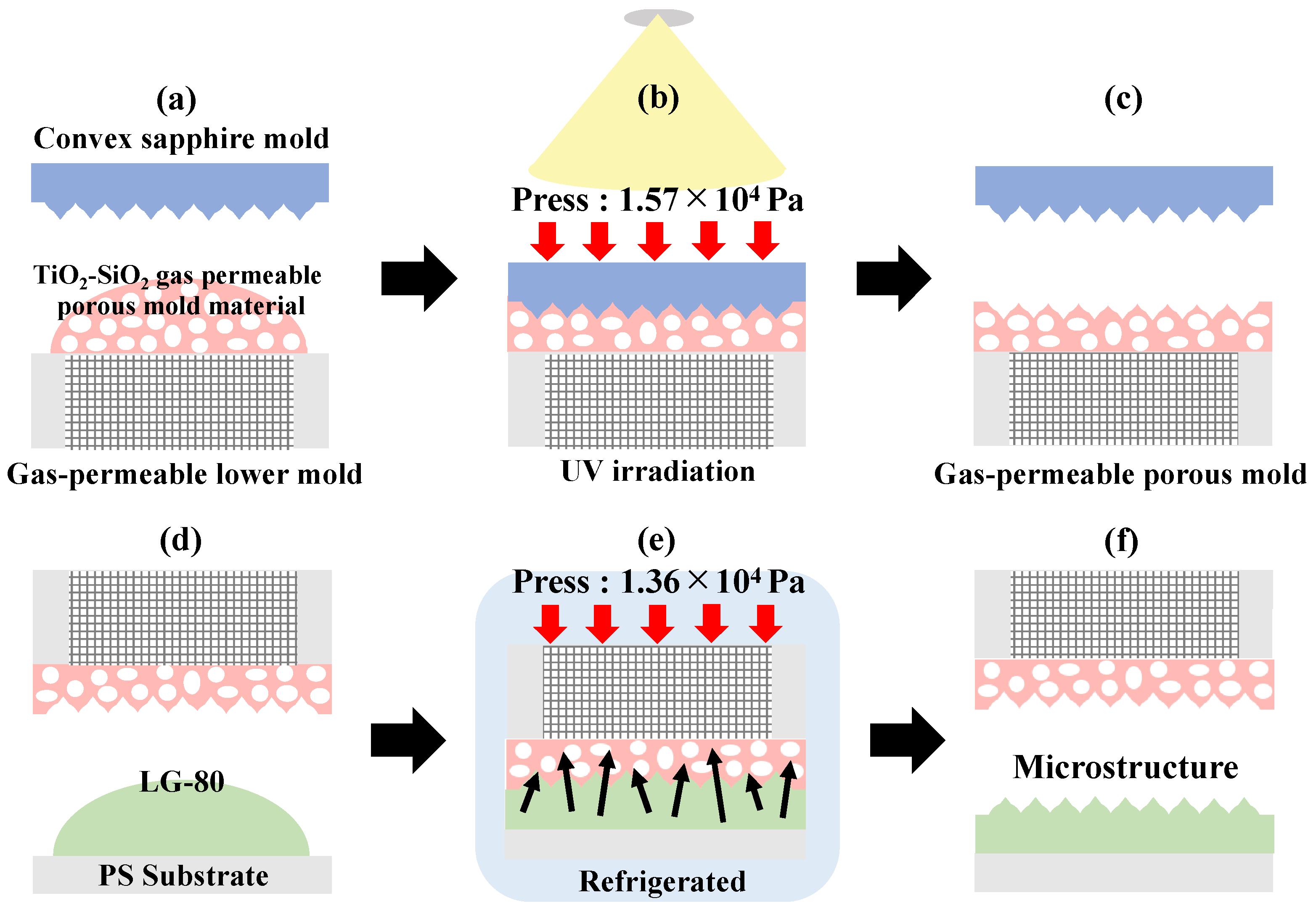
2.4. Surface Micromachining Process

Figure 2 shows the surface micromachining process used in this study.
The surface microfabrication process utilized a double micro-imprint lithography method consisting of two steps: a 1.3-μm-high, 3-μm-pitch convex sapphire mold was transferred onto a TiO2-SiO2 gas-permeable porous mold material applied on top of a gas-permeable lower-layer mold to fabricate a concave gas-permeable porous mold (Figure 2a–c), and the gas-permeable porous mold was transferred onto the LG-80 (Figure 2d–f). A double-micro-imprint lithography method was employed [37,38,39]. The gas-permeable lower mold was made of maraging steel with an average grain size of 20–30 μm, and the mold was fabricated by 3D photoengraving using LUMEX Avance-25 (Matsuura Manufacturing Co., Ltd., Tokyo, Japan) to provide permeability.
First, a mold release agent (DURASURF DS-831TH, Harves, Saitama, Japan) was applied to the surface of the convex sapphire mold and allowed to dry for 2 h. The TiO2-SiO2 gas-permeable porous mold material was applied on top of the gas-permeable lower layer mold (Figure 2a), the convex sapphire mold was placed on top, and then light irradiated with LED (AC90V-240V, 365 nm, 72 W) for 8 s under pressure at 1.57 × 10⁴ Pa to light cure the TiO2-SiO2 gas-permeable porous mold material (Figure 2b). The convex sapphire mold was then removed to fabricate a gas-permeable porous mold, in which the gas-permeable lower mold and TiO2-SiO2 gas-permeable porous mold material were bonded (Figure 2c).
Next, the surface of the TiO2-SiO2 gas-permeable porous mold material was coated with an organic fluorine compound to improve mold release from the LG-80. The LG-80, mixed as described in Section 2.2, was applied to a polystyrene (PS) substrate that had been cleaned with ethanol (Figure 2d), and the gas-permeable porous mold was placed on top of the LG-80 and pressurized with a weight of 1.36 × 10⁴ Pa (Figure 2e). After refrigeration for 4 h in a refrigerator to slowly remove solvents, gases, bubbles, etc., the gas-permeable porous mold was removed (Figure 2f), and the surface microfabrication of the LG-80 was performed. To check the transferability, (Figure 2d–f) were repeated 20 times.
The same procedure was used for the surface microfabrication of the LG-80 using the non-gas-permeable mold material described in Section 2.3. A non-gas-permeable mold material was applied to a glass substrate, a convex sapphire mold was placed on top, and light irradiation using an LED was applied for 5 s while applying pressure at 1.57 × 10⁴ Pa to harden the material.

2.5. Surface Micromachining SEM Observation

Steps (Figure 2d–f) in Section 2.4 were repeated to observe the 10th, 15th, and 20th transfers of the LG-80 with surface microfabrication using the same gas-permeable porous mold and the LG-80 with surface microfabrication using a non-gas-permeable mold by SEM images using a Regulus8100 (Hitachi High-Tech, Tokyo, Japan). No deposition was performed, and the observation conditions were accelerating voltage = 3000 volts, deceleration voltage = 0 volts, magnification = 5000, working distance = 31.6 mm, and emission current = 9800 nA.

2.6. Oxygen and Carbon Dioxide Gas Permeability Measurement

Oxygen gas permeability measurements were performed on the TiO2-SiO2 gas-permeable porous mold material, quartz, polyethylene, polypropylene, polystyrene, and polymethyl methacrylate using a differential pressure gas permeability measuring system (GTR-11, GTR Tec, Kyoto, Japan) at a sample thickness of approximately 100 μm and a temperature of 40 °C [40,41].

2.7. Contact Angle Measurement

Water repellency was evaluated by measuring the water contact angle of the flat LG-80 without surface microfabrication and the LG-80 with surface microprocessing. The water contact angle was measured using the 0/2 analysis method with a fully automatic contact angle meter (Dropmaster DM500, Kyowa Surfaces Science Co., Ltd., Saitama, Japan). Measurements were carried out in an environment with an air temperature of 25 °C. The amount of drop was 1.0 mL, the time immediately after dropping was set as 0 s, and measurements were taken from 0 to 9 s. Three measurements were taken for each angle, and 0 and 9 s measurements were omitted to calculate average values.

2.8. FT-IR Measurement

FT-IR measurements of the LG-80 without surface microfabrication, the LG-80 with surface microfabrication, and dichloromethane were performed using a Fourier transform infrared spectrophotometer (Spectrum Two, Perkin Elmer, Waltham, MA, USA). Data were recorded at a resolution of 4 cm−1, with 10 integrations and a frequency range of 400–4000 cm−1.

3. Results

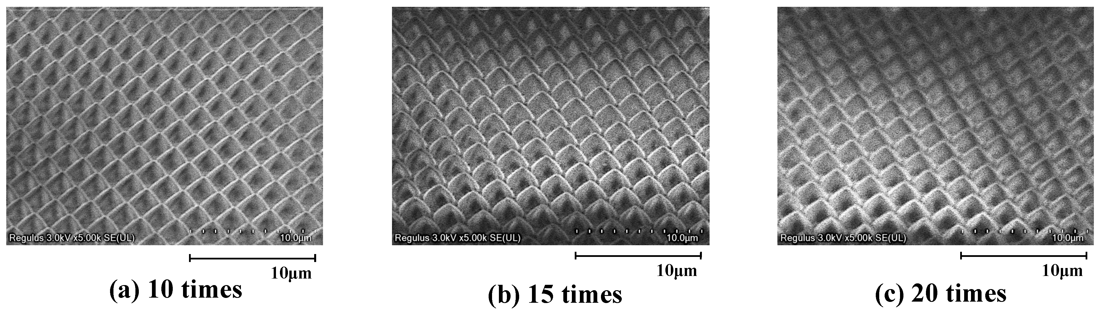
3.1. Surface Micromachining Results for LG-80 Using Gas-Permeable Porous Mold

Figure 3 shows SEM observation results for the LG-80 processed using the gas-permeable porous mold.
Because the same single mold can be used for repeated transcriptions, the gas-permeable porous mold was considered to have excellent durability. In addition, an organic fluorine compound was applied to the surface of the TiO2-SiO2 gas-permeable porous mold material to further improve the mold release from the LG-80, which is thought to prevent contamination and peeling of the mold. Prior studies of the surface microfabrication of bioabsorbable materials have published processing techniques using beams and lasers [42,43,44]. These have disadvantages, such as a high processing cost, thermal denaturation of the material around the processing, non-uniform processing, such as melting or peeling on the surface of the material, and difficulty processing large areas. However, the micro-imprinting method selected for this study is relatively inexpensive and can be modified to process complex shapes and large areas, primarily by designing the mold. In addition, the micro-imprinting process used in this study does not use heat; therefore, there was no risk of thermal denaturation of the bioabsorbable material, which has a low melting point. Furthermore, the gas-permeable porous mold remained clean after imprinting, although the imprinting process was performed only 20 times because it was a hand press imprinting process.

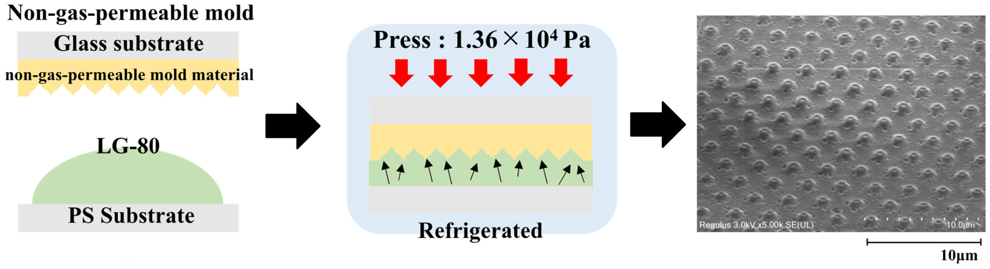
3.2. Results of Surface Micromachining of LG-80 Using Non-Gas-Permeable Mold

Figure 4 shows an SEM image of the LG-80 with surface microfabrication machined using a non-gas-permeable mold.
In LG-80 surfaces microfabricated using non-gas-permeable molds used in conventional micro-imprint lithography, it has been observed that formation defects occur due to volatile solvents and air entrained during pressurization. This is thought to be due to the fact that volatile solvents and air lose their escape route during the microfabrication process and remain in the mold, degrading the transfer quality and making it difficult to accurately form the desired pattern. Such molding defects can seriously affect product performance and reliability.
On the other hand, it was confirmed that molding defects were greatly improved in the LG-80 that was surface micro-machined using a gas-permeable porous mold. This means that the gas-permeable porous mold can be used to form the target micro-pattern with high accuracy without affecting the surface of the LG-80, which is the target of transfer.
These results suggest that gas-permeable porous molds are effective in removing volatile solvents and gases generated in micro-imprint lithography. This in turn improves the accuracy and reliability of the molding process. The introduction of this technology will enable the stable production of higher-quality products and further the development of micro-imprinting technology.

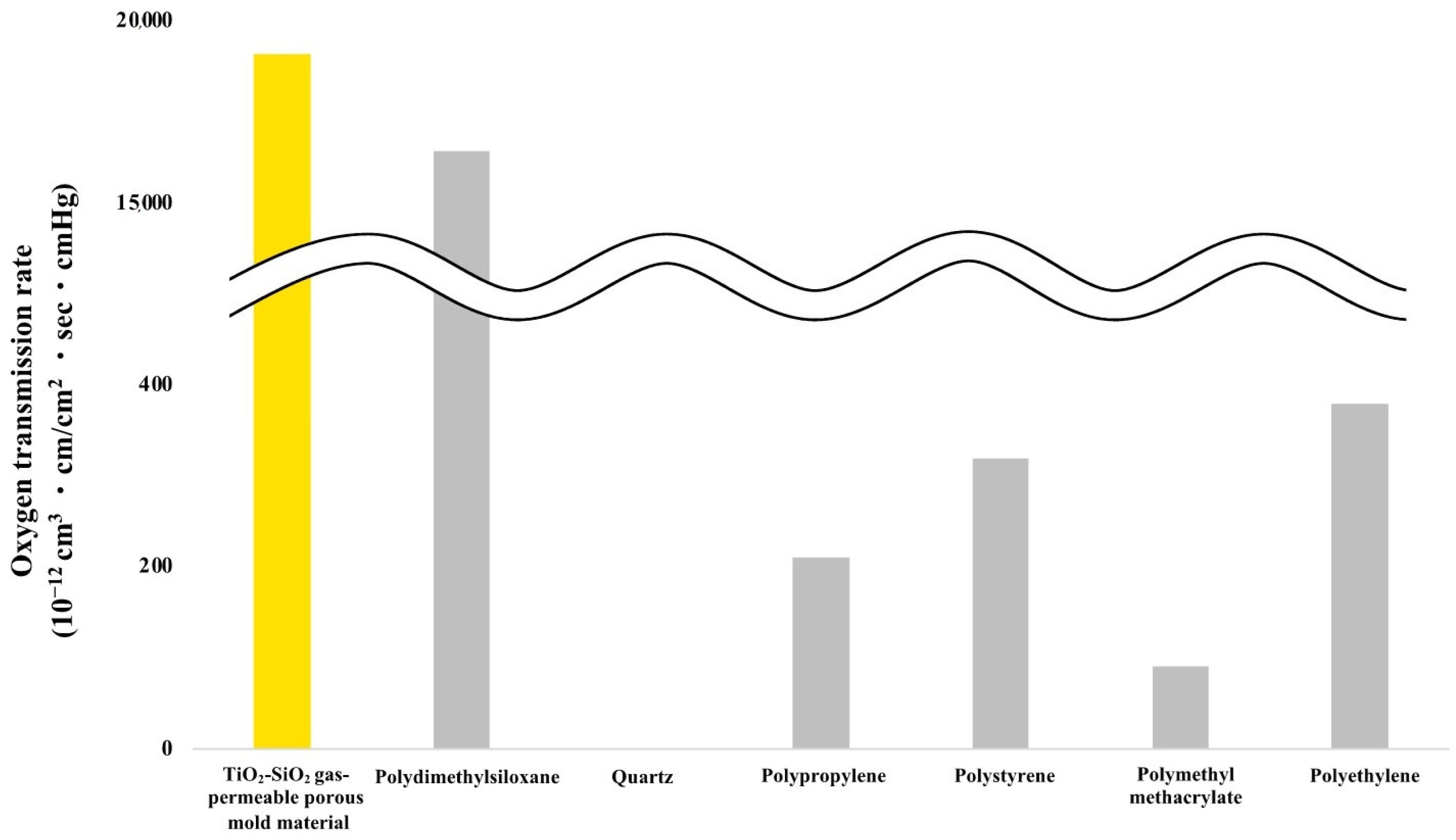
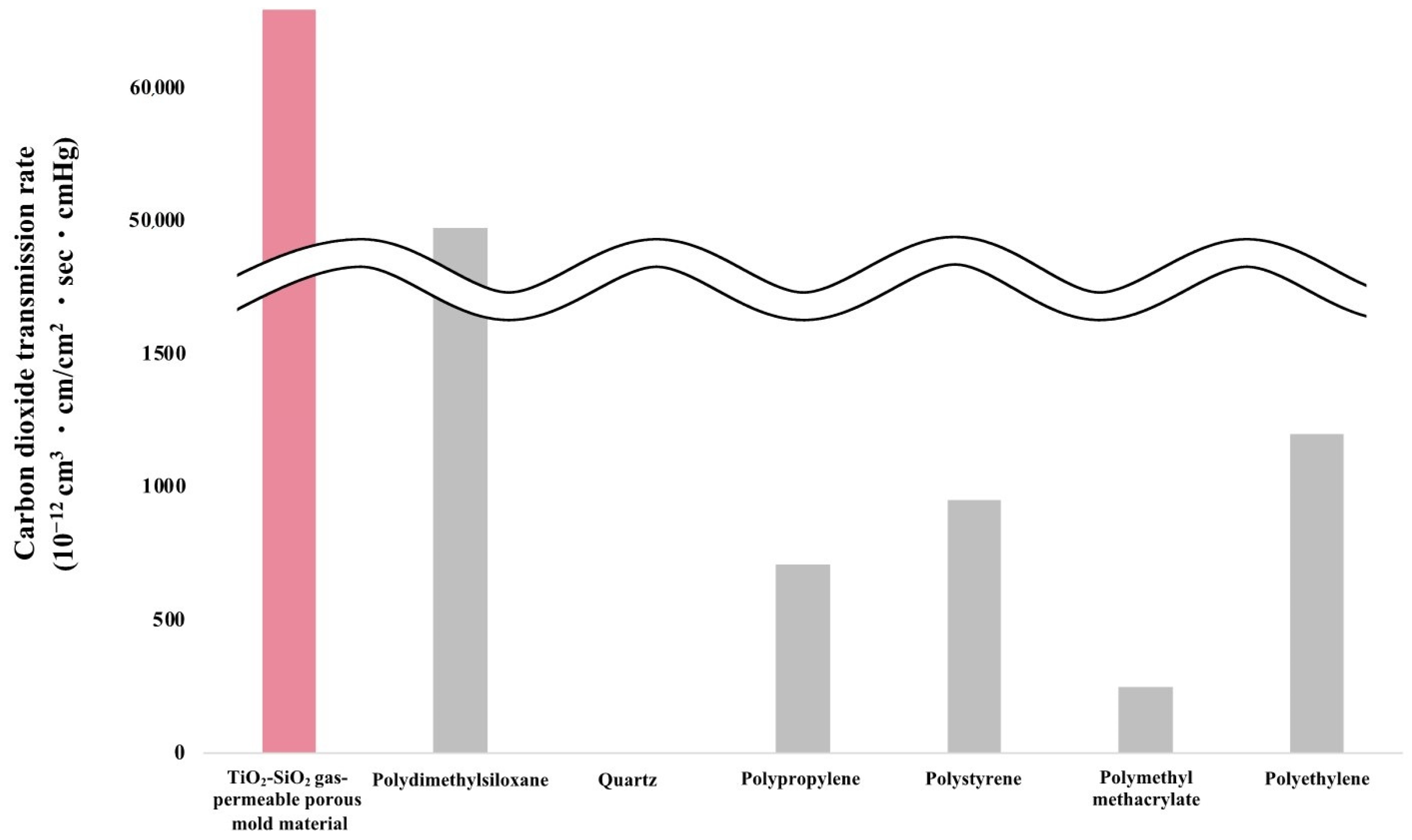
3.3. Oxygen Gas Permeability Measurement Results

Figure 5 shows results of oxygen gas permeability measurements for TiO2-SiO2 gas-permeable porous mold material, quartz, polyethylene, polypropylene, polystyrene, and polymethyl methacrylate.
Figure 5 and Figure 6 show that quartz, which has been used conventionally as a mold for microimprint lithography, is completely impermeable to gas. The TiO2-SiO2-based gas-permeable porous mold material also exhibited higher gas permeability than polydimethylsiloxane (PDMS), which is widely used as a mold in many fields. Furthermore, the TiO2-SiO2-based gas-permeable porous mold material exhibited higher gas permeability than polyethylene and polystyrene, which are general-purpose plastics with high gas permeability, and higher gas permeability than polymethyl methacrylate, a transparent material.
In addition, Demko, Roh, and Selyanchyn reported results of gas permeability measurements of PDMS membranes [45,46,47]. In these studies, the permeability of the gas molecules passing through the PDMS membrane was measured, and it was shown that the permeability of CO2 was higher than that of O2. Based on results of this study, gas permeability does not depend on the size of molecules, but rather on the solubility and diffusivity of gas molecules, their chemical properties, and their interaction with the membrane material.
Furthermore, the variation in error bars for these gas permeabilities is small, indicating that the measured gas permeability values were consistent, highly reproducible, and had excellent measurement accuracy. As the TiO2-SiO2 gas-permeable porous mold material is similar to PDMS in terms of structure and gas-permeation mechanism, we believe that error bars in this measurement result are also small. In future research, we will also measure the oxygen gas permeability of dichloromethane through the TiO2-SiO2 gas-permeable porous mold material and conduct detailed analysis based on these data. In addition, it is important to set a new theme to investigate how gas permeability changes by changing the size and polarity of permeating molecules. This is expected to lead to a deeper understanding of the performance of the TiO2-SiO2 gas-permeable porous mold material and to explore further application possibilities.

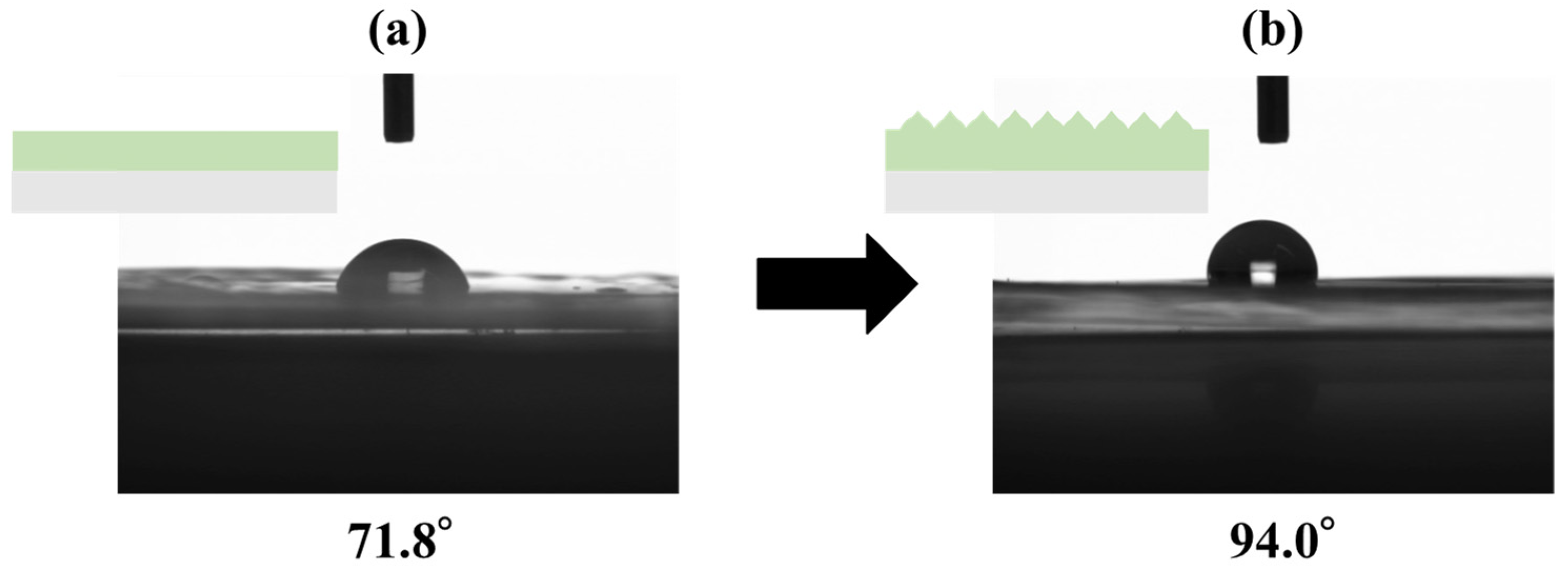
3.4. Contact Angle Measurement Results

Figure 7 shows the results of water contact angle measurements of the flat LG-80 without surface microfabrication and the LG-80 with surface microfabrication.
The water contact angle of the flat LG-80 (Figure 7a) without surface microfabrication was 71.8°, indicating that this material has some water repellency. On the other hand, the water contact angle of the LG-80 (Figure 7b) with surface microfabrication reached 94°, which indicates that its contact angle increased by more than 20° compared to the LG-80 without surface microfabrication. This significant increase in contact angle suggests that surface microfabrication improves the water repellency of the LG-80. Specifically, surface microfabrication increased the contact angle by forming microscopic protrusions and irregularities on the surface of the material. This microfabrication reduced the area where water droplets contact the surface, making it easier for the droplets to maintain a more spherical shape. This is an important factor in improving water repellency. Furthermore, microscopic protrusions are thought to provide added antimicrobial properties. There are several mechanisms for these properties: protrusions physically damage cell walls and membranes of bacteria and kill them; protrusions change the surface energy, making it difficult for bacteria to adhere to the surface; and protrusions prevent the movement of bacteria, making it difficult for them to migrate and multiply [48,49,50]. Therefore, it is necessary to evaluate the antimicrobial properties of this material in the future.

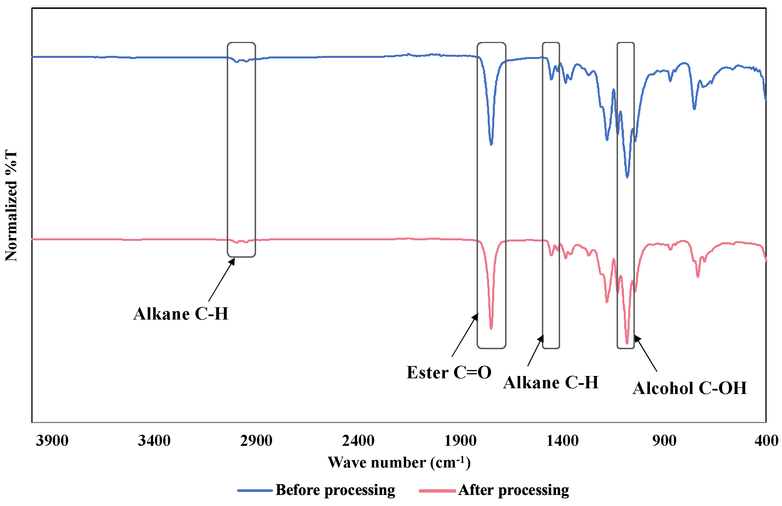
3.5. FT-IR Measurement Results

Figure 8 shows FT-IR measurement results for the LG-80 before and after processing, and Figure 9 shows FT-IR measurement results for dichloromethane.
Peaks at 995 cm−1 (alkane CH bond), 2947 cm−1 (alkane CH bond), 1747 cm−1 (ester C=O bond), 1454 cm−1 (alkane CH bond), and 1081 cm−1 (alcohol C-OH bond) were observed in the LG-80 before processing, while 2995 cm−1 (alkane CH bond), 2942 cm−1 (alkane CH bond), 1749 cm−1 (ester C=O bond), 1453 cm−1 (alkane CH bond), and 1083 cm−1 (alcohol C-OH bond) peaks were observed in the LG-80 after surface refinement. Peaks before and after processing were found to be almost identical. Although minor, these changes indicate that the effect of surface microfabrication on the chemical structure of the material is limited, suggesting that surface microfabrication improves surface properties without significantly altering the original chemical composition.
The characteristic peak of dichloromethane, 730 cm−1 (C-Cl bond), was not observed in the LG-80 with surface microfabrication. This observation indicates that surface microfabrication effectively removed dichloromethane, leaving no residue on the material’s surface, and maintaining the adequacy of the treatment and the cleanliness of the material.

4. Discussion and Conclusions

This study made new progress in the surface microfabrication of a bioabsorbable lactic acid–glycolic acid copolymer (LG-80) by making full use of micro-imprint lithography technology at 5 °C or lower. In particular, the development and application of a TiO2-SiO2 gas-permeable porous mold material realized high-resolution surface micromachining with a height of 1.26 μm and a pitch of 2.97 μm using a convex sapphire mold with a height of 1.3 μm and a pitch of 3 μm.
As a result of this experiment, we found that when a gas-permeable porous mold is used, the effects of volatile solvents and gases generated during pressurization can be effectively suppressed, while at the same time, a process was found that steadily transmits highly volatile dichloromethane by refrigerated drying, and we succeeded in forming a uniform microstructure on the surface of the LG-80. However, we confirmed that molding defects were significantly improved compared to when a conventional non-gas-permeable mold was used. Permeability measurements for oxygen and carbon dioxide also revealed that the TiO2-SiO2 gas-permeable porous mold material showed higher gas permeability than polyethylene and polystyrene, thereby improving the efficiency of the processing process. In addition, the water contact angle of the LG-80 with surface microfabrication increased by more than 20°, confirming an improvement in water repellency. This is expected to improve antibacterial and antifouling properties; therefore, further evaluation, such as antibacterial testing, is necessary. FT-IR measurements confirmed that there was almost no change in the chemical structure of the LG-80 before and after processing, indicating that dichloromethane was removed during gas permeation. This supports the safety and efficiency of the process.
In conclusion, micro-imprint lithography technology using gas-permeable porous molds is extremely promising for the surface microfabrication of bioabsorbable materials, and is expected to have a wide range of applications in the medical and life sciences. This research demonstrates the possibility of contributing to the establishment of a new technology that realizes precise microstructures while maintaining biocompatibility.

Author Contributions

Conceptualization, M.A. and S.T.; data curation, M.A. and S.T.; formal analysis, N.S., T.K. and Y.Y.; funding acquisition, S.T.; investigation, M.A., R.Y., S.M., Y.H. and S.T.; methodology, M.A., R.Y., S.M. and S.T.; project administration, S.T.; resources, S.T.; supervision, S.T.; validation, M.A., R.Y., S.M.,Y.H. and S.T.; writing—original draft preparation, M.A. and S.T.; writing—review and editing, M.A. and S.T. All authors have read and agreed to the published version of the manuscript.

Funding

The authors wish to acknowledge the funding received from the Japan Science and Technology Tech Startup HOKURIKU Program No. TeSH2024-17, Nakato Scholarship Foundation 2024, the Japan Society for the Promotion of Science Bilateral Joint Research Projects No. 120229937 in conjunction with Belgium, the Toyama Prefecture Grant 2024, the Suzuki Foundation 2023, Sango Monozukuri Foundation 2023, OSG Foundation 2023, Amano Institute of Technology Foundation 2023, Takeuchi Foundation 2022, Amada Foundation 2022, Die and Mould Technology Promotion Foundation 2022, Hayashi Rheology Memorial Foundation 2022, Lotte Foundation 2022, KOSE Cosmetology Research Foundation 2022, TAU Scholarship 2023, TOBE MAKI Scholarship Foundation 2022-2024, Marubun Research Promotion Foundation 2023, Fujikura Foundation 2024 and Murata Science and Education Foundation 2024.

Data Availability Statement

Datasets generated and/or analyzed during the current study are not publicly available because they are associated with ongoing research, but are available from the corresponding author upon reasonable request.

Acknowledgments

The authors appreciate the valuable and practical contributions of the Takikagaku Corporation.

Conflicts of Interest

Author Naoto Sugino and Takao Kameda were employed by the company Sanko Gosei Ltd. The remaining authors declare that the research was conducted in the absence of any commercial or financial relationships that could be construed as a potential conflict of interest.

References

  1. Pisecky, L.; Luger, M.; Klasan, A.; Gotterbarm, T.; Klotz, M.C.; Hochgatterer, R. Bioabsorbable implants in forefoot surgery: A review of materials, possibilities and disadvantages. EFORT Open Rev. 2021, 6, 1132–1139. [Google Scholar] [CrossRef]
  2. On, S.W.; Cho, S.W.; Byun, S.H.; Yang, B.E. Bioabsorbable osteofixation materials for maxillofacial bone surgery: A review on polymers and magnesium-based materials. Biomedicines 2020, 8, 300. [Google Scholar] [CrossRef] [PubMed]
  3. Figueiredo, L.; Fonseca, R.; Pinto, L.F.; Ferreira, F.C.; Almeida, A.; Rodrigues, A. Strategy to improve the mechanical properties of bioabsorbable materials based on chitosan for orthopedic fixation applications. J. Mech. Behav. Biomed. Mater. 2020, 103, 103572. [Google Scholar] [CrossRef] [PubMed]
  4. Boland, E.L.; Shine, C.J.; Kelly, N.; Sweeney, C.A.; McHugh, P.E. A review of material degradation modelling for the analysis and design of bioabsorbable stents. Ann. Biomed. Eng. 2016, 44, 341–356. [Google Scholar] [CrossRef] [PubMed]
  5. An, Y.H.; Woolf, S.K.; Friedman, R.J. Pre-clinical in vivo evaluation of orthopaedic bioabsorbable devices. Biomaterials 2000, 21, 2635–2652. [Google Scholar] [CrossRef] [PubMed]
  6. Morsada, Z.; Hossain, M.M.; Islam, M.T.; Mobin, M.A.; Saha, S. Recent progress in biodegradable and bioresorbable materials: From passive implants to active electronics. Appl. Mater. Today 2021, 25, 101257. [Google Scholar] [CrossRef]
  7. Adekomaya, O.; Majozi, T. Bioresorbable polymers and their composites for biomedical applications. In Bioresorbable Polymers and Their Composites; Woodhead Publishing: Cambridge, UK, 2024; pp. 23–40. [Google Scholar]
  8. Jurak, M.; Wiącek, A.E.; Ładniak, A.; Przykaza, K.; Szafran, K. What affects the biocompatibility of polymers? Adv. Colloid Interface Sci. 2021, 294, 102451. [Google Scholar] [CrossRef] [PubMed]
  9. Ramot, Y.; Haim-Zada, M.; Domb, A.J.; Nyska, A. Biocompatibility and safety of PLA and its copolymers. Adv. Drug Delivery Rev. 2016, 107, 153–162. [Google Scholar] [CrossRef] [PubMed]
  10. Liu, C.; Huang, L.; Zhang, H.; Chang, F.; Li, S.; Ma, S.; Zhang, Y.; Ren, L. Biomechanical comparison between bioabsorbable and medical titanium screws in distal chevron osteotomy of first metatarsal in hallux valgus treatment. J. Mech. Behav. Biomed. Mater. 2022, 131, 105260. [Google Scholar] [CrossRef]
  11. Sheik-Ali, S.; Guets, W. Absorbable vs non absorbable sutures for wound closure. Systematic review of systematic reviews. Wound Med. 2018, 23, 35–37. [Google Scholar] [CrossRef]
  12. Gillanders, S.L.; Anderson, S.; Mellon, L.; Heskin, L. A systematic review and meta-analysis: Do absorbable or non-absorbable suture materials differ in cosmetic outcomes in patients requiring primary closure of facial wounds? JPRAS 2018, 71, 1682–1692. [Google Scholar] [CrossRef] [PubMed]
  13. Seitz, J.M.; Durisin, M.; Goldman, J.; Drelich, J.W. Recent advances in biodegradable metals for medical sutures: A critical review. Adv. Healthc. Mater. 2015, 4, 1915–1936. [Google Scholar] [CrossRef] [PubMed]
  14. Zhang, Y.; Liu, Y.; Jiang, Z.; Wang, J.; Xu, Z.; Meng, K.; Zhao, H. Poly (glyceryl sebacate)/silk fibroin small-diameter artificial blood vessels with good elasticity and compliance. Smart Mater. Med. 2021, 2, 74–86. [Google Scholar] [CrossRef]
  15. Cheng, S.; Jin, Y.; Wang, N.; Cao, F.; Zhang, W.; Bai, W.; Zheng, W.; Jiang, X. Self-adjusting, polymeric multilayered roll that can keep the shapes of the blood vessel scaffolds during biodegradation. Adv. Mater. 2017, 29, 1700171. [Google Scholar] [CrossRef] [PubMed]
  16. Prajapati, V.D.; Jani, G.K.; Kapadia, J.R. Current knowledge on biodegradable microspheres in drug delivery. Expert Opin. Drug Delivery 2015, 12, 1283–1299. [Google Scholar] [CrossRef] [PubMed]
  17. Srivastava, A.; Yadav, T.; Sharma, S.; Nayak, A.; Kumari, A.A.; Mishra, N. Polymers in drug delivery. J. Biosci. Med. 2015, 4, 69–84. [Google Scholar] [CrossRef]
  18. Hogan, K.J.; Mikos, A.G. Biodegradable thermoresponsive polymers: Applications in drug delivery and tissue engineering. Polymer 2020, 211, 123063. [Google Scholar] [CrossRef]
  19. Asadi, N.; Del Bakhshayesh, A.R.; Davaran, S.; Akbarzadeh, A. Common biocompatible polymeric materials for tissue engineering and regenerative medicine. Mater. Chem. Phys. 2020, 242, 122528. [Google Scholar] [CrossRef]
  20. Pina, S.; Ribeiro, V.P.; Marques, C.F.; Maia, F.R.; Silva, T.H.; Reis, R.L.; Oliveira, J.M. Scaffolding strategies for tissue engineering and regenerative medicine applications. Materials 2019, 12, 1824. [Google Scholar] [CrossRef]
  21. Pina, S.; Joaquim, M.O.; Rui, L.R. Natural-based nanocomposites for bone tissue engineering and regenerative medicine: A review. Adv. Mater. 2015, 27, 1143–1169. [Google Scholar] [CrossRef]
  22. Takei, S.; Hanabata, M. High-resolution nanopatterning of biodegradable polylactide by thermal nanoimprint lithography using gas permeable mold. AIP Adv. 2017, 7, 035110. [Google Scholar] [CrossRef]
  23. Budak, K.; Oguz, S.; Umran, A.S. A review on synthesis and biomedical applications of polyglycolic acid. J. Polym. Res. 2020, 27, 208. [Google Scholar] [CrossRef]
  24. Wang, R.; Sun, X.; Chen, L.; Liang, W. Morphological and mechanical properties of biodegradable poly (glycolic acid)/poly (butylene adipate-co-terephthalate) blends with in situ compatibilization. RSC Adv. 2021, 11, 1241–1249. [Google Scholar] [CrossRef] [PubMed]
  25. Li, J.X.; Niu, D.Y.; Liu, B.; Xu, P.W.; Yang, W.J.; Lemstra, P.J.; Ma, P.M. Improvement on the mechanical performance and resistance towards hydrolysis of poly (glycolic acid) via solid-state drawing. Chin. J. Polym. Sci. 2023, 41, 14–23. [Google Scholar] [CrossRef]
  26. Deng, H.; Yu, J.; Liu, C.; Zhao, Y.; Pan, H.; Ni, H.; Wang, Z.; Bian, J.; Han, L.; Zhang, H. Crystallization and. heat resistance properties of poly (glycolic acid) reinforced poly (lactic acid)/poly (butylene adipate-co-terephthalate) blends. Thermochim. Acta 2024, 731, 179628. [Google Scholar] [CrossRef]
  27. Jem, K.J.; Bowen, T. The development and challenges of poly (lactic acid) and poly (glycolic acid). Adv. Ind. Eng. Polym. Res. 2020, 3, 60–70. [Google Scholar] [CrossRef]
  28. Piyasin, P.; Rattakarn, Y.; Supree, P. Size-controllable melt-electrospun polycaprolactone (PCL) fibers with a sodium chloride additive. Polymers 2019, 11, 1768. [Google Scholar] [CrossRef] [PubMed]
  29. Mamun, A.; Bazuin, C.G.; Prud’homme, R.E. Morphologies of various polycaprolactone/polymer blends in ultrathin films. Macromolecules 2015, 48, 1412–1417. [Google Scholar] [CrossRef]
  30. Sugioka, K. Progress in ultrafast laser processing and future prospects. Nanophotonics 2017, 6, 393–413. [Google Scholar] [CrossRef]
  31. Li, M.; Chen, Y.; Luo, W.; Cheng, X. Nanoindentation behavior of UV-curable resist and its correlation with patterning defect in nanoimprint lithography. J. Micromech. Microeng. 2020, 30, 065010. [Google Scholar] [CrossRef]
  32. Park, W.I.; Park, T.W.; Choi, Y.J.; Lee, S.; Ryu, S.; Liang, X.; Jung, Y.S. Extreme-Pressure Imprint Lithography for Heat and Ultraviolet-Free Direct Patterning of Rigid Nanoscale Features. ACS Nano 2021, 15, 10464–10471. [Google Scholar] [CrossRef] [PubMed]
  33. Hager, A.; Güniat, L.; Morgan, N.; Ramanandan, S.P.; Rudra, A.; Piazza, V.; Morral, A.I.; Dede, D. The implementation of thermal and UV nanoimprint lithography for selective area epitaxy. Nanotechnology 2023, 34, 445301. [Google Scholar] [CrossRef] [PubMed]
  34. Takei, S. Nanoimprinting of TiO2–SiO2 photocurable materials with high titanium concentration for CF4/O2 etch selectivity. Micro Nano Lett. 2023, 8, 1–4. [Google Scholar] [CrossRef]
  35. Miura, S.; Yamagishi, R.; Sugino, N.; Yokoyama, Y.; Miyazaki, R.; Yasuda, K.; Ando, M.; Hachikubo, Y.; Murashita, T.; Kameda, T.; et al. Nanoimprint lithography and microinjection molding using gas-permeable hybrid mold for antibacterial nanostructures. J. Photopolym. Sci. Technol. 2023, 36, 183–190. [Google Scholar] [CrossRef]
  36. Miura, S.; Yamagishi, R.; Miyazaki, R.; Yasuda, K.; Kawano, Y.; Yokoyama, Y.; Sugino, N.; Kameda, T.; Takei, S. Fabrication of high-resolution fine microneedles derived from hydrolyzed hyaluronic acid gels in vacuum environment imprinting using water permeable mold. Gels 2022, 8, 785. [Google Scholar] [CrossRef]
  37. Takei, S. Direct nanoimprint lithography of polyethersulfone using cellulose-based mold. Macromol. Mater. Eng. 2020, 305, 1900853. [Google Scholar] [CrossRef]
  38. Takei, S. Fabrication of moth-eye gold nanostructures by nanoimprint lithography using solvent-permeable porous cross-link molds derived from hydroxypropyl-cyclodextrin. Appl. Phys. Express 2019, 12, 046501. [Google Scholar] [CrossRef]
  39. Yamagishi, R.; Miura, S.; Ando, M.; Hachikubo, Y.; Murashita, T.; Sugino, N.; Kameda, T.; Yokoyama, Y.; Kawano, Y.; Yasuda, K.; et al. Ultraviolet-curable material with high fluorine content for biomimetic functional structures achieved by nanoimprint lithography with gas-permeable template for life science and electronic applications. J. Photopolym. Sci. Technol. 2023, 36, 83–90. [Google Scholar] [CrossRef]
  40. Yamagishi, R.; Miura, S.; Yasuda, K.; Sugino, N.; Kameda, T.; Kawano, Y.; Yokoyama, Y.; Takei, S. Thermal nanoimprint lithography of sodium hyaluronate solutions with gas permeable inorganic hybrid mold for cosmetic and pharmaceutical applications. Appl. Phys. Express 2022, 15, 046502. [Google Scholar] [CrossRef]
  41. Ando, M.; Yamagishi, R.; Miura, S.; Hachikubo, Y.; Murashita, T.; Sugino, N.; Kameda, T.; Yokoyama, Y.; Kawano, Y.; Yasuda, K.; et al. Surface nanopatterning of bioabsorbable materials using thermal imprinting technology. J. Photopolym. Sci. Technol. 2023, 36, 277–282. [Google Scholar] [CrossRef]
  42. Oyama, T.G.; Kimura, A.; Nagasawa, N.; Oyama, K.; Taguchi, M. Development of advanced biodevices using quantum beam microfabrication technology. Quantum Beam Sci. 2020, 4, 14. [Google Scholar] [CrossRef]
  43. Malinauskas, M.; Lukosevicius, L.; Butkus, S.; Paipulas, D. Femtosecond pulse light. filament-assisted microfabrication of biodegradable polylactic acid (PLA) material. J. Laser Micro/Nanoeng. 2015, 10, 222–228. [Google Scholar] [CrossRef]
  44. Aguilar, C.A.; Lu, Y.; Mao, S.; Chen, S. Direct micro-patterning of biodegradable polymers using ultraviolet and femtosecond lasers. Biomaterials 2005, 26, 7642–7649. [Google Scholar] [CrossRef] [PubMed]
  45. Demko, M.T.; Cheng, J.C.; Pisano, A.P. Rigid, vapor-permeable poly (4-methyl-2-pentyne) templates for high resolution patterning of nanoparticles and polymers. ACS Nano 2012, 6, 6890–6896. [Google Scholar] [CrossRef] [PubMed]
  46. Roh, E.; Subiyanto, I.; Choi, W.; Park, Y.C.; Cho, C.H.; Kim, H. CO2/N2 and O2/N2 separation using mixed-matrix membranes with MOF-74 nanocrystals synthesized via microwave reactions. Bull. Korean Chem. Soc. 2021, 42, 459–462. [Google Scholar] [CrossRef]
  47. Selyanchyn, R.; Fujikawa, S. Molecular hybridization of polydimethylsiloxane with zirconia for highly gas permeable membranes. ACS Appl. Polym. Mater. 2019, 1, 1165–1174. [Google Scholar] [CrossRef]
  48. Pogodin, S.; Hasan, J.; Baulin, V.A.; Webb, H.K.; Truong, V.K.; Boshkovikj, V.; Fluke, C.J.; Watson, G.S.; Crawford, R.J.; Ivanova, E.P. Biophysical model of bacterial cell interactions with nanopatterned cicada wing surfaces. Biophys. J. 2013, 104, 835–840. [Google Scholar] [CrossRef] [PubMed]
  49. Wu, S.; Zhang, B.; Liu, Y.; Suo, X.; Li, H. Influence of surface topography on bacterial adhesion: A review. Biointerphases 2018, 13, 060801. [Google Scholar] [CrossRef]
  50. Hasan, J.; Crawford, R.J.; Ivanova, E.P. Antibacterial surfaces: The quest for a new generation of biomaterials. Trends Biotechnol. 2013, 31, 295–304. [Google Scholar] [CrossRef]
Figure 1. Chemical structures of TiO2-SiO2 gas-permeable porous mold material and mechanism of cross-linking reaction.
Macromol 04 00032 g001
Figure 2. Surface microfabrication processes. (a) TiO2-SiO2 gas-permeable porous mold material applied on top of gas permeable lower mold. (b) TiO2-SiO2 gas-permeable porous mold material is light cured. (c) Completion of gas-permeable porous mold. (d) LG-80 is applied on polystyrene substrate. (e) Gas-permeable porous mold is placed and pressurized. (f) Release the gas-permeable porous mold.
Macromol 04 00032 g002
Figure 3. SEM images of LG-80 processed using gas-permeable porous mold. Results of the (a) tenth, (b) fifteenth, and (c) twentieth transcriptions. (a) Height 1.24 μm, pitch 2.96 μm; (b) height 1.27 μm, pitch 2.98 μm; and (c) height 1.27 μm, pitch 2.98 μm. Using a convex sapphire mold with a height of 1.3 μm and a pitch of 3 μm, we successfully performed high-precision LG-80 surface micromachining with an average height of 1.26 μm and a pitch of 2.97 μm in (ac).
Macromol 04 00032 g003
Figure 4. SEM image of LG-80 processed using a non-gas-permeable mold.
Macromol 04 00032 g004
Figure 5. Oxygen gas permeability measurement results.
Macromol 04 00032 g005
Figure 6. Carbon dioxide gas permeability measurement results.
Macromol 04 00032 g006
Figure 7. Contact angle measurement results. (a) Contact angle of flat LG-80 without surface microfabrication. (b) Contact angle of LG-80 with surface microfabrication.
Macromol 04 00032 g007
Figure 8. FT-IR measurement results for LG-80 before and after processing.
Macromol 04 00032 g008
Figure 9. FT-IR measurement results for dichloromethane.
Macromol 04 00032 g009
Disclaimer/Publisher’s Note: The statements, opinions and data contained in all publications are solely those of the individual author(s) and contributor(s) and not of MDPI and/or the editor(s). MDPI and/or the editor(s) disclaim responsibility for any injury to people or property resulting from any ideas, methods, instructions or products referred to in the content.

