Abstract
Background/Objectives: Lutathera® ([177Lu]Lu-DOTA-TATE) is the first radiolabelled somatostatin (SST) analog approved for the treatment of patients with well-differentiated (G1 and G2) unresectable or metastatic gastro-entero-pancreatic neuro-endocrine-neoplasms (GEP-NENs). The bone marrow and kidneys are critical organs for RLT with [177Lu]Lu-DOTA-TATE. Our purpose was to evaluate hematological and renal toxicity in 29 patients (18 males, 11 females) treated with Lutathera®. Methods: According to standard protocols, four cycles of (177Lu)Lu-DOTA-TATE were administered every eight/nine weeks. Patients received pre-medication with anti-emetic and anti-acid drugs and a slow amino acid infusion for renal protection. Blood count and serum creatinine data were collected at three time points: before the first cycle, after the second cycle, and at the end of treatment. Results: We found that almost all hematological parameters significantly decreased between the baseline and/or interim and post-therapy evaluation, although without a clinical impact. The presence of total tumor load or bone metastases had no influence on these findings, while male patients showed less hematological toxicity than females. Conversely, creatinine levels did not vary during treatment. Conclusions: Our study confirms that [177Lu]Lu-DOTATATE RLT is safe and well tolerated despite some minor (grade 1) hematological toxicity.
1. Introduction
Neuroendocrine neoplasms (NENs) belong to a group of tumors originating from neuroendocrine cells that are distributed throughout the human body. Approximately two-thirds of NENs are derived from the gastrointestinal system and represent a group of gastro-enteropancreatic tumors (GEP-NENs). Over the last few years, more classifications of GEP-NENs have been introduced to identify reproducible criteria for diagnosis and prognostic significance; however, all these classifications have in common the identification of two NENs families: well-differentiated NENs and poorly differentiated NENs [1]. NENs have a variable high expression level of somatostatin receptors (SSTRs), and most of them are eligible for tumor imaging and therapy with radiolabelled somatostatin (SST) analogs [2].
Indium-111 radiolabelled octreotide, using the chelator diethylenetriaminepentaacetic acid (DTPA), was the first synthetic somatostatin analog used in nuclear medicine in the 1980s to visualize SSTR expression. In the 1990s, two new 99mTc-labeled SST analogs were introduced: [99mTc]Tc-N4-[Tyr3] Octreotate (Demotate®, POLATOM, Otwock, Poland) and [99mTc]Tc-EDDA/HYNIC-[Tyr3] Octreotide (Tektrotyd®, POLATOM, Otwock, Poland), which have been broadly used in clinical practice in patients with several SSTR+ neoplasms [3,4,5]. Subsequently, the introduction of PET/CT revolutionized the imaging of malignant tumors, especially for the detection of lymph nodal involvement and distant metastases [6]. In particular, PET with 68Gallium-labeled SST analogs has indeed improved the diagnostic work-up for the evaluation of neuroendocrine tumors, becoming the gold standard in the diagnosis and management of well-differentiated neuroendocrine tumors. The use of this new molecular imaging modality for the diagnosis of NENs provides higher sensitivity and specificity for visualizing primary tumors and their metastases compared to SRS by gamma camera [7,8,9]. Three DOTA-peptides (DOTA-TOC, DOTA-NOC, and DOTA-TATE) have been proposed in clinical practice for the diagnosis of NENs [10,11]. DOTA-TATE is also suitable for radioligand therapy (RLT) when labeled with 177Lutetium (Lutathera) [12,13]. Lutathera® ([177Lu]Lu-DOTA-TATE) is the first peptide receptor radionuclide therapy approved by both the U.S. Food and Drug Administration (FDA) and the European Medicines Agency (EMA) in 2018 [14]. It is indicated for the treatment of adult patients with GEP-NENs, somatostatin receptor positive, unresectable or metastatic, well-differentiated (G1 and G2 grade), progressing after cold somatostatin analog [15]. The treatment schedule for Lutathera® consists of four infusions of 7.4 GBq of radiopharmaceutical every eight weeks, with the possibility of extending the interval up to 16 weeks if dose-modifying toxicity occurs. Indeed, critical organs are the kidneys and the bone marrow.
Renal protection is crucial during treatment, as [177Lu]Lu-DOTA-TATE undergoes renal excretion. Usually, a slow infusion of amino acids (L-lysine and L-arginine) should be administered 30 min before Lutathera® administration in order to prevent radiolabelled peptide reabsorption through their competitive binding of proximal tubule receptors, thus limiting renal radionuclide retention. Amino acid administration can increase serum potassium levels; therefore, patients should be checked for pre-existing hyperkalemia before infusion. However, hyperkalemia episodes are generally mild and transient. The vital signs of every patient should be monitored during infusion, regardless of their baseline potassium levels.
Contraindications to treatment include pregnancy; hypersensitivity to the active substance or excipients; severe associated diseases (e.g., serious cardiac, hepatic, and hematological alterations); and critical psychiatric disorders. [177Lu]Lu-DOTA-TATE is also contraindicated in patients with severe renal impairment and creatinine clearance <30 mL/min and is not recommended in patients with a creatinine clearance at treatment initiation <40 mL/min [16,17]. The European Neuroendocrine Tumor Society (ENETS) Consensus Guidelines for the Standards of Care in Neuroendocrine Neoplasms recommend that patients have a creatinine clearance of ≥50 mL/min [17,18]. Treatment monitoring should be conducted to assess the condition of the patient and, if necessary, to adapt the treatment schedule.
At least 2–4 weeks before and a few days before administration of each Lutathera® dose, liver (aspartate aminotransferase, alanine aminotransferase, serum albumin, and bilirubin), kidney (glomerular filtration rate, creatinine clearance), and hematological (hemoglobin, white blood cell count, platelet count) parameters must be checked.
For bone marrow protection there are no specific protocols. Prior to any infusion of Lutathera®, the patient must have a hemoglobin value ≥8 g/dL, white blood cell count ≥2000/mm3, and platelet count ≥75,000/mm3.
In 2020, the French Nuclear Safety Authority (ASN) published new recommendations for Lutathera® treatment [19]. When a patient’s clinical condition allows it, a minimum hospitalization of 24 h is no longer required, and patients can be admitted to a day-hospital regimen for at least 6 h after the end of radiopharmaceutical administration. During this period, the patient should urinate frequently to reduce total body irradiation. If a complication occurs, the patient must be hospitalized in a Nuclear Medicine unit [19,20].
Lutathera® treatment, due to metabolic acidosis, may also cause gastroenteric complications such as nausea and vomiting, which can usually be easily controlled by medical therapy [15].
If renal or hematological toxicity occurs, the interval between infusions must be prolonged, and if serious toxicities occur, the treatment should be discontinued.
A serious but infrequent complication of [177Lu]Lu-DOTA-TATE therapy is the occurrence of myeloid neoplasms, including acute myeloid leukemia (AML) and myelodysplastic syndrome (MDS) [15].
Furthermore, several studies have reported that AML and MDS can occur concomitantly with autoimmune disorders and have a significant impact on survival [21,22].
The purpose of our study was to evaluate the hematological and renal toxicity that developed over a period of 1 year in a series of 29 patients, all treated with two to four doses of Lutathera® in our institution in the last 5 years.
2. Materials and Methods
We retrospectively enrolled a total of 29 patients with advanced, well-differentiated, metastatic, SSTR-positive GEP-NENs who were treated with Lutathera® ([177Lu]Lu-DOTA-TATE) at our institution between May 2019 and April 2024. This work is an ancillary project of study with EudraCT number 2019-001562-15. The cohort consisted of 18 (62.07%; mean age: 62.51 ± 10.08 years) males and 11 (37.93%; mean age: 61.75 ± 13.16 years) females, with a mean age at the start of the treatment of 62.22 ± 11.22 years (range from 33.23 to 75.73 years). All patients had previously received a histopathological diagnosis of NEN, with lesions showing high somatostatin receptor (SSTR) expression on PET/CT with 68Ga-DOTA-conjugated peptides (Krenning score at least 3). The primary origin of the NENs varied among the patients. Specifically, 15 patients had a tumor originating from the pancreas, 12 from the small intestine, 1 from the large intestine, and 1 from the stomach. Tumor grading, based on histopathology, revealed that 15 patients had grade 2 (G2) NEN, while the remaining 14 patients had grade 1 (G1) NEN. (177Lu)Lu-DOTA-TATE was performed according to the standard procedure consisting of four cycles with a dose of 7.4 GBq per cycle (single infusion). Each cycle was administered at intervals of eight/nine weeks unless timing changes were required due to specific clinical needs. According to the protocols, patients received pre-medication with anti-emetic and anti-acid drugs, while for renal protection, a slow intravenous administration of amino acid solution with lysine and arginine starting 30 min before radiopharmaceutical injection for at least 4 h was performed.
Twenty-six patients completed all four cycles, while 2 patients interrupted treatment after the second cycle due to heart failure, and 1 patient discontinued treatment after the third cycle due to the development of autoimmune hemolytic anemia. A retrospective evaluation was conducted to analyze hematological and renal complications in patients who completed at least two cycles of (177Lu)Lu-DOTA-TATE (total dose of 14.8 GBq).
Complete blood count and serum creatinine data were collected at three time points: ahead of treatment (baseline, within one week before the first cycle), after the second cycle (within one week before the start of the third cycle), and at the end of treatment (between three and six months after completion of the last cycle).
In order to achieve an estimation of tumor burden in each patient, 68Ga-DOTA-NOC PET scans performed before starting RLT were examined to calculate through visual assessment the total number of lesions, both hepatic and extra-hepatic.
3. Statistical Analysis
Continuous variables are presented as mean ± standard deviation (SD) and 95% confidence interval (CI). Categorical variables are presented as absolute frequencies and percentages, n (%). Longitudinal data analyses of RBCs, Hb, HCT, MCV, MCH, MCHC, PLT, WBCs, neutrophils, lymphocytes, and creatinine (Table 1 and Table 2) were performed using a generalized linear mixed model (GLIMMIX) with repeated measures. In the GLIMMIX model, the gamma distribution was chosen to avoid the non-normality of the residuals. The results in Table 3 were obtained by GLIMMIX, in which both the temporal difference in the two subgroups (female and male) and the difference between female vs. male at baseline, interim analysis, and end therapy were evaluated. Baseline measurements were considered in the model and not as covariates because this was a non-randomized study. The choice to not adjust for baseline covariates follows Lord’s paradox [23]. The Shapiro–Wilk test was used to verify the normality of the distribution of residuals, while homoscedasticity was verified by checking studentized residuals. Post-hoc analysis was performed using the Tukey and Benjamini-Hochberg method to correct multiple comparisons. The correlation between age, tumor grade, and tumor burden vs. hematological parameters was evaluated by Kendall’s tau-b coefficient. A p-value < 0.05 was considered statistically detectable.

Table 1.
Differences in complete blood count and creatinine over time for n = 29 patients.

Table 2.
Patients with bone metastases (all males; n = 5 patients). No differences were observed in the blood parameters.

Table 3.
Differences in complete blood count and creatinine over time for n = 29 patients relative to females and males and comparisons between sexes at individual time points.
All statistical analyses were performed using SAS v.9.4 TS level 1M8 and JMP PRO v.17.1 (SAS Institute Inc., Cary, NC, USA).
4. Results
Table 1 shows the complete blood count parameters and creatinine values divided into three time points (baseline, interim analysis, and end therapy).
Only mean corpuscular hemoglobin concentration (MCHC) and creatinine do not show any significant difference over time; conversely, other parameters show statistically detectable differences with p-values between <0.0001 and 0.04.
The post-hoc analysis highlights significant differences between the baseline and/or interim analysis and end therapy.
In Table 2, no detectable differences were observed in any of the parameters analyzed in a subgroup of patients (n = 5, 17.24%, all males and with mean age: 65.94 ± 6.33) who presented with bone metastases.
Table 3 shows the differences between the three time points for males and females, and the comparisons between males and females for all parameters of the blood count and creatinine.
For females, differences are observable in seven parameters (RBCs, MCV, MCH, PLT, WBCs, neutrophils, and lymphocytes) out of 11, with p-values between <0.0001 and 0.04.
In the male subgroup, only WBCs (p = 0.006) and lymphocytes (p = 0.001) show differences over time.
Post-hoc analyses for both females and males highlight detectable differences between baseline and/or interim analysis and end therapy.
When comparing males vs. females at baseline, differences are evident for Hb (p = 0.009), MCH (p = 0.009), and PLT (p = 0.03). At interim analysis, differences are observed for Hb and HCT (p = 0.002 for both). At the end of the study, RBCs (p = 0.03), Hb (p = 0.005), HCT (p = 0.008), and creatinine (p = 0.04) show detectable significance.
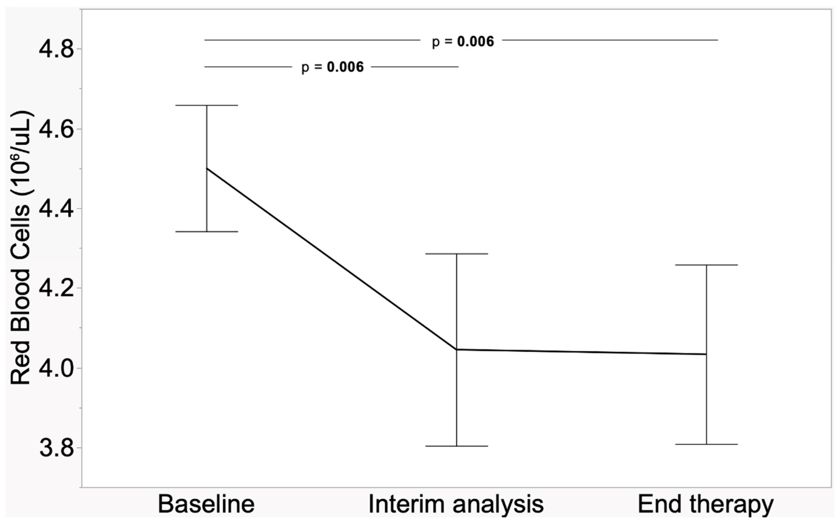
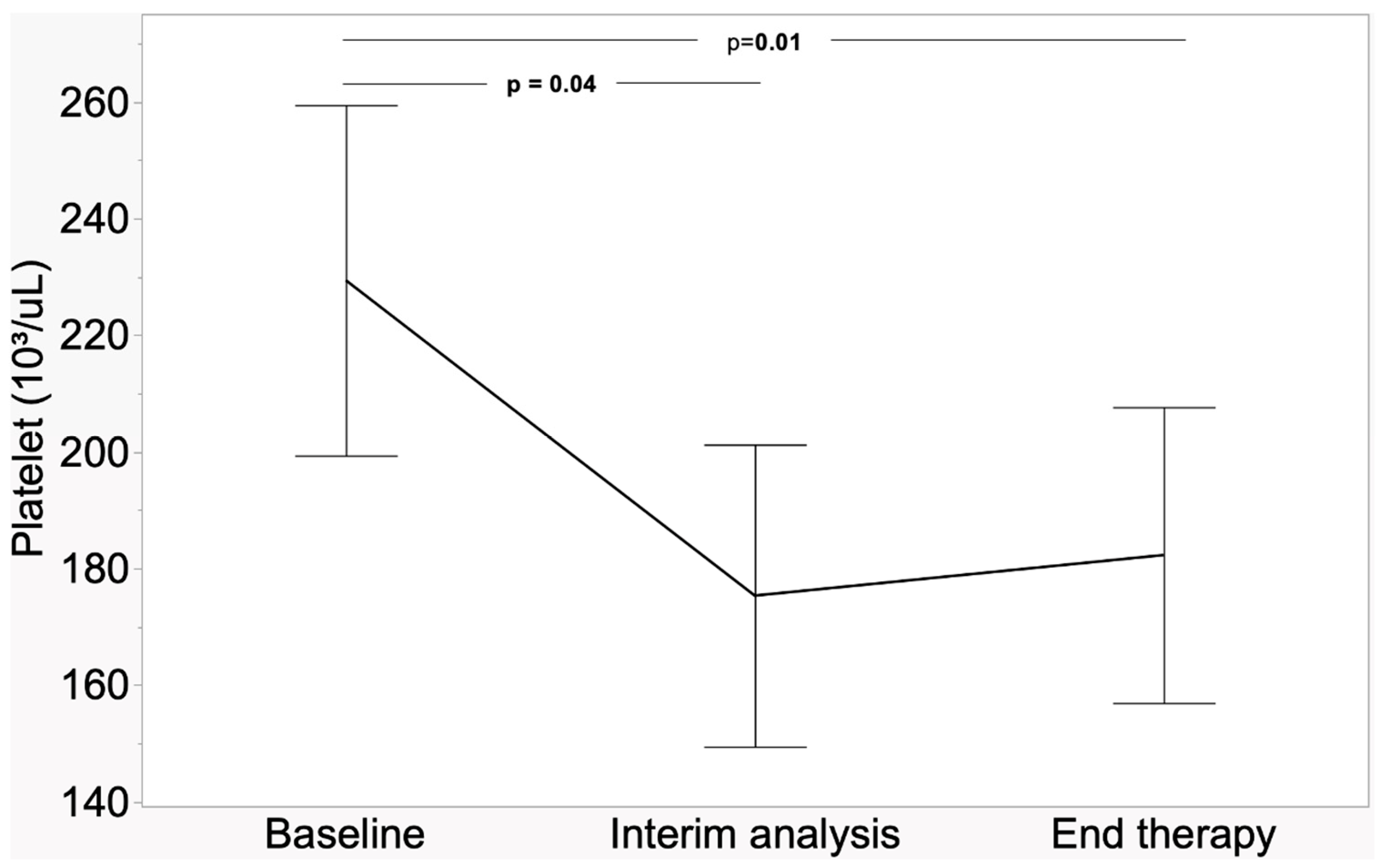
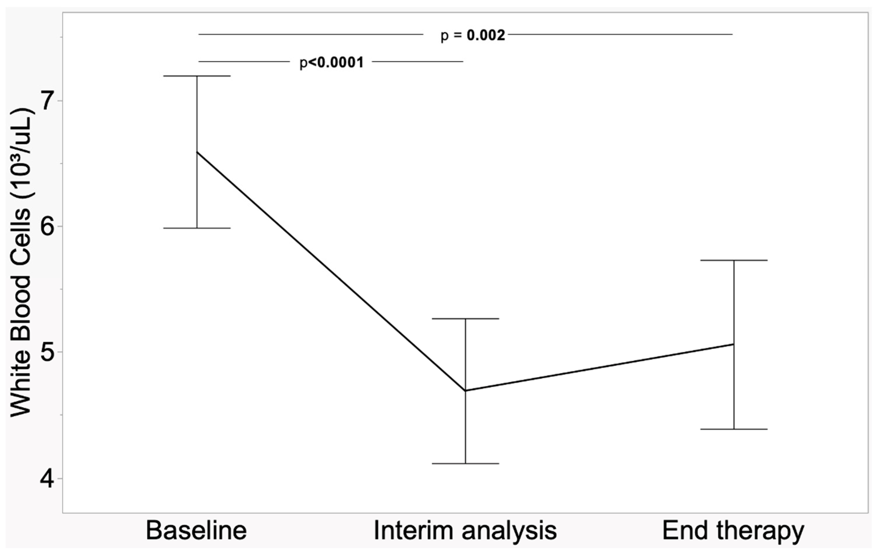
In Figure 1, Figure 2 and Figure 3, the trends in RBCs, PLTs, and WBCs are highlighted. All figures have in common the differences between baseline vs. (interim analysis and end therapy), with p-values between <0.0001 and 0.04.
Figure 1.
Trend of red blood cells in 29 patients.
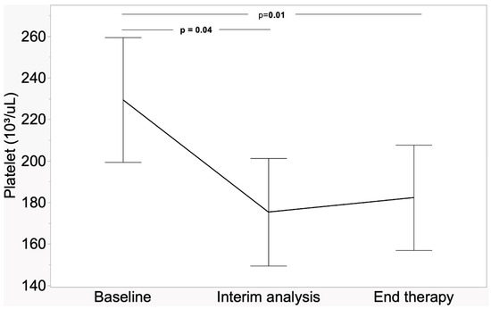
Figure 2.
Trend of platelet count in 29 patients.
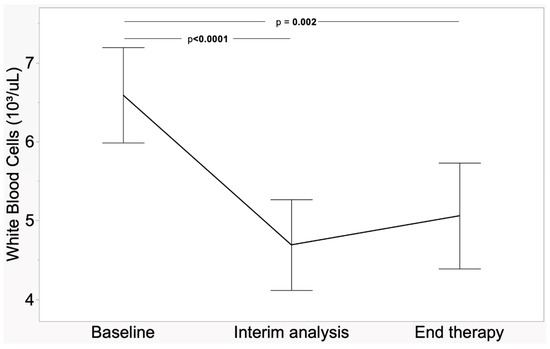
Figure 3.
Trend of white blood cells in 29 patients.
No significant correlation was found between both hematological parameters and creatinine serum levels at each time point versus age, Ki-67 index, and total tumor load.
5. Discussion
In our cohort of 29 patients treated with Lutathera®, almost all hematological parameters (except MCHC) showed a significant decrease between the baseline and/or interim and post-therapy evaluations, although without a clinical impact (grade 1 toxicity). In the subgroup analysis, the presence of bone metastases did not significantly affect these values. Conversely, unlike what we found in other research papers, gender-influenced blood parameters differ. In particular, male patients had less hematological toxicity compared to female patients, in as much as only WBC and lymphocytes dropped over time. The significantly lower baseline levels of Hb, MCH, and PLT that we found in females, together with the smaller sample size, probably had an impact on these results. Nevertheless, we think that this finding should be taken into account in future studies and, if confirmed, can be useful from the perspective of personalized dosimetry. Moreover, age, tumor grade, and tumor burden showed no correlation with hematological parameter variations during therapy, suggesting that blood count reduction probably relies only on the patient’s hematological status before starting RLT.
Finally, we did not find signs of renal toxicity in our study population because creatinine levels did not vary over time during treatment.
Our study is not the first to analyze hematological and renal toxicity in patients treated with radioligand therapy. Several other groups have reported that [177Lu]Lu-DOTATATE RLT is safe and well tolerated. It is important for clinicians to know the incidence of these adverse events during treatment to better manage them.
In a large cohort of 504 patients, mostly affected by GEP-NENs, who underwent a total of 1772 [177Lu]Lu-DOTATATE RLT treatments, Kwekkeboom et al. found that subacute hematological toxicity (WHO grade 3 or 4) occurred in 9.5% of patients and after 3.6% of infusions. Only two patients had renal insufficiency: one had pre-existent deterioration of kidney function, and the other one had tricuspid valve insufficiency; therefore, renal failure was likely unrelated to RLT. Regarding serious delayed toxicities, four patients developed myelodysplastic syndrome (MDS); in one of them, previous chemotherapy with alkylating agents was probably the cause of MDS, while in the other three patients, it was likely an effect of RLT [24].
Similar results were reported by Brabander et al., who observed grade 3/4 overall hematological toxicity in 61 of 582 patients (10%) treated with [177Lu]Lu-DOTATATE for NENs. In particular, 5% of the patients showed grade 3/4 thrombocytopenia, 5% showed grade 3/4 total WBC toxicity, and 4% showed grade 3 hemoglobin toxicity. At three months follow-up evaluation, values normalized in 77% of patients. Acute leukemia occurred in four patients (0.79%) and MDS in nine patients (1.59%). There were six cases of renal failure during follow-up, but probably unrelated to RLT [25].
In a phase-3 trial to assess the efficacy of [177Lu]Lu-DOTATATE therapy plus octreotide compared to cold somatostatin analogs alone in 221 patients with midgut neuroendocrine tumors, Strosberg et al. reported that the incidence of hematological toxicity (WHO grade 3 or 4) was 1% for neutropenia, 2% for thrombocytopenia, and 9% for lymphopenia in the group treated with radioligand therapy, while nobody showed renal toxicity. One patient in the RLT group, who had a previous history of monoclonal gammopathy of unknown significance (MGUS), subsequently developed myelodysplastic syndrome [26].
In their retrospective study of 126 patients treated with [177Lu]Lu-DOTATATE, Voter et al. noticed that hematological adverse events occurred in 14 (11%) patients and were strongly correlated with elevated baseline mean corpuscular volume (>95 fL). Interestingly, bone marrow metastases are not associated with an increased risk of hematological toxicity [27].
[90Y-DOTA]-peptides have also been used for RLT of neuroendocrine neoplasms, despite the longer positron range and higher energy of Yttrium-90 making it more myelotoxic and nephrotoxic compared to Lutetium-177, as previously reported [28].
Goncalves et al. reported from a single institution series of 521 RLT-treated patients that the median overall survival was 62 months, but only 13 months for patients who presented with therapy-related myeloid neoplasms, whose death was attributed primarily to hematological disease progression [29]. The World Health Organization pharmacovigilance database collected data indicating that AML and MDS represent around 1% of declared side effects of therapy with [177Lu]Lu-DOTATATE, which is significantly higher compared to that of other drugs [30]. Treatment with [177Lu]Lu-DOTATATE should be discontinued upon the occurrence of unacceptable toxicity or disease progression as assessed by clinical examination, imaging, or biomarkers [15].
Imhof et al., in a large population of 1109 patients affected by neuroendocrine tumors treated with [90Y-DOTA]-TOC, showed that 12.8% of them (n = 142) presented transient WHO grade 3 and grade 4 hematological toxicity (67 leukopenia; 11 anemia; 64 thrombocytopenia). Regarding myeloproliferative diseases, one patient subsequently developed myelodysplastic syndrome, while another patient developed acute myeloid leukemia. A total of 9.2% of patients (n = 102) had permanent severe renal toxicity (grade 4.67 patients; grade 5.35) [31].
Saracyn et al., in their study, compared complications of RLT in 42 patients treated with [177Lu]Lu-DOTATATE alone (n = 31) and combined with [90Y]Y-DOTATATE (n = 11). They noticed a decrease in all blood cell counts during treatment, in particular, lymphocyte numbers. All these parameters improved during follow-up but remained lower than the pre-therapy values. Long-term observation showed renal toxicity as well, with a significant decrease in GFR and an increase in serum creatinine concentration. No statistical difference was found between the two treatment groups. The authors also found a correlation between low GFR values (<60 mL/min/1.73 mq) and bone marrow function deterioration, probably related to longer circulation of the radioisotope in the blood and reduced hematopoiesis. Furthermore, previous chemotherapy affects hematological parameters [32].
Recently, the preliminary results of an Italian multicenter long-term observational study of patients with unresectable, metastatic, or progressive G1-G2 GEP-NENs selected for therapy with [177Lu]Lu-DOTATATE were published. In the early data analysis conducted at the end of population enrollment, only one of the 161 patients included in the study developed thrombocytopenia [33].
Moreover, in a recently published Netter-2 international multicenter randomized trial, whose purpose was to investigate the efficacy of [177Lu]Lu-DOTATATE therapy plus octreotide versus octreotide only in patients affected by grade 2 and grade 3 unresectable GEP NENs regarding grade 3 or worse hematological toxicity, the authors found that in the RLT group (n = 147), only three patients had leukopenia (2%), one patient had anemia (<1%), and three had thrombocytopenia (2%), while one patient developed MDS during follow-up. Lastly, grade 3 or worse nephrotoxicity occurred in three patients (2%) [34].
Interestingly, Scalorbi et al., in a cohort of 67 patients treated with RLT with an incidence of 7.5% of hematological grade 3/4 toxicity, found a correlation between splenectomy and the risk of leukopenia and thrombocytopenia, suggesting that surgical spleen removal before therapy can prevent these adverse events. As expected, performing RLT as a third- or further-line treatment, especially after chemotherapy, increases the risk of developing anemia, thrombocytopenia, and leukopenia [35].
Currently, as previously reported, a standard schedule of four cycles with fixed doses of 200 mCi (29.6 GBq) each is administered every four to eight weeks, regardless of sex, age, weight, and other patient characteristics, with the possibility of retreatment. In the future, internal dosimetry may play a big role in improving RLT with [177Lu]Lu-DOTATATE, as recently claimed also by this consensus paper [36]. Classic dosimetric calculations of adsorbed doses in healthy tissues and bone marrow have been based on serial planar scans obtained using gamma cameras, together with various measurements of radioactivity in urine and blood samples after injection [37]. Subsequently, a new approach using hybrid SPECT/CT imaging has been proposed in order to achieve a more accurate patient-specific determination of activity in the bone marrow, especially in patients with bone metastases who receive higher adsorbed doses and, consequently, more hematological toxicity [38]. Personalized dosimetry will allow adjustment of the dose for each single patient in order to optimize the therapeutic effect on tumor lesions and minimize the adsorbed dose on healthy organs, thus reducing the incidence of adverse events, although these are rare, as shown by our study and all other studies mentioned above.
6. Conclusions
RLT with [177Lu]Lu-DOTATATE, despite some minor (grade 1) hematological toxicity with no significant clinical impact, is safe and well tolerated, as already demonstrated by a large number of studies available in the literature. These findings may lead to an optimization of treatment in terms of administered dose and number of cycles in order to improve the efficacy of RLT even more in patients affected by NENs.
Author Contributions
Conceptualization: D.P. and L.C.; writing—original draft preparation: L.C., D.P., R.M., V.M.R. and E.D.; writing—review and editing: L.C. and D.P.; supervision: L.C., D.P. and F.P.; validation: L.C., D.P., R.M. and F.P.; formal analysis: G.C.; software: G.C.; data curation: R.M., V.M.R. and E.D.; visualization: M.R. All authors have read and agreed to the published version of the manuscript.
Funding
This research received no external funding.
Institutional Review Board Statement
The study was conducted in accordance with the Declaration of Helsinki. This paper is an ancillary project of study with EudraCT number 2019-001562-15.
Informed Consent Statement
Informed consent was obtained from all subjects involved in the study.
Data Availability Statement
The original data presented in the study are openly available in a publicly accessible repository.
Acknowledgments
All the authors want to express their deepest gratitude to Alberto Signore for his guidance and the support received throughout completion of this research paper.
Conflicts of Interest
The authors declare no conflict of interest.
References
- Klöppel, G. Neuroendocrine Neoplasms: Dichotomy, Origin and Classifications. Visc. Med. 2017, 33, 324–330. [Google Scholar] [CrossRef] [PubMed]
- Xu, C.; Zhang, H. Somatostatin receptor-based imaging and radionuclide therapy. BioMed Res. Int. 2015, 2015, 917968. [Google Scholar] [CrossRef] [PubMed]
- Gabriel, M.; Decristoforo, C.; Maina, T.; Nock, B.; von Guggenberg, E.; Cordopatis, P.; Moncayo, R. 99mTc-N4-[Tyr3]Octreotate Versus 99mTc-EDDA/HYNIC-[Tyr3]Octreotide: An intrapatient comparison of two novel Technetium-99m labeled tracers for somatostatin receptor scintigraphy. Cancer Biother. Radiopharm. 2004, 19, 73–79. [Google Scholar] [CrossRef] [PubMed]
- Czepczyński, R.; Parisella, M.G.; Kosowicz, J.; Mikołajczak, R.; Ziemnicka, K.; Gryczyńska, M.; Sowiński, J.; Signore, A. Somatostatin receptor scintigraphy using 99mTc-EDDA/HYNIC-TOC in patients with medullary thyroid carcinoma. Eur. J. Nucl. Med. Mol. Imaging 2007, 34, 1635–1645. [Google Scholar] [CrossRef]
- Pavlovic, S.; Artiko, V.; Sobic-Saranovic, D.; Damjanovic, S.; Popovic, B.; Jakovic, R.; Petrasinovic, Z.; Jaksic, E.; Todorovic-Tirnanic, M.; Saranovic, D.; et al. The utility of 99mTc-EDDA/HYNIC-TOC scintigraphy for assessment of lung lesions in patients with neuroendocrine tumors. Neoplasma 2010, 57, 68–73. [Google Scholar] [CrossRef][Green Version]
- Meucci, R.; Prosperi, D.; Lauri, C.; Campagna, G.; Nayak, P.; Garaci, F.; Signore, A. Peritoneal Carcinomatosis of Malignant Gynecological Origin: A Systematic Review of Imaging Assessment. J. Clin. Med. 2024, 13, 1254. [Google Scholar] [CrossRef]
- Gabriel, M.; Decristoforo, C.; Kendler, D.; Dobrozemsky, G.; Heute, D.; Uprimny, C.; Kovacs, P.; Von Guggenberg, E.; Bale, R.; Virgolini, I.J. 68Ga-DOTA- Tyr3-octreotide PET in neuroendocrine tumors: Comparison with somatostatin receptor scintigraphy and CT. J. Nucl. Med. 2007, 48, 508–518. [Google Scholar] [CrossRef]
- De Camargo Etchebehere, E.C.S.; de Oliveira Santos, A.; Gumz, B.; Vicente, A.; Hoff, P.G.; Corradi, G.; Ichiki, W.A.; Filho, J.G.d.A.; Cantoni, S.; Camargo, E.E.; et al. 68Ga-DOTATATE PET/CT, 99mTc-HYNIC-octreotide SPECT/CT, and whole-body MR imaging in detection of neuroendocrine tumors: A prospective trial. J. Nucl. Med. 2014, 55, 1598–1604. [Google Scholar] [CrossRef]
- Sharma, P.; Arora, S.; Mukherjee, A.; Pal, S.; Sahni, P.; Garg, P.; Khadgawat, R.; Thulkar, S.; Bal, C.; Kumar, R. Predictive value of 68Ga-DOTANOC PET/CT in patients with suspicion of neuroendocrine tumors: Is its routine use justified? Clin. Nucl. Med. 2014, 39, 37–43. [Google Scholar] [CrossRef]
- Prosperi, D.; Gentiloni Silveri, G.; Panzuto, F.; Faggiano, A.; Russo, V.M.; Caruso, D.; Polici, M.; Lauri, C.; Filice, A.; Laghi, A.; et al. Nuclear Medicine and Radiological Imaging of Pancreatic Neuroendocrine Neoplasms: A Multidisciplinary Update. J. Clin. Med. 2022, 11, 6836. [Google Scholar] [CrossRef]
- Prosperi, D.; Carideo, L.; Russo, V.M.; Meucci, R.; Campagna, G.; Lastoria, S.; Signore, A. A Systematic Review on Combined [18F]FDG and 68Ga-SSA PET/CT in Pulmonary Carcinoid. J. Clin. Med. 2023, 12, 3719. [Google Scholar] [CrossRef] [PubMed]
- Rinzivillo, M.; Prosperi, D.; Bartolomei, M.; Panareo, S.; Iannicelli, E.; Magi, L.; Panzuto, F. Efficacy of Lutetium-Peptide Receptor Radionuclide Therapy in Inducing Prolonged Tumour Regression in Small-Bowel Neuroendocrine Tumours: A Case of Favourable Response to Retreatment after Initial Objective Response. Oncol. Res. Treat. 2021, 44, 276–280. [Google Scholar] [CrossRef] [PubMed]
- Dell’unto, E.; Rinzivillo, M.; Esposito, G.; Iannicelli, E.; Prosperi, D.; Panzuto, F.; Annibale, B. Metastatic Type 1 low-grade gastric neuroendocrine tumor treated with peptide receptor radionuclide therapy in a young adult: A case report. Gastroenterol. Rep. 2024, 12, goae023. [Google Scholar] [CrossRef] [PubMed]
- Hennrich, U.; Kopka, K. Lutathera®: The First FDA- and EMA-Approved Radiopharmaceutical for Peptide Receptor Radionuclide Therapy. Pharmaceuticals 2019, 12, 114. [Google Scholar] [CrossRef]
- CADTH Recommendation. Lutetium (177Lu) Oxodotreotide (Lutathera). Can. J. Health Technol. 2022, 2, 1–24. [Google Scholar]
- Hope, T.A.; Bodei, L.; Chan, J.A.; El-Haddad, G.; Fidelman, N.; Kunz, P.L.; Mailman, J.; Menda, Y.; Metz, D.C.; Mittra, E.S.; et al. NANETS/SNMMI Consensus Statement on Patient Selection and Appropriate Use of 177Lu-DOTATATE Peptide Receptor Radionuclide Therapy. J. Nucl. Med. 2020, 61, 222–227. [Google Scholar] [CrossRef]
- Ambrosini, V.; Zanoni, L.; Filice, A.; Lamberti, G.; Argalia, G.; Fortunati, E.; Campana, D.; Versari, A.; Fanti, S. Radiolabeled Somatostatin Analogues for Diagnosis and Treatment of Neuroendocrine Tumors. Cancers 2022, 14, 1055. [Google Scholar] [CrossRef]
- Hicks, R.J.; Kwekkeboom, D.J.; Krenning, E.; Bodei, L.; Grozinsky-Glasberg, S.; Arnold, R.; Borbath, I.; Cwikla, J.; Toumpanakis, C.; Kaltsas, G.; et al. ENETS Consensus Guidelines for the Standards of Care in Neuroendocrine Neoplasms: Peptide Receptor Radionuclide Therapy with Radiolabelled Somatostatin Analogues. Neuroendocrinology 2017, 105, 295–309. [Google Scholar] [CrossRef]
- Autorité de Sureté Nucléaire, A.S.N. Lettre Circulaire Du 12 Juin 2020—Evolution Des Conditions D’autorisation Des Services de Médecine Nucléaire par L’ASN Pour la Détention Et L’utilisation Du Lutétium-177; Autorité de Sureté Nucléaire ASN: Montrouge, France, 2020. [Google Scholar]
- Herrmann, K.; Giovanella, L.; Santos, A.; Gear, J.; Kiratli, P.O.; Kurth, J.; Denis-Bacelar, A.M.; Hustinx, R.; Patt, M.; Wahl, R.L.; et al. Joint EANM, SNMMI and IAEA enabling guide: How to set up a theranostics centre. Eur. J. Nucl. Med. Mol. Imaging 2022, 49, 2300–2309. [Google Scholar] [CrossRef]
- Saowapa, S.; Polpichai, N.; Tanariyakul, M.; Suenghataiphorn, T.; Kulthamrongsri, N.; McCullough, M.; Damasceno Moreira, M.G.; Siladech, P.; Tijani, L. Effects of Autoimmune Disorders on Myelodysplastic Syndrome Outcomes: A Systematic Review. Hemato 2024, 5, 208–219. [Google Scholar] [CrossRef]
- Pizzi, M.; Gurrieri, C.; Orazi, A. What’s New in the Classification, Diagnosis and Therapy of Myeloid Leukemias. Hemato 2023, 4, 112–134. [Google Scholar] [CrossRef]
- Glymour, M.M.; Weuve, J.; Berkman, L.F.; Kawachi, I.; Robins, J.M. When is baseline adjustment useful in analyses of change? An example with education and cognitive change. Am. J. Epidemiol. 2005, 162, 267–278. [Google Scholar] [CrossRef] [PubMed]
- Kwekkeboom, D.J.; de Herder, W.W.; Kam, B.L.; van Eijck, C.H.; van Essen, M.; Kooij, P.P.; Feelders, R.A.; van Aken, M.O.; Krenning, E.P. Treatment with the radiolabeled somatostatin analog [177Lu-DOTA 0,Tyr3]octreotate: Toxicity, efficacy, and survival. J. Clin. Oncol. 2008, 26, 2124–2130. [Google Scholar] [CrossRef] [PubMed]
- Brabander, T.; van der Zwan, W.A.; Teunissen, J.J.M.; Kam, B.L.R.; Feelders, R.A.; de Herder, W.W.; van Eijck, C.H.J.; Franssen, G.J.H.; Krenning, E.P.; Kwekkeboom, D.J. Long-Term Efficacy, Survival, and Safety of [177Lu-DOTA0,Tyr3]octreotate in Patients with Gastroenteropancreatic and Bronchial Neuroendocrine Tumors. Clin. Cancer Res. 2017, 23, 4617–4624. [Google Scholar] [CrossRef]
- Strosberg, J.; El-Haddad, G.; Wolin, E.; Hendifar, A.; Yao, J.; Chasen, B.; Mittra, E.; Kunz, P.L.; Kulke, M.H.; Jacene, H.; et al. Phase 3 Trial of 177Lu-Dotatate for Midgut Neuroendocrine Tumors. N. Engl. J. Med. 2017, 376, 125–135. [Google Scholar] [CrossRef]
- Voter, A.F.; Gafita, A.; Werner, R.A.; De Jesus-Acosta, A.; Rowe, S.P.; Solnes, L.B. Elevated Baseline Mean Corpuscular Volume Predicts the Development of Severe Hematologic Toxicity After 177Lu-DOTATATE Therapy. J. Nucl. Med. 2024, 65, 1423–1426. [Google Scholar] [CrossRef]
- Bodei, L.; Kidd, M.; Paganelli, G.; Grana, C.M.; Drozdov, I.; Cremonesi, M.; Lepensky, C.; Kwekkeboom, D.J.; Baum, R.P.; Krenning, E.P.; et al. Long-term tolerability of RLT in 807 patients with neuroendocrine tumours: The value and limitations of clinical factors. Eur. J. Nucl. Med. Mol. Imaging 2015, 42, 5–19. [Google Scholar] [CrossRef]
- Goncalves, I.; Burbury, K.; Michael, M.; Iravani, A.; Ravi Kumar, A.S.; Akhurst, T.; Tiong, I.S.; Blombery, P.; Hofman, M.S.; Westerman, D.; et al. Characteristics and outcomes of therapy-related myeloid neoplasms after peptide receptor radionu-clide/chemoradionuclide therapy (RLT/PRCRT) for metastatic neuroendocrine neoplasia: A single-institution series. Eur. J. Nucl. Med. Mol. Imaging 2019, 46, 1902–1910. [Google Scholar] [CrossRef]
- Vigne, J.; Chrétien, B.; Bignon, A.-L.; Bouhier-Leporrier, K.; Dolladille, C. [177Lu]Lu-DOTATATE peptide receptor radionuclide therapy–associated myeloid neoplasms: Insights from the WHO pharmacovigilance database. Eur. J. Nucl. Med. Mol. Imaging 2022, 49, 3332–3333. [Google Scholar] [CrossRef]
- Imhof, A.; Brunner, P.; Marincek, N.; Briel, M.; Schindler, C.; Rasch, H.; Mäcke, H.R.; Rochlitz, C.; Müller-Brand, J.; Walter, M.A. Response, survival, and long-term toxicity after therapy with the radiolabeled somatostatin analogue [90Y-DOTA]-TOC in metastasized neuroendocrine cancers. J. Clin. Oncol. 2011, 29, 2416–2423. [Google Scholar] [CrossRef]
- Saracyn, M.; Durma, A.D.; Bober, B.; Kołodziej, M.; Lubas, A.; Kapusta, W.; Niemczyk, S.; Kamiński, G. Long-Term Complications of Radioligand Therapy with Lutetium-177 and Yttrium-90 in Patients with Neuroendocrine Neoplasms. Nutrients 2022, 15, 185. [Google Scholar] [CrossRef] [PubMed]
- Lastoria, S.; Rodari, M.; Sansovini, M.; Baldari, S.; D’agostini, A.; Cervino, A.R.; Filice, A.; Salgarello, M.; Perotti, G.; Nieri, A.; et al. Lutetium [177Lu]-DOTA-TATE in gastroenteropancreatic-neuroendocrine tumours: Rationale, design and baseline characteristics of the Italian prospective observational (REAL-LU) study. Eur. J. Nucl. Med. Mol. Imaging 2024, 51, 3417–3427. [Google Scholar] [CrossRef] [PubMed]
- Singh, S.; Halperin, D.; Myrehaug, S.; Herrmann, K.; Pavel, M.; Kunz, P.L.; Chasen, B.; Tafuto, S.; Lastoria, S.; Capdevila, J.; et al. [177Lu]Lu-DOTA-TATE plus long-acting octreotide versus high-dose long-acting octreotide for the treatment of newly diagnosed, advanced grade 2-3, well-differentiated, gastroenteropancreatic neuroendocrine tumours (NETTER-2): An open-label, randomised, phase 3 study. Lancet 2024, 403, 2807–2817. [Google Scholar] [PubMed]
- Scalorbi, F.; Argiroffi, G.; Baccini, M.; Gherardini, L.; Fuoco, V.; Prinzi, N.; Pusceddu, S.; Garanzini, E.M.; Centonze, G.; Kirienko, M.; et al. Application of FLIC model to predict adverse events onset in neuroendocrine tumors treated with RLT. Sci. Rep. 2021, 11, 19490, Erratum in: Sci. Rep. 2023, 13, 1759. [Google Scholar] [CrossRef]
- Panzuto, F.; Albertelli, M.; De Rimini, M.L.; Rizzo, F.M.; Grana, C.M.; Cives, M.; Faggiano, A.; Versari, A.; Tafuto, S.; Fazio, N.; et al. Radioligand therapy in the therapeutic strategy for patients with gastro-entero-pancreatic neuroendocrine tumors: A consensus statement from the Italian Association for Neuroendocrine Tumors (Itanet), Italian Association of Nuclear Medicine (AIMN), Italian Society of Endocrinology (SIE), Italian Association of Medical Oncology (AIOM). J. Endocrinol. Investig. 2024. [Google Scholar] [CrossRef]
- Bergsma, H.; Konijnenberg, M.W.; Kam, B.L.; Teunissen, J.J.; Kooij, P.P.; de Herder, W.W.; Franssen, G.J.; van Eijck, C.H.; Krenning, E.P.; Kwekkeboom, D.J. Subacute haematotoxicity after RLT with (177)Lu-DOTA-octreotate: Prognostic factors, incidence and course. Eur. J. Nucl. Med. Mol. Imaging 2016, 43, 453–463. [Google Scholar] [CrossRef]
- Hagmarker, L.; Svensson, J.; Rydén, T.; van Essen, M.; Sundlöv, A.; Gleisner, K.S.; Gjertsson, P.; Bernhardt, P. Bone Marrow Absorbed Doses and Correlations with Hematologic Response During 177Lu-DOTATATE Treatments Are Influenced by Image-Based Dosimetry Method and Presence of Skeletal Metastases. J. Nucl. Med. 2019, 60, 1406–1413. [Google Scholar] [CrossRef]
Disclaimer/Publisher’s Note: The statements, opinions and data contained in all publications are solely those of the individual author(s) and contributor(s) and not of MDPI and/or the editor(s). MDPI and/or the editor(s) disclaim responsibility for any injury to people or property resulting from any ideas, methods, instructions or products referred to in the content. |
© 2024 by the authors. Licensee MDPI, Basel, Switzerland. This article is an open access article distributed under the terms and conditions of the Creative Commons Attribution (CC BY) license (https://creativecommons.org/licenses/by/4.0/).

