Review

Clonal Hematopoiesis, a Risk Condition for Developing Myeloid Neoplasia

Ugo Testa, Department of Oncology, Istituto Speriore di Sanità, Viale Regina Elena 299, 00161 Rome, Italy
*
Author to whom correspondence should be addressed.
Hemato 2025, 6(2), 10; https://doi.org/10.3390/hemato6020010
Submission received: 11 February 2025 / Revised: 18 April 2025 / Accepted: 19 April 2025 / Published: 22 April 2025
(This article belongs to the Section Leukemias)

Abstract

:
Clonal hematopoiesis (CH) is an age-related process in which hematopoietic stem/progenitor cells increase their fitness due to the acquisition of mutations that lead to a proliferative advantage and to clonal expansion. Its frequency increases with age, and it mostly affects people older than 70 years. The most mutated genes in CH are epigenetic regulators, DNA damage response genes, and splicing factors, which are all involved in the development of myeloid neoplasia. Some risk factors, including age, smoking, and prior cytotoxic therapy, increase the risk of developing CH or increase the fitness of CH. Various types of CH have been observed, associated or not with cytopenias or monocytosis. CH represents a risk factor for many pathological conditions and particularly for hematologic malignancies. A better understanding of the risks related to CH has triggered the development of research, translational, and clinical programs for the monitoring, prevention, and treatment of CH.

1. Introduction

The acquisition of mutations in healthy human tissues is commonly observed during aging and must be considered as an inevitable consequence of aging [1]. DNA sequencing studies have shown that there is a ubiquitous increase in somatic mutations with age, at the level of both dividing and nondividing tissues, ranging from 2.4 mutations/cell/year in the testes and 56 mutations/cell/year in the intestine [1]. Thus, these studies have shown a progressive accumulation of mutations in normal tissues with age.
If a given mutation increases cell fitness and provides a selective advantage, this cell will amplify and will undergo a clonal expansion.
The hematopoietic tissue is characterized by the continuous production of blood cell elements through a complex process that implies the controlled proliferation of 50,000–200,000 hematopoietic stem cells (HSCS), which initially differentiate into more committed progenitor cells (HPCs), which, in turn generate, through differentiation/maturation processes, all blood cell lineages. During their lifespan, somatic mutations may be acquired by all blood cell elements, including HSCs. Most of these mutations are neutral mutations and do not have physiological consequences; more rarely, some mutations can confer a fitness advantage, allowing a selective advantage and enabling the progressive clonal expansion of a mutated HSC and of its progeny. This process of clonal expansion is defined as clonal hematopoiesis (CH).
The discovery of CH has revolutionized our understanding of the mechanisms underlying the aging of hematopoietic tissues and the development of hematologic malignancies. CH has many important implications for human health in that it represents a condition associated with an increased risk of developing hematologic malignancies and many age-related diseases (such as cardiovascular disease, diabetes, and autoimmune disorders); CH is associated with an increased risk of chronic disease and all-cause mortality in the general population. Only a minority of individuals with CH develop a hematologic malignancy. The frequency of occurrence of CH is considerably influenced by environmental exposures, such as smoking or cytotoxic anticancer therapy.
About 30% of myelodysplastic syndromes (MDS) and acute myeloid leukemia (AML) originate from a prior CH condition. CH increases the risk of developing a therapy-related myeloid neoplasia after cytotoxic chemo-radiation therapy or after autologous stem cell transplantation. Although the role of CH in the pathogenesis of hematologic malignancies has considerably improved in recent years, many knowledge gaps remain to be addressed. In particular, the understanding of the cellular and molecular mechanisms driving CH evolution remains largely incomplete but is necessary for the development of preventive and therapeutic approaches. The features defining CH and the implications of the molecular heterogeneity of CH must be better dissected particularly because they concern the risk stratification of CH patients. The criteria for clinical interventions in CH patients and the possible management strategies for these patients remain to be explored and appropriately defined.
Current research based on experimental studies, longitudinal studies, and clinical interventional approaches aims to bridge these gaps by understanding the CH that still exists. This review will try to analyze the recent developments in the definition and characterization of various types of CH existing in humans and to discuss current and future studies, aiming to bypass the knowledge gaps remaining to be addressed in this field.

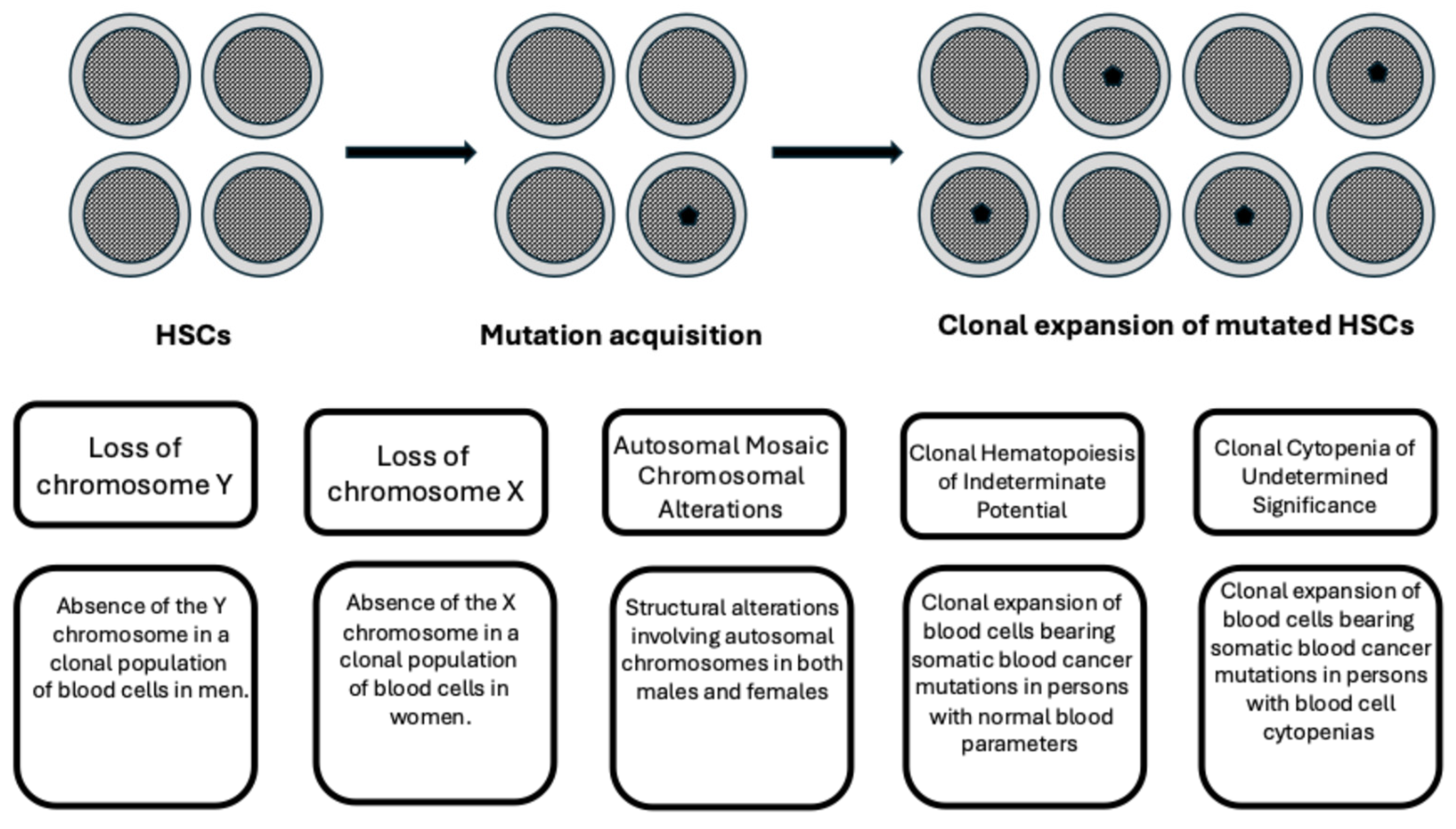
2. Definition of Clonal Hematopoiesis

During their lifespan, cells contained in tissues with rapid renewal, such as hematopoietic tissue, continuously divide and may progressively accumulate somatic mutations. Most of these mutations are neutral and do not have consequences at the level of cellular dynamics; however, a minority of mutations are not neutral and may alter cellular fitness, conferring a growth advantage and thus causing a clonal expansion.
Single-cell studies have shown that hematopoiesis in individuals less than 65 years of age is polyclonal, with high clonal diversity, and is supported by a population of 50,000-200,000 pluripotent HSCs contributing to blood production; however, in individuals aged over 75 years, 30–60% of hematopoiesis was ensured by 12–18 independent clones, each contributing to 1–34% of blood production [2]. Most of these clones started their expansion before 40 years of age, and only 22% of them had known driver mutations [2].
The development of gene sequencing studies has made it possible to quantify and track somatic mutations in normal hematopoietic cells, providing an estimate of a mutational rate between 11.7 and 14.6 mutations per year [3,4,5]. Mitchell and coworkers estimated that HSCs/HPCs accumulate a mean of 17 mutations per year after birth and lose 30 base pairs per year of telomere length [2].
Lineage differentiation of both lymphoid-biased and myeloid-biased HSC subsets progressively shifts to a higher myeloid cellular output during aging; furthermore, HSCs selectively undergo age-dependent gene expression and gene regulatory changes in a progressive manner [6]. With advanced age, there is a progressive shift in the mechanisms of platelet production, with the progressive activation of a differentiation pathway that shortcuts the canonical progenitor cascade to generate megakaryocyte precursor cells directly from HSCs [7]. At the molecular level, aged HSCs typically display metabolic dysregulation, upregulation of inflammatory pathways, and downregulation of DNA repair pathways [8].
DNA lesions emerge from continuous cellular processes; most of these lesions are efficiently repaired. However, a recent study showed that a family of DNA lesions present in HSCs in low numbers persist for months to years, generating a sizeable fraction of cellular mutation burdens; some of these DNA lesions generate a characteristic mutational signature called SBS19, responsible for about 16% of the mutations in blood cells [9].
Initial studies provide evidence of clonally expanded hematopoietic cells through the presence of skewed X chromosome inactivation. Subsequent studies have shown the existence of loss of chromosome Y (absence of the Y chromosome in a clonal population of blood cells in men), loss of chromosome X (absence of an X chromosome in a clonal population of blood cells in women), or autosomal mosaic chromosomal alterations (large structural alterations present across all autosomal chromosomes in clonal blood cells in both males and females). More recently, clonal expansion in blood cells bearing recurrent somatic driver mutations currently observed in hematologic malignancies has been identified in individuals with otherwise normal hematologic parameters and has been defined as clonal hematopoiesis of indetermined potential (CHIP). Finally, in some individuals, a form of CHIP associated with cytopenia was identified and defined as clonal cytopenia of undetermined significance (CCUS) (Figure 1).
Clonal hematopoiesis was discovered in studies focused on measuring the ratios of X chromosome inactivation in females; these studies led to the identification of age-associated skewing (AAS) in blood cells; AAS may be related to the progressive acquisition in HSCs of somatic mutations conferring a growth advantage and thus causing a clonal expansion of some hematopoietic cells: in line with this hypothesis, Busque and coworkers identified TET2 and DNMT3A mutations in one of three individuals with AAS; subsequently, the study of 179 older women with AAS showed the existence of TET2 mutations in 5.6% of cases [10]. As initially shown by four pivotal studies, clonal hematopoiesis (CH) is characterized by the presence of recurrent mutations of some genes (DNMT3A, TET2, and ASXL1) that increase the fitness of hematopoietic stem cells in individuals without any apparent blood neoplasia [10,11,12,13]. The prevalence of CH clearly increases with age, being observed in 10–20% of individuals over the age of 70 years [11,12,13,14]. Studies using very sensitive next-generation sequencing techniques detecting very small mutated clones showed that CH is virtually ubiquitous in individuals over 50–60 years [15].
The terms CH or “age-related clonal hematopoiesis” (ARCH) imply any type of clonal expansion of hematopoietic stem cells, independent of the size of the mutated clone. However, the term clonal hematopoiesis of indeterminate potential (CHIP) refers to the presence of a CH clone expanded at a variant allele fraction ≥2% (thus involving ≥4% of nucleated blood cells); although this level of allelic frequency is based on an arbitrary threshold, it implies that large clones may have a potential clinical implication.
In most studies on the identification and molecular characterization of CH, DNA sequencing was restricted to the detection of mutations in genes involved in myeloid malignancies, and this has precluded a whole definition of the spectrum of gene mutations involved in CH. To bypass these limitations, an alternative method of CH detection is based on the observation that HSCs accumulate mutations during their lifespan, and most of these mutations have no effect on their phenotype; thus, each HSC and its clonal descendants are “barcoded” with a unique spectrum of mutations; thus, if a particular clone iss sufficiently expanded to give a significant contribution to hematopoiesis, it can be identified by its unique barcode [11]. The use of this technique showed that CH is very common in the elderly, and most CH cases do not carry an obvious candidate preleukemic driver mutation [16,17]. The same conclusion was reached through single-cell sequencing studies showing the existence in individuals over 70 years of numerous clones (15–21), accounting for 30–60% of hematopoiesis; most of these clones lack known driver mutations [2].

3. Mutational Spectrum of CHIP

Many studies have explored the mutational spectrum of driver mutations observed in CHIP. The most recurrent driver mutations observed in CHIP affect different functional categories of genes: epigenetic regulators (DNMT3A, TET2, ASXL1); spliceosome genes (SF3B1, SRSF2, U2AF1); signal transduction genes (JAK2, CALR, MPL); and DNA damage repair genes (TP53, PPM1D).
A recent study provided evidence about the existence of additional driver gene mutations occurring in CHIP. Bernstein et al. performed an analysis of somatic mutations in whole blood in a population of 200,618 individuals of the UK Biobank and determined the mutated genes under positive selection as inferred through the analysis of the ratio of nonsynonymous mutations to synonymous mutations (dN/dS) [18]. This analysis led to the identification of a set of genes already known as drivers of CH, more frequently mutated (DNMT3A, TET2, ASXL1, PPM1D, TP53, SRFSF2, SF3B1, and JAK2) or less frequently mutated (BRCC3, CBL, GNB2, KMD6A, and PHIP) [18]. Furthermore, this analysis led to the identification of a set of 17 new genes evaluated as possible drivers according to their fitness (BAX, CCD115, CCL22, CHEK2, IGL55, MAGEC3, MTA2, MYD88, SHB2D3, SIK3, SPRED2, SRCAP, SRSF1, YLPM1, ZBTB33, ZNF234, and ZNF318) [18]. In the whole population analyzed, 23% displayed CH, 48% with classical drivers and 5% with new drivers. Clones bearing new drivers increase in frequency and size according to age, and their presence is associated with increased risks of developing hematologic malignancies and of death [18].
The type of genetic mechanism is related to the type of genetic alteration observed in CHIP. Thus, for point mutations, the most common mutational event, age-related, consists in the spontaneous deamination of 5-methylcytosine to thymine. Less frequently, the mutations may be acquired by errors occurring during the process of repair of double-stranded DNA breaks, generating small insertions/deletions. Recurrent gene deletions observed in CHIP involve CALR, ASXL1, and SRSF2 loci; these deletions seem to be related to events of double strand break repair mediated by a PARP1-dependent microhomology-mediated and joining pathway [19].

3.1. DNMT3A Mutations

DNMT3A encoding a DNA methyltransferase is the most recurrently mutated gene in CHIP. The functional consequences of DNMT3A mutations consist of a loss of function of the mutated protein and induce a condition of global hypomethylation (focal methylation loss and attenuation of hypermethylation) and enhanced self-renewal of HSCs.
DNMT3A loss stimulates HSC self-renewal and increases the repopulating capacity of these cells, as supported by serial experiments of transplantation in mice [20,21]. DNMT3A-mutant HSCs only slowly outcompete normal HSCs. However, the clonal expansion of mutant HSCs may be considerably enhanced by environmental factors: a chronic mycobacterial infection [22] and inflammatory stress induced by interferon-gamma (IFN-γ) [23] promote clonal expansion of DNMT3A-mutant HSCs.
Studies in an experimental model of CHIP generated by a DNMT34A R878H mutant displayed a dysregulated activity of APOBEC 3, thus generating enhanced DNA damage repair activity [21]. These findings were validated through analysis of patient bone marrow cells showing increased expression of all DNA repair-related gene clusters, except for ATM exhibiting increased expression [24]. According to these observations it was suggested that DNMT3A mutations may induce DNA/RNA damage through APOBEC3, leading to secondary genetic mutations through interference of the cellular repair machinery [24].
Several observations suggest that the major defect in DNMT3A-mutant cells could be related to the generation of defective T cells. In fact, transplantation studies have shown that DNMT3A-mutant recipient patients have hyperactive T cells that may have a role in initiating graft versus host disease (GVHD), as shown by the observation that patients with donor-derived DNMT3A-mutant CH have an increased risk of acute [25] or chronic [26] GVHD; or graft vs. leukemia effect, as supported by the observation that recipients of DNMT3A-mutant donor-derived CH have a decreased leukemia relapse risk [27]. A recent experimental study further supported the conclusion that DNMT3A-mutant HSCs produce hyperactive T cells with increased alloimmune and anti-leukemic activity [28].
Studies in primary CH samples and in an engineered mouse model for DNMT3A-mutant CH provided evidence that HSCs carrying DNMT3AR882 hotspot mutations in CH exhibited a downregulation of MHC-II molecules, thus leading to an altered immunosurveillance mechanism in DNMT3A-mutant CH [29]. Finally, a recent study supported both by clinical observations and by an experimental model showed that DNMT3A mutations associated with CH determine elevated numbers of osteoclast precursors in the bone marrow and osteoclastogenic macrophages in peripheral tissues, together with increased neutrophilic inflammation and impaired T-cell regulatory activity, promoting at a clinical level increased prevalence of periodontitis [30].
The studies carried out on CHIP and on AML patients support the view that DNMT3A mutations play a relevant role in leukemia initiation, but their role in leukemia maintenance is less clear. To investigate this issue, Kohnke et al. developed a CRISPR-based method to directly correct DNMT3AR882 mutations in leukemic blasts obtained from patients: replacing DNMT3AR882 with wild-type DNMT3A did not inhibit the capacity of leukemic blasts to engraft immunodeficient mice and minimally altered DNA methylation [31]. According to these observations, it was concluded that DNMTAR882 mutations are required for AML initiation but are dispensable for disease maintenance [31].

3.2. TET2 Mutations

TET2 is the second most frequently mutated gene in CH; TET2 is a ketoglutarate-dependent dioxygenase promoting DNA demethylation by converting 5-methylcytosine to 5-hydroxymethylcytosine; loss-of-function mutations observed in CH confer a competitive advantage to HSCs in transplantation experiments and induce a granulocyte-monocyte lineage bias.
Genetically mediated deficiency determines a condition of unexpected global hypomethylation of the heterochromatin compartment coupled to regional hypermethylation largely confined to the active euchromatic compartment; because of these chromatin methylation changes, TET2 promotes an increased HSC self-renewal and determines a progressive enlargement of the HSC pool. Chromatin opening is mediated by TET2 deficiency through the inhibition of a process involving recognition of chromatin-associated 5-methylcytosine by the methyl-CpG-binding domain protein MBD6, which guides Lys119 deubiquitination of histone H2A to promote an open chromatin state; the oxidation of 5-mehylcytosine catalyzed by TET2 antagonizes MBD6-dependent deubiquitination [32].
TET2 mutations are associated with DNA hypermethylation occurring at the level of some specific DNA enhancers, associated with myeloid differentiation (leukocyte function and immune response) and with the transcription factors ETS and C/EBP. Most of these TET2-associated hypermethylated enhancer sites are shared between CHIP, CCUS, and AML, but some hypermethylated sites are specifically related to AML and are shared between AML and HSCs [33].
In murine models, TET2 deficiency increases the self-renewal of HSCs, as shown by competitive transplantation assays, and leads to an expansion of the HSC compartment over time, seemingly due to induction of enhanced expression and signaling of the thrombopoietin receptor on HSCs [34].
TET2 loss drives aberrant self-renewal without inducing leukemic transformation. In experimental mouse models, heterozygous and homozygous TET2 deletion cooperates with reduction of PU.1 activity in inducing leukemic transformation in an age-dependent context [35].
A remarkable difference between DNMT3A and TET2 is that while DNMT3A-mutant HSCs expand with similar rates in both young and adult recipient mice, TET2-mutated HSCs expand at a significantly faster rate in old mice compared to young recipient mice [36]. These observations support the view that TET2 mutations observed in CH are selectively advantageous in aging. Other studies support the strong link between TET2 and aging in CH. Thus, studies of single-cell multiomic and flow cytometry characterization have contributed to show that TET2 deficiency, in an age-related manner, mitigates the process of HSC aging at transcriptomic, epigenomic, and cellular levels [37]. Finally, competitive bone marrow repopulation assays showed that the rate of TET2-deficient clone was significantly accelerated by competitor grafts derived from old donors compared to those from young donors, thus suggesting that the lack of fitness of old HSCs may significantly contribute to the emergence and expansion of CH [38].
TET2 loss confers a myeloid bias to HSCs, with reduction in megakaryocytic-erythroid and lymphoid progenitors. TET2 loss in human HSCs causes the generation of a heterogeneous neutrophil population through increased repopulating capacity of neutrophil progenitors; these neutrophils have a low granule content and an exacerbated response to inflammatory stimulations [39].
Other studies have supported a link between aging and inflammation in the mechanisms underlying clonal expansion of TET2-CH. TET2−/+ HSCs showed reduced susceptibility to IL-1α-mediated downregulation of self-renewal genes and showed increased DNA replication [40]. Genetic deletion of IL-1R1 in TET2−/+ HSCs or pharmacologic inhibition of IL-1 signaling impaired TET2-mediated clonal expansion [40]. Using aged mouse models, it was shown that in normal HSCs, IL-1β administration induces loss of DNA methylation, while TET2-deficient HSCs are resistant to this effect, with consequent expression of self-renewal genes and inhibition of differentiation-related transcription factors [41].
Single-cell analysis of human TET2 or DNMT3A-mutated CH bone marrow samples showed that normal HSCs contained in these samples display an enrichment in inflammatory and aging transcriptomic signatures in comparison with HSCs from normal bone marrow samples; however, TET2- and DNMT3A-mutated HSCs have an attenuated inflammatory response relative to WT-HSCs within the same sample [42].
In addition to the control of HSC self-renewal, TET2 also acts as a repressor of inflammatory signaling via histone deacetylase. In fact, malignant progression of TET2-mutant CH is driven by dysregulated HSC function, myeloid expansion, and increased inflammation. Interestingly, the pharmacologic inhibition of the epigenetic reader protein bromo-and extra-terminal (BET) domain 4 (BNRD4) reduces self-renewal, inflammation, and myeloid expansion in a murine model of TET2-driven CH [43].

3.3. ASXL1 Mutations

ASXL1, a gene encoding a chromatin-binding protein that interacts with several histone-modifying complexes, is the third most recurrent driver gene mutated in CHIP. ASXL1 mutations observed in CH are nonsense or frameshift mutations localized at the level of the penultimate or final exon of the ASXL1 gene, generating a truncated protein lacking the C-terminal histone binding domain of the ASXL1 protein. ASXL1 mutations result in the generation of a truncated protein that possesses enhanced biological activity, forming a stable complex with the deubiquitinylase BAP1, causing the reduction of H2AK119Ub, resulting in activation of a myeloid gene expression program [44]. Analysis of a large set of individuals with CH present in a UK biobank showed the existence of a strong association between current and post smoking status and CH with ASXL1 mutations [41]. This observation suggests that the inflammatory environment generated by smoking may induce the outgrowth of ASXL1-mutant clones [45].
The mechanisms through which ASXL1 drives CH remained elusive for long time, but a recent study in part clarified the effects of ASXL1 mutations on HSC compartment [46]. Using knock-in mice expressing ASXL1-mutant, it was shown that HSCs expressing mutant ASXL1 have a reduced repopulating capacity after transplantation; however, in genetic mouse models, mutant HSCs acquire clonal advantage during aging, thus recapitulating a condition observed in human CH, and this effect seems to be mediated through cooperation with BAP1 to deubiquitinate and activate AKT; mutant ASXL1 induces an overactivation of AKT/mTOR signaling and through this mechanism induces an increased proliferation and dysfunction of HSCs associated with age-dependent accumulation of DNA damage events [46].
A recent study showed that ASXL1 mutant protein interacts with EHMT1 and EHMT2 methyltransferase complex, which generates H2K9me1 and H3K9me2, a repressive modification in constitutive heterochromatin [47]. This interaction compromises chromatin integrity in an age-dependent manner by decreasing H3K9me2/3 levels in constitutive heterochromatin and H3K9me1 levels in facultative heterochromatin [47].

3.4. Splicing Factor Mutations

The splicing factors SF3B1, SRSF2, and U2AF1 represent a group of genes mutated in CHIP, particularly in older individuals. Specifically, McKerrel et al. showed that CH driven by mutated spliceosome genes SF3B1 and SRSF2 was particularly observed in individuals 70 years or older [14]. Mutations in spliceosome genes (SF3B1, SRSF2, U2AF1) were observed with a higher frequency in thrombocytopenia cases; the combination of thrombocytopenia and CH with spliceosome-mutated genes resulted in an increased risk of death [48]. Splicing factor gene mutations typically involve single amino acid substitutions at hot spot sites, including SRSF2-P95, UA2F1-Q157, SF3B1-K666, Sf3B1-K700, and SF3B1-R625. Spliceosome gene mutations at SRSF2-P95, SF3B1-K666, and SF3B1-K700 were exclusively observed over 70 years, their frequency rising from 1.8% in those aged 70–79 years to 8.3% in the 90–98 years group [14]. These mutations alter protein function, including aberrant splicing of large number of transcripts and the generation of novel mRNA isoforms.
Additional mutations have also been detected more rarely in the splicing factor ZRSF2, under the forms of nonsense, frameshift, or substitution mutations disrupting the function of the minor spliceosome. ZRSF2 mutations are observed in 4% of MDS, and among individuals with clonal hematopoiesis, they are observed particularly in individuals with CCUS, frequently in association with TET2 mutations and more rarely with ASXL1 mutations [49].
Mutations in UAF1 or U2AF1 are identified in about 3% of healthy individuals with CH aged >70 years [14]; their frequency is 10% in younger individuals whose CH converts to AML, thus suggesting that mutations in SRSF2 or U2AF1 are associated with a high risk of AML transformation [50,51].
Mutations in spliceosome components such as SF3B1, SRSF2, U2AF1, and ZRSF2 alter RNA splicing, determining the generation of aberrant mRNA transcripts and dysfunctional proteins that disrupt cellular homeostasis and differentiation. Spliceosome mutations are frequent in AMLs and particularly in MDS, highlighting their central role in the pathogenesis of these hematological malignancies.
The study on the effects of splicing factor mutations exerted in knock-in mouse models showed significant perturbations of HSC/HPC regulation but frequently failed to show an effect on clonal expansion of HSCs. Mutations in splicing factors have widespread effects implicating a multitude of cellular processes. A consistent number of studies have shown that SF mutations deregulate HSCs and HPCs. Mice expressing mutant U2AF1 display altered hematopoiesis, with alterations of the number of HSCs/HPCs, without leukemic development and changes in pre-mRNA splicing in HPCs [52,53]. Using a conditional knockout model, it was further shown that U2AF1 controls HSC survival and function, inducing a reduction in HSCs and HPCs [54]. Using a bone marrow transplant model, it was shown that when a U2AF1 mutation is introduced into young and old recipient mice, there was significant preferential outgrowth when the donor mice were aged [55]. These observations provided strong support for the effects of splicing factor mutations in an aged context [55].
Mutant SF3B1 promotes aberrant splicing of core transcriptional regulations, which, in turn, disrupt gene regulatory networks and drive biased hematopoietic differentiation [56]. The engineering of human HSPCs CD34+ cells with SF3B1 K700E mutations induced miss-splicing and reduced expression of genes regulating mitosis and genome maintenance, leading to altered differentiation and delayed GM2/M progression [57]. SRSF2 mutations drive oncogenesis, activating a global program of aberrant alternative splicing in hematopoietic stem cells, inducing impaired stem cell functions and altered cell differentiation [58,59,60,61]. Cortés-Lopez developed GoT-Splice, a technique allowing the simultaneous profiling of gene expression, cell surface protein markers, somatic mutations genotyping, and RNA splicing within the same single cell. The application of GoT-Splice to the analysis of bone marrow cells of individuals with SF3B1mut CH revealed that while SF3B1 mutations arise in HSCs, their enrichment increases along the differentiation trajectory along committed erythroid progenitors, associated with overexpression of cell-cycle and mRNA translation genes, in line with the SF3B1mut dyserythropoiesis phenotype [62]. Importantly, this study showed that the cell-type specific mis-splicing caused by SF3B1 mutations is already detectable in CH [62].
Time- and tissue-specific deletion of ZRS2 in hematopoietic cells caused a global impairment of minor intron excision and an increase in HSC self-renewal [63]. A key target of impaired minor intron processing is LZTR1, a regulator of RAS-related GTPases [63].

3.5. TP53 and PPM1D Mutations

Aging HSCs are exposed to various types of DNA damage that may generate oncogenic mutations; in response to DNA damage, HSCs activate the DNA damage response pathways with activation of protective effects, including cell cycle arrest, activation of DNA damage response (DDR), and activation of apoptosis if DDR signaling persists. Three genes of the DDR signaling pathway are frequently mutated in CH: TP53, PPM1D, and ATM/ATRX. TP53 mutations are observed in 4–5% of CHIP, and their presence is associated with increased risk of developing AML and MDS [13,50,51]. The mutation type (missense and truncating mutations) and specific mutation spots are similar in CHIP and in patients with MDS or AML, thus supporting the view that mutations present in leukemic cells are already detectable in CHIP [64].
TP53 plays an important role in normal HSCs, maintaining their quiescence; mutant TP53 promotes the self-renewal of HSCs, increasing the repopulating potential of HSCs/HPCs, but fails to induce a leukemic phenotype. Interestingly, a recent study showed that only adult and not young mice engineered to express TP53 mutations and TP53 deletion develop myeloid leukemia, while a lymphoma was generated in young mice [65,66]. This observation suggests that age-related changes in HSCs collaborate with mutant TP53 in the development of CH and myeloid leukemia [65,66].
Therapy-related AML (t-AML) and t-MDS usually develop a few years after exposure to radiotherapy or chemotherapy; t-AML and t-MDS are characterized by frequent TP53 mutations. The evaluation of 7 t-AML patients with clonal TP53-specific mutations showed that in four out of seven, the same TP53 mutations were detected at very low clonal frequency in bone marrow cells years before the development of t-AML, prior to the initiation of anticancer chemotherapy [67]. Using an ultrasensitive NGS technique, TP53 mutations were detected at a variant allele frequency from 0.01% to 0.37% in 9 out 19 normal elderly individuals [67]. Studies in mixed bone marrow chimeric mice showed that TP53 mutations confer a survival advantage after chemotherapy [67].
Other studies have shown that mutant p53 induces a competitive repopulating advantage in mutant HSCs/HPCs following transplantation and promotes HSCs/HPCs expansion after radiation exposure; these effects are mediated by the interaction of TP53 with EZH2, enhancing its association with chromatin [68]. These findings support the existence of epigenetic mechanisms by which mutant TP53 drives clonal hematopoiesis. In this context, it is of interest to note that other studies have shown that aging of human HSCs exhibits a marked epigenetic reprogramming [69] that seemingly cooperates with the epigenetic mechanisms driven by mutant TP53 in promoting CH and leukemia development.
Another major determinant in the development and progression of TP53-driven CH is represented by inflammation. In fact, in mice, inflammation promotes the clonal expansion of TP53-mutant HSCs/HPCs in transplantation assays; this effect seems to be related to induction of inflammasomes Nlrp1a and Nlrp1b promoting the secretion of multiple cytokines from macrophages expressing mutant TP53 [70]. Among the various cytokines, a key role is played by the pro-inflammatory cytokine IL-1β [70]. Inflammation is a driver of TP53-mutant leukemic evolution not only at early but also at later stages of leukemia development [71]. Gene editing of human HSCs/HPCs at the TP53 locus showed that the generation of either monoallelic TP53 aberrations or bi-allelic TP53 aberrations caused the induction of an inflammatory signature [72].
Thus, the development of leukemia requires either additional mutations other than TP53 or a selective pressure favoring the expansion and the transformation of TP53-mutant clones, and this condition was clinically observed in cancer patients undergoing radiation and/or chemotherapy treatments. Bolton et al. have analyzed CH in a large cohort of cancer patients undergoing chemo-radiation cancer therapy and have explored a possible effect of cancer therapy as a driver selecting CH clones [73]. Some environmental and clinical factors showed a strong association with CH: SRSF2 and SF3B1 mutations were linked to exposure to prior anticancer therapy; ASXL1 mutations were markedly associated with smoking [73]. Among the various anticancer therapies, radiation therapy, radionuclide therapy, and chemotherapy were most strongly associated with CH with mutations in myeloid driver genes [54]. Sequential analysis of blood samples of 525 patients with solid tumors undergoing either cytotoxic therapy or external beam irradiation (61%) or targeted therapies or immunotherapy (31%) was performed; 74% of these patients had CH [73]. Some patients receiving chemo-radiation therapy showed a clear increase in CH clonal size, particularly evident for those bearing CH with mutations of DDR genes TP53, PPM1D, and CHECK2 [73]. The competing clonal dynamics were explored in 34 patients bearing CH with one mutation in a DDR gene and one mutation in a non-DDR gene: CH clones bearing DDR mutations displayed faster growth compared to those without DDR mutations in the same patient [73]. An additional 34 patients evolving after therapy from CHIP to myeloid neoplasia (tMN) were studied: 59% of t-MN displayed at least one driver mutation in CH, with 41% exhibiting two or more mutations; 44% of tMN displayed TP53 mutations, with 73% of them showing TP53 mutations in CHIP; complex karyotype alterations were observed in 73% of tMN but were absent in corresponding CHIP [73]. In conclusion, this study showed that some molecular characteristics represent clinical determinants of the risk of CHIP to transform in tMN following anticancer therapy: the type of CHIP mutations, represented by DDR and spliceosome gene mutations, the number of mutations present in CHIP, and their clonal size [73]
Nead et al. have explored CHIP mutations in 29 cancer patients before and after anticancer treatment, showing an increase in the frequency of TP53 mutations after treatment; other CHIP-mutated genes did not show any increase in their mutational frequency [53]. Thirty-eight percent of TP53-mutated clones showed an increase in variant allele frequency (VAF), and 5% showed a decrease [74]. An increase in the number and the size of TP53 clones after therapy was associated with adverse clinical outcomes [74].
PPM1D is another DDR gene frequently mutated in CH. Its expression is modulated by TP53, and it acts as a repressor of p53 activity, inducing its phosphorylation and suppressing DDR-induced apoptosis. Frameshift and nonsense PPM1D mutations are observed in CH and generate a truncated protein with increased stability consequent to dephosphorylation of several DDR components, with inhibition of DDR-dependent apoptosis induced by chemotherapy or by radiation therapy [75]. It was estimated that PPM1D mutations are present in about one-fifth of patients with tMDS or t-AML [56]. Using conditional mouse models, it was shown that PPM1D regulates the competitive fitness and self-renewal of HSCs with or without genotoxic stress [76].

4. Heritability of CHIP

The heritability of CHIP was evaluated through the analysis of a group of 299 twin pairs ≥70 years (mean age 77 years), 43% monozygotic and 57% dizygotic; 36% had CHIP, whose frequency increased with age [77]. Only 20 twin pairs had CHIP mutations within the same genes, but the same mutation was observed among two twin pairs [77]. The concordance rates for carrying a mutation in any of the explored CHIP-related genes showed an identical concordance rate. For monozygotic and dizygotic twins, very comparable concordance rates for TET2 and DNMT3A were observed [77]. Biometrical models showed that a genetic predisposition to CHIP development was not detectable in this cohort of elderly twins, neither overall nor for specific mutations or subgroups [56]. In 127 twin pairs discordant for CHIP mutations, it was observed that in 48% of cases, the affected twin died first, not supporting a negative role for CHIP mutations in survival [58]. In conclusion, this study showed that CHIP driver mutations are usually not concordant between monozygotic twins, thus suggesting that driver mutations arise after birth and that monozygotic twins do not share a genetic predisposition for specific mutations.
A second study largely confirmed the findings of this study. Thus, Fabre et al. explored by deep targeted sequencing of blood DNA 52 monozygotic and 27 dizygotic elderly twin pairs (aged 70–99 years) [78]. Using this approach, higher concordance for CHIP within monozygotic twin pairs was not observed as compared with that observed within dizygotic twin pairs or that expected by chance [78]. Despite the overall lack of concordance of CHIP, two twin monozygotic pairs displayed in both twins identical rare mutations, suggesting a common cellular origin either at the level of a cell before twinning or of a HSC whose progeny reached both twins through shared circulation in utero [78]. In three monozygotic twin pairs harboring mutations in the same driver genes, follow-up for 4–5 years provided evidence about a substantial twin-to-twin variability in clonal trajectories [78]. These findings support the conclusion that CHIP is related to nonheritable mechanisms and CHIP mutations may be acquired early during development.
Several studies support the existence of genetic factors (germline risk) that predispose some individuals to the development of CH. Germline risk can be subdivided into high population frequency/low penetrance alleles (such as TERT) and low population frequency/high penetrance alleles (such as TP53 and ATM) [79]. Genome-wide association studies (GWAS) in large cohorts of individuals have identified germline variants in the TERT locus associated with increased risk of developing CH [80,81,82]. The TERT gene encodes a catalytic (reverse transcriptase) subunit of telomerase, whose activity is responsible for maintaining the length of telomeres. Bick et al. identified two allele variants at the TERT locus associated with increased risk of developing CHIP and with increased leukocyte telomere length [80]. Other studies confirmed the existence of three TERT variants associated with increased risk of CHIP [81].
A familial clonal hematopoiesis was observed in individuals carrying heterozygous loss-of-function mutations in the telomere-related gene POT1 (protection of telomeres protein 1), associated with long telomeres; 28% of the carriers had T-cell clonality, and 67% had CHIP (mostly with DNMT3A and JAK2 mutations). Thus, POT1 mutations associated with long telomere length confer a predisposition to a familial clonal hematopoiesis syndrome, associated with a variety of benign and malignant solid tumors [83]. The studies showing an association between variants in the TERT locus with an increased risk of CHIP and POT1 mutations with familial clonal hematopoiesis support a role of telomeres in facilitating clonal expansion of mutant HSCs. Interestingly, recent studies carried out in spermatogonial stem cells showed that clonal inactivation of TERT impairs stem cell competition through a catalytic-independent mechanism related to MYC activation by TERT [84].
The analysis of the results observed in GWAS studies showed that the relationship between TERT and CHIP is bidirectional: long telomere length induced by some TERT gene variants increases the propensity to develop CHIP, but CHIP then, in turn, hastens to shorten telomere length [85].
In addition to TERT, GWAS analyses have identified additional germline variants associated with CHIP, including PARP1, SMC4, CD164, ATM, and TP53 gene loci [80,81,82]. Particularly, the propensity to develop DNMT3A-CHIP is increased by variants at TCL1A, OBFC1, FLT3, SETBP1, and BCL2L1 gene loci and decreased by variants in PARP1 and LY75 gene loci [80,81,82]. TCLA1 variants are also associated with TET2- and ASXL1-CH; however, the effects of the TCLA1 variant are specific for different CH subtypes, resulting in an increased risk of DNMT3A-CH but a reduced risk of TET2- and ASXL1-CH [80,81,82].
A more recent study showed that a common polymorphism in the TCL1A promoter was associated with a slower expansion rate in clonal hematopoiesis, but the effect changed according to the driver gene: this allele exerted a protective effect on clones with TET2, ASXL1, SF3B1, or SRSF2, reducing the growth rates or prevalence of clones; the protective effect was absent in clones with DNMT3A mutations [86]. Particularly, a genome wide-association study of PACER (passenger-approximated clonal expansion rate) in CHIP carriers identified inherited genetic variation associating with clonal expansion rate; this analysis identified a locus-overlapping TCL1A gene, and genetic fine mapping identified a single variant, rs2887399. The association between rs2887399 and PACER varied by CHIP driver gene: rs2887399 alternative-allele was more protective against clonal expansion in TET2 than DNMT3A CHIP; rs2887399 alt-allele was associated with significantly reduced odds ratios of mutations in TET2, ASXL1, SF3B1, and SRSF2, the risk reduction being particularly pronounced for SF3B1 and SRSF2 mutant clones [86]. The alt-allele rs2887399 reduces expansion rates of TET2, ASXL1, SF3B1, and SRSF2-mutated clones [86]. TCL1A expression was virtually absent in normal human HSCs/HPCs; in contrast, TCL1A was clearly more expressed in HSCs/HPCs in patients with TET2 or ASXL1-muatted myeloid malignancies. According to these findings, it was proposed that normally, TCL1A promoter (standard allele rs2887399) [86] is transcriptionally inaccessible, and its gene expression is repressed in HSCs; in the presence of driver mutations in TET2, ASXL1, SF3B1, and SRSF2, TCL1A is aberrantly expressed and triggers clonal expansion of the mutated HSCs; in the presence of the alt-allele of the rs2887399, the accessibility of chromatin at the TCL1A promoter is impaired, determining a reduced expression of TCL1A RNA and abrogation of the clonal advantage induced by the mutant genes [86]. Necessity and sufficiency experiments based on gene editing support the hypothesis that TCL1A expression is a causal dominant factor for clonal expansion of HSCs [86].
CD164 encodes a transmembrane sialomucin protein that regulates the adhesion and migration of HSCs. The CD164 locus is involved in splicing quantitative trait polymorphism generating two major variant isoforms, which differ by the presence (CD164-202) or the absence (CD164-203) of exon 5; increased exon 5 skipping was associated with the rs3056655_A CH risk allele [17]. Variants in the CD164 locus are associated with an increased risk of DNMT3A- and ASXL1-CH and a trend toward a decreased risk of TET2-CH [81].

5. Lymphoid CHIP

Most studies were limited to the analysis of a set of genes recurrently mutated in myeloid malignancies. Niroula et al. hypothesized that a similar condition of clonal hematopoiesis could also be identified for the lymphoid lineage and could represent a condition for increased risk of developing lymphoid malignancies [87]. Thus, a wide exome sequencing carried out in 46,706 individuals aged 40–70 years included in the UK Biobank resource allowed the identification of the following in 1.3% of these individuals at the level of 235 genes recurrently mutated in lymphoid malignancies: individuals carrying variants in one of these lymphoid driver genes were defined as lymphoid CHIP (L-CHIP) [87]. The incidence of L-CHIP increases with age as well as myeloid-CHIP. A consistent number of genes is involved in L-CHIP, each occurring at a similar frequency; these genes include ARID1A, ATM, DUSP22, FAT1, KMT2C, KMT2D, MGA, NEB, PEN, POLO, and SYNE1 [87]. The incidence of lymphoid malignancies was increased among individuals with L-CHIP compared to those with myeloid CHIP [87]. Rarely, in some individuals, the concomitant presence of both lymphoid and myeloid CHIP is observed: in these individuals, a higher frequency of myeloid than lymphoid malignancies is observed [88]. Importantly, this study showed for the first time the existence of mosaic chromosomal alterations mCAs of lymphoid (L-mCA) and myeloid (M-mCA); L-mCAs were associated with increased risk of CLL, DLBCL, follicular lymphoma, and NHL [87].
M-CHIP and L-CHIP were also studied in a population of individuals who were exposed to carcinogens during the terrorist attack on the World Trade Center [88]. The prevalence of both M-CHIP and L-CHIP was higher in the responders to the terrorist attack compared to a comparable control population [88]. The genes most frequently involved in L-CHIP were EEF1A1, DDX11, KTM2D, ATM, and FAT2 [88].
L-CHIP and L-mCA may in part overlap with monoclonal B-cell lymphocytosis (MBL), a condition characterized by the presence of <5 × 10⁹/L B-lymphocytes in peripheral blood with absence of lymphadenopathy. Several arguments suggest an overlap between L-CHIP and MBL: both conditions are associated with an increased risk of developing CLL; some gene mutations, such as NOTCH1, FBXW7, and POT1 mutations, are observed both in L-CHIP and MBL [89]. L-mCAs are characterized by chromosome abnormalities, such as deletion of 6q, 11q, 13q, 17p, and trisomy 12, typically observed in CLL; L-mCA had an 11-fold increased risk of developing lymphoid malignancies and a 69-fold increased risk of developing CLL compared to individuals without L-mCA [90]. Those with MBL, associated with high B-lymphocyte counts, have an 881-fold probability of harboring CLL-associated mCA abnormalities than those without MBL [90]. The presence of L-mCAs should contribute to stratifying individuals with MBL, identifying those at higher risk of developing CLL [90].
The existence of an M and L form of CHIP led to hypothesizing two different pathways of CH, associated with a different profile of association with diseases [89]. Thus, M-CHIP is associated with increased overall mortality rate, risk of myeloid malignancies, and coronary artery disease risk, while L-CHIP is associated with risk of lymphoid malignancies, autoimmunity, and immunodeficiency.

6. Mosaic Chromosomal Alterations (mCAs)

mCAs are structural alterations (duplications, deletions, copy-neutral loss of heterozygosity) present at the level of all chromosomes in clonal blood cells and represent a condition of CH.
Two pivotal studies in 2012 have reported a first analysis of mCAs in normal individuals showing a frequency increasing with age from 0.5% in individuals before 50 years to 2.2% in individuals with >70 years [91,92]. As for CHIP studies, the occurrence of mCAs was explored through the screening of large cohorts of individuals. A study on the British population evaluated a group of 151,202 UK Biobank participants, reporting 4.94% frequency of mCAs; in 70% of cases, the clonally expanded mCA cell population corresponded to <5% [93]. The mCA events were classified as loss or gains or copy number neutral loss of heterozygosity; in females, loss of chromosome X was the most common event, and in males, loss of chromosome Y was; currently deleted regions of <1 MB involve tumor suppressor genes; focal deletions most frequently involved DNMT3A, TET2, and 13q14; and in elderly males, gains on chromosome 15 were more frequent, while in females, 16p11.2 deletions and 10q terminal deletions were more frequent [93]. CNN-LOH events were frequently associated with germline variants on the same chromosome: CNN-LOH events at chromosome 1p are associated with haplotypes at the MPL locus at 1p34.1; CNN-LOH events at 11q are associated with a haplotype at the ATM locus at 11q22.3 [93].
Terao et al. evaluated the occurrence of mCA events in a population of 179,417 participants in the Japan Biobank, observing a mean frequency of mCA of 15.5%, with values of about 34–41% in women or men, respectively, of over 90 years [94].
Some remarkable differences in the type of mCA were observed in Japanese individuals compared to European individuals [95]. A sub analysis from this study showed that 7% of subjects displayed co-occurrent CHIP mutations and mCAs; this co-occurrence was associated with a higher clone size, more abnormal blood cell counts, and higher mortality from hematological malignancies; particularly, 56% of patients who developed a hematological malignancy had co-occurrence of CHIP mutations and mCAs. CHIP with TP53, TET2, JAK2, SF3B1, and U2AF1 more frequently had co-occurrent mCAs and less frequently had those with DNMT3A, CBL, and SRSF2 mutations [95].
A study based on the analysis of mCAs in a large cohort of individuals (768,762) from five biobanks showed the existence of different mCAs with hematological traits: autosomal mCA, loss of chromosome X (LOX), and loss of chromosome Y (LOY) were associated with hematological abnormalities; autosomal mCAs were associated with increased lymphocyte and white blood cell counts; LOY was associated with increased neutrophil, monocyte, and platelet counts; and LOX was associated with increased neutrophil, monocyte and lymphocyte counts [96]. Autosomal mCAs were associated with increased risk of blood cancer compared to LOY; LOX was associated with an increased risk of CLL, lymphoid leukemia, and AML [96].
Loss of the chromosome Y (LOY) is the most frequent chromosome abnormality in the hematopoietic lineage of aging men. LOY seems to be related to a mechanism of chromosome mis-segregation during mitosis [97]. The study of 222,835 males showed a frequency of LOY corresponding to 20%; the majority (72%) of these individuals had a clonal fraction <10% [98]. Seventy-five percent of men with loss of chromosome Y also carried mutations associated with CHIP, such as TET2, DNMT3A, SF3B1, ASXL1, and TP53 [99]. Another study confirmed these findings, showing that individuals with chromosome Y loss frequently have mutations of DNMT3A, TET2, and ASXL1 genes [100]. A study on 206,353 UK Biobank men showed an association between LOY and reduced erythrocyte count and elevated leukocyte count, independently of the effects of aging and smoking [99]. A study carried out on 44,5658 men with LOY showed a strong association with high levels of sex hormone binding globulin (SHBG), a regulator of testosterone bioavailability; Mendelian randomization suggested an effect of SHBG on LOY, but not of LOY on SHBG [82]. TET2, TP53, and CBL mutations were enriched in LOY cases [101]. Finally, CHIP was associated with LOY at a clonal fraction >30% [101].
Interestingly, Zhang et al. reported the generation of LOY in murine HSCs/HPCs, with CRISPR/Cas9 genome editing showing a dramatic increase in DNA damage in these cells and enhanced reconstitution capacity, giving rise to CH in vivo [102].
Brown and coworkers performed a systematic study exploring genetic and phenotypic associations across all types of clonal hematopoiesis (CHIP, mCAs, LOX, LOY), hematologic malignancies, and hematologic phenotypes in a large population of biobank participants [84]. The study of this widely characterized large population allowed the evaluation of the associations occurring between various types of clonal hematopoiesis; loss of function in TET2, ASXL1, and DNMT3A was less likely to display LOY; individuals with the highest cellular fractions of autosomal mCA events were more likely to have LOX, CHIP, and myeloid neoplasias; individuals with higher VAF of CHIP mutations were more likely to have mCAs and myeloid neoplasias [103]. Six percent of individuals with CHIP and 6.3% of individuals with autosomal mCAs carried both abnormalities; some CHIP mutations were preferentially associated with mCAs [103]. In some individuals with both CHIP mutations and mCAs, genetic events overlap in the same genetic region; in these individuals, the study of CHIP VAF and mCA cellular fraction suggested that the acquisition of CHIP mutation is an early event and the acquisition of autosomal mCA is a later event [84]. Individuals with co-occurrent CHIP mutations and autosomal mCAs have a strong positive association with incident lymphoid and myeloid malignancy risk [103].
Watson and Blundell explored the occurrence of mCA in a population of about 500,000 individuals in the UK Biobank and according to the data obtained estimated the fitness and mutation rates of gains, losses, and copy-neutral loss of heterozygosity (CN-LOH) [104]. Autosomal mCAs were detected in 3.5% of individuals, with CN-LOH events being more frequent than loss or gain events. The highest fitness was observed for 3p and 17p and confers fitness effects of >20% per year, enabling an HSC acquiring these mCAs to clonally expand and to dominate the whole HSC pool over 40 years; the lowest fitness mCAs confer fitness effects of about 6–10% per year, and these variants are unlikely to reach cell fractions >10%, even in elderly individuals [85]. The mutational frequency of CN-LOH was highest (9 × 10⁻⁸ per cell per year), but association with modest fitness effects of losses is higher; gains have a wide range of fitness effects [104]. LOY confers a modest fitness effect of 13% per year and a mutation rate of about 9 × 10⁻⁷ per year, which is 1000-fold higher than the typical mCA mutation rate; the fitness rate of LOX is variable, with one conferring a large fitness effect (16% per year) but a low mutational frequency (1 × 10⁻⁸ per year) and the other conferring a low fitness effect (7% per year) but a higher mutational rate (1.6 × 10⁻⁶ per year) [104].
A recent study reported the profiling of autosomal-mCAs (aut-mCAs) in more than one million individuals using genotyping arrays in four different biobanks [105]. The prevalence of mCAs increased with age. Every gain aut-mCA contained proto-oncogenes as putative drivers, while loss aut-mCAs contained tumor suppressors as putative drivers, and neutral m-CAs contained either proto-oncogeners or tumor suppressor drivers [105]. Many aut-mCA putative drivers were genes implicated in CHIP, such as DNMT3A, TET2, ASXL1, JAK2, Sf3B1, TP53, and RUNX1 [105]. Most common germline variants alter the risk of specific aut-mCAa and may propel clonal selection; frequently, these genetic variants act in cis (the genetic variants increase the risk of an aut-mCA of the chromosome and arm of the variant). Other genetic variants act in trans, affecting the risk of multiple aut-mCAs. Aut-mCAs may affect the risk of incident blood cell abnormalities; in fact, the follow-up of >30,000 individuals with mCA showed an increased risk of cytoses (particularly of lymphocytosis) compared to cytopenias. A long-term follow-up showed that the presence of mCAs was associated with leukemias, particularly CLL and AML [105]. These last results suggest that personalized interventions may enable prevention of progression from aut-mCAs to CLL [105].
Jakabek and coworkers have investigated mCAs in blood by whole-genome sequencing in a population of 67,390 individuals from the National Heart, Lung and Blood Institute Trans-Omics for Precision Medicine program [86]. The rate of mCAs markedly increased with age. The most frequent autosomal mCAs were 14q CN-LOH, 12p and 12q gains, 20q loss, 11q CN-LOH, and 1p CN-LOH; the majority of autosomal mCAs were evaluated to be present in cell fractions less than 10%, and 44% were present at cell fractions <3%, while only 8.5% were present at a cell fraction >20% [86]. Chromosomes 20 and 12 were the most frequent, with a cell fraction >10%, while the mCAs more frequently were on chromosomes 11, 12, and 14; this difference implies diverse fitness advantages across clones with different mCAs [86]. Autosomal mCAs were associated with increased risk for lymphoid and myeloid cancers, with a stronger effect for high clonal fraction mCAs [106]. In a subsequent study, the same authors used the passenger approximated clonal expansion rate (PACER) method to evaluate clonal expansion rates as PACER scores [87]. Interestingly, among individuals with JAK2V617F, CHIP or mCAs affecting the JAK2 gene on chromosome 9 displayed increased erythrocyte counts, correlating with PACER score [87]. Using this approach a TCL1A locus variant associated with mCA clonal expansion rate was identified, with variants in NRIP1 and TERT [107].

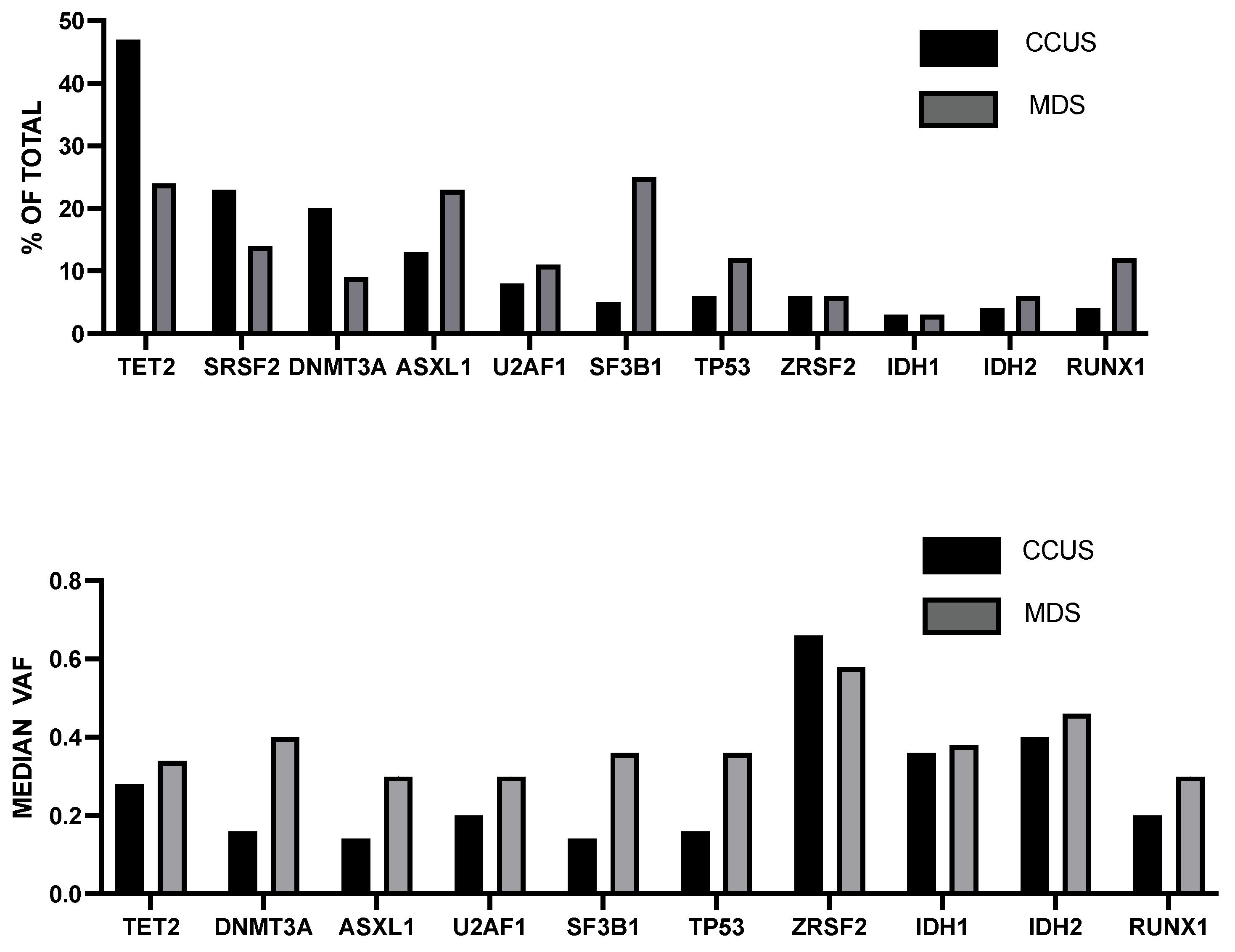
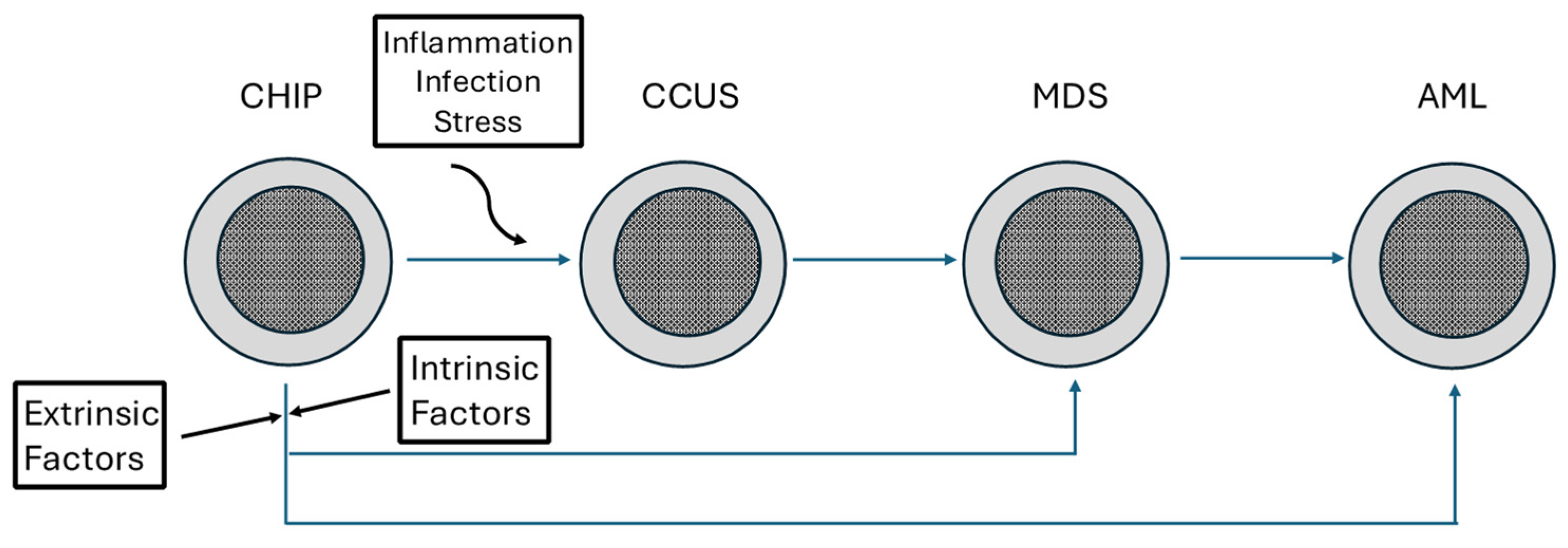
7. Clonal Cytopenia of Undetermined Significance (CCUS)

CCUS has to be considered as a form of CHIP associated with cytopenia. (Table 1). Cytopenia is defined as a condition associated with hemoglobin level, platelet, and neutrophil counts less than 11 g/dL, 100 × 109/L, and 1.5 × 10⁹/L, respectively, and lasting for at least 6 months. If a patient with cytopenia does not meet the criteria for a diagnosis of MDS or of another hematological disorder, the existence of a condition defined as idiopathic cytopenia of undetermined significance (ICUS) is suspected [108].
The prevalence of cytopenias was evaluated in 147,170 individuals living in the northern part of the Netherlands, showing anemia in 4.2% of cases, thrombocytopenia in 1.6%, and neutropenia in 4.8%; anemia and thrombocytopenia increased with aging, while neutropenia did not show an increase in older age; the combined presence of anemia and thrombocytopenia was associated with reduced overall survival; anemia and thrombocytopenia but not neutropenia were associated with a higher incidence of hematological malignancies in individuals aged ≥60 years. The highest incidence of mortality related to hematological malignancies was observed among individuals with >1 cytopenia [109].
The discovery of CCUS was originated in 2015 in studies aiming to characterize ICUS. In these studies, it was shown that over 30% of patients with ICUS not complying with diagnostic criteria for MDS carry MDS-associated somatic mutations [110,111] (Table 1). In particular, Kwok et al. have characterized 484 ICUS and observed that 115, 136, and 233 meet the criteria of MDS, CCUS, and non-clonal ICUS. This classification was based on DNA sequencing studies of bone marrow cells showing that 35% of ICUS patients carried a somatic mutation or chromosomal abnormality indicative of clonal hematopoiesis [110]. (Table 1) ICUS patients were subdivided into those with some dysplasias (62% with MDS-like mutations) and those without dysplasias (20% with MDS-like mutations) [110]. The mutational spectrum observed in CCUS is similar to that observed in MDS, with TET2 and DNMT3A being the most recurrent mutations; SF3B1 mutations are markedly less frequent in CCUS than in MDS [110].
Cargo et al. retrospectively characterized a group of patients who, despite having an initial bone marrow with nondiagnostic features, developed progressive dysplasias or AML; these pre-MDS subjects exhibited an MDS-like mutational profile, except for low mutation frequency of the SF3B1 gene [111] (Figure 2). Similar findings were reported by Malcovati et al., who observed that 36% of ICUS patients displayed somatic mutations; the most frequent mutations in ICUS patients were TET2, ASXL1, DNMT3A, and SRSF2, with a low frequency of SF3B1 mutations [112]. The presence of mutations in ICUS patients (CCUS) was associated with a considerably increased risk of developing a myeloid neoplasia, estimated 14 times higher than in ICUS patients without mutations 112].
Choi et al. have explored the clinical and genetic features of 139 ICUS (78 CCUS and 61 non-clonal ICUS) and 226 MDS: OS of individuals with CCUS was worse than non-clonal ICUS and better than lower-risk MDS; the genetic abnormalities observed in CCUS are similar to those reported for lower-risk MDS, with the exception of a lower frequency of SF3B1 mutations in CCUS compared to MDS (2.6% vs. 18.2%, respectively) [113]. Lower hemoglobin, DDX41 (allelic germline and somatic), ETV6, and RUNX1 mutations were independent prognostic factors for worse OS [113].
Gallì et al. characterized the molecular level in 311 individuals with ICUS and showed that 92 of these individuals carried a genetic abnormality supporting a condition of CCUS [94]. Eighty-seven of these 92 CCUS patients displayed one or more somatic mutations, while five patients harbored a cytogenetic abnormality [114]. Mutational load or VAF > 0.1 or some mutations, such as mutations of splicing factors, ASXL1, or TET2, increased the risk of transformation to myeloid neoplasia (MN) [114]. The analysis of the mutational profile allowed the identification of two mutational profiles, one characterized by isolated DNMT3A mutations (CH-like) and a second characterized by mutations in splicing factors, TP53, SF3B1, TET2, or ASXL1 genes isolated or in combination with other genes (Mn-like) [114]. CCUS patients with MN-like mutational profiles displayed a significantly higher risk of MN progression and a lower OS compared to those with CH-like mutational profiles [114]. CCUS patients within the MN-like cluster showed significantly higher VAF than those included in the CH-like cluster [94]. This study confirmed that SF3B1 gene mutations were observed in CCUS at a lower frequency than in MDS (84.6% vs. 29.5%, respectively) [114].
Additional studies showed a lower frequency of spliceosome mutations in CCUS compared to low-risk MDS [115].
In addition to somatic mutations, CCUSs also display some structural aberrations. In this context, Mikkelson and coworkers explored the occurrence of CNAs and CN-LOHs in a cohort of 153 patients with ICUS: 23/152 patients displayed CNA/CN-LOH, and 52% of these patients also displayed MDS-like mutations; CNA/CN-LOH observed in CCUS patients were similar to those observed in MDS [116]. In the group of CCUS patients, the presence of CNA/CN-LOH was associated with a median OS lower than that observed in CCUS patients without CNA/CN-LOH [116].
Cargo et al. reported a detailed phenotypical, genetical, and clinical analysis of 400 CCUS (64% males and 36% females). TET2, SRFS2, and DNMT3A were more frequently mutated genes in CCUS than in MDS, while SF3B1, ASXL1, RUNX1, TP53, and STAG2 were less frequently mutated in CCUS than in MDS [117] (Figure 3). For most of these genes, the median variant allele fraction of mutations was significantly lower in CCUS than in MDS [97] (Figure 3). Age and sex mutations in ASXL1, BCOR, and TP53 correlated with a worse overall survival; the number of mutations was the strongest predictor for progression of CCUS patients to a myeloid malignancy [117]. Follow-up over time of these patients showed that patients with CCUS displayed a clear tendency to worsen their cytopenia compared to ICUS patients without clonal alterations [117].
A second recent study characterized the genomic landscape of a large cohort of 222 CCUS patients compared to 698 MDS patients [117]. Concerning the mutational profile, this study largely confirmed the study by Cargo et al. [114]. In addition, this study reported a higher mutational frequency of PPM1D in CCUS than in MDS and a lower mutational frequency of STAGH2, NRAS, and CUX1 in CCUS compared to MDS [96]. Variant allele frequencies of the mutated genes showed higher values in MDS than in CCUS for most of these genes (TET2, DNMT3A, ASXL1, SRTSF2, TP53, U2AF1, SF3B1, PPM1D, IDH1, and STAG2) [117]. The analysis of cytogenetic abnormalities in CCUS and MDS showed several remarkable differences: a clearly higher frequency of LOY in CCUS compared to MDS (in males, 26% vs. 9%, respectively), while complex karyotypes and del(5q) were markedly less frequent in CCUS than in MDS [117]. The finding observed for Y-loss is in line with the observation that this cytogenetic alteration is not associated with MN progression.
Morphologic dysplasia is not considered a typical feature of CCUS. However, some studies have shown the existence of low-level bone marrow dysplasia in a portion of patients with CCUS [118]. Interestingly, the classification of CCUS patients into two subgroups, one with BM dysplasia and the other without BM dysplasia, showed that the presence of morphologic dysplasia in BM is associated with increased mutation frequency of some genes, such as spliceosome genes SF3B1, SRSF2, ZRSF2, U2AF1, IDH2, RUNX1, and CBL; furthermore, an association was observed with higher variant allele frequency and co-mutation of DNMT3A, TET2, and ASXL1 gene with other genes [119].
The study of the molecular evolution of CCUS patients is fundamental to evaluating the molecular mechanisms responsible for the progression of myeloid malignancy. However, the studies characterizing this transition of CCUS to MDS at a molecular level are very limited. In this context, Baer et al. explored at two time points (at a median interval of 16 months from diagnosis) 69 patients with CCUS, 94 with ICUS, and 161 with MDS; at the second testing, 11% of ICUS, 26% of CCUS, and 32% of MDS patients acquired at least one mutation [120]. In CCUS patients, the most frequently acquired mutations were RUNX1, ASXL1, CBL, NRAS, and STAG2 [120]. The median VAF of the acquired mutations in CCUS patients was 10% [120]. Interestingly, the pattern of acquired mutations in CCUS was similar to that observed in MDS patients [120]. Mutation acquisition in CCUS patients was highly associated with disease progression, mostly to MDS (79% of cases) [120].
A recent study reported the development of a simple and efficient score for predicting the risk of progression of CCUS patients [121]. This scoring system (CCRS, clonal cytopenia risk score) was based on the analysis of 357 patients with CCUS across 17 academic centers and was based on the identification of three key negative prognostic factors represented by splicing mutations (two points), platelet count <100 × 109/L (2.5 points), and presence of ≥2 mutations (three points); patients with a score of <2 points are classified as low risk (2-year risk of progression 6.4%), patients with a score between 2.5–5 are classified as intermediate risk (2-year risk of progression 14.1%), and patients with a score >5 are classified as high risk (2-year risk of progression 37.2%) [121]. The development of CCRS not only represents a fundamental tool for predicting risk progression of CCUS patients but may also offer invaluable support for the design of therapeutic studies in CCUS patients.
Anemia is a major determinant in the survival of CCUS patients. Thus, Traeden et al. explored 241 patients with CCUS, subdivided into three groups according to the blood cell count phenotype: patients with pancytopenia (14%), anemic patients without pancytopenia (60%), and non-anemic patients without pancytopenia (26%); in the total CCUS population, 76% of patients were anemic [122]. For CCUS patients without anemia, CCUS patients with anemia, and CCUS patients with pancytopenia, the overall survival at 5 years was 85%, 52%, and 55%, respectively; for low-risk MDS patients, the overall survival at 5 years was 45%. The overall survival for CCUS patients with pancytopenia and low-risk MDS was not significantly different [122]. Considerable differences in survival were observed between the CCUS patients within 65 years and within 80 years of age.
A recent study evaluated the risk of incident cytopenia in individuals with CHIP, based on a cohort of 9374 CHIP cases and 24,749 controls; cytopenias occurred in 13.5% of CHIP and 11.6% of controls [103]. Among CHIP, increased risk of incident cytopenia was observed in participants with driver mutations in the TP53 pathway, in spliceosome genes, and in AML-like genes IDH1 and IDH2; individuals with two or more mutations also have a significantly increased risk of developing cytopenia; other factors of risk are represented by MCV ≥ 100 femtoliters, RDW ≥ 15%, and age ≥ 65 years. CHIP patients with no high-risk features had an 8.1% incidence of cytopenia at two years, compared to 21% in those with two or three high-risk factors and 45% in those with four or five high-risk factors [123]. Both controls and CHIP with cytopenia have a significantly higher risk of subsequent myeloid neoplasia compared to those without cytopenia [123]. Individuals with CHIP and cytopenia have a higher risk of developing a myeloid neoplasia compared to controls with cytopenia [103]. Among CHIP individuals who developed an incident cytopenia, 4.4% developed a myeloid neoplasia, compared to 0.4% observed among CHIP individuals not developing cytopenia [123].
The consistent mutational heterogeneity confers to CCUS a considerable biological and clinical heterogeneity and suggests a need for studies aiming to characterize CCUS cases according to their gene drivers. Thus, a recent study reported the characterization of 39 CCUS patients bearing ZRSR2 mutations [104]. Concurrent hematological malignancies were observed in 21% of patients; prior treatment with chemotherapy or immunotherapy was observed in 43% of patients; abnormal cytogenetics were observed in 29% of patients; and 59% off these patients had an absolute monocyte count >0.5 × 10⁹/L, thus supporting the diagnosis of clonal cytopenia with monocytosis of unknown significance (CCMUS) [124]. The median co-mutation number was 1: TET2 and ASXL1 were the most common co-mutations. With a median follow-up of 32 months, 23% of patients progressed to MN (MDS or MDS/MPN) [124]. The median OS was 56 months [124].
Recent studies have described a peculiar form of clonal cytopenia occurring after DNA-damaging therapy, defined as therapy-related clonal cytopenia (t-CCUS), and have proposed that it represents a distinct entity from therapy-related myeloid neoplasia (t-MN) [125]. Shah et al. explored a group of patients developing a condition of unexplained cytopenia following cytotoxic therapy: a portion of these patients were defined as t-MDS for the presence of MDS-defining cytogenetic abnormalities or as t-MN, while the rest of patients were identified as t-CCUS [125]. Some remarkable differences were observed between t-CCUS and t-MN: the interval from primary diagnosis to t-CCUS was shorter compared to t-MN (34.4 vs. 79.8 months, respectively); t-CCUS have a markedly lower incidence of abnormal cytogenetics (24.2 vs. 84.9%, respectively), complex karyotype (none vs. 52.3%, respectively), and monosomal karyotype (none vs. 50.3%, respectively); TET2 and DNMT3A mutation frequency was higher in t-CCUS than in t-MN, while TP53 mutations were more frequent in t-MN than in t-CCUS; WBC counts were low in both t-CCUS and t-MN, but anemia, neutropenia, and thrombocytopenia were more pronounced in t-MN than in t-CCUS [125]. The mean overall survival was markedly longer for t-CCUS than for t-MN patients (124 months vs. 13.2 months) [125].
In a subsequent study, the same authors compared the features of 105 patients who met criteria for CCUS and 46 for t-CCUS; patients with t-CCUS received prior chemotherapy (37%), combined chemotherapy and radiation therapy (37%), or radiation therapy alone (24%) [126]. Peripheral blood cell counts were similar in the two groups of patients. The examination of bone marrow showed some remarkable differences: patients with CCUS were more likely to have hypercellular bone marrows (median, 45% vs. 17%), whereas patients with t-CCUS were more likely to have hypocellular bone marrow blasts (1% vs. 0%) [126]. The median percentage of patients with abnormal cytogenetics was similar in the two groups. The CCUS group was enriched in SRSF2 mutations compared to t-CCUS (30% vs. 9.5%, respectively), whereas TP53 mutations were more frequent among t-CCUS as compared to CCUS (20% vs. 2%, respectively) [104]. Median OS was longer for CCUS than for t-CCUS (not reached vs. 3.6 years, respectively) [126].
DNA methylation studies further supported the strong similarities between CCUS and MDS. Thus, Katrup and coworkers explored the DNA methylation profile occurring in CCIUS and MDS patients bearing DNMT3A, TET2, and IDH2 mutations, showing a high similarity in the DNA methylation profile observed in CCUS and MDS patients [127]. Particularly, in both these conditions, mutation-specific DNA methylation profiles were significantly enriched at enhancer regions and depleted at CpG islands for both hypermethylated sites associated with TET2 and IDH2 mutations and hypomethylated sites associated with DNMT3A mutations [127].
A recently proposed molecular taxonomy classification of myelodysplastic syndromes included 16 molecular groups [107]. One of these subgroups corresponding to 6.9% of all MDS patients was classified as CCUS-like; in this group, 46% of patients had a single mutated gene (TET2 or DNMT3A), 8% had LOY without gene mutations, and 6% had least two DAT mutations; in the group, MDSs had lower cancer cell fractions compared to other MDS groups (42% vs. 84%, respectively), reflecting the low clonal sizes of CCUS [128].

8. Clonal Monocytosis of Undetermined Significance (CMUS) and Clonal Cytopenia and Monocytosis of Undetermined Significance (CCMUS)

The terms CMUS and CCMUS were introduced by the 2022 International Consensus Classification (ICC) of Myeloid Neoplasms to define precursor entities characterized by CH-associated mutations and concurrent monocytosis but not meeting diagnostic criteria for chronic myelomonocytic leukemia (CMML).
CMML is characterized by the proliferation of M1 monocytes, bearing typical mutations of TET2, SRSF2, and ASXL1 genes. The characterization of subjects with absolute monocytosis allowed the identification of CMUS. Thus, Cargo et al. characterized a group of 283 individuals investigated for occurrence of monocytosis for a period of over 2 years: a significant proportion of these individuals had diagnostic bone marrow, mostly related to a CMML diagnosis; 23% of these patients had nondiagnostic BM, and 57% of them had somatic mutations similar to those observed in CMML (TET2, SRFSF2, ASXL1) [129]. The OS of nondiagnostic monocytosis subjects was similar to that observed for CMML patients and significantly worse than in unmutated patients [108]. In these patients, the presence of a mutation was associated with a progressive decrease in hemoglobin/platelet levels and increasing monocyte counts compared with mutation-negative patients [129].
CH in the context of monocytosis could represent an early clonal expansion preceding the development of CMML. Van Zeventer and coworkers tested this hypothesis through the analysis of community-dwelling individuals with monocytosis (≥1 × 1909/L and ≥10% of leukocytes) in the population-based Lifelines cohort (144,676 adults). In this population, the proportion of individuals with monocytosis increased with age (<0.5% in individuals in the 18–59 years range and >2% individuals >80 years) [106]. Monocytosis in older individuals correlated with a higher prevalence of CH (50% in the group with monocytosis compared to 35.5% in the control group); in individuals with monocytosis, the number of monocytes was higher among those with CH than among those without CH [130]. The analysis of the mutational spectrum among individuals with CH and monocytosis or with CH without monocytosis showed higher frequencies of DNMT3A mutations and of spliceosome mutations (SF3B1, SRFSF2, and U2AF1, 7.2% vs. 2.4%) [130]. The follow-up of 21,601 older Lifelines participants for a median of 7.7 years showed that individuals with monocytosis have a higher risk of developing a hematological malignancy compared to controls; all individuals with monocytosis developing a hematological malignancy (mostly AML) carry CH [130]. In these individuals, the presence of monocytosis was associated with lower OS, while in older subjects with monocytosis, spliceosome mutations were linked to a more pronounced risk of death [130].
Dunn and coworkers recently reported the results of a large screening of CMUS in 431,531 UKB participants; in this study, CMUS were defined according to the presence of a CH driver mutation, monocyte count ≥0.5 × 10⁹/L, monocytes ≥10% of total WBCs, and no prevalent hematological malignancy at diagnosis, and CCMUS were defined according to the same criteria as above, plus the presence of cytopenia [131]. CMUS and CCMUS were associated with an increased risk of incident MN, with a 10-year cumulative incidence of 2.4%, 9.1%, and 18.6% for CMUS with monocytes 0.5 × 10⁹/L, CMUS with monocytes >1 × 10⁹/L, and CCMUS, respectively; this risk was estimated as 1.8% and 5.3% for CH and CCUS, respectively [131]. DNMT3A was the most frequent mutation in CMUS and CCMUS; TET2 and particularly SRFSF2 mutations were more frequent in CCMUS than in CMUS [131].
Some remarkable differences exist in the classification of myeloid neoplasms with monocytosis following World Health Organization (WHO) and International Consensus Classification (ICC). Both WHO and ICC have lowered the cutoff for absolute monocytosis to >0.5 × 10⁹/L, thus incorporating oligomonocytic cases into the CMML category, and have included the proof of clonality as an absolute prerequisite for CMML diagnosis [132,133]. However, the two classifications systems have introduced different morphologic criteria, in that for WHO, the documentation of dysplasia in at least one lineage is required, whereas the ICC requires two additional criteria represented by the presence of bone marrow hypercellularity due to myeloid proliferation and peripheral blood cytopenia [132,133]. Furthermore, a relevant difference between the two classification systems is also observed for the definition of precursor lesions relevant to CMML: for the WHO, these precursor lesions are included within the group of CCUS, while for the ICC, these precursor lesions form a specific, separate group defined as CMUS [132,133].
Baumgarten et al. reported the results of a retrospective analysis on a large cohort of 5541 patients with monocytosis, also including cases of mild monocytosis (included with 0.5 and 1.0 × 10⁹/L), and applied both WHO and ICC to the diagnostic definition of these patients [134]. They observed a high rate of concordance between BM dysplasia, required by WHO, and PB cytopenia, required by ICC; however, a typical BM morphology, required by ICC, was observed in only a part of the cases identified as CMMNL by WHO, thus showing that the CMML definition applied by ICC is more restrictive compared to the one applied by WHO [134]. In spite of the existence of these discrepancies in the classification of some patients with monocytosis as MDS with monocytosis or CMML, both WHO and ICC classification systems have identified the key role of clonality proof as an essential element in the diagnosis of CMML and its precursor conditions associated with monocytosis [134].

9. Platelet-Restricted Clonal Hematopoiesis (PLT-CH)

Recent studies have proposed the existence of a peculiar subtype of platelet-restricted clonal hematopoiesis (P-CH).
Kamphuis et al. explored the prevalence of CH-related mutational patterns in individuals ≥60 years of the Life Cohort study (167,729) with abnormal platelet cell counts: thrombocytopenia (631 individuals) and thrombocytosis (178 individuals) [48]. The results of this study showed that the incidence of thrombocytopenia increased with age, while thrombocytosis remained stable; thrombocytopenia was not associated with CH in that the prevalence was similar among individuals with thrombocytopenia (37.9%) or not (39.3%); the prevalence of CH was higher in individuals with thrombocytosis (55.8%) compared to matched controls (37.7%); the mutational profile of individuals with thrombocytopenia was characterized by a higher frequency of spliceosome mutations (SF3B1, SRSF2, and U2AF1); the mutational profile of individuals with thrombocytosis, compared to matched controls, was enriched in JAK2 (12.8 vs. 1.1%) and CALR mutations (4.7% vs. 0.0%); the prevalence of JAK2, CALR, and MPL mutations was still higher in individuals with severe thrombocytosis [48]. A total of 15.4% of individuals with thrombocytopenia had concurrent cytopenia, and 2.1% had a concurrent cytosis; 15.7% of individuals with thrombocytosis had a concurrent cytosis, and 15.2% had a concurrent cytopenia [48]. Importantly, individuals with thrombocytosis and CH displayed an increased risk of developing hematological malignancies, enriched in the case of myeloproliferative neoplasia [48].
A recent study explored the occurrence of CH in 151 individuals with normal blood counts using an assay based on the analysis of genomic DNA isolated from purified granulocytes and from cDNA isolated from purified platelets [114]. Interestingly, GRA-specific CH was observed in 11.9% of individuals, PLT-specific CH was observed in 33%, and combined GRA and PLT-CH was observed in 16.7% [135]. At the level of gene mutations, TET2-mutated and JAK2-mutated CH showed a lineage bias towards platelets; PLT-restricted CH included DNMT3A, TET2, and JAK2 variants with high allele frequency [135]. In line with these observations, a recent study showed the generation of dysfunctional platelets in TET2-mutant CH [136]. Furthermore, another study showed that aging of HSCs in mice is associated with a pathway of megakaryocytic differentiation directly from HSCs, generating dysfunctional platelets [7].

10. Clonal Hematopoiesis in Very Old Individuals, Aged >80 Years

The study of CH in individuals >80 years old is important to evaluate the impact of CHIP mutations on survival.
A study by van den Akker et al. on a small group of individuals of 89 years and older suggested that mutations in genes involved in CH do not seem to compromise the survival of these old individuals [137].
Zink et al. explored by whole-sequencing the occurrence of CH in 11262 Icelanders and showed that CH is very common in the elderly, with a frequency of 12.5 in the whole population and reaching frequencies of 23%, 32%, and 52% in the age groups of 65–75, 75–85, and 85–110 years, respectively [16].
Van Zeventer studied the clinical consequences of CH in the context of aging in a population of 621 individuals ≥80 years using a technique of sensitive sequencing of 27 driver genes involved in hematopoiesis at a VAF ≥1% [138]. Sixty-two percent of these individuals displayed CH, usually with low VAF and with frequent DNMT3A and TET2 mutations [138]. The presence of CH was associated with increased death related only to hematological malignancies. The presence of spliceosome and ASXL1 variants was associated with a prior history of exposure to various types of DNA damaging agents, such as smoking or cancer therapy [138,139].
Rossi et al. reported the results of a mutational screening carried out on 1794 persons aged > 80 years; CHIP (defined as CH with identification of mutated genes at VAF ≥ 1%) occurred in about 32% of subjects, with predominant mutation of DNMT3A and TET2 [140]. A significant increase in the prevalence of TET2 and ASXL1 mutations was observed after the age of 90 years. The majority of patients bear a single mutation. The presence of mutations of JAK2 and splicing genes of multiple mutations affecting DNMT3A, TET2, and ASXL1 with additional genes and variant allele frequency > 9.6% had a positive predictive value for myeloid malignancies [140]. Probability of survival was reduced in individuals with two or more mutations [140]. The exploration over time of 96 individuals showed that the evolution is variable, with some individuals without mutations at baseline acquiring a second mutation during follow-up (13/74) and other individuals positive at baseline acquiring a second mutation during follow-up (2/22) or increasing VAF ≥ 0.05 (10/22) [140].
Studies on two supercentenarian individuals (111 and 115 years, respectively) showed the absence of malignant hematopoiesis and provided evidence that a large part of hematopoiesis is ensured by a single hematopoietic stem cell [141,142]. Particularly, in one of these supercentenarian females, it was shown that DNMT3A-mutated HSCs contributed to the generation of the majority of myeloid cells (78–87%) and to a minority of T-cells (11%) and B-cells (6–7%); various subclones differently contributed to hematopoiesis [142].
Wang and coworkers explored CH in a group of older individuals with ages between 60 and 89 years (defined as common elderly) compared to a group of individuals with ages between 90 and 110 years (defined as longevous) [143]. The prevalence of CH was higher in the longevous group (about 75%) compared to the common elderly group (about 40%); the longevous group exhibited a significant increase in the prevalence of TET2 (44% vs. 19%) and ASXL1 (12% vs. 6%) mutations compared to the common elderly group [143]. Furthermore, the frequency of SF3B1, GNAS, CBL, STAG2, and TP53 mutations was also more frequent in the longevous group compared to the common elderly group [143]. Unexpectedly, no significant correlation was observed between clonal size of CH and age or expression of aging biomarkers in the range of 60 to 110 years [143]. The size of CH correlated with the number of mutations per individual: the size of mutant clones was significantly larger in individuals with two mutant alleles than in those with one mutant allele [143]. These observations strongly support the conclusion that the evolution of CH is influenced by factors in addition to age [143]. The observation that the longevous elderly group display a significantly higher frequency of TET2 and ASXL1 mutations suggests that certain CH could be beneficial to prolonging life.
Interestingly, a recent study explored the mechanisms of HSC aging in mice, particularly in the optics of exploring clonal selection and population dynamics of the HSC pool during murine aging [144]. Mouse stem and progenitor cells accrue approximately 45 somatic mutations per year, a rate about threefold greater than that observed for human HSCs/HPCs. Aging mice did not exhibit the marked loss of clonal diversity typically observed in human hematopoietic aging [144]. Targeted sequencing studies showed the existence of small, expanded clones in aging mice, which enlarged following hematological perturbations, in a manner similar to that observed in humans [144]. These observations suggest conserved properties of population dynamics of blood and distinct patterns of age-associated somatic clonal evolution between mice and humans [144].

11. Longitudinal Dynamics of CH Clones

In order to evaluate the dynamics of CH over time, it is fundamental to infer the fitness of CH-mutant clones. These studies fall into two different categories: (i) studies inferring the fitness of the mutation from measurements carried out at a single time point and (ii) studies based on the direct measurement of the rate of growth of the mutant clone at different time points (longitudinal evaluation).
Two studies have used approaches based on methods to deduce mutant clones’ fitness from measurements performed at a single time point. Thus, Watson et al. analyzed the prevalence of common CH-driven mutations at their variant allele frequency in a large population of healthy individuals to deduce the fitness of the various mutations [145]. This analysis was based on the assumption that a mutation with a high allele frequency may have either low fitness and occurred early in life or has high fitness and occurred late in life. Thus, the analysis of the prevalence of a recurrent mutation in a given population and of the age of the evaluated population provides important information about the fitness of the mutation. This approach showed that the mutant spliceosome genes SF3B1 and SRFSF2 are some of the fittest in CH, with fitness effects as high as 23% per year [146]. Concerning DNMT3A mutations, DNMT3AR882C is fitter than DNMT3AR882H (19% vs. 15% per year) [145].
Weinstock et al. used a similar approach to evaluate the relationship between fitness and age of occurrence of the driver mutation [86]. This evaluation was based on the assumption that if two mutations display the same allele fraction, the mutation with higher fitness would have required less time to expand and was thus acquired later in life. Weinstock et al. developed a peculiar approach based on the evaluation of passenger mutations together with driver mutations; passenger mutations occur in HSCs at a constant rate over time, similar across individuals: therefore, the number of passenger mutations in the founding cell of a CHIP clone can be used to date the acquisition of the driver mutation [86]. The burden of passenger mutations in the HSC founding a CHIP clone can be estimated by the WGS of whole blood DNA; as a mutant clone expands, the VAF of both the driver and passenger mutations increases [86]. The system of using age- and VAF-adjusted passenger mutations to estimate fitness was called the “passenger-approximated clonal expansion rate” (PACER). According to this system, SF3B1, SRSF2, U2AF1, and JAK2V617F were the fastest, while DNMT3AR882 was among the slowest; mutations in TET2, ASXL1, PPM1D, TP53, and GNB1 were intermediate between rapidly and slowly growing mutants [86].
Several studies have explored the longitudinal dynamics of CH clones, aiming to define the main determinants of this process.
Fabre et al. reported an analysis of the development of CH in 385 individuals 55 years of age or older over a period of a median of 13 years. The results of this study showed the following: the large majority (92.4%) of these clones expanded at a sustained exponential rate (clones bearing DNMT3A or TET2 mutation genes expanded at a constant rate, while clones with JAK2 mutations display an irregular growth rate); clones bearing different mutations grow at a different annual growth rate, changing from about 5% in clones with DNMT3A and TP53 mutations to 10% in those with ASXL1 PPM1D, SF3B1, and TET2 mutations and to 20% in those bearing PTPN11, SRSF2, and U2AF1 mutations; different clones with the same mutations display comparable growth rates, usually with low interclonal variability [146]. Lifelong analysis of clonal growth showed remarkable differences between various clones according to their mutation profile: clones bearing DNMT3A mutations decelerate their growth rate before older age, their growth being at least twice as fast in early life compared to more advanced age; clones bearing TET2 mutations are less susceptible to this age-related deceleration than DNMT3A-mutant clones, thus explaining the increasing prevalence of TET2 CH at older age; clones bearing IDH1, PTPN11, SRSF2, and U2AF1 showed no age-related deceleration [146]. Finally, a link between mutation fitness and malignant progression was observed, in that clones growing rapidly are enriched with the highest risk of leukemic progression [146].
Robertson et al., through a longitudinal analysis of 85 individuals of older age with CHIP mutations, reached the conclusion that individual gene mutations have different fitness advantages outweighing the effects related to inter-individual variation [147].
Van Zaventer et al. explored the evolutionary landscape of clonal hematopoiesis in 3359 individuals of the general population; 1320 (39%) of these individuals had CH, 275 with a single mutation and 13% with multiple mutations [148]. VAF was usually low. A minority of these individuals displayed abnormal blood counts, most frequently related to anemia, thrombocytopenia, and thrombocytosis; anemia and thrombocytosis were observed in a minority of CHIP individuals. Higher VAF mutations were observed among individuals with multiple blood count abnormalities [148]. The longitudinal evolution of CHIP was explored through follow-up at 43 months and at 74 months; at first evaluation of follow-up, the prevalence of CHIP increased by 3.8%, with 80% of cases displaying a stable number over time and 20% showing some changes during follow-up; in some instances, the changes were related to the appearance of new mutations, to an increase in the VAF of mutations present at baseline, or to the disappearance of mutations present at baseline [148]. The longitudinal analysis of mutational VAF showed a consistent heterogeneity: an increase was observed for the majority of mutations (61%), while 6% of cases did not show any significant change, and 34% showed a significant decrease; in some instances, in the same individual, the concomitant VAF increase in a given clone and decrease in another clone was observed [148]. The clonal dynamics were related to some specific gene features: thus, predominant growth was observed for the spliceosome genes SF3B1, SRFSF2, and U2AF1; JAK2 clones also showed considerable expansion; DNMT3A and ASXL1 showed considerably lower proportional growth rates; and TET2 clones showed moderate growth [127]. Traditional cancer risk factors, such as age, male sex, smoking, excessive alcohol use, and being overweight, seem to be involved as determinants of clonal expansion. The acquisition of new mutations in clonal evolution seems to be dictated in large part by baseline conditions: pre-existing CHIP at baseline had a higher risk of acquiring new mutations than those without CHIP at baseline; new gene mutations frequently emerged in individuals with baseline TET2 mutations and are usually represented by an additional TET2 mutation or an SRSF2 gene mutation; new TP53 or JAK2 mutations emerged in individuals independently of pre-existing CHIP at baseline. Importantly, the presence of CHIP was associated with an increased risk of developing hematological malignancies (myeloid malignancies, 2.5% per 5 years) [148]. The highest risk of developing myeloid malignancies was observed for JAK2; spliceosome mutations U2AF1, SF3B1, and SRSF2; and KRAS/NRAS mutations. DNMT3A mutations did not significantly increase the risk of developing myeloid malignancies; the risk of myeloid malignancies is higher for the combined presence of blood count abnormalities and CHIP [148].
Uddin and coworkers reported the results of a long-term longitudinal analysis on 4,187 participants in the Atherosclerosis Risk in Community Study (ARIC) explored the incidence and progression of CHIP clones in older adults by whole exome sequencing [149]. Baseline CHIP prevalence was 10.9% at VAF ≥ 2% and 3.8% at VAF ≥ 10%; most of the individuals with CHIP had a single mutation [149]. CHIP was then assessed at a later visit, with a median follow-up duration of 21 years and with a median age of participants of 75.8 years; the prevalence of CHIP at VAF ≥ 2% and ≥ 10% increased to 25% and 11.8%, respectively [149]. The prevalence of DNMT3A CHIP decreased, while mutations in TET2, ASXL1, and splicing factor genes increased; median VAF increased; 269 clones at VAF ≥ 2% disappeared, and 972 clones emerged at the follow-up visit [149]. A total of 19.7% of individuals developed incident CHIP during the follow-up (63% at VAF ≥ 2% and 37% at VAF ≥ 10%); most incident CHIP mutations of large clones involved splicing factor genes, ASXL1 and JAK2 [149]. Clinical predictors of incident CHIP were represented by age, association with a higher incidence of overall CHIP, TET2, and splicing factor mutations; furthermore, male sex was significantly associated with a higher incidence of overall CHIP, ASXL1, and splicing factor mutations [149]. The main determinants of clonal growth rate were represented by driver gene mutation and age. In fact, splicing factor genes, including genes such as SF3B1, SRSF2, U2AF1, and ZRS2 clones, grew significantly faster than other mutant clones [149]. Age was significantly associated with clonal growth rate [149].
Since CH becomes increasing detectable with aging, but in many instances driver mutations are absent and inferred weak selection suggests the existence of mutants arising early in life, a recent study proposed a theory to explain how mutations conferring a low fitness advantage may drive the development of CH. This theory was based on the assumption that weak selection conferred by stem cell variation generated before birth can support the development of CH later in life [150]. Weak selection may generate cloned dominance at a condition of an appropriate long time and in the context of large cell populations; stochastic negative selection of weakly selective variants would be prevented by the expansion of stem cell lineages during development [150]. The dominance of stem cell clones generated before birth is supported by blood fluctuating CpG methylation patterns exhibiting low correlation between unrelated individuals, but high correlation between many elderly monozygotic tweens [128]. Thus, CH driven by the generation of mutants with low fitness and weak selection in later life seems to reflect variation created before birth [150].
A recent study suggested that age-related changes in HSC proteostasis promoted the emergence of clonal hematopoiesis in older adults [151]. This conclusion is supported by the observation that aging HSCs activate HSF1, the key proteostasis sensor and master transcription factor of the heat shock response, to preserve proteostasis and fitness during aging [151]. Studies in experimental murine models provided evidence that age-related HSF1 activation promoted the emergence of CH and leukemia in older mice [151].
Splicing factor gene mutations only initiate clones in old age when they drive rapid clonal growth (>50% per year for some mutant SRTSF2 clones) [146]. Phylogenetic and longitudinal analyses demonstrate that splicing factor-mutant clones only begin to expand after middle age (>50 years), and their expansion rate is rapid and stable after initiation [146]. Thus, it must be assumed that factors facilitating expansion of splicing factor mutant CH are operative only after middle age. There is no evident explanation for the expansion of splicing factor-mutant clones only in old individuals. Fabre and Vassiliou hypothesized a possible role of telomere maintenance mechanisms in this phenomenon, suggesting that splicing gene mutations could protect HSCs from the replicative senescence induced by age-related telomere shortening [152]. In one study of patients with telomere biology disorders in which pathogenic germ line variants cause abnormally rapid telomere shortening, CH bearing U2AF1 mutations was detected in 10% of individuals, a mutational rate much higher than that expected for a median age of 29 years [153]. These observations were confirmed in a study of a larger group of 207 patients with a median age of 27 years: the Ch prevalence was 46%, much higher than that observed in age-matched controls [154]. In these patients, recurrent mutations occurred in TP53, PPM1D, telomere maintenance genes (TERT promoter, POT1, TINF2), and RNA splicing (particularly U2AF1S34); furthermore, gain of chromosome 1q (+1q), encompassing MDM4, was the most recurrent cytogenetic abnormality [154]. RNA expression analysis of CD34+ cells showed that the U2AF1S34 mutations alter p53 and interferon signaling pathways, suggesting that these mutations could compensate for aberrant upregulation of p53 and interferon pathways in telomere-dysfunctional HSCs [154]. The presence of TP53, RNA splicing factors, and +1q was associated with risk of MDS and AML.
In a population screening of telomere length in the UK Biobank and its association with CH, Burren and coworkers observed that SF3B1-mutant CH was associated with short telomeres, while SRSF2-mutant CH was associated with telomeres [155]. Since CH with splicing factor mutations is observed in older individuals, it is expected that it should be associated with short telomeres. The observation that telomeres in SRSF2-mutant CH do not appear to shorten as the clones expand or eventually elongate strongly contrasts with the accelerated attrition of telomeres observed with clonal expansion driven by other CH genes; this intriguing finding suggests that SRSF2 mutations could confer clonal advantage through telomere modulation [155].

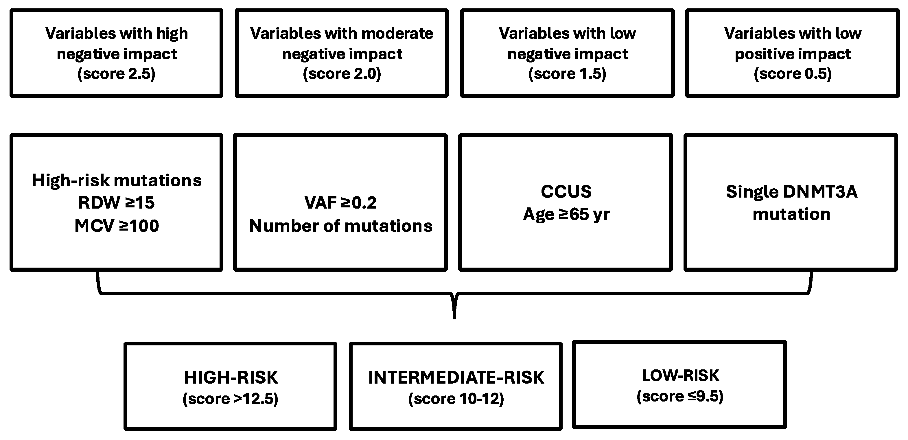
12. Prediction of the Risk of Developing Myeloid Neoplasms in Individuals Bearing CHIP Mutations

Not all the individuals with CHIP develop myeloid neoplasia; thus, it is of fundamental importance to define criteria predicting the risk of leukemia transformation of individuals bearing CHIP.
In order to distinguish individuals at high risk of developing a myeloid malignancy from those with benign CHIP, 95 individuals studied before developing AML (on average 6.3 years before AML diagnosis) were compared to 424 control individuals [51]. Using targeted deep sequencing, CHIP was observed in 73% of pre-AML cases, compared to 36% in the control group; 39% of pre-AML cases displayed a driver mutation with VAF >10%, compared to 4% of controls [51]. The number of mutations, the mutational burden, and the size of the larger driver clone were associated with the risk of progression to AML [51]. Finally, it was observed that some mutations, such as those involving TP53 and U2AF1, impart a relatively high risk of developing AML, while mutations in genes such as DNMT3A and TET2 confer a lower risk of leukemic progression [51].
Gu and coworkers have developed a multiparametric prediction risk of developing myeloid neoplasia, based on the analysis carried out on 454,340 biobank participants, of whom 1808 developed a myeloid neoplasia (AML, MDS, or MPN) in the lapse of time of 0–15 years after recruitment (defined as pre-MN) [130]. Through wide exome sequencing (WES), CH was detected in 28.5% pre-MN individuals, compared to 4.8% in the control group; pre-MN cases exhibited a higher frequency of “high-risk” genes, including JAK2, SRFSF2, SF3B1, and IDH2, while the standard CH mutations DNMT3A, TET2, and ASXL1 are more common in controls; the proportion of pre-MN cases displaying CH driver mutations was similar among individuals who developed AML, MDS, or MPN [156]. CH driver mutations differed in the different types of MNs developed from CH: DNMT3AR882 mutations in AML; TET2, SRFSF2, and SF3B1 mutations in MDS; JAK2, CALR, and MPL mutations in MPN [156]. Clonal sizes increased in all pre-MN cases with advancing age. The analysis of hazard ratios allowed defining individual predictive variables, including DNMT3AR882 mutations for AML, SF3B1 mutations for MDS, and JAK2/CALR mutations for MPN; higher VAF was associated with decreased DFS [156]. The presence of some mCAs was specifically associated with the development of specific neoplasias: pre-AML with 5(-5q), pre-MDS with -5q and 4qLOH, and pre-MPN with 9pLOH and +9 [156]. Using these variables, Cox regression models quantifying the risk of progression to each myeloid neoplasm subtype were developed [156]. These models are based on the observation that some mutations and blood cell count abnormalities are associated with evolution to different subtypes of MN [156]. These models incorporated additional variables, such as platelet volume and distribution of width, as well as blood chemistries (e.g., cholesterol and creatinine); the final “MN-predict” model predicts the risk of progressing from CHIP/CCUS to MDS/AML/MPN [156].
Weeks and coworkers developed a clonal hematopoiesis risk score (CHRS) based on the data derived from the exome sequencing data on 193,743 UK Biobank participants (range of age 40–70 years); 11,337 individuals met the criteria for having CHIP or CCUS [157]. DNMT3A, TET2, and ASXL1 were the most commonly mutated genes in both CHIP and CCUS; individuals with CCUS have a higher VAF compared to those with CHIP; the presence of more than one mutation was more frequent among CCUS than CHIP [157]. Among 11,337 individuals with CHIP/CCUS, 269 individuals (2.37%) had an incipient risk of developing a myeloid neoplasia (MN), with a higher incidence in CCUS than in CHIP. The analysis of these “pre-MN” individuals showed that mutations in splicing factor genes (SRFSF2, SF3B1, ZRSF2) and AML-like genes (IDH1, IDH2, FLT3, and RUNX1) were associated with 926-fold and 13.8-fold increased risks of incident MN relative to other genotypes of CHIP/CCUS, respectively [157]. Individuals with DNMT3A mutant CHIP/CCUS have a 4.5-fold increased risk of developing MN compared to those without CHIP/CCUS [130]. Taking into account these findings integrated with clinical variables, a CHRS was developed; it includes single DNMT3A mutations (being favorable), high-risk mutations (SRFSF2, SF3B1, ZRSF2, IDH1/2, FLT3, TP53, or RUNX1), a VAF of 0.2 or more, 65 years of age or older, having CCUS vs. CHIP, and red cell blood indices (mean corpuscular volume and red cell distribution width), affecting MN risk in variable directions [157] (Figure 4). Following CHRS, three groups of CHIP/CCUS patients were defined: low-risk (88%), intermediate-risk (11%), and high-risk (1%); in clinical cohorts, most MN-related events were observed in the high-risk group [157]. (Figure 4) The CHRS represents an important prognostication tool in CHIP/CCUS, providing aid in clinical decisions and supporting intensive surveillance and therapeutic intervention in the set of CHIP/CCUS patients who are most likely to progress to MN [157].
Recent studies have suggested that the study of mitochondrial DNA mutations may contribute to improving the prediction risk stratification of CHIP patients in developing MNs. Each human cell contains several hundred to 1000 mitochondria, and each mitochondrion contains two to 10 copies of mitochondrial DNA (mtDNA). Therefore, mtDNA can be present at 1000 s to 10,000 s copies in somatic cells, and mutations in mtDNA can exist in a subset of the total cellular mtDNA, a condition defined as mitochondrial heteroplasmy; mtDNA heteroplasmy is present in about 30% of individuals and increases with age [158]. mtDNA heteroplasmy was associated with a 1.5-fold increase in all-cause mortality and the presence of mtDNA mutations at highly constrained sites [132]. Mitochondrial local constraint score sum (MSS) was associated with a fourfold increase in mortality due to leukemia [159]. mtDNA heteroplasmy was explored in 434,304 participants from UK Biobank: in this population, CHIP was present in 7.7% of individuals and mtDNA heteroplasmy was present in 28.3% of cases [160]. A notable number of heteroplasmies identified in adults reflect acquired mutations and likely mark the clonal expansion of HSCs. Concurrent CHIP and mtDNA heteroplasmy was present in 2.6% of individuals, heteroplasmy alone in 24.1%, and CHIP alone in 5% of individuals [160]. Among individuals with CHIP, heteroplasmy was more common in individuals with large CHIP clones, in those with multiple mutations, or in those with spliceosome machinery mutations [159]. Importantly, both the number of mtDNA heteroplasmies and MSS were associated with an increased risk of MN (HR 1.5 and 3.1, respectively); the presence of both heteroplasmies and CHIP increased the risk of developing MN compared to each clone [160]. Heteroplasmic variants with higher predicted deleteriousness increase the risk of MNs [134]. Incorporating mtDNA heteroplasmy in the existing CHRS score model for individuals with CHIP improves sensitivity and detection of high-risk groups [160].
Recent studies have explored the hierarchical role of CH-related mutations (DTA) in the context of MDS and AML. These analyses were based on the VAF model of the hierarchy of mutation evolution, based on the assumption that mutations with the highest VAF are clonal founders. AMLs with MDS features (corresponding to 33% of total AMLs) were subdivided into five groups: del(5q), SF3BA, SP (other spliceosome genes), I/D/T (IDH1/2, DNMT3A, TET2), and none of these; the SP and I/D/T groups displayed a frequent origin from CH [161]. The majority of AMLs with a CH-related founder clone were observed in the groups of NPM1 (49%) and MDS-related (26%) AMLs [162]. Interestingly, a recent study provided evidence that CH may also precede, in some instances, AMLs characterized by defining cytogenetic abnormalities, such as KMT2A-rearranged (9% of cases), CBFB::MYH11 (8% of cases), RUNX1::RUNX1T1 (9% of cases), CEBPA (15% of cases), and MECOM-rearranged (17% of cases) [163].
The presence of pre-leukemic CH prior to cancer therapy is common among patients who later develop therapy-related myeloid disorders, a process seemingly related to increased fitness of underlying CH [164]. Chien et al. retrospectively analyzed 78 of these patients; DNMT3A, TET2, ASXL1, and TP53 were the most common CH mutations in these patients; the 2-year OS was 79%, and the main causes of death were primary malignancy and comorbidities; 20% of deaths were related to the development of therapy-related myeloid neoplasms [165].

13. Effects of Donor-Engrafted Clonal Hematopoiesis in Autologous and Allogeneic Stem Cell Transplantation

Recent studies have analyzed the clonal dynamics observed in long-term survivors after allo-HSCT. These studies have explored the dynamics of CH-related clones in donor-recipient pairs and have explored whether the increased replicative stress exerted on donor cells as a consequence of HSCT could determine mechanisms triggering the preferential proliferation of CH variants. Thus, one of these studies explored clonal dynamics in 16 related donor-recipient pairs at a median follow-up of 33.8 years after allo-HSCT [166]. The study was based on the analysis of mutations in genes recurrently involved in myeloid malignancies and CH; CH-related variants were observed in all donors; the average mutation rates were similar in donors compared to recipients post-HSCT (2.0 vs. 2.6 per year, respectively); only 5.6% of the variants shared between paired donors and recipients showed a >10-fold higher VAF in the recipient compared to the donor [139]. These observations support the conclusion that even a few decades after HSCT, there is no generalized increased clonal expansion in transplanted HSCs [166].
A second study explored long-term stem cell engraftment though genome sequencing of single-cell-derived hematopoietic colonies of 10 donor-recipient pairs obtained 9–31 years after allo-HSCT; when young donors were involved in transplantation, 5000–30,000 HSCs were responsible for engraftment and contributed to hematopoiesis; when older donors were involved in transplantation, a significantly lower number of HSCs were responsible for engraftment, and hematopoiesis shows an accelerated progression towards aged, oligoclonal hematopoiesis [167]. Importantly, recipients had a significantly lower clonal diversity than matched donors, corresponding to about 10–15 years of aging, seemingly due to a greater clonal expansion during repopulation after HSCT [167]. Analysis of phylogenetic trees suggests two different modes of HSC selection by transplantation: one selection operating at the level of HSCs preceding transplantation and operating at the level of selection of cells endowed with preferential mobilization, survival ex vivo, and/or initial homing capacities and a second selection, a growth selection, underlying clonal expansion occurring in the recipient’s marrow after engraftment and mostly pronounced in clones with multiple CH driver mutations [167]. Thus, the lower clonal diversity of hematopoiesis observed in older donors derives from the selection and preferential growth of HSCs adapted to survival in the conditions observed during and after HSCT; this observation suggests that during aging there is not a generalized decline of the fitness of transplanted HSCs, but the acquisition of increased fitness occurring in a minority of HSCs [167].
The health status of donor HSCs is of fundamental importance in the outcome of HSCT.
A recent meta-analysis explored the outcomes of allo-HSCT and auto-HSCT with engraftment with CH: donor-engrafted CH after allo-HSCT was associated with reduced disease-related relapse, but not OS, PFS, or mortality not related to relapse; donor-engrafted CH after auto-HSCT was associated with decreased OS, PFS, and increased risk of developing therapy-related myeloid malignancies [168]. A second meta-analysis of the literature confirmed the results of the first meta-analysis; furthermore, in this second meta-analysis, in the autologous HSCT setting, a decrease in peripheral blood stem cell mobilization and an increase in the time required for platelet engraftment were also noted [169]. The most adverse effect of CH in auto-HSCT was a clear increase in the number of secondary myeloid neoplasms, particularly observed in CH bearing DNA repair pathway mutations (PPM1D, TP53, RAD21, BRCC3) [169].
Some studies are particularly relevant. Gibson et al. explored donor clonal hematopoiesis and recipient outcomes after transplantation in a large cohort of patients undergoing HSCT [170]. CH was detected in 22.5% of donors, and the presence of DNMT3A or TET2 mutations did not adversely affect recipient outcome; donor DNMT3A-CH was associated with reduced relapse and increased chronic GVHD; no recipient of sole DNMT3A or TET2 CH developed donor cell leukemia (DCL); in seven out of eight cases, DCL originated from donor CH associated with TP53 or splicing factor mutations or from donors bearing germline DDX41 mutations [170].
Kim et al. reported a study of 372 patients undergoing allo-HSCT; 25 of these patients displayed donor-related CHIPs successfully engrafted [171]. With a median duration of 13 years among survivors, the presence of CHIP in the donor did not affect OS, relapse incidence, or non-relapse mortality; furthermore, donor CHIP did not affect neutrophil or platelet engraftment; the rates of acute or chronic graft versus host disease were not affected by donor CHIP [171]. In conclusion, this study, based on a long follow-up post-transplantation provided sound evidence that the presence of CHIP in an allo-HSCT setting does not negatively affect transplant outcomes after transplant [143]. Boettcher et al. evaluated CH in 42 long-term survivors of allo-HSCT: this evaluation was performed both in donors and recipients; five cases of donor-engrafted CH were detected, and in 80% of them, there was a consistent increase in clone size, as measured by VAF, in recipients compared with donors; however, this increase was only modest, thus suggesting that in recipients, hematopoiesis is not dominated by a single clone of HSCs [172]. One patient with CH developed MDS in both donor and recipient [172].
Muskens and coworkers explored the occurrence of CH among pediatric patients exhibiting long-term survival after allo-HSCT; CH was detected in 16% of allo-HSCT recipients compared to 8% observed in matched controls [173]. CH mutations were observed in DNMT3A (80%) and TET2 (20%) genes; large clones were exclusively observed in HSCT recipients [145]. Older hematopoietic age of the donor and the HSCT procedure independently increased the risk of CH [173]. These observations support long-term monitoring of allo-transplanted pediatric patients displaying CH [173].
The most adverse event related to CH on auto-HSCT was related to the increased risk of developing t-MN post-HSCT, and this conclusion was supported through the analysis of 1507 patients undergoing auto-HSCT performed in a single institution; particularly, in these patients, PPM1D and TP53 mutations in CH predicted the risk of developing post-HSCT t-MN [174]. Importantly, a recent retrospective analysis on 1931 lymphoma and multiple myeloma patients who underwent auto-HSCT showed that the presence of CHIP was associated with a significant and independent risk of developing non-myeloid secondary malignant neoplasms; patients with high VAF CHIP or TP53 CHIP mutations had a markedly increased risk of developing non-myeloid secondary neoplasms [175].
Other studies have explored the impact of CH mutations (DNMT3A, TET2, ASXL1, SRSF2, SFG3B1, U2AF1, JAK2) in AML patients on the response to standard chemotherapy and to HSCT. CH-associated mutations frequently persist in patients achieving a CR after induction and consolidation chemotherapy, and this does not indicate adverse outcomes [176,177]. However, in the large majority of patients, CH mutations were cleared post allo-HSCT [176]. The persistence of measurable residual disease clonal hematopoiesis (MRCH) or not after allo-HSCT, measured at various times after transplantation (up to 5 years), represents a strong prognostic factor, in that patients with MRCH negativity post-HSCT have a markedly better EFS than those with MRCH positivity [178].
At variance with other studies, a recent study by Imus and coworkers explored in a group of 97 lymphoma patients aged 60 or more years undergoing allo-HSCT the effect of recipient CH [179]. CH was detected in 62% of these patients; pre-transplant CH in recipients was associated with adverse survival after allo-HSCT: 78% for patients without CH and 47% for those with CH [179]. Among patients with CH, adverse OS and non-relapse mortality were associated with CH burden and number of mutations [179]. To explain these results, it was hypothesized that the negative effect of recipient CH could represent the result of persistent CH-related macrophages secreting proinflammatory cytokines, inducing a high inflammatory response and an increase in cytokine release syndrome and c-GVHD [179]. The findings of this study need to be confirmed by additional studies involving other types and higher numbers of patients; furthermore, the mechanisms through which recipient CH may directly or indirectly affect the outcomes of allo-HSCT remain to be evaluated.

14. CH in Bone Marrow Failure Syndromes

Bone marrow failure syndromes (BMFS) are a group of diseases characterized by inefficient hematopoiesis and variable risk of progression to myeloid malignancies. BMFSs can be acquired or inherited and may be due either to intrinsic defects of HSC biologic activity or to extrinsic factors. Acquired BMFSs are often related to immune-mediated inhibition of HSCs, while inherited BMFS are related to various mechanisms, including telomere maintenance, germline defects in DNA damage repair machinery, or ribosome biogenesis. CH is frequently associated with BMFS and represents an important factor in disease evolution.

14.1. CH in Acquired Aplastic Anemia

Acquired aplastic anemia (AAA) is characterized by pancytopenia and BM hypocellularity, resulting from cytotoxic T cell-mediated destruction of HSCs/HPCs. The advent of high-through-put sequencing techniques allowed the discovery of the frequent occurrence of acquired genetic abnormalities in AAA patients, observed in about 50% of these patients [180]. The most frequent mutations observed in these patients were as follows: BCOR or BCORL1 (7–10%), PIGA (5–9%), DNMT3A (6–1259, ASXL1 (6–7%), splicing factor genes (1-4%), TET2 (1–2%), TP53 (1–2%), and RUNX1 (1–2%) [181]. PIGA and BCOR/BCOTL1 mutations displayed a similar frequency in patients of different ages, while the other mutations greatly increased with the age of patients [181]. The PIGA gene is involved in the biosynthesis of glycosylphosphatidylinositol-anchored proteins (GPI-Aps), whose deficient expression in HSCs is responsible for paroxysmal nocturnal hemoglobinuria (PNH). BCOR and BCORL1 are two linked genes that encode transcription factors involved in the control of embryogenesis and hematopoiesis. These mutations display different chronological evolution and clinical impact. Thus, BCOR/BCORL1 and PIGA mutations have a tendency to disappear or to remain stable following immunosuppressive therapy, while DNMT3A, ASXL1, TET2, TP53, and splicing factor mutations tend to expand their clonal size and are associated with a faster progression to MDS/AML [181]. BCOR/BCORL1 and PIGA mutations predict better clinical outcomes compared to CH-related mutations [181]. Thus, CH in AAA is closely linked to the late disease clonal evolution.
The longitudinal exploration of four AAA patients treated with immunosuppressive agents and evolving to MDS/AML showed that mutations in genes recurrently mutated in myeloid malignancies, such as RUNX1, NRAS, NPM1, SXL1, SETBP1, and PHF6, precede leukemic evolution [182]. It remained unclear whether clonal evolution to MDS/AML is a result of genomic instability present at diagnosis or developed after immunosuppressive therapy [182]. Goarke et al. evaluated clonal evolution in 663 AAA patients undergoing immunosuppressive therapy; according to several clinical and laboratory criteria, the patients were classified as low-risk and high-risk. OS was 37% in high-risk clonal evolution by 5 years compared to 94% in low-risk [183]. The presence of ASXL1, RUNX1, or splicing factor mutations predicted high-risk clonal evolution; BCOR/BCORL1, DNMT3A, and TET2 were not predictive of clonal evolution [183]. The characterization of myeloid neoplasia developing in AAA patients showed four molecular subtypes: a group with del7/7q displaying ASXL1, SETBP1, RUNX1, and RAS pathway gene mutations; a group with normal karyotype showing classical leukemogenic drivers, including DNMT3A, FLT3, NPM1, and U2AF1; a group with complex karyotype; and a group classified as others, showing diverse molecular configurations [184].
A recent study explored the longitudinal contribution of different hematopoietic clones during marrow recovery in AAA following immunosuppressive therapy with cyclosporine and eltrombopag; the patients were explored before therapy and at 6 months and 2–5 years after therapy [185]. At baseline, CH was detected in 64% of the patients, most commonly in PIGA (27%), DNMT3A (23%), BCOR (18%), and ASXL1 (14%); 25% of cases had multiple mutations in these genes [184]. After 6 months, 71% of patients had CH, and new mutations were detected in BCOR and ASXL1 [184]. After 2–5 years, most BCOR clones disappeared, and ASXL1 and PIGA clones persisted, with expansion of ASXL1 clones associated in a minority of cases with acquisition of MDS-related mutations (SETBP1, RUNX1, and U2AF1) and with expansion of a portion of PIGA clones associated with signs and symptoms consistent with clinical PNH [185]. A second study longitudinally explored 59 patients with severe AAA enrolled in the context of the EBMT RACE trial and undergoing different immunosuppressive treatments; preliminary results of this study showed a high prevalence (94%) of CH at diagnosis, which tended to increase after therapy, irrespective of the immunosuppressive treatment [186].

14.2. CH in Paroxysmal Nocturnal Hemoglobinuria

Paroxysmal nocturnal hemoglobinuria (PNH) is a nonmalignant clonal disease of HSCs that is associated with hemolysis; the pathogenesis of PNH is related to two components: mutant HSCs bearing a mutation of PIGA gene and expansion of the mutant clone [187]. In rare cases, expansion may be related to independently arisen mutations [187]. Deep sequencing studies have shown that some PNH patients, in addition to PIGA mutation, also have mutations in genes known to be involved in myeloid malignancies, such as TET2, U2AF1, and JAK2 [188]. Lin et al. evaluated 85 PNH cases by NGS and observed mutations in addition to PIGA mutations in 26 cases: five DNMT3A, four ASXL1, and four U2AF1 mutations [189].

14.3. CH in Inherited BMFs

Inherited BMFs are generated by germline HSC defects, involving different biochemical pathways, such as DNA damage repair (Fanconi anemia, FA), impaired ribosome biogenesis (Diamond–Blackfan, Schwachman–Diamond Syndrome, SDS), short telomere syndromes (including dyskeratosis congenita, DC), granulocyte lineage stress (severe congenital neutropenia, SCN), and SAMD9/SAMD9L mutations (MIRAGE syndrome).

14.4. CH in Fanconi Anemia

FA is an inherited DNA-instability disorder caused by biallelic mutations in one of the 22 genes of the FA pathway, a gene network involved in DNA damage repair and stress response. FA affects bone marrow and is characterized by impaired DNA repair mechanisms and accumulation of chromosomal damage, causing a progressive loss of all types of blood cells. HSCs are susceptible to replication stress, which is a major contributor to HSC defects in FA.
FA patients have a high risk of developing MDS or AML and other malignancies. BMF in these patients seems to be caused by an exacerbated basal p53n signaling-elicited unrepaired DNA damage. This sustained p53 response observed in FA HSCs/HPCs drives a potent anti-cancer mechanism to FA-induced genomic instability but contributes to BMF [190]. The acquisition of 1q chromosome trisomy represents an early and frequent event occurring during BM leukemic evolution of FA patients; 1q+ in turn drives enforced MDM4 expression through a gene dosage mechanism and represses high basal p53n response [190]. This mechanism of p53 attenuation confers to 1q+ cells, which are not transforming per se but behave like a pre-leukemic cell population, resembling CHIP [190]. 1q+ clones acquire the capacity to proliferate and to expand in vivo, repopulating the exhausted BM of FA patients and promoting leukemia development [190].

14.5. CH in Diamond–Blackfan Anemia

Diamond–Blackfan anemia (DBA) is a rare congenital BMFS characterized by erythroid hypoplasia and heterogeneous allelic variations in ribosomal protein (RP) genes; in addition, non-RP genes, such as GATA1 and TSR2, are associated with DBA [191]. Mutations in up to 37 genes have been implicated in DBA or DBA-like syndromes; mono-allelic mutations in the ribosomal protein gene RPS19 are observed in approximately 25% of patients with DBA, and mutations in other RP or related genes account for most of the remaining known genotypes [191]. Recently, it was shown that the various RP gene alterations functionally converge to reduced translation of the master erythroid transcription factor GATA1, and preclinical studies have strongly supported lentiviral gene therapy restoring GATA1 levels as a universal gene therapy for DBA [192].
Patients with DBA have an increased risk of developing MDS or AML compared to age-matched controls. A whole exome sequencing study of 65 DBA patients showed that three of these patients had somatic mutations in STAG1, U2AF1, SF3B1, and DNMT3A; one of these three patients developed MDS at the age of 21 years [193]. Recently, Perdigones et al. reported the case of a DBA patient acquiring a mutation in deubiquitinating enzyme USP 11 associated with clonal hematopoiesis, improving the defective hematopoiesis in this patient [194].

14.6. CH in Schwan-Diamond Syndrome (SDS)

SDS is an inherited multisystem ribosomopathy characterized by exocrine pancreatic deficiency, BMF, and predisposition to myeloid malignancies. This syndrome is due to impaired ribosomal maturation due to deficiency of SBDS (Schwan-Diamond Syndrome) ribosomal protein. The outcome of SDS patients who develop myeloid malignancies is extremely poor.
The p53 tumor suppressor pathway is activated in SDS hemopoietic cells due to defective ribosome biogenesis and aberrant protein translation. Somatic TP53 mutations have been observed in SDS patients who develop MDS [195]. However, TP53 mutations have also been identified in SDS patients who do not develop MDS, thus suggesting that other factors must cooperate with TP53 mutations in promoting leukemic progression [196]. In 27 patients, TP53 mutations were observed in 48% of patients, with a low allelic frequency, and none of these patients had evidence of MDS: these observations indicate that TP53 mutations precede MDS development and that their presence in HSCs confers a fitness advantage over their nonmutated counterparts.
Kennedy et al. reported the extensive molecular characterization of 110 SDS patients. The large majority (98%) of these patients had germline BBDS mutations. fifteen of these patients had myeloid malignancies (eight AML and seven MDS) associated with the presence of myeloid-associated mutations, such as TP53, RUNX1, U2AF1, SETBP1, BRAF, and NRAS, and recurrent chromosomal alterations involving chromosomes 3, 5, 7, and 20 [197]. The rest of the 95 patients without myeloid neoplasia possessed somatic mutations in 74% of cases; the most frequently mutated genes were EIF6 (61%), TP53 (45%), PRPF8 (12%), and CSNKA1 (6%); the majority (/”5) of them possessed multiple mutations [197]. The presence of TP53 mutations was associated with myeloid neoplasms, while EIF6, CSNK1A1, and PRPF8 were not associated with MN [195]. EIF6 gene mutations represent the most frequent somatic mutations observed in SDS patients.
SBDS deficiency impairs ribosome assembly and results in reduced abundance of the mature ribosome 80S, with concomitant accumulation of free 60S subunits and upregulation of p53-dependent cell stress pathways in SDS patient bone marrow cells. Both EIF6 and TP53 mutations through different mechanisms reduce p53 activation, as supported by the reduction in CDKN1 expression, which is a marker of p53 pathway activation [197]. About 91% of SDS patients with TP53 mutations have concomitant EIF6 mutations, suggesting that these two mutations cooperate in inducing clonal evolution. The presence of TP53 mutations was associated with the presence of somatic TP53 mutations; however, the leukemogenic activity of mutant TP53 requires the presence of biallelic alterations of TP53 (two TP53 gene mutations or one gene mutation and a CN-LOH) [197]. In conclusion, the results of this study show that SBDS germline mutations induce a fitness constraint that drives selection of mutant clones arising early in life and involving mutations in EIF6 or TP53: EIF6 inactivation mediates a compensatory mechanism with limited leukemic potential by improving the SDS-related ribose defect and enhancing cloning fitness; TP53 inactivation underlines a maladaptation pathway associated with clearly enhanced leukemic potential and limited correction of the ribosome SDS defect.

14.7. CH in SAMD9/SAMD9L Syndrome

Sterile alpha motif domain-containing protein 9 (SAMD9) and SAMD9-like (SAMD9L) are paralogous genes encoding antiviral proteins that negatively regulate cell proliferation. Heterozygous germline gain-of-function SAMD9/SAMD9L variants cause multisystem syndromes with a wide spectrum of carriable clinical manifestations, including bone marrow failure, cytopenia, immunodeficiency, MDS infections, and monosomy 7 [198]. Most patients carry germline missense gain-of-function variants; a minority of patients carry frameshift-truncating variants, associated with a disease presenting with an inflammatory syndrome [198].
The study of a large cohort of pediatric and adult MDS patients showed that 8% of these patients carried germline SAMD9/SAMD9L mutations, and 7% carried germline GATA2 mutations; these patients in 90% of cases presented with refractory cytopenia of childhood and in 10% with MDS with excess ob blasts [199]. Monosomy 7 and del(7q) (-7) were the predominant cytogenetic abnormalities, significantly enriched in SAMD9/SAMD9L germline-mutant (38%) and GATA2 germline-mutant (57%), compared to MDS patients without these genetic abnormalities (8%) [199].
At least two-thirds of patients with germline SAMD9/SAMD9L mutations undergo a process of somatic genetic rescue (SGR), leading to the elimination or inactivation of the mutant allele in hematopoietic cells [199]. SGR induces CH, which can have an adaptive (curative), indeterminate, or maladaptive (premalignant) effect. Three types of SGR-induced CH have been observed in SAMD9/SAMD9L syndromes: monosomy of chromosome 7 carrying the SAMD9 mutant allele; uniparental disomy 7q (UPD7q); and somatic SAMD9/SAMD9L mutations [198,199]. SAMD9/SAMD9L-related monosomy of chromosome 7 has an indetermined risk in that it can spontaneously resolve (transient monosomy 7) or can progress to leukemia [200,201]. Transient monosomy 7 is specifically observed in SAMD9/SAMD9L syndromes. Monosomy 7 can also be associated with the gain of leukemia-related driver gene mutations, including RUNX1, PTPN11, ASXL1, EXH2, and RAS pathway [198,199]. Adaptive CH mutations in SAMD9/SAMD9L syndromes are a typical feature of these syndromes and can occur through two different mechanisms: a first related to the acquisition of somatic loss-of-function SAMD9/SAMD9L mutations and a second related to UPD7q, which corresponds to the duplication of the wild-type SAMD9/SAMD9L allele [198,199].

14.8. CH in Telomere Biology Disorders

Telomeres are nucleoprotein complexes at the end of chromosomes that protect the chromosome ends to be sensed as a free end generated by a DNA double-strand break, which would inappropriately activate the DNA damage repair machinery. In humans, progressive telomere shortening occurs with normal aging. However, germline defects that lead to either accelerated telomere shortening or to telomere inability cause a group of inherited diseases collectively known as telomeropathies or short telomere syndromes (STS) or telomere biology disorders (TBD) [202].
In humans, 16 TBD-causing genes have been identified; these genes affect different processes required for telomere biology, including telomere maintenance, structure, and function [202]. The mutation of some of these genes, such as TERT, TERC, TINF2, NAF1, RPA1, and ACD, is associated with the development of BMF and/or MDS [202].
TBD patients have an increased risk of developing myeloid malignancies, which was estimated to correspond to >20-fold for AML and to >150-fold for MDS compared to the general population [203,204,205]. It was estimated that 8–10% of TBD patients develop myeloid malignancies [203,204,205]. The study of a large cohort of 1514 MDS patients showed a germline TERT variant in 2.7% of cases, associated with increased non-relapse mortality after stem cell transplant [206].
Initial studies have shown that CH mutations were observed in about 390% of TBD patients, with very rare mutations of the age-related DNMT3A, TET2, and ASXL1 genes [205]. Gutierrez-Fernandes et al. explored 1290 TBD patients and showed that 40% of these patients had somatic mutations in peripheral blood; the somatic mutational landscape of these CH clones differed from age-related CH, with recurrent TERT promoter (TERTp), POT1, and truncated PPM1D; among MDS-related genes, only U2AF1 was frequently mutated (10% of cases) [207]. U2AF1 and other splicing factor gene mutations (SRSF2, ZRSR2, and SF3B1) in TBD patients are associated with the development of myeloid malignancies [208]. However, PPM1D mutations, associated with therapy-related MDS, are commonly mutated in TBD, but do not associate with MDS development [208].
A recent study reported a detailed analysis of the molecular abnormalities observed in 14 TBD patients. The analysis of the mutational landscape showed the following: somatic variants in DDR pathway genes in 21.5% of cases, most frequently involving ATM gene 88.5%); PPM1D (7.8%), not associated with a myeloid neoplasm; TP53 (4.9%); U2AF1S34 (8.3%) in 50% of cases associated with a myeloid malignancy; and DNMT3A, TET2, and ASXL1 less frequently mutated (2.1%, 2.8%, and 3.5%, respectively) [209]. The analysis of gene expression profiles showed a marked downregulation of cell proliferation-related pathways, upregulation of senescence-associated genes, and significant activation of the ATM-dependent DDR pathway in HSCs/HPCs [209]. Somatic ATM variants allow TBD mutant cells to overcome hyperactive ATM-dependent DDR by reducing ATM function, and their presence was associated with a more aggressive clinical phenotype [209]. Pharmacological inhibition of ATM improved TBD cell fitness by allowing it to bypass DDR-induced senescence, thus suggesting that dampening hyperactive ATM-dependent DDR may represent a valuable therapeutic intervention in TBD [209].
In conclusion, these studies have shown that in TBD patients, at variance with age-related CH whose variants largely perturb epigenetic regulators (DNMT3A, TET2, and ASXL1), the mutational landscape of TBD largely includes genes regulating mRNA splicing and DNA damage repair, with the most frequent being U2AF1S34 [210]. It is unclear why U2AF1 is predominantly mutated in TBD patients compared to other spliceosome component genes and why U2AF1S34 in TBD are much more frequent than U2AF1Q157 mutations (two U2AF1 mutations equally represented in myeloid malignancies) [210].
A second recent study reported the extensive molecular characterization of 207 TBD patients; 46% of symptomatic patients had one or two somatic mutations in PB, with recurrent mutations in PPM1D, POT1, TERTp, U2AF1, TP53, BCOR, and RUNX1; CH in TERTp and PPM1D were enriched in patients with TERT germline variants, POT1-CH was highly enriched in TINF2 germline variants, splicing tactor mutations were enriched in TERT and TERC patients [154]. U2AF1 mutations almost exclusively occurred at the hot spot S34 and only rarely at the hot spot Q157R [154]. CH incidence in TBD was much higher than in age-matched healthy individuals; TERTp, splicing factor, and PPM1D mutations increased with patients’ age; CH in splicing factor genes or Chr1q+ was associated with development of MDS/AML and with a shorter overall survival [154]. U2AF1S34 mutations were always driver mutations associated with linear trajectories of clonal evolution with acquisition of other MDS-related mutations in the same clone, even in the absence of MDS/AML development [154].
Dyskeratosis congenita (DC) is an inherited syndrome of bone marrow failure, characterized by a triad of muco-cutaneous features, such as abnormal skin pigmentation, nail dystrophy, and oral leukoplakia [211]. BM failure is a significant cause of mortality in individuals with DC and predisposes to leukemia and solid cancer development [211]. Currently, nineteen DC genes/loci have been identified [211]. A study in patients with DC provided evidence that CXH is frequent in these patients: skewed X-chromosome inactivation was observed in eight out of nine female patients compared to three out of ten in controls; 50% of DC patients displayed acquired somatic changes in BM cells, as shown by whole exome sequencing studies [212]. Cryptic DC is a late-onset, so-called cryptic form, with initial manifestation in adults. Gene sequencing studies carried out in 15 adult patients with DC showed MRD-related somatic mutations in only one of these patients; chromosomal aberrations were more frequent in these patients, and their presence correlated with MDS or AML evolution [213]. These observations support the view that clonal evolution of subclones carrying MDS-related mutations is not the predominant mechanism for MDS/AML initiation in adult cryptic DC patients [213]. Chromosomal instability seems to be the predominant mechanism underlying leukemic evolution in these patients.

15. CH in Germline Predisposition Syndromes

Genetic predisposition syndromes (GPS) are inherited disorders associated with the presence of germinal aberrations that increase the risk for malignancies. While aberrations in some genes, such as TP53, ATM, and CDKN2A, increase the risk of developing all types of malignancies, there are numerous genes, such as RUNX1, GATA2, ETV6, SAMD9, SAMD9L, and ANKRD26, whose germline mutations specifically associate with the development of hematological malignancies [214]. Here, we analyze inherited predispositions to myeloid neoplasms.

15.1. CH in Patients with RUNX1 Germline Mutations

RUNX1 is a member of the core-binding factor family of transcription factors and is indispensable for the development of definitive hematopoiesis in vertebrates. RUNX1 is one of the most frequently mutated genes in a variety of hematological malignancies. Germline loss-of-function or dominant negative alterations of RUNX1 are associated with thrombocytopenia and familial platelet disorders that predispose to the development of myeloid neoplasia. Various genetic alterations of the RUNX1 gene are associated with familial platelet disorders. The study of individuals with RUNX1 germline mutations without hematological malignancies showed the presence of frequent somatic mutations, and gene mutations related to CHIP were observed in 49% of these patients; BCOR is the most frequently mutated gene, with multiple alterations of this gene in some patients; other frequently detected mutations involved CHIP or AML-related genes, such as TET2, DNMT3A, KRAS, LRP1B, IDH1, and KMT2C; in few patients, mutations of genes such as NFE2, involved in erythroid and megakaryocytic differentiation, were observed [215]. Other studies confirmed that in RUNX1 germline RUNX1 carriers without hematological malignancies, with CH, the most frequent mutations were BCOR, TET2, and PHF6 genes recurrently mutated in RUNX1-driven hematologic malignancies [216].
It was estimated that about 44% of individuals with germline RUNX1 mutations will develop MDS or AML, with a median age onset of 33 years. RUNX1 is mutated in about 10% of adult AML patients, and in about 30% of these patients, RUNX1 mutations are germline; molecular profiling showed higher frequencies of NRAS mutations and other mutations known to activate various signaling pathways in these patients with RUNX1 germline-mutated AMLs [217]. Leukemic progression of germline RUNX1-mutated individuals can be accompanied by biallelic RUNX1 hits or induced by additional somatic variants [218], such as RUNX1-germline (RUNX1GL) mutations, thus explaining that 13% of RUNX1GL individuals have a biallelic configuration [218]. PHF6 mutations were more frequent in RUNX1GL, while U2AF1 and DNMT3A mutations were more frequent in RUNX1SOM [218]. The top somatic mutations most overexpressed in RUNX1GL compared to RUNX1WT were BCOR, SRSF2, ASXL1, PHF6, and TET2, suggesting that these variants may increase the fitness in a RUNX1GL background [218].

15.2. CH in Familial Thrombocytopenia Related to ANKRD26 and ETV6 Germline Mutations

Germline mutations in the regulatory region of the gene encoding ANKRD26 are associated with thrombocytopenia 2; all these patients display moderate thrombocytopenia, and 10% develop myeloid malignancies. All pathogenic ANKRD26 variants are localized at the level of the 5′ untranslated region of ANKRD26, where the RUNX1 transcription factor binds to suppress expression of ANKRD26. The study of seven patients with germline ANKRD26 germline mutations showed the presence of somatic mutations in only one patient, characterized by the presence of SF3B1 mutations [219].
ETV6 is a transcriptional repressor in the ETS transcription factor family that is essential for hematopoiesis and megakaryopoiesis, and its germline mutations are associated with thrombocytopenia with high penetrance that predisposes to both lymphoid and myeloid malignancies. The study of 16 patients with germline ETV6 variants showed the existence of comatic mutations related to CH in 42% of cases; these patients had at least one mutation in genes KRAS, EZH2, TP53, DNMT3A, JAK2, NFE2, PTPN11, and CBL [220]. It is of interest to note that in this cohort of patients, CH was associated with R399L carriers of ETV6 germline syndrome [220].

15.3. CH in Patients with Germline DDX41 Mutations

One of the most frequently observed germline predisposition syndromes to myeloid neoplasms is related to germline variants in the DEAD-box helicase 41 gene (DDX41), with common clinical phenotypes including MDS and AML. The onset of myeloid-associated neoplasms was late-occurring after 60 years, often in the absence of a family history of hematopoietic malignancy. Interestingly, among the numerous DDX41 mutations identified in myeloid malignancies, more than 80% are germline in nature and only about 15% are somatic. CH was observed in only 3% of patients with DDX41 germline mutations without myeloid neoplasms [216]. Kusne et al. reported the characterization of 195 patients with germline DDX41 mutations and reported that 11% of them displayed CH or CCUS [221]. The most common somatic variants were in TET2 (31%), followed by somatic DDX41 (21%) and DNMT3A (17%) [221]. Patients with CCUS were more likely to have somatic DDX41 mutations [221]. Patients with CCUS and somatic DDX41 mutations have more hypercellular bone marrow and notes of dysmegakaryopoiesis [221].

15.4. CH in Germline GATA2 Deficiency Syndrome

GATAs plays a key role in the maintenance of HSCs and in the differentiation of HSCs into the various hematopoietic lineages. The multiplicity of these functions of GATA2 explains the spectrum of defects observed in patients with germline heterozygous GATA2 mutations, resulting in cytopenias, bone marrow failure, and increased propensity to develop myeloid malignancies. The ensemble of these abnormalities is defined as GATA2 deficiency syndrome [222]. The clinical manifestations of GATA2 deficiency originate from loss of multilineage progenitors generating B-lymphocytes, monocytes, NK cells, and dendritic cells, with consequent cytopenias of these lineages. In a portion of patients, GAAT2 deficiency evolves to myeloid malignancies and particularly to MDS, AML, and chronic myelomonocytic leukemia (CMML): these three malignancies are associated with the acquisition of specific somatic alterations, corresponding to monosomy 7 for MDS, trisomy 8 for AML, and ASXL1 or STAG2 mutations for CMML [222].
Lageaud et al. reported the detailed molecular characterization of 78 GATA2 deficient patients; the mutations of GATA2 were heterogeneous in these patients, with null mutations more frequently associated with chronic infectious complications and with missense mutations more frequently associated with hematological manifestations [223]. Karyotype was normal in 50% of patients, while the other 50% displayed chromosomal abnormalities mainly represented by monosomy 7 (29%) and trisomy 8 (16%) [223]. Sixty-six percent of these patients have somatic mutations in their peripheral blood; the most frequently mutated genes were STAG2 (33%), ASXL1 (22%), EXH2 (8%), RAS pathway (7%), RUNX1 (7%), and CBL (7%); DNMT3A (3%) and TET2 (3%) mutations were rarely observed in these patients [223]. No mutations of NPM1, FLT3, IDH1, and IDH2 were observed [223]. According to the morphologic analysis of bone marrow, three groups of patients were identified: spectrum 0 with no evidence of hematologic transformations (13% of all patients); spectrum 1 with hypoplastic bone marrow and/or low signs of low-grade MDS (61%); and spectrum2 with signs of myeloid transformation [223]. Spectrum 2 patients, enriched in missense GATA2 mutations, have a higher risk of MDS/AML transformation compared to spectrum 1 patients, enriched in GATA2 null mutations [223]. Mutation number increased in spectrum 2 patients compared to spectrum 1 (three vs. one mutation/patient), with an enrichment in SETBP1, RAS pathway, and RUNX1 mutations; the number of patients with chromosome abnormalities increased in spectrum 2 compared to spectrum 1 patients (84% vs. 47%, respectively) [223]. Monosomy 7 increased in the various groups of patients: 0% in spectrum 0, 28% in spectrum 1, and 47% in spectrum 2. STAG2 mutations were significantly enriched in spectrum 1 patients compared to spectrum 2 patients (47% vs. 19%). SETBP1 mutations were clearly enriched in spectrum 2 patients, and their presence seems to be critical for leukemic progression in GAT2-deficient patients; particularly, concurrent SETBP1 mutations and monosomy 7 are enriched in spectrum 2 patients and exhibit increased monocyte counts [223]. These observations strongly support a key role of the different somatic mutations observed in germline mutated GATA2 patients in driving the different types of leukemic progression of these patients [223].

16. Treatment of CHIP and CCUS

High-risk individuals with CHIP or CCUS have a consistent risk of progression to myeloid malignancies, thus supporting intervention studies aiming to prevent or at least delay their progression to malignancy. There are currently no FDA-approved strategies for the prevention of myeloid neoplasms in the setting of CHIP/CCUS.

16.1. Anti-IL-1β

In this context, inflammation represents a potential target for these preventive approaches, given the role of inflammatory pathways in supporting the expansion of CH clones. In fact, CH bearing DNMT3A, TET2, and ASXL1 genes are associated with increased levels of circulating IL-1β and IL-6 [224]. IL-1β and IL-18 levels increased from non-CHIP to CHIP to lower-risk MDS [224]. CCUS patients have inflammatory cytokine levels similar to those observed in low-risk MDS [225]. Studies in experimental models support a key role of IL-1-mediated inflammatory signaling in TET2-mediated leukemogenesis [226,227].
A large clinical study of canakinumab, a monoclonal antibody neutralizing IL-1β, explored the effects of IL-1β blockade in patients with a high cardiovascular risk (history of myocardial infarction and C-reactive protein levels ≥2 mg/L); treatment with canakinumab prevented recurrent cardiovascular events, particularly in patients with TET2 CH [228,229]. During a follow-up of 3.7 years, participants without baseline anemia who received canakinumab had significantly less incidence of anemia than those who received a placebo [230]. A subsequent analysis showed that in patients from the CANTOS study, incident anemia was more common in those who have CH and was reduced by canakinumab: canakinumab treatment was associated with significantly higher hemoglobin increment in patients with concurrent CH mutations and anemia than in those with CH without anemia or without CH mutations [231]. Interestingly, the administration of canakinumab was associated with a decrease in non-hematological malignancies among patients with TET2 CH versus those treated with placebo [232].
A recent phase II clinical trial (NCT 04239157) [233] explored the safety profile and the efficacy of canakinumab in previously treated intermediate-risk MDS patients [142]. The overall response rate was 17%, with all responses including hematological improvement [233]. The median duration of response was 8 months. The stratification of patients according to IPSS-M into higher-risk and lower-risk categories showed that all responding pertains to the lower-risk group [233]. Sequential RNA studies on HSC/HPC samples showed that canakinumab blocked the IL-1β receptor-mediated inflammatory signaling pathway and rescued ineffective erythropoiesis only in the context of lower genetic complexity.
Recently, Borate and coworkers proposed a randomized double-blind placebo-controlled phase II clinical trial to prevent leukemic progression in a population of high-risk CCUS patients, based on treatment with either canakinumab (300 mg subcutaneously every 3 months for two years) or with a placebo [234].

16.2. Ascorbic Acid

Other studies have explored the use of ascorbic acid as a pharmacologic agent to target TET2 and inflammatory response in pre-leukemic and leukemic cells. In animal models, the treatment of TET2-deficient mice with high-dose vitamin C was able to mimic TET2 restoration and reverse proliferation and renewal of HSCs/HPCs [235]. Ascorbic acid (Vit C) is enriched in HSCs and multipotent progenitor cells (MPP) compared to other hematopoietic cells, and ascorbate deficiency increases HSC function by reducing TET2 function [236]. Studies of genetic induction of Vit C deficiency showed that Vit C limits MPP self-renewal and clonal expansion [237].
Two recent studies have explored the clinical use of ascorbic acid in patients with CCUS or low-risk MDS. In particular, the phase II randomized, placebo-controlled NCT 03682029 clinical study enrolled 109 patients with low-risk myeloid malignancies (low-risk MDS) and CCUS, randomized to receive 1000 mg of oral Vit C daily or a placebo for 12 months [238]. Vit C plasmatic levels were low in 57% of the enrolled patients, and Vit C administration significantly increased plasmatic Vit C levels [238]. At the end of the study, with a median follow-up of 33.5 months, 11 deaths occurred in the group treated with Vit C and 24 in the placebo group, with a median OS not reached in the Vit C group and 42.2 months in the placebo group [166]. In multivariate analysis, oral Vit C treatment was associated with significantly improved OS compared to a placebo [238]. The analysis of inflammatory cytokine levels (IL-6, IL-10, CXCL-10) showed a significant decrease in the group of patients treated with Vit C, while an increase was observed in patients treated with a placebo [239]. These findings support a significant anti-inflammatory effect exerted by Vit C.
However, Xie et al. failed to observe any significant effect of ascorbic acid administered by intravenous route at high doses (1g/kg) for 12 weeks to 10 high-risk CCUS patients with TET2 mutations [168]. After a median follow-up of 31.6 months, none of the patients responded to treatment (only five with stable disease) [240]. Three of the treated patients progressed to MN [240]. In patients with stable disease, a change in methylation profile was observed at the level of sites involved in development, function, and survival of HSCs/HPCs, compatible with an effect on TET2 activity [240]. Although it is difficult to offer a plausible explanation for the different results observed in the two studies, it is important to point out remarkable differences in these two studies concerning the route and the dose of ascorbic acid and the length of treatment.
It is of interest to note that a recent study explored the effect of Vit C and Vit D supplementation during induction and consolidation chemotherapy treatment of AML patients, reporting a better OS in patients with NPM1 mutation receiving Vit C and Vit D supplementation compared to patients not receiving the vitamins [241]. As discussed above, NPM1-mutant AMLs represent the AML group bearing the highest frequency of CH-related mutations.
Ascorbic acid is under evaluation in association with either decitabine [242] or azacitidine [243] in CMML patients. Initial results showed that high-dose ascorbate reduces IL-1β secretion in TET2 mutant monocytes and was well tolerated when administered in association with azacitidine; furthermore, in CMML patients treated with ascorbate plus azacitidine, a decrease in leukemic monocytes was noted after three to six cycles of treatment [243].

16.3. IDH Inhibitors

CCUS patients with mutations in high-risk genes, including IDH1 and IDH2, are more likely to transform to AML [50,146,148]. In particular, Desai and coworkers reported a 100% progression of IDH1/IDH2-mutant CHIP patients [50]. Thus, it is fully justified to attempt the treatment of IDH1-mutant CCUS patients with IDH1 inhibitors. Thus, Petrone et al. have proposed the NCT 0503441 decentralized open label, multicenter clinical trial to evaluate the efficacy of ivosidenib in patients with IDH1-mutant CCUS [244]. Given the rarity of the condition, a decentralized trial is expected to reduce the barriers to trial participation [244].
Another clinical trial will evaluate olutasidenib, an oral selective potent IDH1 inhibitor with no off-target activity, in IDH1-mutated CCUS or lower-risk MDS/CMML, either alone or in combination with hypomethylating agents.

16.4. TGFβ Pathway Inhibitors

Luspatercept is an inhibitor of SMAD2/SMAD3 signaling mediated by binding to TGFβ ligand; TGFβ signaling is increased in diseases with ineffective erythropoiesis, such as MDS. Based on the results of the phase III trials MEDALIST (luspartecept vs. placebo for lower-risk transfusion-dependent MDS with ringed sideroblasts that were refractory to or unlikely to respond to erythropoietin stimulating agents) or COMMANDS (luspatercept vs. ESA in frontline lower-risk transfusion-dependent MDS), it was approved by the FDA as a frontline therapy for lower-risk MDS patients who require transfusions [245]. A subgroup analysis explored the association between the most frequent CHIP-related mutations (DNMT3A, TET2, ASXL1, SF3B1) and clinical outcomes in patients treated with luspatercept from the COMMANDS; luspatercept treatment in these patients reduced anemia, improved white blood cell counts, and reduced inflammatory gene signatures [246,247]. These observations provide a rationale for the development of clinical studies exploring luspatercept in high-risk anemic CCUS patients.
Interestingly, a case report study reported a case of rapid, significant, and sustained response to combined treatment with luspatercept and eltrombopag (thrombopoietin receptor agonist) following failure of cyclosporin and androgen therapy; even after luspatercept discontinuation for 10 months, hematopoiesis recovery was maintained [248].

16.5. Decitabine/Cedazuridine

Decitabine/cedazuridine is a DNA metyltransferase inhibitor with the addition of cedazuridine, a cytidine deaminase inhibitor in the gastrointestinal tract that increases systemic exposure of decitabine after oral administration, and represents an alternative to intravenous decitabine, which was approved by FDA for treatment of MDS and CMML [249]. For the treatment of low-risk MDS, low doses of decitabine or azacitidine [250] or of decitabine/cedazuridine [251] showed reduced toxicity and clinical efficacy. These low-dosage regimens could be explored in high-risk CCUS patients as a therapeutic strategy aiming to reduce the risk of leukemic progression and to improve cytopenias.

16.6. Therapeutic Targeting of Splicing Factor Mutations in CH

As discussed above, SF-mutant CH is associated with rapid clonal expansion and with an elevated risk of leukemic transformation. Therefore, there is a strong rationale to develop therapeutic approaches attempting to prevent the leukemic transformation of SF-mutant CH or to pharmacologically target these mutations at initial stages of leukemic development. However, this issue is particularly challenging. The most intriguing issue is related to the difficulty in identifying, among the many altered transcripts generated in pre-leukemic and leukemic cells by altered RNA splicing, which are relevant for cancer initiation and/or maintenance. Furthermore, another critical question for appropriate therapeutic targeting is to determine if the impact of mutations in RNA splicing factors in pre-leukemic cells is attributable to their direct impact on RNA splicing and/or to an indirect impact on splicing-related events, such as R-loop formation, mRNA translation, or stress RNA granule formation [252,253,254]. In spite these consistent difficulties, strategies of pharmacological correction of aberrant splicing have been developed, including small-molecule splicing modulators, splice-switching oligonucleotides, and modified oligonucleotides [252,253,254].
A recent study reported the first clinical results using the protein arginine methyltransferase 5 (PRMT5) inhibitor to target splicing factor (SF)-mutant myeloid malignancies. PRMT5 catalyzes symmetrical demethylation of arginine, a post-translational modification required for normal RNA splicing. Preclinical studies have shown synthetic lethality between PRMT5 inhibition and SF-mutant myeloid malignancies. PRT 543 is a potent and selective oral inhibitor of PRMT5 that displays anti-proliferative activity in in vitro and in vivo models of myeloid leukemia. A phase I clinical study showed that PRT 543 was safe and well tolerated, and 35 mg 5x/week was identified as the recommended phase 2 dose [255]. A phase II study showed that monotherapy with PRT 543 was safe in patients with relapsed/refractory SF-mutant myeloid malignancies and showed clear target engagement and objective responses in 5 out of 40 patients, including two CR in one AML patient and in one HR MDS patient [255].
SF mutations cause distinct alternative splicing patterns with minimal overlap. Only alternative splicing events with significant convergence are those that change intron retention, thus suggesting a unified post-transcriptional mechanism underlying intron retention alterations. A recent study provided evidence about the existence of a unified post-transcriptional mechanism regulating intron retention in SF-mutant MDS. This post-translational mechanism is regulated by the combined activity of two kinases: SRPK1 (serine-arginine protein kinase 1) and CLK1 (CDC2-like kinase 1); SPRK1 interacts with an RS (arginine-serine rich)-like domain in the N-terminus of CLK1 to facilitate the release of phosphorylated SR (serine-arginine) protein, which then promotes efficient splice-site recognition and subsequent spliceosome assembly. This mechanism is deregulated by DNA damage and replicative stress mechanisms activated in SF-mutant leukemic cells [256]. The pharmacological targeting of these kinases could represent a potentially valuable tool for the development of new therapeutic approaches to target SF-mutant MDS/AML [256].
Stress granules are cytoplasmic ribonucleoprotein granules that commonly nucleate from the interaction of translationally stalled mRNA and RNA binding proteins [257]. Studies carried out in U2AF1-mutant leukemic cells have shown a clearly enhanced stress granule response [258]. These observations suggest that stress granule response may represent a potential new therapeutic target in SF-mutant leukemic cells [259].

17. Conclusions

Studies carried out in the last decade have led to the discovery of CH and have greatly contributed to the understanding of the cellular and molecular mechanisms underlying the development and evolution of myeloid malignancies.
Various types of CH have been discovered, all characterized by the presence of typical somatic mutations and associated or not with different risks of developing a myeloid neoplasia over time. Only a minority of individuals with CH develop an MN, and some criteria have been identified in risk stratification of these patients, providing criteria to quantify the extent and the nature of this risk.
In spite of these improvements in the definition of the risk of leukemic progression of CH and in the identification of high-risk patients, no effective therapies have been developed to change the natural history of high-risk CH either to block or to delay MN development, and this represents a major challenge for future studies.
At a biological level, future studies are needed to better understand the mechanisms by which various types of mutations determine a fitness advantage to mutant HSCs, and this is particularly true for splicing factor mutations that drive rapid clonal expansion and confer high risk of progression and whose mechanism of action remains to be elucidated.
A better knowledge of the clinical implications of CH will require a better understanding of the natural history of CH and the development of more robust hemopathology and clinical guidelines. In particular, prospective studies, registries, and biorepositories of CH patients are required. In this context, a recent study reported the development of a biorepository of CH patients, providing a prospective, longitudinal cohort of well-genotyped and -phenotyped patients [260]

Author Contributions

G.C. and E.P. were involved in researching, writing, and editing the manuscript. U.T. was involved in conceptualization, organization, and researching and editing the manuscript. All authors have read and agreed to the published version of the manuscript.

Funding

This research received no external funding.

Institutional Review Board Statement

Not applicable.

Informed Consent Statement

Not applicable.

Data Availability Statement

Not applicable.

Conflicts of Interest

The authors declare no conflicts of interest.

References

  1. Moore, L.; Cagan, A.; Coorens, T.H.H.; Neville, M.D.C.; Snahfvi, R.; Sanders, M.A.; Oliver, T.R.W.; Leongamornlert, D.; Ellis, P.; Noorani, A.; et al. The mutational landscape of human somatic and germline cells. Nature 2021, 597, 381–386. [Google Scholar] [CrossRef] [PubMed]
  2. Mitchell, E.; Spencer Chapman, M.; Williams, N.; Dawson, K.J.; Mende, N.; Calderbank, E.F.; Jung, H.; Mitchell, T.; Coorens, T.; Spencer, D.H.; et al. Clonal dynamics of hematopoiesis across the human lifespan. Nature 2022, 606, 343–350. [Google Scholar] [CrossRef] [PubMed]
  3. Lee-Six, H.; Oebro, N.F.; Shepherd, M.S.; Grossmann, S.; Dawson, K.; Belmonte, M.; Hunlky, B.; Martincorena, I.; Anderson, E.; O’Neill, L.; et al. Population dynamics of normal human blood inferred from somatic mutations. Nature 2018, 561, 473–478. [Google Scholar] [CrossRef] [PubMed]
  4. Osorio, F.G.; Rosendhal Huber, A.; Oka, R.; Vereuln, M.; Patel, S.H.; Hasaart, K.; de la Fonteijne, L.; Varela, I.; Camargo, F.D.; van Broxtel, R. Somatic mutations reveal lineage relationships and age-related mutagenesis in human hematopoiesis. Cell Rep. 2018, 25, 2308–2316. [Google Scholar] [CrossRef]
  5. Brandsma, A.M.; Bertrums, E.; van Rooismalen, M.J.; Horman, D.A.; Oka, R.; Verheulen, M.; Manders, F.; Ubels, J.; Bbelderbos, M.E.; van Boxtel, R. Mutation signatures of pediatric acute myeloid leukemia and normal blood progenitors associated with differential patient outcomes. Blood Cancer Discov. 2021, 2, 484–499. [Google Scholar] [CrossRef]
  6. Su, T.Y.; Hauenstein, J.; Somuncular, E.; Dumral, O.; Leonard, E.; Gustafsson, C.; Tzortis, E.; Forlani, A.; Johansson, A.S.; Qian, H.; et al. Aging is associated with functional and molecular changes in distinct hematopoietic stem cell subsets. Nat. Commun. 2024, 15, 7966. [Google Scholar] [CrossRef]
  7. Poscablo, D.M.; Worthington, A.K.; Smith-Berdan, S.; Rommel, M.; Manso, B.A.; Adili, R.; Mok, L.; Reggiardo, R.E.; Cool, T.; Mogharrab, R.; et al. An age-progressive platelet differentiation path from hematopoietic stem cells causes exacerbated thrombosis. Cell 2024, 187, 3090–3107. [Google Scholar] [CrossRef]
  8. Mansell, E.; Lin, D.S.; Loghran, S.J.; Milsom, M.D.; Trowbridge, J.J. New insight into the causes, consequences, and correction of hematopoietic stem cell aging. Exp. Hematol. 2023, 125–126, 1–5. [Google Scholar] [CrossRef]
  9. Chapman, M.S.; Mitchell, E.; Yoshida, K.; Williams, N.; Fabre, M.A.; Ranzoni, A.M.; Robinson, P.S.; Robinson, P.S.; Kregar, L.; Wilk, C.M.; et al. Mutagenic DNA lesions in normal hematopoietic stem cells persist for years causing distinct patterns of mutation. Blood 2024, 144 (Suppl. 1), 2673. [Google Scholar] [CrossRef]
  10. Busque, L.; Patel, J.P.; Figueroa, M.; Vasanthjakumar, A.; Provost, S.; Hamilou, Z.; Mollica, L.; Li, J.; Viale, A.; Heguy, A.; et al. Recurrent somatic TET2 mutations in normal elderly individuals with clonal hematopoiesis. Nat. Genet. 2012, 44, 1179–1181. [Google Scholar] [CrossRef]
  11. Genovese, G.; Kahler, A.K.; Handsaker, R.E. Clonal hematopoiesis and blood-cancer risk inferred from blood DNA sequence. N. Engl. J. Med. 2014, 371, 2477–2487. [Google Scholar] [CrossRef] [PubMed]
  12. Xie, M.; Lu, C.; Wang, J. Age-related mutations associated with clonal hematopoietic expansion and malignancies. Nat. Med. 2014, 20, 1472–1478. [Google Scholar] [CrossRef] [PubMed]
  13. Jaiswal, S.; Fontanillas, P.; Flannick, J. Age-related clonal hematopoiesis associated with adverse outcomes. N. Engl. J. Med. 2014, 371, 2488–2498. [Google Scholar] [CrossRef]
  14. McKerrell, T.; Park, N.; Moreno, T.; Grove, C.S.; Postingl, H.; Stephens, J.; Understanding Society Scientific Group; Crawley, C.; Craig, J.; Scoot, M.A.; et al. Leukemia-associated somatic mutations drive distinct patterns of age-related clonal hemopoiesis. Cell Rep. 2015, 10, 1239–1245. [Google Scholar] [CrossRef] [PubMed]
  15. Young, A.L.; Challen, G.A.; Birmann, B.M.; Druley, T.E. Clonal hematopoiesis harbouring AML-associated mutations is ubiquitous in healthy adults. Nat. Commun. 2016, 7, 12484. [Google Scholar] [CrossRef]
  16. Zirk, F.; Stacey, S.N.; Nordahhl, G.; Frigge, M.L.; Magnusson, O.; Jansdottir, I.; Thorgeirsson, T.E.; Sigurdsson, A.; Gudjonsson, S.A.; Gudnnond, J.; et al. Clonal hematopoiesis with and without candidate driver mutations, is common in elderly. Blood 2017, 130, 742–752. [Google Scholar]
  17. Stacey, C.N.; Zink, F.; Halidorsson, G.; Stafansdottir, L.; Gudjonsson, S.; Einorsson, G.; Hjörleifsson, G.; Eiriksdottir, T.; Helgadottir, A.; Bjornsdottir, G.; et al. Genetics and epidemiology of mutational barcode-defined clonal hematopoiesis. Nat. Genet. 2023, 55, 2149–2159. [Google Scholar] [CrossRef]
  18. Bernstein, N.; Chapman, M.S.; Nyamondo, K.; Chen, Z.; Williams, N.; Mitchell, E.; Campbell, P.J.; Cohen, R.L.; Nangalia, J. Analysis of somatic mutations in whole blood from 200,618 individuals identifies pervasive positive selection and novel drivers of clonal hematopoiesis. Nat. Genet. 2024, 56, 1147–1155. [Google Scholar] [CrossRef]
  19. Feldman, T.; Bercovich, A.; Moskovitz, Y.; Chapal-Ilani, N.; Mitchell, A.; Medeiros, J.; Biezuner, T.; Kaushansky, N.; Minden, M.D.; Gupta, V.; et al. Recurrent deletions in clonal hematopoiesis are driven by microhomology-mediated end joining. Nat. Commun. 2021, 12, 2455. [Google Scholar] [CrossRef]
  20. Challen, G.A.; Sun, D.; Mayle, A.; Jeong, M.; Luo, M.; Rodriguez, B.; Mallaney, C.; Celik, H.; Yang, L.; Xia, Z.; et al. Dnmt3a and Dnmt3b have overlapping and distinct functions in hematopoietic stem cells. Cell Stem. Cell 2014, 15, 350–364. [Google Scholar] [CrossRef]
  21. Jeong, M.; Park, H.J.; Celik, H.; Ostrander, E.; Reyes, J.; Guzman, A.; Rodriguez, B.; Lei, Y.; Lee, Y.; Ding, L.; et al. Loss of Dnmt3a immortalizes hematopoietic stem cells in vivo. Cell Rep. 2018, 23, 1–10. [Google Scholar] [CrossRef] [PubMed]
  22. Hormachea-Agulkla, D.; Matatall, K.; Le, D.T.; Kain, B.; Long, X.; Kus, P.; Jaksik, R.; Challen, G.A.; Kimmel, M.; King, K.Y. Chronic infection drives Dnmt3a-loss-of-function clonal hematopoiesis via IFNγ signaling. Cell Stem. Cell 2021, 28, 1428–1442. [Google Scholar] [CrossRef] [PubMed]
  23. Zhang, C.; Ostrander, E.; Kukhar, O.; Mallaney, C.; Sun, J.; Haussler, E.; Celik, H.; Koh, W.K.; King, K.; Gontarz, P.; et al. Txnip enhances fitness of Dnmt3a-mutant hematopoietic stem cells via p21. Blood Cancer Discov. 2022, 3, 220–239. [Google Scholar] [CrossRef] [PubMed]
  24. Dai, Y.; Li, X.P.; Liang, Y. Exploring the mechanism of chromatin spatial structure changes and transcriptional regulation in age-related clonal hematopoiesis caused by DNMT3A mutations using chromatin capture technology. Blood 2024, 144 (Suppl. 1), 192–193. [Google Scholar] [CrossRef]
  25. Oran, B.; Champlin, R.E.; Wang, F.; Tanaka, T.; Saliba, R.M.; Al-Atrash, G.; Garcia-Manero, G.; Kantarjian, H.; Cao, K.; Shpall, E.J.; et al. Donor clonal hematopoiesis increases risk of acute graft versus host disease after matched sibling transplantation. Leukemia 2022, 36, 257–262. [Google Scholar] [CrossRef]
  26. Frick, M.; Chan, W.; Arends, C.M.; Hablesreiter, R.; Halik, A.; Heuser, M.; Michonneau, D.; Blau, O.; Hoyer, K.; Christen, F.; et al. Role of donor clonal hematopoiesis in allogeneic hematopoietic stem-cell transplantation. J. Clin. Oncol. 2019, 37, 375–385. [Google Scholar] [CrossRef]
  27. Gibson, C.J.; Kim, H.T.; Zhao, L.; Murdock, H.M.; Hambley, B.; Ogata, A.; Madero-Marroquin, R.; Wang, S.; Green, L.; Fleharty, M.; et al. Donor clonal hematopoiesis and recipient outcomes after transplantation. J. Clin. Oncol. 2022, 40, 189–201. [Google Scholar] [CrossRef]
  28. Wallace, L.; Engelken, M.; Myers, J.A.; Harper, J.; Clark, B.; Cullins, D.; Zoine, J.T.; Shanmugan, R.; Schattgen, S.; Velasquez, P.; et al. Dnmt3a mutant hematopoietic stem cells produce hyperactive T cells with increased alloimmune and anti-leukemic activity. Blood 2024, 144 (Suppl. 1), 1288–1289. [Google Scholar] [CrossRef]
  29. Stelmach, P.; Vonfitch, D.; Merbach, A.K.; Lin, D.; Beneyto-Calabuig, S.; Scheller, M.; Leppa, A.M.; Grunschlager, F.; Vassiliou, G.S.; Haas, S.; et al. Mapping genotype. To phenotype. In clonal hematopoiesis uncovers downregulated MHC-II molecules DNMT3AR882-mutant hematopoietic stem cells. Blood 2024, 144 (Suppl. 1), 1292–1293. [Google Scholar] [CrossRef]
  30. Wang, H.; Divaris, K.; Pan, B.; Li, X.; Lim, J.H.; Sha, G.; Barovic, M.; Giannakou, D.; Korostoff, J.M.; Bing, Y.; et al. Clonal hematopoiesis driven by mutated DNMT3A. promotes inflammatory bone loss. Cell 2023, 187, 3690–3711. [Google Scholar] [CrossRef]
  31. Kohnke, T.; Karigane, D.; Hilgart, E.; Fan, A.C.; Kayamari, K.; Miyauchi, M.; Collins, C.T.; Suchy, F.P.; Rangavajhula, A.; Feng, Y.; et al. DNMT3AR882H is not required for disease maintenance in primary human AML, but is associated with increased leukemic stem cell frequency. bioRxiv 2024. [Google Scholar] [CrossRef]
  32. Zou, Z.; Dou, X.; Li, Y.; Zhang, Z.; Wang, J.; Gao, B.; Xiao, Y.; Wang, Y.; Zhao, L.; Sun, C.; et al. RNA m5C oxidation by TET2 regulates chromatin state and leukemogenesis. Nature 2024, 634, 986–993. [Google Scholar] [CrossRef] [PubMed]
  33. Tuslstrup, M.; Soerensen, M.; Hansen, J.W.; Gillberg, L.; Needhamsen, M.; Kaastrup, K.; Helin, K.; Christensen, K.; Weinschfeldt, J.; Gronbaeck, K. TET2 mutations are associated with hypermethylation at key regulatory enhancers in normal and malignant hematopoiesis. Nat. Commun. 2021, 12, 6061. [Google Scholar] [CrossRef]
  34. Yang, Y.; Cathelin, S.; Liu, A.; Subedi, A.; Maher, A.; Hasseini, M.; Ayyathan, D.M.; Vanner, R.; Chan, A.M. TET2 deficiency increases the competitive advantage of hematopoietic progenitor and stem cells through upregulation of thrombopoietic receptor signaling. Nat. Commun. 2025, 16, 2384. [Google Scholar] [CrossRef] [PubMed]
  35. Aivalioti, M.; Bartholdy, B.; Pradhan, K.; Bhagat, T.; Zintiridou, A.; Jeong, J.J.; Tiruthiuvanathan, V.; Pujato, M.; Paranjpe, A.; Chang, C.; et al. PU.1-dependent enhancer inhibition separates Tet2-deficient hematopoiesis from Malignant Transformation. Blood Cancer Discov. 2022, 3, 444–4676. [Google Scholar] [CrossRef]
  36. De Dominici, M.; Naddaf, L.; Zaberezhnyy, V.; Gooidspeed, A.; Li, S.; Degregori, J. Hematopoietic stem cell aging promotes expansion of Tet2 mutant clones by cell intrinsic mechanisms. Blood 2024, 144 (Suppl. 1), 191. [Google Scholar] [CrossRef]
  37. Li, S.S.; Karmakar, S.; Motakis, E.; Liu, Y.; Sengupta, K.; Naddaf, L.; Roeder, T.; Vu, B.; Mujica, K.; Pietras, E.M.; et al. Tet2 deficiency mitigates epigenetic aging in clinical hematopoiesis. Blood 2024, 144 (Suppl. 1), 5638–5639. [Google Scholar] [CrossRef]
  38. Vail, D.J.; Jiang, D.; Chung, K.; Zhang, Y.; Martinez, S.; Guarnera, L.; Parker, Y.; Melenhorst, J.J.; Lindner, D.; Maciejewski, J.; et al. Age-related cellular factors facilitate TET2 mutant clone hematopoiesis. Blood 2024, 144 (Suppl. 1), 1278–1279. [Google Scholar] [CrossRef]
  39. Encabo, H.H.; Aramburu, I.V.; Garcia-Albornoz, M.; Piganeau, M.; Wood, H.; Song, A.; Ferrelli, A.; Sharma, A.; Minutti, C.M.; Domart, M.C.; et al. Loss of TET2 in human hematopoietic stem cells alters the development and function of neutrophils. Cell Stem. Cell 2023, 30, 781–799. [Google Scholar] [CrossRef]
  40. Caiado, F.; Kovtonyuk, L.V.; Gonullu, N.G.; Fullin, J.; Boettcher, S.; Manz, M.G. Aging drives Tet2+/− clonal hematopoiesis via IL-1 signaling. Blood 2023, 141, 886–903. [Google Scholar] [CrossRef]
  41. McClatchy, J.; Strogantsev, R.; Wolfe, E.; Lin, H.Y.; Mohammadhosseini, M.; Davis, B.A.; Eden, C.; Goldman, D.; Fleming, W.H.; Conley, P.; et al. Clonal hematopoiesis related TET2 loss-of-function impedes IL1β-mediated epigenetic reprogramming in hematopoietic stem and progenitor cells. Nat. Commun. 2023, 14, 8102. [Google Scholar] [CrossRef] [PubMed]
  42. Jakobsen, N.A.; Turkalj, S.; Zeng, A.; Stoilova, B.; Metzner, M.; Rahmig, S.; Nagree, M.; Shah, S.; Moore, R.; Usukhbayar, B.; et al. Selective advantage of mutant stem cells in human clonal hematopoiesis is associated with attenuated response to inflammation and aging. Cell Stem. Cell 2024, 31, 1127–1144. [Google Scholar] [CrossRef] [PubMed]
  43. Marbr, A.R.; Halpin, M.; Corbin, D.; Gordon, B.; Benrashid, S.; Whipp, E.C.; Truxall, J.; Cahn, O.; Mundy-Bosse, B.; Cooper, J.; et al. Targeting inflammation in Tet2 mutant clonal hematopoiesis via inhibition of Brd4 demonstrates activity in both murine and human models. Blood 2024, 144, 2661–2662. [Google Scholar] [CrossRef]
  44. Kohnke, T.; Nuno, K.A.; Alder, C.C.; Gars, E.J.; Phan, P.; Fan, A.C.; Majeti, R. Human ASXL1-mutant hematopoiesis is driven by a truncated protein associated with aberrant deubiquitination of H2AK119. Blood Cancer Discov. 2024, 5, 202–223. [Google Scholar] [CrossRef]
  45. Dawoud, A.A.; Tapper, W.J.; Cross, N. Clonal myelopoiesis in the UK biobank cohort: ASXL1 mutations are strongly associated with smoking. Leukemia 2020, 34, 2660–2672. [Google Scholar] [CrossRef] [PubMed]
  46. Fujino, T.; Goyama, S.; Sugiura, Y.; Inoue, D.; Asada, S.; Yamasaki, S.; Matsumoto, A.; Yamaguchi, K.; Isobe, Y.; Tsuchida, A.; et al. Mutant ASXL1 induces age-related expansion of phenotypic hematopoietic stem cells through activation of Akt/mTOR pathway. Nat. Commun. 2021, 12, 1826. [Google Scholar] [CrossRef]
  47. Dong, Z.; Sepulveda, H.; Artega-Vazquez, L.J.; Blouin, C.; Fernandez, J.; Binder, M.; Chou, W.C.; Tien, H.F.; Patnaik, M.M.; Faulker, G.J.; et al. A mutant ASXL1-EHMT complex contributes to heterochromatin dysfunction in clonal hematopoiesi and chronic myelomonocytic leukemia. Proc. Natl. Acad. Sci. USA 2025, 122, e241330210121. [Google Scholar] [CrossRef]
  48. Kamphuis, P.; van Bergen, M.; van Zeventer, I.; deGraal, A.; Dimohamed, A.; Zalzbrum, J.; Schuring, J.J.; van der Rajiden, B.; Huls, G.; Jansen, J.H. Abnormal platelet counts and clonal hematopoiesis in the general population. Hemasphere 2023, 7, e821. [Google Scholar] [CrossRef]
  49. Yacout, M.; Katamesh, B.; Jabban, Y.; He, R.; Viswanatha, D.; Jevremovic, D.; Greipp, P.; Bosonen, K.; Palmer, J.; Foran, J.; et al. Characterisation and prognostic impact of ZRSF2 mutations in myeloid neoplasms. Leukemia 2024, 38, 2727–2730. [Google Scholar] [CrossRef]
  50. Desai, P.; Mencia-Trinchant, N.; Savenkov, O.; Simon, M.S.; Cheang, G.; Lee, S.; Samuel, M.; Ritchie, E.K.; Guzman, E.L.; Ballman, K.V.; et al. Somatic mutations precede acute myeloid leukemia years before diagnosis. Nat. Med. 2018, 24, 1015–1023. [Google Scholar] [CrossRef]
  51. Abelson, S.; Collord, G.; Ng, S.; Weissbrod, O.; Cohen, N.M.; Niemeyer, E.; Barda, N.; Zuzarte, P.C.; Heisler, L.; Sundaravadanam, Y.; et al. Prediction of acute myeloid leukemia risk in healthy individuals. Nature 2018, 559, 400–404. [Google Scholar] [CrossRef] [PubMed]
  52. Shirai, C.L.; Ley, J.N.; White, B.S.; Kim, S.; Tibbitts, J.; Shao, J.; Ndonwi, M.; Wadugu, B.; Duncavage, E.J.; Okeyo-Owuor, T.; et al. Mutant U2AF1 expression alters hematopoiesis and pre-mRNA splicing in vivo. Cancer Cell 2015, 27, 631–643. [Google Scholar] [CrossRef] [PubMed]
  53. Fei, D.L.; Zhen, T.; Durham, B.; Feerone, J.; Zhang, T.; Garrett, L.; Yoshimi, A.; Abdel-Wahab, O.; Bradley, R.K.; Liu, P.; et al. Impaired hematopoiesis and leukemia development in mice with a conditional knock-in allele of a mutant splicing factor gene U2AF1. Proc. Natl. Acad. Sci. USA 2018, 115, E10437–E10446. [Google Scholar] [CrossRef]
  54. Dutta, A.; Yang, Y.; Le, B.; Zhang, Y.; Abdal-Wahab, O.; Zang, C.; Mahi, G. U2af1 is required for survival and function of hematopoietic stem/progenitor cells. Leukemia 2021, 35, 2382–2398. [Google Scholar] [CrossRef]
  55. Hoag, B.; De Dominici, M.; Alberti, M.O.; DeGregori, J. Exploring aging dependent adaptations of splicing factor mutations in clonal hematopoiesis. Blood 2023, 142 (Suppl. 1), 5597. [Google Scholar] [CrossRef]
  56. Bonner, E.A.; Song, A.; Arriaga-Gomez, E.; Gadek, K.; Galvis, R.B.; Linton, J.; Venkataram, R.A.; Sinha, S.; Rongvaux, A.; Evan, M.; et al. MDS-associated SF3B1 mutations promote aberrant hematopoietic cell fate choice by disrupting mediator kinase module component CDK8. Blood 2024, 144 (Suppl. 1), 344–345. [Google Scholar] [CrossRef]
  57. Sarchi, M.; Clogh, C.A.; Crosse, E.I.; Aydinyan, N.; Wellington, R.; Yang, F.; Gallì, A.; Creamer, P.; Stewart, S.; Bardley, R.K.; et al. Mis splicing of mitotic regulators sensitizes SF3B1-mutated human HSCs to CHK1 inhibition. Blood Cancer J. 2024, 5, 353–370. [Google Scholar] [CrossRef]
  58. Liang, Y.; Tebaldi, T.; Tejeski, K.; Joshi, P.; Stefani, G.; Taylor, A.; Song, Y.; Vasic, R.; Maziarz, J.; Balasubramian, K.; et al. SRSF2 mutations drive oncogenesis by activating a global program of aberrant alternative splicing in hematopoietic cells. Leukemia 2018, 32, 2659–2671. [Google Scholar] [CrossRef]
  59. Kon, A.; Yamazaki, S.; Nannya, Y.; Kataoka, K.; Ota, Y.; Nakagawa, M.M.; Yoshida, K.; Shiozawa, Y.; Morita, M.; Yoshizato, T.; et al. Physiological Srsf2 P95H expression causes impaired hematopoietic stem cell functions and aberrant RNA splicing in mice. Blood 2018, 131, 621–635. [Google Scholar] [CrossRef]
  60. Bapat, A.; Keita, N.; Martelly, W.; Kang, P.; Seet, C.; Jacobsen, J.R.; Stoilov, P.; Hu, C.; Crooks, G.M.; Sharma, S.; et al. Myeloid disease mutations of splicing factor SRSF2 cause G2-M arrest and skewed differentiation of human hematopoietic stem and progenitor cells. Stem. Cells 2018, 36, 1663–1675. [Google Scholar] [CrossRef]
  61. Bapat, G.; Fobelteder, J.; Schlacher, A.; Auinger, L.; Martinez-Krams, D.; Edrickrema, A.; Kashofer, K.; Beham-Schmid, C.; Greinks, T.; Woelber, A.; et al. Modeling the development of SRSF2mutated myeloid malignancies by CRISPR/Cas9 mediated genome engineering of primary human hematopoietic stem and progenitor cells. Blood 2021, 138 (Suppl. 1), 2180. [Google Scholar]
  62. Cortes-Lopez, M.; Chamely, P.; Hawkinz, A.G.; Stanley, R.F.; Swett, A.D.; Ganesan, S.; Moulheddine, T.H.; Dai, X.; Kluegel, L.; Chen, C.; et al. Single-cell multi-omics defines impact of splicing aberrations in human hematopoietic clonal outgrowths. Cell Stem. Cell 2023, 30, 1262–1281. [Google Scholar] [CrossRef] [PubMed]
  63. Inoue, D.; Polaski, J.T.; Taylor, J.; Costal, P.; Chen, S.; Kobayashi, S.; Hogg, S.J.; Hayacvhi, Y.; Pined, J.M.B.; Marabti, E.E.; et al. Minor intron retention drives clonal hematopoietic disorders and diverse cancer predisposition. Nat. Genet. 2021, 33, 707–718. [Google Scholar] [CrossRef]
  64. Boettcher, S.; Miller, P.G.; Sharma, R.; McConkey, M.; Leventhal, M.; Krivstov, A.V.; Giacomelli, A.O.; Wong, W.; Kim, J.; Chao, C.; et al. A dominant-negative effect drives selection of TP53 missense mutations in myeloid malignancies. Science 2019, 365, 599–604. [Google Scholar] [CrossRef] [PubMed]
  65. Pourebrahim, R.; Khazaei, S.; Ostermann, L.; Zhao, R.; Muftuoglu, M.; Andreef, M. Age-specific induction of mutant p53 drives clonal hematopoiesis in adult mice leading to acute myeloid leukemia. Blood 2023, 142 (Suppl. 1), 1382–1383. [Google Scholar] [CrossRef]
  66. Pourebrahim, R.; Montoya, R.H.; Akiyama, H.; Ostermann, L.; Khazaei, S.; Muftuouglu, M.; Baran, N.; Zhao, R.; Lesluyes, T.; Liu, B.; et al. Age-specific induction of mutant p53 drives clonal hematopoiesis and acute myeloid leukemia in adult mice. Cell Rep. Med. 2024, 5, 101558. [Google Scholar] [CrossRef]
  67. Wong, T.N.; Ramsinghj, G.; Young, A.L.; Miller, C.A.; Touma, W.; Welch, J.S.; Lamprecht, T.L.; Shen, D.; Hundal, J.; Fulton, R.S.; et al. Role of TP53 mutations in the priming and evolution of therapy-related acute myeloid leukemia. Nature 2015, 518, 552–555. [Google Scholar] [CrossRef]
  68. Chen, S.; Wang, Q.; Yu, H.; Capitano, M.L.; Vemula, S.; Nabinger, S.C.; Gao, R.; Yao, C.; Kobayashi, M.; Geng, Z.; et al. Mutant p53 drives clonal hematopoiesis through modulating epigenetic pathway. Nat. Commun. 2019, 10, 5649. [Google Scholar] [CrossRef]
  69. Adelman, E.R.; Huang, H.T.; Roisman, A.; Olsson, A.; Colaprico, A.; Qin, T.; Lindsley, R.C.; Bejar, R.; Salomonis, N.; Grimes, H.L.; et al. Aging human hematopoietic stem cells manifest profound epigenetic reprogramming of enhancers that may predispose to leukemia. Cancer Discov. 2019, 99, 1080–1101. [Google Scholar] [CrossRef]
  70. Barajas, S.; Vemula, S.; Chen, S.; Chen, H.; Cai, W.; Xiao, S.; Halene, S.; Abdel-Wahab, O.; Mayo, L.; Savage, S.A.; et al. Chronic inflammation drives p53 mutant clonal hematopoiesis via activating the NLRP1 inflammosome. Blood 2022, 140 (Suppl. 1), 5743–5744. [Google Scholar] [CrossRef]
  71. Rodriguez-Meira, A.; Norfo, R.; Wen, S.; Chédiville, A.L.; Rahman, H.; O’Sullivan, J.; Wang, G.; Louka, E.; Kretschemar, W.W.; Paterson, A.; et al. Single-cell multi-omics identifies chronic inflammation as a driver of TP53-mutant leukemic evolution. Nat. Genet. 2023, 55, 1531–1541. [Google Scholar] [CrossRef] [PubMed]
  72. Lind, K.; Dutta, S.; Cosenza, M.; Eder, T.; Fobeltedere, J.; Pabst, G.; Scholl, W.; Prokesch, A.; Reinisch, A.; Woelfer, A.; et al. Impact of mono- and bi-allelic TP53 aberrations on transformation of human HSPCs. Blood 2024, 144 (Suppl. 1), 5631–5632. [Google Scholar] [CrossRef]
  73. Bolton, K.L.; Ptashkin, R.N.; Gao, T.; Braunstein, L.; Devlin, S.M.; Kelly, D.; Patel, M.; Berthon, A.; Syed, A.; Yabe, M.; et al. Cancer therapy shapes the fitness landscape of clonal hematopoiesis. Nat. Genet. 2020, 52, 1219–1226. [Google Scholar] [CrossRef]
  74. Nead, K.T.; Kim, T.; Joo, L.; McDowell, T.; Wong, J.; Chan, I.; Brock, E.; Zhao, J.; Xu, T.; Tang, C.; et al. Impact of cancer therapy on clonal hematopoiesis mutations and subsequent clinical outcomes. Blood Adv. 2024, 8, 5215–5224. [Google Scholar] [CrossRef]
  75. Hsu, J.I.; Dayaram, T.; Tovy, A.; De Brakeleer, E.; Jeong, M.; Wang, F.; Zhang, J.; Hoffernan, T.P.; Gera, S.; Kovacs, J.J.; et al. PPM1D mutations drive clonal hematopoiesis in response to cytotoxic chemotherapy. Cell Stem. Cell 2018, 23, 700–713. [Google Scholar] [CrossRef]
  76. Miller, P.G.; Sperling, A.S.; Meyerhofer, C.; McConkey, M.E.; Ellegast, J.M.; Da Silva, C.; Cohen, D.N.; Wang, C.; Sharda, A.; Yan, N.; et al. PPM1D modulates. Hematopoietic cell fitness and response to DNA damage and is a therapeutic target in myeloid malignancy. Blood 2023, 142, 2079–2088. [Google Scholar] [CrossRef] [PubMed]
  77. Hansen, J.W.; Pedersen, D.A.; Larsen, L.A.; Husby, S.; Clemmensen, S.B.; Hjelmborg, J.; Favero, F.; Weischendfeldt, J.; Christense, K.; Gronbaek, K. Clonal hematopoiesis in elderly twins: Concordance, discordance, and mortality. Blood 2020, 135, 261–268. [Google Scholar] [CrossRef]
  78. Fabre, M.A.; Mekerrel, T.; Zwiebel, M.; Vijayabaskar, M.S.; Park, N.; Wells, P.M.; Rad, R.; Deloukas, P.; Small, K.; Steves, C.J.; et al. Concordance for clonal hematopoiesis is limited in elderly twins. Blood 2020, 135, 269–273. [Google Scholar] [CrossRef]
  79. Franco, S.; Godley, L.A. Genetic and environmental risks for clonal hematopoiesis and cancer. J. Exp. Med. 2025, 222, e20230931. [Google Scholar] [CrossRef]
  80. Bick, A.G.; Weinstock, J.S.; Nandakumar, S.K. Inherited causes of clonal hematopoiesis in 97,691 whole genomes. Nature 2020, 586, 763–768. [Google Scholar] [CrossRef]
  81. Kar, S.P.; Quiros, P.M.; Gu, M.; Jiang, T.; Mitchell, J.; Langdon, R.; Iyer, V.; Barcena, C.; Vijayabaskar, M.S.; Fabre, M.A.; et al. Genome-wide analyses of 200,453 individuals yield new insights into the causes and consequences of clonal hematopoiesis. Nat. Genet. 2022, 54, 1155–1166. [Google Scholar] [CrossRef] [PubMed]
  82. Kessler, M.D.; Damask, A.; O’Kaeffe, S.; Banerjee, N.; Li, D.; Watanabe, K.; Marketta, A.; Van Meter, M.; Semrau, S.; Horowitz, J.; et al. Common and rare variant associations with clonal haematopoiesis phenotypes. Nature 2022, 612, 301–309. [Google Scholar] [CrossRef] [PubMed]
  83. De Boy, E.A.; Tassia, M.G.; Schrantz, K.E.; Yan, S.M.; Ciosner, Z.L.; McNally, E.J.; Gable, D.L.; Xiang, Z.; Lombard, D.B.; Antonarakis, E.S.; et al. Familial clonal hematopoiesis in a long telomere syndrome. N. Engl. J. Med. 2023, 388, 2422–2433. [Google Scholar] [CrossRef] [PubMed]
  84. Hasegawa, K.; Zhao, Y.; Garbuzov, A.; Corces, M.R.; Neuhofer, P.; Gillespie, V.M.; Cheung, P.; Belk, J.A.; Huang, Y.H.; Wei, Y.; et al. Clonal inactivation of TERT impairs stem cells competition. Nature 2024, 632, 201–208. [Google Scholar] [CrossRef]
  85. Nakao, T.; Bick, A.G.; Taub, M.A.; Zekavat, S.M.; Uddin, M.M.; Niroula, A.; Carty, C.L.; Lane, J.; Honogberg, M.C.; Weinstock, J.S.; et al. Mendelian randomization supports. Bidirectional causality between telomere length and clonal hematopoiesis of indeterminate potential. Sci. Adv. 2022, 8, eabl6579. [Google Scholar] [CrossRef]
  86. Weinstock, J.S.; Gopakumar, J.; Burugula, B.B.; Uddin, M.M.; Jahn, N.; Belk, J.A.; Bouzid, H.; Daniel, B.; Miao, Z.; Ly, N.; et al. Aberrant activation of TCL1A promotes stem cell expansion in clonal hematopoiesis. Nature 2023, 616, 755–763. [Google Scholar] [CrossRef]
  87. Niroula, A.; Sekar, A.; Murakami, M.A.; Trinder, M.; Agrawal, M.; Wong, W.J.; Bick, A.G.; Uddin, M.M.; Gibson, C.J.; Griffin, G.K.; et al. Distinction of lymphoid and myeloid clonal hematopoiesis. Nat. Med. 2021, 27, 1921–1927. [Google Scholar] [CrossRef]
  88. Selvan, M.E.; Kuan, P.F.; Yang, X.; Mascarenhas, J.; Klein, R.J.; Luft, B.J.; Boffetta, P.; Gumus, Z. Distinct characteristics of lymphoid and myeloid clonal hematopoiesis in World Trade Center first responders. medRxiv 2024. [Google Scholar] [CrossRef]
  89. Von Boeck, K.; von Boeck, T.; Ferrell, P.B.; Bick, A.G.; Kishtagari, A. Lymphoid clonal hematopoiesis: Implications for malignancy, immunity, and treatment. Blood Cancer 2024, 13, 5. [Google Scholar] [CrossRef]
  90. Sekar, A.; Griffin, R.; Parikh, S.A.; Genovese, G.; Robinson, D.P.; Norman, A.D.; Olson, J.E.; Rabe, K.G.; Hoel, M.S.; Boddicker, N.J.; et al. Mosaic chromosomal alterations (mCAs) in individuals with monoclonal B-cell lymphocytosis (MBL). Blood Cancer J. 2024, 14, 193. [Google Scholar] [CrossRef]
  91. Laurie, C.C.; Laurie, C.A.; Rice, K.; Doheny, K.F.; Zelnick, L.R.; McHugh, C.P.; ling, H.; Hetrick, K.N.; Pugh, E.W.; Amos, C.; et al. Detectable clonal mosaicism from birth to old age and its relationship to cancer. Nat. Genet. 2012, 44, 642–650. [Google Scholar] [CrossRef] [PubMed]
  92. Jacobs, K.B.; Yeager, M.; Zhou, W.; Wacholder, S.; Wang, Z.; Rodriguez-Santiago, B.; Hutchinson, A.; Deng, X.; Liu, C.; Horner, M.J.; et al. Detectable clonal mosaicism and its relationship to aging and cancer. Nat. Genet. 2012, 44, 651–658. [Google Scholar] [CrossRef] [PubMed]
  93. Loh, P.R.; Genovese, G.; Handsaker, R.E.; Finucane, H.K.; Reshf, J.A.; Palamara, P.F.; Birmann, B.M.; Talkowski, M.E.; Bakoum, S.F.; McCarroll, S.A.; et al. Insights into clonal hematopoiesis from 8,432 mosaic chromosomal alterations. Nature 2018, 559, 350–355. [Google Scholar] [CrossRef] [PubMed]
  94. Terao, C.; Suzuki, A.; Mmomozawa, Y.; Akiytama, M.; Ishigaki, K.; Yamamoto, K.; Matsuda, K.; Murakami, Y.; NcCarroll, S.A.; Kubo, M.; et al. Chromosomal alterations among age-related hematopoietic clones in Japan. Nature 2020, 584, 130–134. [Google Scholar] [CrossRef]
  95. Saiki, R.; Momozawa, Y.; Nannya, Y.; Nakagawa, M.M.; Ochi, Y.; Yoshizato, T.; Terao, C.; Korada, Y.; Shiraishi, Y.; Chiba, K.; et al. Combined landscape of single-nucleotide variants and copy number alterations in clonal hematopoiesis. Nat. Med. 2021, 27, 1239–1249. [Google Scholar] [CrossRef]
  96. Zekavat, S.M.; Lin, S.H.; Bick, A.G.; Liu, A.; Paruchuri, K.; Wang, C.; Uddin, M.; Ye, Y.; Liu, X.; Kamatani, Y.; et al. Hematopoietic mosaic chromosomal alterations increase the risk for diverse types of infection. Nat. Med. 2021, 27, 1012–1024. [Google Scholar] [CrossRef]
  97. Thompson, D.J.; Genovese, G.; Ulirsch, J.C.; Wright, D.J.; Tyerao, C. Genetic predisposition to mosaic Y chromosome loss in blood. Nature 2019, 575, 652–657. [Google Scholar] [CrossRef]
  98. Dawouid, A.; Tapper, W.; Cross, N. Age-related loss of chromosome Y is associated with levels of hormone binding globulin and clonal hematopoiesius defined by TET2, TP53 and CBL mutations. Sci. Adv. 2023, 9, eade9746. [Google Scholar] [CrossRef]
  99. Ljunstrom, V.; Martisson, J.; Halvardson, J.; Pandzic, T.; Davies, H.; Rychlicka-Buiniowska, E.; Lacaze, P.; Cavelier, L.; Dumanski, J.P.; Baliakis, P.; et al. Loss of Y and clonal hematopoiesis in blood-two sides of the same coin? Leukemia 2022, 36, 889–891. [Google Scholar] [CrossRef]
  100. Ouseph, M.M.; Hassejian, R.P.; Dal Cin, P.; Lovitch, S.B.; Steensma, D.P.; NMardi, V.; Weinberg, O.K. Genomic alterations in patients with somatic loss of the Y chromosome as the sole cytogenetic finding in the bone marrow cells. Haematologica 2021, 106, 555–564. [Google Scholar] [CrossRef]
  101. Lin, S.H.; Loftfiueld, E.; Sampson, J.N.; Zhou, W.; Yeager, M.; Freedman, N.D.; Chanock, S.J.; Machiela, M.J. Mosaic chromosome Y loss is associated with alterations in blood cell counts in the UK Biobank men. Scient. Rep. 2020, 10, 3655. [Google Scholar] [CrossRef] [PubMed]
  102. Zhang, Q.; Zhao, L.; Yang, Y.; Li, S.; Liu, Y.; Chen, C. Mosaic loss of chromosome Y promotes leukemogenesis and clonal hematopoiesis. JCI Insight. 2022, 7, e153768. [Google Scholar] [CrossRef]
  103. Brown, D.W.; Cato, L.D.; Zhao, Y.; Nandakumar, S.K.; Bao, E.L.; Gardner, E.J.; Hubbard, A.K.; De Paulis, A.; Rahling, T.; Song, L.; et al. Shared and distinct etiologies for different types of clonal hematopoiesis. Nat. Commun. 2023, 14, 5536. [Google Scholar] [CrossRef] [PubMed]
  104. Watson, C.J.; Blandell, J.R. Mutational rates and fitness consequences of mosaic chromosomal alterations in blood. Nat. Genet. 2023, 55, 1677–1685. [Google Scholar] [CrossRef]
  105. Zhao, K.; Pershad, Y.; Poisner, H.M.; Ma, X.; Quade, K.; Vlasschaert, C.; Mack, T.; Khankary, N.K.; von Beck, K.; Brogan, J.; et al. Genetic drivers and clinical consequences of mosaic chromosomal alterations in 1 million individuals. medRxiv 2025, in press. [Google Scholar]
  106. Jakubek, J.A.; Zhou, Y.; Shep, A.; Bacon, J.; Wang, J.W.; Orcan, Z.; Arnett, D.; Barbes, K.; Bis, J.C.; Boerwinkle, E. Mosaic chromosomal aberrations on blood across ancestries using whole genome sequencing. Nat. Genet. 2023, 55, 1912–1919. [Google Scholar] [CrossRef]
  107. Pershad, Y.; Mack, T.; Poisner, H.; Jakubek, Y.; Stilp, A.M.; Mitchell, B.; Lewis, J.; Boerwinkle, E.; Loos, R.; Chami, N.; et al. Determinants of mosaic chromosomal alterations fitness. Nat. Commun. 2024, 15, 3800. [Google Scholar] [CrossRef] [PubMed]
  108. De Zern, A.E.; Malcovati, L.; Ebert, B.L. CHIP, CCUS, and other acronyms: Definition, implications, and impact on practice. ASCO Educ. Book 2019, 39, 400–410. [Google Scholar]
  109. Van Zeventer, I.A.; de Graaf, A.O.; van der Klauw, M.M.; Vellenga, E.; van der Reihden, B.A.; Schuringa, J.J.; Diepstra, A.; Malcovati, L.; Jansen, J.H.; Huls, G. Peripheral blood cytopenias in the aging general population and risk of incident hematological disease and mortality. Blood Adv. 2021, 5, 3266–3278. [Google Scholar] [CrossRef]
  110. Kwok, B.; Hall, J.M.; Witte, J.S.; Xu, Y.; Reddy, P.; Lin, K.; Flamholz, R.; Dabbas, B.; Yung, A.; Al-Hafidh, J.; et al. MDS-associated somatic mutations and clonal hematopoiesis are common in idiopathic cytopenias of undetermined significance. Blood 2015, 126, 2355–2361. [Google Scholar] [CrossRef]
  111. Cargo, C.A.; Rowbotham, N.; Evans, P.A.; Barrans, S.L.; Bowen, D.T.; Crouch, S.; Jack, A.S. Targeted sequencing identifies patients with preclinical MDS at high risk of disease progression. Blood 2015, 126, 2362–2365. [Google Scholar] [CrossRef] [PubMed]
  112. Malcovati, L.; Galli, A.; Travaglino, E.; Ambaglio, I.; Rizzo, E.; Molteni, E.; Elena, C.; Ferretti, V.V.; Catricalà, S.; Bono, E.; et al. Clinical significance of somatic mutation in unexplained blood cytopenia. Blood 2017, 129, 3371–3378. [Google Scholar] [CrossRef]
  113. Choi, E.J.; Cho, Y.U.; Hur, E.H.; Park, H.S.; Choi, Y.; Lee, J.H.; Lee, K.H.; Kim, M.; Hwang, H.S.; Jong, S.; et al. Clinical implications and genetic features of clonal cytopenia of undetermined significance compared to lower-risk myelodysplastic syndromes. Brit. J. Haematol. 2022, 198, 703–712. [Google Scholar] [CrossRef]
  114. Gallì, A.; Todisco, G.; Catamo, E.; Sala, C.; Elena, C.; Pozzi, S.; Bono, E.; Ferretti, V.V.; Rizzo, E.; Molteni, E.; et al. Relationship between clone metrics and clinical outcome in clonal cytopenia. Blood 2021, 138, 965–978. [Google Scholar] [CrossRef]
  115. Ferrone, C.K.; McNaughton, A.; Rashedi, I.; Ring, B.; Buckstein, R.; Tsui, H.; Rauh, M.J. A lower frequency of spliceosome mutations distinguishes cytopenias of undetermined significance from low-risk myelodysplastic syndromes, despite inherent similarities in genomic, laboratory, and clinical features. Mod. Pathol. 2023, 36, 100068. [Google Scholar] [CrossRef] [PubMed]
  116. Mikkelsen, S.U.; Safavi, S.; Dimopoulos, K.; O’Rourke, C.J.; Andersen, M.K.; Holm, M.S.; Marcher, C.W.; Andersen, J.B.; Hansen, J.W.; Grinboeck, K. Structural aberrations are associated with poor survival in patients with clonal cytopenia of undetermined significance. Haematologica 2021, 106, 1762–1766. [Google Scholar] [CrossRef] [PubMed]
  117. Cargo, C.; Bernard, E.; Beinertas, T.; Bolton, K.L.; Glover, P.; Wanen, H.; Payne, D.; Ali, R.; Khan, A.; Short, M.; et al. Predicting cytopenias, progression, and survival in patients with clonal cytopenia of undetermined significance: A prospective cohort study. Lancet Hematol. 2024, 11, e51–e60. [Google Scholar] [CrossRef]
  118. Huber, S.; Baer, C.; Hutter, S.; Wossidilo, N.; Hormann, G.; Pohlkamp, C.; Walter, W.; Meggendorfer, M.; Kan, W.; Haferlach, T.; et al. Genomic landscape of CCUS compared to MDS and its implications on risk prediction. Leukemia 2024, 38, 1634–1637. [Google Scholar] [CrossRef]
  119. Jajosky, A.N.; Sadri, N.; Meyerson, H.J.; Oduro, K.A.; Kelkar, A.; Fitzgerald, B.; Tomlinson, B.; Moore, E.M.; Beck, R.C. Clonal cytopenia of undetermined significance (CCUS) with dysplasia in enriched for MDS-type molecular finding compared to CCUS without dysplasia. Eur. J. Haematol. 2021, 106, 500–507. [Google Scholar] [CrossRef] [PubMed]
  120. Baer, C.; Meggendorfer, M.; Kern, W.; Haferlach, C.; Haferlach, T.; Hoermann, G. Molecular evolution of CCUS already follows the same rules as MDS progression. Blood 2022, 140 (Suppl. 1), 8598–8599. [Google Scholar] [CrossRef]
  121. Xie, Z.; Komrokji, R.; Al Ali, N.; Regelson, A.; Geter, S.; Patel, A.; Sasygin, C.; Zeidan, A.M.; Zeidan, A.M.; Bewersdorf, J.P.; et al. Risk prediction for clonal cytopenia: Multicenter real-world evidence. Blood 2024, 144, 2033–2044. [Google Scholar] [CrossRef] [PubMed]
  122. Traeden, D.; Tulsrtup, M.; Johansen, M.M.; Orskov, A.D.; Marcher, C.; Raauschu-Jensen, K.; Roug, A.S.; Severnsin, M.T.; Porse, B.; Theigaard-Monch, K.; et al. Overall survival in patients with CCUS depends on presence of anemia. Blood 2023, 142 (Suppl. 1), 1864–1865. [Google Scholar] [CrossRef]
  123. Broyan, J.; Kishtagari, A.; Carty, R.W.; Pershad, Y.; Vlaschaert, C.; Sharber, B.; Heimlich, B.; Luo, L.; Xu, y.; Bick, A. Risk of incident cytopenia in clonal hematopoiesis. medRxiv 2024. [Google Scholar]
  124. Yacout, M.; Katamesh, B.; Jabban, Y.; He, R.; Greipp, P.; Jevremovic, D.; Foran, J.M.; Arana Yi, C.; Saliba, A.N.; Mangaonkar, A.A.; et al. A clinical overview of clonal cytopenia of undetermined significance in the presence of mutations. Blood 2024, 144, 2667–2668. [Google Scholar] [CrossRef]
  125. Shah, M.V.; Mangaonkar, A.A.; Begna, K.H.; Alkhaqteeb, H.B.; Greipp, P.; Nanaa, A.; Elliott, M.A.; Hogan, W.G.; Litzow, M.R.; McCollough, K.; et al. Therapy-related clonal cytopenia as a precursor to therapy-related myeloid neoplasms. Blood Cancer J. 2022, 12, 106. [Google Scholar] [CrossRef]
  126. Li, M.; Baranwal, A.; Gurney, M.; Shah, S.N.; Al_kali, A.; Alkhateeb, H.; Foran, J.; Arana Yi, C.; Ongie, L.; Chen, D.; et al. The impact of cytotoxic therapy on the risk of progression and death in clonal cytopenia(s) of undetermined significance. Blood Adv. 2024, 8, 3130–3138. [Google Scholar] [CrossRef] [PubMed]
  127. Kaastrup, K.; Tulstrup, M.; Hansen, J.W.; Schollopf, C.; Raaschou-Jensen, K.; Orskov, A.D.; Pose, B.; Jones, P.A.; Weinschenfeldt, J.; Gillberg, L.; et al. Overlapping DNA methylation changes in enhancers in clonal cytopenia of undetermined significance and myelodysplastic neoplasm patients with TET2, IDH2 or DNAMT3A mutations. Haematologica 2025, in press. [Google Scholar] [CrossRef] [PubMed]
  128. Bernard, E.; Hasserjian, R.P.; Greenberg, P.L.; Arango Ossa, J.E.; Creignou, M.; Tuechler, H.; Gutierrez-Abril, J.; Domenico, D.; Medina-Martinez, J.S.; Lavine, M.; et al. Molecular taxonomy of myelodysplastic syndromes and its clinical implications. Blood 2024, 144, 1617–1630. [Google Scholar] [CrossRef]
  129. Cargo, C.; Cullen, M.; Taylor, J.; Short, M.; Glover, P.; Van Happe, S.; Smith, A.; Evans, P.; Crouch, S. The use of targeted sequencing and flow cytometry to identify patients with a clinically significant monocytosis. Blood 2019, 133, 1325–1334. [Google Scholar] [CrossRef]
  130. Van Zeventer, I.; de Graaf, A.; Koorenhof-Schoele, T.; van der Reijden, B.; van der Klouw, M.; Dinmohamed, A.; Diepstra, A.; Schuringa, J.J.; Malcovati, L.; Hals, G. Monocytosis and its association with clonal hematopoiesis in community-dwelling individuals. Blood Adv. 2022, 6, 4174–4184. [Google Scholar] [CrossRef]
  131. Dunn, W.G.; Gu, M.; Quiros, P.; Mohorianu, I.; Wiseman, D.H.; Vassiliou, G.S. Prevalence and significance of clonal monocytosis of undetermined significance (CMUS) amongst 431,531 United Kingdom biobank participants. Blood 2024, 144, 4048–4049. [Google Scholar] [CrossRef]
  132. Khoury, J.D.; Solary, E.; Abla, O.; Akkari, Y.; Alaggio, R.; Apperley, J.F.; Bejar, R.; Berti, E.; Busque, L.; Chan, J.; et al. The 5th edition of the World Health Organization classification of hematolymphoid tumors: Myeloid and histiocytic/dendritic neoplasms. Leukemia 2022, 36, 1703–1719. [Google Scholar] [CrossRef] [PubMed]
  133. Arber, D.A.; Orazi, A.; Hasserjian, R.P.; Borowitz, M.J.; Calvo, K.R.; Kvasnicka, A.M.; Wang, S.A.; Bugga, H.; Barbui, T.; Barnford, S.; et al. International Consensus Classification of myeloid neoplasms and acute leukemias: Integrating morphologic, clinical, and genomic data. Blood 2022, 140, 1220–1228. [Google Scholar] [CrossRef] [PubMed]
  134. Baumgartner, F.; Baer, C.; Bamopoulos, S.; Ayoub, E.; Truger, M.; Meggendorfer, M.; Lenk, M.; Hoermann, G.; Hutter, S.; Muller, H.; et al. Comparing malignant monocytosis across the updated WHO and ICC classifications of 2022. Blood 2024, 143, 1139–1156. [Google Scholar] [CrossRef] [PubMed]
  135. Sousos, N.; Murphy, L.; Wen, S.; Hamblin, A.; Clark, S.A.; Karali, C.S.; Whittle, J.; Ren, Z.; Rahman, H.; Hayder, N.; et al. Platelet-restricted clonal hematopoiesis. Blood 2023, 142 (Suppl. 1), 814. [Google Scholar] [CrossRef]
  136. Camacho, V.; Craminita, E.; Barrachina, M.; Becker, I.C.; Italiano, J.E.; Nachius, K.R. Tet2 age-associated somatic mutations impair megakaryopoiesis and platelet function in clonal hematopoiesis. Blood 2024, 144 (Suppl. 1), 422–423. [Google Scholar] [CrossRef]
  137. Van den Akker, E.B.; Pitts, S.J.; Deelen, J.; Moed, M.H.; Potluri, S.; van Rooij, J.; Suchiman, H.E.; Lakenberg, N.; de Dijcekr, W.J.; Uitterlinden, A.G.; et al. Uncompromised 10-year survival of oldest old carrying somatic mutations in DNMT3A and TET2. Blood 2016, 127, 1512–1515. [Google Scholar] [CrossRef]
  138. Van Zeventer, I.A.; Salzbrunn, J.B.; de Graaf, A.O.; van der Reijden, B.A.; Boezen, H.M.; Vonk, H.M.; van der Harst, P.; Schuringa, J.J.; Jansen, J.H.; Huls, G. Prevalence, predictors, and outcomes of clonal hematopoiesis in individuals aged >80 years. Blood Adv. 2021, 5, 2115–2122. [Google Scholar] [CrossRef]
  139. Van Zeventer, I.A.; de Graaf, A.; Wouters, H.; van der Reijden, B.; van der Klkauw, M.; de Witte, T.; Jonker, M.A.; Malcovati, L.; Jansen, J.H.; Huls, G. Mutational spectrum and dynamics of clonal hematopoiesis in anemia of older individuals. Blood 2020, 135, 1161–1170. [Google Scholar]
  140. Rossi, M.; Merggendorfer, M.; Zampini, M.; Tettamanti, M.; Riva, E.; Travaglino, E.; Bersanelli, M.; Mandelli, S.; Galbussera, A.A.; Tosca, E.; et al. Clinical relevance of clonal hemaatopoiesis in persons aged >80 years. Blood 2021, 138, 2093–2105. [Google Scholar] [CrossRef]
  141. Holstege, H.; Pfeiffer, W.; Sie, D.; Hulsman, M.; Nicholas, T.J.; Lee, C.C. Somatic mutations found in the healthy blood compartment of a 115 yr-old woman demonstrate oligoclonal hematopoiesis. Genome Res. 2014, 24, 733–742. [Google Scholar] [CrossRef]
  142. Van den Akker, E.; Makrodimitris, S.; Hulsman, M.; Brugman, M.H.; Nikolic, T.; Bradley, T.; Waisfisz, Q.; Baas, F.; Jakobs, M.E.; de Jong, D.; et al. Dynamic clonal hematopoiesis and functional T-cell immunity in a supercentenarian. Leukemia 2021, 35, 2125–2129. [Google Scholar] [CrossRef]
  143. Wang, K.; Zhang, W.; Yi, L.; Zhao, M.; Li, P.-Y.; Fu, M.-H.; Lin, R.; Zhu, Y.-M.; Li, J.-F.; Yang, W.-P.; et al. The impact of age and number of mutations on the size of clonal hematopoiesis. Proc. Natl. Acad. Sci. USA 2024, 121, e2319364121. [Google Scholar] [CrossRef] [PubMed]
  144. Kapadia, C.D.; Williams, N.; Dawson, K.J.; Watson, C.; Yousefzadeh, M.; Lo, D.; Nyamondo, K.; Kadavali, S.; Cagan, A.; Waldvogel, S.; et al. Clonal dynamics and somatic evolution of hematopoiesis in mouse. Nature 2025, in press. [Google Scholar] [CrossRef]
  145. Watson, C.J.; Papula, A.L.; Poon, G.V.P.; Wong, W.H.; Young, A.L.; Druley, J.E.; Fider, D.S.; Blundell, J.R. The evolutionary dynamics and fitness landscape of clonal hematopoiesis. Science 2020, 367, 1449–1454. [Google Scholar] [CrossRef] [PubMed]
  146. Fabre, M.A.; De Almeida, J.G.; Fiorillo, E.; Mitchell, E.; Damaskou, A.; Rak, J.; Orrù, V.; Marongiu, M.; Viyabaskar, M.S.; Baxter, J.; et al. The longitudinal dynamics and natural history of clinical hematopoiesis. Nature 2022, 606, 335–342. [Google Scholar] [CrossRef] [PubMed]
  147. Robertson, N.A.; Lattore-Crespo, E.; Terrades-Fernandes, M.; Lemos-Portela, G.; Purcell, A.G.; Livosey, B.J.; Hillary, R.F.; Murphy, L.; Fawkes, A.; MacGillivroy, L. Longitudinal dynamics of clonal hematopoiesis identifies gene-specific fitness effects. Nat. Med. 2022, 28, 1439–1446. [Google Scholar] [CrossRef]
  148. Van Zaventer, I.; de Graaf, A.; Salzbrunn, J.; Noklte, I.; Kamphuis, P.; Dinmohamed, A.; van der Rejden, B.; Schuring, J.J.; Jansen, J.; Huls, G. Evolutionary landscape of clonal hematopoiesis in 3,359 individuals from the general population. Cancer Cell 2023, 41, 1017–1031. [Google Scholar] [CrossRef]
  149. Uddin, M.M.; Saadatagah, S.; Niroula, A.; Hornsby, W.E.; Geneh, S.; Lannery, K.; Schuermans, A.; Honigberg, M.C.; Bick, A.G.; Libbi, P.; et al. Long-term longitudinal analysis of 4,187 participants reveals insights into determinants of clonal hematopoiesis. Nat. Commun. 2024, 15, 7858. [Google Scholar] [CrossRef]
  150. Kreger, J.; Mooney, J.A.; Shibita, D.; MacLean, A. Developmental hematopoietic stem cell variation explains clonal hematopoiesis later in life. Nat. Commun. 2024, 15, 10268. [Google Scholar] [CrossRef]
  151. Zhou, F.J.; Wang, H.C.; Le, M.; Wang, W.; Sunshine, M.J.; Magee, J.A.; Signer, R. Age-related changes in hematopoietic stem cell proteostasis promote the emergence of clonal hematopoiesis and leukemia in older adults. Blood 2024, 144, 4047. [Google Scholar] [CrossRef]
  152. Fabre, M.A.; Vassiliou, G.S. The lifelong natural history of clonal hematopoiesis and its links to myeloid neoplasia. Blood 2024, 143, 573–581. [Google Scholar] [CrossRef]
  153. Ferrel, A. Clonal hematopoiesis and myeloid neoplasms in the context of telomere biology disorders. Curr. Hematol. Malig. Rep. 2022, 17, 61–68. [Google Scholar] [CrossRef] [PubMed]
  154. Gutierrez-Rodrigues, F.; Groarke, E.; Tongon, N.; Rodriguez-Sevilla, J.J.; Catto, L.F.; Niewisch, R.M.; Shalhoub, R.; McReynolds, L.; Clé, D.V.; Patel, B.A.; et al. Clonal landscape and clinical outcomes of telomere biology disorders: Somatic recsue and cancer mutations. Blood 2024, 144, 2402–2416. [Google Scholar] [CrossRef] [PubMed]
  155. Burren, O.S.; Dhindsa, R.S.; Deevi, S.; Wen, S.; Nag, A.; Mitchell, J.; Hu, F.; Loesch, D.P.; Smith, K.R.; Razdan, N.; et al. Genetic architecture of telomere length in 462,666 Uk Biobank whole-genome sequences. Nat. Genet. 2024, 56, 1832–1850. [Google Scholar] [CrossRef] [PubMed]
  156. Gu, M.; Kovilakam, S.C.; Dunn, W.; Marande, L.; Bencena, C.; Mahorianu, I.; Smith, A.; Kar, S.P.; Fabre, M.A.; Gerstung, M.; et al. Multiparameter prediction of myeloid neoplasia risk. Nat. Genet. 2023, 55, 1523–1530. [Google Scholar] [CrossRef]
  157. Weeks, L.D.; Niroula, A.; Neuberg, D.; Wong, W.; Lindsley, R.C.; Luskin, M.R.; Berliner, N.; Store, R.M.; DeAngelo, D.J.; Soiffer, R.J.; et al. Prediction of risk for myeloid malignancy in clonal hematopoiesis. N. Engl. J. Med. Evid. 2023, 2, 5. [Google Scholar] [CrossRef]
  158. Hong, Y.S.; Battle, L.; Shi, W.; Puiu, D.; Pillalamarri, V.; Xie, J.; Pankratz, N.; Lake, N.J.; Lek, M.; Rotter, J.L.; et al. Deleterious heteroplasmic mitochondrial mutations are associated with an increased risk of overall and cancer-specific mortality. Nat. Commun. 2023, 14, 6113. [Google Scholar] [CrossRef]
  159. Lake, N.J.; Ma, K.; Liu, W.; Battle, S.L.; Laricchia, K.M.; Tiao, G.; Puiu, D.; Ng, K.K.; Cohen, J.; Compton, A.G.; et al. Quantifying constraint in the human mitochondrial genome. Nature 2024, 635, 390–397. [Google Scholar] [CrossRef]
  160. Hong, Y.S.; Pasca, S.; Shi, W.; Puiu, D.; Lake, N.J.; Lek, M.; Ru, M.; Grove, M.L.; Prizment, A.; Joshu, C.E.; et al. Mitochondrial heteroplasmy improves risk prediction for myeloid neoplasms. Nat. Commun. 2024, 15, 10133. [Google Scholar] [CrossRef]
  161. Haferlach, C.; Huber, S.; Baer, C.R.; Hutter, S.; Meggendorfer, M.; Kern, W.; Hoermann, G.; Haferlach, T. A proposal for a classification of Ccus, MDS and AML primarily based on genetic abnormalities considering the biological continuum of these entities. Blood 2023, 142 (Suppl. 1), 6492–6494. [Google Scholar] [CrossRef]
  162. Baer, C.R.; Huber, S.; Hoermann, G.; Meggendorfer, M.; Hutter, S.; Kern, W.; Haferlach, T.; Haferlach, C. The frequency of clonal hematopoiesis prior to AML and MDS varies among the different molecularly defined WHO subtypes. Blood 2023, 142 (Suppl. 1), 725–727. [Google Scholar] [CrossRef]
  163. Marcinek, A.; Baer, C.R.; Hutter, S.; Meggendorfer, M.; Haferlach, T.; Kern, W.; Haferlach, C. Clonal hematopoiesis can precede AML and defining cytogenetic abnormalities—The frequencies vary considerably. Blood 2024, 144 (Suppl. 1), 4318–4319. [Google Scholar] [CrossRef]
  164. Chien, K.S.; Braish, J.S.; Li, Z.; Loghavi, S.; Bataller, A.; Montalban-Bravo, G.; Sasaki, K.; Kanagal-Shamanna, R.; Takahashi, K.; DiNardo, C.D.; et al. Clinicopathologic characteristics and outcomes of patients with clonal hematopoiesis who progress to myeloid neoplasms. Leukemia 2025, in press. [Google Scholar] [CrossRef]
  165. Chien, K.S.; Ong, F.; Kim, K.; Li, Z.; Kanagal-Shamanna, R.; DiNardo, C.; Takahashi, K.; Montablan-Bravo, G.; Hammond, D.; Sasaki, K.; et al. Cancer patients with clonal hematopoiesis die from primary malignancy or comorbiodities despite higher rates of transformation to myeloid neoplasms. Cancer Med. 2024, 13, e7093. [Google Scholar] [CrossRef]
  166. Oshima, M.U.; Higgins, J.; Jenkins, I.; Randolph, T.; Smith, T.; Valentine, C.; Salk, J.; Yeung, C.; Beppu, L.; Campbell, J.; et al. characterization and clonal dynamics using duplex sequencing in donor-recipient pairs decades after hematopoietic cell transplantation. Sci. Transl. Med. 2024, 16, eado5108. [Google Scholar] [CrossRef] [PubMed]
  167. Chapman, M.S.; Wilks, M.; Boettcher, S.; Mitchell, E.; Dawson, K.; Williams, N.; Muller, J.; Kovtonyuk, L.; Jung, H.; Caiado, F.; et al. Clonal dynmamics after allogeneic haematopoietic cell transplantation. Nature 2024, 635, 926–934. [Google Scholar] [CrossRef] [PubMed]
  168. Xie, Y.; Kazakova, V.; Weeks, L.; Greber, J.; Tai, J.; Zhang, T.; Lowsky, R.; Wu, X.; Yang, C.; Patel, S. Effects of donor -engrafted clonal hematopoiesis in allogeneic and autologous stem cell transplantation: A systematic review and meta-analysis. Bone Marrow Transpl. 2024, 59, 1585–1593. [Google Scholar] [CrossRef]
  169. Gillis, N.; Abied, A.; Thompson, Z.; Pidala, J. Clinical impact of clonal hematopoiesis in hematopoietic cell transplantation: A review, meta-analysis, and call to action. Haematologica 2024, 109, 3952–3964. [Google Scholar] [CrossRef]
  170. Gibson, C.J.; Lindsley, R.C.; Gondek, L.P. Clonal hematopoiesis in the setting of hematopoietic cell transplantation. Semin Hematol 2024, 61, 9–15. [Google Scholar] [CrossRef]
  171. Kim, K.H.; Kim, T.H.; Novitzky-Basso, I.; Lee, H.; Yoo, Y.; Ahn, J.S.; Pasic, I.; Law, A.; Lam, W.; Michelis, F.W.; et al. Clonal hematopoiesis in the donor does not adversely affect long-term outcomes following allogeneic hematopoietic stem cell transplantation: Result from a 13-year follow-up. Haematologica 2023, 108, 1817–1826. [Google Scholar] [CrossRef] [PubMed]
  172. Boettcher, S.; Wilk, C.M.; Singer, J.; Beier, F.; Burcklen, E.; Beisel, C.; Ferreira, M.V.; Gourri, E.; Gassner, C.; Frey, B.M.; et al. Clonal hematopoiesis in donors and long-term survivors of related allogeneic hematopoietic stem cell transplantation. Blood 2020, 135, 1548–1559. [Google Scholar] [CrossRef]
  173. Muskens, K.; Wieringa, N.; van Bergen, M.; Bense, J.E.; Te Pas, B.; de Patger, A.; Lankester, A.C.; Bierings, M.B.; Neuberg, D.S.; Kaitjema, S.; et al. Increased clonal hematopoiesis in long-term survivors of pediatric hematopoietic cell transplantation. Blood Cancer Discov. 2025, in press. [Google Scholar] [CrossRef] [PubMed]
  174. Awada, H.; Gurnari, C.; Visconte, V.; Durmaz, A.; Kuzmanovic, T.; Awada, H.; Tu, Z.J.; Cook, J.R.; Bolwell, B.J.; Sobecks, R.; et al. Clonal hematopoiesis-derived therapy-related myeloid neoplasms after autologous hematopoietic cell transplant for lymphoid and non lymphoid disorders. Leukemia 2024, 38, 1266–1274. [Google Scholar] [CrossRef]
  175. Rhee, J.W.; Chen, S.; Pillai, R.; Boswoth, A.; Oganesyan, A.; Atenzio, L.; Estrada, C.; Kassabian, M.; Lindenfeld, L.; Goldsmith, S.R.; et al. Clonal hematopoiesis is associated with non-myeloid subsequent malignant neoplasms after autologous hematopoietic cell transplantation. Blood 2024, 144 (Suppl. 1), 947–948. [Google Scholar] [CrossRef]
  176. Tanaka, T.; Morita, K.; Loghavi, S.; Wang, F.; Funudate, K.; Sasaki, Y.; Little, L.; Gumbs, C.; Matthews, J.; Daver, N.; et al. Clonal dynamics and clinical implications of postremission clonal hematopoiesis in acute myeloid leukemia. Blood 2021, 138, 1733–1736. [Google Scholar] [CrossRef]
  177. Heuser, M.; Heida, B.; Buttner, K.; Wienecke, C.P.; Teich, K.; Funke, C.; Brandes, M.; Klement, P.; Liebich, A.; Wichmann, M.; et al. Posttransplantation MRD monitoring in patients with AML by next-generation sequencing using DTA and non-DTA mutations. Blood Adv. 2021, 5, 2294–2304. [Google Scholar] [CrossRef] [PubMed]
  178. Bischof, L.; Ussmann, J.; Grimm, J.; Bill, M.; Brauer, D.; Backaus, D.; Hermann, L.; Merz, M.; Herling, M.; Metzler, K.H.; et al. Prognostic impact of measurable residual clonal hematopoiesis in acute myeloid leukemia patients after allogeneic hematopoietic stem cell transplantation. Leukemia 2024, 38, 198–201. [Google Scholar] [CrossRef]
  179. Imus, P.H.; Pasca, S.; Tsai, H.L.; Aljawai, Y.M.; Cooke, K.R.; Waleton, J.D.; Gocke, C.D.; Varadhan, R.; Jones, R.J.; Gondek, L.P. Recipient clonal hematopoiesis in allogeneic bone marrow transplantation for lymphoid malignancies. Blood Adv. 2024, 6, 3849–3857. [Google Scholar] [CrossRef]
  180. Ogawa, S. Clonal hematopoiesis in acquired aplastic anemia. Blood 2016, 128, 337–347. [Google Scholar] [CrossRef]
  181. Yoshizato, T.; Mumitriu, B.; Hosokawa, K.; Makishima, H.; Yoshida, K.; Toownsley, D.; Sato-Otsubo, A.; Liu, D.; Suzuki, H.; Wu, C.O.; et al. Somatic mutations and clonal hematopoiesis in aplastic anemia. N. Engl. J. Med. 2015, 373, 35–47. [Google Scholar] [CrossRef]
  182. Patel, B.A.; Ghannam, J.; Groarke, E.; Goswami, M.; Dillon, L.; Gutierrez-Rodrigues, F.; Rios, O.; Raffo, D.Q.; Lotter, J.; Young, N.S.; et al. Detectable mutations precede late myeloid neoplasia in aplastic anemis. Haematologica 2021, 106, 647–650. [Google Scholar] [CrossRef] [PubMed]
  183. Groarke, E.M.; Patel, B.A.; Shalhoub, R.; Gutierrez-Rodrigues, F.; Desai, P.; Leuva, H.; Zaimoku, Y.; Paton, C.; Spitofsky, N.; Lotter, J.; et al. Predictors of clonal evolution and myeloid neoplasia following immunosuppressive therapy in severe aplastic anemia. Leukemia 2022, 36, 2328–2337. [Google Scholar] [CrossRef] [PubMed]
  184. Gurnari, C.; Pagliuca, S.; Prata, P.H.; Galimard, J.E.; Catto, L.F.; Larcher, L.; Sebert, M.; Allain, V.; Patel, B.; Durmaz, A.; et al. Clinical and molecular determinants of clonal evolution in aplastic anemia and paroxysmal nocturnal hemoglobinuria. J. Clin. Oncol. 2022, 41, 132–142. [Google Scholar] [CrossRef] [PubMed]
  185. Gutierrez-Rodrigues, F.; Groarke, E.M.; Alemu, L.; Rios, O.; Raffo, Q.D.; Lotter, J.; Young, N.S.; Patel, B.A. Longitudinal contribution of distinct hematopoietic clones during marrow recovery in immune aplastic anemia. Blood 2024, 144 (Suppl. 1), 32–33. [Google Scholar] [CrossRef]
  186. Kaya, D.; Cook, R.; Iacobelli, S.; Kulasekararaj, A.G.; Napolitani, G.; Gervelik, S.; Seymen, N.; Sicre de Fontbrune, F.; Griffin, M.; Frieri, C.; et al. Dynamics of oligoclonal hematopoiesis in severe aplastic anemia undergoing immunosuppressive treatment: Longitudinal somatic mutation analysis from the EBMT race trial. Blood 2024, 144 (Suppl. 1), 33–35. [Google Scholar] [CrossRef]
  187. Luzzatto, L.; Nakao, S. Pathogenesis of paroxysmal nocturnal hemoglobinuria. Blood 2025, in press. [Google Scholar] [CrossRef]
  188. Shan, W.; Clemente, M.J.; Hosono, N.; Yoshida, K.; Przychodzen, B.; Yoshizato, T.; Shiraishi, Y.; Miyano, S.; Ogawa, S.; Maciejewski, J.P.; et al. Deppe sequencing reveals stepwise mutation acquisition in paroxysmal hemoglobinuria. J. Clin. Investig. 2014, 124, 4529–4538. [Google Scholar] [CrossRef]
  189. Li, J.; Lin, Y.; Chen, L.; Qin, L.; Tan, H.; Zou, J.; Zhang, D.; Nie, Y.; Wang, G.; Zhang, H.; et al. Identification of acquired PIGA mutations and additional variants by next-generation sequencing in paroxysmal nocturnal hemoglobinuria. Int. J. Lab. Hematol. 2020, 42, 473–481. [Google Scholar] [CrossRef]
  190. Sebert, M.; Gachet, S.; Leblanc, T.; Rousseau, A.; Bluteu, O.; Kim, R.; Abdelali, R.B.; Sicre de Fontbrune, F.; Maillard, L.; Fredonie, C.; et al. Clonal hematopoiesis driven by chromosome 1q/MDM4 trisomy defines a canonical route toward leukemia in Fanconi anemia. Cell Stem. Cell 2023, 30, 153–170. [Google Scholar] [CrossRef]
  191. Liu, Y.; Karlson, S. Perspectives of current understanding and therapeutics of Diamond-Blackfan anemia. Leukemia 2024, 38, 1–9. [Google Scholar] [CrossRef] [PubMed]
  192. Voit, R.A.; Liao, X.; Antoszewski, M.; Cohen, B.; Armant, M.; Lu, H.Y.; Fleming, T.J.; Kamal, E.; Wahlster, L.; Roche, A.M.; et al. Regulated GATA1 expression as a universal gene therapy for Diamond-Blackfan anemia. Cell Stem. Cell 2025, 32, 38–52. [Google Scholar] [CrossRef] [PubMed]
  193. Nash, M.; bVlachos, A.; Wlodarski, M.W.; Lipton, J.M. Clonal hematopoiesis in patients with Diamond Blackfan anemia. Blood 2020, 136 (Suppl. 1), 25–26. [Google Scholar] [CrossRef]
  194. Pedigones, N.; Babushok, D.V.; Tian, L.; Bradfield, J.; Kim, C.E.; Perin, J.C.; Hakonarson, H.; Bessler, M.; Mason, P.J. An acquired mutation in deuquitinating enzyme USP11 associated with clonal hematopoiesis in Diamond Blackfan anemia. Blood 2014, 124, 1596. [Google Scholar] [CrossRef]
  195. Lindsley, R.C.; Saber, W.; Mar, B.G.; Redd, R.; Wang, T.; Haagenson, M.D.; Grauman, P.V.; Hu, Z.H.; Spellman, S.R.; Lee, S.J.; et al. Prognostic mutations in myelodysplastic syndrome after stem-cell transplantation. N. Engl. J. Med. 2017, 376, 536–547. [Google Scholar] [CrossRef]
  196. Xia, J.; Miller, C.A.; Baty, J.; Ramesh, A.; Jotte, M.; Fulton, R.S.; Vogel, T.P.; Cooper, M.A.; Wankovich, K.J.; Makaryan, V.; et al. Somatic mutations and clonal hematopoiesis in congenital neutropenia. Blood 2018, 131, 408–416. [Google Scholar] [CrossRef]
  197. Kennedy, A.L.; Myers, K.C.; Bowman, J.; Gibson, C.J.; Camarda, N.D.; Furutani, E.; Muscato, G.M.; Klein, R.H.; Ballotti, K.; Liu, S.; et al. Distinct genetic pathways define pre-malignant versus compensatory clonal hematopoiesis in Shwachman-Diamond syndrome. Nat. Commun. 2021, 12, 1334. [Google Scholar] [CrossRef]
  198. Sahoo, S.S.; Erlacher, M.; Wlodarski, M.W. Genetic and clinical spectrum of SAMD9 and SAMD9L syndromes: From variant interpretation to patient management. Blood 2025, 145, 475–485. [Google Scholar] [CrossRef]
  199. Sahoo, S.S.; Pastor, V.B.; Goodings, C.; Voss, R.K.; Kozyra, E.J.; Svetnik, A.; Noelike, P.; Dworzak, M.; Stary, J.; Locatelli, F.; et al. Clinical evolution, genetic landscape landscape and trajectories of clonal hematopoiesis in SAMD9/SAMD9L syndromes. Nat. Med. 2021, 27, 1806–1817. [Google Scholar] [CrossRef]
  200. Pastor, V.B.; Sahoo, S.S.; Boklan, J.; Schwabe, G.C.; Saribeyoglu, E.; Strahm, B.; Lebrecht, D.; Voss, M.; Bryceson, Y.Y.; Erlacher, M.; et al. Constitutional SAMD9L mutations cause familial myelodysplastic syndrome and transient monosomy 7. Haematologica 2018, 103, 427–437. [Google Scholar] [CrossRef]
  201. Wong, J.C.; Bryant, V.; Lamprecht, T.; Ma, J.; Walsh, M.; Schwartz, J.; Alzamora, M.; Mullighan, C.G.; Loh, M.L.; Ribeiro, R.; et al. Germline SAMD9 and SAMD9L mutations are associated with extensive genetic evolution and diverse hematologic outcomes. JCI Insight. 2018, 3, e121086. [Google Scholar] [CrossRef] [PubMed]
  202. Revy, P.; Kannengiesser, C.; Bertuch, A.A. Genetics of human telomere biology disorders. Nat. Rev. Genet. 2013, 24, 86–108. [Google Scholar] [CrossRef] [PubMed]
  203. Alter, B.P.; Giri, N.; Savage, S.A.; Rosenberg, P.S. Cancer in the National Cancer Institute inherited bone marrow failure syndrome cohort after fifteen years of follow-up. Haematologica 2018, 103, 30–39. [Google Scholar] [CrossRef] [PubMed]
  204. Schratz, K.E.; Armanios, M. Cancer and myeloid clonal evolution in the short telomere syndromes. Curr. Opin. Genet. Dev. 2020, 60, 112–118. [Google Scholar] [CrossRef]
  205. Schratz, K.E.; Haley, L.; Danoff, S.K.; Blackford, A.L.; DeZern, A.E.; Gocke, C.D.; Duffield, A.S.; Armanios, M. Cancer spectrum and outcomes in the Mendelian short telomere syndromes. Blood 2020, 135, 1946–1956. [Google Scholar] [CrossRef]
  206. Reilly, C.R.; Myllymaki, M.; Redd, R.; Padmanaban, S.; Karunakaran, D.S.; Tesmer, V.; TsaI, F.D.; Gibson, C.J.; Rana, H.Q.; Zhanbg, L. The clinical and functional effects of TERT variants in myelodysplastic syndrome. Blood 2021, 138, 898–911. [Google Scholar] [CrossRef]
  207. Gutierrez-Rodrigues, F.; Groarke, E.M.; Clé, D.V.; Patel, B.A.; Donaires, F.S.; Spitofsky, N.; Alemu, L.; Santana, B.A.; Kajigaya, S.; Colla, S.; et al. Clonal hematopoiesis in telomere biology disorders associated with the underlying germline defect and somatic mutations in POT1, PPM1D and TERT promoter. Blood 2021, 138, 111–1112. [Google Scholar] [CrossRef]
  208. Groarke, A.M.; Gutierrez-Ropdrigues, F.; Ma, X.; Patel, B.A.; Spitofsky, N.; Browne, P.E.; Chien, K.S.; Wu, C.O.; DiNardo, C.D.; Colla, S.; et al. U2AF1 and the splicing factor gene mutations in telomere biology disorders are associated with hematologic neoplasis and worse overall survival. Blood 2021, 138 (Suppl. 1), 862. [Google Scholar] [CrossRef]
  209. Sande, C.M.; Chen, S.; Mitchell, D.V.; Liu, P.; SAbraham, D.M.; Cheng, J.; Gebhard, T.; Deolikan, R.J.; Freeman, C.; Zhou, M. ATM-dependent DNA damage response constrains cell growth and drives clonal hematopoiesis in telomere biology disorders. J. Clin. Investig. 2025, 135, e181659. [Google Scholar] [CrossRef]
  210. Ferrer, A.; Lasho, T.; Fernandez, J.A.; Steinauer, N.P.; Simon, R.A.; Finke, C.M.; Carmona, E.M.; Wylam, M.E.; Ongle, L.J.; Burnap, B.N.; et al. Patients with telomere biology disorders show context specific somatic mosaic states with high frequency of U2AF1 variants. Am. J. Hematol. 2023, 98, E357–E359. [Google Scholar] [CrossRef]
  211. Tummala, H.; Walne, A.J.; Badat, M.; Patel, M.; Walne, A.M.; Ainajar, J.; Chow, C.C.; Alburson, I.; Frost, J.M.; Ballard, D.; et al. The evolving genetic landscape of telomere biology disorder dyskeratosis congenita. EMBO Mol. Med. 2024, 16, 2560–2582. [Google Scholar] [CrossRef] [PubMed]
  212. Perdigones, N.; Perin, J.C.; Schiano, I.; Nicholas, P.; Biegel, J.A.; Mason, P.J.; Babushock, D.V.; Bessler, M. Clonal hematopoiesis in patients with dyskeratosis congenita. Am. J. Hematol. 2016, 91, 1227–1233. [Google Scholar] [CrossRef] [PubMed]
  213. Krischner, M.; Maurer, A.; Wodarski, M.W.; Ventura Ferreira, M.; Bouillon, A.S.; Halfmeyer, I.; Blau, W.; Kreuter, M.; Rosewitch, M.; Carbacioglu, S.; et al. Recurrent somatic mutations are rare in patients with cryptic dyskeratosis congenita. Leukemia 2018, 32, 1762–1767. [Google Scholar] [CrossRef]
  214. Liu, Y.C.; Eldomery, M.K.; Maciaszek, J.L.; Klco, J.M. Inherited predisposition to myeloid neoplasms: Pathogenesis and clinical implications. Ann. Rev. Pathol. Mech. Dis. 2025, 20, 87–114. [Google Scholar] [CrossRef]
  215. Yu, K.; Deuitch, N.; Merguerian, M.; Cunningham, L.; Davis, J.; Bresciani, E.; Diemer, J.; Andrews, E.; Young, A.; Donovan, F.; et al. Genomic landscape of patients with germline RUNX1 variants and familial platelet disorder with myeloid malignancy. Blood Adv. 2024, 6, 497–505. [Google Scholar] [CrossRef] [PubMed]
  216. Homan, C.C.; Drazer, M.; Yu, K.; Lawrence, D.M.; Feng, J.; Arriola-Martinez, L.; Pozsgai, M.J.; McNeely, K.E.; Ha, T.; Venugopal, P.; et al. Somatic mutational landscape of hereditary hematopoietic malignancies caused by germline variants in RUNX1, GATA2, and DDX1. Blood Adv. 2023, 7, 6092–6102. [Google Scholar] [CrossRef]
  217. Simo, L.; Spinella, J.F.; Yao, C.Y.; Lavallée, V.P.; Boivin, I.; Boucher, G.; Audemard, E.; Bordeleau, M.E.; Lemieux, S.; Hébert, J.; et al. High frequency of germline RUNX1 mutations in patients with RUNX1-mutated AML. Blood 2020, 135, 1882–1886. [Google Scholar] [CrossRef]
  218. Haddad, C.; Guarnera, L.; Kubota, Y.; Bravo-Perez, C.; Gurnari, C.; Ogbue, O.; Durmaz, A.; Unlu, S.; Mandala, A.; Orland, M.; et al. Genetic landscape of germline RUNX1 variants in adult neoplasia according to functional impact. Blood 2024, 144 (Suppl. 1), 1328–1329. [Google Scholar] [CrossRef]
  219. Drazer, M.W.; Homan, C.C.; Yu, K.; Silva, M.C.; McNeely, K.E.; Poszgai, M.J.; Acevedo-Mendez, M.G.; Segal, J.P.; Wang, P.; Feng, J.; et al. Clonal hematopoiesis in patients with ANKRD26 or ETV6 germline mutations. Blood Adv. 2022, 6, 4357–4366. [Google Scholar] [CrossRef]
  220. Torres-Esquius, S.; Ramil Lopez, G.; Sabira, M.G.; Acha, P.; Jauregui, S.N.; Pratcorona, M.; Saumell, S.; Valcarcel, D.; Jerez, A.; Montoro, M.J. Association of clonal hematopoiesis and dysgranulopoiesis with germline ETV6 carriers without hematological malignancies. Blood 2024, 144 (Suppl. 1), 2709–2710. [Google Scholar] [CrossRef]
  221. Kusne, Y.; Bactor, T.; Lasho, T.; Marando, L.; Mangaonkar, A.A.; Finke, C.; Foran, J.M.; Al-Kali, A.; Palmer, J.; Yi, C.A.; et al. Prevalence of cytopenia(s) and somatic variants in patients with DDX41 mutant germline predisposition syndrome. Brit. J. Haematol. 2025, 206, 1109–1120. [Google Scholar] [CrossRef] [PubMed]
  222. Calvo, K.R.; Hickstein, D.D. The spectrum of GATA2 deficiency syndrome. Blood 2023, 141, 1524–1532. [Google Scholar] [CrossRef]
  223. Largeud, L.; Collin, M.; Mouselet, N.; Vergez, F.; Fregona, V.; Larcher, L.; Hircsh, P.; Duployez, N.; Bidet, A.; Luquet, I.; et al. Somatic genetic alterations predict hematological progression in GATA2 deficiency. Haematologica 2023, 108, 1515–1529. [Google Scholar] [CrossRef] [PubMed]
  224. Schneider, M.; Rofls, C.; Trumpp, M.; Winter, S.; Fischer, L.; Richter, M.; Menger, V.; Nenoff, K.; Grieb, N.; Kubasch, A.S.; et al. Activation of distinct inflammatory pathways in LR-MDS is determined by genetics. Blood 2022, 140 (Suppl. 1), 4011–4012. [Google Scholar] [CrossRef]
  225. Nielsen, A.B.; Hansen, J.W.; Orskov, A.D.; Dimopoulos, K.; Salem, M.; Grigorian, M.; Bruunsgard, H.; Gronbaek, K. Inflammatory cytokine profiles do not differ between patients with idiopathic cytopenias of undetermined significance and myelodysplastic syndromes. Hemasphere 2022, 6, e713. [Google Scholar] [CrossRef]
  226. Cai, Z.; Kotzin, J.J.; Ramdas, B.; Chen, S.; Nelanithale, S.; Palam, L.R.; Pandey, R.; Mali, R.S.; Liu, Y.; Kelley, M.R.; et al. Inhibition of inflammatory signaling in Tet2 mutant preleukemic cells mitigates stress-induced abnormalities and clonal hematopoiesis. Cell Stem. Cell 2018, 23, 833–849. [Google Scholar] [CrossRef]
  227. Burns, S.S.; Kumar, R.R.; Pasupuleti, S.K.; So, K.; Zhang, C.; Kapur, T. ILK1r1 drives leukemogenesis induced by Tet2 loss. Leukemia 2022, 36, 2531–2534. [Google Scholar] [CrossRef]
  228. Ridker, P.M.; Everett, B.M.; Thuren, T.; MacFadyen, J.G.; Chang, W.H.; Ballatntyne, C.; Fonseca, F.; CANTOS trial group. Anti-inflammatory therapy with canakinumab for atherosclerotic disease. N. Engl. J. Med. 2017, 377, 1119–1131. [Google Scholar] [CrossRef] [PubMed]
  229. Svensson, E.C.; Madar, A.; Campbell, C.D.; He, Y.; Sultan, M.; Healey, M.L.; Xu, H.; D’Aco, K.; Fernandez, A.; Wache-Mainier, C.; et al. TET2-driven clonal hematopoiesis and response to canakinumab: An exploratory analysis of the CANTOS randomized clinical trial. JAMA Cardiol. 2022, 7, 521–528. [Google Scholar] [CrossRef] [PubMed]
  230. Vallurupalli, M.; MacFadyen, J.G.; Glynn, R.J.; Thuren, T.; Libby, P.; Berliner, N.; Ridker, P.M. Effects of interleukin-1β inhibition on incident anemia: Exploratory analyses from a randomized trial. Ann. Intern. Med. 2020, 172, 523–532. [Google Scholar] [CrossRef]
  231. Woo, J.; Lu, D.; Lewandoski, A.; Xu, H.; Serrano, P.; Healey, M.; Yata, D.P.; Baste, M.T.; Libby, P.; Ridker, P.M.; et al. Effects of IL-1β inhibition on anemia and clonal hematopoiesis in the randomized CANTOS study. Blood Adv. 2023, 7, 7471–7484. [Google Scholar] [CrossRef]
  232. Woo, J.; Zhai, T.; Yang, F.; Xu, H.; Healey, M.L.; Yates, D.P.; Beste, M.T.; Steensma, D.T. Effect of clonal hematopoiesis mutations and canakinumab treatment on incidence of solid tumors in the CANTOS randomized clinical trial. Cancer Prev. Res. 2024, 17, 429–436. [Google Scholar] [CrossRef]
  233. Rodriguez-Seville, J.J.; Adema, V.; Chien, K.S.; Loghavi, S.; Ma, F.; Yang, H.; Montalban-Bravo, G.; Huang, X.; Calvo, X.; Joseph, J.; et al. The IL-1β inhibitor canakinumab in previously treated lower-risk myelodysplastic syndromes: A phase 2 clinical trial. Nat. Commun. 2024, 15, 9840. [Google Scholar] [CrossRef]
  234. Borate, U.M.; Dvorak-Kornaus, K.M.; Zhao, Q.; Eisfeld, A.K.; Mundy-Bosse, B.L. A randomized double-blind placebo-controlled phase II multi-center study of inflammation modification of canakinumab to prevent leukemic progression of clonal cytopenias of unknown significance (CCUS): Impact study. Blood 2023, 142 (Suppl. 1), 1327–1329. [Google Scholar] [CrossRef]
  235. Cimino, L.; Dolgalev, I.; Wang, Y.; Yoshimi, A.; Martin, G.H.; Wang, J.; Ng, V.; Xia, B.; Witkowski, M.T.; Mitchell-Flack, M.; et al. Restoration of TET2 function blocks aberrant self-renewal and leukemia progression. Cell 2017, 170, 1079–1095. [Google Scholar] [CrossRef] [PubMed]
  236. Agathocleous, M.; Meacham, C.E.; Burgess, R.J.; Piskounova, E.; Zhao, Z.; Crane, G.M.; Cowin, B.L.; Bruner, E.; Murphy, M.M.; Chen, W.; et al. Ascorbate regulates hematopoietic stem cell function and leukemogenesis. Nature 2017, 549, 476–481. [Google Scholar] [CrossRef] [PubMed]
  237. Comazzetto, S.; Cassidy, D.; Davis, B.; Reyes, A.; Morrison, S. Vitamin C limits multipotent progenitor self-renewal and clonal expansion. Blood 2023, 142 (Suppl. 1), 4048. [Google Scholar] [CrossRef]
  238. Mikkelsen, S.U.; Vallentin, A.; Nielsen, A.; Hansen, J.; Mortensen, B.; Severinsen, M.; O’Connell, C.; Andersen, M.; Likkesfeldt, I.; Jones, P.; et al. Supplementation in patients with clonal cytopenia of undetermined significance or low-risk myeloid malignancies: Results from EVI-2, a randomized, placebo-controlled phase 2 study. In Proceedings of the he 29th Congress of the European Hematology Association, Brussels, Belgium, 13–16 June 2024. [Google Scholar]
  239. Al-Mousawi, A.; Mikkelsen, S.U.; Nielsen, A.B.; Vallentin, A.P.; Mortensen, A.; Madaj, Z.; Jespersen, J.; Gillberg, L.; Hansen, J.W.; Andersen, M.K.; et al. Oral vitamin C supplementation modulates inflammatory cytokines in clonal cytopenia of undetermined significance and low-risk myeloid malignancies: Results from Evi-2 trial. Blood 2024, 144 (Suppl. 1), 3201. [Google Scholar] [CrossRef]
  240. Xie, Z.; Fernandez, J.; Lasho, T.; Finke, C.; Amundson, M.; McCullogh, K.B.; LaPlant, B.R.; Mangaonkar, A.; Gangat, N.; Reichard, K.K.; et al. High-dose ascorbic acid therapy for patients with CCUS with TET2 mutations. Blood 2024, 144, 2456–2461. [Google Scholar] [CrossRef]
  241. Mouchel, P.L.; Bérard, E.; Tavitian, S.; Gadaud, N.; Vergez, F.; Rieu, J.B.; Luquet, I.; Sarry, A.; Huguet, F.; Largeaud, L.; et al. Vitamin C and D supplementation in acute myeloid leukemia. Blood Adv. 2023, 7, 6886–6894. [Google Scholar] [CrossRef]
  242. McCullogh, K.B.; Xie, Z.; Lasho, T.L.; Finke, C.; Fernandez, J.A.; Amundson, R.N.; LaPlant, B.; Witzing, T.E.; Paatnaik, M.M. Phase II trial assessing safety and preliminary efficacy of high-dose intravenous ascorbic acid and decitabine in TET2-mutant chronic myelomonocytic leukemia. Blood 2024, 144 (Suppl. 1), 6699–6700. [Google Scholar] [CrossRef]
  243. Kamel, M.M.; Robinson, N.N.; Kutyna, M.; Lim, K.; Thompson-Peach, C.; Lane, S.W.; Yeung, D.T.; Yong, A.; Ross, D.M.; Hiwase, D.; et al. High dose ascorbate reduces interleukin-1 beta secretion in TET2 mutant monocytes and demonstrates excellent safety and tolerability in AMML patients in combination with azacitidine. Blood 2024, 144 (Suppl. 1), 1817. [Google Scholar] [CrossRef]
  244. Petrone, G.; Stein, E.M. Ivosidenib for patients with clonal cytopenia of undetermined significance and mutations in IDH1. Blood 2023, 142 (Suppl. 1), 3253–3254. [Google Scholar] [CrossRef]
  245. Della Porta, M.G.; Garcia-Manero, G.; Santini, V.; Zeidan, A.M.; Komrokji, R.S.; Shortt, J.; Valcarel, D.; Jonasova, A.; Dimicoli-Salazar, S.; Tiong, I.S.; et al. Luspatercept versus epoietin alfa in erythropoiesis-stimulating agent-naïve, transfusion-dependent, lower-risk myelodysplastic syndromes (COMMANDS): Primary analysis of a phase 3, open-label, randomized, trial. Lancet Hematol. 2024, 11, e646–e658. [Google Scholar] [CrossRef]
  246. Hasan, M.; Vodala, S.; Hayati, S.; Garcia-Manero, G.; Gandhi, A.K.; Suragani, R. Clinical hematopoiesis-related mutations are associated with favorable clinical benefit following luspatercept treatment in patients with lower-risk myelodysplastic syndromes: A subgroup analysis from the phase 3 COMMANDS trial. Blood 2013, 142 (Suppl. 1), 3214–3216. [Google Scholar] [CrossRef]
  247. Hasan, M.; Tannu, S.; Saini, S.; Ugidos, M.; Garcia-Manero, G.; Platzbecker, U.; Chang, C.P.; Gandhi, A.K.; Garcia, R.; Suragani, R. Luspatercept reduces clonal hematopoiesis-associated cardiac stress via modulation of mTORC1 and inflammatory signaling pathways. Blood 2024, 144 (Suppl. 1), 4046–4047. [Google Scholar] [CrossRef]
  248. Xu, J.; Yan, Y.; Zonfg, S.; Ye, W.; Zheng, J.; Min, C.; Wang, Q.; Li, Z. Rapid and sustained response to luspaterceprt and eltrombopag combined treatment in one case of clonal cytopenias of undetermined significance with prior failure to cyclosporin and androgen therapy: A case report. Ther. Adv. Hematol. 2024, 15, 20406207241260353. [Google Scholar] [CrossRef]
  249. Garcia-Manero, G.; McCloskey, J.; Griffiths, E.A.; Yee, K.; Zeidan, A.M.; Al-Kali, A.; Deeg, H.J.; Patel, P.A.; Sabloff, M.; Kaeting, M.M.; et al. Oral decitabine-cedazuridine versus intravenous decitabine for myelodysplastic syndromes and chronic myelomonocytic leukemia (ASCERTAIN): A registrational, randomized, crossover, pharmacokinetics, phase 3 study. Lancet Hematol. 2024, 11, e15–e26. [Google Scholar] [CrossRef] [PubMed]
  250. Sasaki, K.; Jabbour, E.; Montalban-Bravo, G.; Darbaniyan, F.; Do, K.A.; Class, C.; Short, N.J.; Kanagal-Shamana, R.; Kadia, T.; Borthakur, G.; et al. Low-dose decitabine versus low-dose azacitidine in lower-risk MDS. NEJM Evid. 2022, 1, EVIDoa220034. [Google Scholar] [CrossRef]
  251. Garcia-Manero, G.; Bachiashvili, K.; Griffiths, E.A.; Zeidan, A.M.; Traer, E.; Saini, L.; Amin, H.; Mohan, S.; Lubbert, M.; Maness-Harris, L.; et al. MDS-641 randomized phase 1-2 study to assess safety and efficacy of low-dose (LD) oral decitabine/cedazuridine (ASTX727) in lower-risk myelodysplastic syndromes (LR-MDS) patients: Interim safety analysis. Clin. Lymph. Myel. Leuk. 2023, 23 (Suppl. 1), S376–S377. [Google Scholar] [CrossRef]
  252. Szlest, M.; Giannopoulos, K. Targeting splicing for hematological malignancies therapy. BMC Genomics. 2024, 25, 1067. [Google Scholar] [CrossRef]
  253. Biswas, J.; Boussi, L.; Abdel-Wahab, O. Aberrant pre-mRNA processing in cancer. J. Exp. Med. 2024, 221, e20230891. [Google Scholar] [CrossRef]
  254. Zhang, Q.; Abdel-Wahab, O. Molecular impact of mutations in RNA splicing factors in cancer. Mol. Cell 2024, 84, 3667–3680. [Google Scholar] [CrossRef] [PubMed]
  255. Bewersdorf, J.P.; Mi, X.; Lu, B.; Kuykendall, A.T.; Sallamn, D.; Patel, M.; Stevens, D.; Philipovskiy, A.; Sutamtewagul, G.; Masarova, L.; et al. Phase Ib study of PRT543, an oral protein arginine methytransferase 5 (PRMT5) inhibitor, in patients with relapsed or refractory, splicing factor-mutant myeloid malignancies. Blood 2024, 144, 3215. [Google Scholar] [CrossRef]
  256. Boddu, P.; Roy, R.; Baumgartner, F.; Hutter, S.; Haferlach, T.; Pillai, M. A unified post-transcriptional mechanism regulates intron retention in splicing factor-mutant MDS. Blood 2024, 144 (Suppl. 1), 2732–2733. [Google Scholar] [CrossRef]
  257. Biancon, G.; Busanello, E.; Cheng, M.; Halene, S.; Tebaldi, T. Dissecting the stress granule RNA world: Dynamics, strategies and data. RNA 2025, in press. [Google Scholar] [CrossRef]
  258. Biancon, G.; Joshl, P.; Zimmer, J.T.; Hunck, T.; Gao, Y.; Lessard, M.D.; Corchaine, E.; Barentine, A.; Machyna, M.; Botti, V.; et al. Precision analysis of mutant U2AF1 activity reveals deployment of stress granules in myeloid malignancies. Mol. Cell 2022, 82, 1107–1122. [Google Scholar] [CrossRef] [PubMed]
  259. Biancon, G.; Busanello, E.; Chdeng, M.; Sidoli, S.; VanOudenhove, J.; Bucciarelli, G.; Tebaldi, T.; Halene, S. Unravelling the drivers of the stress granule signature in splicing factor-mutant myeloid malignancies. Blood 2024, 144, 4117–4118. [Google Scholar] [CrossRef]
  260. Shannon, M.L.; Heimlich, J.B.; Olson, S.; Debevec, A.; Copeland, Z.; Kishtagari, A.; Vlasschaert, C.; Snider, C.; Silver, A.J.; Brown, D.; et al. Clonal Hematopoiesis and Inflammation in the VasculaturE (CHIVE): A prospective, longitudinal cohort and biorepository. Blood Adv. 2024, 8, 3453–3463. [Google Scholar] [CrossRef]
Figure 1. Early steps in the generation and evolution of somatic clonal alterations in HSCs. Different types of genetic alterations may occur in HSCs, including chromosomal abnormalities (loss of chromosome Y or X and autosomal chromosomal alterations) or gene mutations (generating CHIP or CCUS). If these genetic alterations confer a fitness advantage to mutated HSCs, a clonal expansion of these cells is observed under the influence of intrinsic and extrinsic factors.
Hemato 06 00010 g001
Figure 2. Comparison of the mutational profile (top panel) and VAF (bottom panel) observed in CCUS and in MDS. TET2, SRSF2, and DNMT3A mutational frequency is higher in CCUS than in MDS, while ASXL1, U2AF1, SF3B1, and TP53 mutational frequency is higher in MDS than in CCUS.
Hemato 06 00010 g002
Figure 3. Evolution of clonal hematopoiesis to myeloid malignancies. Most patients with CHIP do not develop a myeloid malignancy; however, the risk of developing a myeloid malignancy in CHIP and particularly in CCUS individuals is increased, and a minority of them develop progression to a myeloid malignancy (MDS, AML, MPN). Some intrinsic factors, such as unfavorable genetic, aging, telomere attrition, DNA mismatch repair, or extrinsic factors, such as smoking, chemotherapy, ionizing radiation, and occupational exposures, favor the progression of CHIP or CCUS to myeloid malignancy. Inflammation, infection, and stress conditions favor the progression of CHIP to CCUS.
Hemato 06 00010 g003
Figure 4. Model of clonal hematopoiesis risk score (CHRS) developed by Weeks et al. [130]. This mode is based on the evaluation of several variables with a negative effect or positive effect on risk assessment; the presence of each variable received 1 point of risk score, while the absence of the variable received -1 point of risk score. The sum of risk scores provides a final score evaluation, allowing the stratification of the CHIP/CCUS individuals into high-, intermediate- and low-risk groups.
Hemato 06 00010 g004
Table 1. Main genetic features of CHIP, ICUS, and CCUS compared to MDS.
Healthy
Helthy
Controls
CHIP
CHIP
ICUS
ICUS
CCUS
CCUS
MDS
MDS
Cytopenia --+++
Dysplasia ----+
Frequently
mutated
genes
-DNMT3A
TET2
ASXL1
PPM1D
JAK2
TP53
-TET2
DNMT3A
ASXL1
SRSF2
ZRSR2
TP53
U2AF1
SF3B1
TET2
ASXL1
SRSF2
TP53
RUNX1
STAG2
U2AF1
ZRSR2
Average number of
mutated genes
-≈1-≈2≈3
Typical allele
Frequency
-0.1–0.15-0.2–0.40.3–0.5
Risk of MN Very lowLowLowLow/ModerateHigh
Disclaimer/Publisher’s Note: The statements, opinions and data contained in all publications are solely those of the individual author(s) and contributor(s) and not of MDPI and/or the editor(s). MDPI and/or the editor(s) disclaim responsibility for any injury to people or property resulting from any ideas, methods, instructions or products referred to in the content.

