Review

Digital Model in Orthodontics: Is It Really Necessary for Every Treatment Procedure? A Scoping Review

by
Ioanna Pouliezou
1,
Anastasia Panagiota Gravia
2 and
Michail Vasoglou
3,*
1
Medical Research Methodology, School of Medicine, Aristotle University of Thessaloniki, 54124 Thessaloniki, Greece
2
Metabolic Bone Diseases, School of Medicine, National and Kapodistrian University of Athens, 75 Mikras Asias Str., Goudi, 11527 Athens, Greece
3
Department of Orthodontics, School of Dentistry, National and Kapodistrian University of Athens, 2 Thivon Str., Goudi, 11527 Athens, Greece
*
Author to whom correspondence should be addressed.
Oral 2024, 4(2), 243-262; https://doi.org/10.3390/oral4020020
Submission received: 21 March 2024 / Revised: 19 May 2024 / Accepted: 28 May 2024 / Published: 3 June 2024

Abstract

:
Digital models and three-dimensional technology in orthodontics have become an integral part of everyday clinical practice. Nevertheless, there is currently no consensus regarding in which cases a digital model is really necessary. Therefore, this scoping review aims to identify and assess which orthodontic procedures require a digital model. This review’s reporting was based on PRISMA guidelines. A literature search was undertaken using five electronic databases on 17 February 2024. A total of 87 studies met the inclusion criteria and were qualitatively analyzed by three reviewers. The following aspects of orthodontic treatment were identified and discussed with regard to digital model application: diagnosis, treatment procedures, retention, and outcome evaluation in orthodontics. Based on the studies assessed, despite some limitations regarding radiation exposure justification and the accuracy of the integration methods of 3D data, there is evidence that digital models lead to more accurate orthodontic diagnosis and treatment planning. In cases of impacted canines, aligner treatment, mini-implants insertion (when angle definition and orientation are essential), and primary care for cleft lip and palate, a partial digital model produced by an integration of some of the 3D data (face scan, intraoral scan, CBCT) that we can acquire is beneficial. A full digital model that combines all the 3D information should be used in orthognathic surgery cases, in which prediction and accurate performance are highly advocated.

1. Introduction

The analysis of the dentition along with the patient’s face is of paramount importance for a proper orthodontic diagnosis and effective treatment planning. Traditional methods for evaluating dentofacial morphology rely on two-dimensional (2D) imaging obtained from photographs and 2D radiography [1]. However, the complexity of the human face as a geometric structure renders realistic recreation with merely a 2D image challenging. Thus, orthodontics is currently transitioning from a two-dimensional (2D) to a three-dimensional (3D) approach due to advancements in digital technology. The introduction of cone-beam computed tomography (CBCT), 3D intraoral, and face scanners into everyday clinical practice has not only contributed to enhanced diagnostic and performing workflow, with improved speed and accuracy of the treatment, but also has elevated treatment outcomes in facial esthetics. Creating the digital model of the patient and the final smile design enables better patient communication [1,2].
The ideal patient file would ideally consist of a complete 3D craniofacial record with individual and conjunctive access to facial soft tissue, craniofacial bone, and dentition. We can only have such a record in digital format [3]. Therefore, to obtain a complete 3D digital model for orthodontic purposes, adequate and precise image acquisition is essential. Images of the maxillofacial region are generated in DICOM (Digital Imaging and Communications in Medicine) files obtained from CBCT; images of the dentition and occlusion are produced in STL (stereolithography) files using intraoral scanning; and image acquisition of soft facial tissues is achieved with facial 3D scanning [4,5].
The most common imaging methods for extraoral (facial) scanning are magnetic resonance imaging, 3D ultrasound, 3D surface laser scanning, and stereophotogrammetry. Stereophotogrammetry is considered the most indicated system, as it can capture the correct geometry and texture of the face in an accurate and photorealistic perspective without the drawbacks of the other techniques, i.e., ionizing radiation or movement artifacts. This method involves photographing an object from two coplanar planes; namely, two cameras are arranged as a stereo pair, and software can dynamically analyze various geometric relationships between facial points to produce a 3D reconstruction of the images. Facial models can be acquired by one or more stereo pairs of photographs simultaneously. As a result, a polygonal mesh with textured color information is obtained [4,5].
Since the human face is highly dynamic and deformable, dynamic 4D facial image acquisition has been recently introduced using video or functional records to facilitate achieving highly demanding treatment outcomes, such as in orthognathic cases. Functional records include jaw range of motion, jaw path of opening–closing, smile animation, speech, muscle tonicity, and soft tissue evaluation. These data, if considered essential for a specific case, should be thoroughly recorded and synchronized to the rest of the database in order to establish a comprehensive virtual setup that allows for meeting treatment objectives [5].
Several systems have been developed to automatically merge data provided by CBCT, intra-oral and facial scanning, or any combination of them in order to create a full or partial digital model, respectively. Individual digital records of the face, craniofacial structures, and dentition of the patient must be synchronized to create a 3D digital combined model. Consequently, a clinician can easily generate a “virtual patient” or “digital clone” [1] that allows the development of a personalized 3D digital treatment plan and improved patient assessment and communication [2,5].
To create a 3D digital combined model, rigid registration, with or without markers based on points, surfaces, or voxels, must be performed [5,6]. Integration of digital dental casts into the CBCT scan can be achieved with a surface matching method, especially by using an open mouth posture, small voxel size, and specific segmentation threshold selection [7]. Intraoral reference devices, or bite jigs, can be used to locate fiducial markers outside the occlusal area. Although this process improves the integration of digital dental casts into CBCT scans, ultimately, it is time-consuming [8]. Alternatively, titanium markers can be glued onto the gingiva, which may then be used for the matching process [7].
Surface-based automatic registration of 3D facial images on CBCT scans provides a precise and photorealistic digital 3D dataset of a patient’s face [5]. It has been recommended that consecutively taken CBCT scans and 3D facial surface scans, which are then integrated to correct any discrepancies in the facial surface rendering, could provide more reliable diagnostic information for complex treatment planning that affects the aesthetic appearance of the face [9].
Additionally, accurate matching of intraoral and facial scans can be achieved without the risks of radiation exposure from CBCT. The facial image can be registered with the corresponding intra-oral image by superimposing the area of the dental arches acquired by a face scanner with the same area derived by an intraoral scanner [10]. Tooth surfaces must be acquired with a facial scanner, at least from the upper right first premolar to the upper left first premolar, to generate a 3D alignment between the facial and intraoral scans, and not only from the frontal aspect. This can resolve problems of misfit and error in the alignment of the occlusal plane, rotation, and palatal inclination of the teeth, which are very common in orthodontic cases [2].
Digital models’ applicability is versatile in the field of orthodontics, from facilitating diagnostic processes and decision-making to enhancing several orthodontic procedures. Segmentation and evaluation of specific teeth, arch form, crowding or spacing, type of malocclusion, as well as measurements of overjet, overbite, transverse distances, and Bolton discrepancy, are feasible. Thus, the clinician can obtain a virtual diagnostic setup, present recommended treatment plans, and perform bracket placement and indirect bonding [11]. Furthermore, the superimposition of study casts on facial scans can be especially useful for closely monitoring longitudinal therapy and growth controls in children and adolescents [12]. CBCT and digital models can be incorporated to aid in the treatment planning of orthognathic cases, the manufacture of surgical guides, the placement of mini-implants, and the exposure of impacted teeth. Digital models can also be 3D printed into a physical model of the dentition when the fabrication of orthodontic appliances is required [11].
Although digital models and 3D technology in the craniofacial field have now become an indispensable part of everyday clinical practice, there is no consensus on when they are really needed. It is crucial to recognize the value of well-established traditional approaches rather than hastily discard them in pursuit of new technologies. Therefore, the aim of this scoping review was to examine the necessity and applications of the digital model in terms of diagnosis, treatment procedures, treatment monitoring, retention, and outcome evaluation in orthodontics.

2. Materials and Methods

The present scoping review was conducted according to PRISMA (Preferred Reporting Items for Systematic Reviews and Meta-Analyses) guidelines for the Scoping Reviews Checklist [13]. The following five electronic databases were comprehensively searched without additional filters up to 17 February 2024: PubMed, Cochrane Library, Web of Science, ScienceDirect, and Google Scholar.
The systematic search was performed by two examiners (I.P. and M.V.) using appropriate medical subject headings (MeSH) and free text words. The following search terms were utilized: “digital combined model”, “CBCT”, “intraoral scan”, “face scan”, and “orthodontics”. A partial gray literature search was undertaken using Google Scholar to identify additional eligible studies with the first 50 relevant results to be considered for inclusion. Hand-searching of reference lists of selected articles and relevant reviews was conducted to identify any possible pertinent studies that may not have been detected by the electronic search. Details of the complete electronic search strategy are provided in Supplementary Table S1.
Articles included in this review were studies (research, reviews, or case reports) that presented a digital model that combined DICOM files (CBCT) and/or STL files (intraoral scanning) and/or files from face scanning or any combination of them for diagnostic reasons or any kind of treatment type in orthodontics. Studies written in any language other than English were not eligible for this review.
A total of 345 records was retrieved from the five databases. After duplicates’ removal (18 studies), 327 studies were thoroughly screened for eligibility based on their title and abstract independently by two reviewers (I.P. and M.V.), of which 250 studies were excluded. Six records could not be retrieved for full-text evaluation and were removed. As a result, 71 full-text articles were assessed for inclusion based on the eligibility criteria independently by two reviewers (I.P. and M.V.). Any disagreement concerning study inclusion was resolved with discussion until a consensus was reached. Seven studies were eliminated for various reasons, as agreed by the two reviewers, while 23 records were identified through citation searching. Finally, 87 studies met the eligibility criteria and were included in the qualitative analysis. The PRISMA flowchart is shown in Figure 1.

3. Diagnosis

During the last decades, rapid advancements in the field of digital dentistry have made possible applications of three-dimensional technologies and digital combined models in orthodontics. The development of cone-beam computed tomography (CBCT) offers numerous significant advantages as far as orthodontics is concerned. The craniofacial structure can be evaluated in more detail with a CBCT exam. CBCT allows the clinician to easily detect and accurately measure craniofacial asymmetries, longitudinal growth, and mild occlusal alterations, as well as improve the localization of impacted teeth and provide visualization of pharyngeal airway abnormalities, including enlarged adenoids that might lead to the development of pathological conditions like obstructive sleep apnea (OSA) [14,15,16].
The use of a 3D scanner is another technological modality that is crucial in the treatment, diagnosis, and planning of orthodontic cases. The clinician can create a three-dimensional image of the dental arches from plaster models or impressions or by directly scanning the oral cavity. The digital model that is produced may be preferred when compared to a conventional setup due to higher tolerance, increased comfort, a lesser risk of allergic reactions, and ease of data storage, recovery, and transfer [14,17,18]. However, the prediction of dental arch widths in cases of crowded or spaced dentition and class II or class III malocclusions is an area that requires further research [19].
Digital technology has greatly enhanced the field of dentistry and orthodontics in particular. Recent studies have confirmed the diagnosis precision and measurement sensitivity of orthodontic digital models when compared to conventional techniques. Digital models (Figure 2) are not only highly accurate and reliable but also easy to reproduce as they have high repeatability and are overall more efficient than plaster models [14,17].

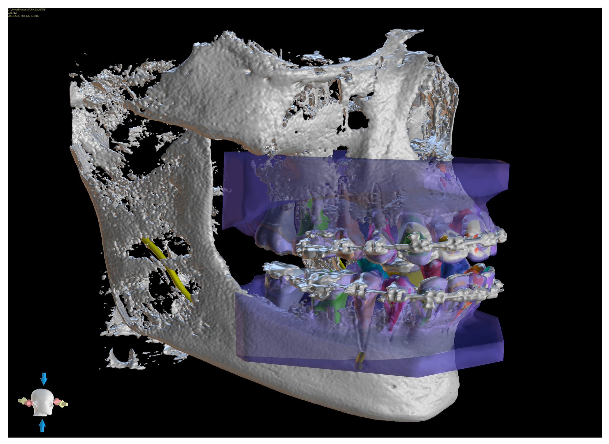
4. Orthodontic Treatment

Clinicians may implement digital technology not only as a diagnostic tool but also in numerous procedures during orthodontic treatment. These procedures will be further analyzed in the following sections and include designing and producing indirect bonding trays, orthodontic braces, clear aligners, surgical guides or splints (manufactured with 3D printed CAD-CAM technology), planning placement sites for orthodontic mini-implants, localization of impacted canines, management of cleft lip and palate cases, and even predicting the postoperative outcomes of orthognathic treatment [14].

4.1. Treatment Procedures

4.1.1. Direct—Indirect Bonding

Proper bracket placement is of paramount importance for an effective orthodontic movement; however, this process is subject to human error. Indirect bracket bonding techniques have been developed with the aim of minimizing human errors associated with variations in the anatomy and morphology of crowns.
Three-dimensional images of each bracket in a bracket kit and of the patient that will undergo treatment are obtained from CBCT scanning. The teeth are then isolated in the software to gain a clear view of the roots. Afterward, each bracket is accurately positioned on the designated crown and thoroughly adjusted in order to achieve post-treatment root parallelism. An image of a U-shaped molding tray is then added to the projection and subsequently adapted to cover the occlusal half of the teeth and brackets while leaving the other half uncovered. In the next step, the images of brackets and teeth are subtracted so that a negative replica ready for printing is produced. Following printing and adjusting the bonding tray, indirect bonding can take place, minimizing human error [20].
Digital positioning of the brackets and indirect bonding techniques, which utilize digital setup models, may greatly facilitate the application of lingual orthodontic appliances. The development of techniques for CAD/CAM fabrication of lingual orthodontic appliances based on 3D models and CBCT scans is emerging, as it provides improved design accuracy [21].

4.1.2. Designing and Manufacturing of Devices

Traditional metallic-based orthodontic appliances, such as hyrax, Herbst appliances, and lingual and transpalatal arches, can be manufactured via CAD/CAM technology without analog impressions and conventional models. The workflow (Figure 3) includes intraoral scanning, digital design of the appliance and incorporation of any additional part, for instance, an expansion screw, direct 3D metal printing via laser melting, welding of any additional part, insertion, and finally activation in the patients’ oral cavity [22,23].
Following testing of these appliances, it has been concluded that they can withstand the stress generated during activation. Therefore, digitally produced orthodontic appliances can clinically function as conventional ones. The application of the digital model offers many advantages, including faster workflow, high predictability, and quicker replacement in case of loss or breakage of appliances. Finally, by facilitating and enhancing communication between the laboratory technician and the orthodontist during appliance design, this approach may ultimately result in a better course of treatment for the patient [22].

4.1.3. Orthodontic Treatment Using Custom-Made Brackets

The development of computer-aided design software has enabled the clinician to design and print in-office orthodontic brackets customized for a specific patient. The design workflow is quick and easy as it follows a standard protocol. The protocol consists of the following steps: intraoral scanning, import of dental scans in the software, teeth segmentation and setup, customized brackets positioning on the teeth, positioning keys designing, bracket files export, ceramic resin 3D printing or zirconia slurry 3D printing, resin brackets UV curing or zirconia brackets sintering, and finally bonding. Three-dimensional printing of brackets can help to deliver a more predictable orthodontic outcome and treat the patient as a unique individual [24].

4.2. Treatment of Impacted Canines

In cases of impacted canines, it is crucial to carefully plan and execute an effective treatment after a meticulous assessment of the exact localization of the impacted teeth, the arrangement of the adjacent teeth, and the condition of the soft tissues. Treatment may consist of either an orthodontic approach or a combination of surgical exposure and orthodontic traction. The utilization of an orthodontic digital model that represents teeth, alveolar bone, and gingiva (Figure 4) would be a valuable tool in the process of comprehensive treatment planning. In order to digitally reconstruct full dentition, gingiva, and alveolar bone into an accurate model, we should integrate data from two imaging techniques. Tooth crowns and gingiva data are obtained from intraoral optical scanning, and tooth roots and bone are obtained from CBCT imaging [25]. This 3D model can be utilized in the exact spotting of the impacted tooth and in deciding the direction of traction. Even a 3D-printed metal device has been proposed for proper traction of palatal-impacted canines [23].
However, the benefits of CBCT scans must be carefully balanced against the radiation risk to pediatric patients and the intricacy of the underlying pathology [15].

4.3. Cleft Lip and Palate Treatment

Three-dimensional reconstruction for patients with cleft lips and palates is useful for both preoperative and therapeutic assessments. Preoperative evaluations of the cleft palate involve an assessment of the volume and location of the bone defect, the presence of supernumerary teeth, and an evaluation of permanent teeth and alveolar bone morphology [15]. Prior to surgical intervention for alveolar bone defects, patients with cleft lip and palate are subjected to a primary repair, which consists of an acrylic intra-oral passive molding plate called Nasoalveolar Molding (NAM). This device is delivered early in the patient’s life in order to separate the oral cavity from the nasal cavity, allow feeding, and reorient the growth process of misaligned bone and soft tissue in the maxilla. The latter contributes to better results following surgery, as NAM can minimize the space across the cleft, hence alleviating the tension throughout the final healing [26,27].
Digital models can also be utilized before the orthognathic surgery of cleft cases in order to establish a 3D surgical occlusion setup. This digital approach may minimize errors from conventional analog models, the risks of dental casts breaking, and the requirement for dental cast storage space. Furthermore, it improves the efficiency and accuracy of the setup, which is crucial for the proper prediction of tooth movements required and the development of the occlusal splint/guide used during orthognathic surgical procedures [28].

4.4. Mini-Implant Placement

Data from CBCT imaging for creating a replica of teeth and surrounding bone in conjunction with plaster models have been used for template fabrication for mini-implant placement [29]. A CBCT-guided surgical stent for a mini-implant was tested for accuracy more than a decade ago [30].
Furthermore, 3D models with the integration of CT scans and intraoral scans for designing templates for accurate mini-implant placement have been proposed. The stereolithography templates are fabricated with CAD/CAM procedures using a stereolithography apparatus (SLA), which is the process of using photosensitive resins cured by a laser layer by layer [31].
CBCT data and 3D laser-scanned images from cast models imported into special software using several points on occlusal dental surfaces for accurate superimposition were also used for surgical stent fabrication with photopolymerized resin using a stereolithographic appliance (SLA) [32].
The same method, after designing the optimal insertion of mini-implants on 3D images created by the fusion of CBCT and digital model data, was used by some researchers [33], while others [34], after creating a digital model of the patient, proposed a special 3D printed surgical guide (Figure 5) incorporating grooves and holes leading to accurate positioning of the mini-implant and convenient release of the guide.
Concerning the exact positioning of mini-implants in the palate as an adjunct procedure for fabricating special orthodontic devices for molar distalization or widening the median suture of the palate, templates advocating information from CBCT data and STL models of the oral cavity have been fabricated with CAD/CAM procedures [35,36].
All these methods lead to accurate positioning of mini-implants regarding adjacent teeth, anatomic structures, and even positioning at exact angles regarding the occlusal plane for enhanced stability.

4.5. Orthognathic Surgery

Designing an orthognathic surgery case is one of the most challenging procedures in orthodontics. Up until recent years, this was mostly conducted by using conventional 2D radiographic examinations such as lateral and anteroposterior cephalograms, panoramic X-rays, and photographs of the patient for surgery treatment planning. The surgery splints that reproduced the planned surgery reposition of the upper and lower jaw anatomic structures were manufactured with auto- or photo-polymerized resins and acrylic materials after articulation of the patient’s casts on a semi-adjustable articulator and the performance of the model surgery [37].
Nowadays, the full digital model of the patient created by the integration of the CBCT images with the face scan (acquired by stereophotogrammetry, laser scanning, or structured light scanning) and the intraoral scanning allows for the more accurate and predictable design of the surgery [5]. Several methods for merging 3D intraoral models and 3D facial models have been proposed [38]. Rigid registration of the 3D acquired images is of great importance in surgery cases and is performed by point-based, surface-based, and voxel-based registration, which are the three most commonly used methods [39].
In the designing stage of the surgery, a 3D model with the new anatomic relationships and a prediction of facial appearance can help the orthodontist communicate the whole treatment plan to the patient and present the procedure in detail. However, caution should be taken, as the patient must be warned that this is a rough and not exact simulation of his face appearance. Today, special algorithms (called approximation models) are used for three-dimensional simulations of soft tissues, and though the result is satisfying, improvements must be made [40].
Acquiring a digital model of the dentition and the occlusion enables the clinician to evaluate in detail the current situation and the predicted final occlusion. In that field, techniques for virtual bite registration in order to inspect bones and teeth simultaneously with the lower jaw in a central relationship have been proposed [41].
In CAD/CAM procedures, the wafer that the surgeon will use to reposition the bony segments is 3D-designed and printed. This leads to more accurate and predictable dental relationships [42]. In addition, the wafer itself is more precise in transferring the maxillary surgical plan to the true operating procedure room and needs fewer adjustments in order to fit on the teeth without any collisions [43]. A meta-analysis in that field reported that 3D surgical cutting guidance ensured precise positioning of the maxillomandibular complex as the virtual plan [44].
Inspection of some anatomic structures like the inferior alveolar nerve, interdental spaces, and the dimensions of the upper and lower jaw, which is easy to do in the digital patient, is of great importance as it may lead the surgeon to differentiate the osteotomy cuts. In special surgeries in which teeth are not involved, such as genioplasty, a digitally created surgical template can help clinicians achieve a more aesthetic result [5].
In cases that present asymmetry, a better outcome is feasible through repositioning the maxillomandibular complex with translational and rotational (pitch, yaw, and roll directions) movements in order to achieve an ideal occlusion and a symmetric and balanced facial appearance. Additionally, identifying possible bony overlaps or gaps during virtual surgery is essential in order to avoid failures and unfavorable situations and thus reduce the possibility of revisionary surgical interventions [45].
It is even proposed that with the use of a surgical splint (that is 3D designed during virtual surgery and computer-aided manufactured), the lines of the Le Fort I osteotomy are marked with a piezoelectric saw to enhance safety. In addition, the digital model on which the surgery is designed enables proper positioning and angulation of fixation screws in areas with the biggest bone thickness. It is also estimated that the accuracy between virtual planning and final surgical results is around 1 mm [46].
In order to overcome problems that arise in designing digital wafers for orthognathic surgery in the vertical direction (z-axis), repositioning guides are used instead. Those are computer-aided designed guides and 3D printed in resin or metal that are embedded in the patient’s bone during surgery and are accurate in transferring the virtual surgery plan to the operating field [39].

4.6. Clear Aligner Design and Printing

The use of digital technology in orthodontics has become increasingly widespread nowadays, especially in allowing planning and carrying out treatment using aligners [47]. Measuring the amount and direction of tooth movement has been considered challenging as it has to be conducted in three different planes of space. Furthermore, three-dimensional superimposition of the initial and final models, tooth segmentation, and landmark registration are necessary for a more accurate quantitative assessment of the orthodontic modifications in all dimensions for each tooth. Automated methods using deep learning technology allow orthodontists to make precise assessments of each tooth movement in a three-dimensional manner with a significant quantity of measurements, a measurement strategy that is very difficult to accomplish using manual methods. This approach was developed to reduce human error and save time [48].
Since the invention of aligners, manufacturing processes have been developed to include vacuum forming, plaster molding, and—most recently—3D printing. Comparing 3D printing with traditional methods reveals several benefits: faster manufacture, better patient fit, less invasive modeling methods, etc. Digital light processing (DLP), stereolithography (SLA), polyjet (PJ), and other specialized vat polymerization (VP) methods are commonly used to print aligners. In these applications, safety and strength are of utmost importance [49].
When 3D printing, computer-aided design (CAD) software, and 3D scanning were brought to the field of orthodontics, the creation of aligners underwent a significant change. Initially, the only resin used in 3D printing was for dental models. However, as the years passed, more resins were added for occlusal splints, indirect bonding trays (IDB), and, most recently, brackets. Up until now, numerous companies have produced plastic foils of varying thickness and composition to be used in the creation of aligners [50]. Furthermore, via 3D printing, orthodontics are able to produce aligners more quickly, accurately, and efficiently than conventional methods.
As directly printed aligners are digitally defined, undercuts do not exist, borders are replicated exactly on each aligner set, and the edges are smooth and do not require polishing or trimming [51]. Color stability and customizable intra-aligner thickness are also some of the benefits that 3D printing provides. It is finally important to mention that 3D-printed aligners provide higher geometric precision and can be tailored to any patient’s needs. Continuous contact between the aligner and the teeth is necessary to ensure its ideal performance since any gaps could prevent the aligner from reaching its maximum mechanical capability [49]. A printed aligner should ideally be recyclable [50].

5. Retention and Outcome Evaluation

5.1. Retainer Fabrication Using Digital Technology

As mentioned previously, doubling a digital model of teeth with the suitable 3D software offers numerous possibilities, one of which is the designing and manufacturing of a retainer after the end of the orthodontic treatment [52].
Permanent retention has lately been recommended by a growing percentage of clinicians. As a consequence, the demands placed on retaining appliances are accordingly increasing. The use of computer-aided design and manufacturing (CAD/CAM) is gradually replacing manual, technician-made retention wires. With this approach, the position, form, and contact of the retainers with the teeth can be enhanced, thereby improving both the retentive effect and the patient’s comfort. Specifically, a 3D analysis of the available space and a deliberate assessment of local conditions enable a fabrication procedure that can enhance the precision of fixed retainer placement in an area with restricted space or unusual tooth shapes, as frequently observed in the upper anterior segment [53].
After performing intraoral scanning or scanning of the impressions of the dental arches, data are transferred to special software, and an orthodontist designs a virtual draft of the retainer. Precise data on the retainer’s distance from the gingiva or opposing dentition might be collected, which would help the doctor plan the bonding connection, especially in cases where there is a significant overbite that creates difficult occlusal relationships. Following virtual retainer planning in 3D modeling software, the retainer is laser-cut from a nitinol blank and electropolished to create a refined, bacteria-resistant surface. A high-precision vinyl polysiloxane transfer coping facilitates handling and serves as a positioning aid for clinical insertion. The retainer’s proper position is obvious due to its excellent passive adaptation [53].
Precise data on the retainer’s distance from the gingiva or opposing dentition might be collected, which would help the doctor plan the bonding connection, especially in cases where there is a significant overbite that creates difficult occlusal relationships. Due to a high risk of bonding-site defects, significant wire-tooth distances, decreased patient comfort, and limited access to hygiene instruments (since there are thick layers of material at bonding sites), many of the inserted retainers remain considered temporary even nowadays. These disadvantages can be significantly mitigated by creating delicate wire designs that closely adapt to the tooth surface, leading to retainer appliances that accomplish the recommended goal of permanent retention [53].
Another advantage is that the patient’s digital data are stored in a more convenient way and can be easily sent to any laboratory or an in-office machine, allowing the practitioner or the technician to fabricate appliances directly on the patient’s digital models obtained by a simple scan. It also provides simplicity, speed, accuracy, and patient satisfaction [52].
Orthodontists are seriously concerned about plaque/calculus accumulation and gingival irritation that are caused by the lingual retainers in the bonded area, as specific surface characteristics of the retainer and its bonding sites would favor plaque formation. However, flatter and more delicate bonding sites, along with the bacteria-resistant character of the wire (due to its electropolished surface), would be less susceptible to plaque formation on exposed wire portions than multistranded wires [53,54].
Furthermore, even in anatomically difficult situations when the conventional retainer technique would reach its limitations, a digitally designed retainer, given this level of precision and predictability, can be easily fitted. The 3D datasets provide extremely accurate views of potential bonding surfaces, enabling the clinician to determine whether these locations would provide enough adhesive strength. The exact fit that CAD/CAM retainers provide almost exactly matches the requirements that apply to crowns and permanent dental prostheses [53].
However, it is necessary to mention that the stability, longevity, and patient satisfaction with CAD/CAM in comparison to traditional multistranded fixed retainers were investigated. It has been found that, after six months, there were no clinically meaningful differences in Little’s irregularity index, arch breadth, or length between CAD/CAM and traditional retainers. Additionally, there was no distinction between the two types of fixed retainers in terms of failures or patient satisfaction [18].

5.2. Outcome Evaluation

As conventional 2D technologies are being replaced by 3D imaging modalities like stereometric surface imaging and cone-beam computed tomography [55], accurate data on 3D changes in the jaws, facial soft tissue, and dentition after orthognathic surgery are provided. Respecting the three-dimensional nature of the face, a three-dimensional method to measure factors associated with changes in soft tissue, bony tissue, and dentition has been proposed [55]. The ability to generate 3D head models gives CBCT a significant advantage over traditional radiography (orthopantomograph and lateral cephalogram), enabling measurements to be made without over-projection of anatomical structures or magnification mistakes. The postoperative soft and hard tissue changes should be taken into consideration during treatment planning and communicated to patients accordingly before surgery [56].
Three-dimensional documentation of orthognathic surgical interventions, as mentioned before, is now achievable since there are numerous 3D imaging possibilities available today. Maal et al. [57] compared the precise 3D soft tissue changes resulting from skeletal transformations following a bilateral sagittal split osteotomy (BSSO) one year following surgery using different types of imaging. In this study, the preoperative and 1-year postoperative CBCT scans were registered with each other using voxel-based registration. Following image registration, the mandible was segmented in both scans. The precise volumetric changes brought on by the BSSO could be calculated by deducting the preoperative hard tissue data from the postoperative hard tissue data. Surface-based registration was used to register the preoperative and postoperative 3D photos in order to examine the soft tissue changes one year after surgery. The overall soft tissue volumetric difference was obtained by subtracting the preoperative surface from the postoperative surface, as determined by the 3D picture following registration. A single, unified dataset of precise hard and soft tissue information could be produced by registering the 3D photos with the CBCT scan’s soft tissue data. Through this procedure, surgeons and orthodontists are able to acknowledge the actual outcomes of soft tissue changes following a specific mandibular transformation [57].
It is also possible to estimate changes in soft tissue at the midsagittal area using typical lateral cephalometric analysis in Class III patients who will undergo mandibular setback surgery (MSS). Nonetheless, patients prefer to assess soft tissue esthetics and changes based mainly on frontal views. Lim et al. [58] analyzed and assessed the soft tissues of the entire face using 3D imaging techniques, such as 3D computed tomography and three-dimensional facial scan images (3D-FSIs). They took lateral cephalograms immediately, both before and 6 months after MSS, with centric occlusion, reposed lip, and natural head position. Cephalometric tracing and measurements were performed by a single operator using the V-Ceph program. In this study, the 3D-FSIs were taken before and 6 months after the MSS from three different horizontal angles and from two different vertical angles to scan the entire face without shadows. The 3D-FSIs were reconstructed using Rapidform 2006 and Rapidform XO scanning software [58].
However, when the sagittal plane is mainly examined, 2D information might be efficient. Legal et al. [59] examined the differences between postoperative outcomes and preoperative virtual planning in orthognathic surgery using the 2D-Onyx Ceph® System. The researchers assessed vertical parameters as well as sagittal angle parameters. As a result, it provided a standard 2D method for planning orthognathic surgery that produces accurate outcomes, especially related to sagittal parameters (SNA, SNB).
Yuan et al. [60] aimed to evaluate changes in the lateral facial shape resulting from surgical treatment of skeletal Class III malocclusion using 3D laser scanning. In this study, three-dimensional data were acquired by a Vivid 910 laser scanner. This is a laser-line triangulation scanner that produces a 640 × 480-pixel 3D image. Data sets were loaded with the appropriate computer software. Each scanned image was reconstructed into 3D images for analysis and measurement using a reverse engineering software system. All of these procedures were undertaken by a single operator. The lateral face shape outline measurement technique utilized in this study could provide quantitative information for clinical evaluation and objective analysis of the human face when viewed from a full-facial perspective.
The measurement of changes in tooth position caused by orthodontic therapy, growth, or software-suggested orthodontic treatment is difficult and complicated due to the lack of consistent reference points. In the absence of these comparatively permanent landmarks, it is hard to forecast the accuracy of the orthodontic appliance system or distinguish between growth, relative movement, and treatment effects.
Alwafi et al. [61] introduced a novel methodology to assess differences between the predicted and achieved mandibular tooth movement with clear aligner therapy using stable mandibular landmarks and a tooth-specific coordinate system. Nevertheless, due to the small sample size and the fact that ClinCheck is mainly a tool for designing clinicians’ biomechanics rather than visualizing the predicted treatment outcomes when planning clear aligner therapy, the results were weak.
Finally, by measuring tooth movement or predicting treatment outcomes, clinicians are able to visualize treatment innervations, make real-time adjustments to simulations, and easily project corrections. They can also have a better judgment on choosing an appropriate treatment plan, build necessary compensations into the virtual plan, provide better interaction and communication with patients, and help with extraction or interproximal reduction decisions.
In Table 1, we summarize the orthodontic devices that may be digitally designed and manufactured.

6. Discussion

6.1. Drawbacks and Limitations Concerning the Digital Model in Orthodontics

In the field of orthodontics and maxillofacial surgery, the use of three-dimensional (3D) information through a digital model (full or partial) tends to become the norm for treatment planning and evaluation. Digital three-dimensional analysis significantly elevates accuracy compared to traditional cephalometric analysis in simple or complicated cases. Intraoral scans and matching CBCT scans, as well as face 3D documentation, are necessary in order to create the 3D model at pretreatment. Accuracy in registering those digital files is of paramount importance, as errors in the procedure could affect treatment planning and the final outcome of orthodontic or orthognathic treatment [65].
Point-based, surface-based, and voxel-based rigid registration are the three most commonly used methods. Combinations are also suggested, such as surface-to-image registration (STI), which combines the surface and voxel-based techniques [5].
In order to assess the accuracy of deep learning-based integrated tooth models (ITMs) for 3D evaluation of root position during orthodontic treatment, Lee et al. [48] combined intraoral and CBCT scans. They also compared the manual and integrated tooth model (ITM) fabrication processes. Using a Trios scanner (3Shape, Copenhagen, Denmark), intraoral scans of the maxillary and mandibular arches were acquired. The intraoral scans were trimmed to the clinical crown by deleting the gingival area. They were then submitted into the OrthoAnalyzer (3Shape) software and reprocessed in stereolithography (STL) file format. The manual approach required more time than the automatic method to complete the procedure and collect the measurements. The final predictability of tooth root position is affected by the inaccurate integration of two imaging modalities. Pretreatment 3D tooth models were therefore created from pretreatment CBCT and pretreatment intraoral scans in this study before the beginning of orthodontic treatment. By obtaining the tooth roots from the CBCT images and the tooth crowns from the intraoral scans, the possibility of an overlapping error (integration error) resulting from the artifacts of the crown appearing in the CBCT image during the pretreatment stage were reduced. There are numerous computer algorithms available for automatically segmenting teeth. It is, therefore, beneficial to use a technique that separates the tooth, including the root, from the alveolar bone in CBCT pictures without removing the alveolar bone. The software involved in this research is typically used to generate 3D models and evaluate medical pictures. In contrast to the medical segmentation process of other anatomical parts, it proved difficult to segment teeth from alveolar bone. In essence, the software carries out segmentation by distinguishing between and using various levels of various anatomic features. Nonetheless, because of the extremely small periodontal ligament space between the tooth and alveolar bone, the software makes it difficult to separate the two due to their identical contrast levels. Therefore, it was not possible to isolate the tooth from the alveolar bone using fully automatic segmentation. Region expanding, the program’s automatic segmentation feature, was mainly used for rough segmentation; the slice edit tool was used to manually tweak it for precision. The ability to divide the segmentation into distinct items is offered by the region-growing tool. All slices underwent morphology operations before region-growing in order to extract the intrinsic tooth structure from the bone. The accuracy of integrating intraoral scans and CBCT images was comparable for both automated and manual techniques. For clinical practice, the automatic approach for ITMs is highly recommended because of its efficiency and time-saving qualities [48].
Manual, semi-automated, and even automated segmentation of anatomic structures in the maxillofacial region has also been suggested [66].
Higher costs that arise from adopting those digital procedures (CBCT imaging instead of conventional radiographs, intraoral scanners, and equipment for 3D photographs or face scans) should be considered a limitation, as should the fact that 3D data are manipulated on a 2D screen [5].
Even more, significant drawbacks associated with the ergonomic limitations of stereophotogrammetry have been raised, leading to the quest for alternative modalities. Such an alternative is the Bellus3D Dental Pro app, which utilizes Apple’s TrueDepth sensor and can reconstruct a digital, high-resolution version of the face. TrueDepth technology is an effective substitute, as smartphone applications are portable, less expensive, and more accessible, allowing every orthodontist to reap the benefits of facial scanning in their everyday practice [67]. Nevertheless, evidence suggests that the current TrueDepth sensor from Apple has limited clinical applicability for orthodontic evaluation due to decreased accuracy below the 3 mm threshold [68].
Simulations produced by those digital models are generally satisfactory, with doubts concerning the labial region, as modeling of the sliding of the lips over the teeth is not yet fully reliable [40].
When surgery procedures are involved, one should keep in mind that CBCT imaging should adopt a large field of view in order to inspect the entire anatomic structure. Unfortunately, this exposes the patient to a greater amount of radiation, and this must be contrasted with the accurate recording of craniofacial abnormalities [16].

6.2. Necessity of a Partial 3D Model

6.2.1. Impacted Canines

In cases where impacted canines are involved, although the type of exposure technique (closed or open) is generally determined by the surgeon’s preference [69], and for initial evaluation, there is no significant difference between 2D and 3D information [70], when a decision is to be made for determining the place on the canine’s surface on which the eyelet of the chain apparatus will be placed and the direction of the traction, a 3D model is advantageous [71,72]. CBCT images can increase the clinician’s confidence regarding the canine location, contact with the adjacent teeth, and the presence or absence of root resorption, thus leading to a successful operation [69,70].
Research has shown that CBCT can provide more information regarding the canine position, detection of root resorption, estimation of space in the arch, overall severity, and overlap than conventional 2D radiography [73,74]. Furthermore, compared to 2D imaging, 3D imaging allows quicker resolution and a better overall tooth prognosis since it can facilitate a more precise and less traumatic surgical exposure, along with more efficient and proper orthodontic traction [73]. Thus, the use of CBCT contributes to a more clinically oriented approach, and it may play a key role in the success of the treatment with a substantial impact on clinicians’ decisions on exposure and traction, biomechanics, and treatment time estimates [15,73,74].

6.2.2. Aligners

A partial digital model created by the integration of face and intraoral scans is essential for clear aligner treatment (CAT), as monitoring facial appearance, soft tissue condition, and smile line simultaneously with intraoral information such as crowding, protruded or retruded teeth, and any deviation of the occlusal plane helps the clinician establish an effective treatment plan.
We have entered the era of face-driven orthodontics, and this was at first facilitated by the use of digital cameras. However, there are limitations to the two-dimensional “reality” [75].
Adding 3D facial information to an intraoral scan benefits a more detailed analysis in cases of asymmetry and cleft lip and/or palate patients [76,77]. Research has underlined that soft tissue information derived from facial scans favors the reliability of the evaluation of the outcome in orthognathic and extraction cases [78,79].
Usually, new technologies also present some disadvantages that, over time, are reduced. For example, a major disadvantage of 3D-printed aligners is the fact that the printing and post-printing procedures depend on multiple steps. An error in one step affects the following stages and can create problems. Another disadvantage is the somewhat higher cost of equipment compared to thermoformed aligner production. A printer and a UV curing unit should be acquired in addition to the software that comes with both thermoformed and directly 3D-printed aligners [50]. There are also some primary difficulties in producing aligners through 3D printing. These arise from the lack of materials that satisfy essential requirements such as biocompatibility, translucency, 3D printability, and suitable mechanical characteristics. Several challenges need to be resolved before the product is used in a clinical setting. These issues fall into the following categories: (1) workflows, (2) anisotropic behavior, (3) characteristics, (4) precision, (5) cost, (6) tooth movement effectiveness, and (7) hygiene of 3D-printed clear aligners [49].

6.2.3. Mini Implants

Placement of mini-implants among the roots of the teeth can be effectively conducted only by utilizing conventional X-rays and plaster models. However, when it comes to exact placement in regions where anatomic limitations occur (not enough space between roots, neighboring inferior alveolar nerve, or greater palatine artery) or when angle to the occlusal plane is essential to enhance stability, then a partial 3D model by fusing CBCT imaging and digital models of the dentition and surrounding tissues is necessary for individuality in treatment planning and safe and accurate implementation [34,36].

6.2.4. Cleft Lip and Plate (Initial Stages)

As mentioned before, passive molding plates are used in the initial stages of dealing with cleft lip and palate patients in order to facilitate feeding. Although such devices have been produced with traditional cast models, the use of a fully digital workflow based on intraoral scanning, 3D printing, and CAD software is gaining more and more attention due to its numerous advantages that benefit both the patient and the clinician. It has been shown that plate manufacturing accuracy, based on digital models, is optimized compared to conventional techniques, offering a more customized appliance for the patient [26,27]. In addition, digital protocol accelerates production time, decreases chairtime, costs, and the invasiveness of impression-taking, avoiding any risk of aspiration of impression materials [26,80].

6.3. Necessity of a Full Digital Model

Orthognathic Surgery Cases (Upper and Lower Jaw) and Cleft Lip and Palate Patients (Later Stages)

Fusion of all 3D information acquired and the construction of a full “virtual patient” allows an in-depth study of an orthognathic surgery case, considering all the parameters that are necessary. It is a superimposition of images from different sources (facial scan, intraoral scan, and CBCT imaging) in a new coordinate system [40]. The superimposition procedure can now be automatically performed [81]. Nowadays, there are also software programs that are capable of tissue 3D simulation when skeletal recontraction is planned with orthognathic surgery [82]. All these possibilities and simulations are essential when planning such serious interventions and prevail over traditional procedures when soft tissue changes are involved. In order to improve the prediction of these changes, orofacial functions might need to be recorded. Some new stereophotogrammetry techniques allow this dynamic analysis by creating “4D” models [83].
In the case of only a setback of the lower jaw, a full 3D digital model of the patient may not be necessary, as the new position of the mandible is dictated by the occlusion. If genioplasty is also planned, then, as mentioned, a digitally created surgical template contributes to a more aesthetic result [5]. However, even with a great number of developing new technologies, it is not yet routine to use them in everyday clinical procedures, mainly due to complexity, training requirements, and elevated costs [84,85]. Even if, with CBCT imaging, the exposure of the patient to radiation is reduced in contrast to CT, each CBCT scan should be acquired for well-justified reasons, especially in children and adolescents [86].
The development of AI can help orthodontics eliminate difficulties associated with the integration of 3D data. Automated identification of landmarks for 3D cephalometric analysis and automated segmentation of teeth and anatomic structures by AI can be very helpful in diagnosis, treatment planning, and outcome prediction, especially for the new clinician who lacks experience but also for the experienced one, in testing several treatment possibilities [75].
In the future, with the further development of 4D printing technology [87,88], which uses time as the fourth dimension and introduces the change in the shape or behavior of a 3D-printed object in reaction to certain stimuli like light, pressure, or magnetic field, many other ”clever” orthodontic devices will be manufactured.
A possible limitation of this review and its conclusions is the differentiation of the digital procedures available in partial and full digital models of the patient. This was chosen in order to present the cases where some or full digital information is needed.

7. Conclusions

Digital procedures in orthodontics lead to more accurate diagnosis and treatment planning. However, we should consider that exposure to radiation must be justified when using CBCT imaging and that the integration methods of 3D data must be accurate. Full and partial digital models of the patient can be acquired by fusion of all or some of the 3D information (face scan, intraoral scan, CBCT).
A partial digital model is needed in impacted canines’ cases, aligner treatment, mini-implant insertion (when angle definition and orientation are essential), and in the early stages of cleft lip and palate cases.
A full digital model should be used in orthognathic surgery cases in which prediction and accurate performance are highly advocated.
It is documented that capable and experienced clinicians can successfully deal with all aspects discussed above, even before the advent of the new digital era. However, it seems that adopting the digital model of a patient in daily practice can give the orthodontist the opportunity to focus on details that otherwise would be impossible to identify. This can lead to more detailed procedures while executing the treatment plan and, thus, a higher quality of outcome. In addition, new orthodontists can benefit from the diagnostic tools that digital means offer and, in this way, avoid mistakes and become more effective.

Supplementary Materials

The following supporting information can be downloaded at: https://www.mdpi.com/article/10.3390/oral4020020/s1, Table S1: Electronic Search Strategy.

Author Contributions

Conceptualization, M.V.; methodology, M.V. and I.P.; investigation, M.V. and I.P.; data curation, M.V., I.P., and A.P.G.; writing—original draft preparation, I.P., A.P.G., and M.V.; writing—review and editing, M.V.; supervision, M.V. All authors have read and agreed to the published version of the manuscript.

Funding

This research received no external funding.

Institutional Review Board Statement

The images used in this research are from a study that was conducted in accordance with the Declaration of Helsinki and approved by the Scientific and Ethics Committee of 401 General Army Hospital in Athens, Greece (ref: No. 10/8-12-2020).

Informed Consent Statement

Written informed consent to publish the images in this paper has been obtained from the patients.

Data Availability Statement

No new data were created or analyzed in this study. Data sharing is not applicable to this article.

Conflicts of Interest

The authors declare no conflicts of interest.

References

  1. Conejo, J.; Dayo, A.F.; Syed, A.Z.; Mupparapu, M. The Digital Clone: Intraoral Scanning, Face Scans and Cone Beam Computed Tomography Integration for Diagnosis and Treatment Planning. Dent. Clin. N. Am. 2021, 65, 529–553. [Google Scholar] [CrossRef] [PubMed]
  2. Campobasso, A.; Battista, G.; Lo Muzio, E.; Lo Muzio, L. The Virtual Patient in Daily Orthodontics: Matching Intraoral and Facial Scans without Cone Beam Computed Tomography. Appl. Sci. 2022, 12, 9870. [Google Scholar] [CrossRef]
  3. Palomo, J.M.; Yang, C.-Y.; Hans, M.G. Clinical Application of Three-Dimensional Craniofacial Imaging in Orthodontics. J. Med. Sci. 2005, 25, 269–278. [Google Scholar]
  4. Carvalho, P.E.G.; Ortega, A.D.O.; Maeda, F.A.; da Silva, L.H.; Carvalho, V.G.G.; Torres, F.C. Digital Scanning in Modern Orthodontics. Curr. Oral. Health Rep. 2019, 6, 269–276. [Google Scholar] [CrossRef]
  5. Elnagar, M.H.; Aronovich, S.; Kusnoto, B. Digital Workflow for Combined Orthodontics and Orthognathic Surgery. Oral Maxillofac. Surg. Clin. N. Am. 2020, 32, 1–14. [Google Scholar] [CrossRef] [PubMed]
  6. Almukhtar, A.; Ju, X.; Khambay, B.; McDonald, J.; Ayoub, A. Comparison of the Accuracy of Voxel Based Registration and Surface Based Registration for 3D Assessment of Surgical Change Following Orthognathic Surgery. PLoS ONE 2014, 9, e93402. [Google Scholar] [CrossRef] [PubMed]
  7. Rangel, F.A.; Maal, T.J.J.; de Koning, M.J.J.; Bronkhorst, E.M.; Bergé, S.J.; Kuijpers-Jagtman, A.M. Integration of Digital Dental Casts in Cone Beam Computed Tomography Scans—A Clinical Validation Study. Clin. Oral Investig. 2018, 22, 1215–1222. [Google Scholar] [CrossRef] [PubMed]
  8. Yang, W.M.; Ho, C.T.; Lo, L.J. Automatic Superimposition of Palatal Fiducial Markers for Accurate Integration of Digital Dental Model and Cone Beam Computed Tomography. J. Oral Maxillofac. Surg. 2015, 73, 1616.e1–1616.e10. [Google Scholar] [CrossRef] [PubMed]
  9. Nahm, K.Y.; Kim, Y.; Choi, Y.S.; Lee, J.; Kim, S.H.; Nelson, G. Accurate Registration of Cone-Beam Computed Tomography Scans to 3-Dimensional Facial Photographs. Am. J. Orthod. Dentofac. Orthop. 2014, 145, 256–264. [Google Scholar] [CrossRef]
  10. Li, M.; Xu, X.; Punithakumar, K.; Le, L.H.; Kaipatur, N.; Shi, B. Automated Integration of Facial and Intra-Oral Images of Anterior Teeth. Comput. Biol. Med. 2020, 122, 103794. [Google Scholar] [CrossRef]
  11. Taneva, E.; Kusnoto, B.; Evans, C.A.; Taneva, E.; Kusnoto, B.; Evans, C.A. 3D Scanning, Imaging, and Printing in Orthodontics. Issues Contemp. Orthod. 2015, 148, 862–867. [Google Scholar] [CrossRef]
  12. Bechtold, T.E.; Göz, T.G.; Schaupp, E.; Koos, B.; Godt, A.; Reinert, S.; Berneburg, M. Integration of a Maxillary Model into Facial Surface Stereophotogrammetry. J. Orofac. Orthop. 2012, 73, 126–137. [Google Scholar] [CrossRef] [PubMed]
  13. Tricco, A.C.; Lillie, E.; Zarin, W.; O’Brien, K.K.; Colquhoun, H.; Levac, D.; Moher, D.; Peters, M.D.J.; Horsley, T.; Weeks, L.; et al. PRISMA Extension for Scoping Reviews (PRISMA-ScR): Checklist and Explanation. Ann. Intern. Med. 2018, 169, 467–473. [Google Scholar] [CrossRef] [PubMed]
  14. Francisco, I.; Ribeiro, M.P.; Marques, F.; Travassos, R.; Nunes, C.; Pereira, F.; Caramelo, F.; Paula, A.B.; Vale, F. Application of Three-Dimensional Digital Technology in Orthodontics: The State of the Art. Biomimetics 2022, 7, 23. [Google Scholar] [CrossRef] [PubMed]
  15. Machado, G.L. CBCT Imaging—A Boon to Orthodontics. Saudi Dent. J. 2015, 27, 12–21. [Google Scholar] [CrossRef] [PubMed]
  16. Damstra, J.; Fourie, Z.; Ren, Y. Evaluation and Comparison of Postero-Anterior Cephalograms and Cone-Beam Computed Tomography Images for the Detection of Mandibular Asymmetry. Eur. J. Orthod. 2013, 35, 45–50. [Google Scholar] [CrossRef] [PubMed]
  17. Baan, F.; de Waard, O.; Bruggink, R.; Xi, T.; Ongkosuwito, E.M.; Maal, T.J.J. Virtual Setup in Orthodontics: Planning and Evaluation. Clin. Oral Investig. 2020, 24, 2385–2393. [Google Scholar] [CrossRef] [PubMed]
  18. Fawaz, P.; Sayegh, P.E.; Vannet, B. Vande. What Is the Current State of Artificial Intelligence Applications in Dentistry and Orthodontics? J. Stomatol. Oral Maxillofac. Surg. 2023, 124, 101524. [Google Scholar] [CrossRef]
  19. Mahmood, T.M.A.; Noori, A.J.; Aziz, Z.H.; Rauf, A.M.; Kareem, F.A. Scan Aided Dental Arch Width Prediction via Internationally Recognized Formulas and Indices in a Sample of Kurdish Population/Iraq. Diagnostics 2023, 13, 1900. [Google Scholar] [CrossRef]
  20. El-Timamy, A.M.; El-Sharaby, F.A.; Eid, F.H.; Mostafa, Y.A. Three-Dimensional Imaging for Indirect-Direct Bonding. Am. J. Orthod. Dentofac. Orthop. 2016, 149, 928–931. [Google Scholar] [CrossRef]
  21. Kwon, S.Y.; Kim, Y.; Ahn, H.W.; Kim, K.B.; Chung, K.R.; Kim, S.H. Computer-Aided Designing and Manufacturing of Lingual Fixed Orthodontic Appliance Using 2D/3D Registration Software and Rapid Prototyping. Int. J. Dent. 2014, 2014, 164164. [Google Scholar] [CrossRef]
  22. Graf, S.; Cornelis, M.A.; Hauber Gameiro, G.; Cattaneo, P.M. Computer-Aided Design and Manufacture of Hyrax Devices: Can We Really Go Digital? Am. J. Orthod. Dentofac. Orthop. 2017, 152, 870–874. [Google Scholar] [CrossRef] [PubMed]
  23. Vasoglou, G.; Lyros, I.; Patatou, A.; Vasoglou, M. Orthodontic Treatment of Palatally Impacted Maxillary Canines with the Use of a Digitally Designed and 3D-Printed Metal Device. Dent. J. 2023, 11, 102. [Google Scholar] [CrossRef]
  24. Panayi, N.C. In-House Three-Dimensional Designing and Printing Customized Brackets. J. World Fed. Orthod. 2022, 11, 190–196. [Google Scholar] [CrossRef]
  25. Barone, S.; Paoli, A.; Razionale, A.V. Creation of 3D Multi-Body Orthodontic Models by Using Independent Imaging Sensors. Sensors 2013, 13, 2033–2050. [Google Scholar] [CrossRef] [PubMed]
  26. Dalessandri, D.; Tonni, I.; Laffranchi, L.; Migliorati, M.; Isola, G.; Bonetti, S.; Visconti, L.; Paganelli, C. Evaluation of a Digital Protocol for Pre-Surgical Orthopedic Treatment of Cleft Lip and Palate in Newborn Patients: A Pilot Study. Dent. J. 2019, 7, 111. [Google Scholar] [CrossRef]
  27. Ahmed, M.K.; Ahsanuddin, S.; Retrouvey, J.M.; Koka, K.S.; Qureshi, H.; Bui, A.H.; Taub, P.J. Fabrication of Nasoalveolar Molding Devices for the Treatment of Cleft Lip and Palate, Using Stereolithography Additive Manufacturing Processes and Computer-Aided Design Manipulation Software. J. Craniofac. Surg. 2019, 30, 2604–2608. [Google Scholar] [CrossRef]
  28. Seo, H.J.; Denadai, R.; Pai, B.C.J.; Lo, L.J. Digital Occlusion Setup Is Quantitatively Comparable with the Conventional Dental Model Approach: Characteristics and Guidelines for Orthognathic Surgery in Patients with Unilateral Cleft Lip and Palate. Ann. Plast. Surg. 2020, 85, 171–179. [Google Scholar] [CrossRef] [PubMed]
  29. Kim, S.H.; Choi, Y.S.; Hwang, E.H.; Chung, K.R.; Kook, Y.A.; Nelson, G. Surgical Positioning of Orthodontic Mini-Implants with Guides Fabricated on Models Replicated with Cone-Beam Computed Tomography. Am. J. Orthod. Dentofac. Orthop. 2007, 131 (Suppl. S4), S82–S89. [Google Scholar] [CrossRef]
  30. Yu, J.J.; Kim, G.T.; Choi, Y.S.; Hwang, E.H.; Paek, J.; Kim, S.H.; Huang, J.C. Accuracy of a Cone Beam Computed Tomography–Guided Surgical Stent for Orthodontic Mini-Implant Placement. Angle Orthod. 2012, 82, 275–283. [Google Scholar] [CrossRef]
  31. Liu, H.; Liu, D.X.; Wang, G.; Wang, C.L.; Zhao, Z. Accuracy of Surgical Positioning of Orthodontic Miniscrews with a Computer-Aided Design and Manufacturing Template. Am. J. Orthod. Dentofac. Orthop. 2010, 137, 728.e1–728.e10. [Google Scholar] [CrossRef] [PubMed]
  32. Qiu, L.; Haruyama, N.; Suzuki, S.; Yamada, D.; Obayashi, N.; Kurabayashi, T.; Moriyama, K. Accuracy of Orthodontic Miniscrew Implantation Guided by Stereolithographic Surgical Stent Based on Cone-Beam CT-Derived 3D Images. Angle Orthod. 2012, 82, 284–293. [Google Scholar] [CrossRef] [PubMed]
  33. Bae, M.J.; Kim, J.Y.; Park, J.T.; Cha, J.Y.; Kim, H.J.; Yu, H.S.; Hwang, C.J. Accuracy of Miniscrew Surgical Guides Assessed from Cone-Beam Computed Tomography and Digital Models. Am. J. Orthod. Dentofac. Orthop. 2013, 143, 893–901. [Google Scholar] [CrossRef] [PubMed]
  34. Vasoglou, G.; Stefanidaki, I.; Apostolopoulos, K.; Fotakidou, E.; Vasoglou, M. Accuracy of Mini-Implant Placement Using a Computer-Aided Designed Surgical Guide, with Information of Intraoral Scan and the Use of a Cone-Beam CT. Dent. J. 2022, 10, 104. [Google Scholar] [CrossRef]
  35. Wilmes, B.; Tarraf, N.E.; de Gabriele, R.; Dallatana, G.; Drescher, D. Procedure Using CAD/CAM-Manufactured Insertion Guides for Purely Mini-Implant-Borne Rapid Maxillary Expanders. J. Orofac. Orthop. 2022, 83, 277–284. [Google Scholar] [CrossRef] [PubMed]
  36. Weismann, C.; Heise, K.; Aretxabaleta, M.; Cetindis, M.; Koos, B.; Schulz, M.C. Mini-Implant Insertion Using a Guide Manufactured with Computer-Aided Design and Computer-Aided Manufacturing in an Adolescent Patient Suffering from Tooth Eruption Disturbance. Bioengineering 2024, 11, 91. [Google Scholar] [CrossRef] [PubMed]
  37. Proffit, W.; White, R. Surgical-Orthodontic Treatment; Mosby: St. Louis, MO, USA, 2008. [Google Scholar]
  38. Sigouin, A.J. A Novel Method to Integrate Intra-Oral Scan Models with 3D Facial Images. Doctoral Dissertation, University of British Columbia, Vancouver, BC, Canada, 2023. [Google Scholar] [CrossRef]
  39. Muthuswamy Pandian, S.; Gandedkar, N.H.; Palani, S.K.; Kim, Y.J.; Adel, S.M. An Integrated 3D-Driven Protocol for Surgery First Orthognathic Approach (SFOA) Using Virtual Surgical Planning (VSP). Semin. Orthod. 2022, 28, 320–333. [Google Scholar] [CrossRef]
  40. Rasteau, S.; Sigaux, N.; Louvrier, A.; Bouletreau, P. Three-Dimensional Acquisition Technologies for Facial Soft Tissues—Applications and Prospects in Orthognathic Surgery. J. Stomatol. Oral Maxillofac. Surg. 2020, 121, 721–728. [Google Scholar] [CrossRef]
  41. Nilsson, J.; Richards, R.G.; Thor, A.; Kamer, L. Virtual Bite Registration Using Intraoral Digital Scanning, CT and CBCT: In Vitro Evaluation of a New Method and Its Implication for Orthognathic Surgery. J. Cranio-Maxillofac. Surg. 2016, 44, 1194–1200. [Google Scholar] [CrossRef]
  42. Hanafy, M.; Akoush, Y.; Abou-ElFetouh, A.; Mounir, R.M. Precision of Orthognathic Digital Plan Transfer Using Patient-Specific Cutting Guides and Osteosynthesis versus Mixed Analogue-Digitally Planned Surgery: A Randomized Controlled Clinical Trial. Int. J. Oral Maxillofac. Surg. 2020, 49, 62–68. [Google Scholar] [CrossRef]
  43. Chen, C.M.; Lai, S.; Lee, H.E.; Chen, K.K.; Hsu, K.J. Soft-Tissue Profile Changes after Orthognathic Surgery of Mandibular Prognathism. Kaohsiung J. Med. Sci. 2012, 28, 216–219. [Google Scholar] [CrossRef] [PubMed]
  44. Van den Bempt, M.; Liebregts, J.; Maal, T.; Bergé, S.; Xi, T. Toward a Higher Accuracy in Orthognathic Surgery by Using Intraoperative Computer Navigation, 3D Surgical Guides, and/or Customized Osteosynthesis Plates: A Systematic Review. J. Cranio-Maxillofac. Surg. 2018, 46, 2108–2119. [Google Scholar] [CrossRef] [PubMed]
  45. Ho, C.T.; Lai, H.C.; Lin, H.H.; Denadai, R.; Lo, L.J. Outcome of Full Digital Workflow for Orthognathic Surgery Planning in the Treatment of Asymmetric Skeletal Class III Deformity. J. Formos. Med. Assoc. 2021, 120, 2100–2112. [Google Scholar] [CrossRef] [PubMed]
  46. Ebker, T.; Korn, P.; Heiland, M.; Bumann, A. Comprehensive Virtual Orthognathic Planning Concept in Surgery-First Patients. Br. J. Oral Maxillofac. Surg. 2022, 60, 1092–1096. [Google Scholar] [CrossRef] [PubMed]
  47. Arveda, N.; Colonna, A.; Palone, M.; Lombardo, L. Aligner Hybrid Orthodontic Approach to Treat Severe Transverse Divergence in an Adolescent Girl: A Case Report. Int. Orthod. 2022, 20, 100686. [Google Scholar] [CrossRef] [PubMed]
  48. Lee, S.; Wu, T.-H.; Deguchi, T.; Ni, A.; Lu, W.-E.; Minhas, S.; Murphy, S.; Ko, C.-C. Assessment of Malalignment Factors Related to the Invisalign Treatment Time Aided by Automated Imaging Processes. Angle Orthod. 2022, 93, 144–150. [Google Scholar] [CrossRef] [PubMed]
  49. Narongdej, P.; Hassanpour, M.; Alterman, N.; Rawlins-Buchanan, F.; Barjasteh, E. Advancements in Clear Aligner Fabrication: A Comprehensive Review of Direct-3D Printing Technologies. Polymers 2024, 16, 371. [Google Scholar] [CrossRef]
  50. Panayi, N.C. Directly Printed Aligner: Aligning with the Future. Turk. J. Orthod. 2023, 36, 62–69. [Google Scholar] [CrossRef] [PubMed]
  51. Tartaglia, G.M.; Mapelli, A.; Maspero, C.; Santaniello, T.; Serafin, M.; Farronato, M.; Caprioglio, A. Direct 3D Printing of Clear Orthodontic Aligners: Current State and Future Possibilities. Materials 2021, 14, 1799. [Google Scholar] [CrossRef] [PubMed]
  52. Nasef, A.A.; El-Beialy, A.R.; Mostafa, Y.A. Virtual Techniques for Designing and Fabricating a Retainer. Am. J. Orthod. Dentofac. Orthop. 2014, 146, 394–398. [Google Scholar] [CrossRef]
  53. Wolf, M.; Schumacher, P.; Jäger, F.; Wego, J.; Fritz, U.; Korbmacher-Steiner, H.; Jäger, A.; Schauseil, M. Novel Lingual Retainer Created Using CAD/CAM Technology: Evaluation of Its Positioning Accuracy. J. Orofac. Orthop. 2015, 76, 164–174. [Google Scholar] [CrossRef] [PubMed]
  54. Sifakakis, I.; Iijima, M.; Brantley, W. Wires Used in Fixed Retainers. In Debonding and Fixed Retention in Orthodontics: An Evidence-Based Clinical Guide; John Wiley & Sons Ltd.: Hoboken, NJ, USA, 2023; pp. 227–247. [Google Scholar] [CrossRef]
  55. Zupan, J.; Ihan Hren, N.; Verdenik, M. An Evaluation of Three-Dimensional Facial Changes after Surgically Assisted Rapid Maxillary Expansion (SARME): An Observational Study. BMC Oral Health 2022, 22, 155. [Google Scholar] [CrossRef] [PubMed]
  56. Xi, T.; Laskowska, M.; van de Voort, N.; Ghaeminia, H.; Pawlak, W.; Bergé, S.; Maal, T. The Effects of Surgically Assisted Rapid Maxillary Expansion (SARME) on the Dental Show and Chin Projection. J. Cranio-Maxillofac. Surg. 2017, 45, 1835–1841. [Google Scholar] [CrossRef] [PubMed]
  57. Maal, T.J.J.; De Koning, M.J.J.; Plooij, J.M.; Verhamme, L.M.; Rangel, F.A.; Bergé, S.J.; Borstlap, W.A. One Year Postoperative Hard and Soft Tissue Volumetric Changes after a BSSO Mandibular Advancement. Int. J. Oral Maxillofac. Surg. 2012, 41, 1137–1145. [Google Scholar] [CrossRef] [PubMed]
  58. Lim, Y.K.; Chub, E.H.; Leea, D.Y.; Yangc, I.H.; Baekd, S.H. Three-Dimensional Evaluation of Soft Tissue Change Gradients after Mandibular Setback Surgery in Skeletal Class III Malocclusion. Angle Orthod. 2010, 80, 896–903. [Google Scholar] [CrossRef] [PubMed]
  59. Legal, S.; Moralis, A.; Waiss, W.; Zeman, F.; Winkler, C.; Müller, S.; Reichert, T.E.; Proff, P.; Meier, J.; Klingelhöffer, C.; et al. Accuracy in Orthognathic Surgery─comparison of Preoperative Plan and Postoperative Outcome Using Computer-Assisted Two-Dimensional Cephalometry by the Onyx Ceph® System. J. Cranio-Maxillofac. Surg. 2018, 46, 1793–1799. [Google Scholar] [CrossRef] [PubMed]
  60. Yuan, L.; Shen, G.; Wu, Y.; Jiang, L.; Yang, Z.; Liu, J.; Mao, L.; Fang, B. Three-Dimensional Analysis of Soft Tissue Changes in Full-Face View after Surgical Correction of Skeletal Class III Malocclusion. J. Craniofac. Surg. 2013, 24, 725–730. [Google Scholar] [CrossRef] [PubMed]
  61. Alwafi, A.A.; Hannam, A.G.; Yen, E.H.; Zou, B. A New Method Assessing Predicted and Achieved Mandibular Tooth Movement in Adults Treated with Clear Aligners Using CBCT and Individual Crown Superimposition. Sci. Rep. 2023, 13, 4084. [Google Scholar] [CrossRef] [PubMed]
  62. Eliades, T.; Panayi, N.; Papageorgiou, S.N. From Biomimetics to Smart Materials and 3D Technology: Applications in Orthodontic Bonding, Debonding, and Appliance Design or Fabrication. Jpn. Dent. Sci. Rev. 2023, 59, 403–411. [Google Scholar] [CrossRef]
  63. Panayi, N.C. In-Office Customized Brackets: Aligning with the Future. Turk. J. Orthod. 2023, 36, 143–148. [Google Scholar] [CrossRef]
  64. Zarean, P.; Zarean, P.; Thieringer, F.M.; Mueller, A.A.; Kressmann, S.; Erismann, M.; Sharma, N.; Benitez, B.K. A Point-of-Care Digital Workflow for 3D Printed Passive Presurgical Orthopedic Plates in Cleft Care. Children 2022, 9, 1261. [Google Scholar] [CrossRef] [PubMed]
  65. Naudi, K.B.; Benramadan, R.; Brocklebank, L.; Ju, X.; Khambay, B.; Ayoub, A. The Virtual Human Face: Superimposing the Simultaneously Captured 3D Photorealistic Skin Surface of the Face on the Untextured Skin Image of the CBCT Scan. Int. J. Oral Maxillofac. Surg. 2012, 42, 393–400. [Google Scholar] [CrossRef] [PubMed]
  66. Nogueira-Reis, F.; Morgan, N.; Suryani, I.R.; Tabchoury, C.P.M.; Jacobs, R. Full Virtual Patient Generated by Artificial Intelligence-Driven Integrated Segmentation of Craniomaxillofacial Structures from CBCT Images. J. Dent. 2024, 141, 104829. [Google Scholar] [CrossRef] [PubMed]
  67. Beretta, M.; Canova, F.F.; Zaffarano, L.; Gianolio, A. Face Scan for Ceph 3D: A Green Way for Diagnosis in Children. Eur. J. Paediatr. Dent. 2022, 23, 201–203. [Google Scholar] [CrossRef] [PubMed]
  68. Thurzo, A.; Strunga, M.; Havlínová, R.; Reháková, K.; Urban, R.; Surovková, J.; Kurilová, V. Smartphone-Based Facial Scanning as a Viable Tool for Facially Driven Orthodontics? Sensors 2022, 22, 7752. [Google Scholar] [CrossRef] [PubMed]
  69. Wriedt, S.; Jaklin, J.; Al-Nawas, B.; Wehrbein, H. Impacted Upper Canines: Examination and Treatment Proposal Based on 3D versus 2D Diagnosis. J. Orofac. Orthop. 2012, 73, 28–40. [Google Scholar] [CrossRef] [PubMed]
  70. Alqerban, A.; Hedesiu, M.; Baciut, M.; Nackaerts, O.; Jacobs, R.; Fieuws, S.; Consortium, S.; Willems, G. Pre-Surgical Treatment Planning of Maxillary Canine Impactions Using Panoramic vs Cone Beam CT Imaging. Dentomaxillofac. Radiol. 2013, 42, 20130157. [Google Scholar] [CrossRef] [PubMed]
  71. Walker, L.; Enciso, R.; Mah, J. Three-Dimensional Localization of Maxillary Canines with Cone-Beam Computed Tomography. Am. J. Orthod. Dentofac. Orthop. 2005, 128, 418–423. [Google Scholar] [CrossRef] [PubMed]
  72. Alqerban, A.; Jacobs, R.; Fieuws, S.; Willems, G. Comparison of Two Cone Beam Computed Tomographic Systems versus Panoramic Imaging for Localization of Impacted Maxillary Canines and Detection of Root Resorption. Eur. J. Orthod. 2011, 33, 93–102. [Google Scholar] [CrossRef]
  73. Becker, A.; Chaushu, S.; Casap-Caspi, N. Cone-Beam Computed Tomography and the Orthosurgical Management of Impacted Teeth. J. Am. Dent. Assoc. 2010, 141 (Suppl. S3), 14S–18S. [Google Scholar] [CrossRef]
  74. Keener, D.J.; de Oliveira Ruellas, A.C.; Aliaga-Del Castillo, A.; Arriola-Guillén, L.E.; Bianchi, J.; Oh, H.; Gurgel, M.L.; Benavides, E.; Soki, F.; Rodríguez-Cárdenas, Y.A.; et al. Three-Dimensional Decision Support System for Treatment of Canine Impaction. Am. J. Orthod. Dentofac. Orthop. 2023, 164, 491–504. [Google Scholar] [CrossRef] [PubMed]
  75. Tomášik, J.; Zsoldos, M.; Oravcová, Ľ.; Lifková, M.; Pavleová, G.; Strunga, M.; Thurzo, A. AI and Face-Driven Orthodontics: A Scoping Review of Digital Advances in Diagnosis and Treatment Planning. AI 2024, 5, 158–176. [Google Scholar] [CrossRef]
  76. Hallac, R.R.; Feng, J.; Kane, A.A.; Seaward, J.R. Dynamic Facial Asymmetry in Patients with Repaired Cleft Lip Using 4D Imaging (Video Stereophotogrammetry). J. Craniomaxillofac. Surg. 2017, 45, 8–12. [Google Scholar] [CrossRef] [PubMed]
  77. Xue, Z.; Wu, L.; Qiu, T.; Li, Z.; Wang, X.; Liu, X. Three-Dimensional Dynamic Analysis of the Facial Movement Symmetry of Skeletal Class III Patients With Facial Asymmetry. J. Oral Maxillofac. Surg. 2019, 78, 267–274. [Google Scholar] [CrossRef] [PubMed]
  78. Rongo, R.; Nissen, L.; Leroy, C.; Michelotti, A.; Cattaneo, P.M.; Cornelis, M.A. Three-Dimensional Soft Tissue Changes in Orthodontic Extraction and Non-Extraction Patients: A Prospective Study. Orthod. Craniofac. Res. 2021, 24 (Suppl. S2), 181–192. [Google Scholar] [CrossRef]
  79. Perrotti, G.; Reda, R.; Rossi, O.; D’apolito, I.; Testori, T.; Testarelli, L. A Radiation Free Alternative to CBCT Volumetric Rendering for Soft Tissue Evaluation. Braz. Dent. Sci. 2023, 26, 1–7. [Google Scholar] [CrossRef]
  80. Gong, X.; Dang, R.; Xu, T.; Yu, Q.; Zheng, J. Full Digital Workflow of Nasoalveolar Molding Treatment in Infants With Cleft Lip and Palate. J. Craniofac. Surg. 2020, 31, 367–371. [Google Scholar] [CrossRef] [PubMed]
  81. Kau, C.H. Creation of the Virtual Patient for the Study of Facial Morphology. Facial Plast. Surg. Clin. N. Am. 2011, 19, 615–622. [Google Scholar] [CrossRef] [PubMed]
  82. Swennen, G.R.J.; Gaboury, M. 3D Virtual Treatment Planning of Orthognathic Surgery, 1st ed.; Swennen, G.R.J., Ed.; Springer: Berlin/Heidelberg, Germany, 2016. [Google Scholar] [CrossRef]
  83. Shujaat, S.; Khambay, B.S.; Ju, X.; Devine, J.C.; McMahon, J.D.; Wales, C.; Ayoub, A.F. The Clinical Application of Three-Dimensional Motion Capture (4D): A Novel Approach to Quantify the Dynamics of Facial Animations. Int. J. Oral Maxillofac. Surg. 2014, 43, 907–916. [Google Scholar] [CrossRef]
  84. Xia, J.J.; Gateno, J.; Teichgraeber, J.F.; Yuan, P.; Li, J.; Chen, K.C.; Jajoo, A.; Nicol, M.; Alfi, D.M. Algorithm for Planning a Double-Jaw Orthognathic Surgery Using a Computer-Aided Surgical Simulation (CASS) Protocol. Part 2: Three-Dimensional Cephalometry. Int. J. Oral Maxillofac. Surg. 2015, 44, 1441–1450. [Google Scholar] [CrossRef]
  85. Stokbro, K.; Aagaard, E.; Torkov, P.; Bell, R.B.; Thygesen, T. Virtual Planning in Orthognathic Surgery. Int. J. Oral Maxillofac. Surg. 2014, 43, 957–965. [Google Scholar] [CrossRef] [PubMed]
  86. Abdelkarim, A. Cone-Beam Computed Tomography in Orthodontics. Dent. J. 2019, 7, 89. [Google Scholar] [CrossRef] [PubMed]
  87. Cataldi, P.; Liu, M.; Bissett, M.; Kinloch, I.A.; Cataldi, P.; Liu, M.; Bissett, M.; Kinloch, I.A. A Review on Printing of Responsive Smart and 4D Structures Using 2D Materials. Adv. Mater. Technol. 2022, 7, 2200025. [Google Scholar] [CrossRef]
  88. Saritha, D.; Boyina, D. A Concise Review on 4D Printing Technology. Mater. Today Proc. 2021, 46, 692–695. [Google Scholar] [CrossRef]
Figure 1. Flowchart of study selection according to PRISMA statement.
Oral 04 00020 g001
Figure 2. A digital model of the patient was created by combining intraoral scanning and CBCT imaging. The blue color represents the STL model of the intraoral scan, while the gray color represents the 3D model acquired by CBCT imaging.
Oral 04 00020 g002
Figure 3. (a) The design of a metal device (blue color) for traction of an impacted canine (brown color) on a digital model (green color). (b) The device, after 3D printing and implementation in the patient’s mouth.
Oral 04 00020 g003
Figure 4. A digital model (green color) on which the detection of the exact location of a palatally impacted canine (brown color) is easy.
Oral 04 00020 g004
Figure 5. (a) Insertion of a virtual mini-implant (small green conical-shaped feature) using a 3D designed and printed surgical guide (blue color) on the patient’s digital model (green color). (b) the mini-implant in the patient’s mouth between the roots of the second premolar (red color) and the first molar (yellow color).
Oral 04 00020 g005
Table 1. Orthodontic devices that may be produced by digital technology (CAD/CAM).
DevicePrinting MaterialPrinting Technology
trays for indirect bondingBiocompatible liquid photopolymers (resins)Vat photopolymerisation technology (Stereolithography/SLA)
hyrax device/
metal device for impacted canine traction/
transpalatal arch/
Fixed retainers
Biocompatible dental alloys (stainless steel, titanium, and mainly cobalt–chromium [CoCr] alloys) [62]Selective Laser Melting (SLM)and Selective laser sintering (SLS)/metal additive manufacturing technology for all parts except the screw in the hyrax device
ceramic bracketsHybrid ceramic permanent crown resins or zirconia slurryLaser stereolithography (laser-SLA) or Direct Light Processing (DLP) (varies according to the used printer) [63]
intra-oral passive molding plate in cleft palate casesBiocompatible liquid photopolymers (acrylates)Stereolithography/SLA [64]
templates for mini-implant placement/orthognathic surgery splintsBiocompatible liquid photopolymers (resins)Stereolithography/SLA
models for aligners (vacuum forming)Liquid photopolymers (resins)Stereolithography/SLA
direct printed alignersAligner resin called TC-85DAC (Graphy, Seoul, Republic of Korea)Stereolithography/SLA [50]
Disclaimer/Publisher’s Note: The statements, opinions and data contained in all publications are solely those of the individual author(s) and contributor(s) and not of MDPI and/or the editor(s). MDPI and/or the editor(s) disclaim responsibility for any injury to people or property resulting from any ideas, methods, instructions or products referred to in the content.

