Article

Finding Hidden Outliers to Promote the Consistency of Key Morphological Traits and Phylogeny in Dennstaedtiaceae

1
The Orchid Conservation and Research Centre of Shenzhen, Shenzhen 518114, China
2
Key Laboratory of National Forestry and Grassland Administration for Orchid Conservation and Utilization, Shenzhen 518114, China
3
CAS Center for Excellence in Molecular Plant Sciences, Institute of Plant Physiology and Ecology, Chinese Academy of Sciences, Shanghai 200032, China
4
Shanghai Chenshan Plant Science Research Center, Chinese Academy of Sciences, Shanghai Chenshan Botanical Garden, Shanghai 201602, China
5
College of Life Sciences, Shanghai Normal University, Shanghai 200234, China
6
State Key Laboratory of Systematic and Evolutionary Botany, Institute of Botany, Chinese Academy of Sciences, Beijing 100093, China
*
Author to whom correspondence should be addressed.
These authors contributed equally to this work.
Taxonomy 2021, 1(3), 256-265; https://doi.org/10.3390/taxonomy1030019
Submission received: 12 July 2021 / Revised: 31 July 2021 / Accepted: 9 August 2021 / Published: 23 August 2021

Abstract

:
With the development of open science and technological innovation, using sharing data and molecular biology techniques in the study of taxonomy and systematics have become a crucial component of plants, which undoubtedly helps us discover more hidden outliers or deal with difficult taxa. In this paper, we take Dennstaedtia smithii as an example, based on sharing molecular database, virtual herbarium and plant photo bank, to clarify the outliers that have been hidden in Dennstaedtia and find the key morphological traits with consistent of molecular systematics. In molecular phylogenetic analyses, we used rbcL, rps4, psbA-trnH and trnL-F sequences from 5 new and 49 shared data; the results showed that Dennstaedtia smithii is nested within Microlepia rather than Dennstaedtia. We further studied the morphological characters based on the phylogeny result and found that D. smithii is distinguished from other species of Dennstaedtia by spore ornamentation and the unconnected of grooves between rachis and pinna rachis. According to morphological and molecular phylogenetic studies, our results supported that D. smithii should be a new member of Microlepia and renamed Microlepia smithii (Hook.) Y.H. Yan. Finding hidden outliers can promote the consistency of morphological and molecular phylogenetic results, and make the systematic classification more natural.

1. Introduction

From the evolutionary emergence of primitive organisms to today’s broad variety of organisms, people have been constantly exploring how many species there are on the earth and what kind of evolutionary relationship among species. With the development of open science and technological innovation, methods of species identification range from using morphological characteristics to the integration of various methods (e.g., molecular biology, bioinformatics, bionomics) [1,2,3,4,5], which help us gain a more in-depth understanding of the evolutionary process between organisms and their accurate position in the tree of life. Due to the multi-disciplines combination and the improvement of sharing databases, many misclassifications hidden in the past have been gradually discovered, and their key morphological boundaries have also been redefined. For example, Typhonium giganteum Engler 1883 had long been recognized as a member of Typhonium Schott 1829 according to the morphological characteristics, but the molecular phylogenetic evidence indicated that it should belong to the Sauromatum Schott 1832 and was renamed as Sauromatum giganteum (Engl.) Cusimano and Hetterscheid 2010 [1]. Ferns, an ancient group, also have similar examples, one of which is Athyrium niponicum (Mett.) Hance 1873. A. niponicum had been treated as a member of Athyrium Roth 1875, but was later confirmed to be within Anisocampium C. Presl 1851 based on rbcL and trnL-F region sequences [2]. At the end of the paper, the author revised the morphological boundaries of Athyrium and Anisocampium according to the results of systematics [2].
Microlepia C. Presl 1836, comprising about 60 species in the world, is mostly distributed in tropics and subtropics [6,7]. In the past, there had been much controversy over the relationships of Microlepia and Dennstaedtia Bernh. 1800. Some species of Dennstaedtia, including the type species Dennstaedtia flaccida (J.R. Forst.) Bernh. 1801, had been placed in Microlepia by Smith [8]. However, in previous molecular phylogenetic studies, Microlepia was monophyletic [6,9,10,11] with the type species of Microlepia speluncae (L.) T. Moore 1857 and was sister to the old-world clade of Dennstaedtia [10]. We can distinguish Microlepia and Dennstaedtia from the following characteristics: the abaxial condition of sori, the shallow costal grooves and the finely echinate spores [7,12].
Dennstaedtia smithii (Hook.) T. Moore 1861 was first described as Dicksonia Smithii Hooker 1846 because all ferns with bivalved indusia were originally united under Dicksonia L’Héritier 1789 [13]. Many genera were later segregated from Dicksonia (mostly by Smith 1875), and subse-quently Dicksonia smithii was treated as a synonym of Dennstaedtia smithii by Moore [14]. In 1904, Christ published a new species Dennstaedtia formosae Christ 1904 based on a Taiwan specimen [15], but was later renamed Culcita formosae (Christ) Maxon 1922 by Maxon [16]. In 1988, Richard and Melvin thought that Culcita formosae belonged to Dennstaedtia according to the morphological features, and treated it as a synonym of Dennstaedtia smithii [17]. Nowadays, Dicksonia smithii, Dennstaedtia formosae, D. leptophylla and Culcita formosae are merged as synonyms of Dennstaedtia smithii in Flora of China [7].
During our field investigation in Taiwan, we collected two population samples of Dennstaedtia and identified it as Dennstaedtia smithii based on the literature [7,8,13,14,15,16,17,18] and type specimens of virtual herbarium (e.g., CVH, GBIF, JSTOR). Using scanning electron microscopy (SEM) observation, we found that D. smithii resembled those of Microlepia rather than of Dennstaedtia based on the spore micro-morphological characteristics [8,9,12,18,19,20,21,22,23,24]. To further confirm the phylogenetic and taxonomical position of D. smithii, we collected 5 new and 49 shared data of Dennstaedtiaceae for morphological and systematic studies.

2. Materials and Methods

2.1. Morphological Observation

By means of JSZ-6 anatomical lens (Nanjing Jiangnan Novel Optica Co., Ltd., Nanjing, China), virtual herbarium (e.g., CVH, GBIF, JSTOR), plant photo bank (e.g., PPBC, CUGB, GBIF, Ferns) and the literature [6,7,8,10,12,18,19,20,21,22,23,24,25,26,27], we observed and compared the morphological characteristics (e.g., leaf shape, the position of sori, the grooves between rachis and pinna rachis) of all samples between Microlepia and Dennstaedtia.
The spores of Dennstaedtia smithii (Yan 1706Y021) were dispersed directly on stubs and observed using SEM (FEI, The United States of America) at 10 kV, and their sizes were measured using the ruler tool in Adobe Photoshop CS3’s (Adobe Systems, San Jose, CA, USA). From the samples of the field of vision, a total of 109 spores were measured from the specimen. Spore terminology follows Wang and Dai [27] and Luo et al. [9].

2.2. DNA Extraction, Polymerase Chain Reaction and Sequencing

The total genomic DNA of five samples was extracted from silica gel-dried leaves with a DNA Secure Plant Kit (Tiangen Biotech, Beijing, China), according to the manufacturer’s protocols. The rbcL gene, rps4 gene, psbA-trnH intergenic spacer and trnL-F intergenic spacer were amplified using primers and PCR protocols designed in previous studies as follows: AF and 1379R for rbcL [28], rps4.5 [29] and trnS [30] for rps4, psbA and turH2 for psbA-trnH [31], f and FernLr1 for trnL-F [32] and amplicons were sequenced with an ABI 3730xl DNA analyzer (Applied Biosystems, Foster City, CA, USA). Each of the four chloroplast DNA regions for 49 samples were downloaded from GenBank, and five samples were newly generated in this study. We have included a list of 54 samples; their voucher information and GenBank accession numbers are presented in Table 1.

2.3. Phylogenetic Analyses

Sequence assembly and editing were performed using SeqMan [33]. The four genes (rbcL, psbA-trnH, rps4 and trnL-F) were aligned using ClustalW and manually edited using BioEdit v.7.1.11 [34]. Phylogenetic trees of the combined cpDNA data set were constructed using the Maximum likelihood (ML) and Bayesian inference (BI) methods. The ML tree was constructed using IQ-TREE 2 [35] with the K3Pu + F + I + G4 model by ModelFinder based on Akaike information criterion (AIC) [36]. To calculate maximum likelihood bootstrap values (BSML), 1000 replicates were run under the same criteria. BI analysis was performed with MrBayes 3.1.2 [37] and the K3Pu + F + G4 model recommended by ModelFinder based on Bayesian information criterion (BIC) [36]. Two simultaneous runs were performed with four chains. Each chain had 1,000,000 generations and was sampled every 100 generations. The first 25% of the samples were discarded as burn-in and the others were used for the calculation of the majority-rule consensus tree. Then, Tracer ver.1.4 was used to make a convergence test.

3. Results

3.1. Morphological Observation

The spores of the specimen (Yan 1706Y021) are trilete and tetrahedral-globose (Figure 1). The equatorial and polar diameters are 35.2 ± 0.9 and 25.9 ± 1.7 µm, respectively. The perispore shows the following two distinct layers: the inner layer, which is irregularly reticulated with small areolae, and the outer layer, which is granular (Figure 1g,h).
By comparing the costal grooves between rachis and pinna rachis of Microlepia and the other clade of Dennstaedtia, we found that the rachis of Microlepia were unconnected with the pinna rachis (Figure 2B). This morphological characteristic can be used as one of the species boundaries between Microlepia and Dennstaedtia.

3.2. Molecular Phylogenetic Analyses

We used the four-gene (rbcL, trnL-F, psbA-trnH and rps4) combined matrix to reconstruct the phylogenetic tree. The combined matrix was 3320 bp long and included 1043 variable sites, 753 of which were parsimony-informative. The topologies of the ML and BI trees were consistent with one another, but some branches had different statistical values (Figure 2A). The results showed high support for the nesting of the two specimens (Yan 1706Y008, Yan 1706Y021) in Microlepia (BSML = 100%; Bayesian posterior probabilities (PPBI) = 1.0). The position of this species is not well-resolved, and it is found to be sister to M. todayensis, M. hancei and M. speluncae with weak support values (BSML = 78%; PPBI = 0.79).

3.3. Taxonomy

Microlepia smithii (Hook.) Y.H. Yan, comb. nov.
Dicksonia smithii Hooker: Species Filicum 1: 80. 1846
Dennstaedtia smithii (Hooker) T. Moore: Index Filicum 308. 1861
Dennstaedtia formosae Christ: Bulletin de l’ Herbier Boissier, sér. 2, 4(7): 617. 1904
Culcita formosae (Christ) Maxon: Journal of the Washington Academy of Sciences 12: 456. 1922 Basionym
Type. Luzon, Manilla, Cuming, n. 108, 145 and 222 (Isosyntype: RBGE-E00348832!, RBGE-E00348833!, NBC-L0051505!, NBC-L0051509!).
Distribution: China (Taiwan), Indonesia (Sulawesi), Philippines (Mindanao, Calabarzon)
Additional specimens examined: China, Taiwan, Chiayi, 23 June 2017, Yan 1706Y021, Yan 1706Y008 (CSH); Taitung, ChengKung, 4 March 2002, 172660 (TAIF). Indonensia, Sulawesi, 16 May 1979, 3101538 (US National Herbarium); Philippines, Mindanao, Zamboanga, San Ramon, 12 February 1905, 1190334 (University of Michigan Herbarium); Calabarzon, Rizal, Luzon, 1 January 1907, 2987818 (US National Herbarium).

4. Discussion

4.1. Molecular Systematics and Morphological Analysis Support Dennstaedtia smithii Belongs to Microlepia

According to previous studies, Microlepia and Dennstaedtia differ in perispore characteristics: the perispore of Microlepia shows distinct two layers, the inner layer is irregular reticulation, while the outer layer is capillate, sericate [9,18,19,21] or verrucae [20]; the perispore of Dennstaedtia is composed of one or two layers, which are often verrucae or tuberculate and sometimes coarsely ridged to reticulate [12,18]. Observing the spore micro-morphological characteristics of D. smithii, we found that the perispore has two layers, and the inner-perispore exhibits irregular reticulation (Figure 1g,h). By comparison of the spores’ ornamentation (Figure 1g,h, [8,9,12,18,19,20,21,22,23,24]) and the observation result of the connection of costal grooves between rachis and pinna rachis (Figure 2B), it can be seen that D. smithii is similar to those in Microlepia rather than in Dennstaedtia. Moreover, the molecular systematics results also supported that D. smithii (Yan 1706Y008, Yan 1706Y021) was included in the Microlepia clade (Figure 2). Thus, based on the results of morphology and molecular systematics, we transferred D. smithii from Dennstaedtia to Microlepia and renamed Microlepia smithii (Hook.) Y.H. Yan.

4.2. Redefining the Distinguishing Morphological Characteristics of Dennstaedtia and Microlepia

In previous studies, the position of sori or the indusium shape of Dennstaedtia have been used to distinguish the genus from Microlepia. In Microlepia, the cup-shaped or half-cup-shaped indusium usually attaches to the base or on the side, and only the outer edge is free; in Dennstaedtia, the bowl-shaped indusium attaches to the base only and usually reflexes at maturity [25,38]. This is why M. smithii was generally regarded as a member of Dennstaedtia [7,26,38,39]. In fact, the sorus position and indusium form tend to blur the classification boundaries between genera and may be not applicable to some species of Microlepia, such as M. smithii. Therefore, we redefine the distinguishing morphological characteristics of Microlepia and Dennstaedtia.
Spore characteristics, such as spore ornamentation, are relatively conserved traits in ferns [12,24,27]. A two-layered perispore and a reticulate inner-perispore are the common traits in Microlepia (Figure 1g, h, [8,9,12,18,19,20,21,22,23,24]), and it was inferred as a synapomorphy for this genus [9,20,21]. According to the above morphological analysis results, we found that spore ornamentation and the connection of grooves between rachis and pinna rachis were relatively reliable distinguishing character between Microlepia and Dennstaedtia.

4.3. Finding Key Morphological Traits with Consistent of Molecular Systematics

For hundreds of years, botanists used morphology, or overall appearance, to identify and classify species [40]. However, due to subjectivity or artifact, it was easy to produce wrong reports, or the selected morphological feature was not the critical dividing line. With the development of open science and technological innovation, using molecular biology techniques and shared data in the study of taxonomy and systematics have become a crucial component of plants. Having genetic characterization at the disposal of researchers has produced mostly useful, and arguably more objective, conclusions than those only based on morphological characters [41]. The advantage of this method is that it can reduce the error caused by subjectivity or artifact and establish a more natural classification framework.
In the past, people thought that sporangium and indusium were the key traits for the division of genera; therefore, the taxonomic status of Microlepia smithii had been classified in Dennstaedtia [13,14,15,16]. However, with the help of molecular systematics, we found that M. smithii belongs to Microlepia not to Dennstaedtia. According to this result, we searched again for key traits between Microlepia and Dennstaedtia, in order to make the morphological classification of Dennstaedtia more natural. For the taxa whose morphology is difficult to define or whose genera relationships are complex, we encourage the use of stable phylogenetic results for detecting key characteristics of the study group, thus reducing erroneous revision.

4.4. Open Science and Technological Innovation Are Accelerating the Discovery of Hidden Outliers in Taxonomy

Open science and technological innovation have promoted the co-development of different disciplines, including taxonomy. We can obtain global specimens and data from virtual herbarium (e.g., CVH, GBIF, JSTOR), plant photo bank (e.g., PPBC, CUGB, GBIF, Ferns) and obtain genetic data of different species from molecular databases (e.g., NCBI, CNGBdb), which greatly facilitates the taxonomic processing of target taxa. However, among the tens of thousands of species on Earth, how to quickly find the hidden outliers requires more technology and standards. For example, to make digital specimens truly digital, the standard of species description and corresponding detailed data should be unified, such as the morphology, size and proportion of plants, leaves, pinnae, scales, sporangia, spores, pollen and fruit. We can use this digitized information to initially identify the ‘outliers’ of a taxa by programming language (e.g., python, perl, java, C++) and verify them through the material and molecular biological technique. At the same time, we can also use the shared molecular data and computer language to automatically search for the groups with obvious conflicts or low support in the phylogenetic structure, and re-expand the sample according to the results to find the natural taxonomic boundaries that are consistent with the phylogeny and morphology.
Technological advances allow for unprecedented taxonomic approaches [42], and the integration of artificial intelligence methods to guide species delimitation analyses will enable the faster implementation of natural systems of taxonomy, which may be the trend of the taxonomy of the future.

Author Contributions

Conceptualization, Y.-H.Y., B.L., T.W. and L.L.; Writing—Original Draft Preparation, L.L.; Writing—Review & Editing, T.W. and S.-S.C.; Formal Analysis, J.-J.L. and H.S.; Investigation, Y.-F.G. All authors have read and agreed to the published version of the manuscript.

Funding

This study was jointly supported by the National Natural Science Foundation of China (No. 31370234).

Institutional Review Board Statement

Not applicable.

Informed Consent Statement

Not applicable.

Data Availability Statement

The data presented in this study are available in article.

Acknowledgments

We thank Yea-Chen Liu, Hui Shen, Dong-Mei Jin and Kuan-Yu Shen for their kind assistance during the field investigation and for providing the photographs of the specimens in Taiwan.

Conflicts of Interest

The authors declare no conflict of interest; the funders had no role in the design of the study; in the collection, analyses, or interpretation of data; in the writing of the manuscript, or in the decision to publish the results.

References

  1. Cusimano, N.; Barrett, M.D.; Hetterscheid, W.L.A.; Renner, S.S. A Phylogeny of the Areae (Araceae) Implies That Typhonium, Sauromatum, and the Australian Species of Typhoniumare Distinct Clades. Taxon 2010, 59, 439–447. [Google Scholar] [CrossRef]
  2. Liu, Y.C.; Chiou, W.L.; Kato, M. Molecular Phylogeny and Taxonomy of the Fern Genus Anisocampium (Athyriaceae). Taxon 2011, 60, 824–830. [Google Scholar] [CrossRef]
  3. Prasanna, R.; Kumar, R.; Sood, A.; Prasanna, B.M.; Singh, P.K. Morphological, Physiochemical and Molecular Characterization of Anabaena Strains. Microbiol. Res. 2006, 161, 187–202. [Google Scholar] [CrossRef] [PubMed]
  4. Pfingstl, T.; Lienhard, A.; Baumann, J.; Koblmuller, S. A Taxonomist’s Nightmare—Cryptic Diversity in Caribbean Intertidal Arthropods (Arachnida, Acari, Oribatida). Mol. Phylogenet. Evol. 2021, 163, 107240. [Google Scholar] [CrossRef]
  5. Zhao, C.F.; Wei, R.; Zhang, X.C.; Xiang, Q.P. Backbone Phylogeny of Lepisorus (Polypodiaceae) and a Novel Infrageneric Classification Based on the Total Evidence from Plastid and Morphological Data. Cladistics 2019, 36, 235–258. [Google Scholar] [CrossRef]
  6. PPGI. A Community-Derived Classification for Extant Lycophytes and Ferns. J. Syst. Evol. 2016, 54, 563–603. [Google Scholar] [CrossRef]
  7. Yan, Y.H.; Qi, X.P.; Liao, W.B.; Xing, F.W.; Ding, M.Y.; Wang, F.G.; Zhang, X.C.; Wu, Z.H.; Shunshuke, S.; Jefferson, P.; et al. Dennstaedtiaceae. In Flora of China; Yi, W.Z., Hi, R.P., Yuan, H.D., Eds.; Science Press: Beijing, China, 2013; pp. 147–168. [Google Scholar]
  8. Hooker, S.W.J. The London Journal of Botany: Containing Figures and Descriptions of Such Plants as Recommend Themselves by Their Novelty, Rarity, History, or Uses; Together with Botanical Notices and Information, and Occasional Portraits and Memoirs of Eminent Botanists; Hippolyte Baillière: Paris, France, 1842; p. 427. [Google Scholar]
  9. Luo, J.J.; Wang, Y.; Shang, H.; Zhou, X.L.; Wei, H.J.; Huang, S.; Gu, Y.F.; Jin, D.M.; Dai, X.L.; Yan, Y.h. Phylogeny and Systematics of the Genus Microlepia (Dennstaedtiaceae) Based on Palynology and Molecular Evidence. Chin. Bull. Bot. 2018, 53, 782. [Google Scholar]
  10. Perrie, L.R.; Shepherd, L.D.; Brownsey, P.J. An Expanded Phylogeny of the Dennstaedtiaceae Ferns: Oenotrichia Falls within a Non-Monophyletic Dennstaedtia, and Saccoloma Is Polyphyletic. Aust. Syst. Bot. 2015, 28, 256–264. [Google Scholar] [CrossRef]
  11. Wolf, P.G. Phylogenetic Analyses of Rbcl and Nuclear Ribosomal Rna Gene Sequences in Dennstaedtiaceae. Am. Fern J. 1995, 85, 306–327. [Google Scholar] [CrossRef]
  12. Yañez, A.; Marquez, G.J.; Morbelli, M.A. Palynological Analysis of Dennstaedtiaceae Taxa from the Paranaense Phytogeographic Province That Produce Trilete Spores Ii: Microlepia Speluncae and Pteridium Arachnoideum. An. Acad. Bras. Ciências 2016, 88, 877–890. [Google Scholar] [CrossRef] [Green Version]
  13. Hooker, W.J. Species Filicum; William Pamplin: London, UK, 1846; Volume 1, pp. 80–81. [Google Scholar]
  14. Moore, T. Index Filicum: A Synopsis, with Characters, of the Genera, and an Enumeration of the Species of Ferns, with Synonymes, References; Pamplin: London, UK, 1861; Volume 1, p. 308. [Google Scholar]
  15. Christ, K.H.H. Bulletin De L’herbier Boissier; Table of Contents Plantæ Hasslerianæ soit Enumeration des Plantes Récoltées au Paraguay par le Dr Èmile Hassler, d’Aarau: Cham, Switzerland, 1904; Volume 4, p. 617. [Google Scholar]
  16. Maxon, W.R. The Genus Culctia. In Journal of the Washington Academy of Sciences; Washington Academy of Sciences: Washington, DC, USA, 1922; Volume 12, p. 456. [Google Scholar]
  17. White, R.A.; Turner, M.D. Calochlaena, a New Genus of Dicksonioid Ferns. Am. Fern J. 1988, 78, 86–95. [Google Scholar] [CrossRef]
  18. Tryon, R.M.; Tryon, A.F. Ferns and Allied Plants: With Special Reference to Tropical America; Springer Science & Business Media: Berlin/Heidelberg, Germany, 2012. [Google Scholar]
  19. Tryon, A.F.; Lugardon, B. Spores of the Pteridophyta: Surface, Wall Structure, and Diversity Based on Electron Microscope Studies; Springer: New York, NY, USA, 1991. [Google Scholar]
  20. Wang, Y. Molecular Phylogeny of the Genus Microlepia (Dennstaedtiaceae); Shanghai Normal University: Shanghai, China, 2017. [Google Scholar]
  21. Zhang, Y. Biological Characteristics and Systematic Significance of Microlepia. Master’s Thesis, Guangxi Normal University, Guilin, China, 2020. [Google Scholar]
  22. Yañez, A.; Arana, M.; Marquez, G.J.; Oggero, A. The Genus Dennstaedtia Bernh. (Dennstaedtiaceae) in Argentina. Phytotaxa 2014, 174, 69–81. [Google Scholar] [CrossRef] [Green Version]
  23. Giudice, G.E.; Morbelli, M.A.; Macluf, C.C.; Hernández, M.; Ruiz, A. Morphology and Ultrastructure of the Spores of Dennstaedtiaceae from North West Argentina. Rev. Palaeobot. Palynol. 2006, 141, 245–257. [Google Scholar] [CrossRef]
  24. Cao, J.G.; Yu, J.; Wang, Q.X. Spore Morphology of Ferns from China Vii. Cyatheaceae. Acta Bot. Yunnan. 2007, 29, 7–12. [Google Scholar]
  25. Ching, R.C. Flora Reipublicae Popularis Sinicae; Science Press: Beijing, China, 1959; Volume 2. [Google Scholar]
  26. Kramer, K.U.; Green, P.S. Pteridophytes and Gymnosperms. In The Families and Genera of Vascular Plants; Springer Science & Business Media: Berlin/Heidelberg, Germany, 1990. [Google Scholar]
  27. Wang, Q.X.; Dai, X.L. Spores of Polypodiales (Filicales) from China; Science Press: Beijing, China, 2010. [Google Scholar]
  28. Little, D.P.; Barrington, D.S. Major Evolutionary Events in the Origin and Diversification of the Fern Genus Polystichum (Dryopteridaceae). Am. J. Bot. 2003, 90, 508–514. [Google Scholar] [CrossRef] [PubMed]
  29. Souza-Chies, T.T.; Bittar, G.; Nadot, S.; Carter, L.; Besin, E.; Lejeune, B. Phylogenetic Analysis Ofiridaceae with Parsimony and Distance Methods Using the Plastid Generps4. Plant Syst. Evol. 1997, 204, 109–123. [Google Scholar] [CrossRef]
  30. Shaw, J.; Small, R.L. Chloroplast DNA Phylogeny and Phylogeography of the North American Plums (Prunus Subgenus Prunus Section Prunocerasus, Rosaceae). Am. J. Bot. 2005, 92, 2011–2030. [Google Scholar] [CrossRef] [Green Version]
  31. Tate, J.A.; Simpson, B.B. Paraphyly of Tarasa (Malvaceae) and Diverse Origins of the Polyploid Species. Syst. Bot. 2003, 28, 723–737. [Google Scholar]
  32. Li, F.W.; Kuo, L.Y.; Rothfels, C.J.; Ebihara, A.; Chiou, W.L.; Windham, M.D.; Pryer, K.M. rbcl and matk Earn Two Thumbs up as the Core DNA Barcode for Ferns. PLoS ONE 2011, 6, e26597. [Google Scholar] [CrossRef]
  33. Burland, T.G. Dnastar’s Lasergene Sequence Analysis Software. In Bioinformatics Methods and Protocols; Springer: Berlin/Heidelberg, Germany, 2000; pp. 71–91. [Google Scholar]
  34. Hall, T. Bioedit: A User-Friendly Biological Sequence Alignment Editor and Analysis Program for Windows 95/98/Nt. Nucleic Acids Symp. Ser. 1999, 41, 95–98. [Google Scholar]
  35. Minh, B.Q.; Schmidt, H.A.; Chernomor, O.; Schrempf, D.; Woodhams, M.D.; von Haeseler, A.; Lanfear, R. Iq-Tree 2: New Models and Efficient Methods for Phylogenetic Inference in the Genomic Era. Mol. Biol. Evol. 2020, 37, 1530–1534. [Google Scholar] [CrossRef] [Green Version]
  36. Kalyaanamoorthy, S.; Minh, B.Q.; Wong, T.K.; Von Haeseler, A.; Jermiin, L.S. Modelfinder: Fast Model Selection for Accurate Phylogenetic Estimates. Nat. Methods 2017, 14, 587–589. [Google Scholar] [CrossRef] [Green Version]
  37. Ronquist, F.; Teslenko, M.; Van Der Mark, P.; Ayres, D.L.; Darling, A.; Höhna, S.; Larget, B.; Liu, L.; Suchard, M.A.; Huelsenbeck, J.P. Mrbayes 3.2: Efficient Bayesian Phylogenetic Inference and Model Choice across a Large Model Space. Syst. Biol. 2012, 61, 539–542. [Google Scholar] [CrossRef] [PubMed] [Green Version]
  38. Rohwer, J.; Camus, J.; Bittrich, V. Pteridophytes and Gymnosperms; Springer Science & Business Media: Berlin/Heidelberg, Germany, 1990; Volume 1. [Google Scholar]
  39. Copeland, E. Genera Filicum, Chronica Botanica; Chronica Botanica Company: Waltham, MA, USA, 1947; 247p. [Google Scholar]
  40. Smith, J.F. Systematics Molecular. Available online: https://www.encyclopedia.com/plants-and-animals/botany/botany-general/molecular-systematics (accessed on 27 June 2018).
  41. Santos, Q.M.D.; Avenant-Oldewage, A. Review on the Molecular Study of the Diplozoidae: Analyses of Currently Available Genetic Data, What It Tells Us, and Where to Go from Here. Parasites Vectors 2020, 13, 539. [Google Scholar] [CrossRef] [PubMed]
  42. Orr, M.C.; Ferrari, R.R.; Hughes, A.C.; Chen, J.; Ascher, J.S.; Yan, Y.-H.; Williams, P.H.; Zhou, X.; Bai, M.; Rudoy, A. Taxonomy Must Engage with New Technologies and Evolve to Face Future Challenges. Nat. Ecol. Evol. 2021, 5, 3–4. [Google Scholar] [CrossRef] [PubMed]
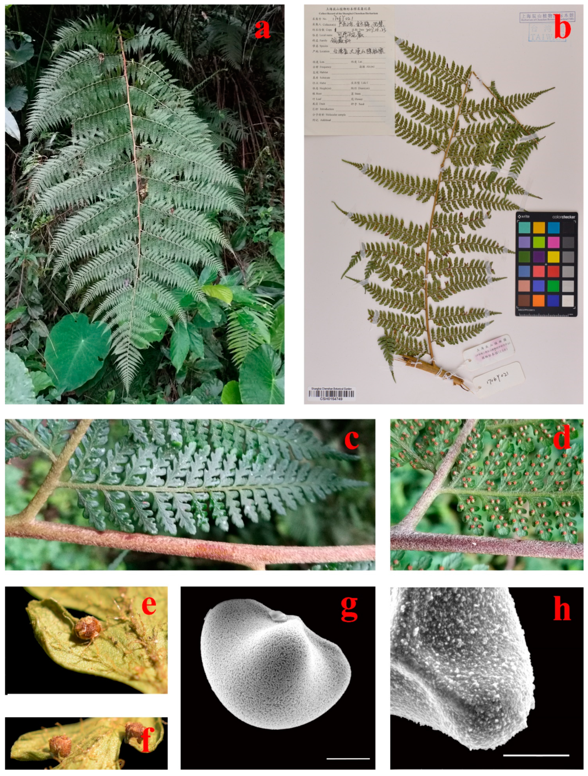
Figure 1. Morphological characters in Microlepia smithii. (a): M. smithii in the forest; (b): Herbarium specimen of M. smithii; (c): Ultimate-pinnule in live specimen; (d): Position and shape of sori in life specimen; (e,f): Indusia in dried herbarium specimen; (g): SEM of equatorial view of spore showing inner perispore reticulation with small areolae; (h): SEM of outer perispore showing granular. Scale bar in (g,h) = 10 μm.
Taxonomy 01 00019 g001
Figure 2. (A): Phylogenetic tree from ML and BI analyses of combined data from four chloroplast regions (rbcL, rps4, psbA-trnH and trnL-F). Both analyses have the same topology. Dennstaedtia smithii and Microlepia speluncae (the type of Microlepia) are marked in red text. Support values beside each node represent bootstrap support for ML (BSML) followed by posterior probabilities for BI (PPBI). Asterisks (*) indicate BSML = 100% and PPBI = 1.0. (B): The connection of costal grooves between rachis and pinna rachis of Microlepia and Dennstaedtia.
Taxonomy 01 00019 g002
Table 1. List of 54 Specimens information and GenBank accessions.
No.SpeciesVoucher No.LocalityHerbariumGenBank Accession No.
rbcLrps4trnL-FpsbA-trnH
1Microlepia. strigosaSG272Jiangxi, ChinaCSHMK051745MK051993MK052534MK052254
2M. strigosaYYH11609Taiwan, ChinaCSHMK051843MK052104MK052649MK052373
3M. khasiyanaZXL5742Yunnan, ChinaCSHMK051616MK052063MK052601MK052325
4M. khasiyanaZXL7194Yunnan, ChinaCSHMK051627MK052087MK052625MK052349
5M. obtusilobaWYD098Guangdong, ChinaCSHMK051755MK052006MK052547MK052267
6M. obtusilobaSG2854Hainan, ChinaCSHMK051664MK051913MK052443MK052163
7M. lofoushanensisWYD642Guangdong, ChinaCSHMK051675MK051924MK052454MK052174
8M. lofoushanensisWYD641Guangdong, ChinaCSHMK051674MK051923MK052453MK052173
9M. trichosoraWYD445Guangdong, ChinaCSHMK051855MK052110MK052662MK052386
10M. trichosoraWYD389Guangdong, ChinaCSHMK051829MK052091MK052635MK052359
11M. marginataWZS006Hainan, ChinaCSHMK051696MK051947MK052477MK052197
12M. marginataWYG156Guizhou, ChinaCSHMK051771MK052024MK052563MK052286
13M. szechuanicaWYG056Guizhou, ChinaCSHMK051677MK051926MK052456MK052176
14M. szechuanicaYanYH13825Sichuan, ChinaCSHMK051732MK051980MK052521MK052241
15M. rhomboideaWYD529Guangdong, ChinaCSHMK051763NAMK052555MK052278
16M. rhomboideaSG2641Yunnan, ChinaCSHMK051806MK052059MK052597MK052321
17M. yaoshanicaYYH12136Yunnan, ChinaCSHMK051834MK052095MK052640MK052364
18M. yaoshanicaWYD303Guangdong, ChinaCSHMK051667MK051916MK052446MK052166
19M. firmaZXL6895Yunnan, ChinaCSHMK051813MK052070MK052608MK052332
20M. firmaZXL6882Yunnan, ChinaCSHMK051812MK052069MK052607MK052331
21M. kurziiZXL7021Yunnan, ChinaCSHMK051815MK052080MK052618MK052342
22M. kurziiYYH12098Yunnan, ChinaCSHMK051631MK051874MK052404MK052124
23M. platyphyllaWYD609Guangdong, ChinaCSHMK051831MK052092MK052637MK052361
24M. platyphyllaYYH12394Yunnan, ChinaCSHMK051634MK051878MK052408MK052128
25M. hanceiYanYH13703Guangdong, ChinaCSHMK051642MK051886MK052416MK052136
26M. hanceiSG258Jiangxi, ChinaCSHMK051661MK051908MK052438MK052158
27M. todayensisINA-BL49Bali, IndonesiaCSHMK051733MK051981MK052242MK052242
28M. todayensisINA-BL44Bali, IndonesiaCSHMK051646MK051890MK052420MK052140
29M. speluncaeZXL09896Chiang Mai, ThailandCSHMK051795MK052048MK052587MK052310
30M. speluncaeYYH12379Yunnan, ChinaCSHMK051712MK051965MK052501MK052221
31M. hookerianaWYD218Guangdong, ChinaCSHMH289650MH289714MK052488MK052208
32M. hookerianaZXL5886Yunnan, ChinaCSHMK051810MK052064MK052326MK052602
33M. teneraKY1426Taiwan, ChinaNAMK051802MK052055MK052593MK052317
34M. teneraSG1026Yunnan, ChinaCSHMK051801MK052054MK052592MK052316
35DennstaedtiawilfordiiJSL2982Anhui, ChinaCSHMK051796MK052049MK052588MK052311
36D. smithiiYan 1706Y021Taiwan, ChinaCSHMZ959179MZ983428MZ959174MZ983423
37D. smithiiYan 1706Y008Taiwan, ChinaN/AMZ959180MZ983429MZ959175MZ983424
38D. appendiculaZhangXC5294Tibet, ChinaPEMK051807MK052060MK052598MK052322
39D. scabraYYH12150Yunnan, ChinaCSHMH289649MH289713MK052490MK052210
40D. scabraYYH11627Hainan, ChinaCSHMK051705MK051958MK052489MK052209
41D. hirsutaSG159Fujian, ChinaCSHMK051800MK052053MK052591MK052315
42D. punctilobulaN/AN/AN/AKP644118AY459159MT633781N/A
43D. scandensYYH16230Taiwan, ChinaCSHMH289628MH289707N/AN/A
44D. cornuta4374N/AN/AMT416335MT559747MT633779N/A
45D. spinosa5045N/AN/AMT416337MT593216MT633782N/A
46D. distenta4998N/AN/AMT633748MT559732MT633780N/A
47D. cicutaria3866N/AN/AMT633747MT593213MT633776N/A
48Leptolepia novae-zelandiae12400New ZealandDUKEEF463168N/AN/AN/A
49Leptolepia novae-zelandiaeP027279New ZealandN/AKT983829N/AN/AN/A
50Leptolepia novae-zelandiaeWolf 682New ZealandUTCU18639N/AN/AN/A
51Oenotrichia maximaP026233New CaledoniaN/AKT983830N/AN/AN/A
52Pteridium aquilinumBJZ003Guangxi, ChinaCSHMZ959183MZ983432MZ959178MZ983427
53Hypolepis punctataMS067Hunan, ChinaCSHMZ959182MZ983431MZ959177MZ983426
54Histiopteris incisaYYH11645Hainan, ChinaCSHMZ959181MZ983430MZ959176MZ983425
N/A = not available.
Publisher’s Note: MDPI stays neutral with regard to jurisdictional claims in published maps and institutional affiliations.