Article

Life-History Traits and Genetic Characterization of Polystoma borellii (Monogenea, Polystomatidae), a Parasite of Pleurodema borellii (Anura, Leptodactylidae)

by
Carolina Davies
1,*,
Juan José Lauthier
2,
Matías Martín Renfijes
3,
Ivanna Gabriela Cruz
4 and
Dora Davies
5
1
Instituto de Biología de Organismos Marinos (IBIOMAR)—Consejo Nacional de Investigaciones Científicas y Técnicas (CONICET), Puerto Madryn 9120, Argentina
2
Instituto de Medicina Traslacional e Ingeniería Biomédica (IMTIB), CONICET—Universidad Hospital Italiano—Hospital Italiano de Buenos Aires, Ciudad Autónoma de Buenos Aires C1199ACL, Argentina
3
Facultad de Medicina, Instituto de Biología Celular y Neurociencia “Prof. E. De Robertis” (IBCN), Universidad de Buenos Aires-CONICET, Ciudad Autónoma de Buenos Aires C1199ACL, Argentina
4
Museo de Ciencias Naturales, Facultad de Ciencias Naturales, Universidad Nacional de Salta, Salta 4400, Argentina
5
Instituto para el Estudio de la Biodiversidad de Invertebrados, Facultad de Ciencias Naturales, Universidad Nacional de Salta, Salta 4400, Argentina
*
Author to whom correspondence should be addressed.
Parasitologia 2025, 5(2), 17; https://doi.org/10.3390/parasitologia5020017
Submission received: 10 March 2025 / Revised: 21 March 2025 / Accepted: 10 April 2025 / Published: 17 April 2025

Abstract

:
The genus Polystoma includes parasites with direct life cycles that involve a short-lived free-living, aquatic oncomiracidia, post-larvae infecting the gill chambers of tadpoles, and adults parasitizing the urinary bladder of adult anurans. Despite the high diversity of anurans in the South American sub-continent, less than 20 species of Polystoma have been reported to date, and, particularly, in Argentina, only five species have been described from adult frogs. The aim of this work was to describe and characterize taxonomically the specimens found parasitizing tadpoles and frogs living in a well in Chicoana, Salta province, Argentina. Parasites were observed under optical and scanning electron microscopy and characterized genotypically by sequencing ITS1 and COI fragments. Frogs and tadpoles were characterized by morphology and sequencing a partial fragment of the cytochrome B region, confirming that the hosts corresponded to Pleurodema borellii. Given their morphology and the strict specificity of Polystoma species for their hosts, the identity of the parasites was established as P. borellii. The morphology of oncomiracidia and post-larvae was described, expanding adults’ description with insights provided by COI and ITS1 molecular analysis. The present work summarizes a complete description of the life cycle, with the genetic characterization of Polystoma borellii in Salta, Argentina.

1. Introduction

The current context of biodiversity loss has many facets, from climate change to the overexploitation of natural resources and the illegal trade of wildlife [1,2]. In this sense, wildlife trade also comprises the hidden diversity of parasites that is surreptitiously transported to new places with their hosts, including aquatic ornamental species [3]. This situation highlights the need to know the parasitic fauna of wildlife animals, since these species not only hold information about themselves but also provide clues about their co-evolution with their hosts. With a few exceptions, polystomatids are parasites of aquatic and semi-aquatic tetrapods which have a direct life cycle and high specificity towards their hosts. They belong to the family Polystomatidae, which comprises more than 200 species [4]. In particular, the genus Polystoma has been cited in the scientific literature since the 1800s, having its life cycle described by the end of that century [5]. Even though polystomatids have direct life cycles, Polystoma and Metapolystoma developed an additional step in their transmission pathway, showing two phenotypes depending on the eco-physiological stage of their host: the post-larva or branchial phenotype found in the gills of tadpoles, and the adult or bladder phenotype in the urinary bladder of adult anurans [5,6,7]. Briefly, the adult parasite lays eggs that are flushed out with the urine of its hosts; then, the oncomiracidium hatches from the eggs, and follows one of two development pathways: if it infects a tadpole in pro-metamorphosis, it attaches to the gills and slowly develops, migrating towards the urinary bladder as the tadpole completes its metamorphosis. On the contrary, if the oncomiracidium infects a tadpole in pre-metamorphosis, it quickly develops into the branchial phenotype, reproduces sexually, and dies when the gills are reabsorbed. Additionally, it can also directly infect the urinary bladder of the adult anuran [5,6,7,8] (Figure 1).
The taxonomic classification of polystomatids is based on the morphology (of the haptor or attachment organ, describing the number of suckers and hooks, the digestive tract, male and female genitalia, including the number of testicles, vaginal apertures, and length of the uterus) [7] and, in recent years, genetic markers. To differentiate species within genera, the main diagnostic characteristics are the haptoral hooks’ morphometry and host systematics. In particular, the genus Polystoma Zeder, 1800 has a haptor with three pairs of suckers and one pair of hooks (hamuli); the adults are found in the urinary bladder of frogs [5].
Polystoma comprises more than 60 species, of which 14 were reported from South America [9,10]. Out of these, only five species have been described in adult frogs from Argentina: P. borellii Combes and Laurent, 1974 from Pleurodema borellii [11]; P. andinum Combes and Laurent, 1978 from Melanophryniscus rubriventris [12]; P. praecox Combes and Laurent, 1978 from Telmatobius oxycephalus [12]; P. guevarai Combes and Laurent, 1979 from Boana riojana (formerly Hyla pulchella); and P. lopezromani Combes and Laurent, 1979 from Trachycephalus typhonius (formerly Phrynohyas venulosa) [5]. In spite of this diversity, there have been no new reports of Polystoma from Argentina in recent years, with the exception of a study on the morphological variation in P. andinum [13]. Particularly, the P. borellii parasite of Pl. borellii, a frog with wide distribution in north and central Argentina [14,15], has not been the subject of any new update since its description in 1974. In this context, the aim of the present work was to provide new information about the life cycle, morphology, and molecular markers of adults and larvae of Polystoma borellii.

2. Materials and Methods

2.1. Collection and Treatment of Hosts and Parasites

Hosts were collected from an artificial well in a rural area of the Chicoana department, province of Salta, northwest Argentina (24°08′08″ S, 65°36′19″ W), at 1484 m a.s.l. The average temperature in this area is 17 °C (6–36), 20–80% relative humidity, and rainfall higher than 800 mm annually. The vegetation is semi-jungle, with anthropic impacts, since raspberries and citrus fruits are grown around the well.
Tadpoles (N = 188) were sampled during Spring and Summer 2023, maintained in dechlorinated and aerated water, and fed with lettuce until their sacrifice and dissection. Tadpoles at the metamorphic climax (N = 9) were kept in individual containers until they completed the metamorphosis, and at that moment, they were euthanized to search their urinary bladder for parasites. In addition, adult frogs (N = 4) were collected during Spring 2023 and kept in individual containers with water from their original environment. Tadpoles and adults were euthanized with benzocaine prior to dissection. The gills of the tadpoles and the urinary bladder of metamorphic frogs and adults were examined under a stereoscopic microscope, and once found, monogeneans were carefully removed. Two pools of polystomid post-larvae were kept in 99% ethanol until their use for DNA extraction, while the rest were processed according to the requirements of different techniques, as follows.

2.2. Optical Microscopy

Mature (N = 6) and immature (N = 12) post-larvae were placed between a slide and coverslip and observed in vivo directly under an optical microscope (Zeiss 47-30-28, Oberkochen, Germany). Additionally, a hydrochloric carmine staining protocol was carried out on ovigerous post-larvae (N = 8) and an adult (N = 1) to measure the length and width of the body, haptor with its suckers, oral sucker, pharynx, ovary, and eggs. The analysis of the haptor hooks, the branches of the digestive tube, and the genital spines was carried out on post-larvae (N = 4) mounted on Hoyer’s medium [16,17]. Haptoral suckers and haptor hooks were measured for each specimen, and we reported the average for the total number of specimens. All measurements were expressed in micrometers (µm) with the mean ± standard deviation and the minima and maxima within parentheses. Drawings were made using a camera lucida.
Voucher specimens were deposited in the collection of the Museo de Ciencias Naturales de Salta “Miguel Ángel Arra” (MCN, Natural Sciences Museum of Salta). MCNSInv05 (adult), MCNSInv06 (post-larva), MCNSInv07 (post-larva), MCNSInv08 (oncomiracidium), MCNSHe 334 (tadpole), and MCNSHe 335 (frog).

2.3. Scanning Electron Microscopy (SEM)
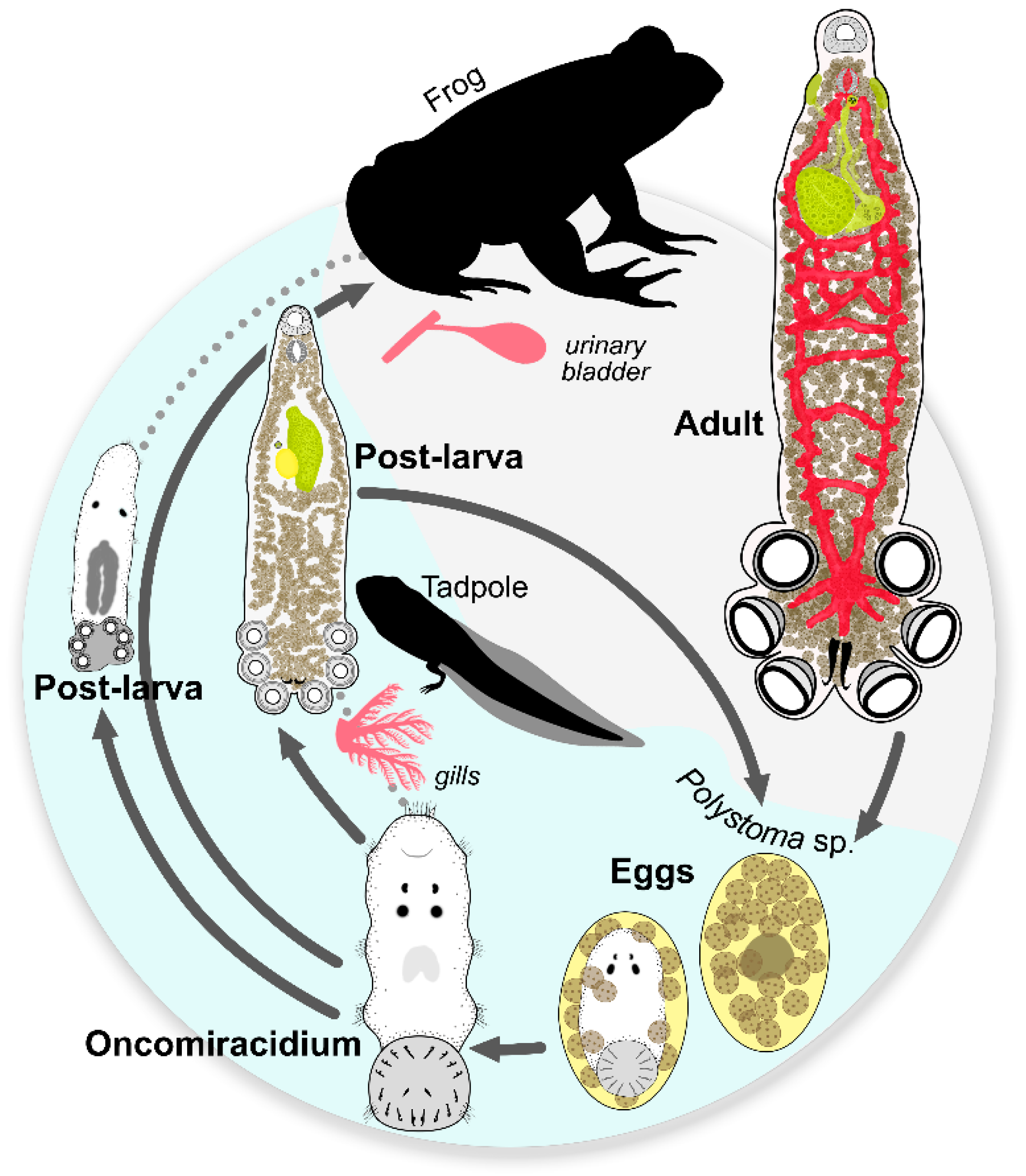
Ovigerous post-larvae (N = 2), and 1 adult were submerged in hot water, fixed with 10% formalin, and stored in 70% ethanol until use. For SEM analysis, they were dehydrated in solutions with increasing ethanol concentration, dried by critical point, coated in gold, and observed with Zeiss Supra 55VP SEM.

2.4. Life Cycle

Ovigerous post-larvae (N = 4) were placed individually in small Petri dishes for two hours with dechlorinated water; after that time, free eggs in the water were counted and measured. Additionally, the water in which adult frogs were kept was filtered through a 125 µm sieve. The retained eggs were measured and incubated in dechlorinated water at room temperature (19–26 °C). Eggs were then placed in excavated slides to follow their development and observed under an optical microscope until hatching. After hatching, the oncomiracidia were observed in vivo or stained for microscopy analysis.

2.5. Molecular, Phylogenetic, and Distance Analyses

DNA extraction was performed from two pools of post-larvae (N = 8 each) using the ThermoFisher PureLink® Genomic DNA Mini kit (Waltham, MA, USA) following the manufacturer’s protocol. The nuclear ribosomal internal transcribed spacer 1 (ITS1) and the mitochondrial cytochrome oxidase subunit 1 (COI) were amplified using the following primer sets: ITS1 (forward) 5′-ACC TGG TTG ATC CTG CCA GTA G-3′ and (reverse) 5′-TAC CGG TGT ACT ATT TAG CAG-3′ [18]; COI (forward) 5′-TTT TTT GGG CAT CCT GAG GTT TAT-3′ and (reverse) 5′-TAA AGA AAC AAC ATA ATG AAA ATG-3′ [19]. Each PCR was performed using TaKaRa Ex Taq DNA polymerase, hot start version (Takara-Bio, Kusatsu, Japan) following the manufacturer’s instructions. The cycling conditions were as follows: initial denaturation at 94 °C for 3 min, 35 cycles of denaturation for 1 min at 94 °C, annealing for 1 min at 45.7 °C, extension for 1 min at 72 °C, and final extension at 72 °C for 5 min. The amplification products were visualized on 1% agarose gels under ultraviolet light, purified, and sequenced using an Applied Biosystems Hitachi 3110 Genetic Analyzer. Additionally, to confirm the hosts’ identity, DNA extraction from parasitized tadpoles and frogs was carried out following Aljanabi and Martínez [20]. A partial fragment of cytochrome b was amplified using primers MVZ15/MVZ16 [21] and sent to Macrogen, Korea, for standard sequencing. The obtained sequences were edited, assembled, and compared to those available in GenBank using the Standard Nucleotide Search without further analysis.
Sequences from Polystoma specimens were edited in Chromas Lite v.2.6.6 and compared with GenBank sequences using nucleotide Basic Local Alignment Search Tool (BLAST, blastn suite v. 2.15.0) (https://blast.ncbi.nlm.nih.gov/Blast.cgi, accessed on 20 January 2024). Sequences of the molecular markers 18S, 28S, ITS1, and COI for 19 polystomatid species were obtained from GenBank (Table 1) and aligned with Clustal W [22] in BioEdit v.7.0.5.3 [23] under default parameters. For Bayesian inference (BI) analysis, the nucleotide substitution model with the best fit to these data sets was determined using MEGA software v12 [24], which held the jModeltest 2.1.1 [25], with model selection based on the Akaike information criterion (AIC). Models with the lowest Bayesian information criterion (BIC) scores were considered to describe the best substitution pattern. Non-uniformity of evolutionary rates among the sites were modeled using a discrete gamma distribution (+G) with 5 rate categories and by assuming that a certain fraction of sites was evolutionarily invariable (+I). For BI, the substitution model was 4 by 4, while the rate variation across sites was fixed, depending on the gene fragment analyzed. Using MrBayes 3.2 [26], four Markov Chain Monte Carlo (MCMC) chains were run for 10,000,000 generations, sampling every 1000 generations, with the first 25% sampled trees discarded as “burn-in”. Finally, a 50% majority rule consensus tree was constructed. The pairwise distance (p-distance) analysis of COI and ITS1 sequences among relatives of P. borellii was carried out in MEGA12 [24].
Procedures related to the sampling and manipulation of animals were approved by the “Secretaría de Ambiente y Desarrollo Sustentable, Ministerio de Producción y Desarrollo Sustentable, Salta, Argentina” (Environment and Sustainable Development Secretariat, Ministry of Production and Sustainable Development, Province of Salta, Argentina, Authorization No. 00203/23).

3. Results

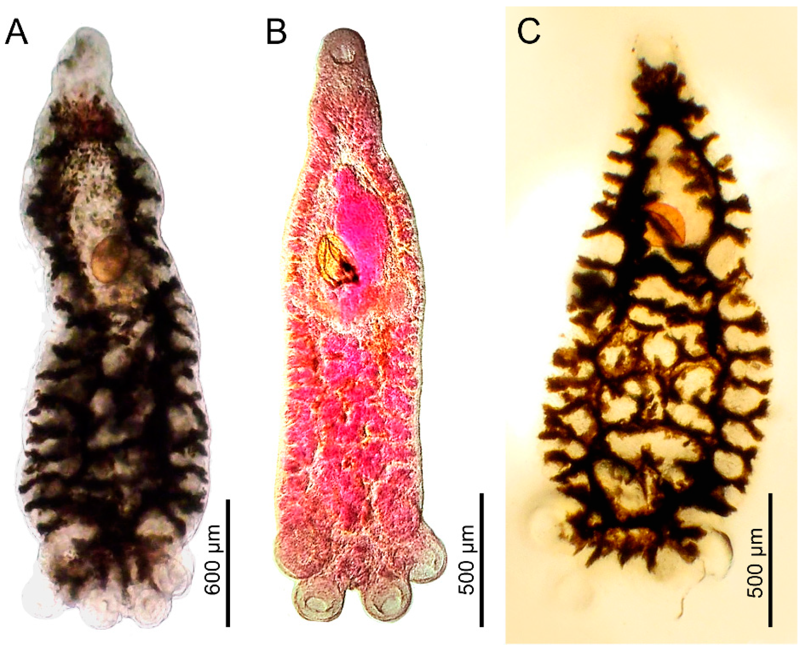
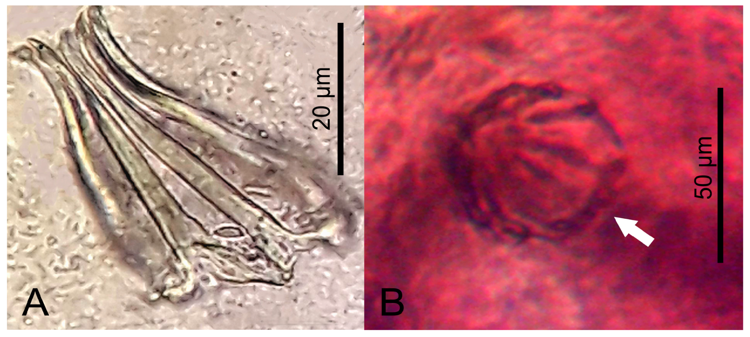
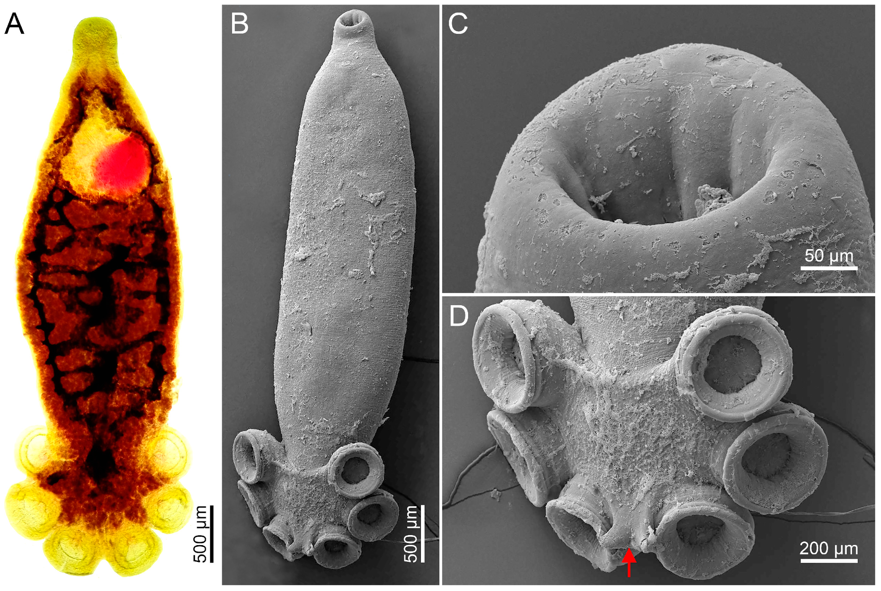
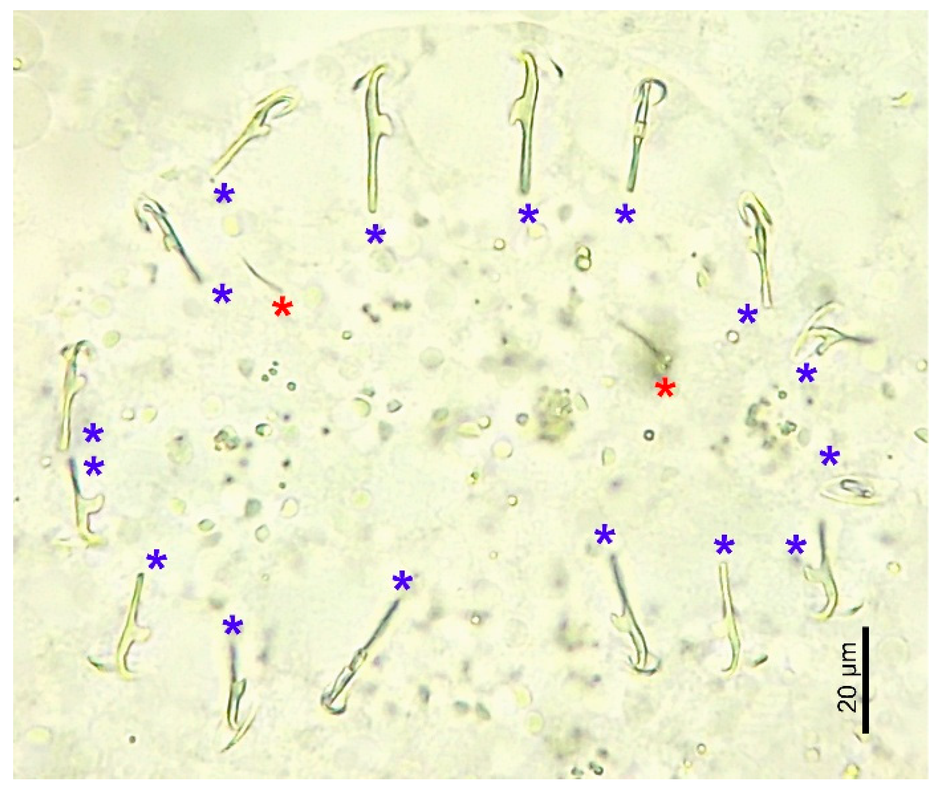
3.1. Morphological Analysis by Optical Microscopy and SEM

Descriptions of the post-larvae (Figure 2C, Figure 3 and Figure 4) were based on ovigerous specimens: six of them observed in vivo, eight carmine-stained, one observed with SEM, and four mounted with Hoyer’s medium, in addition to immature post-larvae observed in vivo (N = 12). Body measurements were based on ovigerous individuals stained with carmine, while hook measurements were based on specimens mounted in Hoyer’s medium. Figure 2(C1), Figure 3A and Figure 4 show that the body is elongated and the maximum width occurs at the level immediately anterior to the haptor. In the ovigerous post-larvae, there are six haptoral suckers (Figure 2(C1) and Figure 3C) and a pair of hamuli. Its external and internal roots have numerous longitudinal striations and lack a dividing notch between them (Figure 2(C4)).
In very small, immature post-larvae, two hamuli, and 16 hooklets can be distinguished. The pair of hooklets 1 and 2 is observed posterior to the hamuli, pairs 3 to 5 lateral to the haptor, and pairs 6 to 8 anterior to the haptor, between the third pair of suckers; these can also be observed in ovigerous post-larvae (Figure 3C), while the hooklets 3 to 5 progressively become embedded in the developing suckers. There are two pairs of pigmented eyes in the pre-pharyngeal region, the anterior pair smaller than the posterior one. The oral sucker has numerous glands and papillae arranged around the opening (Figure 3B). The mouth is sub-terminal, and the pharynx is almost spherical.
The intestine is bifurcated with numerous lateral ramifications on both sides of the main branches and has pre-haptoral anastomoses. The branches join immediately before the haptor and penetrate it (Figure 4A,C).
The ovary is in the left field of the anterior half of the body, with smooth edges, curved posteriorly. The uterus is short, with no more than one egg, without vagina (Figure 4A–C). There are vitelline glands distributed from the pharyngeal region to the interior of the haptor; arranged in the lateral fields up to the level of the posterior border of the ovary, posteriorly to the ovary, they cover almost the entire body (Figure 4B). Testicles are not observed. The genital bulb is located at the level of the anterior third of the ovary and has a crown of spines (Figure 2(C1–C3) and Figure 5A).
We hereby present an expanded description of the adult, shown in Figure 2D, Figure 5B and Figure 6. The description and measurements are based on one specimen stained with carmine and one observed with SEM. The body is elongated (Figure 2D and Figure 6A). The haptor has three pairs of suckers and one pair of hamuli (Figure 2D and Figure 6B). The oral sucker does not have noticeable glands or papillae (Figure 6C), and the pharynx is oval. The intestinal branches has numerous ramifications: the external ones are short and the internal are long, some of which are anastomosed (Figure 6A). The ovary is obliquely arranged and the uterus is long and sinuous. There are two vaginas with not very prominent mamelons at the genital pore level (Figure 2D and Figure 6A). The genital bulb is located immediately posterior to the intestinal bifurcation and has a crown of spines (Figure 2D and Figure 5B).
The egg description was based on N = 8 observed in the uterus of post-larvae stained with hydrochloric carmine, eggs released by post-larvae observed in vivo (N = 8), and eggs laid by the adults (N = 10), observed in vivo in water recovered from adult frogs’ containers. The eggs are elliptical, yellow, not embryonated, without filaments, and operculated (Figure 2(A1) and Figure 4B).
The oncomiracidium was described based on four specimens observed in vivo and three stained with hydrochloric carmine (Figure 2B and Figure 7). The latter were also used for body measurements. The body is cylindrical with five ciliated areas (one anterior, three lateral, and one lateral–posterior) and two pairs of pigmented ocelli (Figure 2(B1)). The haptor has sixteen marginal hooklets (Figure 2(B2)): three anterior pairs, three lateral pairs, and two posterior pairs. There is a pair of small hamuli (Figure 7).
Table 2 summarizes the body measurements for each developmental stage of P. borellii specimens described in the present study, as well as the measurements of the adult in the first report of the species by Combes and Laurent, in 1974 [11].

3.2. Life Cycle

The egg production rate for post-larvae was estimated at 6–54 eggs/parasite/day, with each specimen producing between 0.24 and 2.25 eggs/h. In addition, the time elapsed between successively laid eggs was measured in four post-larvae, resulting in an average of 31 min between one egg laid and the next (N = 11 eggs). The adult frog parasitized by two adult specimens laid several eggs/day, and it was possible to observe oncomiracidia developing inside the eggs. On day 1, the zygote and numerous vitelline cells were visible, while on day 10, it was possible to distinguish the ocelli and the haptor with its 16 hooklets. On day 14, the oncomiracidium was completely formed, presenting two small hamuli and five ciliated bands just before hatching (Figure 2(A1–A3)).
From nine tadpoles at their metamorphosis climax held in individual containers, two were parasitized with small sub-adults found in their urinary bladder. In one of these metamorphic tadpoles, there were three parasites, while in the other, there were five; thus, the mean infection intensity was four.

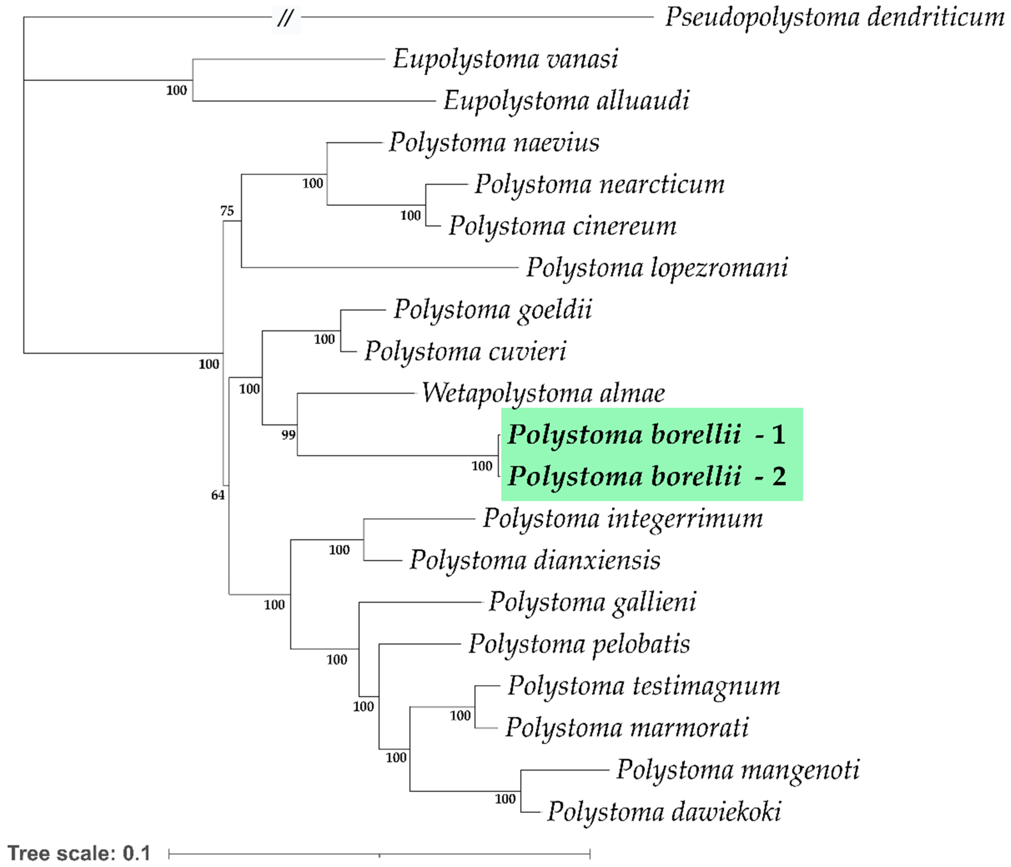
3.3. Molecular, Phylogenetic, and Distance Analyses

The hosts’ identity was confirmed with a morphological analysis of adult frogs and the genetic analysis of parasitized tadpoles and adult frogs. The nucleotide search in GenBank of the partial fragment of cytochrome b resulted in 100% similarity with sequences of Pleurodema borellii (GenBank accession number JQ937113).
The post-larvae samples had identical sequences for the 274 bp COI fragment (deposited in GenBank with accession numbers OK489454 and OK489455) and were also identical regarding the 597 bp ITS1 marker (OK491390 and OK491391). For BI analysis, the nucleotide substitution model selected by JModelTest was GTR + G + I. Tree topologies suggest that P. borellii is a sister group of Wetapolystoma almae. In turn, this clade is closely related to the group formed by P. cuvieri and P. goeldii, while P. lopezromani (another Polystoma from South America with sequences available in GenBank) is related to the clade containing Nearctic species (P. naevius, P. nearcticum, and P. cinereum) (Figure 8).
The p-distance among COI sequences of P. borellii and its closest relatives ranged between 14.2% and 16.6%, whereas the ITS1 range between 13.2% and 14.7%, as shown in Table 3.

4. Discussion

In the present study, we found the morphological characteristics corresponding to the branchial and bladder phenotypes of Polystoma, i.e., the latter having two vaginas and a long uterus, while the former is without a vagina and has a short uterus. These characters are in agreement with the findings from other authors for species of this genus [5,6]. However, since there were no molecular data available to assign a specific taxon to the Polystoma specimens described here, we used morphological and molecular analysis to identify their hosts. Thus, given the strict host-specificity of Polystoma, the fact that the parasitized adult frogs and tadpoles were characterized as Pleurodema borellii, and the morphological characteristics of the Polystoma found herein, we inferred that the specimens here described were Polystoma borellii.
Morphologically, the ovigerous post-larvae were smaller than the adult, reaching no more than 53% of the adult’s body length. The gripping structures were bigger in the adult than in the post-larvae, evidenced by the haptor, which was 31% of the adult’s body length vs. 22% in the larva. Noteworthy, the post-larvae had two pairs of ocelli and glands and papillae arranged in a circle over the oral sucker that were absent in the adult, as shown by SEM in Figure 2 and Figure 5.
We also found some morphological differences with the specimens described by Combes and Laurent [11] in Tucumán, type locality being about 200 Km south from the site of the present work. For example, the adults here described were thinner (1.3 vs. 2.3–3.2), had six pre-haptor intestinal anastomoses in contrast to an anastomosed net, and ten genital spines instead of the eight described previously. These contrasts could be due to intra-specific variation, different fixing and staining protocols, or the developmental stage of the adults, as observed by Rubstova and Heckmann [27]. These authors reported morphological variations in P. integerrimum mature adults and immature sub-adults isolated from the same host collected in Ukraine and Lithuania. They also observed that the intestinal caecum occupied the same position in adults and sub-adults, in agreement with our observations of adults and post-larvae. Similarly, Vaira [13] observed low variation in the shape of the intestinal anastomoses in different populations of P. andinum, suggesting that this character has limited taxonomic value. However, Rubstova and Heckmann [27] proposed that the shape and number of spines in the copulatory pore is fixed and therefore could be used as a taxonomic character. In this sense, other neotropical species such as P. andinum, P. cuvieri, P. knoffi, and P. travassosi have eight spines, P. goeldii has nine, and P. stellai has twenty-one, while for other species, such as P. diptychi, P. guevarai, P. lopezromani, P. naevius, P. napoensis, P. touzeti, and P. praecox, it is not reported [10]. The observation of 10 spines in the genital pore in both the adult and post-larvae described here contrasts with the description of Combes and Laurent [11]. Along with the other morphological differences mentioned earlier, these suggest the need for further studies to establish morphological and genetic variation within P. borellii species.
Combes and Laurent [11] documented variations in the shape of the hamuli in different P. borellii specimens, reporting hooks without notches between the guard and handle or hooks with profound notches. Vaira [13] also found differences in the shape of hamuli of P. andinum adult specimens from different northwest Argentine localities. He proposed that, given the little variation in the spike length, this character could be used for species identification. In the present work, there were differences in the hamuli of the post-larvae, even though they always showed longitudinal striations throughout the guard and handle area. These striations were not observed in the adults described here or in those described by Combes and Laurent [11].
Additionally, one adult specimen had no eggs in the uterus, and the other had only one, whereas the adults described by Combes and Laurent [11] had several eggs in the uterus. The number of eggs in an individual depends on its developmental stage, infection intensity, light, and temperature, among other factors [28,29]. It also depends on the season of material collection, since the reproductive activity of Polystoma is synchronized with the sexual activity of their hosts. In this regard, Rubtsova and Heckmann [27] documented the maximum number of eggs inside P. integerrimum during the host’s reproductive season in the Spring. Studies on P. umthakathi showed that egg production was low in young specimens who had just started to produce them (around day 16 post-infection), while it rose and stabilized at 5 or 6 days after the first laying. In the same work, it was reported that egg production was also linked to infection intensity (I): since when there was only one parasite (I = 1), egg production was at maximum (15 eggs/parasite/day), while egg production decreased as infection intensity increased (14 eggs/parasite/day at I = 2, and 5 eggs/parasite/day at I = 3) [29]. Sales et al. (2023) [10] also found a great variability in egg production rate for the neotropical P. goeldii (1 to 325 eggs/day). Thus, the adults described here may correspond to young specimens.
The Bayesian inference analyses using COI, ITS1, 18S, and 28S recovered P. borellii in the same clade with Wetapolystoma almae, while P. cuvieri and P. goeldii grouped together in a related clade. It is worth noting that P. lopezromani, the other species known from South America, has never been recovered in this clade, suggesting that at least two lineages of Polystoma coexist in South America ([10,30,31], this study). Additionally, given its phylogenetic position in different studies (e.g., Refs. [10,30,31], this study), the DNA samples identified in GenBank as W. almae from French Guiana belong to the genus Polystoma. However, it must be considered that W. almae was originally described parasitizing Rhinella margaritifera in Cusco, Peru [32], based on morphological characters, while the DNA sequences in GenBank come from specimens collected in French Guiana [31]. Given the long geographical distance between these localities and the complex taxonomy of Rhinella margaritifera [33], we consider that the taxonomic identity of Wetapolystoma (Gray, 1993) must be evaluated in future studies. Finally, our results recovered the Nearctic clade grouping P. naevius, P. cinereum, and P. nearticum and the clade formed by the species P. testimagnum, P. marmorati, P. dawiekoki, and P. mangenoti that parasitize frogs from the African continent, while the species from Europe (P. pelobatis, P. gallieni, and P. integerrimum) were not recovered as a monophyletic group ([10,30], this study).
Following standard practices in flatworm systematics, we used mitochondrial (COI) and nuclear (ITS1) markers to explore low and high taxonomic levels [34]. The minimum genetic distances obtained for COI among P. borellii relatives were found between P. cuvieri and P. goeldii (2.6%), while P. borellii presented p-distances higher than 14% with its closest species, but the ITS1 ranged between 12.6% and 14.7% among the species of this clade. These values were equal to or higher than those proposed by Du Preez et al. [35] to differentiate at the species level. These results highlight the need for more integrative taxonomic studies on polystomatids from South America.

5. Conclusions

The present work updates the characterization of Polystoma borellii with the description of each stage of its life cycle, presenting morphological information about the eggs, the oncomiracidia from its development inside the egg to its short period as a swimming larva, the post-larvae in the gills of tadpoles, and the adults in the urinary bladder of froglets and frogs. Additionally, we provided partial sequences of COI and ITS1 markers and widened the geographical distribution of the species 200 km further north from the type locality.

Author Contributions

Conceptualization, D.D. and C.D.; methodology, J.J.L., M.M.R., I.G.C., C.D. and D.D.; formal analysis, C.D. and J.J.L.; writing—original draft preparation, C.D.; writing—review and editing, D.D.; visualization, I.G.C. and J.J.L.; supervision, D.D.; project administration, D.D.; and funding acquisition, D.D., J.J.L. and C.D. All authors have read and agreed to the published version of the manuscript.

Funding

This research was funded by the Research Council of Salta National University (CIUNSa), Grant 2481/0.

Institutional Review Board Statement

The study was conducted in accordance with the Animal Care Guidelines for Herpetology developed by the American Society of Ichthyologists and Herpetologists in 2004 (https://www.asih.org/resources accessed during 20 January 2023), and the protocol was approved by the Environment and Sustainable Development Secretariat, Ministry of Production and Sustainable Development, Province of Salta, Argentina (Authorization #00203) on 17 March 2023.

Informed Consent Statement

Not applicable.

Data Availability Statement

The original contributions presented in this study are all included in the paper.

Acknowledgments

The present work was possible thanks to Lilian Davies and Norberto Bonini for the steady supply of tadpoles and frogs, the generous contribution of Diego Barrasso to their taxonomic identification, the technical help of Daniel Paredes, and the assistance of Gabriela Chauque and Carla Gómez with taking SEM photographs.

Conflicts of Interest

The authors declare no conflicts of interest.

Abbreviations

BIBayesian Inference
COICytochrome Oxidase I
ITS1Internal Transcribed Spacer 1

References

  1. Maxwell, S.L.; Fuller, R.A.; Brooks, T.M.; Watson, J.E.M. Biodiversity: The ravages of guns, nets and bulldozers. Nature 2016, 536, 143–145. [Google Scholar] [CrossRef]
  2. Mozer, A.; Prost, S. An introduction to illegal wildlife trade and its effects on biodiversity and society. Forensic Sci. Int. Anim. Environ. 2023, 3, 100064. [Google Scholar] [CrossRef]
  3. Schoeman, A.L.; Du Preez, L.H.; Kmentová, N.; Vanhove, M.P.M. A monogenean parasite reveals the widespread translocation of the African clawed frog in its native range. J. Appl. Ecol. 2022, 59, 2670–2687. [Google Scholar] [CrossRef]
  4. Verneau, O.; Bentz, S.; Sinnappah, N.D.; Du Preez, L.; Whittington, I.; Combes, C. A view of early vertebrate evolution inferred from the phylogeny of polystome parasites (Monogenea: Polystomatidae). Proc. Biol. Sci. Lond. Ser. B 2002, 269, 535–543. [Google Scholar] [CrossRef]
  5. Du Preez, L.H.; Landman, W.J.; Verneau, O. Polystomatid Flatworms: State of Knowledge and Future Trends, 1st ed.; Feldhaar, H., Schmidt-Rhaesa, A., Eds.; Springer: Cham, Switzerland, 2023; pp. 29–59, 209–369. [Google Scholar] [CrossRef]
  6. Combes, C. Biologie, écologie des cycles et biogéographie de Digènes et Monogènes d’Amphibiens dans l’est des Pyrénées. Mém. Mus. Natl. Hist. Nat. Sér. A Zool. 1968, 51, 1–195. [Google Scholar]
  7. Badets, M.; Verneau, O. Origin and evolution of alternative developmental strategies in amphibious sarcopterygian parasites (Platyhelminthes, Monogenea, Polystomatidae). Org. Divers. Evol. 2009, 9, 155–164. [Google Scholar] [CrossRef]
  8. Badets, M.; Du Preez, L.H.; Verneau, O. Alternative development in Polystoma gallieni (Platyhelminthes, Monogenea) and life cycle evolution. Exp. Parasitol. 2013, 135, 283–286. [Google Scholar] [CrossRef]
  9. Du Preez, L.H.; Dominguez, M.V. Polystoma knoffi n. sp. and Polystoma travassosi n. sp. (Monogenea: Polystomatidae): Naming museum-archived specimens from Brazil. Syst. Parasitol. 2019, 96, 755–765. [Google Scholar] [CrossRef]
  10. Sales, A.N.; Du Preez, L.; Verneau, O.; Domingues, M.V. Morphology and molecular characterization of Polystoma goeldii n. sp. (Monogenea, Polystomatidae) parasite from the urinary bladder of Physalaemus ephippifer (Steindachner) (Anura, Leptodactylidae). Parasitol. Int. 2023, 92, 102685. [Google Scholar] [CrossRef]
  11. Combes, C.; Laurent, R.F. Polystoma borellii n. sp. (Monogenea, Polystomatidae) parasite de Pleurodema borellii Peracca (Anura, Leptodactylidae) en Republique Argentine. Acta Zool. Lilloana 1974, 31, 57–64. [Google Scholar]
  12. Combes, C.; Laurent, R.F. Deux nouveaux Polystomatidae (Monogenea) de Republique Argentine. Acta Zool. Lilloana 1978, 33, 85–91. [Google Scholar]
  13. Vaira, M. Population morphological variation of the monogenean Polystoma andinum, parasitic in Melanophryniscus rubriventris (Anura, Bufonidae). Acta Parasitol. 2004, 49, 281–291. [Google Scholar]
  14. Ferraro, D.; Casagranda, M. Geographic distribution of the genus Pleurodema in Argentina (Anura: Leiuperidae). Zootaxa 2009, 2024, 33–55. [Google Scholar] [CrossRef]
  15. Vaira, M.; Akmentins, M.S.; Attademo, M.; Baldo, D.; Barrasso, D.; Barrionuevo, S.; Basso, N.; Blotto, B.; Cairo, S.; Cajade, R.; et al. Categorización del estado de conservación de los anfibios de la República Argentina. Cuad. Herpetol. 2012, 26, 131–159. [Google Scholar]
  16. Du Preez, L.H.; Maritz, M.F. Demonstrating morphometric protocols using polystome marginal hooklet measurements. Syst. Parasitol. 2006, 63, 1–15. [Google Scholar] [CrossRef]
  17. Du Preez, L.H.; Raharivololoniaina, L.; Verneau, O.; Vences, M. A new genus of polystomatid parasitic flatworm (Monogenea: Polystomatidae) without free-swimming life stage from the Malagasy poison frogs. Zootaxa 2010, 2722, 54–68. [Google Scholar] [CrossRef]
  18. Bentz, S.; Leroy, S.; Du Preez, L.; Mariaux, J.; Vaucher, C.; Verneau, O. Origin and evolution of African Polystoma (Monogenea: Polystomatidae) assessed by molecular methods. Int. J. Parasitol. 2001, 31, 697–705. [Google Scholar] [CrossRef]
  19. Littlewood, D.T.J.; Rohde, K.; Clough, K.A. Parasite speciation within or between host species? Phylogenetic evidence from site-specific polystome monogeneans. Int. J. Parasitol. 1997, 27, 1289–1297. [Google Scholar] [CrossRef]
  20. Aljanabi, S.M.; Martínez, I. Universal and rapid salt-extraction of high quality genomic DNA for PCR-based techniques. Nucleic Acids Res. 1997, 25, 4692–4693. [Google Scholar] [CrossRef]
  21. Moritz, C.; Schneider, C.J.; Wake, D.B. Evolutionary relationships within the Ensatina eschscholtzii complex confirm the ring species interpretation. Syst. Biol. 1992, 41, 273–291. [Google Scholar] [CrossRef]
  22. Thompson, J.D.; Higgins, D.G.; Gibson, T.J. CLUSTAL W: Improving the sensitivity of progressive multiple sequence alignment through sequence weighting, position–specific gap penalties and weight matrix choice. Nucleic Acids Res. 1994, 22, 4673–4680. [Google Scholar] [CrossRef]
  23. Hall, T.A. BioEdit: A user-friendly biological sequence alignment editor and analysis program for Windows 95/98/NT. Nucleic Acids Symp. Ser. 1999, 41, 95–98. [Google Scholar]
  24. Kumar, S.; Stecher, G.; Suleski, M.; Sanderford, M.; Sharma, S.; Tamura, K. MEGA12: Molecular Evolutionary Genetic Analysis Version 12 for Adaptive and Green Computing. Mol. Biol. Evol. 2024, 41, msae263. [Google Scholar] [CrossRef]
  25. Darriba, D.; Taboada, G.L.; Doallo, R.; Posada, D. jModelTest 2: More models, new heuristics and parallel computing. Nat. Methods 2012, 9, 772. [Google Scholar] [CrossRef]
  26. Ronquist, F.; Teslenko, M.; van der Mark, P.; Ayres, D.L.; Darling, A.; Höhna, S.; Larget, B.; Liu, L.; Suchard, M.A.; Huelsenbeck, J.P. MrBayes 3.2: Efficient Bayesian phylogenetic inference and model choice across a large model space. Syst. Biol. 2012, 61, 539–542. [Google Scholar] [CrossRef]
  27. Rubtsova, N.Y.; Heckmann, R.A. Morphological and structural differences of normal adult and sub-adult bladder forms of Polystoma integerrimum (Fröhlich, 1798) (Monogenea: Polystomatidae) from the common frog. Rana temporaria. Sci. Parasitol. 2017, 18, 38–53. [Google Scholar]
  28. MacDonald, S.; Combes, C. The hatching rhythm of Polystoma integerrimum, a monogenean from the frog Rana temporaria. Chronobiol. 1978, 5, 277–285. [Google Scholar]
  29. Kok, D.J. Polystoma umthakathi (Monogenea): Establishment, mortality and reproduction of neotenic parasites in experimentally infected Natalobatrachus bonebergi tadpoles. S. Afr. J. Zool. 1990, 25, 26–30. [Google Scholar] [CrossRef]
  30. Héritier, L.; Badets, M.; Du Preez, L.H.; Aisien, M.S.; Lixian, F.; Combes, C.; Verneau, O. Evolutionary processes involved in the diversification of chelonian and mammal polystomatid parasites (Platyhelminthes, Monogenea, Polystomatidae) revealed by palaeoecology of their hosts. Mol. Phylogenet. Evol. 2015, 92, 1–10. [Google Scholar] [CrossRef]
  31. Bentz, S.; Sinnappah-Kang, N.D.; Lim, L.H.S.; Lebedev, B.; Combes, C.; Verneau, O. Historical biogeography of amphibian parasites, genus Polystoma (Monogenea: Polystomatidae). J. Biogeog. 2006, 33, 742–749. [Google Scholar] [CrossRef]
  32. Gray, M.E. Wetapolystoma almae n. gen., n. sp. (Monogenea: Polystomatidae) Parasite of Bufo typhonius (Linnaeus, 1758) (Amphibia: Bufonidae) from Tropical Peru. Trans. Kans. Acad. Sci. 1993, 96, 181–185. [Google Scholar] [CrossRef]
  33. Pereyra, M.O.; Blotto, B.L.; Baldo, D.; Chaparro, J.C.; Ron, S.R.; Elias-Costa, A.J.; Iglesias, P.P.; Venegas, P.J.; Thomé, M.T.C.; Ospina-Sarria, J.J.; et al. Evolution in the genus Rhinella: A total evidence phylogenetic analysis of Neotropical True Toads (Anura: Bufonidae). Bull. Am. Mus. Nat. Hist. 2021, 447, 1–156. [Google Scholar] [CrossRef]
  34. Blasco-Costa, I.; Cutmore, S.C.; Miller, T.L.; Nolan, M.J. Molecular approaches to trematode systematics: ‘best practice’ and implications for future study. Syst. Parasitol. 2016, 93, 295–306. [Google Scholar] [CrossRef]
  35. Du Preez, L.H.; Verneau, O.; Gross, T.S. Polystoma floridana n. sp. (Monogenea: Polystomatidae) a parasite in the green tree frog, Hyla cinerea (Schneider), of North America. Zootaxa 2007, 1663, 33–45. [Google Scholar] [CrossRef]
Figure 1. General life cycle of Polystoma sp. The scheme shows the adult in the urinary bladder of a frog. The polystomid eggs are flushed with the frog’s urine into the water and develop into free-living oncomiracidium. This larva can directly infect the urinary bladder of a frog and develop into an adult or adhere to the gills of a tadpole. If the infected tadpole is in pro-metamorphosis, the post-larva slowly develops and migrates to the urinary bladder once the tadpole completes its metamorphosis. If the tadpole is in pre-metamorphosis, it quickly develops into an ovigerous post-larva that dies when its host completes the metamorphosis.
Parasitologia 05 00017 g001
Figure 2. Polystoma borellii life stages. (A1A3) Eggs. The sequence shows larval development on days 1, 10, and 14. (B1,B2) Oncomiracidum, and haptor hooklet. (C1C4) Ovigerous post-larva (branchial phenotype) with details of the crown of spines in the lateral view, apical view, and detail of the hamuli. (D) Adult.
Parasitologia 05 00017 g002
Figure 3. Ovigerous post-larvae of Polystoma borellii, SEM photographs. (A) Ventral view. (B) Oral sucker detail: white arrows show the glands, and yellow arrows show the papillae. (C) Haptor detail: the blue arrows show hooklets, and the red arrow shows the hamulus.
Parasitologia 05 00017 g003
Figure 4. Ovigerous post-larvae (branchial phenotype) of Polystoma borellii. (A). Specimen photographed in vivo, latero-dorsal view. (B). Hydrochloric carmine staining, ventral view. (C). Hoyer’s medium, ventral view.
Parasitologia 05 00017 g004
Figure 5. Crown of spines. (A) Spine crown of post-larvae, lateral view photographed in vivo. (B) Genital bulb of the adult with spine crown, latero-apical view of a hydrochloric carmine-stained specimen.
Parasitologia 05 00017 g005
Figure 6. Adult of Polystoma borellii (bladder phenotype). (A). Specimen stained with hydrochloric carmine, ventral view. (B). SEM photography, ventral view. (C). Oral sucker detail. (D). Haptor detail, the arrow shows the hamuli.
Parasitologia 05 00017 g006
Figure 7. Oncomiracidium hooks of Polystoma borellii photographed in vivo. Hooklets are shown with blue asterisks and hamuli with red ones.
Parasitologia 05 00017 g007
Figure 8. Bayesian inference tree showing the phylogenetic relationships of Polystoma borellii. Posterior probability values (percent) are shown.
Parasitologia 05 00017 g008
Table 1. Parasite species * included in the phylogenetic analysis, their host species, geographic origin, and GenBank accession numbers for partial COI, ITS1, 28S, and 18S sequences +.
TaxonHost SpeciesGeographic OriginGenBank Accession Number
COIITS128S18S
Pseudopolystoma dendriticumOnychodactylus japonicusJapanKR856180---FM992707FM992700
Eupolystoma alluaudiSclerophrys regularisKenyaFR667558AJ301695AM157199AM051066
Eupolystoma vanasiSchismaderma carensSouth AfricaFR667559---AM157200AM157185
Metapolystoma cachaniPtychadena longirostrisIvory CoastKR856163---FM897262FM897280
Polystoma borellii 1Pleurodema borelliiArgentinaOK489454OK491390------
Polystoma borellii 2Pleurodema borelliiArgentinaOK489455OK491391------
Polystoma cinereumDryophytes cinereusUSAAM913869---AM157211AM157188
Polystoma cuvieriPhysalaemus cuvieriParaguayAM913862AJ301691AM157203AM051068
Polystoma dawiekokiPtychadena anchietaeSouth AfricaAM913856AJ310405AM157204AM051069
Polystoma gallieniHyla meridionalisFranceJF699305AJ301687AM157205AM051070
Polystoma goeldiiPhysalaemus ephippiferBrazilOP537251---------
Polystoma mangenotiPtychadena superciliarisIvory Coast---AJ310408------
Polystoma dianxiensisRana chaochiaoensisChinaKR856167---KR856143KR856125
Polystoma integerrimumRana temporariaEuropeJF699306AJ301688AM157206AM051071
Polystoma lopezromaniTrachycephalus typhoniusParaguayAM913863AJ301690AM157207AM051072
Polystoma marmoratiHyperolius marmoratusSouth AfricaAM913858AJ310396AM157208AM051073
Polystoma naeviusSmilisca baudiniiCosta RicaAM913864---AM157209AM157187
Polystoma nearcticumDryophytes versicolorUSAAM913865AJ301692AM157210AM051074
Polystoma pelobatisPelobates cultripesFranceKR856168FR821519KR856144AM051076
Polystoma testimagnumStrongylopus fasciatusSouth AfricaAM913860AJ310397AM157217AM157194
Wetapolystoma almaeRhinella margaritiferaFrench GuianaAM913866AJ301693AM157220AM051081
* Species names follow the update of Du Preez et al. (2023) [5]. While the geographic origin maintains the GenBank information about the collection site, it may not match the species’ type location. + Missing data from sequences not available in GenBank are denoted with “---”.
Table 2. Adult, post-larva, and oncomiracidium measurements for P. borellii described in the present study and the adult described by Combes and Laurent, in 1974 [11]. The mean and standard deviation are shown, with minimum and maximum measurements in brackets.
AdultsPost-LarvaeOncomiracidia
CharacterThis study aCombes and Laurent, 1974 [11]This studyThis study
Body length (BL)50405100
(4200–5600)
1962 ± 414
(1440–2650)
259 ± 5.67
(255–265)
Body at maximum width 13252500
(2000–3200)
506 ± 82
(412–638)
96 ± 9
(86–103)
Haptor length (HL)15841500
(1300–1700)
425 ± 111
(265–658)
73 ± 6
(69–81)
Haptor width16422600
(2300–3200)
568 ± 88
(432–697)
83 ± 4
(81–88)
HL/BL ratio0.310.29 b0.16–0.280.28
Haptoral suckers666---
Haptoral suckers’ average length476533 c164 ± 26
(118–209)
---
Haptoral suckers’ average width457---164 ± 26
(118–209)
---
Hamulus length412430
(350–530)
96 ± 32
(51–137)
---
Internal root length------91 ± 29
(51–127)
---
External root length------96 ± 32
(51–137)
---
Hamulus tip length39---46 ± 8
(37–61)
---
Hooklet length------24 ± 2
(20–27)
---
Sickle length of hooklets------11 ± 1
(10–12)
---
Oral sucker length285----138 ± 29
(98–184)
---
Oral sucker width334---139 ± 30
(98–189)
---
Pharynx length236250
(229–274)
114 ± 20
(81–145)
---
Pharynx width177240
(200–285)
106 ± 17
(73–122)
---
Intestinal anastomoses6Reticulated4–6---
Ovary length (OL)638---367 ± 62
(265–461)
---
Ovary width (OW)511---133 ± 47
(42–184)
---
OL/BL ratio0.126---0.174–0.184---
Vagina220---
No. of genital spines10810---
Genital spine length---5435
(24–49)
---
Egg length
carmine-stained
---230164 ± 14
(147–184)
---
Egg width
carmine-stained d
---120120 ± 11
(103–135)
---
Egg length
in vivo
237 ± 9 (226–246)---193 ± 17
(157–226)
---
Egg width
in vivo
164 ± 13 (147–189)---162 ± 10
(147–179)
---
a Adult measurements in the present study were based on one specimen. b Calculated by us based on data by Combes and Laurent, 1974 [11]. c Average calculated based on the diameters of 3 pairs of suckers. d Measurements are based on the eggs present in the uterus of ovigerous post-larvae stained with hydrochloric carmine.
Table 3. Percentage of base differences per site from COI/ITS1 * sequences between P. borellii’s closely related species.
p-distances COI/ITSPolystoma borelliiPolystoma cuvieriPolystoma goeldii
Polystoma cuvieri14.2/14.7
Polystoma goeldii15.3/--2.6/--
Wetapolystoma almae16.4/13.213.3/13.412.6/--
* Missing data from ITS1 sequences are denoted with “--”.
Disclaimer/Publisher’s Note: The statements, opinions and data contained in all publications are solely those of the individual author(s) and contributor(s) and not of MDPI and/or the editor(s). MDPI and/or the editor(s) disclaim responsibility for any injury to people or property resulting from any ideas, methods, instructions or products referred to in the content.

