Skip Content
You are currently on the new version of our website. Access the old version .

Parasitologia

Parasitologia is an international, peer-reviewed, open access journal on parasitology published bimonthly online by MDPI.

All Articles (204)

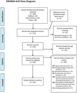
Xenomas are distinctive hypertrophic host–cell lesions caused by intracellular parasites and represent a recurrent pathological finding in wild and farmed fish. Their presence has implications for fish health, diagnostic workflows, aquaculture productivity, and in some cases product quality and consumer acceptability. Despite this relevance, information on xenoma diversity, associated pathogens, and diagnostic practices remains fragmented across decades of literature. This scoping review synthesised available evidence on naturally occurring xenomas in fish, following PRISMA-ScR guidelines. Eligible sources included studies reporting clinical, pathological, or diagnostic information on xenomas in fish. Data were charted on host species and families, taxonomic identification of the aetiological agents, xenoma morphology, and diagnostic approaches. Across 114 publications published between 1968 and 2024, xenomas were reported in a wide range of teleost families and were attributed mostly to microsporidian infections, particularly species of Glugea, Loma, Spraguea, Pleistophora, and Microgemma, although myxosporean-associated cases (Kudoa, Myxidium, Nephrocystidium) were also documented. Light and electron microscopy were the most frequently applied diagnostic methods, whereas molecular techniques were used less consistently, with increasing use in the most recent decade. Macroscopic xenomas were typically described as whitish, rounded to oval, and well delimited, yet substantial morphological variation occurred across hosts and tissues. Overall, the review highlights major heterogeneity in pathogen identification and diagnostic pathways, underscoring the need for more standardised and integrative approaches.

6 February 2026

PRISMA-ScR Flow Diagram.

A supplemental description of Lamproglena barbicola Fryer, 1961 is provided based on specimens collected from the gills of Labeobarbus altianalis (Boulenger, 1900) from the Nyando River, Lake Victoria Basin, Kenya, using an integrated approach of scanning electron microscopy (SEM) and molecular analysis (18S, 28S rDNA, and cox1 gene regions). Morphologically, the specimens conform to L. barbicola and closely resemble Lamproglena hoi Dippenaar, Luus-Powell & Roux, 2001; however, SEM revealed a previously undescribed feature on the uniramous antennule in L. barbicola, namely indistinctly three-segmented, tapering from a broad base to the apex, basal segment much longer than distal, comprising 14 setae of varying sizes, ventral laterally, absence of distinctive anterior fringe of setae on the antennule, as well as several characters that differentiate L. barbicola from L. hoi, including 5 setae at the basal endopod of leg one, five cuticular protuberances in the oral region, 19 setae on the basal antennular segment, and 10 setae on the distal segment, with 1 seta on each ramus. The phylogenetic analysis confirms L. barbicola as a sister taxon of L. hoi, supporting their close relationship. The genetic divergence presented as the uncorrected genetic p-distances between L. barbicola and L. hoi are 23.1% and 0.45% for cox1 and 28S rDNA regions, respectively, with observed nucleotide differences of 145 and 3 bp between the sequences, respectively. There was no interspecific variability detected in the 18S rDNA sequences. This study provides novel molecular sequences and the first high-resolution SEM images, which reveal additional taxonomic features for L. barbicola, establishing a robust reference for future identification.

3 February 2026

Scanning electron micrographs of Lamproglena barbicola Fryer, 1961, adult female ex Labeobarbus altianalis (Boulenger, 1900). (A)—ventral view of mature adult; (B)—ventral view of cephalothorax showing antennules, antennae, oral region, maxillae, maxillipeds, and first pair of legs; (C)—detailed antennae and antennules; (D)—maxilliped; (E)—first thoracic leg; (F)—second thoracic leg; (G)—third thoracic leg; (H)—fourth thoracic leg; (I)—fifth thoracic leg; (J)—caudal rami.

Prevalence and Risk Analysis of Human Geohelminths in Rural Communities of Ilalo, Ecuador

  • Fernando Pazmiño,
  • Gissela García and
  • Stefan Michael Geiger
  • + 1 author

Soil-transmitted helminthiases (STH) represent the most prevalent helminth infections worldwide and are closely associated with inadequate sanitation and poverty. During 2020 and 2021, a significant information gap emerged as a consequence of the SARS-CoV-2 pandemic. Therefore, the objective of this study was to provide an updated post-pandemic overview by estimating the prevalence of STH, identifying the etiological agents involved, and analyzing the risk factors associated with these infections. The study was conducted in Ilaló, Pichincha, Ecuador, where a total of 320 individuals were examined using three diagnostic methods: Kato-Katz, McMaster, and Mini-FLOTAC. Of these, 73 participants tested positive (22.81%, 95% CI: 22.3–23.4). The most frequently identified parasite was Ascaris lumbricoides (74.73%, 95% CI: 73.7–75.8). The main risk factors identified were pig farming (OR: 4.16; 95% CI: 2.34–7.42) and vegetable and fruit cultivation (OR: 11.66; 95% CI: 4.32–41.08). These findings provide relevant epidemiological data on STH in the region, demonstrating a reduction in infection prevalence likely associated with improved prevention and control measures implemented during the COVID-19 pandemic.

2 February 2026

Location of the communities of the Ilaló Strip, Pichincha Province, Ecuador.

Clonorchis sinensis and Metagonimus spp. are prevalent parasites in Korea, and accurate diagnosis is essential because treatment dosages differ between infections. However, their eggs are morphologically similar under light microscopy, making differentiation difficult and dependent on examiner expertise. To address this limitation, we evaluated an artificial intelligence (AI)-based automated microscope solution for the simultaneous detection and discrimination of both parasites. Microscopic images from 170 stool samples were analyzed using an AI system based on You Only Look Once version 5. The annotated dataset comprised 9455 egg images (6494 C. sinensis and 2961 Metagonimus spp.), randomly divided at the slide/patient level into training (6862), validation (1301), and test (1292) sets. Diagnostic performance was evaluated using mean average precision, confusion matrix analysis, and correlation with conventional microscopy. The model achieved a classification accuracy of up to 97.8%. C. sinensis showed higher recall and F1 scores, whereas Metagonimus spp. showed higher precision and specificity. Species identification showed complete concordance with conventional microscopy, and egg quantification was strongly correlated. These results indicate that the proposed AI system may serve as a supportive diagnostic tool comparable to conventional microscopy.

28 January 2026

Workflow map for dataset generation, training, and validation.

News & Conferences

Issues

Open for Submission

Editor's Choice

Get Alerted

Add your email address to receive forthcoming issues of this journal.

XFacebookLinkedIn
Parasitologia - ISSN 2673-6772