Article

Anticancer Activity of Cerium Oxide Nanoparticles Towards Human Lung Cancer Cells

by
Nithin Krisshna Gunasekaran
1,2,
Nicole Nazario Bayon
1,2,
Prathima Prabhu Tumkur
1,2,
Krishnan Prabhakaran
1,
Joseph C. Hall
1 and
Govindarajan T. Ramesh
1,2,*
1
Molecular Toxicology Laboratory, Center for Biotechnology and Biomedical Sciences, Norfolk State University, 700 Park Avenue, Norfolk, VA 23504, USA
2
Center for Materials Research, Norfolk State University, 555 Park Avenue, Norfolk, VA 23504, USA
*
Author to whom correspondence should be addressed.
Nanomanufacturing 2025, 5(2), 6; https://doi.org/10.3390/nanomanufacturing5020006
Submission received: 6 December 2024 / Revised: 12 March 2025 / Accepted: 24 March 2025 / Published: 3 April 2025

Abstract

:
Cerium oxide nanoparticles (CeO2 NPs) have gained significant attention in various fields, including biomedicine, semiconductors, cosmetics, and fuel cells, due to their unique physico-chemical properties. Notably, green-synthesized CeO2 NPs have demonstrated enhanced potential as drug carriers, particularly in biomedical applications such as anti-inflammatory, anticancer, antimicrobial, and anti-oxidant therapies. This study aimed to investigate the anticancer effects of cerium oxide nanoparticles synthesized using turmeric rhizomes on human lung cancer cells. The cytotoxicity and proliferation inhibition of these nanoparticles were assessed using MTT and Live/Dead assays, revealing a dose-dependent reduction in cell viability. Additionally, reactive oxygen species (ROS) generation was quantified through ROS assays, confirming oxidative stress induction as a key mechanism of cytotoxicity. Cell proliferation analysis further demonstrated that increasing concentrations of CeO2 NPs significantly reduced the multiplication of healthy lung cancer cells. These findings highlight the potential of turmeric-derived CeO2 NPs as a promising therapeutic agent for lung cancer treatment, warranting further exploration of their mechanism of action and in vivo efficacy.

1. Introduction

Nanoparticles play a crucial role across multiple scientific and technological domains, offering unique properties that enhance applications ranging from semiconductors to medicine. They have significantly contributed to semiconductor technology by improving thermal stability, conductivity, and optoelectronic applications, such as with quantum dots for display technologies [1]. Their synthesis, characteristics, and applications in areas like solar cells, LEDs, and miniaturized electronics have been widely explored, alongside the challenges associated with their large-scale integration [2]. The green synthesis of metal nanoparticles has emerged as an eco-friendly approach, utilizing biological agents for sustainable synthesis. These nanoparticles demonstrate significant potential in environmental remediation and medical applications, including antimicrobial and anticancer therapies [3]. Their classification and intrinsic properties have been examined, explaining how morphology, composition, and size-dependent behavior impact applications in drug delivery and advanced materials [4]. In the medical field, nanoparticles have been extensively researched for cancer therapy, particularly for targeted drug delivery and controlled release mechanisms, although concerns regarding their toxicity and large-scale production remain [5]. Nanomaterials are defined to be in the range of 1 to 100 nm, based on materials such as metals, ceramics, polymers, and several others [6]. Based on their distinct physical properties, nanomaterials have potential applications in various industries, such as cosmetics, electronics, biomedicine, and food processing [7]. In the pharmaceutical industry, various nanoparticles have been studied for their efficiency in being used as drug carriers to the target sites [8]. Nanoparticle synthesis methods are broadly categorized into physical, chemical, and biological approaches, each with its own advantages and challenges [9]. The physical methods include ball milling, laser ablation, and sputtering, which are effective for producing nanoparticles of a controlled size but often require high energy inputs [10]. Chemical methods such as sol–gel synthesis, chemical vapor deposition, and precipitation techniques offer greater control over nanoparticle properties and are widely used in industrial applications [11]. However, concerns regarding the environmental impact and toxicity of chemical synthesis byproducts have led to the development of green synthesis methods, which utilize biological sources like plants, bacteria, fungi, and algae, thereby providing cost-effective and biocompatible nanoparticles suitable for medical, agricultural, and environmental applications [12]. Additionally, the fabrication of nanocomposites integrates nanoparticles into different matrices, further enhancing their mechanical, electrical, and thermal properties for use in advanced engineering and biomedical fields [13]. Cerium oxide nanoparticles (CeO2 NPs) exhibit exceptional biomedical and industrial potential due to their unique redox properties, biocompatibility, and enzyme-mimicking behavior. These nanoparticles have shown promising applications in cancer therapy by selectively inducing oxidative stress in cancer cells while protecting normal cells, making them a valuable tool in oncology [14]. Their antioxidant capabilities play a significant role in mitigating oxidative stress-related diseases such as neurodegenerative disorders, cardiovascular diseases, and ischemic injuries [15]. Additionally, CeO2 NPs enhance biosensing technologies by improving sensitivity and stability in biosensors, facilitating medical diagnostics and environmental monitoring [16]. Their regenerative redox cycling between Ce3⁺ and Ce4⁺ contributes to their catalytic activity, which is leveraged in drug delivery systems, regenerative medicine, and antibacterial coatings [17]. Conventional methods are less effective in the treatment of cancer, and extensive research in the field of nanotechnology has been conducted to identify effective drugs to treat various types of cancer, which can be determined by the induction of oxidation stress in the cancer cells [18]. A nanoceria–curcumin conjugate was synthesized and shown to exhibit selective cytotoxic effects against cancer cells under oxidative stress conditions, a property attributed to the redox nature of cerium oxide, which oscillates between Ce3⁺ and Ce4⁺ states to enhance the antioxidant and pro-oxidant balance in tumor microenvironments [19]. Similarly, cerium–curcumin complexes were prepared and investigated for their anticancer effects under LED irradiation on MDA-MB-231 (breast cancer) and A375 (melanoma) cell lines, demonstrating enhanced therapeutic efficacy through photodynamic effects that further promote apoptotic pathways in cancer cells [20]. In this research, cerium oxide nanoparticles were synthesized using turmeric rhizomes and studied for their anticancer activities.
Cultured cells are used to analyze the cytotoxicity of nanoparticles using assays that use a dye-based component to measure viable cells with respect to the control and a fluorescent compound to observe the live and dead cells [21]. The MTT assay involves the use of 3-(4,5-dimethylthiazol-2-yl)-2,5-diphenyltetrazolium bromide dye, which is converted to formazan crystals when added to the cells [22]. In the LIVE/DEAD assay, the green component of the dye stains the live cells, in which the calcein acetoxymethyl is converted to a fluorescent calcein compound, and the red component of the dye, ethidium homodimer, stains the dead cells, indicated by cells taking up the red color [23]. The MTT assay is a widely used colorimetric method for assessing cell viability and cytotoxicity by measuring metabolic activity through the reduction of tetrazolium salts into formazan crystals [24]. It provides a quantitative evaluation of cell viability and is extensively utilized in pharmacological and toxicological studies to determine the cytotoxic potential of drugs in a dose-dependent manner [25]. On the other hand, the LIVE/DEAD assay is a fluorescence-based method that distinguishes live cells from dead ones using membrane-permeable and impermeable dyes [26]. This assay is particularly useful for real-time viability assessments in flow cytometry and fluorescence imaging [27].
It has been previously mentioned that a lack of antioxidants induces oxidative stress, which may be the cause of various diseases, including cancer [28]. Increased reactive oxygen species levels are observed in cancer cells because of the abnormal function of their mitochondria, their modification in metabolism, and their unstable genome [29]. The induction of oxidative stress in cells interferes with their function, causing the DNA and membrane of the cells to be damaged [30]. The pro-oxidant activity of cerium oxide nanoparticles shows an increased level of oxidative stress that leads to the death of cancer cells [31]. The anticancer activity of the drugs towards various cell lines has been studied using different kinds of viability assays and proliferation assays to assess their efficiency to be used for different biomedical applications [32]. In this study, cell viability assays such as MTT, Live/Dead, and ROS were conducted to assess the cytotoxicity of cerium oxide nanoparticles toward human lung cancer (H460) cells. A cell proliferation assay was further conducted to confirm the results of the viability assays.

2. Materials and Methods

2.1. Materials

Cerium nitrate hexahydrate (99.99%-Ce) was procured from Thermo Fisher Scientific (Rockford, IL, USA), turmeric rhizomes were brought from the fresh market (Norfolk, VA, USA), and Dulbecco’s Modified Eagle Medium (DMEM), Roswell Park Memorial Institute Medium (RPMI) and Dulbecco’s Phosphate-Buffered Saline (DPBS) solution were procured from VWR (Bridgeport, NJ, USA). Fetal Bovine Serum (FBS) and penicillin–streptomycin were obtained from Atlanta Biologicals (Atlanta, GA, USA). Trypsin, 3-(4,5-dimethylthiozol-2-yl)-2, 5-diphenyl tetrazolium bromide (MTT) dye, Live/Dead assay kits, ROS assay kits, and cell proliferation assay kits were purchased from Thermo Fischer Scientific (Rockford, IL, USA). Dimethyl sulfoxide (DMSO) was procured from Fisher Scientific. The human lung cancer (H460) cells were procured from ATCC (Manassas, VA, USA).

2.2. Experimental Section

The green synthesis of cerium oxide nanoparticles (CeO2 NPs) has gained significant attention due to its environmentally friendly and cost-effective approach. Among various plant-based methods, turmeric (Curcuma longa) rhizomes stand out as a promising biomaterial for synthesizing CeO2 NPs due to their high concentration of bioactive compounds, particularly curcumin, which serve as both reducing and stabilizing agents [33]. Additionally, when incorporated into a dextran-based amphiphilic nano-hybrid hydrogel system, turmeric-derived CeO2 NPs have demonstrated remarkable efficacy in wound healing, significantly enhancing anti-oxidant activity and cellular proliferation [34]. Compared to other green synthesis methods, including those using Origanum majorana leaves and Oroxylum indicum fruit extract, turmeric rhizomes offer a unique phytochemical composition that influences the morphology, stability, and bioactivity of the nanoparticles [35]. A review has highlighted various plant-based green synthesis methods, including those using olive leaf, Gloriosa superba, and Hibiscus sabdariffa extracts, producing nanoparticles with diverse properties, while turmeric-based CeO2 NPs stand out due to their curcumin content, which enhances anti-oxidant potential more effectively than many other botanical sources [36]. In this research, cerium oxide nanoparticles were synthesized using turmeric rhizomes, which were thoroughly cleaned with deionized water, crushed into a paste, and boiled in deionized water at 90 °C before being filtered and centrifuged; the extract was then mixed with a homogeneous solution of 0.1 M cerium nitrate hexahydrate in deionized water, stirred continuously, heated until the supernatant evaporated, and finally calcinated at 600 °C for 2 h. The cerium oxide nanoparticles synthesized using this method were characterized using various techniques and reported in the previous paper from our laboratory [37]. To report the size of the particles at the nanoscale, characterization of the nanoparticles using field emission scanning electron microscopy (FESEM) and Fourier Transform Infrared Spectroscopy (FTIR) are mentioned in this study. The anticancer activity of the cerium oxide nanoparticles was assessed using cell viability assays such as MTT, the Live/Dead assay, the ROS assay, and the cell proliferation assay.

2.2.1. Field Emission Scanning Electron Microscopy (FESEM)

This characterization was performed using a Hitachi S-4700 scanning electron microscope (Hitachi, Tokyo, Japan) at William and Mary ARC core labs. A carbon tape was attached to the sample holder, and a required amount of finely dispersed cerium oxide nanoparticles were drop-cast onto it and dried overnight. The sample was then loaded into the equipment, and further analysis was conducted. The energy-dispersive X-ray spectrometer (EDS) integrated into the FESEM equipment was used to determine the elemental composition of the nanoparticles.

2.2.2. Fourier Transform Infrared Spectroscopy (FTIR)

Fourier Transform Infrared (FTIR) Spectroscopy is an essential analytical technique used for identifying and characterizing molecular structures based on their infrared absorption spectra [38]. The method relies on the principle that molecular bonds absorb infrared radiation at specific frequencies, allowing for the determination of functional groups and material composition [39]. Unlike traditional dispersive infrared spectroscopy, FTIR employs an interferometer, typically based on the Michelson design, to collect spectral data more efficiently and at a higher resolution [40]. The powdered sample was prepared for further analysis and the resulting spectra revealed distinct absorption peaks recorded within the range of 4000 to 650 cm−1.

2.2.3. Cell Culture

The human lung epithelial (Baeas-2B) cells and human lung cancer (H460) cells were cultured, respectively, using Dulbecco’s Modified Eagle Medium (DMEM) and Roswell Park Memorial Institute (RPMI 1640) medium prepared by adding 5% Fetal Bovine Serum (FBS) and 1% penicillin–streptomycin. The cultured cells were stored in an incubator for 48 h, maintained at 37 °C and 5% CO2. After reaching confluency, the cells were then subcultured and further used to conduct the viability and proliferation assays.

2.2.4. MTT Assay

The efficiency of cerium oxide nanoparticles in inhibiting the growth of H460 cells was studied using the MTT assay. After the subculture, the required number of cells to plate 10,000 cells in each well was calculated from the total number of cells counted using a hemocytometer. After this process, the H460 cells were seeded in a 96-well microtiter plate and incubated for 48 h. The cerium oxide stock solution was prepared using RPMI medium in a ratio of 1:1. The solution was then sonicated for 60 min. After incubation, the existing media were removed and the cells were washed with DPBS. The cells were then treated with various concentrations of cerium oxide nanoparticles (5 µg/µL, 10 µg/µL, 15 µg/µL, 25 µg/µL, 50 µg/µL, 75 µg/µL, 100 µg/µL) and incubated for 48 h. After this process, the cells were treated with MTT dye and incubated for 3 h.

2.2.5. Live/Dead Assay

The Live/Dead assay was performed to visually observe and assess the dosage-based cytotoxicity effect of the cerium oxide nanoparticles toward the H460 cells. A total of 250,000 cells from the subcultured cells were segregated and seeded in each well of a 6-well microtiter plate, which was then incubated for 48 h. After this process, the cells were treated with 10–100 µg/µL concentrations of cerium oxide nanoparticles and incubated for 48 h. On the last day of the process, the red and green components of the dye were mixed together, and an equal amount of this mixture was added to the cells in all the wells, along with RPMI medium. The 6-well plate was then incubated in the dark for 60 min.

2.2.6. ROS Assay

The ROS assay was conducted to study the effect of oxidative stress induced in the cells. A total of 10,000 H460 cells were plated in a 96-well black microtiter plate and incubated for 48 h. After the treatment of cells with different concentrations of cerium oxide nanoparticles (5 to 100 µg/µL) on the third day, the cells were incubated for 48 h. After this process, DCF-DA was added to all the wells and incubated for 3 h. The DCF-DA-based ROS assay detected intracellular reactive oxygen species (ROS) by utilizing the non-fluorescent molecule 2′,7′-dichlorodihydrofluorescein diacetate (DCFH-DA), which enters cells, is hydrolyzed by intracellular esterases to form 2′,7′-dichlorodihydrofluorescein (DCFH), and, upon oxidation by ROS, converts into highly fluorescent 2′,7′-dichlorofluorescein (DCF), allowing ROS quantification through fluorescence measurement.

2.2.7. Cell Proliferation Assay

The cell proliferation assay was conducted to ensure the results of the viability assays by observing the multiplication of healthy cells treated with various concentrations of cerium oxide nanoparticles. The H460 cells were plated in a 6-well microtiter plate and incubated for 48 h. After this process, the cells were treated with 10 to 100 µg/µL concentrations of cerium oxide nanoparticles. After 48 h of incubation, the cells with cerium oxide nanoparticles were treated with a detection reagent containing nucleic acid stain, background suppressor, and supplemented RPMI medium.

3. Results

3.1. FESEM-EDS Analysis

As a result of the FESEM analysis, an image was obtained at a high resolution. This image, shown in Figure 1, was analyzed using ImageJ version 1.54, and the particles were determined to be at the nanoscale and their size was determined to be around 70 nm. A spectrum with the elemental composition of the nanoparticles was obtained from the EDS analysis and confirmed the presence of cerium and oxygen.

3.2. FTIR Analysis

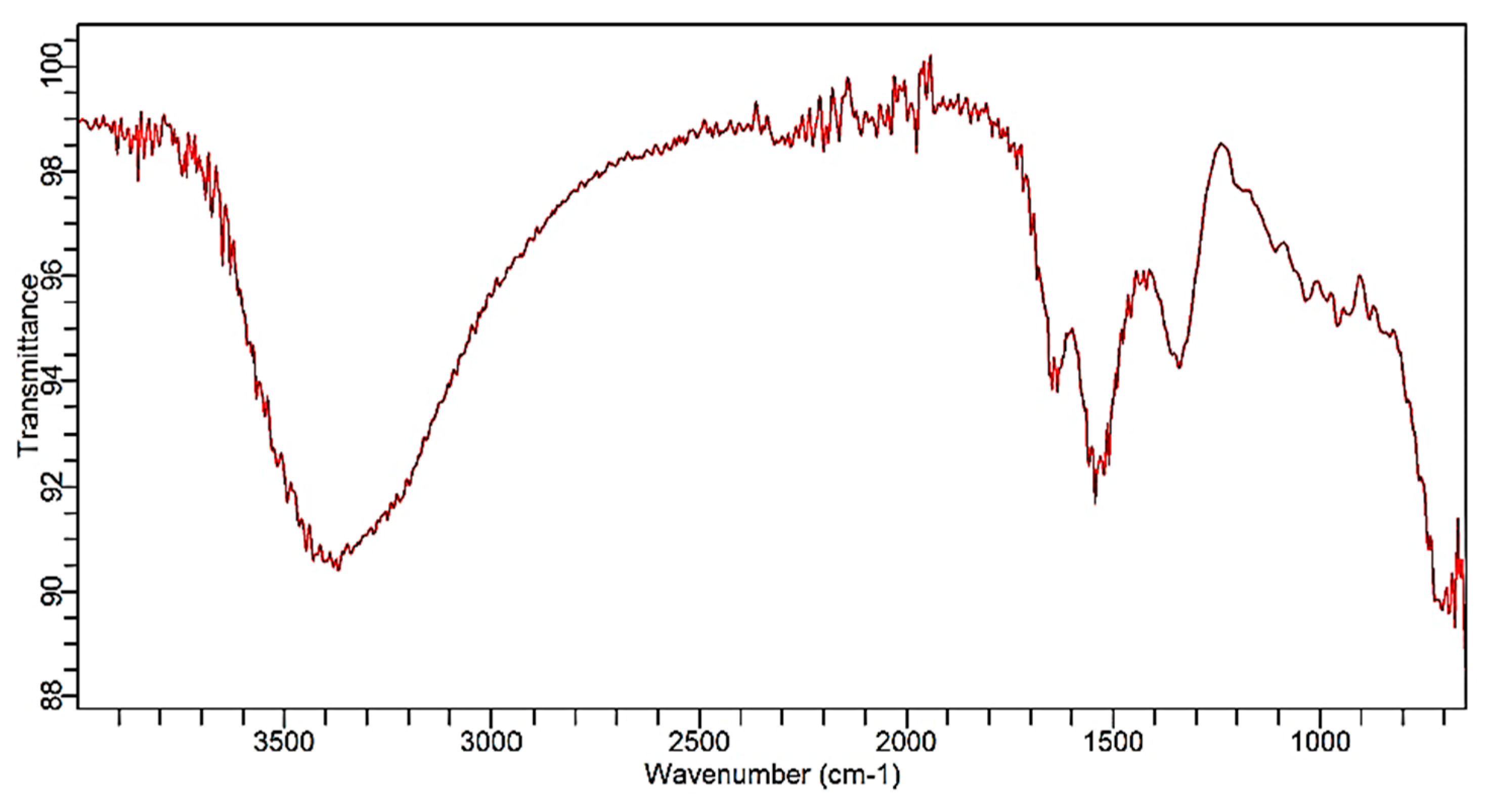
In this analysis, the functional groups were identified in the wavenumber range of 4000 to 1500 cm−1 and the corresponding compound was identified in the fingerprint region of 1500 to 650 cm−1, as shown in Figure 2. OH stretching was identified in the range of 3500 to 3000 cm−1, and the wavenumber of around 1500 cm−1 indicated C-H vibration. The peak around the wavenumber of 700 cm−1 confirms the presence of cerium oxide bonds.

3.3. MTT Assay Analysis

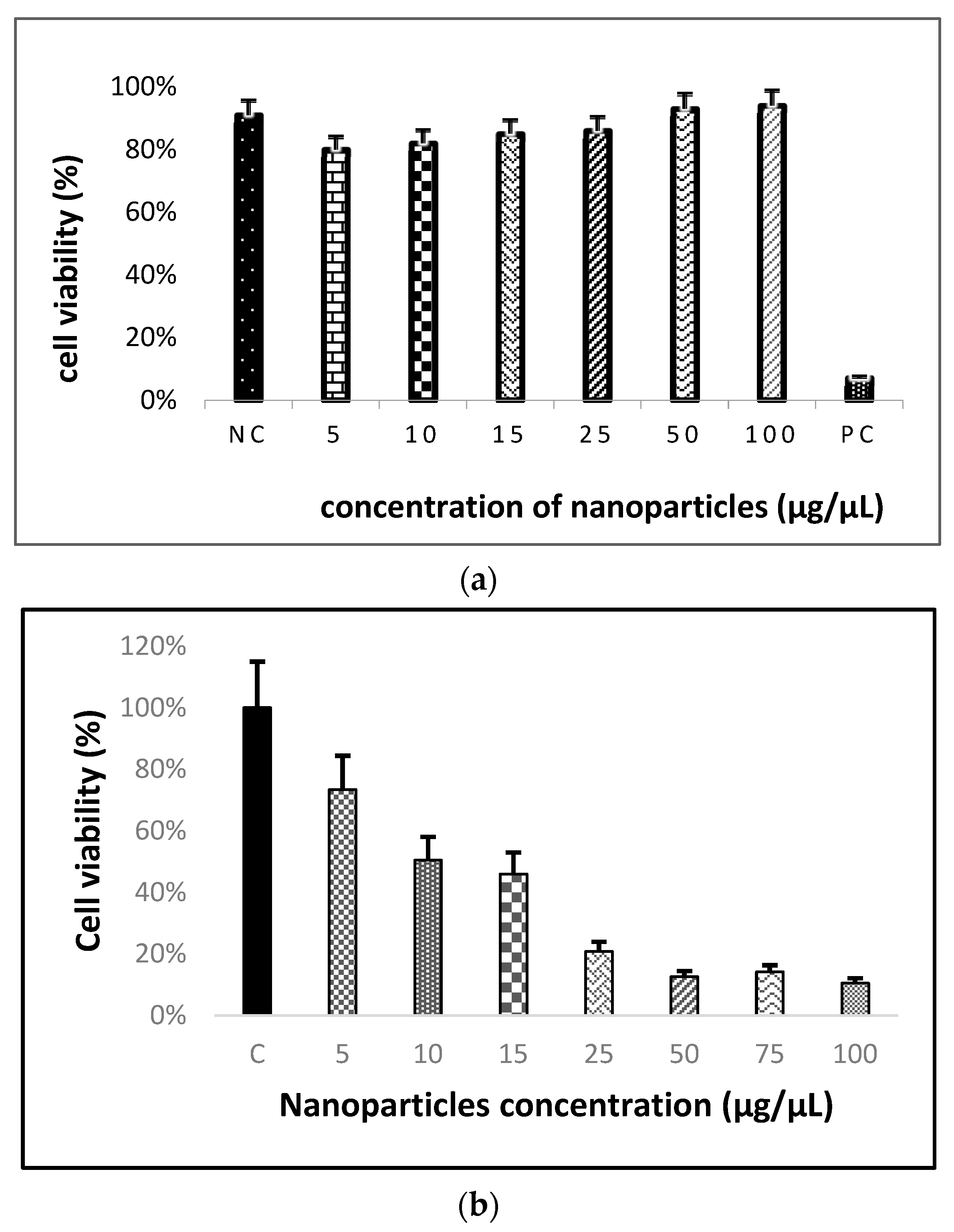
MTT dye was added to the H460 cells treated with the cerium oxide nanoparticles; during the process of incubation, the MTT dye was absorbed by the metabolically active cells and it was reduced to soluble formazan crystals, indicated by their purple color. After this process, DMSO was added to the cells, and the absorbance was measured using a spectrophotometer at 570 nm. Various concentrations of cerium oxide nanoparticles synthesized using turmeric rhizomes, such as 5, 10, 15, 25, 50, 75, and 100 μg/μL, were used to treat the Beas-2B cells and H460 cells. One-way analysis of variance (ANOVA) was conducted with a significance level of 0.05 to statistically analyze the data with respect to the control (cells without nanoparticles). The results in Figure 3a determine that the BEAS-2B cells treated with 5 to 100 μg/μL of CeO2 show a significant difference in viability when compared to the positive control (SWCNT), and no viability decrease was identified when compared to the negative control (cells without nanoparticles). The results in Figure 3b reveal that cell viability began to decrease at a concentration of 10 μg/μL, and a drastic reduction was observed at concentrations of 25 μg/μL and higher.

3.4. Live/Dead Assay Analysis

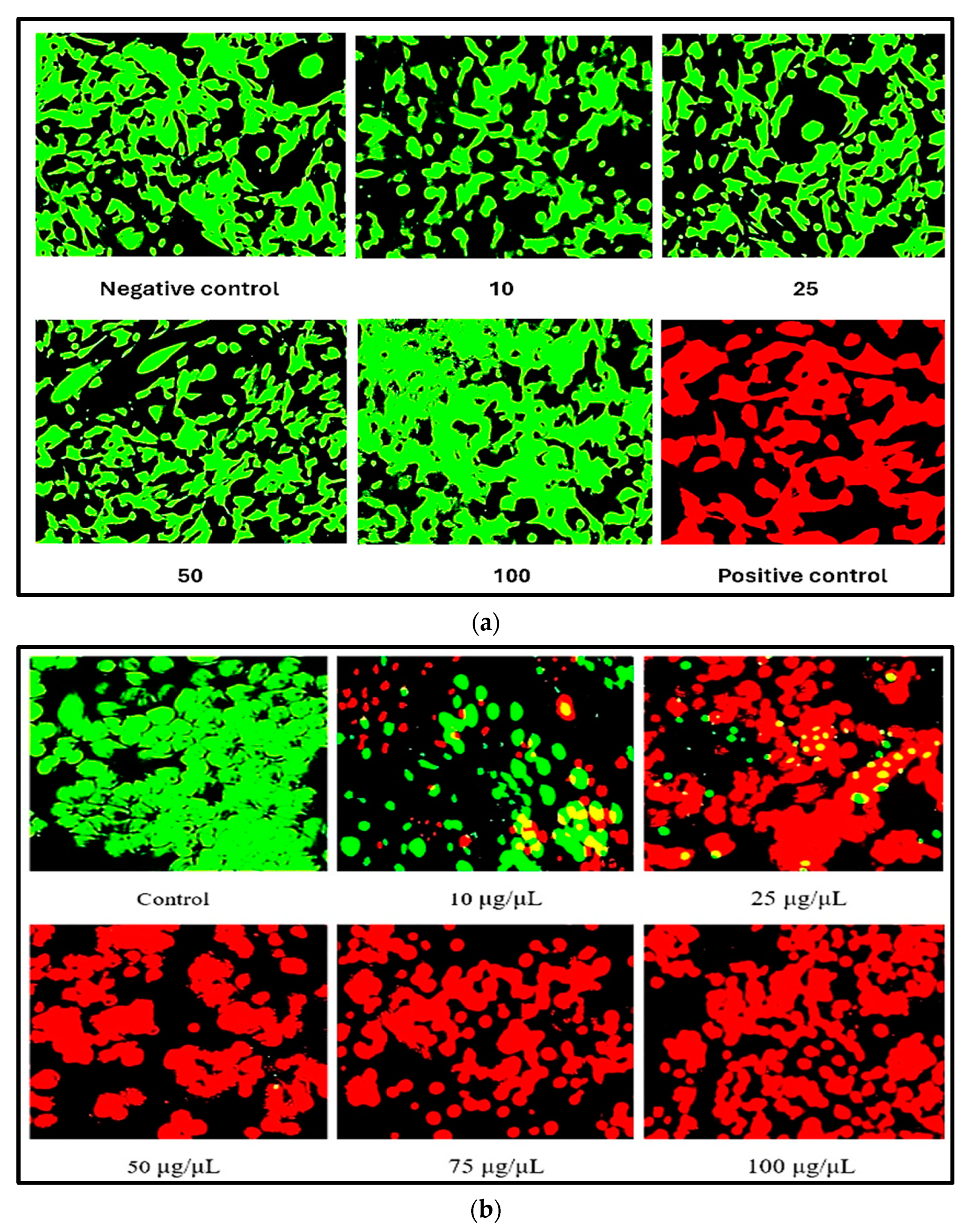
The MTT results were supported by the Live/Dead assay results. The live component was added to the H460 cells treated with cerium oxide nanoparticles, and during this process, Calcein-AM was converted to green fluorescent dye, indicating intracellular esterase activity in live cells, and the red component bound to nucleic acids, producing enhanced red fluorescence and thereby indicating the loss of plasma membrane integrity in the dead cells [41]. A fluorescent microscope was used to analyze the viability of Beas-2B and H460 cells using the green (FITC) filters and red (TRITC) filters. The fluorescent microscopic images are shown in Figure 4a,b. For all the doses of CeO2 nanoparticles, the Beas-2B cells show no cell death, which coincides with the results of MTT assay. At the nanoparticle concentration of 10 μg/μL, the H460 cells that have taken up the green and red components of the dye are observed, thereby indicating the reduction in viability. For the 25–100 μg/μL concentrations of cerium oxide nanoparticles, the H460 cells that have absorbed the red component are majorly observed, indicating a further decrease in cell viability.

3.5. ROS Assay Analysis

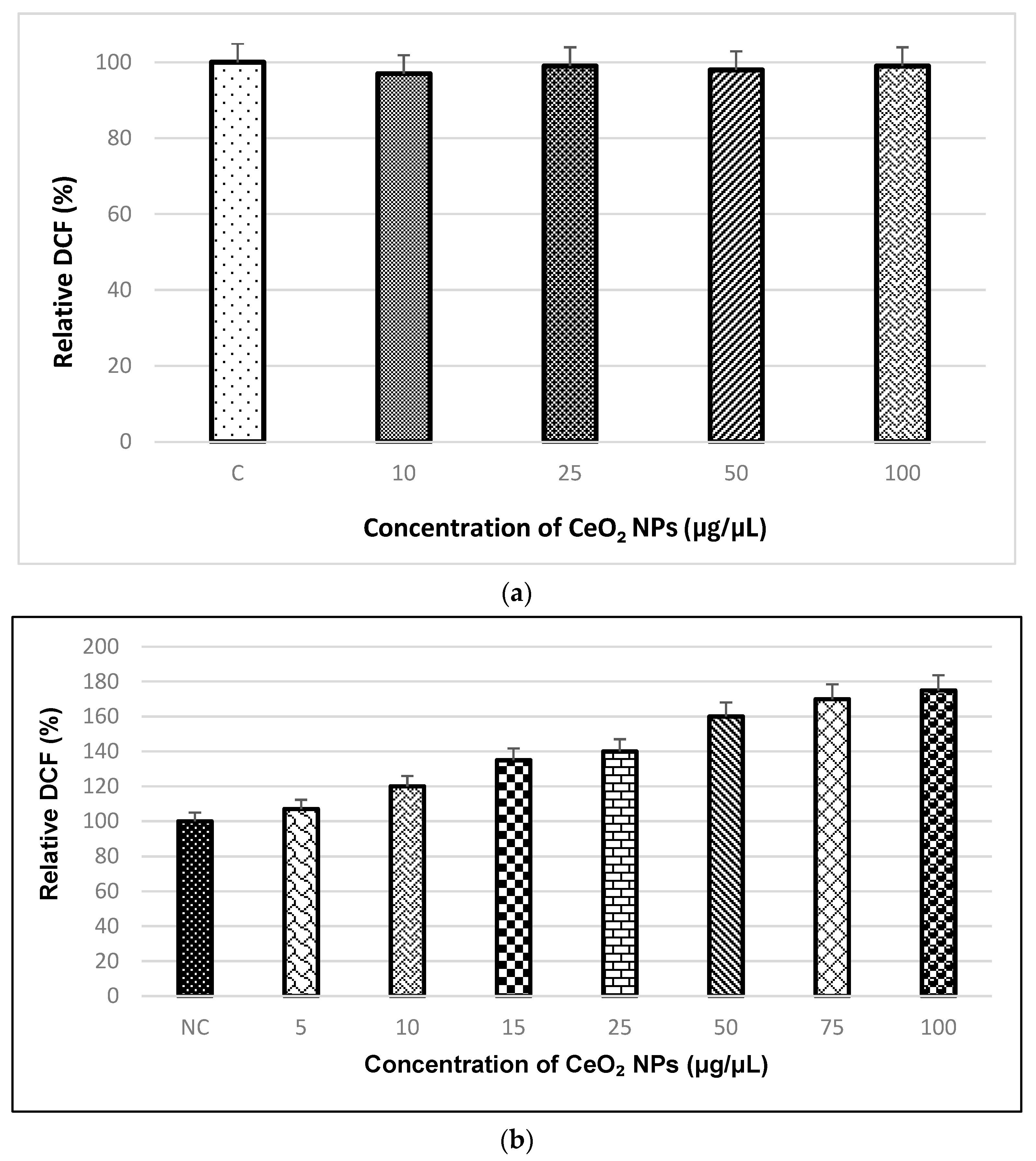
This assay used a cell-permeable compound, 2′,7′-dichlorofluorescin diacetate (DCF-DA), which after diffusion into the cells was deacetylated by cellular esterases to a non-fluorescent compound and later oxidized by ROS into a highly fluorescent molecule, 2′,7′-dichlorofluorescein (DCF). This fluorescence was then measured using a microplate reader at the ex/em wavelengths of 485/527 nm. When compared to the control, no significant difference in DCF levels was observed in the nanoparticles-treated Beas-2B cells, as shown in Figure 5a. A statistical evaluation of differences was made using ANOVA at significance levels of p 0.05 with respect to the control. The cerium oxide nanoparticles showed an increase in DCF levels from the lower concentration of 10 μg/μL, thereby inducing oxidative stress in the H460 cells, as shown in Figure 5b. For the higher nanoparticle concentrations of 50 to 100 μg/μL, a significant increase in the oxidative stress levels in human lung cancer cells was observed.

3.6. Cell Proliferation Assay Analysis

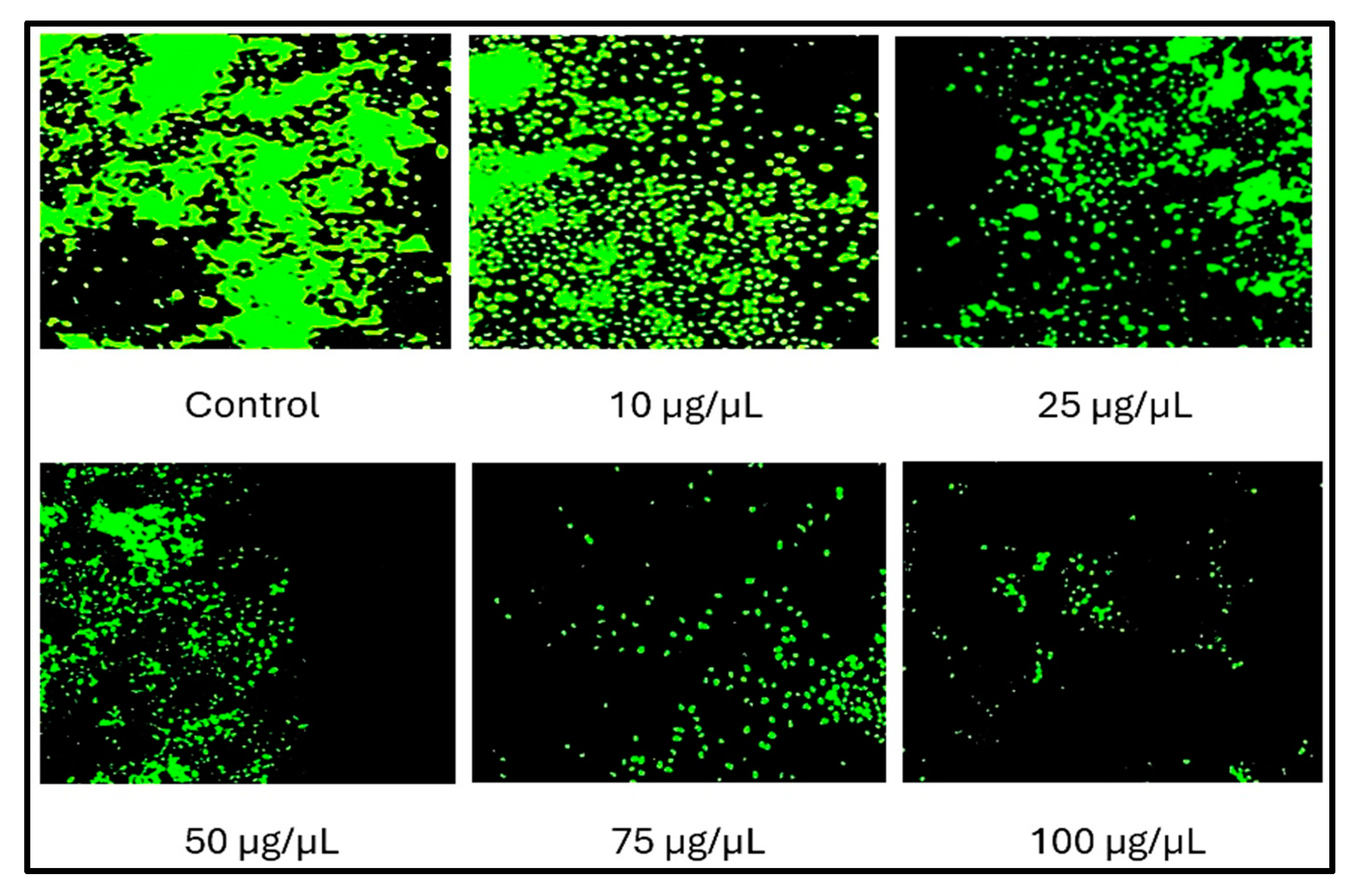
The cell proliferation assay was conducted only for H460 cells to further confirm the anticancer potential of the CeO2 NPs. In the cell proliferation assay, after the treatment of H460 cells with cerium oxide nanoparticles, the microtiter plate was incubated for 1 h. During this process, the cell-permeant nucleic acid binding dye along with the background suppression reagent blocks the staining of the dead cells and stains only the healthy cells. The fluorescence was analyzed using a fluorescence microscope at the ex/em wavelengths of 508/527 nm. The healthy H460 cells treated with cerium oxide nanoparticles were stained by the detection reagent, which was indicated by the fluorescent microscope image, as shown in Figure 6. The multiplication of active cells began to decrease from the 10 μg/μL concentration, and a significant decrease in the cell population was identified, corresponding to the nanoparticle concentrations of 50 μg/μL and higher.

4. Conclusions

The anticancer activity of cerium oxide nanoparticles synthesized using turmeric rhizomes was successfully analyzed using cell viability assays and cell proliferation assays. MTT assay and Live/Dead assay results determined that the Beas-2B cells treated with CeO2 NPs were viable and the cerium oxide nanoparticles were cytotoxic toward the H460 cells, indicating their anticancer potential. The results of the ROS assay confirmed the increase in oxidative stress levels in H460 cells treated with cerium oxide nanoparticles, that further confirmed the results of the viability assays. Based on the cell proliferation assay results, a dosage-dependent decrease in the multiplication of H460 cells treated with cerium oxide nanoparticles was observed, thereby confirming their anticancer potential. This study determined cerium oxide nanoparticles synthesized using turmeric rhizomes to be an efficient anticancer agent.

Author Contributions

Conceptualization, N.K.G. and G.T.R.; methodology, N.K.G.; validation, N.K.G.; formal analysis, N.K.G., P.P.T. and N.N.B.; data curation, N.K.G.; writing—original draft preparation, N.K.G.; writing—review, J.C.H. and K.P.; supervision, G.T.R.; project administration, G.T.R.; funding acquisition, G.T.R. All authors have read and agreed to the published version of the manuscript.

Funding

This research was supported by the National Science Foundation (NSF) CREST #2112595.

Data Availability Statement

All data shown in this manuscript are available upon request from the corresponding author.

Acknowledgments

The characterization of nanoparticles by scanning electron microscopy was assisted by Olga Trofimova at the College of William and Mary, Williamsburg, Virginia.

Conflicts of Interest

The authors declare no conflicts of interest.

References

  1. Hossain, N.; Mobarak, M.H.; Mimona, M.A.; Islam, M.A.; Hossain, A.; Zohura, F.T.; Chowdhury, M.A. Advances and significances of nanoparticles in semiconductor applications—A review. Results Eng. 2023, 19, 101347. [Google Scholar]
  2. Altammar, K.A. A review on nanoparticles: Characteristics, synthesis, applications, and challenges. Front. Microbiol. 2023, 14, 1155622. [Google Scholar]
  3. Vijayaram, S.; Razafindralambo, H.; Sun, Y.Z.; Vasantharaj, S.; Ghafarifarsani, H.; Hoseinifar, S.H.; Raeeszadeh, M. Applications of green synthesized metal nanoparticles—A review. Biol. Trace Elem. Res. 2024, 202, 360–386. [Google Scholar] [PubMed]
  4. Khan, S.; Hossain, M.K. Classification and properties of nanoparticles. In Nanoparticle-Based Polymer Composites; Woodhead Publishing: Sawston, UK, 2022; pp. 15–54. [Google Scholar]
  5. Gavas, S.; Quazi, S.; Karpiński, T.M. Nanoparticles for cancer therapy: Current progress and challenges. Nanoscale Res. Lett. 2021, 16, 173. [Google Scholar] [PubMed]
  6. Adan, A.; Kiraz, Y.; Baran, Y. Cell proliferation and cytotoxicity assays. Curr. Pharm. Biotechnol. 2016, 17, 1213–1221. [Google Scholar]
  7. Datta, A.; Mishra, S.; Manna, K.; Saha, K.D.; Mukherjee, S.; Roy, S. Pro-oxidant therapeutic activities of cerium oxide nanoparticles in colorectal carcinoma cells. ACS Omega 2020, 5, 9714–9723. [Google Scholar]
  8. Gao, Y.; Chen, K.; Ma, J.L.; Gao, F. Cerium oxide nanoparticles in cancer. OncoTargets Ther. 2014, 7, 835–840. [Google Scholar]
  9. Patil, N.; Bhaskar, R.; Vyavhare, V.; Dhadge, R.; Patil, Y. Overview on Methods of Synthesis of Nanoparticles. Int. J. Curr. Pharm. Res. 2021, 13, 11–16. [Google Scholar]
  10. Rajput, N. Methods of Preparation of Nanoparticles—A Review. Int. J. Adv. Eng. Technol. 2015, 7, 1806–1811. [Google Scholar]
  11. Namakka, M.; Rahman, M.R.; Said, K.A.M.B.; Mannan, M.A.; Patwary, A.M. A Review of Nanoparticle Synthesis Methods, Classifications, Applications, and Characterization. Environ. Nanotechnol. Monit. Manag. 2023, 20, 100900. [Google Scholar]
  12. Goornavar, V.; Biradar, S.; Ezeagwu, C.; Ezeagwu, D.; Hall, J.C.; Ramesh, G.T. Toxicity of raw and purified single-walled carbon nanotubes in rat’s lung epithelial and cervical cancer cells. J. Nanosci. Nanotechnol. 2015, 15, 2105–2114. [Google Scholar] [PubMed]
  13. Rane, A.V.; Kanny, K.; Abitha, V.K.; Thomas, S. Methods for Synthesis of Nanoparticles and Fabrication of Nanocomposites. In Synthesis of Inorganic Nanomaterials; Woodhead Publishing: Sawston, UK, 2018; pp. 121–139. [Google Scholar]
  14. Wason, M.S.; Zhao, J. Cerium oxide nanoparticles: Potential applications for cancer and other diseases. Am. J. Transl. Res. 2013, 5, 126–131. [Google Scholar] [PubMed]
  15. Nelson, B.C.; Johnson, M.E.; Walker, M.L.; Riley, K.R.; Sims, C.M. Antioxidant cerium oxide nanoparticles in biology and medicine. Antioxidants 2016, 5, 15. [Google Scholar] [CrossRef]
  16. Charbgoo, F.; Ramezani, M.; Darroudi, M. Bio-sensing applications of cerium oxide nanoparticles: Advantages and disadvantages. Biosens. Bioelectron. 2017, 96, 33–43. [Google Scholar]
  17. Walkey, C.; Das, S.; Seal, S.; Erlichman, J.; Heckman, K.; Ghibelli, L.; Traversa, E.; McGinnis, J.F.; Self, W.T. Catalytic properties and biomedical applications of cerium oxide nanoparticles. Environ. Sci. Nano 2015, 2, 33–53. [Google Scholar] [PubMed]
  18. Huston, M.; DeBella, M.; DiBella, M.; Gupta, A. Green synthesis of nanomaterials. Nanomaterials 2021, 11, 2130. [Google Scholar]
  19. Zholobak, N.M.; Shcherbakov, A.B.; Ivanova, O.S.; Reukov, V.; Baranchikov, A.E.; Ivanov, V.K. Nanoceria-curcumin conjugate: Synthesis and selective cytotoxicity against cancer cells under oxidative stress conditions. J. Photochem. Photobiol. B Biol. 2020, 209, 111921. [Google Scholar]
  20. Hosseinzadeh, R.; Khorsandi, K.; Esfahani, H.S.; Habibi, M.; Hosseinzadeh, G. Preparation of cerium-curcumin and cerium-quercetin complexes and their LEDs irradiation assisted anticancer effects on MDA-MB-231 and A375 cancer cell lines. Photodiagnosis Photodyn. Ther. 2021, 34, 102326. [Google Scholar]
  21. Javid, H.; Hashemy, S.I.; Heidari, M.F.; Esparham, A.; Gorgani-Firuzjaee, S. The anticancer role of cerium oxide nanoparticles by inducing antioxidant activity in esophageal cancer and cancer stem-like ESCC spheres. BioMed Res. Int. 2022, 2022, 3268197. [Google Scholar]
  22. Jiang, H.; Zuo, J.; Li, B.; Chen, R.; Luo, K.; Xiang, X.; Lu, S.; Huang, C.; Liu, L.; Tang, J.; et al. Drug-induced oxidative stress in cancer treatments: Angel or devil? Redox Biol. 2023, 63, 102754. [Google Scholar]
  23. Lin, W.; Huang, Y.W.; Zhou, X.D.; Ma, Y. Toxicity of cerium oxide nanoparticles in human lung cancer cells. Int. J. Toxicol. 2006, 25, 451–457. [Google Scholar] [PubMed]
  24. Kumar, P.; Nagarajan, A.; Uchil, P.D. Analysis of cell viability by the MTT assay. Cold Spring Harb. Protoc. 2018, 2018, pdb-prot095505. [Google Scholar]
  25. Bahuguna, A.; Khan, I.; Bajpai, V.K.; Kang, S.C. MTT assay to evaluate the cytotoxic potential of a drug. Bangladesh J. Pharmacol. 2017, 12, 115–118. [Google Scholar]
  26. Johnson, S.; Nguyen, V.; Coder, D. Assessment of cell viability. Curr. Protoc. Cytom. 2013, 64, 9.2.1–9.2.26. [Google Scholar]
  27. Zhou, S.; Cui, Z.; Urban, J. Dead Cell Counts During Serum Cultivation Are Underestimated by the Fluorescent Live/Dead Assay; WILEY-VCH Verlag: Weinheim, Germany, 2011. [Google Scholar]
  28. Najahi-Missaoui, W.; Arnold, R.D.; Cummings, B.S. Safe nanoparticles: Are we there yet? Int. J. Mol. Sci. 2020, 22, 385. [Google Scholar] [CrossRef]
  29. Sullivan, L.B.; Chandel, N.S. Mitochondrial reactive oxygen species and cancer. Cancer Metab. 2014, 2, 17. [Google Scholar]
  30. Lewinski, N.; Colvin, V.; Drezek, R. Cytotoxicity of nanoparticles. Small 2008, 4, 26–49. [Google Scholar] [PubMed]
  31. Riss, T.; Niles, A.; Moravec, R.; Karassina, N.; Vidugiriene, J. Cytotoxicity assays: In vitro methods to measure dead cells. In Assay Guidance Manual; NIH: Bethesda, MD, USA, 2019. [Google Scholar]
  32. Wiesenthal, A.; Hunter, L.; Wang, S.; Wickliffe, J.; Wilkerson, M. Nanoparticles: Small and mighty. Int. J. Dermatol. 2011, 50, 247–254. [Google Scholar]
  33. Kalaycıoğlu, Z.; Geçim, B.; Erim, F.B. Green synthesis of cerium oxide nanoparticles from turmeric and kinds of honey: Characterisations, antioxidant and photocatalytic dye degradation activities. Adv. Nat. Sci. Nanosci. Nanotechnol. 2022, 13, 015016. [Google Scholar]
  34. Andrabi, S.M.; Majumder, S.; Gupta, K.C.; Kumar, A. Dextran based amphiphilic nano-hybrid hydrogel system incorporated with curcumin and cerium oxide nanoparticles for wound healing. Colloids Surf. B Biointerfaces 2020, 195, 111263. [Google Scholar]
  35. Aseyd Nezhad, S.; Es-haghi, A.; Tabrizi, M.H. Green synthesis of cerium oxide nanoparticle using Origanum majorana L. leaf extract, its characterization and biological activities. Appl. Organomet. Chem. 2020, 34, e5314. [Google Scholar]
  36. Nadeem, M.; Khan, R.; Afridi, K.; Nadhman, A.; Ullah, S.; Faisal, S.; Mabood, Z.U.; Hano, C.; Abbasi, B.H. Green synthesis of cerium oxide nanoparticles (CeO2 NPs) and their antimicrobial applications: A review. Int. J. Nanomed. 2020, 15, 5951–5961. [Google Scholar]
  37. Gunasekaran, N.K.; Tumkur, P.P.; Bayon, N.N.; Prabhakaran, K.; Arasho, W.D.; Hall, J.C.; Ramesh, G.T. Cerium Oxide Nanoparticles: Synthesis and Characterization Using Curcuma longa (Turmeric Rhizome). Energy Environ. Focus 2023, 7, 83–89. [Google Scholar]
  38. Dutta, A. Fourier transform infrared spectroscopy. In Spectroscopic Methods for Nanomaterials Characterization; Elsevier: Amsterdam, The Netherlands, 2017; pp. 73–93. [Google Scholar]
  39. Jaggi, N.; Vij, D.R. Fourier transform infrared spectroscopy. In Handbook of Applied Solid State Spectroscopy; Springer: Boston, MA, USA, 2006; pp. 411–450. [Google Scholar]
  40. Ismail, A.A.; van de Voort, F.R.; Sedman, J. Fourier transform infrared spectroscopy: Principles and applications. In Techniques and Instrumentation in Analytical Chemistry; Elsevier: Amsterdam, The Netherlands, 1997; Volume 18, pp. 93–139. [Google Scholar]
  41. Zamborini, F.P.; Bao, L.; Dasari, R. Nanoparticles in measurement science. Anal. Chem. 2012, 84, 541–576. [Google Scholar] [PubMed]
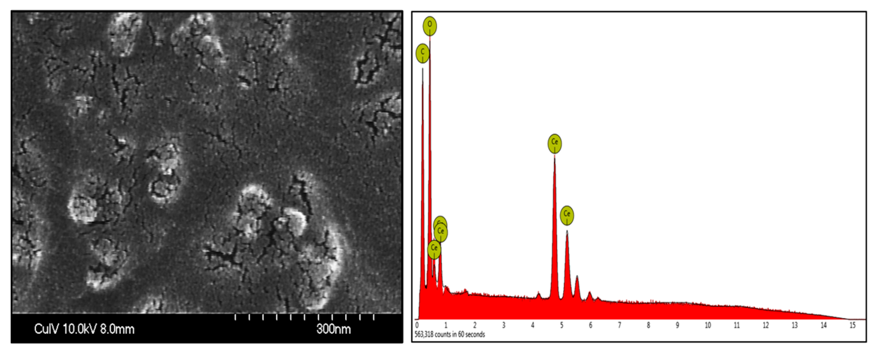
Figure 1. FESEM-EDS image of cerium oxide nanoparticles.
Nanomanufacturing 05 00006 g001
Figure 2. FTIR spectra of cerium oxide nanoparticles.
Nanomanufacturing 05 00006 g002
Figure 3. (a) Viability of Beas-2B cells towards cerium oxide nanoparticles synthesized using turmeric rhizomes. (b) Viability of H460 cells towards cerium oxide nanoparticles synthesized using turmeric rhizomes.
Nanomanufacturing 05 00006 g003
Figure 4. (a) Fluorescence-based viability of Beas-2B cells treated with CeO2 nanoparticles synthesized from turmeric rhizomes. (b) Fluorescence-based viability of H460 cells treated with CeO2 nanoparticles synthesized from turmeric rhizomes.
Nanomanufacturing 05 00006 g004
Figure 5. (a) Oxidative stress levels of Beas-2B cells treated with CeO2 nanoparticles synthesized from turmeric rhizomes. (b) Oxidative stress levels of H460 cells treated with CeO2 nanoparticles synthesized from turmeric rhizomes.
Nanomanufacturing 05 00006 g005
Figure 6. Cell proliferation assay results of H460 cells treated with CeO2 nanoparticles synthesized from turmeric rhizomes.
Nanomanufacturing 05 00006 g006
Disclaimer/Publisher’s Note: The statements, opinions and data contained in all publications are solely those of the individual author(s) and contributor(s) and not of MDPI and/or the editor(s). MDPI and/or the editor(s) disclaim responsibility for any injury to people or property resulting from any ideas, methods, instructions or products referred to in the content.