Article

Genome-Wide Identification, Bioinformatic Characterization, and Expression Profiling of Starch Synthase (SS) Genes in Foxtail Millet under Drought Condition

by
Joseph N. Amoah
1,*,
Monica Ode Adu-Gyamfi
2 and
Albert Owusu Kwarteng
3
1
School of Life and Environmental Sciences, University of Sydney, 380 Werombi Road, Brownlow Hill, Camden, NSW 2570, Australia
2
Plant Biotechnology Department, CSIR—Crop Research Institute, Kumasi 233, Ghana
3
Department of Plant Sciences, Kimberly Research and Extension Center, University of Idaho, Moscow, ID 83341, USA
*
Author to whom correspondence should be addressed.
Stresses 2024, 4(3), 518-533; https://doi.org/10.3390/stresses4030033
Submission received: 19 July 2024 / Revised: 8 August 2024 / Accepted: 14 August 2024 / Published: 16 August 2024
(This article belongs to the Topic Plant Responses to Environmental Stress)

Abstract

:
Millet, a vital and nutritionally dense cereal extensively cultivated in Sub-Saharan Africa, plays a key role in ensuring food security. This study investigates the starch synthase (SS) gene family, which is crucial for starch biosynthesis and influences various plant functions and stress responses. While the specific roles of SS genes in millet under drought conditions are not fully elucidated, this research provides a thorough analysis of the SS gene family in millet. A total of twelve millet SS genes (SiSSs) were identified and classified into four subfamilies (I–IV) through gene structure and phylogenetic analysis. The SiSS genes were unevenly distributed across millet chromosomes, with cis-acting elements associated with plant growth and stress defense being identified. Quantitative PCR (qPCR) revealed dynamic and varied expression patterns of SiSSs in different tissues under drought stress. Millet plants subjected to drought conditions showed higher tissue starch content and increased starch synthase activity compared to controls. Importantly, the expression levels of the twelve SiSSs were positively correlated with both starch content and synthase activity, suggesting their significant role in drought tolerance. This study enhances our understanding of the millet SS gene family and highlights the potential of these genes in breeding programs aimed at developing drought-resistant millet varieties. Further research is recommended to validate these findings and delve deeper into the mechanisms underlying drought tolerance.

1. Introduction

Starch is the most abundant carbohydrate reserve in plants that serves as their source of carbon and energy and its synthesis and metabolism play an essential role in plant growth, development, and defense, as well as in responding to stress such as drought and other environmental conditions [1]. Starch synthases, classified as soluble starch synthase (SSS) and granule-bound starch synthase (GBSS), comprise a class of enzymes involved in starch metabolism. They elongate glucan chains by adding glucosyl units, transferred from ADP-Glc to the non-reducing end of the glucan chains. Specifically, SSS synthesizes amylopectin, while GBSS synthesizes both amylopectin and the extra-long fraction of amylopectin [2,3].
Starch is synthesized and stored in the endosperm, where it is categorized into rapidly digestible, slowly digestible, and resistant starch. Resistant starch, unlike the former types, withstands digestion by human pancreatic amylase in the small intestine [4]. As it progresses into the large intestine, it undergoes fermentation by gut microbiota, resulting in a gradual and sustained release of glucose. This unique digestion pattern exhibited by resistant starch not only promotes gut health, mitigates adiposity, and reduces insulin resistance but also diminishes the risk of cardiovascular disease and colon cancer [5]. Notably, commonly consumed cereals such as rice, wheat, maize, sorghum, etc., are predominantly composed of rapidly and slowly digestible starch. In contrast, millet grains are notable for their richness in resistant starch [6]. This underscores the significance of millets in forming low glycemic index foods, a crucial requirement considering the rising numbers of individuals with diabetes and prediabetes [7]. Starch acts as a vital energy reservoir in plants, providing essential energy and carbon skeletons during drought stress when photosynthesis is compromised. The conversion of starch into soluble sugars supports osmotic regulation, maintaining cell turgor and safeguarding cellular structures against dehydration. Furthermore, starch metabolism impacts stress signaling pathways, aiding in plant growth and recovery by ensuring a continuous energy supply for crucial processes and minimizing damage to the photosynthetic system.
As an orphan crop, millet is notable for its richness in protein, micronutrients, and high starch content (approximately 75%), a level comparable to other cereals, making it an economically significant cereal [8]. It has garnered considerable attention from plant scientists and breeders due to its resilience to high-stress conditions brought about by climate change. Millet is in high demand, both in the Sub-Saharan region and globally, owing to its numerous nutritional benefits [6,9]. It plays a crucial role in addressing food security and sustainability, contributing significantly to the goal of achieving Zero Hunger globally. Despite being considered a climate-resilient crop, global millet production is hindered by drought. The onset of drought stress affects various metabolic processes in millet, significantly impacting its growth, development, and harvestable yield [10]. Therefore, it is imperative to understand the physiological and molecular mechanisms that contribute to drought tolerance in millet. This understanding is crucial for informing breeding and engineering efforts aimed at developing millet genotypes with enhanced drought tolerance.
Genome-wide analysis of gene families has emerged as a widely employed molecular approach that has revolutionized cultivar development over the years. This technique offers a holistic view of the genetic landscape, facilitating a comprehensive understanding of the functions of individual genes and their interactions. Such knowledge is instrumental in unraveling complex biological processes, ultimately contributing to the development of cultivars with optimal performance under diverse environmental conditions [11,12]. Among various starch-metabolism-related genes, the starch synthase family stands out due to its unique response under drought conditions. Members of this family play key roles in synthesizing and polymerizing starch molecules, extending chains during starch biosynthesis, and influencing the structure and properties of starch granules. While starch synthase genes have been well studied in rice and cassava using the genome-wide approach [13,14,15], a gap exists in our understanding, particularly in millet under drought conditions. Hence, there is a critical need to understand the profile of these valuable starch-related genes, given their potential application in developing plants with enhanced drought tolerance. By identifying and examining these genes in foxtail millet, researchers can gain valuable insights into the dynamics of starch metabolism under water scarcity conditions. This knowledge will help breeders create foxtail millet varieties that maintain stable yields even in drought-prone areas, thereby enhancing food security.
Additionally, conducting a comparative study on the starch synthase gene family in millet is important due to the functional versatility of SS genes. In this study, SiSS genes were identified using bioinformatics methods and subjected to phylogenetic analysis, categorizing them into four subfamilies. Additionally, we characterized SiSSs based on their distribution on chromosomes, gene and motif structure, protein structure, qPCR expression analysis, and physiological assays of starch and starch synthase activity. This comprehensive approach aims to shed light on the potential impact of SS genes on millet. These findings have the potential to contribute to the development of drought-tolerant millet genotypes through both forward and reverse molecular and genetic approaches.

2. Results

2.1. Identification and Phylogenetic Analysis of SS in Millet

Twelve (12) starch synthase (SS) proteins, identified in foxtail millet and containing the conserved domain Glyco_transf_5, SS catalytic domain (PFAM accession no. PF08323), were designated as SiSS1-SiSS12 based on their chromosomal locations (see Table S1). Regarding the putative SiSSs’ physiochemical properties, the coding sequence (CDS) lengths ranged from 1242 (SiSS12) to 4629 (SiSS8), protein lengths from 414 (SiSS12) to 1543 (SiSS8) amino acids, with relative molecular weights ranging from 46.42 (SiSS12) kDa to 174.78 (SiSS8) kDa. Based on the pI values, eight SiSSs exhibited an acidic nature with pIs ≤ 6.5, one was alkaline with pIs > 7.5, and two were neutral with 6.5 < pI < 7.5 (refer to Table 1). The instability index suggested that SiSSs were unstable, while the GRAVY values indicated their hydrophilic nature. Additionally, SiSSs were found to be in the chloroplast (Table 1).
Furthermore, a neighbor-joining (NJ) tree was constructed using the aligned protein sequences of the 12 putative SiSSs alongside 7 SS from wheat (TaSSs), 2 SS from barley (HvSSs), 14 SS from oryza sativa (OsSS), 7 SS from Arabidopsis thaliana (AtSSs), and 4 SS from Manihot esculenta (MeSSs). In contrast to previous findings in Manihot esculenta [14] and Oryza sativa [15], SiSSs were categorized into four subfamilies (I–IV) alongside SS from these plant species. Specifically, subfamily I comprised two SiSSs (three TaSSs, one HvSSs, three OsSSs, two AtSSs, and three MeSSs), subfamily II included three SiSSs (four TaSSs, one HvSSs, one AtSSs, and one MeSSs), subfamily III contained four SiSSs (five OsSSs and three AtSSs), and subfamily IV consisted of two SiSSs (three OsSSs and one AtSSs) (Figure 1).

2.2. Gene Structure and Motif Analysis

The configuration of exons and introns within a gene is a pivotal evolutionary trait, offering valuable insights into its functional diversity (Figure 2A). An analysis of the exon-intron structure was conducted to elucidate this aspect in putative SiSSs. According to the results, the SiSSs were categorized into three subfamilies based on their gene structure (exon-intron), with subfamily I containing six SiSS genes, and subfamilies II and III each comprising three SiSS genes (Figure 2A). The putative genes exhibited varying exon counts: 8 exons (17%), 9 exons (8%), 11 exons (17%), 15 exons (17%), 16 exons (25%), 17 exons (8%), and 20 exons (8%) (Figure 2B). Additionally, the Multiple Em for Motif Elicitation (MEME) software identified ten (10) conserved motifs in SiSS proteins. Motifs 1, 2, 3, and 6 were present in all SiSSs. Subfamily I members displayed 7 or 8 motifs, subfamily II had 3 motifs, subfamily III had 7 motifs, and subfamily IV contained all 10 motifs (Figure 2C).

2.3. Chromosomal Location, Gene Duplication of SiSSs and Synteny Analysis of SiSSs in Four Species

The SiSSs were unevenly distributed across the millet chromosomes. Chromosome 1 contained two SiSSs (SiSS1 and SiSS2), chromosome 3 had SiSS3, chromosome 4 had three SiSSs (SiSS4, SiSS5, and SiSS6), chromosome 5 had SiSS7, chromosome 6 had two SiSSs (SiSS7 and SiSS8), chromosome 7 had SiSS10, and chromosome 9 had two SiSSs (SiSS11 and SiSS12) (Figure 3). Gene duplication has been shown to provide the raw material (catalysts) for evolutionary innovations [16]. Gene duplication events occurred in millet (Figure 3B) and were detected in subfamilies I and II. Four paralogous gene pairs (SiSS1-SiSS6, SiSS7-SiSS12, SiSS2-SiSS4, and SiSS8-SiSS9) indicated that their expansion occurred through whole-genome or segmental (WG/SD) duplication events.
Furthermore, the divergent times and selection pressures of the paralogous gene pairs were assessed using the Ka/Ks values, which ranged from 0.065 (SiSS7-SiSS12) to 0.681 (SiSS2-SiSS4) with divergence times of 10.313 to 2.313 MYA, respectively (Table 2). The results suggest that the paralogous gene pairs underwent purifying selection, as evidenced by Ks values less than 1 (Table 2). The syntenic relationships of SSs in millet, Arabidopsis, barley, tomato, and rice were examined (Figure 4). There were five, seven, and one SiSSs that showed collinearity with the HvSSs, OsSSs, and SiSSs, respectively. While the SiSSs shared some collinearity with different plant species, no collinearity was observed between the SiSSs and SSs of Arabidopsis thaliana (Figure 4).

2.4. Promoter Analysis of SiSS Genes

To deepen our understanding of the transcriptional regulation and potential functions of the SiSSs, we analyzed their 2k bp promoter regions using the PlantCare online website to predict cis-regulatory elements. The results showed different cis-acting elements involved in various plant processes, such as growth and development responsiveness, phytohormone responsiveness, abiotic stress responsiveness, and light responsiveness (Figure 5 and Table S3). SiSSs had elements such as CAT-box, GCN4_motif, GC-motif, O2-site, RY-element, circadian motif I, and MSA-like involved in various growth and developmental functions of plants (Figure 5A and Table S3). The ABRE was identified in all SiSSs except SiSS3, highlighting the regulatory function of SSs in starch metabolism under stress conditions and their influence by ABA signaling, as part of a broader stress response [17,18]. Furthermore, 12 SiSSs had MeJA-responsive motifs (TGACG and CGTCA-motif), 9 and 1 SiSSs had auxin (TGA-element and AuxRR-core)-responsive elements, 3, 2, and 8 SiSSs had gibberellin-responsiveness (TATC-box, GARE-motif, and P-box, respectively), and 1 SiSSs had TCA-element (salicylic acid responsiveness) (Figure 5B and Table S3), suggesting the participatory role of SiSSs in hormone metabolism processes and signal transduction networks. Several stress-related elements, such as STRE, WRE3, W box, TC-rich repeats, LTR, MBS, GC-motif, DRE core, WUN-motif, and ARE, highlight that SSs play an active role in abiotic and biotic stress response (Figure 5C and Table S3). Additionally, various light-responsive elements were identified in the putative SiSSs, suggesting the involvement of SiSSs in light response (Figure 5D and Table S3).

2.5. Three-Dimensional Protein Structure of SiSSs

The Protein Homology/Analogy Recognition Engine V 2.0 (Phyre2) website was used to acquire the three-dimensional (3D) structure of SiSS. SiSSs were shown to have a conserved protein structure. The putative SiSSs exhibited an α-helix and β-sheet, while six SiSSs had a transmembrane (Tm) helix as part of their secondary structure (Figure 6) and membrane topology (Figure 7). Furthermore, the 3D structure analysis predicted three different models: c4hlnA_, c6gneB_, and c3c4vB_, based on the template (Table S4). The models were predicted with 100% confidence, suggesting that the stable conformation of the SiSS’s structure makes it applicable for further analysis. The disordered values of SiSSs varied (Table S4), highlighting the versatility and complexity of the SiSS’s structure and function.

2.6. Expression Analysis of Putative SiSSs and Cluster Analysis under Drought Conditions

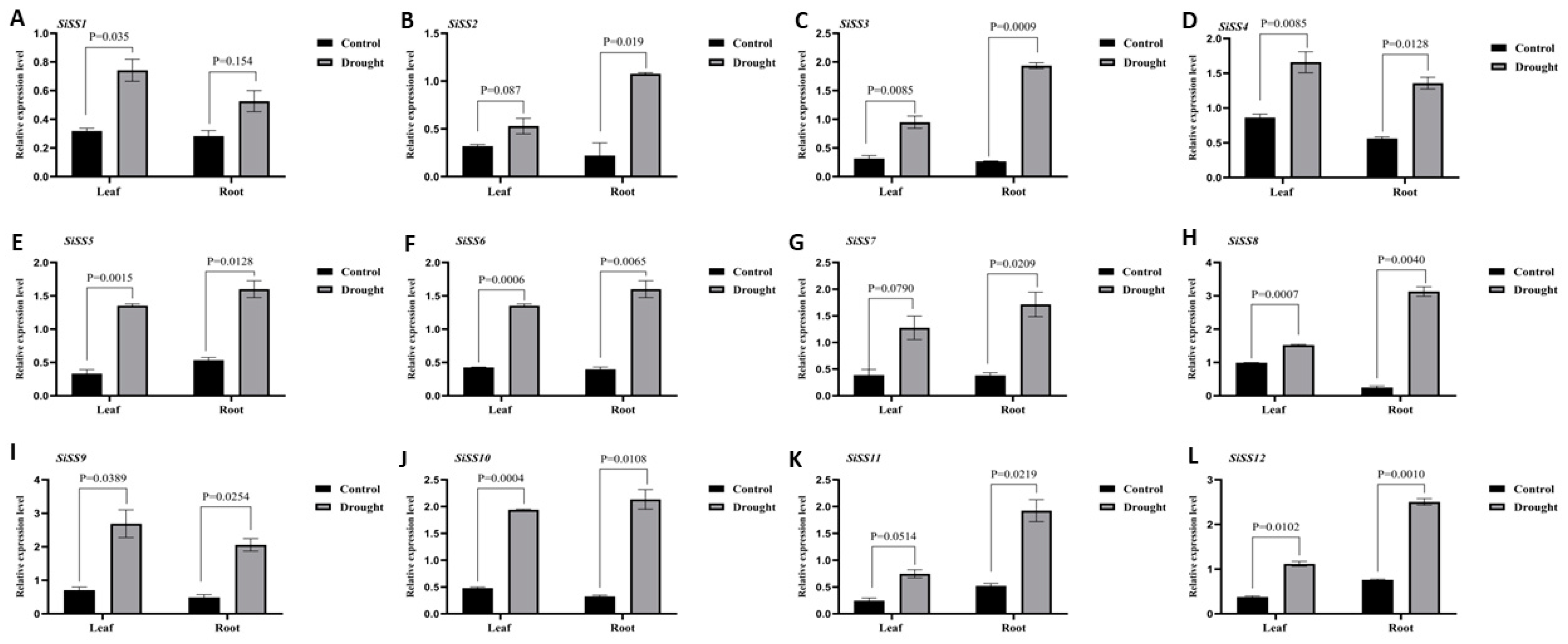
To understand the regulation of the putative SiSSs under drought conditions, the quantitative polymerase chain reaction (qPCR) approach was employed. The results revealed that the putative SiSS genes were differentially regulated in the leaves and roots of millet seedlings under drought conditions. Compared to the control (CK) plants, the expression levels of these genes increased in the leaf and root tissues of drought-stressed plants. For example, in the leaf, the expression of these genes ranged from 1.5-fold (SiSS8) to 4-fold (SiSS5) (Figure 8 and Table S5). In the root, the expression levels ranged from 2.3-fold (SiSS4) to 12-fold (SiSS8) (Figure 7 and Table S5). Additionally, the average tissue (leaf and root) expression of SiSSs ranged from 2.1-fold (SiSS4) to 8-fold (SiSS8), demonstrating that the putative SiSSs may contribute to drought tolerance in millet.
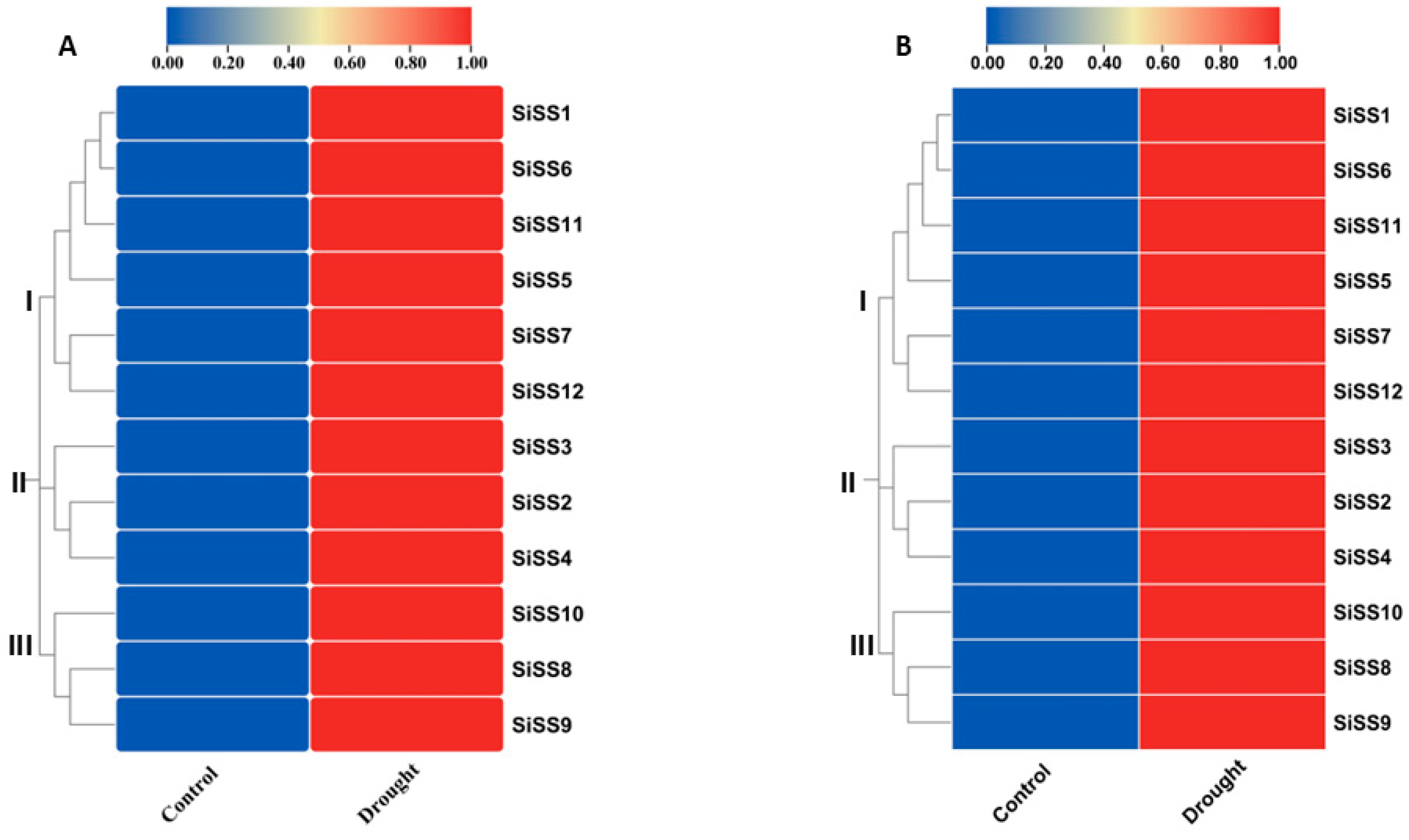
Furthermore, the relative expression values of the putative SiSSs were used to create a cluster (heat map) aimed at understanding the relationship between these SiSSs under drought conditions. In this analysis, the 12 putative SiSSs were categorized into three distinct groups (I, II, and III) based on the magnitude of their expression in different millet tissues (Figure 9). In the leaves, group I comprised six SiSSs that were moderately expressed, group II included three SiSSs with low expression, and group III consisted of three SiSSs that were highly upregulated after drought stress treatment (Figure 9A). Similarly, in the roots, the six SiSSs in group I, three SiSSs in group II, and three SiSSs in group III exhibited moderate, low, and high upregulation, respectively, under drought conditions (Figure 9B).

3. Discussion

Starch plays a crucial role as a primary source of energy and carbon storage in plants, and its metabolic equilibrium is influenced by various environmental factors such as drought [19,20]. The starch synthase (SS) gene family plays an active role in the synthesis and regulation of starch and is essential for plant growth and seed development. The discovery and functional characterization of SSs in the signal transduction pathway [21,22] and starch metabolism hold the promise of enhancing the breeding and development of plant species with superior performance, improving starch content, composition, and overall plant resilience in agricultural settings to mitigate global climate change [23,24]. The starch gene family has been characterized in different species, such as rice [13,15], potato [25], and cassava [26] but not in millet. Twelve (12) starch synthase (SS) genes were identified in the foxtail millet genome using a genome-wide approach. The putative SSs from millet were grouped into four subfamilies (I–IV), along with representatives from five other plant species. Subfamilies I, II, III, and IV contained 15, 12, 13, and 6 SSs, respectively. In millet, we identified four orthologous SS gene pairs, with three demonstrating segmental duplication and one showing tandem duplication. The functional roles of SS genes encompass various metabolic processes, primarily associated with starch synthesis, metabolism, and responses to environmental stresses [1,19,27]. The phylogenetics, gene structure, and conserved motifs analysis supported the findings that the three-dimensional structures of SiSSs were conserved. The protein structure was predicted with 100% confidence, and up to 90% of the residues were positioned in the most preferred location, supporting the accuracy of the predicted 3D structures. In addition, the putative SiSSs contained the conserved Glyco_transf_5 domain, which has been shown to uniquely characterize SS genes [15].
SSs exhibited differential expressions in various plant tissues under different environmental conditions [15,28,29,30,31]. In rice, two isoforms of SSs (SS1 and SS4) were highly induced in the leaves, roots, and grains of rice under drought conditions, and the relatively increased expression correlated with starch content and starch synthase activity [32]. While this finding was confirmed in wheat [24], in maize, SS4 and SS5 were highly upregulated in the kernel and ear leaf during the grain-filling stage [33]. The overexpression of SS1 and SS2 in kidney beans was associated with increased transitory starch synthesis and promoted early seed development in kidney beans [34]. Recently, the regulation of an SS identified in cotton (GhSS), in response to drought stress, enhanced starch synthesis and decomposition [35]. Although starch synthase genes have been demonstrated to undergo differential regulation under drought stress conditions, deepening our understanding of the mechanisms associated with starch synthesis, metabolism, and their contribution to drought tolerance in various plant species, as highlighted above, this mechanism remains unelucidated in millet. Interestingly, the starch synthase (SS) gene of rice (OsSS1) shares the same subfamily (III) with SiSS5, SiSS11, SiSS6, and SiSS1. Considering the previous finding of enhanced regulation of OsSS1 under drought stress, it is not surprising that these genes were also highly upregulated in millet tissue after 15 days of drought treatment. In support of this finding, these genes belonged to the same subfamily, as indicated by their gene and protein 3-dimensional structure and motif patterns. Similarly, OsSS4, an isoform of rice that has also been shown to be highly induced under drought stress, shared the same subfamily with SiSS8, SiSS9, and SiSS10. These genes exhibit the same structure, protein 3-dimensional structure and model, and motif patterns, and are clustered together based on their expression levels, further supporting the notion that these genes may also play a role in the drought stress response in millet.
Furthermore, to investigate the potential involvement of the 12 putative SiSSs in starch synthesis and metabolism under drought stress conditions, we assayed both tissue starch content and starch synthase activity. We then conducted a Pearson correlation analysis, correlating the obtained values of starch content and starch synthase activity with the relative expression values of the 12 putative SiSSs. Significantly higher starch content and starch synthase activity were observed in the leaf compared to the roots of millet plants subjected to a 15-day drought treatment (Figure S1A,B). This observation aligns with previous findings of increased leaf starch contents and starch synthase activity in soybeans and rice under drought stress conditions [36,37]. Interestingly, the tissue starch contents and starch synthase activity positively correlated with each other, as well as with the relative expression of all 12 putative SiSSs (Figure S1C,D). These findings, indicating higher tissue starch content, starch synthase activity, and induced expression of the 12 putative starch synthase (SS) genes, have been shown in different plant species [23,24,32,38], indicating the possible involvement of these genes in starch synthesis, metabolism, and conferring drought tolerance in millet. However, further functional analysis is required to validate these findings.

4. Materials and Methods

4.1. Identification of the Starch Synthase (SS) Genes in Millet

To identify starch synthase (SS) genes in millet, whole-genome data from millet (Setaria italica v2.2) were downloaded from the Phytozome13 website (https://phytozome-next.jgi.doe.gov/blast-search), accessed on 20 March 2024. The Pfam hidden Markov model (HMMER) profile of the Glyco_transf_5 domain (pfam08323) was utilized to search the millet protein database with a standard E-value of <1 × 10⁻⁵ [39]. Protein sequences lacking the Glyco_transf_5 domain were excluded. This search resulted in the identification of 12 putative SS proteins. During the search. Additionally, the protein sequences of 14 rice (OsSS), 7 Arabidopsis thaliana (AtSS), 7 Triticum aestivum (TaSS), 2 Hordeum vulgare (HvSS), and 4 Manihot esculenta (MeSS) proteins were subjected to BLAST searches against the 12 putative SS proteins of millet to identify the best-matching sequences. The starch synthase (SS) genes exhibited a non-uniform distribution across the millet chromosome, and they were designated as SiSS1 to SiSS12 based on their respective positions on the millet chromosome. To evaluate the conserved domains of these genes, a search was conducted using the NCBI Conserved Domain Database (CDD) (https://www.ncbi.nlm.nih.gov/cdd), accessed on 20 March 2024. The physicochemical properties, including the theoretical isoelectric point (pI) and molecular weight (MW) of SiSS proteins, were analyzed using the ProtParam server (https://web.expasy.org/protparam/) [40] accessed on 20 March 2024. Additionally, the sub-cellular localizations of the SiSS proteins were predicted using the TargetP-2.0 Server (http://www.cbs.dtu.dk/services/TargetP/) [41], accessed on 20 March 2024.

4.2. Multiple Sequence Alignment, Gene Structure, Motifs, Gene Duplication, and Phylogenetic Analysis

The BioEdit v7.2.5 software was used to generate multiple sequence alignments and sequence identity matrices [42]. The Gene Structure Display Server GSDS 2.0 online tool (http://gsds.cbi.pku.edu.cn/) aided in determining the gene structures [43], accessed on 20 March 2024. For identifying conserved protein motifs, the Multiple EM for Motif Elicitation (MEME) software server v5.3.3 (https://meme-suite.org/meme/) [44], accessed on 20 March 2024 was used with specific parameters: an optimum motif width ranging from 6 to 200 residues, allowance for any number of repeated motif sites, and a maximum of 10 motifs. To map the SiSS genes onto chromosomes and identify their chromosomal positions, the TBtools program was utilized, and gene duplication analysis was conducted using Wang’s mature method [45]. Subsequently, TBtools was employed to visualize the chromosomal localizations and duplicated regions of all SiSSs [46]. The diverse roles of duplicated genes are shaped by natural selection. The Ka/Ks Calculator 2.0 was used to compute the non-synonymous (Ka) and synonymous (Ks) ratio (Ka/Ks) for each aligned codon in pairs of duplicated SiSSs, aiming to examine the influence of sequence duplication on the function of SiSS [23]. The divergence time was calculated as T  =  Ks/(2 × 1.5 × 10−8) × 10−6 million years ago (MYA) [47].

4.3. Identification of Cis-Regulatory Elements and Prediction of Three-Dimensional Modeling

The promoter regions for each gene were defined as the sequences 2000 bp upstream of the start codon, and these promoter sequences were extracted from each genome using the SAMtools program [48]. To predict putative transcriptional response elements within these gene promoters, the PlantCARE server, a database of plant cis-acting regulatory elements, was used (http://bioinformatics.psb.ugent.be/webtools/plantcare/html/) [49], accessed on 20 March 2024. To identify variations in structure and assess their impact on functions, the three-dimensional structure of a representative SS protein from each subfamily was determined using the Phyre2 server (http://www.sbg.bio.ic.ac.uk/phyre2/html/page.cgi?id=index) [50], accessed on 20 March 2024.

4.4. Plant Material and Growth Conditions

The experimental material consisted of the ‘PI 662292’ foxtail millet genotype, renowned for its enhanced growth and elevated levels of polyphenol accumulation and antioxidant enzyme activity under drought stress conditions [51]. The seeds were supplied by the United States Department of Agriculture (USDA). For surface sterilization, seeds were first washed in an 8% bleach (sodium hypochlorite solution) for 5 min, followed by five washes for 5 min each in ultrapure water. Subsequently, they were germinated on moist filter paper placed in a Petri dish and kept in the dark for 3 days. The seedlings were then transferred into pots (10 × 10 × 8 cm) containing soil (sunshine mix #2) and allowed to grow for 10 days in a temperature-controlled glasshouse. Once the seedlings reached the fully expanded third leaf stage, as defined by Zadok’s scale of cereal development (#13) [52], they were divided into two distinct groups: control (CK) and drought stress (DS). The CK plants were provided with daily watering, while the DS plants underwent a drought stress treatment involving a 15-day period of water withholding, based on our previous study [53]. The conditions in the glasshouse were a photocycle of 16:8 h (day/night), 25–22 °C (day-night), 80% relative humidity (RH), and active photosynthetic radiation at 600 μmol m−2 s−1 (Campbell Computer, Bourne, MA, USA). After 15 d, leaf and root tissues were collected and promptly frozen in liquid nitrogen (N2) and stored at −80 °C for the analysis of starch content, starch synthase activity, and gene expression analysis.

4.5. RNA Isolation and Gene Expression Analysis

Tissue (leaf and root) total RNA was extracted using TRIzol reagent (Invitrogen, Carlsbad, CA, USA). RNA was quantified spectrophotometrically, and the quality was evaluated by agarose gel electrophoresis. Synthesis of cDNA was performed using a Power cDNA Synthesis Kit (Intron Biotechnology Inc., Seoul, Republic of Korea). Quantitative real-time polymerase chain reaction (qPCR) was performed using a CFX 96 Real-Time system (Bio-Rad, Richmond, CA, USA) with SYBR-green fluorescence (Bio-Rad, Richmond, CA, USA), and the results were analyzed using the ΔΔCT method. Gene-specific primers (Table S6) for qPCR were used to evaluate the genes’ activity under progressive drought conditions. The thermal cycle employed was 95 °C for 5 min and 40 cycles of 95 °C for 15 s, 55 °C for 15 s, and 72 °C for 30 s. All experiments were conducted with three biological replicates, and the relative transcript levels were standardized using SiActin1 and SiUBQ1 as the internal control.

4.6. Assay of Starch Content and Starch Synthase Activity

Total starch content was extracted according to previous methods by [36] with minor modifications. Briefly, 100 mg of ground samples were homogenized in 1 mL of 80% (v/v) ethanol, and the mixture was heated at 80 °C for 30 min. The mixture was allowed to cool for 5 min and centrifuged at 12,000× g for 10 min. The supernatants were separated, and the ethanol was allowed to evaporate from the residues, and 1 mL of ultrapure water was added to the sample and heated in a water bath at 100 °C for 20 min. After starch was hydrolyzed with 9.2 M HClO4, centrifuged at 10,000× g for 10 min, and starch content was determined from the supernatant with anthrone reagent at 620 nm wavelength with a spectrophotometer (UV-2550, Shimadzu, Tokyo, Japan).
The activity of sucrose synthase (SS) was determined using ELISA Kits (ThermoFisher Scientific, Waltham, MA, USA). One unit of enzyme activity was defined as the amount that induces a one-unit absorbance increment per gram of fresh weight per minute. Employing the double antibody sandwich method, the optical density of the samples was measured at 450 nm with a microplate reader (Multiskan SkyHigh, Thermo Fisher Scientific, Waltham, MA, USA). Subsequently, the concentration of enzyme activity in the samples was calculated based on the standard curve.

4.7. Data Analysis

Data were analyzed with the R Statistical Software (v4.3.0). The differences between means were assessed using Turkey’s multiple range test (p < 0.05), and the results are indicated by values above the bars. The correlations between physio-biochemical and molecular indicators of millet leaves and roots were determined with Pearson’s correlation matrix, using the R Statistical Software (v4.3.0). Each result was summarized by the mean ± standard error (SE) of three independent experiments. Bar graphs were made with the GraphPad Prism software v9.51 (733) and heat maps were made using TBTools Software (v1.108) [23].

5. Conclusions

In this study, we performed a comprehensive analysis of the starch synthase (SS) gene family in millet at the whole-genome level. A total of 12 SiSSs were identified and categorized into four groups characterized by distinct gene structures and functions. Subsequent phylogenetic analysis comparing SSs in dicotyledons, and monocotyledons revealed a high degree of conservation in SiSSs. The tissue expression profile analysis of SiSSs following a 15-day drought stress treatment indicated potential involvement in conferring drought tolerance in millet. This inference is supported by a strong correlation observed between tissue starch content, starch synthase activity, and the expression of putative SiSSs after drought stress treatment. The study presents these SiSSs as potential candidates for enhancing millet yield under drought conditions through genetic and breeding approaches. This study offers potential candidate genes for enhancing millet yield through genetic and breeding approaches. It is essential to note that this work is theoretical in nature, and the conclusions drawn are yet to be validated. Future experimental investigations are necessary to elucidate the functions of these genes.

Supplementary Materials

The following supporting information can be downloaded at: https://www.mdpi.com/article/10.3390/stresses4030033/s1.

Author Contributions

Conceptualization, J.N.A.; Methodology, J.N.A. and M.O.A.-G.; Software, M.O.A.-G.; Validation, J.N.A., M.O.A.-G. and A.O.K.; Formal analysis, J.N.A., M.O.A.-G. and A.O.K.; Investigation, J.N.A. and M.O.A.-G.; Resources, J.N.A.; Data curation, J.N.A.; Writing—original draft, J.N.A.; Writing—review and editing, A.O.K.; Visualization, J.N.A.; Supervision, J.N.A. All authors have read and agreed to the published version of the manuscript.

Funding

This research received no external funding.

Data Availability Statement

Data is contained within the article or Supplementary Material.

Conflicts of Interest

The authors declare no conflict of interest.

References

  1. Thalmann, M.; Santelia, D. Starch as a determinant of plant fitness under abiotic stress. New Phytol. 2017, 214, 943–951. [Google Scholar] [CrossRef]
  2. Georgelis, N.; Shaw, J.R.; Hannah, L.C. Phylogenetic analysis of ADP-glucose pyrophosphorylase subunits reveals a role of subunit interfaces in the allosteric properties of the enzyme. Plant Physiol. 2009, 151, 67–77. [Google Scholar] [CrossRef]
  3. Qu, J.; Xu, S.; Zhang, Z.; Chen, G.; Zhong, Y.; Liu, L.; Zhang, R.; Xue, J.; Guo, D. Evolutionary, structural and expression analysis of core genes involved in starch synthesis. Sci. Rep. 2018, 8, 12736. [Google Scholar] [CrossRef] [PubMed]
  4. Cummings, J.H.; Englyst, H.N. Gastrointestinal effects of food carbohydrate. Am. J. Clin. Nutr. 1995, 61, 938–945. [Google Scholar] [CrossRef] [PubMed]
  5. Keenan, M.J.; Zhou, J.; Hegsted, M.; Pelkman, C.; Durham, H.A.; Coulon, D.B.; Martin, R.J. Role of resistant starch in improving gut health, adiposity, and insulin resistance. Adv. Nutr. 2015, 6, 198–205. [Google Scholar] [CrossRef] [PubMed]
  6. Annor, G.A.; Tyl, C.; Marcone, M.; Ragaee, S.; Marti, A. Why do millets have slower starch and protein digestibility than other cereals? Trends Food Sci. Technol. 2017, 66, 73–83. [Google Scholar] [CrossRef]
  7. Dhaka, A.; Muthamilarasan, M.; Prasad, M. A comprehensive study on core enzymes involved in starch metabolism in the model nutricereal, foxtail millet (Setaria italica L.). J. Cereal Sci. 2021, 97, 103153. [Google Scholar] [CrossRef]
  8. Muthamilarasan, M.; Dhaka, A.; Yadav, R.; Prasad, M. Exploration of millet models for developing nutrient rich graminaceous crops. Plant Sci. 2016, 242, 89–97. [Google Scholar] [CrossRef]
  9. Saleh, A.S.; Zhang, Q.; Chen, J.; Shen, Q. Millet grains: Nutritional quality, processing, and potential health benefits. Compr. Rev. Food Sci. Food Saf. 2013, 12, 281–295. [Google Scholar] [CrossRef]
  10. Chandra, A.K.; Chandora, R.; Sood, S.; Malhotra, N. Global production, demand, and supply. In Millets and Pseudo Cereals; Elsevier: Amsterdam, The Netherlands, 2021; pp. 7–18. [Google Scholar]
  11. Muthamilarasan, M.; Singh, N.K.; Prasad, M. Chapter One—Multi-omics approaches for strategic improvement of stress tolerance in underutilized crop species: A climate change perspective. In Advances in Genetics; Kumar, D., Ed.; Academic Press: Cambridge, MA, USA, 2019; pp. 1–38. [Google Scholar]
  12. Derbyshire, M.C.; Batley, J.; Edwards, D. Use of multiple ‘omics techniques to accelerate the breeding of abiotic stress tolerant crops. Curr. Plant Biol. 2022, 32, 100262. [Google Scholar] [CrossRef]
  13. Hirose, T.; Terao, T. A comprehensive expression analysis of the starch synthase gene family in rice (Oryza sativa L.). Planta 2004, 220, 9–16. [Google Scholar] [CrossRef]
  14. Yang, Z.; Wang, Y.; Xu, S.; Xu, C.; Yan, C. Molecular evolution and functional divergence of soluble starch synthase genes in cassava (Manihot esculenta crantz). Evol. Bioinform. Online 2013, 9, 239–249. [Google Scholar] [CrossRef]
  15. Zhang, H.; Jang, S.G.; Lar, S.M.; Lee, A.R.; Cao, F.Y.; Seo, J.; Kwon, S.W. Genome-Wide Identification and Genetic Variations of the Starch Synthase Gene Family in Rice. Plants 2021, 10, 1154. [Google Scholar] [CrossRef] [PubMed]
  16. Brosius, J. Gene duplication and other evolutionary strategies: From the RNA world to the future. J. Struct. Funct. Genom. 2003, 3, 1–17. [Google Scholar] [CrossRef]
  17. Chen, H.-J.; Chen, J.-Y.; Wang, S.-J. Molecular regulation of starch accumulation in rice seedling leaves in response to salt stress. Acta Physiol. Plant. 2008, 30, 135–142. [Google Scholar] [CrossRef]
  18. Wei, L.; Wang, L.; Yang, Y.; Wang, P.; Guo, T.; Kang, G. Abscisic acid increases leaf starch content of polyethylene glycol-treated wheat seedlings by temporally increasing transcripts of genes encoding starch synthesis enzymes. Acta Physiol. Plant. 2015, 37, 206. [Google Scholar] [CrossRef]
  19. Thalmann, M.; Pazmino, D.; Seung, D.; Horrer, D.; Nigro, A.; Meier, T.; Kölling, K.; Pfeifhofer, H.W.; Zeeman, S.C.; Santelia, D. Regulation of leaf starch degradation by abscisic acid is important for osmotic stress tolerance in plants. Plant Cell 2016, 28, 1860–1878. [Google Scholar] [CrossRef] [PubMed]
  20. Thitisaksakul, M.; Jiménez, R.C.; Arias, M.C.; Beckles, D.M. Effects of environmental factors on cereal starch biosynthesis and composition. J. Cereal Sci. 2012, 56, 67–80. [Google Scholar] [CrossRef]
  21. Cho, Y.-G.; Kang, K.-K. Functional Analysis of Starch Metabolism in Plants. Plants 2020, 9, 1152. [Google Scholar] [CrossRef]
  22. Wang, S.-J.; Yeh, K.-W.; Tsai, C.-Y. Regulation of starch granule-bound starch synthase I gene expression by circadian clock and sucrose in the source tissue of sweet potato. Plant Sci. 2001, 161, 635–644. [Google Scholar] [CrossRef]
  23. Prathap, V.; Ali, K.; Singh, A.; Vishwakarma, C.; Krishnan, V.; Chinnusamy, V.; Tyagi, A. Starch accumulation in rice grains subjected to drought during grain filling stage. Plant Physiol. Biochem. 2019, 142, 440–451. [Google Scholar]
  24. Lu, H.; Hu, Y.; Wang, C.; Liu, W.; Ma, G.; Han, Q.; Ma, D. Effects of High Temperature and Drought Stress on the Expression of Gene Encoding Enzymes and the Activity of Key Enzymes Involved in Starch Biosynthesis in Wheat Grains. Front. Plant Sci. 2019, 10, 1414. [Google Scholar] [CrossRef]
  25. Van Harsselaar, J.K.; Lorenz, J.; Senning, M.; Sonnewald, U.; Sonnewald, S. Genome-wide analysis of starch metabolism genes in potato (Solanum tuberosum L.). BMC Genom. 2017, 18, 37. [Google Scholar] [CrossRef]
  26. Huang, T.; Luo, X.; Fan, Z.; Yang, Y.; Wan, W. Genome-wide identification and analysis of the sucrose synthase gene family in cassava (Manihot esculenta Crantz). Gene 2021, 769, 145191. [Google Scholar] [CrossRef]
  27. Dong, S.; Beckles, D.M. Dynamic changes in the starch-sugar interconversion within plant source and sink tissues promote a better abiotic stress response. J. Plant Physiol. 2019, 234, 80–93. [Google Scholar] [CrossRef]
  28. Vrinten, P.L.; Nakamura, T. Wheat granule-bound starch synthase I and II are encoded by separate genes that are expressed in different tissues. Plant Physiol. 2000, 122, 255–264. [Google Scholar] [CrossRef] [PubMed]
  29. Guo, J.; Li, H.; Liu, J.; Liu, A.; Cao, X.; Liu, C.; Cheng, D.; Zhao, Z.; Song, J. Genome-Wide Identification and Expression Profiling of Starch-Biosynthetic Genes in Common Wheat. Russ. J. Genet. 2020, 56, 1445–1456. [Google Scholar] [CrossRef]
  30. Schwarte, S.; Brust, H.; Steup, M.; Tiedemann, R. Intraspecific sequence variation and differential expression in starch synthase genes of Arabidopsis thaliana. BMC Res. Notes 2013, 6, 84. [Google Scholar] [CrossRef] [PubMed]
  31. Patron, N.J.; Smith, A.M.; Fahy, B.F.; Hylton, C.M.; Naldrett, M.J.; Rossnagel, B.G.; Denyer, K. The altered pattern of amylose accumulation in the endosperm of low-amylose barley cultivars is attributable to a single mutant allele of granule-bound starch synthase I with a deletion in the 5′-non-coding region. Plant Physiol. 2002, 130, 190–198. [Google Scholar] [CrossRef]
  32. Prathap, V.; Aruna, T. Correlation between expression and activity of ADP glucose pyrophosphorylase and starch synthase and their role in starch accumulation during grain filling under drought stress in rice. Plant Physiol. Biochem. 2020, 157, 239–243. [Google Scholar]
  33. Liu, H.; Yu, G.; Wei, B.; Wang, Y.; Zhang, J.; Hu, Y.; Liu, Y.; Yu, G.; Zhang, H.; Huang, Y. Identification and phylogenetic analysis of a novel starch synthase in maize. Front. Plant Sci. 2015, 6, 1013. [Google Scholar] [CrossRef] [PubMed]
  34. Senoura, T.; Isono, N.; Yoshikawa, M.; Asao, A.; Hamada, S.; Watanabe, K.; Ito, H.; Matsui, H. Characterization of starch synthase I and II expressed in early developing seeds of kidney bean (Phaseolus vulgaris L.). Biosci. Biotechnol. Biochem. 2004, 68, 1949–1960. [Google Scholar] [CrossRef] [PubMed]
  35. Dai, M.; Yang, X.; Chen, Q.; Bai, Z. Comprehensive genomic identification of cotton starch synthase genes reveals that GhSS9 regulates drought tolerance. Front. Plant Sci. 2023, 14, 1163041. [Google Scholar] [CrossRef] [PubMed]
  36. Du, Y.; Zhao, Q.; Chen, L.; Yao, X.; Zhang, W.; Zhang, B.; Xie, F. Effect of drought stress on sugar metabolism in leaves and roots of soybean seedlings. Plant Physiol. Biochem. 2020, 146, 1–12. [Google Scholar] [CrossRef] [PubMed]
  37. Chen, G.; Peng, L.; Gong, J.; Wang, J.; Wu, C.; Sui, X.; Tian, Y.; Hu, M.; Li, C.; He, X.; et al. Effects of water stress on starch synthesis and accumulation of two rice cultivars at different growth stages. Front. Plant Sci. 2023, 14, 1133524. [Google Scholar] [CrossRef] [PubMed]
  38. Yi, B.; Zhou, Y.-f.; Gao, M.-y.; Zhang, Z.; Han, Y.; Yang, G.-d.; Xu, W.; Huang, R.-d. Effect of drought stress during flowering stage on starch accumulation and starch synthesis enzymes in sorghum grains. J. Integr. Agric. 2014, 13, 2399–2406. [Google Scholar] [CrossRef]
  39. Finn, R.D.; Bateman, A.; Clements, J.; Coggill, P.; Eberhardt, R.Y.; Eddy, S.R.; Heger, A.; Hetherington, K.; Holm, L.; Mistry, J.; et al. Pfam: The protein families database. Nucleic Acids Res. 2014, 42, D222–D230. [Google Scholar] [CrossRef] [PubMed]
  40. Gasteiger, E.; Hoogland, C.; Gattiker, A.; Duvaud, S.; Wilkins, M.R.; Appel, R.D.; Bairoch, A. Protein identification and analysis tools on the ExPASy server. In The Proteomics Protocols Handbook; Springer: Totowa, NJ, USA, 2005. [Google Scholar]
  41. Yu, C.S.; Chen, Y.C.; Lu, C.H.; Hwang, J.K. Prediction of protein subcellular localization. Proteins Struct. Funct. Bioinform. 2006, 64, 643–651. [Google Scholar] [CrossRef] [PubMed]
  42. Hall, T.A. BioEdit: A user-friendly biological sequence alignment editor and analysis program for Windows 95/98/NT. In Nucleic Acids Symposium Series; Oxford University Press: Oxford, UK, 1999. [Google Scholar]
  43. Hu, B.; Jin, J.; Guo, A.Y.; Zhang, H.; Luo, J.; Gao, G. GSDS 2.0: An upgraded gene feature visualization server. Bioinformatics 2015, 31, 1296–1297. [Google Scholar] [CrossRef]
  44. Bailey, T.L.; Williams, N.; Misleh, C.; Li, W.W. MEME: Discovering and analyzing DNA and protein sequence motifs. Nucleic Acids Res. 2006, 34 (Suppl. 2), W369–W373. [Google Scholar] [CrossRef]
  45. Wang, Y.; Tang, H.; Debarry, J.D.; Tan, X.; Li, J.; Wang, X.; Lee, T.H.; Jin, H.; Marler, B.; Guo, H.; et al. MCScanX: A toolkit for detection and evolutionary analysis of gene synteny and collinearity. Nucleic Acids Res. 2012, 40, e49. [Google Scholar] [CrossRef]
  46. Chen, C.; Chen, H.; Zhang, Y.; Thomas, H.R.; Frank, M.H.; He, Y.; Xia, R. TBtools: An Integrative Toolkit Developed for Interactive Analyses of Big Biological Data. Mol. Plant 2020, 13, 1194–1202. [Google Scholar] [CrossRef] [PubMed]
  47. Koch, M.A.; Haubold, B.; Mitchell-Olds, T. Comparative evolutionary analysis of chalcone synthase and alcohol dehydrogenase loci in Arabidopsis, Arabis, and related genera (Brassicaceae). Mol. Biol. Evol. 2000, 17, 1483–1498. [Google Scholar] [CrossRef]
  48. Li, H. A statistical framework for SNP calling, mutation discovery, association mapping and population genetical parameter estimation from sequencing data. Bioinformatics 2011, 27, 2987–2993. [Google Scholar] [CrossRef]
  49. Lescot, M.; Déhais, P.; Thijs, G.; Marchal, K.; Moreau, Y.; Van de Peer, Y.; Rouzé, P.; Rombauts, S. PlantCARE, a database of plant cis-acting regulatory elements and a portal to tools for in silico analysis of promoter sequences. Nucleic Acids Res. 2002, 30, 325–327. [Google Scholar] [CrossRef]
  50. Kelley, L.A.; Mezulis, S.; Yates, C.M.; Wass, M.N.; Sternberg, M.J. The Phyre2 web portal for protein modeling, prediction and analysis. Nat. Protoc. 2015, 10, 845–858. [Google Scholar] [CrossRef]
  51. Amoah, J.N.; Adu-Gyamfi, M.O.; Kwarteng, A.O. Effect of drought acclimation on antioxidant system and polyphenolic content of Foxtail Millet (Setaria italica L.). Physiol. Mol. Biol. Plants 2023, 29, 1577–1589. [Google Scholar] [CrossRef]
  52. Zadoks, J.C.; Chang, T.T.; Konzak, C.F. A decimal code for the growth stages of cereals. Weed Res. 1974, 14, 415–421. [Google Scholar] [CrossRef]
  53. Amoah, J.N.; Seo, Y.W. Effect of progressive drought stress on physio-biochemical responses and gene expression patterns in wheat. 3 Biotech 2021, 11, 440. [Google Scholar] [CrossRef]
Figure 1. The phylogenetic relationships among SS proteins in five plant species are depicted through a phylogenetic tree. SS proteins from millet (SiSS1-SiSS12), Arabidopsis thaliana (AtSS1-AtSS7), wheat (TaSS1-TaSS7), rice (OsSS1-OsSS14), and cassava (MeSS1-MeSS4) are highlighted in red, blue, yellow, pink, and cyan-blue colors, respectively. The neighbor-joining phylogenetic tree of SS protein sequences was constructed with 1000 bootstrap replicates using MEGA v10.0 (Pennsylvania State University, Philadelphia, PA, USA).
Stresses 04 00033 g001
Figure 2. (A) Gene structure of SiSS genes generated using the GSDS 2.0 (Gene Structure Display Server) website (http://gsds.cbi.pku.edu.cn (accessed on 20 March 2024)). (B) Exon numbers of p-tative SiSSs. (C) Conserved motifs of the SiSSs identified by MEME (Multiple EM for Motif Elicitation). Each motif is represented by a colored box numbered at the bottom, and the length of the motifs in each protein is proportional. The phylogenetic tree was constructed using the maximum likelihood method with 1000 bootstrap replicates by MEGA v10.0 (Pennsylvania State University, Philadelphia, PA, USA).
Stresses 04 00033 g002
Figure 3. (A) Distribution of SiSSs on the millet chromosome, and (B) gene duplication analysis of SiSSs. The putative whole genome duplicated (WGD) genes are connected by various lines. Numbers 1-9 indicate the positions of SSs on chromosomes 1 through 9 in millet.
Stresses 04 00033 g003
Figure 4. Synteny analysis of SiSSs among millet, Arabidopsis, rice, tomato, and barley was conducted using the one-step MCScanX on TBtools for gene duplication analysis. Grey lines in the background represent collinear blocks within the genomes of different plant species, and blue lines indicate syntenic SS gene pairs. Numbers 1-9 indicate the positions of SSs on chromosomes 1 through 9 across various plant species, while the triangles mark the exact locations of the SSs genes on each chromosome.
Stresses 04 00033 g004
Figure 5. Cis-acting regulatory element (CARE) analysis identified in the putative starch synthesis genes. The CAREs were analyzed from the upstream (2000 bp) promoter sequence of each gene. Identified CAREs were classified based on their function into (A) growth and development, (B) phytohormones, (C) stress and defense, and (D) light-responsive elements.
Stresses 04 00033 g005
Figure 6. Three-dimensional (3D) modeling of starch synthase (SS) Models were predicted and displayed at a confidence level > 90%. Green and blue helix structures denote alpha helix (%) and beta strand (%), respectively.
Stresses 04 00033 g006
Figure 7. Transmembrane topology prediction of SS proteins in millet. Models were predicted and displayed at a confidence level > 90%.
Stresses 04 00033 g007
Figure 8. Quantitative polymerase chain reaction (qPCR) expression analysis of putative SiSSs under different drought conditions. (AL) represent the relative tissue expression (leaf and root) of putative SiSSs (SiSS1-SiSS12) after a 15-day drought stress treatment. The data represent the mean and standard errors of biological triplicates. Significance (p < 0.05, p < 0.001) was determined using t-test.
Stresses 04 00033 g008
Figure 9. Heat map depicting the expression profiles of SiSS genes after a 15-day drought treatment in (A) leaf and (B) root tissues of the ‘PI 662292’ millet genotype. The heat map was computed using the mean expression values of each putative gene. I–III denotes the three different groups of classification for SiSSs.
Stresses 04 00033 g009
Table 1. Information about starch synthases (SSs) identified in foxtail millet.
Gene LocusGene NameChrom IDCDS (bp)Peptide(aa)MW(kDa)pIInstability IndexGRAVYAliphatic Index
Seita.1G318200.1SiSS11212170776.586.0841.92−0.26480.62
Seita.1G359000.1SiSS21211570579.176.7451.62−0.31791.69
Seita.3G167300.1SiSS332733911102.925.9141.41−0.39985.05
Seita.4G007700.1SiSS44180360167.635.9445.76−0.13499.58
Seita.4G065500.1SiSS54192964371.085.5340.46−0.2979.61
Seita.4G099700.1SiSS64232274484.066.244.58−0.33777.02
Seita.5G098100.1SiSS75131443849.206.8538.44−0.09688.58
Seita.6G036700.1SiSS8646291543174.785.2147.18−0.43181.67
Seita.6G036800.1SiSS9642451415159.955.0247.63−0.53177.69
Seita.7G243900.1SiSS10734621154130.655.3949.59−0.58249.59
Seita.9G243600.1SiSS119256885695.235.944.48−0.31944.48
Seita.9G456900.1SiSS129124241446.428.9644.82−0.2583.39
Table 2. Ks, Ka, and Ka/Ks calculation and divergent time of the duplicated millet SS gene pairs.
Seq_1Seq_2KaKsKa/KsTime (Mya)DuplicationPurification
SiSS1SiSS60.2580.6800.37919.632SegmentalYes
SiSS7SiSS120.1352.0950.06510.313SegmentalYes
SiSS2SiSS40.0300.0450.6812.313SegmentalYes
SiSS8SiSS90.1470.3200.45911.174TandemYes
Disclaimer/Publisher’s Note: The statements, opinions and data contained in all publications are solely those of the individual author(s) and contributor(s) and not of MDPI and/or the editor(s). MDPI and/or the editor(s) disclaim responsibility for any injury to people or property resulting from any ideas, methods, instructions or products referred to in the content.