Review

Stress Responses and Mechanisms of Phytopathogens Infecting Humans: Threats, Drivers, and Recommendations

1
Department of Plant Pathology, Bangabandhu Sheikh Mujibur Rahman Agricultural University, Gazipur 1706, Bangladesh
2
College of Agricultural Sciences, International University of Business Agriculture and Technology, Dhaka 1230, Bangladesh
*
Author to whom correspondence should be addressed.
Stresses 2025, 5(2), 28; https://doi.org/10.3390/stresses5020028
Submission received: 19 January 2025 / Revised: 23 March 2025 / Accepted: 31 March 2025 / Published: 18 April 2025
(This article belongs to the Collection Feature Papers in Human and Animal Stresses)

Abstract

:
Cross-kingdom infections, where pathogens from one kingdom infect organisms of another, were historically regarded as rare anomalies with minimal concern. However, emerging evidence reveals their increasing prevalence and potential to disrupt the delicate balance between plant, animal, and human health systems. Traditionally recognized as plant-specific, a subset of phytopathogens, including certain fungi, bacteria, viruses, and nematodes, have demonstrated the capacity to infect non-plant hosts, particularly immunocompromised individuals. These pathogens exploit conserved molecular mechanisms, such as immune evasion strategies, stress responses, and effector proteins, to breach host-specific barriers and establish infections. Specifically, fungal pathogens like Fusarium spp. and Colletotrichum spp. employ toxin-mediated cytotoxicity and cell-wall-degrading enzymes, while bacterial pathogens, such as Pseudomonas syringae, utilize type III secretion systems to manipulate host immune responses. Viral and nematode phytopathogens also exhibit molecular mimicry and host-derived RNA silencing suppressors to facilitate infections beyond plant hosts. This review features emerging cases of phytopathogen-driven animal and human infections and dissects the key molecular and ecological determinants that facilitate such cross-kingdom transmission. It also highlights critical drivers, including pathogen plasticity, horizontal gene transfer, and the convergence of environmental and anthropogenic stressors that breach traditional host boundaries. Furthermore, this review focuses on the underlying molecular mechanisms that enable host adaptation and the evolutionary pressures shaping these transitions. To address the complex threats posed by cross-kingdom phytopathogens, a comprehensive One Health approach that bridges plant, animal, and human health strategies is advocated. Integrating molecular surveillance, pathogen genomics, AI-powered predictive modeling, and global biosecurity initiatives is essential to detect, monitor, and mitigate cross-kingdom infections. This interdisciplinary approach not only enhances our preparedness for emerging zoonoses and phytopathogen spillovers but also strengthens ecological resilience and public health security in an era of increasing biological convergence.

1. Introduction

Traditionally, microbial pathogens were categorized based on their host specificity- phytopathogens were considered exclusive to plant hosts, while pathogens of animals and humans were believed to operate within clearly defined biological boundaries. Under this paradigm, phytopathogens were viewed strictly as agricultural threats, impacting crop productivity, food security, and global trade without posing risks to animal or human health. However, recent studies have begun to challenge this assumption, uncovering certain phytopathogens with adaptive traits that allow them to breach host kingdom barriers. These cross-kingdom pathogens can survive, colonize, and even cause disease in non-plant hosts, including animals and humans [1,2].
Although such cross-kingdom adaptability is relatively restricted, its implications are significant. By infecting non-plant hosts, these pathogens expand their ecological niches, introducing new hosts to previously unencountered infections and complicating diagnosis and treatment [3,4]. Moreover, adaptation to multiple host types may drive genetic changes that enhance pathogenicity, resilience, and stress tolerance across diverse environments [5]. These traits often involve the activation of stress-response pathways, the production of secondary metabolites, and the modulation of host immune defenses. Such cross-kingdom infections highlight an evolving understanding of phytopathogens, suggesting a broader potential for pathogenicity than previously recognized.
The drivers behind the emergence of cross-kingdom infections by phytopathogens are complex and multifactorial. Globalization, climate change, and intensive agricultural practices have intensified pathogen spread and diversity, increasing the likelihood of interactions between phytopathogens and non-plant hosts [4]. Global warming, for instance, alters the geographical distribution and lifecycle of many pathogens, enabling them to thrive in new environments, experience novel abiotic stresses, and encounter new hosts [1,6]. Furthermore, the increased use of broad-spectrum pesticides and antibiotics in agriculture and healthcare may inadvertently select resilient pathogen strains capable of surviving diverse conditions, including those within animal and human hosts [3,7]. Stress-induced adaptability in these pathogens, such as the ability to withstand oxidative stress and nutrient limitations, may further facilitate their survival and infection in new hosts. In parallel, the surge in international trade has facilitated the widespread distribution of plant pathogens to new regions, amplifying their chances to interact with animal and human populations [8,9].
Understanding the molecular mechanisms enabling phytopathogens to adapt and thrive in non-plant hosts is critical to studying cross-kingdom infections. Pathogenicity by a microbe relies on invasiveness, mechanisms for colonization, defense evasion, tissue penetration, and toxinogenesis, or toxin production, which damages host cells [10]. For successful infection, a phytopathogen must colonize the host, compete for nutrients, evade immune defenses, reproduce, and spread [11]. Due to the structural and immune differences between plant and animal cells, plant and animal diseases have evolved along divergent paths shaped by their respective hosts’ distinct structural and immune characteristics. However, certain phytopathogens have evolved mechanisms, such as effector proteins, secondary metabolites, and stress-response pathways, to manipulate plant immunity and bypass physical barriers like cell walls [12]. For instance, fungal pathogens like Colletotrichum spp. produce pectate lyases, cutinases, and polygalacturonases to degrade host cell walls [13], while bacterial phytopathogens, such as Agrobacterium tumefaciens, hijack host cellular machinery using T-DNA transfer mechanisms, a process that shares similarities with horizontal gene transfer events in bacterial–human interactions [14]. These evolutionary expeditions have led to generalized pathogenic traits that make cross-kingdom infections relatively common.
The consequences of cross-kingdom infections extend beyond individual cases, with potential implications for both public health and the environment. Outbreaks of such infections in humans or animals can burden healthcare systems substantially, particularly when they involve pathogens resistant to conventional treatments. Additionally, these infections raise concerns regarding biosecurity and pathogen evolution, as genetic exchanges between plant, animal, and human pathogens could give rise to novel, more virulent strains. For example, the co-infection of humans or animals with plant pathogens could lead to the horizontal transfer of resistance genes, resulting in more resilient pathogens that complicate treatment options.
In light of these complexities, this review aims to explore the evolutionary pathways that allow pathogens to overcome host-specific barriers, analyze the molecular and physiological mechanisms that support their adaptability, and examine the environmental and human-induced factors that drive their emergence as public health threats. By integrating insights from molecular pathogenesis, comparative genomics, and host–pathogen interaction studies, it also seeks to establish a clearer understanding of cross-kingdom infections and emphasize the importance of integrative approaches that encompass plant, animal, and human health within a “One Health” framework. Such integration is essential for developing predictive models, effective surveillance strategies, and innovative therapeutics to confront the complex challenge of cross-kingdom infections.

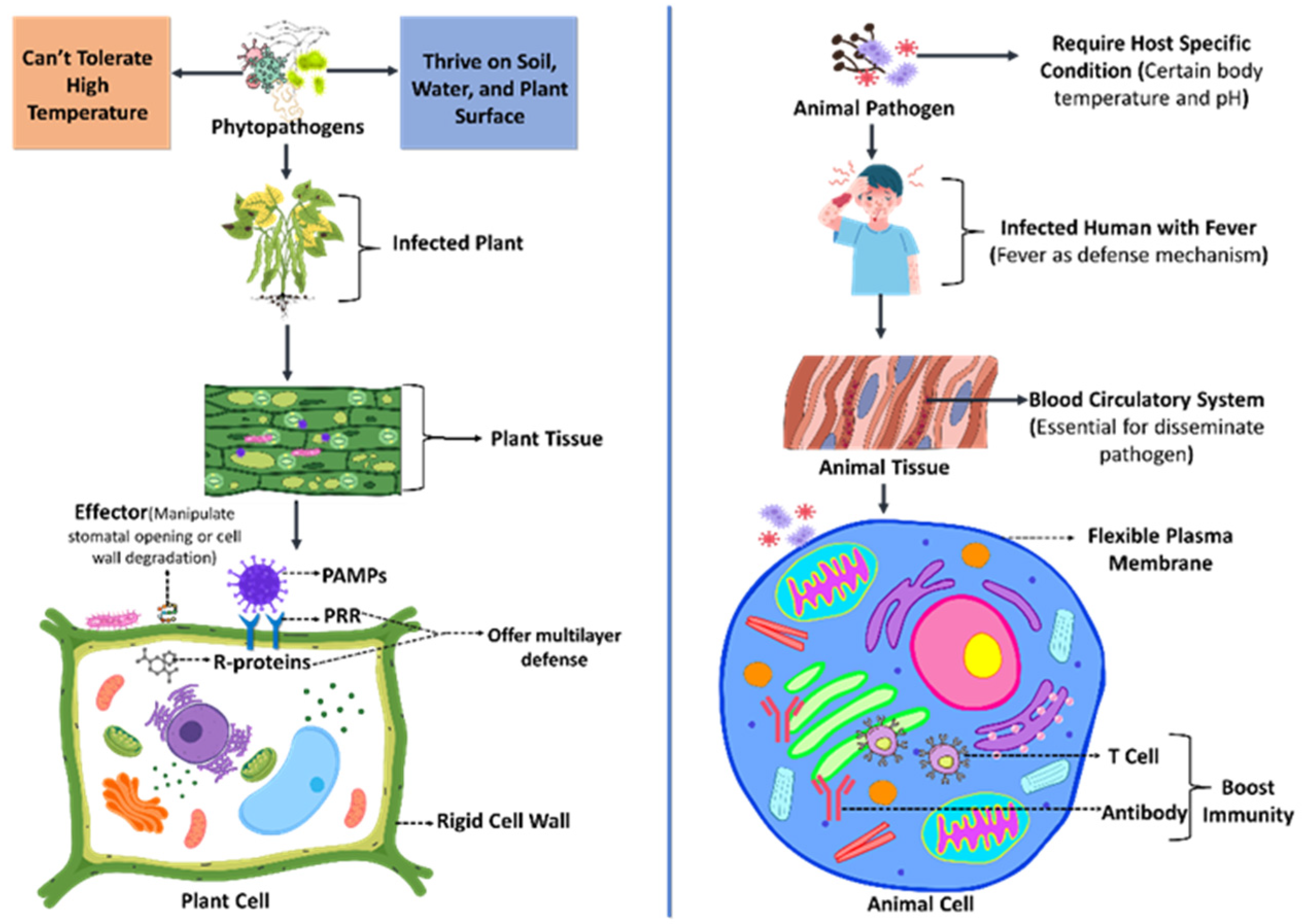
2. Natural Barriers Between Phytopathogens and Animal Pathogens to Prevent Spillover

The natural barriers that segregate phytopathogens and animal pathogens play a critical role in preventing the spillover of diseases between plants and animals. The primary natural barrier lies in the fundamental structural and biological differences between plant and animal cells (Figure 1). Typically, plant and animal cells share fundamental structural similarities within the cytosol, with the exception of certain organelles, such as chloroplasts, that are unique to plant cells. However, plant cells are encased in rigid cell walls made of cellulose, hemicellulose, and lignin, which provide a strong physical barrier against pathogen entry [15,16]. In contrast, animal cells have more flexible plasma membranes, making them vulnerable to pathogens that use different modes of invasion. Furthermore, many animal pathogens depend on circulatory systems that are crucial for their propagation, dissemination, or colonization of the gut or bloodstream, which do not exist in plants [17,18].
The immune systems of plants and animals are distinct and serve as a vital natural barrier that limits the spillover of pathogens between these two kingdoms. Unlike animal cells, plants do not possess immune cells; instead, they rely exclusively on innate immunity, which encompasses several key components [19]. These include pattern recognition receptors (PRRs) that identify pathogen-associated molecular patterns (PAMPs), resistance (R) proteins that detect specific pathogen effectors, and the synthesis of secondary metabolites and reactive oxygen species to restrict pathogen proliferation [20]. In contrast, animals possess both innate and adaptive immune systems, offering a multi-layered defense. The adaptive immune system, characterized by its ability to generate antibodies and memory T-cells, enables highly specific and long-lasting immunity against recurring threats [21]. Due to these fundamental differences, pathogens that have evolved to evade or suppress immune responses in one kingdom often lack the necessary adaptations to exploit the immune pathways of the other. This divergence in immune strategies effectively minimizes cross-kingdom pathogen spillover.
Like their hosts, both phytopathogens and animal pathogens show specialization and only undergo co-evolution with their respective hosts. Plant pathogens produce effectors tailored to manipulate plant-specific processes, such as stomatal opening or cell wall degradation [22]. These adaptations are ineffective in animal hosts. Conversely, animal pathogens require host-specific receptors, enzymes, or conditions absent in plants [2]. Additionally, the ecological separation of plants and animals also reduces spillover risks. Phytopathogens thrive in environments like soil, water, or plant surfaces, where their survival strategies are optimized for plant tissues [23]. Animal pathogens typically need specific conditions found in their hosts, such as a particular body temperature or pH level, which are not present in plants. For instance, many animal pathogens thrive at an optimal temperature of about 37 °C, whereas most plant pathogens cannot endure such high temperatures. This limitation reduces their ability to infect humans and animals [24]. As a result, fever serves as a common defensive mechanism in various animals against invading microorganisms [25]. As shown by the above examples, the natural barriers separating phytopathogens from animal pathogens are robust and primarily upheld by fundamental differences in cellular structure, immune mechanisms, ecological niches, and evolutionary adaptations. However, these barriers can be weakened by human physical conditions, activities, climate change, and habitat overlap, creating significant opportunities for spillover events.

3. Requirements for Cross-Kingdom Pathogenesis

Pathogens specializing in plant or animal hosts have evolved highly specific virulence and survival strategies to colonize their primary hosts effectively. These adaptations are tailored to exploit the distinct physiological and immune characteristics of plants or animals. However, substantial physiological and immunological differences between these host systems generally act as natural barriers against cross-kingdom infections. To establish an infection in a non-primary host, a phytopathogen must overcome significant challenges, including adapting to an unfamiliar biochemical environment, evading immune responses, and acquiring the essential nutrients necessary for survival. Cross-kingdom pathogenesis typically involves three major stages: entry, immune evasion, and adaptation (Figure 2). Phytopathogens can enter the host via multiple transmission routes, such as airborne inhalation, direct contact, or ingesting contaminated food. Upon breaching the epithelial barrier—either through wounds or enzymatic degradation of host tissues—they engage in immune evasion strategies, including chemotaxis and biofilm formation, to persist within the host. Adaptation to the new host environment is facilitated by key molecular pathways, such as the mitogen-activated protein kinase (MAPK) and cyclic AMP/protein kinase A (cAMP/PKA) pathways, which regulate fungal penetration, appressorium formation, and responses to host-derived cues. Iron acquisition mechanisms, including siderophores and iron transporters, play a crucial role in nutrient competition with the host, ensuring pathogen survival. The calcineurin signaling pathway also helps phytopathogens endure environmental stress within nutrient-limited host conditions. These sophisticated adaptation strategies enable phytopathogens to bypass cross-kingdom barriers and successfully infect non-primary hosts. The ability of plant pathogens to exploit these mechanisms in human hosts poses a significant public health risk as it compromises the body’s ability to anticipate and mount effective immune defenses against these emerging threats.

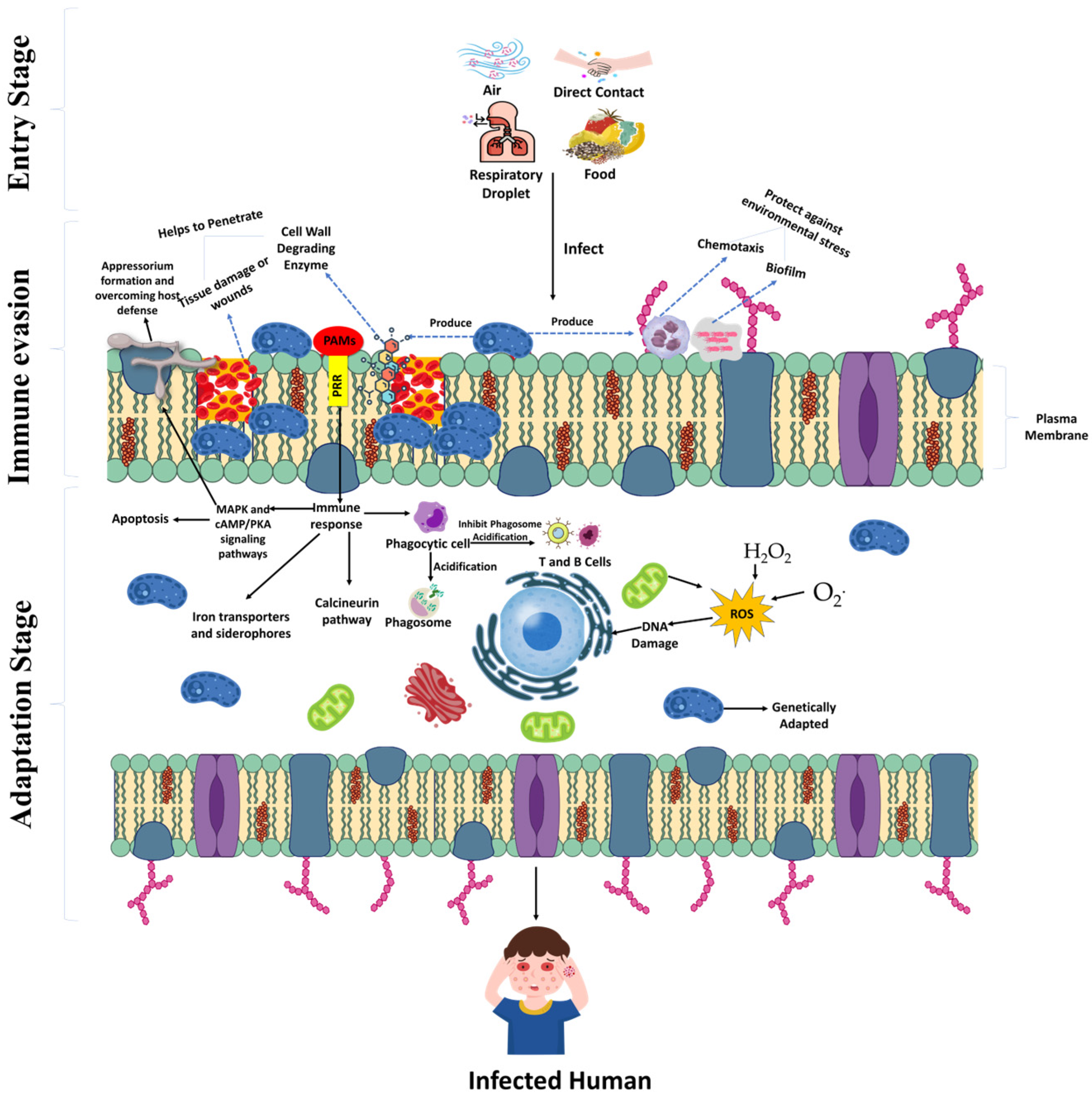
3.1. Breaching the Exterior Physical Barrier

The ability of pathogens to breach external barriers is critical for establishing infections across plant, animal, and human hosts. These barriers, including the epidermis, cuticles, or cell walls in plants and the skin or mucosal layers in animals, serve as the first line of defense. Phytopathogens that infect across kingdoms often share mechanisms enabling them to overcome these obstacles, highlighting similarities between host and pathogen interactions across diverse systems.
Pathogens must first withstand external environmental challenges before reaching potential infection sites. Mechanisms like biofilm formation and chemotaxis provide protection against environmental stressors and help pathogens secure essential nutrients. These adaptations are common to bacterial pathogens, which use these strategies to approach their host effectively [26].
Most animal pathogens require tissue damage to penetrate the host’s physical barriers. Common entry points include wounds caused by insect bites or medical interventions, such as catheter insertions. Some pathogens, however, circumvent these requirements; for example, Mycobacterium tuberculosis infects hosts through respiratory droplets, while Salmonella spreads via air, food, or direct contact [26,27]. Unlike animals, plants possess rigid cell walls, an additional structural defense. Certain phytopathogens, such as species of Pectobacterium (formerly Erwinia), produce cell-wall-degrading enzymes that allow them to penetrate these defenses actively.
Once the external barrier is breached, pathogen behavior diverges depending on the host type. Some bacterial pathogens invade and multiply within animal host cells (e.g., Salmonella, Mycoplasma, and Listeria), while others remain extracellular, surviving within host tissues despite immune responses [2]. On the contrary, in plant hosts, intracellular bacterial invasion is rare. A notable exception is the establishment of Rhizobium bacteria within root nodules as bacteroids [28]. Most bacterial phytopathogens, however, proliferate within the intercellular spaces, where they must contend with plant defense mechanisms.

3.2. Conquering the Principal Host Immunological Response

Conquering the principal host immunological response is crucial for phytopathogens to achieve cross-kingdom infection. The immune systems of animal hosts are designed to detect and respond to pathogens through a complex interplay of innate and adaptive immunity. Innate immunity is the first line of defense, employing PRRs to identify PAMPs. Key components of this system in animals include phagocytic cells like macrophages, neutrophils, and dendritic cells, which engulf and degrade pathogens through phagocytosis [29]. During phagocytosis, the vacuolar ATPase enzyme acidifies the phagosome, creating low pH conditions suitable for hydrolytic enzyme activation to dismantle the pathogen [30,31].
Despite these defenses, some pathogens have evolved sophisticated mechanisms to evade immune responses. For example, Mycobacterium tuberculosis and Legionella pneumophila inhibit phagosomal acidification to survive within macrophages, while pathogens like Pseudomonas aeruginosa and Listeria monocytogenes escape phagosomes by manipulating the host’s actin cytoskeleton [32,33]. Adaptive immunity, unique to animals, further enhances defense through T and B cells, which produce antibodies and facilitate long-term immune memory [34]. These adaptations highlight the dynamic evolutionary arms race between hosts and pathogens and exemplify the dynamic nature of immune evasion. To infect animal hosts, phytopathogens must navigate how to beat robust defense mechanisms. They often adopt specialized strategies to either suppress or bypass immune responses. These adaptations underscore the complexity of cross-kingdom infection and highlight the ongoing struggle between host defenses and pathogen survival strategies.

3.3. Oxidative Stress Adaptations

The oxidative burst produced by the host, characterized by the generation of both radical and non-radical reactive oxygen species (ROS), such as singlet oxygen, superoxide anion, hydrogen peroxide (H2O2), and hydroxyl ion, is recognized as a common defense mechanism against pathogens in both plants and animals [35]. To sustain redox balance, oxidative stress defense systems frequently utilize peroxiredoxins to reduce H2O2, nitric oxide (NO), and alkyl hydroperoxides, using reducing power supplied by NADPH [36,37]. In addition to peroxiredoxins, fungal phytopathogens possess other antioxidant mechanisms, including catalases and glutathione peroxidases [37,38]. The animal-pathogenic fungus Candida albicans expresses an NADPH oxidase, Fre8, which works with Sod5 to create an extracellular H2O2 gradient that promotes hyphal growth [39]. Similarly, in Fusarium oxysporum, H2O2 serves as a chemotropic signal guiding growth toward a host-derived peroxidase gradient [40]. Mild oxidative stress has also been found to enhance polarized growth under in vitro conditions in a thioredoxin-dependent manner [41]. During B. cinerea infection, O2 accumulates at the fungal hyphal tips, and H2O2 is produced via Nox complexes at the interface of the host cell wall and the plasma membrane. Disruption of the Nox complex leads to delayed appressoria formation and abnormal morphology [42]. While there is strong evidence that ROS contributes to fungal differentiation, the targets of ROS and their precise role in fungal development are still unclear.

3.4. Nutritional Adaptation

Nutritional adaptation is a crucial factor in determining the survival of phytopathogens in cross-kingdom infections. This adaptation enables phytopathogens to survive and grow in a wide range of susceptible hosts. A key aspect of this adaptation is the ability of pathogens to utilize host-derived nutrients (Figure 2). For example, L. theobromae has been shown to metabolize salicylic acid and phenylpropanoid pathway precursors from grapevine as carbon sources, exploiting host defense metabolites for its growth [43]. This capacity to exploit the plant’s defense responses demonstrates the dual role of nutrient acquisition in overcoming host resistance mechanisms.
Advanced regulatory pathways, such as the mitogen-activated protein kinase (MAPK) pathway and the cyclic AMP/protein kinase A (cAMP/PKA) signaling pathway, are crucial for nutritional flexibility [44]. If deprived of essential nutrients and limited to a particular region, the pathogen undergoes rapid cellular alterations, leading to cell death. The activation of MAPK leads to the transmission of signals to intrinsic apoptotic proteins, ultimately resulting in cell death [45]. These pathways help organisms recognize the host’s conditions and respond to nutrient availability. One study delves into the distinct functions of three Mitogen-Activated Protein Kinases (MAPKs)—Fmk1, Mpk1, and Hog1—in Fusarium oxysporum’s stress responses and pathogenicity across plant and animal hosts. The study uncovers the vital roles of these MAPKs in developmental regulation, stress mitigation, and virulence mechanisms [46]. They play essential roles in processes like fungal penetration, appressorium formation, and overcoming host defenses.
Iron acquisition is another critical nutritional strategy for phytopathogens. These phytopathogens are able to express a variety of iron transporters and siderophores. These apparatuses solubilize and transport iron, a vital nutrient for their development in human cells [47,48]. In addition, the calcineurin pathway provides support for morphogenesis, cell wall integrity, and adaptability to elevated temperatures and alkaline pH in fungal pathogens, which contributes to their survival in host habitats [49,50]. This route also helps maintain cation homeostasis, essential for the pathogens to survive. This pathway is crucial for both plant and human pathogenicity. The interplay of these nutritional and adaptation mechanisms highlights the sophisticated strategies employed by phytopathogens for cross-kingdom pathogenesis.

3.5. Thermal Adaptation

Thermal adaptation is a critical factor enabling phytopathogens to infect mammalian cells, as it involves overcoming the thermal barriers conferred by mammalian endothermy. Cross-kingdom pathogenic fungi exhibit remarkable thermal resilience, with species like Aspergillus fumigatus capable of growth at extreme temperatures up to 70 °C [51]. Species like Lasiodiplodia theobromae and L. hormozganensis display temperature-specific adaptations that modulate their pathogenicity and virulence. L. theobromae causes more extensive lesions in grapevines at 35 °C compared to 25 °C, demonstrating a clear correlation between thermal conditions and pathogenic severity [52]. Similarly, L. theobromae isolates from coconut trees exhibit mammalian cell toxicity only at growth temperatures of 25–30 °C [53,54], while L. hormozganensis induces substantial cell mortality at 37 °C [54]. These examples reflect an enhanced adaptation of cross-kingdom fungi to mammalian thermal conditions.
Such thermal adaptation is supported by molecular evidence. For instance, L. hormozganensis shows a greater abundance of pathogenesis-related proteins when grown at 37 °C [48]. This indicates a temperature-driven enhancement of its virulence machinery. Notably, Nudix hydrolase effectors, such as YSA1 and NUDT1, are upregulated at 37 °C, contributing to pathogenicity by suppressing host defenses [55]. This capability aligns with transcriptomic and proteomic data showing the upregulation of stress response and pathogenesis-related proteins at higher temperatures, indicating molecular adaptation to stressful conditions [43,54].
Moreover, heat shock proteins are a class of molecular chaperones that are highly conserved and ubiquitous in organisms ranging from microorganisms to plants and humans. Thermal stress triggers the expression of heat shock proteins (HSP60, HSP70), which facilitate protein folding and contribute to thermotolerance, potentially enhancing infection in mammalian hosts [56]. The modulation of heat shock factors (HSFs) in C. albicans is crucial for regulating its transition from yeast to hyphal forms, which is linked to its virulence in a Drosophila infection model [57]. Aspergillus spp. produce aflatoxins, which are toxic compounds that can lead to severe health issues, including liver cancer in humans. The expression of HSP70 in Aspergillus spp. fungi is related to their ability to produce aflatoxins, especially under varying temperatures. Interestingly, higher temperatures can reduce HSP70 expression, which in turn affects aflatoxin production [56,57,58]. Cryptococcus neoformans is known for causing serious infections, particularly in immunocompromised individuals. Heat shock proteins (Hsps), particularly Hsp90, are crucial for maintaining cellular homeostasis in fungal pathogens and play a significant role in infection [59].
Additionally, the fungal ability to sense and adapt to host conditions, including temperature, is mediated by regulatory pathways like the MAPK and cAMP/PKA pathways, which are essential for host penetration and dissemination [44]. These pathways, combined with stress-responsive proteins and effectors, enable phytopathogens to overcome host defenses and establish infections in mammalian systems.

3.6. Genetic Adaptations in Cross-Kingdom Infection

The genes that code for several traits involved in pathogenicity or virulence of pathogens have been cloned and characterized, as have the evolutionary relationships of a few genes for enzymes and toxins known to play roles in the occurrence of diseases. Genetic adaptations enabling cross-kingdom infection are remarkable examples of evolutionary innovation, allowing certain pathogens to breach the fundamental biological barriers that typically separate different kingdoms of life. These adaptations involve a suite of molecular strategies that pathogens use to recognize, invade, and manipulate evolutionarily distant hosts.
For instance, a study on the genetic virulence determinants of Fusarium oxysporum in immunosuppressed mice examined the role of specific fungal genes. Mice infected with a wild-type tomato-pathogenic strain developed disseminated infections and died. Knockout mutations in three virulence-related genes showed different effects in plants and animals. The mitogen-activated protein kinase gene, essential for plant virulence, was not required for virulence in mice. The pH response transcription factor was crucial for animal but not plant virulence. Notably, mice infected with chitin synthase knockout mutants died rapidly (within 24 h) due to severe lung damage caused by large, irregular conidia obstructing lung capillaries [60]. Furthermore, the genetic adaptations of Colletotrichum species, such as enzyme production, environmental adaptability, and host specificity, are essential to their ability to infect a diverse range of hosts [13]. Similarly, the genetic traits of Agrobacterium tumefaciens, including its Ti plasmid, virulence genes, and horizontal gene transfer mechanisms, serve as a foundation for understanding its potential for cross-kingdom interactions and their implications for human cells [14]. So, the genetic adaption capacity of certain microbes to breach kingdom barriers raises significant concerns for agriculture, public health, and ecosystem stability.

3.7. Environmental and Anthropogenic Involvement

Environmental reservoirs play a significant role in cross-kingdom pathogenesis involving phytopathogens like Fusarium species, which have been identified in hospital water distribution systems (e.g., drains, faucet aerators, and showerheads) as potential sources of nosocomial infections [61]. Several pathogenic Fusarium species have been isolated from these environmental sources and hospital plumbing systems [62]. Genotyping studies during hospital outbreaks involving immunocompromised patients have linked hospital water systems to patient infections, suggesting that showers may facilitate aerial dispersal of conidia, leading to host transmission [63]. Additionally, airborne conidia, especially in areas with poor air sealing and ventilation, is another infection source [61]. Genetic similarities between Fusarium from indoor hospital air and isolates from hematologic patients’ blood cultures further suggest that airborne transmission contributes to fusariosis [64]. Inhalation of airborne conidia is believed to cause infections, such as sinusitis and pneumonia, even without systemic dissemination [65]. In response to environmental stressors, including desiccation, antimicrobial agents, and fluctuating oxygen availability, phytopathogens like Fusarium develop enhanced survival mechanisms, such as biofilm formation and metabolic dormancy, which increase their persistence in hospital environments. These stress responses not only promote survival outside of the plant host but also facilitate adaptation to human hosts, increasing the risk of opportunistic infections.
Burkholderia infects various plant species, including onion, rice, sorghum, and velvet beans [66], and may also have a direct route to humans. Anthropogenic disturbances, such as urbanization and agricultural intensification, can amplify the spread of these bacterial pathogens by altering microbial ecosystems and increasing pathogen stress responses. Their indirect transmission to humans, or even within healthcare environments, may occur through other organisms, such as ants and flies. These insects act as carriers and facilitate the movement of environmental isolates to different environments and hosts [67].
Human activities have increasingly facilitated cross-kingdom pathogenesis by exerting selective pressures that drive pathogen evolution and stress adaptation. Industrial agriculture, globalization, deforestation, and environmental pollution disrupt ecological balances, creating novel stress conditions that compel phytopathogens to evolve adaptive mechanisms for survival in diverse hosts. For example, exposure to fungicides in agricultural settings triggers stress responses in fungal pathogens, leading to the development of antifungal resistance, which in turn compromises disease management in clinical settings. Managing Fusarium outbreaks is a major agricultural challenge typically addressed through chemical and biological control methods, including the widespread use of azole fungicides, particularly triazoles, at rates around 100 g/ha [68,69]. However, the extensive agricultural application of azoles has raised concerns in clinical settings, as it may contribute to antifungal resistance in human pathogens. Studies have linked azole-resistant Aspergillus fumigatus strains from flower fields to fungicide use [70,71,72], with similar resistance detected in other environments, such as compost sites, gardens, vineyards, and agricultural soils [73,74]. In India, both clinical and environmental isolates of Fusarium falciforme and F. keratoplasticum from keratitis patients and agricultural settings showed high MIC values for fluconazole, ketoconazole, and terbinafine [75]. Colombia, a leading flower exporter, could also be at risk, primarily as the flower industry—centered in the Bogotá savannah—relies on fungicides that inhibit ergosterol synthesis [70,76]. Despite their specialization to plants, species of Pantoea have also been discovered to be pathogenic to humans. Now classified as an opportunistic human pathogen, P. agglomerans was implicated in US and Canadian outbreaks of septicaemia caused by contaminated closures on infusion fluid bottles [67]. P. agglomerans has since been associated with the contamination of intravenous fluid, parenteral nutrition, blood products, propofol, and transference tubes, causing illness and even death [77]. P. agglomerans has also been obtained from the joint fluids of patients with synovitis, osteomyelitis, and arthritis, where infection often occurs following injuries from wood slivers, plant thorns, or wooden splinters [78,79].
From the discussion above, it is evident that environmental stressors, including changes in temperature, moisture availability, and exposure to disinfectants, can induce genetic shifts in phytopathogens, increasing their tolerance to antimicrobial compounds and host immune defenses. These stress-driven adaptations further enhance their ability to infect nonprimary hosts, highlighting the critical role of environmental and anthropogenic factors in shaping pathogen evolution and cross-kingdom transmission.

4. Cross-Kingdom Pathogenicity and Implications for Health

Certain plant pathogens, including fungi, oomycetes, bacteria, viruses, and nematodes, have evolved to overcome kingdom barriers and infect humans and animals. While some of these phytopathogens only cause mild, self-limiting infections, others can lead to severe or systemic disease, especially in immunocompromised individuals or animals.

4.1. Cross-Kingdom Pathogenesis by Fungi and Oomycetes

Many fungal and oomycete pathogens have a rare yet remarkable ability to infect hosts across the plant and animal kingdoms, causing diseases in phylogenetically distant organisms (Table 1). Some can cause disease in both plants and humans, posing unique public health and agricultural challenges. Alternaria species, for instance, are well-known plant pathogens that cause blossom and leaf blight [80,81], but they are also increasingly recognized as human allergens linked to allergic rhinitis and asthma [82]. In immunocompromised individuals, infections by Alternaria, such as A. alternata and A. infectoria, are rising [83]. Notably, these fungi have been linked to severe infections like phaeohyphomycosis in kidney transplant patients and keratitis following eye trauma [83,84]. A. infectoria was also reported to cause cutaneous lesions in a heart transplant patient [85] and a rare brain abscess in a child with chronic granulomatous disease [86].
Bipolaris species also exhibit cross-kingdom pathogenicity. Bipolaris spicifera, a known plant pathogen on Poaceae plants [87,88,89], has been implicated in surgical site infections [90]. Similarly, Bipolaris hawaiiensis (Syn. Curvularia hawaiiensis), which affects Bermuda grass [91], has been reported as a pathogen in human surgical wounds. Additionally, B. australiensis has been associated with corneal ulcers [92].
Fusarium species represent another significant example, with strains like F. graminearum producing mycotoxins toxic to animals and humans (Table 1). F. proliferatum and F. solani, both of which can infect humans through wounds or ocular contact, frequently cause keratitis [93]. This situation is more prevalent in those who wear contact lenses.
The Aspergillus genus encompasses plant pathogens like A. fumigatus, A. flavus, A. niger, and A. terreus, which cause diseases in crops, such as corn, peanuts, and onions [94]. In humans, A. fumigatus is a leading cause of chronic pulmonary aspergillosis, a life-threatening infection in patients with respiratory conditions [95]. Aspergillus infections can also exacerbate asthma, leading to severe respiratory complications. Additionally, these fungi contaminate food supplies with carcinogenic mycotoxin aflatoxins, which present major food safety risks [96].
Cladosporium species, such as C. cladosporioides and C. herbarum, are plant pathogens that can infect humans, causing respiratory allergies and other infections [97]. Similarly, species belonging to the genus Exserohilum, such as E. rostratum, which is found in grasses, pineapple, and other monocots [98], have the potential to infect humans. Infections caused by this fungus can lead to serious illnesses, including fungal meningitis [99]. There was a notable outbreak of E. rostratum meningitis in the United States that resulted in significant mortality [99,100].
L. theobromae is an important plant pathogen, especially in tropical regions, infecting over 500 plant species [101]. But, over the past decades, the fungus has been increasingly recognized in opportunistic human infections, causing conditions like rhinosinusitis, skin infections, and, rarely, death [102]. Related species, such as L. hormozganensis, have also been reported as cross-kingdom pathogens capable of infecting plants and humans [48].
Macrophomina phaseolina is a significant phytopathogenic fungus that affects hundreds of plant species globally [103]. Recently, the first instance of endophthalmitis attributed to M. phaseolina was documented [104]. A literature survey indicated that M. phaseolina was predominantly linked to ocular infections (76.9% of cases), followed by skin and concurrent skin–joint infections. Most individuals with M. phaseolina infection (63.6%) exhibited no identifiable immunosuppressive variables.
Rhizopus oryzae (syn. Rhizopus arrhizus), the causative agent of Rhizopus rot in plants [105,106,107], is also the primary pathogen behind mucormycosis and rhino–orbital–cerebral infections. Known for its aggressive angioinvasive growth, mucormycosis carries high mortality rates. Experimental data show that this fungus is responsible for approximately 70% of human mucormycoses, and mortality rates can reach as high as 68–80% [108]. The high mortality rate prevailed during the co-infection of SARS-CoV-2 and mucormycosis in India amid the 2021 SARS-CoV-2 pandemic [109].
Beyond fungi, the phytopathogenic oomycete, such as Pythium aphanidermatum, has been implicated as a cause of invasive wound infections in a combat male in Afghanistan [110]. A second case of pythiosis caused by P. aphanidermatum has been reported in Thailand [111]. Fungal cultures obtained from the necrotic tissues of both legs, both before and after disarticulation, also yielded Mucor circinelloides and Aspergillus flavus, but it was not known whether the infection caused by P. aphanidermatum was a co-infection. In this case, vascular pythiosis occurred without any known history of trauma, resembling cases caused by P. insidiosum reported in earlier studies.
Another genus of the class Oomycetes, Lagenedium, was reported to cause keratitis mimicking ocular pythiosis caused by Pythium [112]. Notably, these oomycete strains exhibit remarkable heat tolerance [48,51,112], a critical adaptation trait that enhances their ability to establish infections in mammalian hosts. This thermal resilience likely facilitates their survival in febrile conditions and other warm environments within the host, overcoming a key defense mechanism against pathogens. The emergence and spread of such heat-tolerant strains underscore a growing public health concern as they expand the ecological range of these pathogens. This is especially true in the warming climate, which may further select for thermotolerant traits.
Table 1. Fungal and oomycete phytopathogens with documented cases of human and animal infections.
PathogenPlant SymptomsPlant HostsClinical ManifestationsReferences
Alternaria infectoria, A. alternataBlossom blightGuayulePhaeohyphomycosis,
keratitis
[83,84]
Aspergillus fumigatus, A. flavus, A. niger, A. terreusEar rot, boll rot, yellow mold, black mold, fruit rot Corn, cotton, peanut, onion, garlic, grapes, pomegranates, citrus Pulmonary aspergillosis[94,95]
Bipolaris spicifera, B. hawaiiensis,
B. australiensis
Leaf spot, leaf blight, chlorosis, necrotic lesionsSugarcane, switchgrass, buffalograss, bermudagrassSurgical site infection, corneal ulcer[88,90,92]
Colletotrichum truncatumLesion, blight,
necrosis
Strawberry, citrus, cerealsOphthalmic infection[113,114]
Cladosporium allicinum, C. angustisporum, C. cladosporioides, C. flabelliforme, C. funiculosum, C. halotolerans, C. herbarum, C. macrocarpum, C. perangustum, C. ramotenellum, C. sphaerospermum, C. subinflatum, C. subuliformeLeaf spot, blossom blightDendrobium, Echeveria, strawberryRespiratory tract superficial fluid infection[97]
Exserohilum rostratumLeaf spotGrasses and other monocots Meningitis[98,99,100]
Fusarium graminearum, F. proliferatumHead blight, root rotTomato, tobacco, legumes, cucurbitsBlood infection[113,115]
Fusarium solaniFoot and/or root rot, sudden death syndromeLegumes, solanaceous plantsOnychomycosis, keratitis[116,117]
Lasiodiplodia theobromae, L. hormozganensisDieback, stem end rot, seed rot, leaf blight, boll rot, basal stem rotMango, kenaf, snake plant, grape,
papaya, cotton, castor bean
Keratitis, sinusitis,
skin infections, phaeohyphomycotic cyst
[48,102,118,119,120,121]
Macrophomina phaseolinaStem and root rot, charcoal rot, and seedling blightJute, beans, potato, sorghum, corn, wheat, sun Endophthalmitis, skin infections, and skin–joint infections [103,104]
Pythium aphanidermatumNecrosis, rotSoybean, cucurbits,
cotton
Pythiosis[110,111]
Rhizopus arrhizus (syn. oryzae)Rhizopus rot Apple, banana, mulberry, sweet potato, tobacco, tomato, forage grass Mucormycosis[105,106,107,122,123,124]

4.2. Cross-Kingdom Pathogenesis by Bacteria

Bacterial plant pathogens that typically infect plants are increasingly recognized for their ability to cause opportunistic infections in animals, including humans (Table 2). Notably, genera like Agrobacterium, Erwinia, Burkholderia, Pantoea, Ralstonia, and Pseudomonas include species that demonstrate cross-kingdom pathogenicity. Agrobacterium tumefaciens, now reclassified as Rhizobium radiobacter, is widely known for colonizing plant roots and causing crown gall disease [125]. In human health contexts, R. radiobacter has been implicated in catheter-related bacteremia, particularly in neutropenic patients and immunocompromised individuals [126,127]. In addition to these cases, over 20 instances of human infections linked to Agrobacterium species have been reported, including a recent case of contact-lens-associated keratitis caused by R. radiobacter [128].
The plant pathogen Erwinia persinicus exemplifies cross-species pathogenicity, affecting a wide range of plant hosts and demonstrating its potential to infect animals, including humans. In plants, it is known to cause pink-pigmented soft rot in garlic, onions, lettuce, mushrooms, tomato, potato, barley, and parsley root [129,130,131,132]. It has also been identified as the etiological agent of necrotic leaf spots on legumes and leaf wilting in lucerne [133,134]. Beyond plant infections, E. persinicus demonstrates pathogenicity in model organisms, such as Caenorhabditis elegans and Drosophila melanogaster, and it was isolated from a human patient with a urinary tract infection [135]. This bacterium experimentally induced pathological lesions in mice when injected intraperitoneally [136], underscoring its opportunistic and cross-kingdom potential. Another member of the genus, Erwinia billingiae, has been implicated in human health, causing cutaneous infections and bacteremia [137]. These examples underscore the diverse host range and pathogenic versatility of Erwinia species, warranting further exploration of their biology and mechanisms of infection.
In addition to Agrobacterium and Erwinia, some species within the genera Burkholderia and Pantoea also demonstrate the capacity to infect animal hosts. The Burkholderia cepacia complex (BCC) comprises 24 closely related species functioning as both phytopathogens and biocontrol agents [138]. However, during the 1980s, B. cepacia emerged as a lethal pathogen in cystic fibrosis (CF) patients [139]. Experimental studies have revealed that at least three strains of B. cepacia can penetrate airway barriers in mice [140]. Other Burkholderia species, such as B. gladioli and B. glumae, have been implicated in severe human infections, including pneumonia in individuals with chronic granulomatous disease and septicemia in CF patients [141,142]. Despite these documented cases, the genetic distinctions between plant-pathogenic and human-pathogenic Burkholderia strains remain unclear, posing challenges to understanding their cross-kingdom pathogenicity.
Several Pantoea species, primarily recognized as plant pathogens, have also been associated with human infections, highlighting their opportunistic potential. For example, Pantoea agglomerans has been implicated in infections among 12 cancer patients, traced to contamination from a hospital sink or infected intravenous supplies [77,143]. Similarly, Pantoea ananatis has been linked to cases of anal hemorrhage and fever during colonoscopy procedures in diabetic patients [144]. A particularly tragic instance involved eight neonatal intensive care patients who developed sepsis and succumbed to infections caused by an unidentified Pantoea species. Investigations revealed the source to be contaminated parenteral feeding solutions [145].
Ralstonia pickettii, a close relative of R. solanacearum, the causative agent of bacterial wilt in plants, is a rare Gram-negative opportunistic bacterium. The bacterial pathogen has been reported to cause severe leaf spot and blight on the Bird of Paradise tree (Strelitzia alba (L. f.) Skeels) [146]. While primarily associated with environmental and plant habitats, R. pickettii has been implicated in human infections, particularly in hospitalized or immunocompromised patients. These infections include bacteremia, neonatal sepsis, endocarditis, and meningitis [147].
Pseudomonas aeruginosa is a ubiquitous bacterium found in plants, animals, humans, and diverse environmental niches. In agricultural systems, most P. aeruginosa strains are beneficial, promoting plant growth and offering protection against pathogens [148]. However, pathogenic strains have also been reported [149,150]. Beyond plant-associated roles, P. aeruginosa is a significant human pathogen frequently isolated from the respiratory tracts of cystic fibrosis patients [151]. The bacterium was also associated with chronic obstructive lung infections [152]. Severe infections caused by P. aeruginosa include malignant external otitis, endophthalmitis, endocarditis, meningitis, pneumonia, and septicemia [153]. Interestingly, P. aeruginosa PA14 also exhibits pathogenicity in the nematode Caenorhabditis elegans, a widely used model for studying the virulence of clinical isolates [154].
Table 2. A comprehensive list of bacterial phytopathogens with documented cases of human and animal infections.
PathogenPlant DiseasePlant HostsClinical
Manifestations
References
Agrobacterium tumefaciens (Rhizobium radiobacter)Crown gall diseaseEudicotsBacteremia, fetal death[128]
Erwinia persinicus
Erwinia billingiae
Soft rot, leaf spots, root rot, leaf wilting, fire blightPerishable vegetables and fruitsCervical lymphadenitis[129,130,131,132,133,134,137]
Burkholderia cepacian, B. gladioli, B. glumae, B. cenocepaciaGladiolus cormsMaize, onion, rice, tomatoSepticemia[138,141,142,155]
Pantoea agglomerans
Pantoea ananatis
Leaf spots, blotchesFruit-bearing treesSepticemia[77,143,144,145]
Pseudomonas aeruginosaWilt, rotGinseng, wheat, maize, ArabidopsisMalignant external otitis,
endophthalmitis, endocarditis, meningitis, pneumonia, septicemia
[148,149,150,153,156]
Ralstonia pickettiiLeaf spot, leaf blightBird of ParadiseBacteremia, neonatal sepsis, endocarditis, meningitis[147,157]

4.3. Cross-Kingdom Pathogenesis by Viruses

Plant viruses are ubiquitous in the environment, particularly on fruits and vegetables, exposing both livestock and humans to these highly persistent particles. While plant and vertebrate viruses have specialized host ranges that generally limit infection to specific types of organisms, research on interspecies transmission has primarily focused on viruses in vertebrates (e.g., coronaviruses) [158]. Plant viruses, however, are traditionally considered non-pathogenic to humans and other vertebrates. Some plant viruses can replicate within insect hosts, which act as transmission vectors, but their ability to infect vertebrates remains uncertain.
Despite their distinct host specificity, plant and animal viruses exhibit genetic similarities, suggesting an evolutionary link, as demonstrated in genome-wide phylogenetic analyses [159]. It is known that viruses of three families, Bunyaviridae, Rhabdoviridae, and Reoviridae, can potentially infect plants, animals, and humans. For instance, resilient plant viruses, such as the Tobacco mosaic virus (TMV) and the Pepper mild mottle virus (PMMoV), have been identified in human and animal fecal samples (Table 3). A notable study discovered an unexpected predominance of PMMoV in gut virome samples, with 109 PMMoV virions per gram of dry fecal matter [160]. Subsequent studies linked higher PMMoV levels in feces to symptoms like fever and unusual abdominal pain [159,161] (Table 3).
TMV is remarkably resilient, retaining stability in tobacco products, and it has been found in the lungs of both smokers and passive smokers, as well as in the bronchoalveolar fluid of intubated patients [162]. Elevated anti-PMMoV antibodies have been detected in individuals exposed to PMMoV [163]. Similarly, anti-TMV IgG levels were found to be significantly higher in smokers than in non-smokers [164].
Although definitive evidence of plant viruses reproducing in vertebrate cells is lacking, some studies suggest the possibility. Early research demonstrated that TMV RNA could be translated in Xenopus laevis oocytes [165], and more recent studies have shown TMV entering human HeLa cells, with TMV-derived proteins accumulating on autophagosomal membranes [166]. Similarly, the Cowpea mosaic virus was observed attaching to human endothelial cells through surface vimentin proteins [167]. The Providence virus, an insect virus, has even been shown to infect cowpeas and replicate within two mammalian cell lines, suggesting that it could cross cellular barriers across kingdoms [161].
Phytoviruses and their persistence in animal excreta have been well-documented. Earlier studies revealed that 46% of virome sequences from Californian bat guano and 27% from Texan bat guano corresponded to Luteoviridae and Sobemovirus. In comparison, 3.4% of sequences from wild rodent stools were linked to phytoviruses, such as Nanoviridae, Geminiviridae, and Alphaflexiviridae [168]. Members of Partitiviridae, Tymoviridae, and Secoviridae have also been identified in bat and rodent stools [168,169]. Additionally, animal transmission of sobemoviruses has experimentally been reported, such as the Rice yellow mottle virus by cows, donkeys, and grass rats and the Subterranean clover mottle virus by sheep [159].
Interestingly, algae viruses have been identified in humans. For example, viruses cultured on marine diatoms were detected in 39% of cervicovaginal secretions from Ukrainian women with gynecological conditions [170]. A green algae phycodnavirus, Acanthocystis turfacea chlorella virus-1 (ATCV-1), was found in 44% of human oropharyngeal samples, where its presence correlated to cognitive impairments [171]. The experimental inoculation of ATCV-1 in mice confirmed altered cognitive functions and hippocampal gene expression.
This growing body of evidence hints at the possibility of plant viruses infiltrating and replicating within animal cells, although no conclusive evidence exists that these viruses cause illness in vertebrates. However, the potential for plant viruses to cause disease in animals cannot be dismissed and merits further investigation [172].
Table 3. List of viral and nematode phytopathogens with documented cases of human and animal infections.
PathogenPlant SymptomsPlant HostsClinical ManifestationsReferences
PMMoVChlorosis, mottlingPepperFever, abdominal pains[159,160,161,162,163]
TMVStunting, yellowingTobacco, tomatoPulmonary diseases[162,164,166]
Xiphinema brevicollumRoot damage (dagger nematode) Tropical fruits, ornamental plantsSevere abdominal pain [173,174,175]

4.4. Cross-Kingdom Pathogenesis by Nematodes

Phytopathogenic nematodes do not usually pose a threat to humans or animals. They are highly specialized to infect plants and specifically adapted to the physiology of their plant hosts. Their infection mechanisms, such as style-mediated feeding, parasitic strategies, environmental adaptation, and molecular interactions, are tailored to plants [176]. In contrast, some free-living nematodes, including species like Strongyloides, Ancylostoma, and Ascaris, can infect humans and animals [177,178]. Because these nematodes are taxonomically and ecologically distinct, phytopathogenic nematodes have long been regarded as non-threatening to human and animal health. Nevertheless, their impact on crops can indirectly threaten food security and animal well-being by reducing agricultural productivity [179].
However, a recent case report from France provides the first documented evidence of human infection by a phytopathogenic nematode, Xiphinema brevicollum [175]. This nematode, part of the Longidoridae family and commonly known as the dagger nematode, is primarily recognized for its role as a migratory ectoparasite in plants (Table 3). It inflicts economic damage by feeding on plant roots and acting as a vector for plant-pathogenic viruses [174]. Xiphinema brevicollum is frequently associated with tropical fruits and was previously considered exclusively plant-specific [173].
In the reported case, a 55-year-old woman presented with severe abdominal pain following a trip to the Dominican Republic, where she consumed tropical fruits and raw vegetables. Clinical examinations revealed hypereosinophilia and the presence of X. brevicollum eggs and larvae in her stool. Molecular analyses, including mitochondrial DNA sequencing, confirmed the species. Treatment with albendazole resolved symptoms, suggesting that the nematode was viable within the human gastrointestinal tract [175].
This case highlights the previously undocumented pathogenic potential of Xiphinema brevicollum in humans. The patient likely contracted the infection via oral ingestion of contaminated produce, with the eggs having sufficient time to mature and hatch. While nematodes from the Heteroderidae family, such as Meloidogyne and Heterodera species, have occasionally been detected in human feces due to contaminated raw or unwashed vegetables [180], such cases remain rare.
The emergence of such infections raises significant zoonotic and food safety concerns. Changes in dietary habits, including increased consumption of raw produce, and environmental factors, such as urbanization and ecological disruption, may enhance the risk of similar parasitic infections. Although rare and non-transitional, such cases represent an emerging area of concern in parasitology, necessitating increased vigilance from clinicians and public health professionals. This finding underscores the need for further research on the zoonotic potential of plant-parasitic nematodes in the context of global food safety and environmental change.

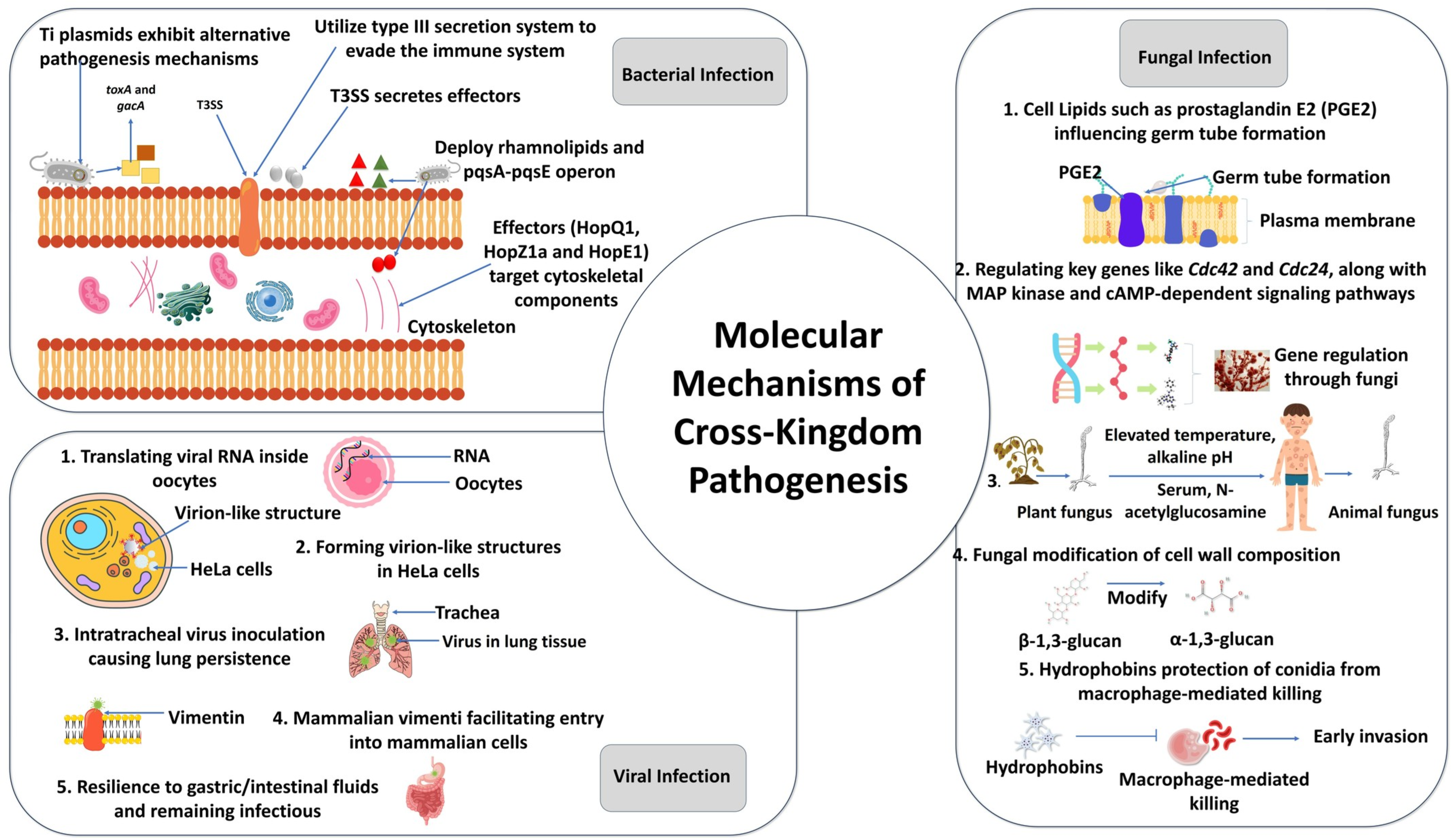
5. Molecular Mechanisms of Cross-Kingdom Pathogenesis

The molecular mechanisms underlying cross-kingdom pathogenesis by phytopathogens in humans and animals remain less understood compared to their well-characterized interactions with plants. Although cross-kingdom infections are often reported as isolated cases, studies suggest that these pathogens exhibit remarkable adaptability by employing distinct pathogenicity factors across different hosts (Figure 3) [2]. Some research indicates that Agrobacterium tumefaciens, which causes crown gall disease in plants through tumor-inducing (Ti) plasmids, may interact with human cells in vitro and exhibit alternative mechanisms of pathogenesis in humans that are unrelated to plant tumorigenesis [181]. It is believed that the bacterium’s T-DNA transfer mechanism could potentially allow it to modify the genome of human cells. However, this does not constitute an infection in the traditional sense.
Experimental studies with Burkholderia plantarii and Burkholderia pseudomallei have shown that these bacteria utilize the type III secretion system (T3SS) to evade immune responses in both plants and humans. In plants, T3SS facilitates the secretion of effector proteins by Burkholderia, which interfere with plant defense mechanisms, such as the MAPK signaling cascade [182]. In humans, these pathogens employ different virulence factors to avoid immune detection. For example, B. plantarii produces rhamnolipids, which disrupt immune cell signaling by inhibiting phagocytosis and promoting biofilm formation [183]. In contrast, B. pseudomallei regulates the expression of the pqsA-pqsE operon, which plays a role in quorum sensing and contributes to its pathogenicity in human hosts [184].
Another study has reported that plant-associated strains of P. aeruginosa share molecular and metabolic traits with clinical isolates [185] and possess multiple virulence factors critical for human infections [150]. These virulence factors include the production of exotoxins, pyocyanin, rhamnolipids, siderophores, biofilms, swarming motility, and lytic enzyme activity [186]. For instance, to infect Arabidopsis thaliana and animal models like mice, P. aeruginosa utilizes pathogenicity factors, such as toxA and gacA [187,188]. This indicates that bacterial pathogens may exploit shared virulence factors for plant and animal hosts. A similar instance is also observed with Pantoea agglomerans. The bacterium employs a T3SS to infect plant crowns and is also associated with human arthritis [189,190]. In experimental studies, P. agglomerans was shown to activate inflammatory pathways in human synovial cells, further suggesting the conserved nature of its virulence factors across hosts. Interestingly, pathogens like Shigella spp., commonly associated with human diseases, produce type III effectors that interact with plant and animal targets, highlighting their ability to cross ecological boundaries [12]. These studies suggest that molecular tools initially evolved for plant infection can be co-opted for animal pathogenesis, potentially contributing to the emergence of zoonotic diseases.
A central target of cross-kingdom phytopathogens is the actin cytoskeleton, which is crucial for immune responses in both plants and animals. In plants, actin filaments facilitate intracellular transport of pathogenesis-related proteins and phytoalexins, critical for inhibiting pathogen growth [191,192]. In animals, actin dynamics are essential for immune processes such as phagocytosis and antigen presentation [193]. Pathogens like Pseudomonas syringae pv. tomato employ various effectors to manipulate host immune responses. Experimental studies have demonstrated that one such effector, HopQ1, disrupts actin rearrangement in both plant cells and murine macrophages, impairing immune defenses [194]. Other effectors, including HopZ1a and HopE1, target cytoskeletal components to inhibit immune signaling in plants, indicating a conserved strategy for immune evasion [195].
Phytopathogenic fungi exhibit a series of conserved and specialized molecular mechanisms. The germination of fungal spores, a critical step in the infection process, is often initiated by environmental cues specific to the host [196]. Experimental studies have shown that the surface hydrophobicity of Candida albicans conidia facilitates recognition by plant receptors, leading to the activation of signaling pathways that promote germination [197]. In humans, lipids, such as prostaglandin E2 (PGE2), act similarly, influencing germ tube formation in Candida albicans. PGE2 has been shown to bind to specific receptors on C. albicans, triggering cellular responses that support its transition to a pathogenic form, akin to the role of surface waxes in plant infections [197]. The transition of C. albicans between plant- and animal-associated states is governed by the regulation of key genes, including Cdc42 and Cdc24, alongside signaling pathways mediated by MAP kinase and cAMP-dependent protein kinases [198]. Studies have demonstrated that factors like elevated temperature (37 °C, a characteristic of the human host), alkaline pH, serum, and N-acetylglucosamine further influence this transition, enabling the fungus to adapt to animal hosts [199]. Similarly, Paracoccidioides brasiliensis undergoes modifications in cell wall composition, notably shifting from β-1,3-glucan to α-1,3-glucan dominance, to evade host immune responses and establish infections [200]. Hydrophobins are another shared molecular feature of fungal virulence in humans. Hydrophobins are believed to prevent immune recognition and protect conidia of pathogens like Aspergillus fumigatus from macrophage-mediated killing, facilitating early invasion events [201,202]. Moreover, the receptor-mediated uptake of fungal conidia by host cells is critical. In A. fumigatus, β-(1,3)-glucan on conidia binds dectin-1 receptors on pneumocytes, triggering internalization and inflammation, while E-cadherin mediates further uptake [203,204], enabling successful colonization of host cells. These adaptive processes reflect a broader capacity of fungi to remodel their cellular architecture in response to different hosts.
Plant viruses have demonstrated the ability to interact with mammalian systems under experimental conditions, revealing mechanisms that could facilitate cross-kingdom pathogenesis. In a study by Li et al. [166], Tobacco Mosaic Virus (TMV) RNA was translated inside of oocytes of the frog Xenopus laevis, and TMV proteins were detected in HeLa cells, where they accumulated on autophagosomal membranes and formed virion-like structures in the cytoplasm. In that study, it was shown that TMV proteins localize to the autophagosomal membranes of HeLa cells, hinting at the potential hijacking of autophagic processes for viral replication or assembly. Furthermore, intratracheal inoculation of TMV in mice resulted in its persistence in lung tissues, with seroconversion and detection in macrophages, confirmed through immunohistochemistry and RT-PCR [205]. Cowpea Mosaic Virus (CPMV), another plant virus, interacts with mammalian vimentin, a conserved surface protein, facilitating entry into various mammalian cells, akin to mechanisms used by animal viruses [167,206]. CPMV has shown resilience in simulated gastric and intestinal fluids, maintaining infectivity and systemically trafficking to multiple organs in mice [207,208]. However, de novo viral protein synthesis or RNA replication is not indicated [206]. While the natural replication of plant viruses in mammalian cells is unproven, the overexpression of specific transcription factors has rendered human cells open to Tospovirus replication [209,210]. These findings highlight the adaptability of plant viruses, their ability to exploit conserved cellular mechanisms, and the potential risks they pose as reservoirs for zoonotic infections.

6. Drivers of Cross-Kingdom Pathogenesis by Phytopathogens

The ability of phytopathogens to infect non-plant hosts, including animals and humans, is an emerging and complex phenomenon. This cross-kingdom pathogenesis challenges the traditional understanding of host specificity and is driven by several interrelated ecological, genetic, and environmental factors.

6.1. Superseded Immune Systems in Non-Plant Hosts

Immunocompromised individuals are highly susceptible to infections from a wide array of pathogens, including bacteria, viruses, fungi, and other microbes, due to their compromised ability to mount effective immune defenses. This vulnerability can arise from conditions like chemotherapy use, HIV/AIDS, advanced age, genetic disorders, or environmental toxins, as well as nutritional deficiencies and chronic stress. Among these pathogens, phytopathogens are increasingly recognized as opportunistic invaders in humans, especially those with weakened immune systems. A striking example of this phenomenon involves filamentous fungal human pathogens (FFHPs), many of which originate in agricultural or food-related environments. Studies have identified about 70 genera of FFHPs, with several frequently associated with food, underscoring the interconnectedness of food safety and human health [211]. Pathogens like Aspergillus and Mucor can transition from environmental reservoirs to human hosts via inhalation or ingestion, causing severe and often fatal infections in immunocompromised individuals [212,213]. Notably, non-Aspergillus mold infections, such as those caused by Fusarium spp., account for approximately 25% of cases in organ transplant recipients [214]. Phytopathogens like Fusarium oxysporum and Fusarium solani can infect humans, especially those with hematological malignancies or undergoing immunosuppressive therapies, leading to systemic infections with high mortality rates [215,216].
Bacterial phytopathogens, too, have demonstrated opportunistic behavior in humans. Pseudomonas aeruginosa, Burkholderia spp., and Rhizobium radiobacter can cause severe infections in humans with weakened immunity or patients with chronic illness [2,188,217]. Furthermore, yeast-like fungi, such as Candida spp., exploit the immune suppression caused by chemotherapy, mucosal damage, and microbiome disruption to cause systemic infections in cancer and transplant patients [199]. This phenomenon suggests that weakened immune systems may lower the barrier to cross-kingdom infections. While healthy immune systems rely on robust mechanisms, including adaptive immunity in animals, compromised systems may fail to recognize or respond effectively to novel pathogens. Consequently, pathogens with plant-specific traits, such as cell-wall-degrading enzymes, may exploit weakened epithelial or mucosal defenses in animal hosts.

6.2. Genomic Adaptability of Phytopathogens

The remarkable genomic adaptability of phytopathogens plays a pivotal role in their ability to overcome host barriers and extend their pathogenicity across kingdoms. Horizontal gene transfer (HGT) enables phytopathogens to acquire genetic material from unrelated organisms, facilitating rapid adaptation to new hosts, including those in the Animalia kingdom. HGT allows phytopathogens to gain genes encoding toxins, barrier-degrading enzymes, or immune-suppressing factors essential for infection in animal hosts. For example, pathogenic bacteria, including Pseudomonas syringae, frequently acquire virulence genes encoding type III secretion systems (T3SS) or effector proteins via HGT [218]. Similarly, comparative genomics of certain fungal pathogens, such as Aspergillus fumigatus, Alternaria alternata, and Fusarium graminearum, has revealed a substantial amount of horizontally acquired genes in their genomes [219,220], suggesting that HGT might have shaped their pathogenicity in non-native hosts. Additionally, mobile genetic elements (MGEs) (e.g., transposons, plasmids) play a crucial role in HGT, promoting adaptability [221]. MGEs can introduce novel virulence genes or regulatory sequences, enhancing the pathogen’s ability to colonize animal hosts. Transposable elements in Colletotrichum species have been implicated in pathogenicity-related gene variation, aiding their ability to infect multiple host types [114,222,223].
Phytopathogens possess hidden genetic pathways that are usually inactive in plant hosts but can be activated in new environments, such as inside an animal host. Environmental stimuli like temperature changes, host pH, or tissue-specific metabolites can activate latent genes encoding virulence factors, such as proteases and toxins, that facilitate animal host colonization [48]. Cryptococcus neoformans activates genes for capsule formation and thermal tolerance when exposed to mammalian body temperatures, enabling infection of immunocompromised hosts [224].
High mutation rates in phytopathogens provide a mechanism for rapid evolution, allowing for adaptation to new hosts and environments. Under selective pressures in animal hosts, such as immune responses or elevated temperatures, mutations in virulence or stress response genes can enhance pathogen survival. In Fusarium spp., mutations in regulatory and metabolic pathways have been linked to opportunistic infections in animals, particularly in hospital environments [225].
Sexual and parasexual recombination leads to the generation of novel genetic combinations, increasing the evolutionary potential of phytopathogens. These recombination events can integrate genetic material that encodes factors for host-specific virulence, such as adhesion molecules or enzymes for penetrating animal barriers [226]. In species of Aspergillus, recombination has played a key role in the emergence of strains capable of causing opportunistic infections in plants and animals [95,227].
Epigenetic changes, such as DNA methylation and histone modifications, may also allow phytopathogens to modulate gene expression rapidly in response to host-specific cues. Epigenetic mechanisms enable pathogens to fine-tune gene expression when transitioning from plant to animal hosts. Exposure to host-derived signals can alter the expression of genes required for immune evasion or nutrient acquisition [228]. These changes allow for rapid phenotypic plasticity, enabling survival in fluctuating environments.
Genomic regions known as “pathogenicity islands or genomic islands” are enriched with virulence-related genes that can enhance cross-kingdom infectivity [229]. These regions harbor clusters of genes encoding toxins, secretion systems, and stress response proteins essential for infection in non-native hosts [230]. For instance, Pseudomonas aeruginosa possesses two pathogenicity islands that enable it to infect both plants and mammals [157,231], illustrating the versatility of these genomic elements. This genetic versatility equips them with dynamic evolutionary toolkits critical for overcoming host-specific barriers and facilitating cross-kingdom infections.

6.3. Microorganisms and Hosts in Close Proximity

The proximity between plant pathogens and humans or animals, whether through direct contact, contamination, or consuming contaminated foods, significantly increases the risk of infections and health complications. This risk is amplified in environments where human activities intersect with plant production, such as agricultural fields, greenhouses, food storage facilities, and healthcare settings. In these shared spaces, humans and animals are vulnerable to infections caused by plant pathogens, which can act as direct pathogens, allergens, or sources of mycotoxin contamination. For instance, while Botrytis cinerea is primarily a plant pathogen, it can cause allergic reactions and hypersensitivity pneumonitis in humans exposed to its spores in occupational environments, such as greenhouses or grain mills [232]. The inhalation of fungal spores from other species like Aspergillus, Penicillium, and Rhizopus, often present on spoiled or decaying agricultural products, has also been linked to serious health issues, including invasive infections and allergic diseases in people working in agricultural settings or exposed to moldy environments [233]. These health risks are not limited to direct exposure in agricultural fields but extend to places where food products are stored or processed, amplifying the potential for widespread contamination and infection.
Moreover, the contamination of drugs, medical devices, and healthcare products by plant pathogens has led to several cross-kingdom infections. A notable example is the 2012–2013 fungal meningitis outbreak, which resulted from contaminated methylprednisolone injections and was linked to poor manufacturing practices [234]. Similarly, Exserohilum contamination was involved in fungal meningitis cases from injectable steroids a decade earlier [235]. In 2006, Fusarium keratitis outbreaks were traced to contaminated ReNu contact lens solution, prompting a global recall [236]. Rhizopus microspores contaminates Allopurinol tablets, leading to infections in hospital patients [237], while Bipolaris hawaiiensis was implicated in 33 cases of fungal endophthalmitis in 2012 [238]. Fungal and mycotoxin contamination is also common in herbal medications, especially in herbal products rich in lipids and polysaccharides [239,240]. These examples underscore the significant public health risks posed by phytopathogens in clinical settings.
The infection risk extends to everyday foods, where phytopathogens in contaminated fruits, vegetables, and grains can pose serious health threats. Rhizopus oryzae, present in fruits like apples and bananas, is a major cause of mucormycosis, a rapid and often fatal infection [106,122]. Plant pathogenic bacteria Pseudomonas aeruginosa, Pantoea agglomerans, Burkholderia cepacia, and Erwinia billingiae have been found in fresh plant and dairy products with documented cases of gastrointestinal infections [78,137,241,242]. Additionally, Ralstonia pickettii, another plant-associated bacterium, has been found in food and water systems, with reports of infections in immunocompromised individuals consuming contaminated sources [243]. Thus, many plant pathogenic microbes have found their way to infiltrate fresh produce, which is alarming for the healthy diet of humans.
Humans and animals can also be exposed to plant viruses through the consumption of contaminated produce or contact with infected plants in the field. Plant viruses, such as TMV and PMMoV, have been detected in human and animal fecal matter [160,163]. TMV is an exceptionally resilient virus that survives in tobacco products and can be found in the lungs of both smokers and passive smokers, raising concerns about respiratory exposure [162]. Additionally, viruses like the Cowpea mosaic virus have been shown to bind to human cells, suggesting that some plant viruses may interact with animal cells when they are in close proximity [167]. While conclusive evidence of plant viruses infecting vertebrates remains scarce, their persistence in animal excreta and the potential for zoonotic transmission warrant further research into the health risks associated with plant virus exposure [159,161]. Finally, rare but significant cases of nematode infections, such as those caused by Xiphinema brevicollum in tropical fruits and vegetables, highlight another potential risk to human health [175]. While such cases are infrequent, they underscore the importance of food safety practices to prevent cross-contamination by plant pathogens.

6.4. Globalization and International Trade

Globalization and the expansion of international trade have dramatically enhanced the movement of phytopathogens, bypassing natural barriers that previously limited their spread [244]. This phenomenon enables cross-kingdom infections, where pathogens traditionally associated with plants adapt to new hosts, including humans and animals. The extensive exchange of agricultural and natural products between developing and developed nations creates persistent pathways for the movement of harmful organisms [245]. Developing countries, often reliant on imports for critical agricultural commodities, face heightened exposure to novel pests and pathogens. These vulnerabilities are exacerbated by limited surveillance and diagnostic capacities, allowing diseases to spread unchecked across vital sectors, such as crops, livestock, and human populations.
The role of trade in disseminating phytopathogens is exemplified by the transport of fungal spores via packaging materials and commodities like bananas, spices, and tobacco. Genera such as Fusarium, Aspergillus, Chaetomium, Paecilomyces, Scopulariopsis, and Trichoderma have been detected in these contexts [246]. Many of these fungi are also linked to human infections, illustrating the interconnected risks to agricultural and public health systems. While most fungi remain host-specific, a small fraction can undergo host shifts, infecting native hosts in new environments [247].
Notable examples include Fusarium musae, which is implicated in human eye infections in non-banana-producing countries and likely spread through contaminated banana trade [248]. Similarly, Fusarium oxysporum, spread via crops like maize, wheat, and barley, poses threats to humans and livestock [249]. Such cases highlight the dual risks posed by phytopathogens to agriculture and human health. The survival and establishment of non-native fungi during transit further elevate biosecurity risks, especially in environments conducive to their proliferation [250]. These challenges demand coordinated international strategies to monitor and manage pathogen movement, ensuring sustainable agricultural practices, protecting public health, and safeguarding global food security.

6.5. Global Warming

Climate change, particularly global warming, is emerging as a key driver of cross-kingdom infections by significantly altering pathogen–host interactions. Historically, the body temperatures of humans and animals have acted as natural barriers to fungal infections, as most phytopathogens are adapted to the ambient temperatures of plant hosts [251]. However, the ongoing rise in global temperatures enables fungi to develop thermal tolerance, an essential adaptation for survival and pathogenesis in warm-blooded hosts. For instance, A. fumigatus, a fungus commonly associated with plants, exhibits increased conidial germination and metabolic adjustments at mammalian body temperatures [252]. These adaptations include the upregulation of genes involved in translation, energy metabolism, and stress responses. Similarly, Cryptococcus neoformans demonstrates enhanced thermal tolerance through mechanisms like the accumulation of trehalose and sorbitol, which act as stress protectants at 37 °C, and the activity of the iron-responsive transcription factor Cir1, which regulates growth at human body temperatures [224,253,254].
Notably, recent research on Lasiodiplodia hormozganensis, a fungal plant pathogen, highlights the capacity for cross-kingdom infections. At elevated temperatures (37 °C), this pathogen upregulates genes associated with virulence in human hosts, underscoring its potential to infect non-plant hosts as temperatures rise [48]. This “cross-kingdom jump” reflects a broader trend where climate-induced changes facilitate pathogen evolution and host range expansion.
Beyond direct impacts on specific pathogens, global warming also alters plant microbiomes [255], potentially destabilizing ecological balances and creating conditions conducive to novel host–pathogen relationships. Elevated temperatures can disrupt existing plant–pathogen dynamics and provide opportunities for opportunistic pathogens to adapt to entirely new hosts, including humans and animals. These perceptions highlight the multifaceted consequences of climate change on pathogen evolution. As global temperatures continue to rise, the risk of cross-kingdom infections and the emergence of novel pathogens are likely to grow, posing significant challenges to public health and ecological stability.

7. Holistic Approach to Addressing Cross-Kingdom Challenges

Broadening our perspective to view cross-kingdom infections holistically rather than through the narrow lens of isolated hazards is essential for understanding their extensive impacts on plant, animal, ecosystem, and human health. Despite increasing recognition of cross-kingdom infections, many epidemiological models fail to incorporate molecular mechanisms of stress adaptation. This requires a paradigm shift in research methodology, especially considering the vast amounts of data available in today’s digital age. Rather than relying solely on broad geographic data, integrating molecular surveillance techniques, such as transcriptomic and proteomic profiling of stress-response pathways, can enhance early pathogen detection and risk assessment. However, much of the data on animal, plant, and human epidemics are still collected using administrative geographic units that often lack optimal resolution for epidemiology and disease management [256]. To achieve this, studying past outbreaks, extracting insights, and examining the intricate relationships among hosts, pathogens, and their environments is crucial. Such analysis can inform enhanced detection and prevention strategies. Collaboration between social and natural sciences can greatly improve our ability to identify, monitor, and manage these emerging issues by integrating insights from various disciplines. This interdisciplinary approach is key to advancing our understanding and stress response capabilities in addressing complex health and environmental challenges.

7.1. Implementing a One-Health Framework

The One Health approach highlights the interconnectedness of human, animal, plant, and environmental health, advocating for an integrated framework to tackle cross-kingdom infections by phytopathogens (Figure 4). This perspective underscores the need for robust biosecurity measures, proper handling practices, and stringent safety protocols to mitigate risks associated with the transmission of pathogens across species. Recognizing that many human pathogens originate from plants or environmental sources, addressing these challenges demands a comprehensive understanding of microbial communities and their potential for cross-species transmission [257]. Research efforts should prioritize mapping pathogen–host networks while integrating stress response profiling to identify key reservoirs and transmission routes. Expanding this focus would aid in detecting early warning signals of pathogen evolution and prevent benign microorganisms from becoming threats. Additionally, incorporating advanced diagnostic tools, pathogen surveillance, and education on safe agricultural practices can bolster efforts to safeguard ecosystems and public health. By fostering collaboration among governments, NGOs, and researchers under the One Health umbrella, we can develop informed, proactive strategies to minimize the emergence and spread of cross-kingdom infections, ultimately ensuring a healthier, more resilient global environment.

7.2. Mapping Interspecies Disease Transmission

Advances in epidemiological techniques have significantly enhanced our understanding of pathogen transfer between natural environments, agricultural zones, and back into semi-natural habitats, providing critical insights into the dynamics of cross-kingdom infections by phytopathogens (Figure 5). Identifying routes of interspecies disease transmission is vital, as many pathogens remain undetectable, lie dormant in reservoirs, or have yet to evolve pathogenic traits [258]. Non-pathogenic organisms in their native environments may acquire pathogenic characteristics upon entering new ecological niches, driven by shifts in environmental conditions, host availability, or genetic changes. Moreover, many pathogens, including viruses, utilize shared transmission pathways, offering a strategic opportunity to disrupt multiple diseases simultaneously by targeting these common routes. However, traditional epidemiological methods may fail to detect hidden transmission routes that remain unexploited by known pathogens, leaving significant gaps in our understanding of disease dynamics. Addressing these challenges requires a robust interdisciplinary approach, bringing together epidemiologists, ecologists, microbiologists, and social scientists to study the complex interplay of transmission pathways across plant, animal, and human systems. Collaborative research efforts are essential for uncovering these hidden pathways and reallocating resources toward proactive surveillance, prevention, and control strategies. Such efforts will bolster our capacity to predict, prevent, and respond to emerging cross-kingdom infections, safeguarding biodiversity, agricultural productivity, and public health in an interconnected world.

7.3. Understanding Cross-Kingdom Pathogenesis Determinants

Understanding the determinants of cross-kingdom pathogenesis is crucial for developing stress-response mechanisms to address the challenges posed by cross-kingdom infections that transcend traditional boundaries between plants, animals, and humans. The growing threat of cross-kingdom diseases underscores the importance of understanding the ecological dynamics and evolutionary mechanisms that enable phytopathogens to adapt and thrive in diverse host species (Figure 6).
Historically, agricultural practices have facilitated the evolutionary adaptation of harmful phytopathogens, allowing them to associate closely with their plant hosts. In animal systems, similar evolutionary processes have enabled pathogens to breach external defenses, establish subclinical infections, and invade critical organ systems, often enhancing host specificity and enabling vertical transmission.
Ecological disruptions, particularly at the interfaces of plant, wildlife, and human populations, have blurred the boundaries between plant, animal, and human health. These disruptions create opportunities for pathogen spillover, species jumps, and the emergence of novel pathogenic strains capable of causing new clinical manifestations in previously unsusceptible hosts. Compounding this risk, excessive antibiotic use in human medicine and animal husbandry alters the gut microbiome, potentially impairing immune function and increasing susceptibility to emerging or mutated pathogens. This highlights the urgency of adopting sustainable antimicrobial practices to mitigate these risks.
Research into the determinants of cross-kingdom pathogenesis, such as how pathogens adapt to new hosts and respond to environmental pressures, is critical for predicting and managing future disease risks. Identifying the genetic, ecological, and physiological factors driving these adaptations will enable scientists to forecast the emergence of novel pathogens and design effective mitigation strategies. A comprehensive understanding of these processes will support efforts to manage cross-kingdom infections, protect biodiversity, and safeguard global health in an era of increasing ecological interconnectedness.

7.4. Horizon Scanning for Emerging Pathogens

Horizon scanning is an essential proactive approach to predict and prepare for the emergence of novel pathogens in animal and public health, addressing the growing threat of cross-kingdom infections by phytopathogens (Figure 7). This method evaluates potential transmission routes, source reservoirs, and geographical origins to identify factors that may contribute to pathogen emergence. Recent advances in metagenomic and genomic technologies have uncovered previously unknown microbial species linked to recognized and novel disease symptoms in animals and humans, emphasizing the need for re-evaluating infection dynamics [259,260].
Large-scale genomic data analysis provides insights into evolutionary drivers and mechanisms of host jumps, shedding light on how pathogens adapt to cross-species boundaries [261]. The emergence of new pathogens is often driven by a complex interplay of genomic, socio-economic, environmental, climatic, and zoological factors, requiring multiple elements to align. Horizon scanning incorporates “complex scenarios” to simulate combinations of these drivers, allowing researchers to explore unlikely yet plausible conditions that could lead to the advent of new diseases. While many scenarios may seem improbable, this approach broadens the scope of potential threats and enhances readiness for emergent challenges.
However, the reliance on intricate networks of assumptions presents challenges, as small initial variations can yield vastly different simulation outcomes, potentially introducing unrecognized uncertainties. Incorporating diverse perspectives and interdisciplinary expertise is crucial to address these limitations. Despite its challenges, horizon scanning serves as a valuable tool to explore novel factor combinations, improve preparedness, and inform policies for the early detection and mitigation of emergent cross-kingdom infections. Continued refinement of methodologies and awareness of limitations will ensure its efficacy in safeguarding public and animal health.

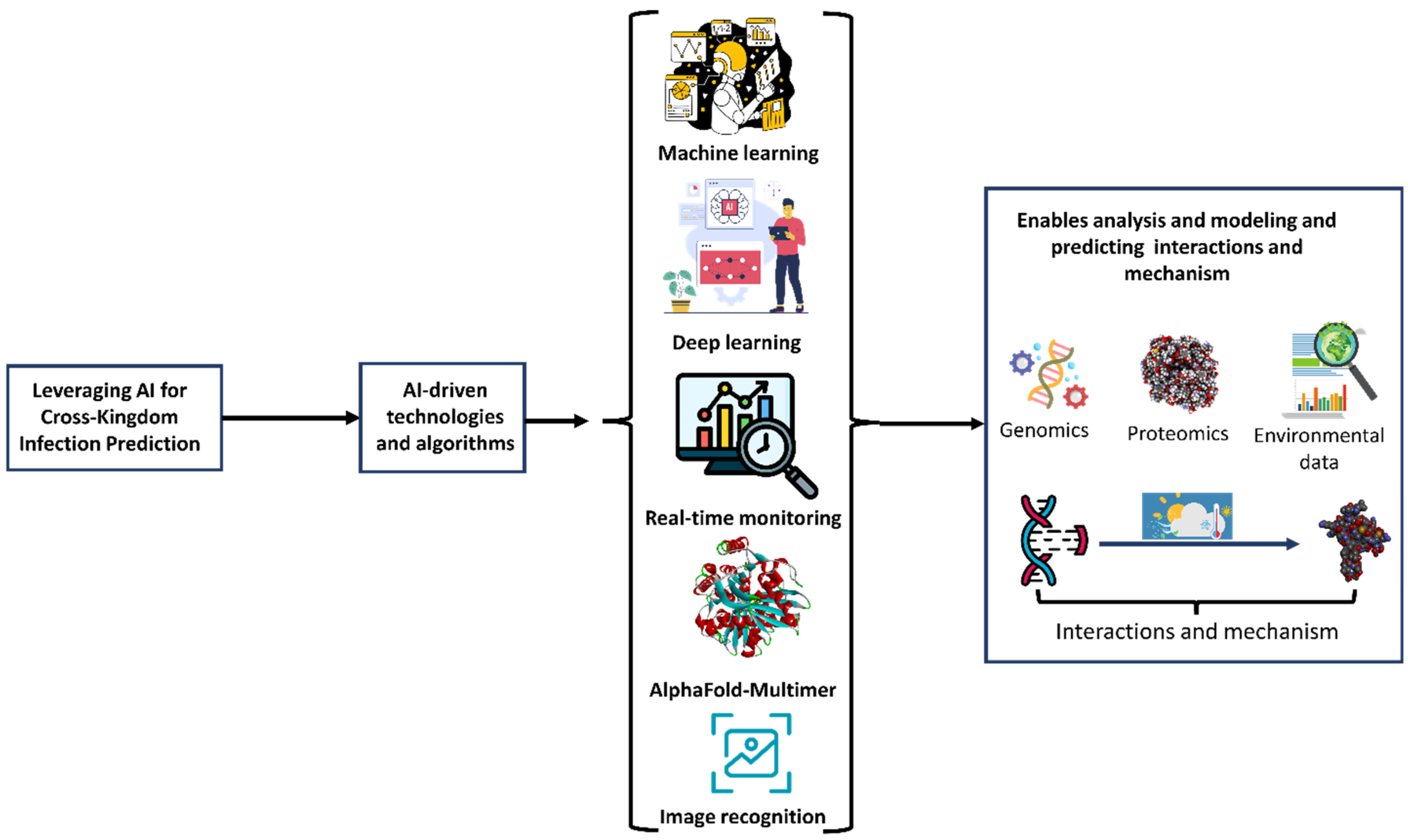
7.5. Leveraging AI for Cross-Kingdom Infection Prediction

Artificial intelligence (AI) has emerged as a transformative tool for predicting cross-kingdom infections by phytopathogens in animals and humans, significantly advancing our understanding of these complex interactions (Figure 8). By leveraging advanced techniques like machine learning (ML), deep learning (DL), and neural networks, AI enables the analysis of genomic, proteomic, and environmental data to uncover hidden patterns indicative of potential cross-kingdom interactions [262]. AI algorithms have demonstrated remarkable capabilities in modeling protein structures and predicting their interactions across kingdoms [263]. For example, tools like AlphaFold-Multimer (AFM) and homology-based threading algorithms can identify conserved motifs and higher-order features in proteins, revealing novel interaction motifs and functional modules relevant to cross-kingdom dynamics [263,264]. AI has also been employed to study symbiotic and pathogenic interactions, as seen in the modeling of fungal microRNAs influencing host genes during symbiosis, which illuminates the mechanisms of cross-kingdom signaling [265]. Furthermore, AI-driven approaches like image recognition and real-time monitoring allow for enhanced early disease detection and diagnosis, surpassing human precision and accuracy capabilities. Such models integrate diverse datasets, including weather, soil, and genomic data, to predict the spatial–temporal risks of outbreaks, enabling timely interventions [266].
AI, particularly through AFM, has demonstrated transformative potential in predicting cross-kingdom interactions at the plant–pathogen interface. A recent study leveraged AFM to analyze 11,274 protein pairs between tomato defense-related hydrolases and small secreted proteins (SSPs) from seven pathogens across bacterial, fungal, and oomycete kingdoms [267]. This approach identified 15 novel SSP–hydrolase complexes, including inhibitors targeting the P69B subtilase, a critical immune protease in tomato. Experimental validation confirmed four inhibitors from bacterial and fungal pathogens, illustrating the utility of AI in elucidating the molecular mechanisms through which pathogens evade plant defenses. Such AI-driven insights offer valuable tools for predicting cross-kingdom interactions at the plant–pathogen interface, as they pinpoint conserved structural features and interaction motifs critical to host–pathogen co-evolution. This not only aids in foreseeing potential threats to human and animal health from phytopathogens but also provides a foundation for developing strategies to mitigate these risks [267].

7.6. Emergency Measures During Cross-Kingdom Phytopathogen Infections

Cross-kingdom infections from phytopathogens to humans can pose a significant threat, especially for immunocompromised individuals. Therefore, it is essential to implement emergency measures to mitigate these risks on a large scale. Concrete steps should be taken to enhance biosecurity protocols to effectively control phytopathogen spread, particularly within agricultural production and food processing facilities (Figure 9). Another critical step is to promote public health awareness. This can be achieved through initiatives that educate the public about the risks associated with phytopathogen infections via comprehensive outreach efforts. Strengthening surveillance and early warning systems is also crucial (Figure 9). This includes developing monitoring networks to promptly detect and alert authorities about potential outbreaks. Most importantly, ensuring emergency medical preparedness is vital for addressing any potential outbreaks. This can involve stockpiling specific medications and establishing treatment protocols for phytopathogen infections, enabling a rapid response in the event of an outbreak. Together, these integrated measures create a comprehensive strategy for preventing and controlling cross-kingdom pathogen transmission, thereby safeguarding both plant and human health.

7.7. Medical Interventions After Cross-Kingdom Phytopathogen Infections

Infections caused by cross-kingdom phytopathogens can have significant effects on human and animal health, necessitating medical interventions (Figure 10). Hospital-acquired infections, in particular, have much higher mortality rates and increased demands for hospitalization. Many of these pathogens exhibit multidrug resistance, posing a serious threat to immunocompromised individuals [268]. Consequently, it is crucial to accurately diagnose the infection’s cause and promptly administer antimicrobial therapy. Using antibiotic treatment can provide an opportunity to neutralize the bacteria and inhibit active infections within the host. Broad-spectrum or specific antibiotics may be employed to treat bacterial phytopathogen infections [269]. Likewise, antifungal therapy with drugs, such as amphotericin B or fluconazole, can be used for fungal phytopathogen infections [270]. However, treatment with these antimicrobial drugs can alter or weaken the patient’s immune system, making immunosuppressed patients more vulnerable to further infections from opportunistic microorganisms. Therefore, immune modulation therapy, combined with existing anti-pathogenic drugs, presents a viable option to enhance the host immune system in combating infections. This can involve the use of immunoglobulins or cytokines [271]. Supportive care, including respiratory and nutritional support for severely ill patients, such as those with acute respiratory distress syndrome (ARDS), is also essential for maintaining vital signs [272]. This support can begin early in hospitalization, when patients are hemodynamically stable and blood gases are adequate. In the acute stage of infection, various surgical interventions are available, which can be used either sequentially or in combination with other treatments, depending on the patient’s response. These interventions may be necessary to surgically remove infected tissues or localized areas of infection when required [273].

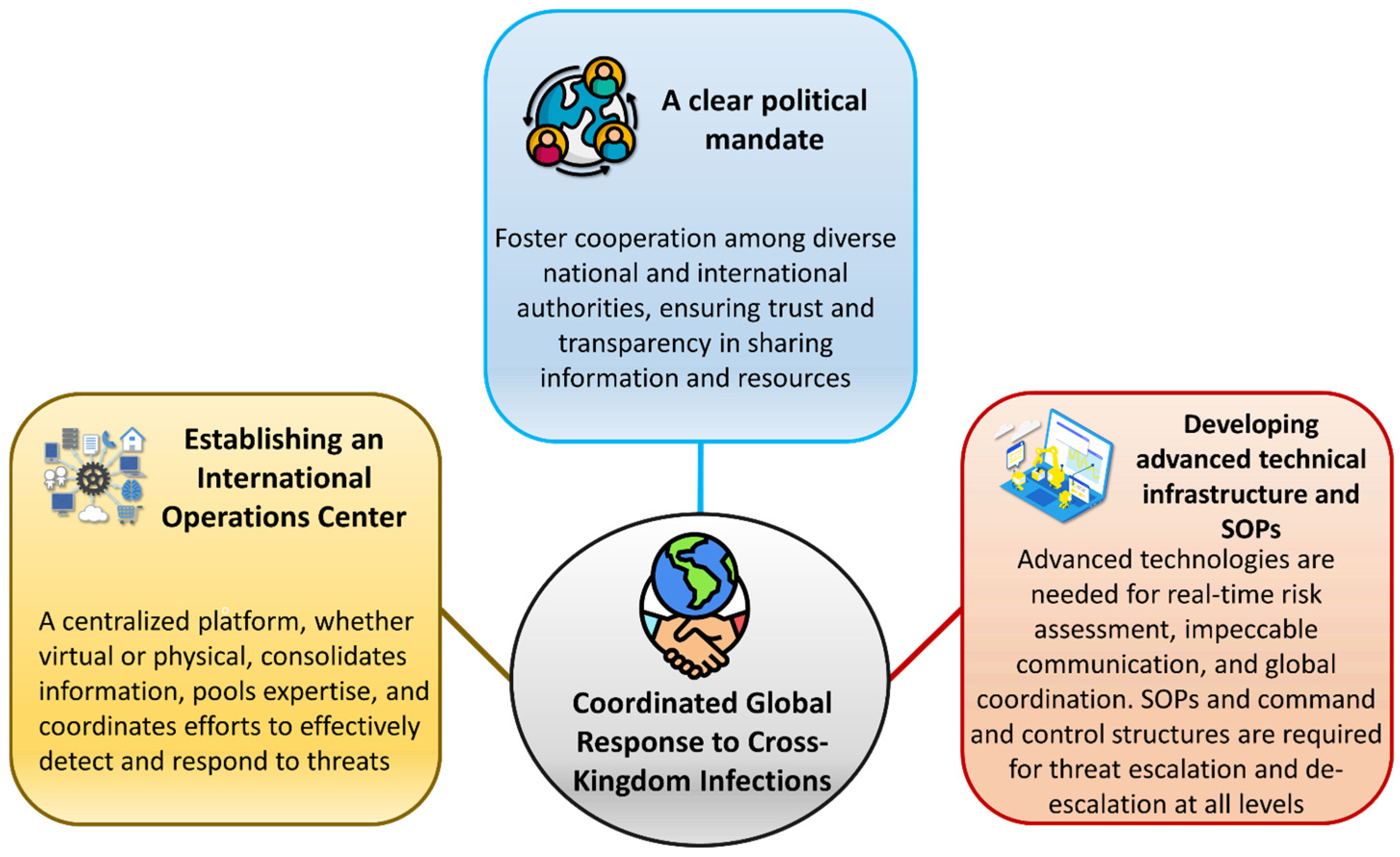
7.8. Coordinated Global Response to Cross-Kingdom Infections

The increasing threat of cross-kingdom infections underscores the urgent need for a globally coordinated response to prevent and control these complex challenges. Establishing an International Operations Center, whether virtual or physical, would provide a centralized platform for consolidating information, pooling expertise, and coordinating technical and organizational frameworks to effectively detect and respond to threats (Figure 11). Such a center would be pivotal in addressing the multifaceted issues associated with cross-kingdom pathogen transmission, leveraging insights from health, agriculture, and environmental sciences to devise comprehensive solutions.
However, establishing this center requires overcoming several significant challenges. A clear political mandate is essential to foster cooperation among diverse national and international authorities, ensuring trust and transparency in sharing information and resources. Building trust across borders is a cornerstone of successful collaboration, particularly in managing cross-border threats where actions in one region can have cascading effects globally. The center would also require a robust network of interdisciplinary expertise, integrating knowledge from various sectors to address emerging threats holistically. Developing advanced technical infrastructure is equally critical, enabling real-time threat detection, seamless communication, and effective coordination across geographies. Standard operating procedures (SOPs) must be established to guide the escalation and de-escalation of responses to threats, incorporating well-defined command and control frameworks to ensure organized action at all levels.
By uniting countries, sectors, and disciplines, an International Operations Center may provide a proactive and coordinated mechanism to mitigate the risks posed by cross-kingdom infections. Such collaborative initiatives would enhance global readiness and ensure more effective management of emerging threats that transcend traditional boundaries of human, plant, and environmental health.

8. Conclusions

Cross-kingdom infections caused by plant pathogens represent a significant and emerging challenge in human health. While these pathogens have traditionally been associated primarily with plants, some have shown remarkable adaptability, crossing biological boundaries to infect humans, particularly in situations involving immune suppression or environmental disturbances. Factors like globalization, climate change, weakened immune systems in non-plant hosts, and genomic plasticity contribute to this issue, reshaping pathogen distribution, ecological niches, and genetic diversity. The molecular mechanisms underlying these infections, including the deployment of effector proteins and the manipulation of host immune responses, reveal shared vulnerabilities between plant and human hosts, highlighting the interconnected nature of these systems.
To effectively address these challenges, a shift towards interdisciplinary collaboration is indispensable. The One Health approach offers a comprehensive framework for integrating health strategies across plant, animal, and human domains. This approach underscores the importance of holistic biosecurity measures, enhanced surveillance, and advanced diagnostic tools. Comprehensive comparative studies of microbial pathogenesis across different kingdoms are vital for recognizing and understanding atypical disease manifestations and novel pathways of interspecies transmission. Furthermore, increasing public awareness and advancing research on potential hazards can significantly mitigate risks associated with contaminated agricultural products.
Future research should prioritize exploring the genetic, ecological, and evolutionary factors influencing cross-kingdom pathogenesis. The development of innovative tools, such as artificial intelligence and genomic technologies, will be vital for predicting and managing emerging threats. Additionally, implementing sustainable agricultural practices and conducting horizon scanning for potential pathogens are essential strategies for risk mitigation. By fostering global collaboration and integrating insights across the health domains of plants, animals, and humans, the complex challenges posed by emerging cross-kingdom pathogens can be effectively tackled.

Author Contributions

Conceptualization, M.M.H. and F.S.; methodology, M.M.; software, H.F.; validation, M.K. and S.A.; formal analysis, N.A.; investigation, A.S.; resources, M.A.A.S.; data curation, F.S.; writing—original draft preparation, M.M.H., F.S. and M.M.; writing—review and editing, M.M.H., M.K., S.A., M.K., N.A., A.S. and M.A.A.S.; visualization, F.S., M.M. and H.F.; supervision, M.M.H.; project administration, M.M.H.; funding acquisition, M.M.H. All authors have read and agreed to the published version of the manuscript.

Funding

This research received no external funding.

Data Availability Statement

Not applicable.

Acknowledgments

We are grateful to the Ministry of Science and Technology, Bangladesh, for supporting this research.

Conflicts of Interest

The authors declare no conflicts of interest.

References

  1. Gauthier, G.M.; Keller, N.P. Crossover Fungal Pathogens: The Biology and Pathogenesis of Fungi Capable of Crossing Kingdoms to Infect Plants and Humans. Fungal Genet. Biol. 2013, 61, 146–157. [Google Scholar] [CrossRef]
  2. Kim, J.S.; Yoon, S.J.; Park, Y.J.; Kim, S.Y.; Ryu, C.M. Crossing the kingdom border: Human diseases caused by plant pathogens. Environ. Microbiol. 2020, 22, 2485–2495. [Google Scholar] [CrossRef]
  3. Casadevall, A.; Pirofski, L.A. What is a Host? Incorporating the Microbiota into the Damage-Response Framework. Infect. Immun. 2018, 86, e00522-17. [Google Scholar] [CrossRef]
  4. Hossain, M.M.; Sultana, F.; Mostafa, M.; Ferdus, H.; Rahman, M.; Rana, J.A.; Islam, S.S.; Adhikary, S.; Sannal, A.; Hosen, M.A.E.; et al. Plant disease dynamics in a changing climate: Impacts, molecular mechanisms, and climate-informed strategies for sustainable management. Discov. Agric. 2024, 2, 132. [Google Scholar] [CrossRef]
  5. Vogt, G. Environmental Adaptation of Genetically Uniform Organisms with the Help of Epigenetic Mechanisms—An Insightful Perspective on Ecoepigenetics. Epigenomes 2022, 7, 1. [Google Scholar] [CrossRef] [PubMed]
  6. Aime, M.C.; Bell, C.D.; Wilson, A.W. Deconstructing the Evolutionary Complexity between Plants and Fungal Pathogens: Coevolutionary Cycles. Annu. Rev. Phytopathol. 2018, 56, 247–266. [Google Scholar] [CrossRef]
  7. Keller, N.P. Fungal secondary metabolism: Regulation, function, and drug discovery. Nat. Rev. Microbiol. 2019, 17, 167–180. [Google Scholar] [CrossRef]
  8. Baker, R.E.; Mahmud, A.S.; Miller, I.F.; Rajeev, M.; Rasambainarivo, F.; Rice, B.L.; Takahashi, S.; Tatem, A.J.; Wagner, C.E.; Wang, L.; et al. Infectious disease in an era of global change. Nat. Rev. Microbiol. 2021, 20, 193–205. [Google Scholar] [CrossRef]
  9. Venbrux, M.; Crauwels, S.; Rediers, H. Current and emerging trends in techniques for plant pathogen detection. Front. Plant Sci. 2023, 14, 1120968. [Google Scholar] [CrossRef]
  10. Kumar, R.; Feltrup, T.M.; Kukreja, R.V.; Patel, K.B.; Cai, S.; Singh, B.R. Evolutionary features in the structure and function of bacterial toxins. Toxins 2019, 11, 15. [Google Scholar] [CrossRef]
  11. Totsline, N.; Kniel, K.E.; Bais, H.P. Microgravity and evasion of plant innate immunity by human bacterial pathogens. npj Microgravity 2023, 9, 1. [Google Scholar] [CrossRef] [PubMed]
  12. Jo, S.H.; Lee, J.; Park, E.; Kim, D.W.; Lee, D.H.; Ryu, C.M.; Choi, D.; Park, J.M. A human pathogenic bacterium Shigella proliferates in plants through adoption of type III effectors for shigellosis. Plant Cell Environ. 2019, 42, 2962–2978. [Google Scholar] [CrossRef]
  13. Velho, A.C.; Mondino, P.; Stadnik, M.J. Extracellular enzymes of Colletotrichum fructicola isolates associated to Apple bitter rot and Glomerella leaf spot. Mycology 2018, 9, 145–154. [Google Scholar] [CrossRef]
  14. Tiwari, M.; Mishra, A.K.; Chakrabarty, D. Agrobacterium-mediated gene transfer: Recent advancements and layered immunity in plants. Planta 2022, 256, 37. [Google Scholar] [CrossRef] [PubMed]
  15. Houston, K.; Tucker, M.R.; Chowdhury, J.; Shirley, N.; Little, A. The plant cell wall: A complex and dynamic structure as revealed by the responses of genes under stress conditions. Front. Plant Sci. 2016, 7, 984. [Google Scholar] [CrossRef]
  16. Wan, J.; He, M.; Hou, Q.; Zou, L.; Yang, Y.; Wei, Y.; Chen, X. Cell wall associated immunity in plants. Stress Biol. 2021, 1, 1. [Google Scholar] [CrossRef] [PubMed]
  17. Tucker, J.S.; Cho, J.; Albrecht, T.M.; Ferrell, J.L.; D’Orazio, S.E.F. Egress of Listeria monocytogenes from Mesenteric Lymph Nodes Depends on Intracellular Replication and Cell-to-Cell Spread. Infect. Immun. 2023, 91, 4. [Google Scholar] [CrossRef]
  18. Worley, M.J. Salmonella bloodstream infections. Trop. Med. Infect. Dis. 2023, 8, 487. [Google Scholar] [CrossRef]
  19. Mermigka, G.; Amprazi, M.; Mentzelopoulou, A.; Amartolou, A.; Sarris, P.F. Plant and animal innate immunity complexes: Fighting different enemies with similar weapons. Trends Plant Sci. 2020, 25, 80–91. [Google Scholar] [CrossRef]
  20. Dalio, R.J.D.; Magalhães, D.M.; Rodrigues, C.M.; Arena, G.D.; Oliveira, T.S.; Souza-Neto, R.R.; Picchi, S.C.; Martins, P.M.M.; Santos, P.J.C.; Maximo, H.J.; et al. PAMPs, PRRs, effectors and R-genes associated with citrus–pathogen interactions. Ann. Bot. 2016, 119, 749–774. [Google Scholar] [CrossRef]
  21. Murphy, K.; Weaver, C. Janeway’s Immunobiology, 9th ed.; Garland Science: New York, NY, USA, 2016; ISBN 978-0815345053. [Google Scholar]
  22. Seong, K.; Krasileva, K.V. Prediction of effector protein structures from fungal phytopathogens enables evolutionary analyses. Nat. Microbiol. 2023, 8, 174–187. [Google Scholar] [CrossRef] [PubMed]
  23. Leonard, S.; Hommais, F.; Nasser, W.; Reverchon, S. Plant–phytopathogen interactions: Bacterial responses to environmental and plant stimuli. Environ. Microbiol. 2016, 19, 1689–1716. [Google Scholar] [CrossRef]
  24. Fisher, M.C.; Gurr, S.J.; Cuomo, C.A.; Blehert, D.S.; Jin, H.; Stukenbrock, E.H.; Stajich, J.E.; Kahmann, R.; Boone, C.; Denning, D.W.; et al. Threats posed by the fungal kingdom to humans, wildlife, and agriculture. mBio 2020, 11, 3. [Google Scholar] [CrossRef]
  25. Casadevall, A. Thermal restriction as an antimicrobial function of fever. PLoS Pathog. 2016, 12, e1005577. [Google Scholar] [CrossRef] [PubMed]
  26. Shiloh, M.U. Mechanisms of mycobacterial transmission: How does Mycobacterium tuberculosis enter and escape from the human host. Future Microbiol. 2016, 11, 1503–1506. [Google Scholar] [CrossRef] [PubMed]
  27. Pal, M.; Merera, O.; Derra, F.; Rahman, M.T.; Hazarika, R. Salmonellosis: A major foodborne disease of global significance. Beverage Food World 2015, 42, 21–24. [Google Scholar]
  28. Lepetit, M.; Brouquisse, R. Control of the rhizobium–legume symbiosis by the plant nitrogen demand is tightly integrated at the whole plant level and requires inter-organ systemic signaling. Front. Plant Sci. 2023, 14, 1114840. [Google Scholar] [CrossRef]
  29. Gordon, S. Phagocytosis: An immunobiologic process. Immunity 2016, 44, 463–475. [Google Scholar] [CrossRef]
  30. Pauwels, A.M.; Trost, M.; Beyaert, R.; Hoffmann, E. Patterns, receptors, and signals: Regulation of phagosome maturation. Trends Immunol. 2017, 38, 407–422. [Google Scholar] [CrossRef]
  31. Lawrence, R.E.; Zoncu, R. The lysosome as a cellular centre for signalling, metabolism and quality control. Nat. Cell Biol. 2019, 21, 133–142. [Google Scholar] [CrossRef]
  32. Queval, C.J.; Song, O.R.; Carralot, J.P.; Saliou, J.M.; Bongiovanni, A.; Deloison, G.; Deboosère, N.; Jouny, S.; Iantomasi, R.; Delorme, V.; et al. Mycobacterium tuberculosis controls phagosomal acidification by targeting CISH-mediated signaling. Cell Rep. 2017, 20, 3188–3198. [Google Scholar] [CrossRef] [PubMed]
  33. Radoshevich, L.; Cossart, P. Listeria monocytogenes: Towards a complete picture of its physiology and pathogenesis. Nat. Rev. Microbiol. 2018, 16, 32–46. [Google Scholar] [CrossRef] [PubMed]
  34. Ratajczak, W.; Niedźwiedzka-Rystwej, P.; Tokarz-Deptuła, B.; Deptuła, W. Immunological memory cells. Cent. Eur. J. Immunol. 2018, 43, 194–203. [Google Scholar] [CrossRef]
  35. Heller, J.; Tudzynski, P. Reactive oxygen species in phytopathogenic fungi: Signaling, development, and disease. Annu. Rev. Phytopathol. 2011, 49, 369–390. [Google Scholar] [CrossRef]
  36. Nelson, K.J.; Knutson, S.T.; Soito, L.; Klomsiri, C.; Poole, L.B.; Fetrow, J.S. Analysis of the peroxiredoxin family: Using active-site structure and sequence information for global classification and residue analysis. Proteins Struct. Funct. Bioinform. 2011, 79, 947–964. [Google Scholar] [CrossRef]
  37. Stincone, A.; Prigione, A.; Cramer, T.; Wamelink, M.M.; Campbell, K.; Cheung, E.; Olin-Sandoval, V.; Gruening, N.M.; Krueger, A.; Tauqeer Alam, M.; et al. The return of metabolism: Biochemistry and physiology of the pentose phosphate pathway. Biol. Rev. 2015, 90, 927–963. [Google Scholar] [CrossRef]
  38. Breitenbach, M.; Weber, M.; Rinnerthaler, M.; Karl, T.; Breitenbach-Koller, L. Oxidative stress in fungi: Its function in signal transduction, interaction with plant hosts, and lignocellulose degradation. Biomolecules 2015, 5, 318–342. [Google Scholar] [CrossRef] [PubMed]
  39. Rossi, D.C.; Gleason, J.E.; Sanchez, H.; Schatzman, S.S.; Culbertson, E.M.; Johnson, C.J.; McNees, C.A.; Coelho, C.; Nett, J.E.; Andes, D.R.; et al. Candida albicans FRE8 encodes a member of the NADPH oxidase family that produces a burst of ROS during fungal morphogenesis. PLoS Pathog. 2017, 13, e1006763. [Google Scholar] [CrossRef]
  40. Nordzieke, D.E.; Fernandes, T.R.; El Ghalid, M.; Turrà, D.; Di Pietro, A. NADPH oxidase regulates chemotropic growth of the fungal pathogen Fusarium oxysporum towards the host plant. New Phytol. 2019, 224, 1600–1612. [Google Scholar] [CrossRef]
  41. da Silva Dantas, A.; Patterson, M.J.; Smith, D.A.; MacCallum, D.M.; Erwig, L.P.; Morgan, B.A.; Quinn, J. Thioredoxin regulates multiple hydrogen peroxide-induced signaling pathways in Candida albicans. Mol. Cell. Biol. 2010, 30, 4550–4563. [Google Scholar] [CrossRef]
  42. Segmüller, N.; Kokkelink, L.; Giesbert, S.; Odinius, D.; van Kan, J.; Tudzynski, P. NADPH oxidases are involved in differentiation and pathogenicity in Botrytis cinerea. Mol. Plant-Microbe Interact. 2008, 21, 808–819. [Google Scholar] [CrossRef] [PubMed]
  43. Paolinelli-Alfonso, M.; Villalobos-Escobedo, J.M.; Rolshausen, P.; Herrera-Estrella, A.; Galindo-Sánchez, C.; López-Hernández, J.F.; Hernandez-Martinez, R. Global transcriptional analysis suggests Lasiodiplodia theobromae pathogenicity factors involved in modulation of grapevine defensive response. BMC Genom. 2016, 17, 615. [Google Scholar] [CrossRef]
  44. Hamel, L.P.; Nicole, M.C.; Duplessis, S.; Ellis, B.E. Mitogen-activated protein kinase signaling in plant-interacting fungi: Distinct messages from conserved messengers. Plant Cell 2012, 24, 1327–1351. [Google Scholar] [CrossRef] [PubMed]
  45. Lin, L.; Tan, R.X. Cross-kingdom actions of phytohormones: A functional scaffold exploration. Chem. Rev. 2011, 111, 2734–2760. [Google Scholar] [CrossRef]
  46. Segorbe, D.; Di Pietro, A.; Pérez-Nadales, E.; Turrà, D. Three Fusarium oxysporum mitogen-activated protein kinases (MAPKs) have distinct and complementary roles in stress adaptation and cross-kingdom pathogenicity. Mol. Plant Pathol. 2017, 18, 912–924. [Google Scholar] [CrossRef]
  47. Kramer, J.; Özkaya, Ö.; Kümmerli, R. Bacterial siderophores in community and host interactions. Nat. Rev. Microbiol. 2020, 18, 152–163. [Google Scholar] [CrossRef]
  48. Félix, C.; Meneses, R.; Gonçalves, M.F.; Duarte, A.S.; Jorrín-Novo, J.V.; Van De Peer, Y.; Deforce, D.; Van Nieuwerburgh, F.; Alves, A.; Esteves, A.C. Lasiodiplodia hormozganensis causing modulation of pathogenesis-related molecules by temperature. Sci. Total Environ. 2024, 927, 171917. [Google Scholar] [CrossRef] [PubMed]
  49. Liu, S.; Hou, Y.; Liu, W.; Lu, C.; Wang, W.; Sun, S. Components of the calcium-calcineurin signaling pathway in fungal cells and their potential as antifungal targets. Eukaryot. Cell 2015, 14, 324–334. [Google Scholar] [CrossRef]
  50. Juvvadi, P.R.; Lee, S.C.; Heitman, J.; Steinbach, W.J. Calcineurin in fungal virulence and drug resistance: Prospects for harnessing targeted inhibition of calcineurin for an antifungal therapeutic approach. Virulence 2016, 8, 186–197. [Google Scholar] [CrossRef]
  51. Paulussen, C.; Hallsworth, J.E.; Álvarez-Pérez, S.; Nierman, W.C.; Hamill, P.G.; Blain, D.; Lievens, B. Ecology of aspergillosis: Insights into the pathogenic potency of Aspergillus fumigatus and some other Aspergillus species. Microb. Biotechnol. 2017, 10, 296–322. [Google Scholar] [CrossRef]
  52. Yan, J.Y.; Zhao, W.S.; Chen, Z.; Xing, Q.K.; Zhang, W.; Chethana, K.W.T.; Xue, M.F. Comparative genome and transcriptome analyses reveal adaptations to opportunistic infections in woody plant degrading pathogens of Botryosphaeriaceae. DNA Res. 2018, 25, 87–102. [Google Scholar] [CrossRef] [PubMed]
  53. Félix, C.; Salvatore, M.M.; DellaGreca, M.; Meneses, R.; Duarte, A.S.; Salvatore, F.; Naviglio, D. Production of toxic metabolites by two strains of Lasiodiplodia theobromae, isolated from a coconut tree and a human patient. Mycologia 2018, 110, 642–653. [Google Scholar] [CrossRef]
  54. Gonçalves, M.F.M.; Nunes, R.B.; Tilleman, L.; Van de Peer, Y.; Deforce, D.; Van Nieuwerburgh, F.; Esteves, A.C.; Alves, A. Dual RNA sequencing of Vitis vinifera during Lasiodiplodia theobromae infection unveils host-pathogen interactions. Int. J. Mol. Sci. 2019, 20, 6083. [Google Scholar] [CrossRef]
  55. Dong, S.; Wang, Y. Nudix effectors: A common weapon in the arsenal of plant pathogens. PLoS Pathog. 2016, 12, e1005704. [Google Scholar] [CrossRef]
  56. Tiwari, S.; Thakur, R.; Shankar, J. Role of heat-shock proteins in cellular function and in the biology of fungi. Biotechnol. Res. Int. 2015, 2015, 132635. [Google Scholar] [CrossRef]
  57. Fu, M.-S.; De Sordi, L.; Mühlschlegel, F.A. Functional characterization of the small heat shock protein Hsp12p from Candida albicans. PLoS ONE 2012, 7, e42894. [Google Scholar] [CrossRef]
  58. Ting, W.; Chang, C.; Szonyi, B.; Gizachew, D. Growth and aflatoxin B1, B2, G1, and G2 production by Aspergillus flavus and Aspergillus parasiticus on ground flax seeds (Linum usitatissimum). J. Food Prot. 2020, 83, 975–983. [Google Scholar] [CrossRef] [PubMed]
  59. Cordeiro, R.D.A.; Evangelista, A.J.D.J.; Serpa, R.; Marques, F.J.D.F.; Melo, C.V.S.D.; Oliveira, J.S.D.; Franco, J.D.S.; Alencar, L.P.D.; Bandeira, T.D.J.P.G.; Brilhante, R.S.N.; et al. Inhibition of heat-shock protein 90 enhances the susceptibility to antifungals and reduces the virulence of Cryptococcus neoformans/Cryptococcus gattii species complex. Microbiology 2016, 162, 309–317. [Google Scholar] [CrossRef] [PubMed]
  60. Ortoneda, M.; Guarro, J.; Madrid, M.P.; Caracuel, Z.; Roncero, M.I.G.; Mayayo, E. Fusarium oxysporum as a multihost model for the genetic dissection of fungal virulence in plants and mammals. Infect. Immun. 2004, 72, 1760–1766. [Google Scholar] [CrossRef]
  61. Sáenz, V.; Alvarez-Moreno, C.; Le Pape, P.; Restrepo, S.; Guarro, J.; Celis Ramírez, A.M. A one health perspective to recognize Fusarium as important in clinical practice. J. Fungi 2020, 6, 235. [Google Scholar] [CrossRef]
  62. Short, D.P.; O’Donnell, K.; Zhang, N.; Juba, J.H.; Geiser, D.M. Widespread occurrence of diverse human pathogenic types of the fungus Fusarium detected in plumbing drains. J. Clin. Microbiol. 2011, 49, 4264–4272. [Google Scholar] [CrossRef] [PubMed]
  63. Litvinov, N.; da Silva, M.T.N.; van der Heijden, I.M.; Graça, M.G.; de Oliveira, L.M.; Fu, L.; Giudice, M.; de Aquino, M.Z.; Odone-Filho, V.; Marques, H.H.; et al. An outbreak of invasive fusariosis in a children’s cancer hospital. Clin. Microbiol. Infect. 2015, 21, 268.e1. [Google Scholar] [CrossRef]
  64. Moretti, M.L.; Busso-Lopes, A.F.; Tararam, C.A.; Moraes, R.; Muraosa, Y.; Mikami, Y.; Gonoi, T.; Taguchi, H.; Lyra, L.; Reichert-Lima, F.; et al. Airborne transmission of invasive fusariosis in patients with hematologic malignancies. PLoS ONE 2018, 13, e0196426. [Google Scholar] [CrossRef]
  65. Nucci, M.; Anaissie, E. Fusarium infections in immunocompromised patients. Clin. Microbiol. Rev. 2007, 20, 695–704. [Google Scholar] [CrossRef]
  66. Coenye, T.; Vandamme, P. Diversity and significance of Burkholderia species occupying diverse ecological niches. Environ. Microbiol. 2003, 5, 719–729. [Google Scholar] [CrossRef]
  67. Kirzinger, M.W.B.; Nadarasah, G.; Stavrinides, J. Insights into cross-kingdom plant pathogenic bacteria. Genes 2011, 2, 980–997. [Google Scholar] [CrossRef] [PubMed]
  68. Singh, R.P.; Singh, P.K.; Rutkoski, J.; Hodson, D.P.; He, X.; Jørgensen, L.N.; Hovmøller, M.S.; Huerta-Espino, J. Disease impact on wheat yield potential and prospects of genetic control. Annu. Rev. Phytopathol. 2016, 54, 303–322. [Google Scholar] [CrossRef] [PubMed]
  69. Azevedo, M.-M.; Faria-Ramos, I.; Costa Cruz, L.; Pina-Vaz, C.; Rodrigues, A.G. Genesis of azole antifungal resistance from agriculture to clinical settings. J. Agric. Food Chem. 2015, 63, 7463–7468. [Google Scholar] [CrossRef]
  70. Alvarez-Moreno, C.; Lavergne, R.-A.; Hagen, F.; Morio, F.; Meis, J.F.; Le Pape, P. Azole-resistant Aspergillus fumigatus harboring TR34/L98H, TR46/Y121F/T289A and TR53 mutations related to flower fields in Colombia. Sci. Rep. 2017, 7, 45631. [Google Scholar] [CrossRef]
  71. Schoustra, S.E.; Debets, A.J.M.; Rijs, A.J.M.M.; Zhang, J.; Snelders, E.; Leendertse, P.C.; Melchers, W.J.G.; Rietveld, A.G.; Zwaan, B.J.; Verweij, P.E. Environmental hotspots for azole resistance selection of Aspergillus fumigatus, the Netherlands. Emerg. Infect. Dis. 2019, 25, 1347. [Google Scholar] [CrossRef]
  72. Godeau, C.; Reboux, G.; Scherer, E.; Laboissiere, A.; Lechenault-Bergerot, C.; Millon, L.; Rocchi, S. Azole-resistant Aspergillus fumigatus in the hospital: Surveillance from flower beds to corridors. Am. J. Infect. Control 2020, 48, 702–704. [Google Scholar] [CrossRef] [PubMed]
  73. Buil, J.B.; Hare, R.K.; Zwaan, B.J.; Arendrup, M.C.; Melchers, W.J.G.; Verweij, P.E. The fading boundaries between patient and environmental routes of triazole resistance selection in Aspergillus fumigatus. PLoS Pathog. 2019, 15, e1007858. [Google Scholar] [CrossRef] [PubMed]
  74. Riat, A.; Plojoux, J.; Gindro, K.; Schrenzel, J.; Sanglard, D. Azole resistance of environmental and clinical Aspergillus fumigatus isolates from Switzerland. Antimicrob. Agents Chemother. 2018, 62, e01128-17. [Google Scholar] [CrossRef] [PubMed]
  75. Homa, M.; Galgóczy, L.; Manikandan, P.; Narendran, V.; Sinka, R.; Csernetics, Á.; Vágvölgyi, C.; Kredics, L.; Papp, T. South Indian isolates of the Fusarium solani species complex from clinical and environmental samples: Identification, antifungal susceptibilities, and virulence. Front. Microbiol. 2018, 9, 1052. [Google Scholar] [CrossRef]
  76. Alvarez-Moreno, C.; Lavergne, R.-A.; Hagen, F.; Morio, F.; Meis, J.F.; Le Pape, P. Fungicide-driven alterations in azole-resistant Aspergillus fumigatus are related to vegetable crops in Colombia, South America. Mycologia 2019, 111, 217–224. [Google Scholar] [CrossRef]
  77. Bicudo, E.L.; Macedo, V.O.; Carrara, M.A.; Castro, F.F.S.; Rage, R.I. Nosocomial outbreak of Pantoea agglomerans in a pediatric urgent care center. Braz. J. Infect. Dis. 2007, 11, 281–284. [Google Scholar] [CrossRef]
  78. Cruz, A.T.; Cazacu, A.C.; Allen, C.H. Pantoea agglomerans, a plant pathogen causing human disease. J. Clin. Microbiol. 2007, 45, 1989–1992. [Google Scholar] [CrossRef]
  79. Kratz, A.; Greenberg, D.; Barki, Y.; Cohen, E.; Lifshitz, M. Pantoea agglomerans as a cause of septic arthritis after palm tree thorn injury; case report and literature review. Arch. Dis. Child. 2003, 88, 542–544. [Google Scholar] [CrossRef]
  80. Hossain, M.M. Diseases of Lablab. In Handbook of Vegetable and Herb Diseases, Handbook of Plant Disease Management; Elmer, W.H., McGrath, M., McGovern, R.J., Eds.; Springer: Cham, Switzerland, 2024. [Google Scholar] [CrossRef]
  81. Islam, S.S.; Adhikary, S.; Mostafa, M.; Hossain, M. Vegetable Beans: Comprehensive insights into diversity, production, nutritional benefits, sustainable cultivation, and future prospects. Online J. Biol. Sci. 2024, 24, 477–494. [Google Scholar] [CrossRef]
  82. Al-Ahmad, M.; Jusufovic, E.; Arifhodzic, N.; Rodriguez, T.; Nurkic, J. Association of Molds and Metrological Parameters to Frequency of Severe Asthma Exacerbation. Allergy Asthma Clin. Immunol. 2019, 15, 29. [Google Scholar] [CrossRef]
  83. Hernandez-Ramirez, G.; Barber, D.; Tome-Amat, J.; Garrido-Arandia, M.; Diaz-Perales, A. Alternaria as an Inducer of Allergic Sensitization. J. Fungi 2008, 7, 838. [Google Scholar] [CrossRef] [PubMed]
  84. NaPier, E.M.; Redd, T.K. Alternaria fungus growing on top of cyanoacrylate glue in a patient with perforated corneal ulcer. Am. J. Ophthalmol. Case Rep. 2022, 28, 101717. [Google Scholar] [CrossRef] [PubMed]
  85. Rammaert, B.; Aguilar, C.; Bougnoux, M.; Noël, N.; Charlier, C.; Denis, B.; Lecuit, M.; Lortholary, O. Success of posaconazole therapy in a heart transplanted patient with Alternaria infectoria cutaneous infection. Med. Mycol. 2012, 50, 518–521. [Google Scholar] [CrossRef] [PubMed]
  86. Hipolito, E.; Faria, E.; Alves, A.F.; Hoog, G.S.; Anjos, J.; Gonçalves, T.; Morais, P.V.; Estevão, H. Alternaria infectoria brain abscess in a child with chronic granulomatous disease. Eur. J. Clin. Microbiol. Infect. Dis. 2008, 28, 377–380. [Google Scholar] [CrossRef]
  87. Vu, A.L.; Dee, M.M.; Gualandi, R.J., Jr.; Huff, S.; Zale, J.; Gwinn, K.D.; Ownley, B.H. First report of leaf spot caused by Bipolaris spicifera on switchgrass in the United States. Plant Dis. 2011, 95, 1191. [Google Scholar] [CrossRef]
  88. Amaradasa, B.S.; Amundsen, K. First Report of Curvularia inaequalis and Bipolaris spicifera Causing Leaf Blight of Buffalograss in Nebraska. Plant Dis. 2014, 98, 279. [Google Scholar] [CrossRef]
  89. Mahadevakumar, S.; Mahesh, M.; Maharachchikumbura, S.S.N.; Lavanya, S.N.; Rajashekara, H.; Prakash, G.; Vikas, K.; Tarasatyavati, C.; Chandranayaka, S. First report of Curvularia spicifera (≡ Bipolaris spicifera) causing spathe blight and leaf spot disease of pearl millet (Cenchrus americanus) in India. Plant Dis. 2024, 108, 3193. [Google Scholar] [CrossRef]
  90. Pham, C.D.; Purfield, A.E.; Fader, R.; Pascoe, N.; Lockhart, S.R. Development of a multilocus sequence typing system for medically relevant Bipolaris species. J. Clin. Microbiol. 2015, 53, 3239–3246. [Google Scholar] [CrossRef]
  91. Manamgoda, D.; Rossman, A.; Castlebury, L.; Crous, P.; Madrid, H.; Chukeatirote, E.; Hyde, K. The genus Bipolaris. Stud. Mycol. 2014, 79, 221–288. [Google Scholar] [CrossRef]
  92. Pai, H.V.; Jamal, E.; Yegneswaran, P.P. Corneal ulcer due to a rare pleosporalean member of the genus Bipolaris following cow tail injury to the eye: A case report and review of literature. Indian J. Ophthalmol. 2017, 65, 403–405. [Google Scholar] [CrossRef]
  93. Cighir, A.; Mare, A.D.; Vultur, F.; Cighir, T.; Pop, S.D.; Horvath, K.; Man, A. Fusarium spp. in Human Disease: Exploring the Boundaries between Commensalism and Pathogenesis. Life 2023, 13, 1440. [Google Scholar] [CrossRef] [PubMed]
  94. Zakaria, L. An Overview of Aspergillus Species Associated with Plant Diseases. Pathogens 2024, 13, 813. [Google Scholar] [CrossRef]
  95. Fukuda, Y.; Homma, T.; Suzuki, S.; Takuma, T.; Tanaka, A.; Yokoe, T.; Ohnishi, T.; Niki, Y.; Sagara, H. High burden of Aspergillus fumigatus infection among chronic respiratory diseases. Chronic Respir. Dis. 2018, 15, 279–285. [Google Scholar] [CrossRef] [PubMed]
  96. Konopka, J.B.; Casadevall, A.; Taylor, J.W.; Heitman, J.; Cowen, L. One Health: Fungal Pathogens of Humans, Animals, and Plants; American Society for Microbiology: Washington, DC, USA, 2019. [Google Scholar] [CrossRef]
  97. Sandoval-Denis, M.; Sutton, D.A.; Martin-Vicente, A.; Cano-Lira, J.F.; Wiederhold, N.; Guarro, J.; Gene, J. Cladosporium species recovered from clinical samples in the United States. J. Clin. Microbiol. 2015, 53, 2990–3000. [Google Scholar] [CrossRef]
  98. Jia, J.; Nan, L.; Song, Z.; Chen, X.; Xia, J.; Cheng, L.; Zhang, B.; Mu, F. Cross-species transmission of a novel bisegmented orfanplasmovirus in the phytopathogenic fungus Exserohilum rostratum. Front. Microbiol. 2024, 15, 1409677. [Google Scholar] [CrossRef] [PubMed]
  99. Sharma, K.; Goss, E.M.; Dickstein, E.R.; Smith, M.E.; Johnson, J.A.; Southwick, F.S.; van Bruggen, A.H.C. Exserohilum rostratum: Characterization of a Cross-Kingdom Pathogen of Plants and Humans. PLoS ONE 2014, 9, e108691. [Google Scholar] [CrossRef]
  100. CDC. Multistate Outbreak of Fungal Meningitis and Other Infections. 2013. Available online: https://archive.cdc.gov/www_cdc_gov/hai/outbreaks/laboratory/index.html (accessed on 10 January 2025).
  101. Coutinho, I.B.L.; Freire, F.C.O.; Lima, C.S.; Lima, J.S.; Gonçalves, F.J.T.; Machado, A.R.; Silva, A.M.S.; Cardoso, J.E. Diversity of genus Lasiodiplodia associated with perennial tropical fruit plants in northeastern Brazil. Plant Pathol. 2017, 66, 90–104. [Google Scholar] [CrossRef]
  102. Maurya, A.K.; Kumari, S.; Behera, G.; Bhadade, A.; Tadepalli, K. Rhino sinusitis caused by Lasiodiplodia theobromae in a diabetic patient. Med. Mycol. Case Rep. 2023, 40, 22–24. [Google Scholar] [CrossRef]
  103. Marquez, N.; Giachero, M.L.; Declerck, S.; Ducasse, D.A. Macrophomina phaseolina: General Characteristics of Pathogenicity and Methods of Control. Front. Plant Sci. 2021, 12, 634397. [Google Scholar] [CrossRef]
  104. Toumasis, P.; Vrioni, G.; Gardeli, I.; Michelaki, A.; Exindari, M.; Orfanidou, M. Macrophomina phaseolina: A Phytopathogen Associated with Human Ocular Infections—A Case Report of Endophthalmitis and Systematic Review of Human Infections. J. Clin. Med. 2025, 14, 430. [Google Scholar] [CrossRef]
  105. Wang, R.Y.; Gao, B.; Chen, S.L.; Ma, J.; Li, X.H.; Xue, C.X. First report of Rhizopus oryzae causing soft rot on storage roots of sweetpotato in China. Plant Dis. 2017, 101, 1039. [Google Scholar] [CrossRef]
  106. Wang, H.C.; Zhang, M.; Zhang, C.Q.; Chen, X.J.; Tan, Q.Q.; Ma, J.; Li, Z. Pathogen detection, carbon metabolic phenotype analysis of Rhizopus oryzae from tobacco and its sensitivity to seven fungicides. J. Pestic. Sci. 2018, 20, 743–748. [Google Scholar] [CrossRef]
  107. Dai, M.; Tan, X.; Chen, X.; Cai, K.; Zhong, Y.; Ye, Z.; Kong, D. Green control for inhibiting Rhizopus oryzae growth by stress factors in forage grass factory. Front. Microbiol. 2024, 15, 1437799. [Google Scholar] [CrossRef]
  108. Osaigbovo, I.I.; Ekeng, B.E.; Davies, A.A.; Oladele, R.O. Mucormycosis in Africa: Epidemiology, diagnosis and treatment outcomes. Mycoses 2023, 66, 13581. [Google Scholar] [CrossRef] [PubMed]
  109. Islam, M.R.; Rahman, M.M.; Ahasan, M.T.; Sarkar, N.; Akash, S.; Islam, M.; Islam, F.; Aktar, M.N.; Saeed, M.; Rashid, H.O.; et al. The impact of mucormycosis (black fungus) on SARS-CoV-2-infected patients: At a glance. Environ. Sci. Pollut. Res. 2022, 29, 69341–69366. [Google Scholar] [CrossRef]
  110. Calvano, T.P.; Blatz, P.J.; Vento, T.J.; Wickes, B.L.; Sutton, D.A.; Thompson, E.H.; White, C.E.; Renz, E.M.; Hospenthal, D.R. Pythium aphanidermatum Infection Following Combat Trauma. J. Clin. Microbiol. 2011, 49, 3710–3713. [Google Scholar] [CrossRef]
  111. Thongsuk, P.; Plongla, R.; Thammahong, A.; Tiewsurin, J.; Worasilchai, N.; Chindamporn, A.; Suankratay, C. Vascular pythiosis caused by Pythium aphanidermatum: The first case report in Asia. Eur. J. Med. Res. 2021, 26, 1. [Google Scholar] [CrossRef]
  112. Vilela, R.; Taylor, J.W.; Walker, E.D.; Mendoza, L. Lagenidium giganteum Pathogenicity in mammals. Emerg. Infect. Dis. 2015, 21, 290–297. [Google Scholar] [CrossRef]
  113. Shivaprakash, M.R.; Appannanavar, S.B.; Dhaliwal, M.; Gupta, A.; Gupta, S.; Gupta, A.; Chakrabarti, A. Colletotrichum truncatum: An unusual pathogen causing mycotic keratitis and endophthalmitis. J. Clin. Microbiol. 2011, 49, 2894–2898. [Google Scholar] [CrossRef]
  114. Rao, S.; Sharda, S.; Oddi, V.; Nandineni, M.R. The landscape of repetitive elements in the refined genome of chilli anthracnose fungus Colletotrichum truncatum. Front. Microbiol. 2018, 9, 2367. [Google Scholar] [CrossRef]
  115. Palmore, T.N.; Shea, Y.R.; Childs, R.W.; Sherry, R.M.; Walsh, T.J. Fusarium proliferatum soft tissue infection at the site of a puncture by a plant: Recovery, isolation, and direct molecular identification. J. Clin. Microbiol. 2010, 48, 338–342. [Google Scholar] [CrossRef]
  116. Muhammed, M.; Anagnostou, T.; Desalermos, A.; Kourkoumpetis, T.K.; Carneiro, H.A.; Glavis-Bloom, J.; Coleman, J.J.; Mylonakis, E. Fusarium infection. Medicine 2013, 92, 305–316. [Google Scholar] [CrossRef]
  117. Coleman, J.J. The Fusarium solani species complex: Ubiquitous pathogens of agricultural importance. Mol. Plant Pathol. 2016, 17, 146–158. [Google Scholar] [CrossRef] [PubMed]
  118. Marques, M.W.; Lima, N.B.; De Morais, M.A.; Barbosa, M.A.G.; Souza, B.O.; Michereff, S.J.; Phillips, A.J.L.; Câmara, M.P.S. Species of Lasiodiplodia associated with mango in Brazil. Fungal Divers. 2013, 61, 181–193. [Google Scholar] [CrossRef]
  119. Netto, M.S.B.; Assunção, I.P.; Lima, G.S.A.; Marques, M.W.; Lima, W.G.; Monteiro, J.H.A.; De Queiroz Balbino, V.; Michereff, S.J.; Phillips, A.J.L.; Câmara, M.P.S. Species of Lasiodiplodia associated with papaya stem-end rot in Brazil. Fungal Divers. 2014, 67, 127–141. [Google Scholar] [CrossRef]
  120. Correia, K.C.; Silva, M.A.; De Morais, M.A.; Armengol, J.; Phillips, A.J.L.; Câmara, M.P.S.; Michereff, S.J. Phylogeny, Distribution and Pathogenicity of Lasiodiplodia Species Associated with Dieback of Table Grape in the Main Brazilian Exporting Region. Plant Pathol. 2015, 65, 92–103. [Google Scholar] [CrossRef]
  121. Custódio, F.A.; Machado, A.R.; Soares, D.J.; Pereira, O.L. Lasiodiplodia hormozganensis Causing Basal Stem Rot on Ricinus communis in Brazil. Australas. Plant Dis. Notes 2018, 13, 25. [Google Scholar] [CrossRef]
  122. Kwon, J.; Ryu, J.; Chi, T.T.P.; Shen, S.; Choi, O. Soft Rot of Rhizopus oryzae as a Postharvest Pathogen of Banana Fruit in Korea. Mycobiology 2012, 40, 214–216. [Google Scholar] [CrossRef]
  123. Khokhar, I.; Wang, J.; Jia, Y.; Yan, Y. First Report of Rhizopus Soft Rot on Tomato (Lycopersicon esculentum) Caused by Rhizopus oryzae in China. Plant Dis. 2019, 103, 1041. [Google Scholar] [CrossRef]
  124. Gnanesh, B.; Tejaswi, A.; Arunakumar, G.; Supriya, M.; Manojkumar, H.; Tewary, P. Molecular phylogeny, identification and pathogenicity of Rhizopus oryzae associated with root rot of mulberry in India. J. Appl. Microbiol. 2020, 131, 360–374. [Google Scholar] [CrossRef]
  125. Holmes, J.E.; Sanghera, H.; Punja, Z.K. Crown gall development on cannabis (Cannabis sativa L., marijuana) plants caused by Agrobacterium tumefaciens species-complex. Can. J. Plant Pathol. 2023, 45, 433–445. [Google Scholar] [CrossRef]
  126. Tiwari, S.; Beriha, S.S. Primary Bacteremia Caused by Rhizobium radiobacter in Neonate: A Rare Case Report. J. Clin. Diagn. Res. 2015, 9, DD01–DD02. [Google Scholar] [CrossRef]
  127. İyigün, F. Rhizobium radiobacter infection in a preterm infant and review of the literature. Ümraniye Pediatri Dergisi J. Umra. Pediatr. 2024, 47, 47–52. [Google Scholar] [CrossRef]
  128. Fenner, B.J.; Kumar, A.; Tan, N.Y.Q.; Ang, M. Case of Isolated Rhizobium radiobacter Contact Lens-Related Infectious Keratitis: A Plant Microbe Now Emerging as a Human Pathogen. Am. J. Ophthalmol. Case Rep. 2019, 15, 100476. [Google Scholar] [CrossRef]
  129. Cho, H.; Park, J.Y.; Kim, Y.K.; Sohn, S.-H.; Park, D.S.; Kwon, Y.-S.; Kim, C.-W.; Back, C.-G. Whole-Genome Sequence of Erwinia persicina B64, Which Causes Pink Soft Rot in Onions. Microbiol. Resour. Announc. 2019, 8, e01302-18. [Google Scholar] [CrossRef] [PubMed]
  130. Canik Orel, D. Erwinia persicina as the New Causal Agent of Lettuce Soft Rot. Eur. J. Plant Pathol. 2020, 158, 223–235. [Google Scholar] [CrossRef]
  131. Kawaguchi, A.; Abe, D.; Saito, T.; Nogata, Y.; Nomiyama, K.; Kohyama, N.; Takahashi, A.; Yoshioka, T.; Ishikawa, N.; Tomioka, K. Pink seed of barley caused by Erwinia persicina. J. Gen. Plant Pathol. 2021, 87, 106–109. [Google Scholar] [CrossRef]
  132. Wasendorf, C.; Schmitz-Esser, S.; Eischeid, C.J.; Leyhe, M.J.; Nelson, E.N.; Rahic-Seggerman, F.M.; Sullivan, K.E.; Peters, N.T. Genome analysis of Erwinia persicina reveals implications for soft rot pathogenicity in plants. Front. Microbiol. 2022, 13, 1001139. [Google Scholar] [CrossRef]
  133. González, A.J.; Tello, J.C.; Rodicio, M.R. Erwinia persicina causing chlorosis and necrotic spots in leaves and tendrils of Pisum sativum in southeastern Spain. Plant Dis. 2007, 91, 460. [Google Scholar] [CrossRef]
  134. Zhang, Z.; Nan, Z. Erwinia persicina, a possible new necrosis and wilt threat to forage or grain legumes production. Eur. J. Plant Pathol. 2014, 139, 349–358. [Google Scholar] [CrossRef]
  135. O’Hara, C.M.; Steigerwalt, A.G.; Hill, B.C.; Miller, J.M.; Brenner, D.J. First report of a human isolate of Erwinia persicinus. J. Clin. Microbiol. 1998, 36, 248–250. [Google Scholar] [CrossRef] [PubMed]
  136. Mohamaden, W.I.; Zhen-Fen, Z.; Hegab, I.M.; Shang-Li, S. Experimental infection in mice with Erwinia persicina. Microb. Pathog. 2019, 130, 38–43. [Google Scholar] [CrossRef] [PubMed]
  137. Prod’homme, M.; Micol, L.A.; Weitsch, S.; Gassend, J.L.; Martinet, O.; Bellini, C. Cutaneous infection and bacteremia caused by Erwinia billingiae: A case report. New Microbes New Infect. 2017, 19, 134–136. [Google Scholar] [CrossRef]
  138. Tavares, M.; Kozak, M.; Balola, A.; Sá-Correia, I. Burkholderia cepacia Complex Bacteria: A Feared Contamination Risk in Water-Based Pharmaceutical Products. Clin. Microbiol. Rev. 2020, 33, 3. [Google Scholar] [CrossRef] [PubMed]
  139. Mahenthiralingam, E.; Baldwin, A.; Dowson, C.G. Burkholderia cepacia complex bacteria: Opportunistic pathogens with important natural biology. J. Appl. Microbiol. 2008, 104, 1539–1551. [Google Scholar] [CrossRef] [PubMed]
  140. Schwab, U.; Leigh, M.; Ribeiro, C.; Yankaskas, J.; Burns, K.; Gilligan, P.; Sokol, P.; Boucher, R. Patterns of epithelial cell invasion by different species of the Burkholderia cepacia complex in well-differentiated human airway epithelia. Infect. Immun. 2002, 70, 4547–4555. [Google Scholar] [CrossRef]
  141. Cui, Z.; Wang, S.; Kakar, K.U.; Xie, G.; Li, B.; Chen, G.; Zhu, B. Genome Sequence and Adaptation Analysis of the Human and Rice Pathogenic Strain Burkholderia glumae AU6208. Pathogens 2021, 10, 87. [Google Scholar] [CrossRef]
  142. Rajendraprasad, S.; Creech, Z.A.; Truong, G.T.D.; Nguyen, T.; Addula, M.; Mendoza, N.; Velagapudi, M. Fatal case of Burkholderia gladioli pneumonia in a patient with COVID-19. Ochsner J. 2022, 22, 349–352. [Google Scholar] [CrossRef]
  143. Yablon, B.R.; Dantes, R.; Tsai, V.; Lim, R.; Moulton-Meissner, H.; Arduino, M.; Jensen, B.; Patel, M.T.; Vernon, M.O.; Grant-Greene, Y.; et al. Outbreak of Pantoea agglomerans bloodstream infections at an oncology clinic-Illinois, 2012–2013. Infect. Control Hosp. Epidemiol. 2017, 38, 314–319. [Google Scholar] [CrossRef]
  144. De Baere, T.; Verhelst, R.; Labit, C.; Verschraegen, G.; Wauters, G.; Claeys, G.; Vaneechoutte, M. Bacteremic Infection with Pantoea ananatis. J. Clin. Microbiol. 2004, 42, 4393–4395. [Google Scholar] [CrossRef]
  145. Habsah, H.; Zeehaida, M.; Van Rostenberghe, H.; Noraida, R.; Wan Pauzi, W.I.; Fatimah, I.; Rosliza, A.; Sharimah, N.N.; Maimunah, H. An outbreak of Pantoea spp. in a neonatal intensive care unit secondary to contaminated parenteral nutrition. J. Hosp. Infect. 2005, 61, 213–218. [Google Scholar] [CrossRef] [PubMed]
  146. Grace, E.R.; Rabiey, M.; Friman, V.; Jackson, R.W. Seeing the forest for the trees: Use of phages to treat bacterial tree diseases. Plant Pathol. 2021, 70, 1987–2004. [Google Scholar] [CrossRef]
  147. Moradi, F.; Behbahani, M.R.; Gorginpour, J.; Dezhkam, A.; Hadi, N. Ralstonia pickettii bloodstream infection in the patient with Guillain-Barre syndrome under plasmapheresis. New Microbes New Infect. 2024, 57, 101218. [Google Scholar] [CrossRef] [PubMed]
  148. Gupta, V.; Buch, A. Pseudomonas aeruginosa predominates as multifaceted rhizospheric bacteria with combined abilities of P-solubilization and biocontrol. J. Pure Appl. Microbiol. 2019, 13, 319–328. [Google Scholar] [CrossRef]
  149. Tiwari, P.; Singh, J.S. A plant growth promoting rhizospheric Pseudomonas aeruginosa strain inhibits seed germination in Triticum aestivum (L) and Zea mays (L). Microbiol. Res. 2017, 8, 7233. [Google Scholar] [CrossRef]
  150. Ambreetha, S.; Marimuthu, P.; Mathee, K.; Balachandar, D. Plant-Associated Pseudomonas aeruginosa Strains Harbor Multiple Virulence Traits Critical for Human Infection. J. Med. Microbiol. 2022, 71. [Google Scholar] [CrossRef] [PubMed]
  151. Bjarnsholt, T.; Jensen, P.O.; Fiandaca, M.J.; Pedersen, J.; Hansen, C.R.; Andersen, C.B.; Pressler, T.; Givskov, M.; Høiby, N. Pseudomonas aeruginosa Biofilms in the Respiratory Tract of Cystic Fibrosis Patients. Pediatr. Pulmonol. 2009, 44, 547–558. [Google Scholar] [CrossRef]
  152. Mulcahy, L.R.; Isabella, V.M.; Lewis, K. Pseudomonas aeruginosa biofilms in disease. Microb. Ecol. 2014, 68, 1–12. [Google Scholar] [CrossRef]
  153. Wilson, M.G.; Pandey, S. Pseudomonas aeruginosa. [Updated 8 August 2023]. In StatPearls [Internet]; StatPearls Publishing: Treasure Island, FL, USA, 2024. Available online: https://www.ncbi.nlm.nih.gov/books/NBK557831/ (accessed on 16 December 2024).
  154. Kirienko, N.V.; Kirienko, D.R.; Larkins-Ford, J.; Wählby, C.; Ruvkun, G.; Ausubel, F.M. Pseudomonas aeruginosa disrupts Caenorhabditis elegans iron homeostasis, causing a hypoxic response and death. Cell Host Microbe 2013, 13, 406–416. [Google Scholar] [CrossRef]
  155. Coutinho, C.P.; de Carvalho, C.C.C.R.; Madeira, A.; Pinto-de-Oliveira, A.; Sá-Correia, I. Burkholderia cenocepacia phenotypic clonal variation during a 3.5-year colonization in the lungs of a cystic fibrosis patient. Infect. Immun. 2011, 79, 2950–2960. [Google Scholar] [CrossRef]
  156. Gao, J.; Wang, Y.; Wang, C.W.; Lu, B.H. First report of bacterial root rot of ginseng caused by Pseudomonas aeruginosa in China. Plant Dis. 2014, 98, 1577. [Google Scholar] [CrossRef] [PubMed]
  157. Grace, A.; Sahu, R.; Owen, D.R.; Dennis, V.A. Pseudomonas aeruginosa reference strains PAO1 and PA14: A genomic, phenotypic, and therapeutic review. Front. Microbiol. 2022, 13, 1023523. [Google Scholar] [CrossRef] [PubMed]
  158. Brüssow, H. The novel coronavirus–a snapshot of current knowledge. Microb. Biotechnol. 2020, 13, 607–612. [Google Scholar] [CrossRef]
  159. Balique, F.; Lecoq, H.; Raoult, D.; Colson, P. Can Plant Viruses Cross the Kingdom Border and Be Pathogenic to Humans? Viruses 2015, 7, 2074–2098. [Google Scholar] [CrossRef]
  160. Zhang, T.; Breitbart, M.; Lee, W.H.; Run, J.Q.; Wei, C.L.; Soh, S.W.; Hibberd, M.L.; Liu, E.T.; Rohwer, F.; Ruan, Y. RNA viral community in human feces: Prevalence of plant pathogenic viruses. PLoS Biol. 2006, 4, e3. [Google Scholar] [CrossRef]
  161. Jiwaji, M.; Matcher, G.F.; de Bruyn, M.M.; Awando, J.A.; Moodley, H.; Waterworth, D.; Jarvie, R.A.; Dorrington, R.A. Providence virus: An animal virus that replicates in plants or a plant virus that infects and replicates in animal cells? PLoS ONE 2019, 14, e0217494. [Google Scholar] [CrossRef]
  162. Bousbia, S.; Papazian, L.; La Scola, B.; Raoult, D. Detection of Plant DNA in the Bronchoalveolar Lavage of Patients with Ventilator-Associated Pneumonia. PLoS ONE 2010, 5, e11298. [Google Scholar] [CrossRef] [PubMed]
  163. Colson, P.; Richet, H.; Desnues, C.; Balique, F.; Moal, V.; Grob, J.J.; Berbis, P.; Lecoq, H.; Harlé, J.-R.; Berland, Y.; et al. Pepper Mild Mottle Virus, a Plant Virus Associated with Specific Immune Responses, Fever, Abdominal Pains, and Pruritus in Humans. PLoS ONE 2010, 5, e10041. [Google Scholar] [CrossRef]
  164. Liu, R.; Vaishnav, R.A.; Roberts, A.M.; Friedland, R.P. Humans have antibodies against a plant virus: Evidence from tobacco mosaic virus. PLoS ONE 2013, 8, e60621. [Google Scholar] [CrossRef]
  165. Knowland, J. Protein synthesis directed by the RNA from a plant virus in a normal animal cell. Genetics 1974, 78, 383–394. [Google Scholar] [CrossRef]
  166. Li, L.; Wang, L.; Xiao, R.; Zhu, G.; Li, Y.; Liu, C.; Yang, R.; Tang, Z.; Li, J.; Huang, W.; et al. The invasion of tobacco mosaic virus RNA induces endoplasmic reticulum stress-related autophagy in HeLa cells. Biosci. Rep. 2012, 32, 171–186. [Google Scholar] [CrossRef] [PubMed]
  167. Koudelka, K.J.; Destito, G.; Plummer, E.M.; Trauger, S.A.; Siuzdak, G.; Manchester, M. Endothelial targeting of cowpea mosaic virus (CPMV) via surface vimentin. PLoS Pathog. 2009, 5, e1000417. [Google Scholar] [CrossRef]
  168. Li, L.; Victoria, J.G.; Wang, C.; Jones, M.; Fellers, G.M.; Kunz, T.H.; Delwart, E. Bat guano virome: Predominance of dietary viruses from insects and plants plus novel mammalian viruses. J. Virol. 2010, 84, 6955–6965. [Google Scholar] [CrossRef]
  169. Phan, T.G.; Kapusinszky, B.; Wang, C.; Rose, R.K.; Lipton, H.L.; Delwart, E.L. The fecal viral flora of wild rodents. PLoS Pathog. 2011, 7, e1002218. [Google Scholar] [CrossRef] [PubMed]
  170. Stepanova, O.A.; Solovyova, Y.V.; Solovyov, A.V. Results of algae viruses search in human clinical material. Ukr. Bioorganica Acta 2011, 2, 53–56. [Google Scholar]
  171. Yolken, R.H.; Jones-Brando, L.; Dunigan, D.D.; Kannan, G.; Dickerson, F.; Severance, E.; Sabunciyan, S.; Talbot, C.C., Jr.; Prandovszky, E.; Gurnon, J.R.; et al. Chlorovirus ATCV-1 is part of the human oropharyngeal virome and is associated with changes in cognitive functions in humans and mice. Proc. Natl. Acad. Sci. USA 2014, 111, 16106–16111. [Google Scholar] [CrossRef]
  172. Garcia-Lopez, R.; Perez-Brocal, V.; Moya, A. Beyond cells—The virome in the human holobiont. Microb. Cell 2019, 6, 373–396. [Google Scholar] [CrossRef]
  173. Magunacelaya, J.C. Concepts in Management of Tree Crops Nematodes in Fruit Production Systems. In Integrated Management of Fruit Crops Nematodes; Ciancio, A., Mukerji, K., Eds.; Integrated Management of Plant Pests and Diseases; Springer: Dordrecht, The Netherland, 2009; Volume 4. [Google Scholar] [CrossRef]
  174. Li, Q.; Liang, W.; Zhang, X.; Mahamood, M. Nematode genera and species description along the transect. In Soil Nematodes of Grasslands in Northern China; Academic Press: London, UK; Zhejiang University Press: Hangzhou, China, 2017; Volume 45, p. 228. [Google Scholar]
  175. Haouchine, D.; Mantelet, S.; Marteau, A.; Brun, S.; Dabi, P.; Abbas, M.; Izri, A.; Akhoundi, M. First evidence of human infection by Xiphinema brevicollum (Nematoda: Longidoridae). Int. J. Infect. Dis. 2022, 122, 609–611. [Google Scholar] [CrossRef]
  176. Jasmer, D.P.; Goverse, A.; Smant, G. Parasitic nematode interactions with mammals and plants. Annu. Rev. Phytopathol. 2003, 41, 245–270. [Google Scholar] [CrossRef]
  177. Stepek, G.; Buttle, D.J.; Duce, I.R.; Behnke, J.M. Human gastrointestinal nematode infections: Are new control methods required? Int. J. Exp. Pathol. 2006, 87, 325–341. [Google Scholar] [CrossRef]
  178. Robinson, M.W.; Dalton, J.P. Zoonotic helminth infections with particular emphasis on fasciolosis and other trematodiases. Philos. Trans. R. Soc. B Biol. Sci. 2009, 364, 2763–2776. [Google Scholar] [CrossRef] [PubMed]
  179. Kantor, M.; Vieira, P.; Handoo, Z. First Report of the Barley Root-Knot Nematode, Meloidogyne naasi, from Pennsylvania, with New Host Report. Plant Dis. 2024, 108, 2583. [Google Scholar] [CrossRef] [PubMed]
  180. Bradbury, R.S.; Speare, R. Passage of Meloidogyne Eggs in Human Stool: Forgotten, but Not Gone. J. Clin. Microbiol. 2015, 53, 1458–1459. [Google Scholar] [CrossRef] [PubMed]
  181. Petrunia, I.V.; Frolova, O.Y.; Komarova, T.V.; Kiselev, S.L.; Citovsky, V.; Dorokhov, Y.L. Agrobacterium tumefaciens-induced bacteremia does not lead to reporter gene expression in mouse organs. PLoS ONE 2008, 3, e2352. [Google Scholar] [CrossRef]
  182. Diggle, S.P.; Lumjiaktase, P.; Dipilato, F.; Winzer, K.; Kunakorn, M.; Barrett, D.A.; Chhabra, S.R.; Cámara, M.; Williams, P. Functional Genetic Analysis Reveals a 2-Alkyl-4-Quinolone Signaling System in the Human Pathogen Burkholderia pseudomallei and Related Bacteria. Chem. Biol. 2006, 13, 701–710. [Google Scholar] [CrossRef]
  183. Kang, M.; Lee, D.; Mannaa, M.; Han, G.; Choi, H.; Lee, S.; Lim, G.; Kim, S.; Kim, T.; Seo, Y. Impact of quorum sensing on the virulence and survival traits of Burkholderia plantarii. Plants 2024, 13, 2657. [Google Scholar] [CrossRef]
  184. Bzdyl, N.M.; Moran, C.L.; Bendo, J.; Sarkar-Tyson, M. Pathogenicity and Virulence of Burkholderia pseudomallei. Virulence 2022, 13, 1945–1965. [Google Scholar] [CrossRef]
  185. Ambreetha, S.; Marimuthu, P.; Mathee, K.; Balachandar, D. Rhizospheric and Endophytic Pseudomonas aeruginosa in Edible Vegetable Plants Share Molecular and Metabolic Traits with Clinical Isolates. J. Appl. Microbiol. 2021, 132, 3226–3248. [Google Scholar] [CrossRef]
  186. Moradali, M.F.; Ghods, S.; Rehm, B.H.A. Pseudomonas aeruginosa lifestyle: A paradigm for adaptation, survival, and persistence. Front. Cell Infect. Microbiol. 2017, 7, 39. [Google Scholar] [CrossRef]
  187. Strateva, T.; Mitov, I. Contribution of an arsenal of virulence factors to pathogenesis of Pseudomonas aeruginosa infections. Ann. Microbiol. 2011, 61, 717–732. [Google Scholar] [CrossRef]
  188. Qin, S.; Xiao, W.; Zhou, C.; Pu, Q.; Deng, X.; Lan, L.; Liang, H.; Song, X.; Wu, M. Pseudomonas aeruginosa: Pathogenesis, virulence factors, antibiotic resistance, interaction with host, technology advances and emerging therapeutics. Signal Transduct. Target. Ther. 2022, 7, 199. [Google Scholar] [CrossRef] [PubMed]
  189. Moretti, C.; Rezzonico, F.; Orfei, B.; Cortese, C.; Moreno-Pérez, A.; Van Den Burg, H.A.; Onofri, A.; Firrao, G.; Ramos, C.; Smits, T.H.M.; et al. Synergistic interaction between the type III secretion system of the endophytic bacterium Pantoea agglomerans DAPP-PG 734 and the virulence of the causal agent of olive knot Pseudomonas savastanoi pv. savastanoi DAPP-PG 722. Mol. Plant Pathol. 2021, 22, 1209–1225. [Google Scholar] [CrossRef] [PubMed]
  190. Mardaneh, J.; Pouresmaeil, O. Pantoea agglomerans: An emerging pathogen in hospitals and foods, A narrative review. Rev. Clin. Med. 2024, 11, 4–12. [Google Scholar] [CrossRef]
  191. Chang, X.; Heene, E.; Qiao, F.; Nick, P. The Phytoalexin Resveratrol Regulates the Initiation of Hypersensitive Cell Death in Vitis Cell. PLoS ONE 2011, 6, e26405. [Google Scholar] [CrossRef]
  192. Chang, X.; Riemann, M.; Liu, Q.; Nick, P. Actin as Deathly Switch? How Auxin Can Suppress Cell-Death-Related Defence. PLoS ONE 2015, 10, e0125498. [Google Scholar] [CrossRef] [PubMed]
  193. Rougerie, P.; Miskolci, V.; Cox, D. Generation of membrane structures during phagocytosis and chemotaxis of macrophages: Role and regulation of the actin cytoskeleton. Immunol. Rev. 2013, 256, 222–239. [Google Scholar] [CrossRef] [PubMed]
  194. Yoon, S.J.; Park, Y.J.; Kim, J.S.; Lee, S.; Lee, S.H.; Choi, S.; Min, J.; Choi, I.; Ryu, C. Pseudomonas syringae evades phagocytosis by animal cells via type III effector-mediated regulation of actin filament plasticity. Environ. Microbiol. 2018, 20, 3980–3991. [Google Scholar] [CrossRef] [PubMed]
  195. Guo, M.; Kim, P.; Li, G.; Elowsky, C.G.; Alfano, J.R. A bacterial effector co-opts calmodulin to target the plant microtubule network. Cell Host Microbe 2016, 19, 67–78. [Google Scholar] [CrossRef]
  196. Doona, C.J.; Feeherry, F.E.; Setlow, P.; Kustin, K. Quasi-chemical Germination Kinetics (QCGK) modeling of HPP germination of bacterial spores and the effects of lowering water activity by nonelectrolytic humectants. In Innovative Food Processing Technologies; Elsevier: Amsterdam, The Netherlands, 2020; pp. 130–140. [Google Scholar] [CrossRef]
  197. Sexton, A.C.; Howlett, B.J. Parallels in fungal pathogenesis on plant and animal hosts. Eukaryot. Cell 2006, 5, 1941–1949. [Google Scholar] [CrossRef]
  198. Kornitzer, D. Regulation of Candida albicans Hyphal Morphogenesis by Endogenous Signals. J. Fungi 2019, 5, 21. [Google Scholar] [CrossRef]
  199. D’Enfert, C.; Kaune, A.; Alaban, L.; Chakraborty, S.; Cole, N.; Delavy, M.; Kosmala, D.; Marsaux, B.; Fróis-Martins, R.; Morelli, M.; et al. The impact of the Fungus-Host-Microbiota interplay upon Candida albicans infections: Current knowledge and new perspectives. FEMS Microbiol. Rev. 2020, 45, 3. [Google Scholar] [CrossRef]
  200. Araújo, D.S.; Pereira, M.; Portis, I.G.; De Castro Moreira Dos Santos, A.; Junior, F.W.; De Sousa, M.V.; Assunção, L.D.P.; Baeza, L.C.; Bailão, A.M.; Ricart, C.A.O.; et al. Metabolic Peculiarities of Paracoccidioides brasiliensis Dimorphism as Demonstrated by iTRAQ Labeling Proteomics. Front. Microbiol. 2019, 10, 555. [Google Scholar] [CrossRef] [PubMed]
  201. De Jesus Carrion, S.; Leal, S.M.; Ghannoum, M.A.; Aimanianda, V.; Latgé, J.; Pearlman, E. The RodA Hydrophobin on Aspergillus fumigatus Spores Masks Dectin-1– and Dectin-2–Dependent Responses and Enhances Fungal Survival In Vivo. J. Immunol. 2013, 191, 2581–2588. [Google Scholar] [CrossRef] [PubMed]
  202. Heinekamp, T.; Schmidt, H.; Lapp, K.; Pähtz, V.; Shopova, I.; Köster-Eiserfunke, N.; Krüger, T.; Kniemeyer, O.; Brakhage, A.A. Interference of Aspergillus fumigatus with the immune response. Semin. Immunopathol. 2014, 37, 141–152. [Google Scholar] [CrossRef]
  203. Bigot, J.; Guillot, L.; Guitard, J.; Ruffin, M.; Corvol, H.; Balloy, V.; Hennequin, C. Bronchial Epithelial Cells on the Front Line to Fight Lung Infection-Causing Aspergillus fumigatus. Front. Immunol. 2020, 11, 1041. [Google Scholar] [CrossRef]
  204. Dash, S.; Duraivelan, K.; Samanta, D. Cadherin-mediated host–pathogen interactions. Cell. Microbiol. 2021, 23, 13316. [Google Scholar] [CrossRef]
  205. Balique, F.; Colson, P.; Barry, A.O.; Nappez, C.; Ferretti, A.; Moussawi, K.A.; Ngounga, T.; Lepidi, H.; Ghigo, E.; Mege, J.; et al. Tobacco Mosaic Virus in the Lungs of Mice following Intra-Tracheal Inoculation. PLoS ONE 2013, 8, e54993. [Google Scholar] [CrossRef]
  206. Omole, A.O.; De Oliveira, J.F.A.; Sutorus, L.; Karan, S.; Zhao, Z.; Neun, B.W.; Cedrone, E.; Clogston, J.D.; Xu, J.; Sierk, M.; et al. Cellular fate of a plant virus immunotherapy candidate. Commun. Biol. 2024, 7, 1. [Google Scholar] [CrossRef]
  207. Zhou, Q.; Verne, G.N. Intestinal hyperpermeability: A gateway to multi-organ failure? J. Clin. Investig. 2018, 128, 4764–4766. [Google Scholar] [CrossRef]
  208. Nkanga, C.I.; Steinmetz, N.F. The pharmacology of plant virus nanoparticles. Virology 2021, 556, 39–61. [Google Scholar] [CrossRef]
  209. De Medeiros, R.B.; Figueiredo, J.; Resende, R.D.O.; De Avila, A.C. Expression of a viral polymerase-bound host factor turns human cell lines permissive to a plant- and insect-infecting virus. Proc. Natl. Acad. Sci. USA 2005, 102, 1175–1180. [Google Scholar] [CrossRef] [PubMed]
  210. Neugebauer, E.; Bastidas-Quintero, A.M.; Weidl, D.; Full, F. Pioneer factors in viral infection. Front. Immunol. 2023, 14, 1286617. [Google Scholar] [CrossRef] [PubMed]
  211. Liu, D. Molecular Detection of Human Bacterial Pathogens; CRC Press, Taylor & Francis Group: Boca Raton, FL, USA, 2011; ISBN 978-1-4398-1239-6. [Google Scholar]
  212. Paterson, R.R.M.; Lima, N. Filamentous fungal human pathogens from food emphasising Aspergillus, Fusarium and Mucor. Microorganisms 2017, 5, 44. [Google Scholar] [CrossRef] [PubMed]
  213. Morogovsky, A.; Handelman, M.; Abou Kandil, A.; Shadkchan, Y.; Osherov, N. Horizontal gene transfer of triazole resistance in Aspergillus fumigatus. Microbiol. Spectr. 2022, 10, e01112-22. [Google Scholar] [CrossRef]
  214. Husain, S.; Alexander, B.D.; Munoz, P.; Avery, R.K.; Houston, S.; Pruett, T.; Jacobs, R.; Dominguez, E.A.; Tollemar, J.G.; Baumgarten, K.; et al. Opportunistic mycelial fungal infections in organ transplant recipients: Emerging importance of non-Aspergillus mycelial fungi. Clin. Infect. Dis. 2003, 37, 221–229. [Google Scholar] [CrossRef]
  215. Nucci, F.; Nouer, S.A.; Capone, D.; Anaissie, E.; Nucci, M. Fusariosis. Semin. Respir. Crit. Care Med. 2015, 36, 706–714. [Google Scholar] [CrossRef]
  216. Lainhart, W. Fusarium spp., a genus of common plant pathogens that can cause devastating, opportunistic human disease. Clin. Microbiol. Newsl. 2018, 40, 1–5. [Google Scholar] [CrossRef]
  217. Wang, D.; Zhao, L.; Li, L.; Zhou, M. Septic shock caused by Rhizobium radiobacter in an elderly woman. Medicine 2019, 98, e18267. [Google Scholar] [CrossRef]
  218. Yan, Q.; Rogan, C.J.; Pang, Y.; Davis, E.W.; Anderson, J.C. Ancient co-option of an amino acid ABC transporter locus in Pseudomonas syringae for host signal-dependent virulence gene regulation. PLoS Pathog. 2020, 16, e1008680. [Google Scholar] [CrossRef]
  219. Mallet, L.V.; Becq, J.; Deschavanne, P. Whole genome evaluation of horizontal transfers in the pathogenic fungus Aspergillus fumigatus. BMC Genom. 2010, 11, 171. [Google Scholar] [CrossRef]
  220. Mehrabi, R.; Bahkali, A.H.; Abd-Elsalam, K.A.; Moslem, M.; Ben M’Barek, S.; Gohari, A.M.; Jashni, M.K.; Stergiopoulos, I.; Kema, G.H.; de Wit, P.J. Horizontal gene and chromosome transfer in plant pathogenic fungi affecting host range. FEMS Microbiol. Rev. 2011, 35, 542–554. [Google Scholar] [CrossRef] [PubMed]
  221. Tokuda, M.; Shintani, M. Microbial evolution through horizontal gene transfer by mobile genetic elements. Microb. Biotechnol. 2024, 17, e14408. [Google Scholar] [CrossRef] [PubMed]
  222. Möller, M.; Stukenbrock, E.H. Evolution and genome architecture in fungal plant pathogens. Nat. Rev. Microbiol. 2017, 15, 756–771. [Google Scholar] [CrossRef]
  223. Tsushima, A.; Gan, P.; Kumakura, N.; Narusaka, M.; Takano, Y.; Narusaka, Y.; Shirasu, K. Genomic plasticity mediated by transposable elements in the plant pathogenic fungus Colletotrichum higginsianum. Genome Biol. Evol. 2019, 11, 1487–1500. [Google Scholar] [CrossRef]
  224. Bloom, A.L.M.; Jin, R.M.; Leipheimer, J.; Bard, J.E.; Yergeau, D.; Wohlfert, E.A.; Panepinto, J.C. Thermotolerance in the pathogen Cryptococcus neoformans is linked to antigen masking via mRNA decay-dependent reprogramming. Nat. Commun. 2019, 10, 12907. [Google Scholar] [CrossRef] [PubMed]
  225. Hof, H.; Schrecker, J. Fusarium spp.: Infections and intoxications. GMS Infect. Dis. 2024, 12, Doc04. [Google Scholar] [CrossRef]
  226. Heitman, J. Sexual reproduction and the evolution of microbial pathogens. Curr. Biol. 2006, 16, R711–R725. [Google Scholar] [CrossRef]
  227. Latgé, J.; Chamilos, G. Aspergillus fumigatus and Aspergillosis in 2019. Clin. Microbiol. Rev. 2019, 33, e00140-18. [Google Scholar] [CrossRef]
  228. Fisher, M.C.; Henk, D.A.; Briggs, C.J.; Brownstein, J.S.; Madoff, L.C.; McCraw, S.L.; Gurr, S.J. Emerging fungal threats to animal, plant, and ecosystem health. Nature 2012, 484, 186–194. [Google Scholar] [CrossRef]
  229. Guo, Y.; Zhai, Y.; Zhang, Z.; Li, D.; Wang, Z.; Li, J.; He, Z.; Hu, S.; Kang, Y.; Gao, Z. Complete genomic analysis of a kingdom-crossing Klebsiella variicola isolate. Front. Microbiol. 2018, 9, 2428. [Google Scholar] [CrossRef]
  230. Ramsay, J.; Colombi, E.; Terpolilli, J.; Ronson, C. Symbiosis Islands. In Reference Module in Life Sciences; Elsevier: Amsterdam, The Netherlands, 2023. [Google Scholar] [CrossRef]
  231. Klockgether, J.; Cramer, N.; Wiehlmann, L.; Davenport, C.F.; Tümmler, B. Pseudomonas aeruginosa Genomic Structure and Diversity. Front. Microbiol. 2011, 2, 150. [Google Scholar] [CrossRef]
  232. Jurgensen, C.W.; Madsen, A.M. Exposure to the airborne mould Botrytis and its health effects. Ann. Agric. Environ. Med. 2009, 16, 183–196. [Google Scholar] [PubMed]
  233. Jabeen, R.; Kizhisseri, M.I.; Mayanaik, S.N.; Mohamed, M.M. Bioaerosol assessment in indoor and outdoor environments: A case study from India. Sci. Rep. 2023, 13, 18066. [Google Scholar] [CrossRef] [PubMed]
  234. CDC. Exserohilum Rostratum. Center for Disease Control and Prevention. 2012. Available online: https://www.cdc.gov/mmwr/preview/mmwrhtml/mm6141a4.htm (accessed on 10 January 2025).
  235. Casadevall, A.; Pirofski, L.A. Exserohilum rostratum fungal meningitis associated with methylprednisolone injections. Future Microbiol. 2013, 8, 135–137. [Google Scholar] [CrossRef] [PubMed]
  236. CDC. Fusarium Keratitis—Multiple States, 2006. MMWR Morb. Mortal. Wkly. Rep. 2006, 55, 400–401. [Google Scholar]
  237. Cheng, V.C.C.; Chan, J.F.W.; Ngan, A.H.Y.; To, K.K.W.; Leung, S.Y.; Tsoi, H.W. Outbreak of Intestinal Infection Due to Rhizopus microsporus. J. Clin. Microbiol. 2009, 47, 2834–2843. [Google Scholar] [CrossRef]
  238. Sheyman, A.T.; Cohen, B.Z.; Friedman, A.H.; Ackert, J.M. An outbreak of fungal endophthalmitis after intravitreal injection of compounded combined bevacizumab and triamcinolone. JAMA Ophthalmol. 2013, 131, 864–869. [Google Scholar] [CrossRef]
  239. Yu, J.; Yang, M.; Han, J.; Pang, X. Fungal and mycotoxin occurrence, affecting factors, and prevention in herbal medicines: A review. Toxin Rev. 2021, 41, 976–994. [Google Scholar] [CrossRef]
  240. Ahmed, M.A.E.G.E.S.; Abbas, H.S.; Kotakonda, M. Fungal Diseases Caused by Serious Contamination of Pharmaceuticals and Medical Devices, and Rapid Fungal Detection Using Nano-Diagnostic Tools: A Critical Review. Curr. Microbiol. 2024, 81, 10. [Google Scholar] [CrossRef]
  241. Parke, J.L.; Gurian-Sherman, D. Diversity of the Burkholderia cepacia complex and implications for risk assessment of biological control strains. Annu. Rev. Phytopathol. 2001, 39, 225–258. [Google Scholar] [CrossRef]
  242. Rasheed, M.U.; Thajuddin, N.; Ahamed, P.; Teklemariam, Z.; Jamil, K. Antimicrobial drug resistance in strains of Escherichia coli isolated from food sources. Rev. Inst. Med. Trop. São Paulo 2014, 56, 341–346. [Google Scholar] [CrossRef] [PubMed]
  243. Ryan, M.P.; Adley, C.C. Ralstonia spp.: Emerging global opportunistic pathogens. Eur. J. Clin. Microbiol. Infect. Dis. 2014, 33, 291–304. [Google Scholar] [CrossRef]
  244. Banks, N.C.; Paini, D.R.; Bayliss, K.L.; Hodda, M. The role of global trade and transport network topology in the human-mediated dispersal of alien species. Ecol. Lett. 2015, 18, 188–199. [Google Scholar] [CrossRef]
  245. Eschen, R.; Grégoire, J.C.; Hengeveld, G.M.; de Hoop, B.M.; Rigaux, L.; Potting, R.P. Trade patterns of the tree nursery industry in Europe and changes following findings of citrus longhorn beetle, Anoplophora chinensis Forster. NeoBiota 2015, 26, 1–20. [Google Scholar] [CrossRef]
  246. Magyar, D.; Tischner, Z.; Páldy, A.; Kocsubé, S.; Dancsházy, Z.; Halász, Á.; Kredics, L. Impact of global megatrends on the spread of microscopic fungi in the Pannonian Biogeographical Region. Fungal Biol. Rev. 2021, 37, 71–88. [Google Scholar] [CrossRef]
  247. Burgess, T.I.; Crous, C.J.; Slippers, B.; Hantula, J.; Wingfield, M.J. Tree invasions and biosecurity: Eco-evolutionary dynamics of hitchhiking fungi. AoB Plants. 2016, 8, plw076. [Google Scholar] [CrossRef]
  248. Triest, D.; Hendrickx, M. Postharvest disease of banana caused by Fusarium musae: A public health concern? PLoS Pathog. 2016, 12, e1005940. [Google Scholar] [CrossRef]
  249. Alkhalifah, D.H.M.; Damra, E.; Beni Melhem, M.; Hozzein, W.N. Fungus under a changing climate: Modeling the current and future global distribution of Fusarium oxysporum using geographical information system data. Microorganisms 2023, 11, 468. [Google Scholar] [CrossRef]
  250. Levine, J.M.; Vilà, M.; Antonio, C.M.D.; Dukes, J.S.; Grigulis, K.; Lavorel, S. Mechanisms underlying the impacts of exotic plant invasions. Proc. R. Soc. Lond. B Biol. Sci. 2003, 270, 775–781. [Google Scholar] [CrossRef]
  251. Robert, V.A.; Casadevall, A. Vertebrate endothermy restricts most fungi as potential pathogens. J. Infect. Dis. 2009, 200, 1623–1626. [Google Scholar] [CrossRef]
  252. Araujo, R.; Rodrigues, A.G. Variability of germinative potential among pathogenic species of Aspergillus. J. Clin. Microbiol. 2004, 42, 4335–4337. [Google Scholar] [CrossRef] [PubMed]
  253. Petzold, E.; Wills, E.; Himmelreich, U.; Mylonakis, E.; Rude, T.; Toffaletti, D.; Cox, G.M.; Miller, J.L.; Perfect, J.R. Characterization and regulation of the trehalose synthesis pathway and its importance in the pathogenicity of Cryptococcus neoformans. Infect. Immun. 2006, 74, 5877–5887. [Google Scholar] [CrossRef]
  254. Jung, W.H.; Sham, A.; White, R.; Kronstad, J.W. Iron regulation of the major virulence factors in the AIDS-associated pathogen Cryptococcus neoformans. PLoS Biol. 2006, 4, e410. [Google Scholar] [CrossRef] [PubMed]
  255. Trivedi, P.; Batista, B.D.; Bazany, K.E.; Singh, B.K. Plant–microbiome interactions under a changing world: Responses, consequences and perspectives. New Phytol. 2022, 234, 1951–1959. [Google Scholar] [CrossRef]
  256. Thompson, R.N.; Cobb, R.C.; Gilligan, C.A.; Cunniffe, N.J. Management of invading pathogens should be informed by epidemiology rather than administrative boundaries. Ecol. Model. 2016, 324, 28–32. [Google Scholar] [CrossRef]
  257. Karesh, W.B.; Dobson, A.; Lloyd-Smith, J.O.; Lubroth, J.; Dixon, M.A.; Bennett, M.; Aldrich, S.; Harrington, T.; Formenty, P.; Loh, E.H.; et al. Ecology of zoonoses: Natural and unnatural histories. Lancet 2012, 380, 1936–1945. [Google Scholar] [CrossRef]
  258. Freer-Smith, P.H.; Webber, J.F. Tree pests and diseases: The threat to biodiversity and the delivery of ecosystem services. Biodivers. Conserv. 2017, 26, 3167–3181. [Google Scholar] [CrossRef]
  259. Wylezich, C.; Papa, A.; Beer, M.; Hoper, D. A versatile sample processing workflow for metagenomic pathogen detection. Sci. Rep. 2018, 8, 13108. [Google Scholar] [CrossRef]
  260. Gu, W.; Miller, S.; Chiu, C.Y. Clinical metagenomic next-generation sequencing for pathogen detection. Annu. Rev. Pathol. 2019, 14, 319–338. [Google Scholar] [CrossRef]
  261. Tan, C.C.S.; Van Dorp, L.; Balloux, F. The evolutionary drivers and correlates of viral host jumps. Nat. Ecol. Evol. 2024, 8, 960–971. [Google Scholar] [CrossRef]
  262. Li, Y.; Teng, Z.; Zhao, D. Plant-derived cross-kingdom gene regulation benefits human health. Trends Plant Sci. 2023, 28, 626–629. [Google Scholar] [CrossRef] [PubMed]
  263. Zhang, Y.; Vass, M.; Shi, D.; Abualrous, E.; Chambers, J.M.; Chopra, N.; Higgs, C.; Kasavajhala, K.; Li, H.; Nandekar, P.; et al. Benchmarking refined and unrefined AlphaFold2 structures for hit discovery. J. Chem. Inf. Model. 2023, 63, 1656–1667. [Google Scholar] [CrossRef] [PubMed]
  264. Uppuluri, P.; Lin, L.; Alqarihi, A.; Luo, G.; Youssef, E.G.; Alkhazraji, S.; Yount, N.Y.; Ibrahim, B.A.; Bolaris, M.A.; Edwards, J.E., Jr.; et al. The Hyr1 protein from the fungus Candida albicans is a cross kingdom immunotherapeutic target for Acinetobacter bacterial infection. PLoS Pathog. 2018, 14, e1007056. [Google Scholar] [CrossRef]
  265. Wong-Bajracharya, J.; Singan, V.R.; Monti, R.; Plett, K.L.; Ng, V.; Grigoriev, I.V.; Martin, F.M.; Anderson, I.C.; Plett, J.M. The ectomycorrhizal fungus Pisolithus microcarpus encodes a microRNA involved in cross-kingdom gene silencing during symbiosis. Proc. Natl. Acad. Sci. USA 2022, 119, e2103527119. [Google Scholar] [CrossRef]
  266. González-Rodríguez, V.E.; Izquierdo-Bueno, I.; Cantoral, J.M.; Carbú, M.; Garrido, C. Artificial Intelligence: A Promising Tool for Application in Phytopathology. Horticulturae 2024, 10, 197. [Google Scholar] [CrossRef]
  267. Homma, F.; Huang, J.; van der Hoorn, R.A.L. AlphaFold-Multimer predicts cross-kingdom interactions at the plant-pathogen interface. Nat. Commun. 2023, 14, 6040. [Google Scholar] [CrossRef]
  268. Bermas, A.; Geddes-McAlister, J. Combatting the evolution of anti-fungal resistance in Cryptococcus neoformans. Mol. Microbiol. 2020, 114, 714–721. [Google Scholar] [CrossRef] [PubMed]
  269. Sukumaran, A.; Ball, B.; Krieger, J.R.; Geddes-McAlister, J. Cross-Kingdom Infection of Macrophages Reveals Pathogen- and Immune-Specific Global Reprogramming and Adaptation. mBio 2022, 13, e01687-22. [Google Scholar] [CrossRef]
  270. Johansen, H.K.; Gøtzsche, P.C. Amphotericin B versus fluconazole for controlling fungal infections in neutropenic cancer patients. Cochrane Database Syst. Rev. 2014, 2014, CD000239. [Google Scholar] [CrossRef]
  271. Sam, Q.H.; Yew, W.S.; Seneviratne, C.J.; Chang, M.W.; Chai, L.Y.A. Immunomodulation as Therapy for Fungal Infection: Are We Closer? Front. Microbiol. 2018, 9, 1612. [Google Scholar] [CrossRef]
  272. Umbrello, M.; Marini, J.J.; Formenti, P. Metabolic Support in Acute Respiratory Distress Syndrome: A Narrative Review. J. Clin. Med. 2023, 12, 3216. [Google Scholar] [CrossRef] [PubMed]
  273. Said, D.G.; Rallis, K.I.; Al-Aqaba, M.A.; Ting, D.S.J.; Dua, H.S. Surgical management of infectious keratitis. Ocul. Surf. 2023, 28, 401–412. [Google Scholar] [CrossRef] [PubMed]
Figure 1. Natural barriers limiting disease spillover between plants and animals arise from structural, immunological, and ecological differences. The left panel focuses on phytopathogens, which thrive in soil, water, and plant surfaces but cannot tolerate high temperatures. When plants become infected, the pathogens invade plant tissues, where effectors manipulate stomatal openings or degrade cell walls to facilitate infection. In response, plants employ multilayer defense mechanisms involving pathogen-associated molecular patterns (PAMPs), pattern recognition receptors (PRRs), and resistance (R) proteins. The rigid plant cell wall also acts as a structural defense barrier against pathogens. The right panel highlights animal pathogens that require specific host conditions, such as body temperature and pH, to survive. When humans become infected, fever serves as a defense mechanism. The pathogens disseminate through the blood circulatory system, reaching animal tissues. Unlike plant cells, animal cells have a flexible plasma membrane. The immune system responds to infections by activating T cells and producing antibodies to boost immunity and combat pathogens effectively. The figure contrasts plant and animal immune responses, showcasing structural and functional differences in pathogen interactions to limit disease spillover between plants and animals.
Stresses 05 00028 g001
Figure 2. Schematic representation of cross-kingdom pathogenesis. The whole infection process comprises three stages: entry, immune evasion, and adaptation by the phytopathogens. Phytopathogens utilize various transmission routes, including airborne inhalation, direct contact (such as handshakes), and ingesting contaminated food. These pathways enable pathogens to enter the body, resulting in infection. Once the pathogens breach the epithelial layer—either through wounds or by producing cell-wall-degrading enzymes—they interact with host cells, evade immune defenses (such as through chemotaxis and biofilm formation), and multiply within the host tissues. The phytopathogen utilizes various mechanisms to acquire essential nutrients and adapt to the host environment. The mitogen-activated protein kinase (MAPK) pathway and the cyclic AMP/protein kinase A (cAMP/PKA) signaling pathway help organisms recognize the host’s conditions and respond to nutrient availability. They play essential roles in fungal penetration, appressorium formation, and overcoming host defenses. Iron acquisition mechanisms, including iron transporters and siderophores, enable phytopathogens to compete with the host for this essential nutrient, which is critical for their growth and metabolism. The calcineurin pathway facilitates adaptation to environmental stress, supporting fungal survival and replication within nutrient-limited host environments. These pathways collectively enable phytopathogens to overcome host-imposed nutritional restrictions and lead to successful infection in the new host.
Stresses 05 00028 g002
Figure 3. This figure illustrates the molecular mechanisms of cross-kingdom pathogenesis by phytopathogens from different biological kingdoms (bacteria, viruses, and fungi). (1) Bacterial pathogens employ various strategies to invade host cells and establish infections. Bacteria attach to the host cell membrane using specialized adhesion molecules and secrete virulence factors, such as toxins and effector proteins, which manipulate host cellular processes. These effectors interfere with host signaling pathways, immune responses, and cellular integrity, leading to disease progression. Some bacteria can also invade cells and survive intracellularly, avoiding immune detection. (2) Fungal pathogens affect both plants and animals by breaching host barriers and manipulating host immune responses. Fungi interact with host cells at the molecular level, where they produce enzymes that break down cell walls and facilitate invasion. Fungi also release secondary metabolites and toxins that disrupt host cellular processes. In plants, fungal infections can lead to tissue decay and reduced productivity, while in humans, they can cause skin infections, respiratory diseases, and systemic illnesses. (3) Viruses infect host cells by binding to specific receptors on the cell membrane, allowing them to enter the host cell. Once inside, they hijack the host’s cellular machinery to replicate and produce new viral particles. Viral infections target different organs, such as the lungs and intestines, where they can cause respiratory or gastrointestinal diseases. Some viruses can also integrate their genetic material into the host genome, leading to persistent infections.
Stresses 05 00028 g003
Figure 4. Implementing a One Health Framework to address cross-species pathogen transmission and promote global health security. The One Health approach recognizes the interconnectedness of human, animal, and environmental health, emphasizing collaboration and preventive strategies. The framework emphasizes robust biosecurity measures, proper handling practices, and stringent safety protocols to reduce the risk of pathogens jumping from one species to another. Many human pathogens originate from plants, and understanding microbial communities is crucial for preventing cross-species transmission. Perusing these interactions helps predict and mitigate emerging infectious diseases. Moreover, effective implementation of the One Health framework requires collaboration among governments, NGOs, and researchers. Working together under the One Health umbrella ensures coordinated efforts in disease prevention, surveillance, and response. There are three pillars of One Health implementation. Mapping: identifying key pathogen reservoirs and transmission routes through pathogen–host network studies. Detecting: developing early warning systems to monitor pathogen evolution and emergence. Combining: using advanced diagnostic tools, pathogen surveillance, and educating stakeholders on safe agricultural and public health practices. These multidisciplinary and proactive approaches aim to mitigate emerging health threats and protect human and environmental well-being.
Stresses 05 00028 g004
Figure 5. Strategies for interspecies transmission mapping across ecological and host boundaries. The approach focuses on identifying routes of interspecies disease transmission through a robust interdisciplinary method and collaborative research effort. Identifying interspecies disease transmission routes is crucial in determining disease distribution to new environments. Undertaking a multidisciplinary approach by bringing together epidemiologists, ecologists, microbiologists, and social scientists helps study the complex interplay among species. It also emphasizes collaborative research efforts to uncover hidden disease transmission pathways and reallocate resources for proactive surveillance and control measures.
Stresses 05 00028 g005
Figure 6. Factors influencing the emergence and adaptation of pathogenic strains and their impacts on human health. Ecological dynamics, evolutionary adaptation, and antibiotic use drive pathogen spillover, species leaps, and increased host susceptibility, leading to potential zoonotic infections and human health challenges.
Stresses 05 00028 g006
Figure 7. The role of horizon scanning in predicting and preparing for the emergence of novel pathogens. This proactive approach evaluates potential transmission routes, source reservoirs, and geographic origins while integrating complex scenario simulations to identify factors contributing to pathogen emergence. The method leverages advances in metagenomic and genomic technologies to uncover novel microbial species and elucidate evolutionary drivers of host jumps to safeguard public and animal health.
Stresses 05 00028 g007
Figure 8. The role of artificial intelligence (AI) in predicting cross-kingdom infections by phytopathogens. Advanced AI techniques, including machine learning, deep learning, real-time monitoring, and image recognition systems, analyze genomic, proteomic, and environmental data to uncover hidden patterns indicative of cross-kingdom interactions. Tools like AlphaFold-Multimer (AFM) and homology-based algorithms have enabled the identification of conserved motifs and interaction modules, as demonstrated by recent studies elucidating protein interactions at the plant–pathogen interface.
Stresses 05 00028 g008
Figure 9. Emergency measures during cross-kingdom phytopathogen infections, emphasizing a multi-layered approach to enhanced biosecurity protocols, surveillance, public awareness, and medical preparedness. The framework begins with enhanced biosecurity measures, which include food processing facility regulations and strategies to control pathogen spread in agriculture. Next, a strengthened surveillance system involving monitoring networks and outbreak alerts is needed to detect and respond to emerging threats. Public health awareness campaigns focusing on risk communication and educational outreach inform communities about potential risks. Lastly, medical preparedness is crucial for response efforts, incorporating medication stockpiling and treatment protocols to mitigate the impact of infections.
Stresses 05 00028 g009
Figure 10. Suggested measures for optimal medical support for patients with acute cross-kingdom infections by phytopathogens. The measures include antifungal, antibiotic, and immunomodulation therapy. Supportive care, including respiratory and nutritional support, is also necessary for severely ill patients. Surgical interventions can be used sequentially or in combination with other treatments to remove infected tissues or localized areas of infection.
Stresses 05 00028 g010
Figure 11. The conceptual framework for addressing cross-kingdom infections through the establishment of an International Operations Center. The figure illustrates a centralized platform designed to consolidate information, integrate interdisciplinary expertise, and coordinate technical and organizational frameworks.
Stresses 05 00028 g011
Disclaimer/Publisher’s Note: The statements, opinions and data contained in all publications are solely those of the individual author(s) and contributor(s) and not of MDPI and/or the editor(s). MDPI and/or the editor(s) disclaim responsibility for any injury to people or property resulting from any ideas, methods, instructions or products referred to in the content.

